VEGF Stimulates Expression of ICAM-1, VCAM-1 and E ... · PDF fileVEGF Stimulates Expression...
37
Kim et al., MS# M0:09705 1 VEGF Stimulates Expression of ICAM-1, VCAM-1 and E-Selectin through Nuclear Factor-κ B Activation in Endothelial Cells Injune Kim a , Sang-Ok Moon a , Sung Hoon Kim a , Hyung Jin Kim b , Young Soon Koh a , Gou Young Koh a.* a National Creative Research Initiatives Center for Cardiac Regeneration, b Department of Urology, Chonbuk National University School of Medicine, Chonju, 560-180, Republic of Korea *To whom correspondence should be addressed: Gou Young Koh, M.D., Ph.D. National Creative Research Initiatives Center for Cardiac Regeneration Chonbuk National University School of Medicine San 2-20, Keum-Am-Dong, Chonju, 560-180, Republic of Korea Phone: 82-63-270-3080 Fax: 82-63-270-4071 e-mail: [email protected] [email protected] Copyright 2000 by The American Society for Biochemistry and Molecular Biology, Inc. JBC Papers in Press. Published on December 6, 2000 as Manuscript M009705200 by guest on May 22, 2018 http://www.jbc.org/ Downloaded from
Transcript of VEGF Stimulates Expression of ICAM-1, VCAM-1 and E ... · PDF fileVEGF Stimulates Expression...
Kim et al., MS# M0:09705
1
VEGF Stimulates Expression of ICAM-1, VCAM-1 and E-Selectin
through Nuclear Factor-κ B Activation in Endothelial Cells
Injune Kima, Sang-Ok Moona, Sung Hoon Kima,
Hyung Jin Kimb, Young Soon Koha, Gou Young Koha.*
aNational Creative Research Initiatives Center for Cardiac Regeneration, bDepartment
of Urology, Chonbuk National University School of Medicine, Chonju, 560-180,
Republic of Korea
*To whom correspondence should be addressed: Gou Young Koh, M.D., Ph.D.
National Creative Research Initiatives Center for Cardiac Regeneration
Chonbuk National University School of Medicine
San 2-20, Keum-Am-Dong, Chonju, 560-180,
Republic of Korea
Phone: 82-63-270-3080
Fax: 82-63-270-4071
e-mail: [email protected]
Copyright 2000 by The American Society for Biochemistry and Molecular Biology, Inc.
JBC Papers in Press. Published on December 6, 2000 as Manuscript M009705200 by guest on M
ay 22, 2018http://w
ww
.jbc.org/D
ownloaded from
Kim et al., MS# M0:09705
2
Running Title: VEGF stimulates expression of adhesion molecules
by guest on May 22, 2018
http://ww
w.jbc.org/
Dow
nloaded from
Kim et al., MS# M0:09705
3
Summary
VEGF induces adhesion molecules on endothelial cells during inflammation. Here we
examined the mechanisms underlying VEGF-stimulated expression of ICAM-1,
VCAM-1 and E-selectin in HUVECs. VEGF (20 ng/ml) increased expression of ICAM-
1, VCAM-1 and E-selectin mRNAs in a time-dependent manner. These effects were
significantly suppressed by Flk-1/KDR antagonist, and by inhibitors of PLC, NF-κB,
sphingosine kinase and PKC, but were not affected by inhibitors of MEK 1/2 or NOS.
Unexpectedly, PI 3’-kinase inhibitor wortmannin enhanced both basal and VEGF-
stimulated adhesion molecule expression, while insulin, a PI 3’-kinase activator,
suppressed both basal and VEGF-stimulated expression. Gel-shift analysis revealed that
VEGF stimulated NF-κB activity. This effect was inhibited by PLC, NF-κB, or PKC
inhibitor. VEGF increased VCAM-1 and ICAM-1 protein levels and increased
leukocyte adhesiveness in a NF-κB dependent manner. These results suggest that
VEGF-stimulated expression of ICAM-1, VCAM-1 and E-selectin mRNAs was mainly
through NF-κB activation with PI 3’ -kinase-mediated suppression, but was independent
of NO and MEK. Thus, VEGF simultaneously activates two signal transduction
pathways that have opposite functions in the induction of adhesion molecule expression.
The existence of parallel inverse signaling implies that the induction of adhesion
molecule expression by VEGF is very finely regulated.
by guest on May 22, 2018
http://ww
w.jbc.org/
Dow
nloaded from
Kim et al., MS# M0:09705
4
Introduction
The adhesive properties of the endothelium, the single-cell lining of the cardiovascular
system, are central to its physiology and pathophysiology (1,2). In health, the luminal
endothelial cell surface is a relatively nonadhesive and nonthrombogenic conduit for the
cellular and macromolecular constituents of the blood. The extracellular matrix holds
the basal endothelial cell surface in a well-arranged array. In certain diseases, various
adhesive interactions between endothelial cells and the constituents of the blood or
extracellular matrix are changed. These diseases include inflammation, atherosclerosis,
pathologic angiogenesis, and vascular injury. During these disease processes, adhesion
molecules are closely involved (1,2). To date, four families of cell adhesion molecules
have been described: integrins, immunoglobulin superfamily members, cadherins and
selectins. Members of each family have been detected in blood vessels during
angiogenesis and inflammation (3).
Vascular endothelial growth factor (VEGF) is a potent angiogenic factor whose
activities include endothelial cell survival, proliferation, migration, and tube formation
(4). VEGF also acts as a proinflammatory cytokine by increasing endothelial
permeability and inducing adhesion molecules that bind leukocytes to endothelial cells
(5,6). The distinct signal transduction mechanisms by which VEGF induces survival,
proliferation, migration, and nitric oxide (NO) production in endothelial cells have been
identified (7-17). However, the signal transduction mechanisms leading to the induction
of adhesion molecules are little known.
by guest on May 22, 2018
http://ww
w.jbc.org/
Dow
nloaded from
Kim et al., MS# M0:09705
5
In this study, we examined signal transduction mechanisms by which VEGF induces
adhesion molecules in human umbilical vein endothelial cells (HUVECs). We
demonstrate that VEGF-stimulated expression of intracellular adhesion molecule-1
(ICAM-1), vascular cell adhesion molecule-1 (VCAM-1), and E-selectin mRNAs is
mediated mainly through nuclear factor-κB (NF-κB) activation with
phosphatidylinositol (PI) 3’-kinase-mediated suppression, but is independent of NO and
mitogen-activated protein/extracellular signal-regulated kinase (ERK) kinase (MEK).
by guest on May 22, 2018
http://ww
w.jbc.org/
Dow
nloaded from
Kim et al., MS# M0:09705
6
Experimental Procedures
Materials and Cell Culture
Recombinant human vascular endothelial growth factor165 (VEGF165), placenta growth
factor (PlGF), and tumor necrosis factor-α (TNF-α ) were purchased from R&D
Systems. Flk-1/kinase- insert domain containing receptor (KDR) antagonist SU1498,
nitric oxide synthase (NOS) inhibitor, NG-nitro-L-arginine methyl ester (L-NAME) and
its inactive isomer, NG-nitro-D-arginine methyl ester (D-NAME) were purchased from
Calbiochem, Inc. PI 3’-kinase inhibitors wortmannin and LY294002 were purchased
from RBI, Inc. MEK 1/2 inhibitor PD98059 was obtained from New England Biolabs.
PLC inhibitor U73122 was purchased from Biomol Research Laboratory Inc.
Sphingosine kinase inhibitor N,N-dimethylsphingosine (DMS) was purchased from ICN
Pharmaceuticals. NF-κB inhibitor pyrrolidine dithiocarbamate (PDTC) and protein
kinase C (PKC) inhibitor chelerythrine chloride were purchased from Sigma. Media and
sera were obtained from Life Technology, Inc. Functional blocking antibodies for
ICAM-1 (clone No. P2A4), VCAM-1 (clone No. P3C4), and E-selectin (clone No.
P2H3) were purchased from Chemicon, Inc. Most other biochemical reagents were
purchased from Sigma, unless otherwise specified. HUVECs were prepared from
human umbilical cords by collagenase digestion and maintained as previously described
(18).
RNase Protection Assay (RPA) for Expression Analysis of ICAM-1, VCAM-1 and
E-selectin mRNA Transcripts
The partial cDNAs of human ICAM-1 (nucleotides 859-1225, GenBank accession
by guest on May 22, 2018
http://ww
w.jbc.org/
Dow
nloaded from
Kim et al., MS# M0:09705
7
NM_000201), human VCAM-1 (nucleotides 538-816, GenBank accession M60335),
and human E-selectin (nucleotides 783-989, GenBank accession M30640) were
amplified by PCR and subcloned into pBluescript II KS+ (Stratagene). After linearizing
with EcoRI, 32P-labeled antisense RNA probes were synthesized by in vitro
transcription using T7 polymerase (Ambion Maxiscript kit) and gel purified. RPA was
performed on total RNAs using the Ambion RPA kit. An antisense RNA probe of
human cyclophilin (nucleotides 135-239, GenBank accession X52856) was used as an
internal control for RNA quantification.
Electrophoretic Gel Mobility Shift Analysis
HUVECs were incubated with the indicated agents for the indicated times and then
washed twice with PBS. Nuclear proteins were extracted as follows. The cells were
scraped into buffer A (10 mmol/L HEPES, 1.5 mmol/L MgCl2, 10 mmol/L KCl) and
centrifuged briefly. The cell pellet was resuspended in buffer A plus 0.1% Nonidet P-40.
After centrifugation at 14,000 rpm for 10 minutes, the nuclear pellet was resuspended in
buffer B (20 mmol/L HEPES, 1.5 mmol/L MgCl2, 0.42 mol/L NaCl, 0.2 mmol/L EDTA,
25% glycerol, DDT, PMSF, and leupeptin). After centrifugation at 14,000 rpm for 10
minutes, the supernatant, which contains the nuclear proteins, was diluted with buffer C
(20 mmol/L HEPES, 50 mmol/L KCl, 0.2 mmol/L EDTA, 20% glycerol, DTT, PMSF,
and leupeptin). The protein concentrations were measured using Coomassie Plus Protein
Assay Reagent (Pierce). The binding reaction was a 30-min incubation of 10 ì g of
nuclear protein with a 32P end-labeled, double-stranded oligonucleotide containing the
NF-κB binding site on the human VCAM-1 promoter (5’-
CCTTGAAGGGATTTCCCTCC -3’ ) (19). Cold competition controls were performed
by guest on May 22, 2018
http://ww
w.jbc.org/
Dow
nloaded from
Kim et al., MS# M0:09705
8
by preincubating the nuclear proteins with unlabeled 20-fold molar excess of the NF-κB
double-stranded oligonucleotide for 20 minutes before the addition of the 32P-labeled
oligonucleotide. As a negative control, a cold competition was also performed with an
irrelevant octamer transcription factor (Oct)-1 oligonucleotide (5’-
TAGAGGATCCATGCAAATGCCGGGTACC-3’ ). In antibody supershift experiments,
nuclear extracts were incubated for 30 min at room temperature with 2 µg of polyclonal
rabbit antibodies to human NF-κB proteins (p65, p50, p52, RelB and c-Rel; Santa Cruz
Biotechnology), then incubated with labeled oligonucleotide. The mixtures were
resolved on native 5% polyacrylamide gels, which were dried and autoradiographed.
Western Blot Analysis
For western blot analysis, samples were mixed with sample buffer, boiled for 10 min,
separated by sodium dodecyl sulfate-polyacrylamide gel electrophoresis under
denaturing conditions, and electro-blotted to nitrocellulose membranes. The
nitrocellulose membranes were blocked by incubation in blocking buffer, incubated
with anti-VCAM-1 polyclonal antibody (Santa Cruz Biotechnology) or anti- ICAM-1
monoclonal antibody (Santa Cruz Biotechnology), washed, and incubated with
horseradish peroxidase-conjugated secondary antibody. Signals were visualized by
chemiluminescent detection according to the manufacturer's protocol (Amersham,
Buckinghamshire, UK). The membrane was re-blotted with anti-actin antibody to
verify equal loading of protein in each lane.
NOS Activity
HUVECs were cultured in 24-well plates. At sub-confluence, the medium was
by guest on May 22, 2018
http://ww
w.jbc.org/
Dow
nloaded from
Kim et al., MS# M0:09705
9
replaced with medium without phenol red in the presence or absence of VEGF, L-
NAME, and D-NAME. After 30 min incubation, this medium was collected and total
NO was measured with a nitrate/nitrite colorimetric assay kit (Cayman Chemical)
according to the manufacturer’s instruction. The measured value was normalized to the
number of HUVECs in the well from which the medium was collected.
Flow Cytometry Analysis
HUVECs were stimulated with VEGF or TNF-α for 8 h. Then, cells were washed
twice with cold PBS, removed by careful trypsinization, and washed again with
Ca2+/Mg2+-free PBS before incubating with 20% FBS for 30 min. Following two
washes, cells were incubated with an antibody against human VCAM-1 or ICAM-1
(Santa Cruz Biotechnology) for 1 hr at 4°C. Cells were then washed twice with
PBS/FBS and incubated for 1 hr at 4°C with a FITC-conjugated secondary antibody.
Cells were then fixed with 2% paraformaldehyde, and analyzed by flow cytometry in a
FACS cytofluorometer (Becton Dickinson). The results were gated for mean
fluorescence intensity above the fluorescence produced by the secondary antibody alone.
Adhesion Assay
Leukocyte-endothelial adhesion was measured by fluorescent labeling of leukocytes
according to the methods of Akeson and Wood (20). Peripheral blood leukocytes were
separated from heparinized peripheral blood of healthy volunteers by Histopaque-1077
density gradient centrifugation. The cells were labeled with Vybrant DiD (5 µM, 20 min,
37oC, Molecular Probes) in phenol red-free RPMI containing 5% FBS. The viability
after labeling was always >95% as judged by trypan blue exclusion test. Cells were
by guest on May 22, 2018
http://ww
w.jbc.org/
Dow
nloaded from
Kim et al., MS# M0:09705
10
washed twice and resuspended in adhesion medium (RPMI containing 2% FBS and 20
mM HEPES). The leukocytes were added (1.5 x 10⁶/ml, 200 µl/well) to confluent
monolayers of HUVECs that had been grown in 24-well plates and treated with various
reagents and blocking antibodies. The amount of labeled cells added was assessed by
recording the fluorescence signal (total signal) using a fluorescence spectrometer
equipped with a microplate reader (Molecular Device). After incubation for 60 min at
37oC, non-adherent cells were removed by washing four times with pre-warmed RPMI.
The fluorescent signal was reassessed by the microplate reader (adherent signal). The
percentage of leukocytes adhering to HUVECs was calculated by the formula: %
adherence = (adherent signal/total signal) x 100.
Densitometric Analyses and Statistics
All signals were visualized and analyzed by densitometric scanning (LAS-1000, Fuji
Film, Tokyo). Data are expressed as mean ± standard deviation. Statistical significance
was tested using 1-way ANOVA followed by the Student-Newman-Keuls test.
Statistical significance was set at p<0.05.
by guest on May 22, 2018
http://ww
w.jbc.org/
Dow
nloaded from
Kim et al., MS# M0:09705
11
Results
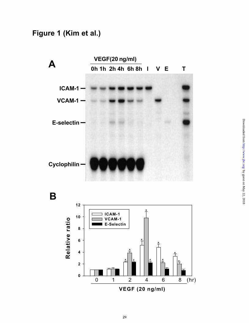
VEGF increased expression of ICAM-1, VCAM-1, and E-selectin mRNAs in
HUVECs
We developed a method of RPA by which we can detect the mRNA levels of ICAM-1,
VCAM-1, E-selectin, and cyclophilin simultaneously. Addition of VEGF (20 ng/ml)
increased the expression of ICAM-1, VCAM-1, and E-selectin mRNAs as early as 2 hr
and produced a maximal effect at 4 hr (Figures 1A and 1B). The higher expression
levels declined thereafter, but the level of ICAM-1 and VCAM-1 continued to be higher
than control for up to 8 hr. The maximum mean increases in ICAM-1, VCAM-1, and E-
selectin were 5.2-, 9.8-, and 2.2-fold, respectively (Figure 1B). As a positive control,
addition of TNF-α (1 ng/ml) for 1 hr also markedly increased the expression of ICAM-1,
VCAM-1, and E-selectin (Figure 1A).
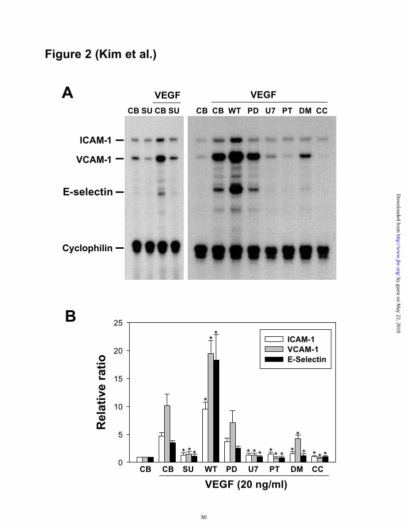
Inhibitors changed VEGF-stimulated expression of ICAM-1, VCAM-1, and E-
selectin mRNAs
To examine the receptor/second messenger mechanisms leading to induction of
adhesion molecules by VEGF, a receptor antagonist and various intracellular kinase
inhibitors were added to VEGF (20 ng/ml)-treated HUVECs. A specific KDR
antagonist (SU1498, 20 µM) completely inhibited VEGF-stimulated expression of the
adhesion molecule mRNAs (Figures 2A and 2B). PlGF is known to be a specific Flt-1
ligand (21). PlGF (10-500 ng/ml) did not produce any effect on expression of the
adhesion molecules (data not shown). MEK 1/2 inhibitor (PD98059, 50 µM) did not
produce any changes, whereas PLC inhibitor (U73122, 1 µM), NF-κB inhibitor (PDTC,
by guest on May 22, 2018
http://ww
w.jbc.org/
Dow
nloaded from
Kim et al., MS# M0:09705
12
50 µg/ml), sphingosine kinase inhibitor (DMS, 5 µM), and PKC inhibitor (chelerythrine
chloride, 5 µM) suppressed VEGF-induced expression of ICAM-1, VCAM-1, and E-
selectin (Figures 2A and 2B). Unexpectedly, PI 3’-kinase inhibitor (wortmannin, 30
nM) enhanced VEGF-induced expression of the three adhesion molecule mRNAs
(Figures 2A and 2B). These results suggested that VEGF-stimulated expression of
ICAM-1, VCAM-1, and E-selectin mRNAs may be mediated mainly through activation
of PLCγ and NF-κB, along with PI 3’-kinase-mediated suppression. The process
appears to be independent of the MEK/ERK pathway.
VEGF-induced expression of ICAM-1, VCAM-1, and E-selectin was correlated
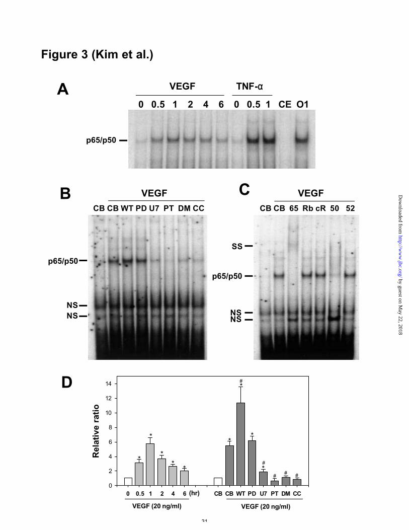
with NF-κB activity
Because the expression of adhesion molecules is mainly regulated by NF-κB (22-24),
we examined NF-κB activity in HUVECs treated with VEGF in the absence or presence
of various intracellular kinase inhibitors. Addition of VEGF (20 ng/ml) increased NF-
κB activity as early as 0.5 hr and produced a maximal effect at 1 hr (Figures 3A and
3D). These effects declined but cont inued to be higher than control levels up to 6 hr.
The maximum mean increase in NF-κB activity was 5.8-fold. As a positive control,
addition of TNF-α (1 ng/ml) for 1 hr increased NF-κB activity. A 20-fold molar excess
of unlabeled competitor almost completely blocked the NF-κB binding site, whereas the
irrelevant oligonucleotide, Oct-1, did not produce any effect on the binding site. MEK
1/2 inhibitor (PD98059, 50 µM) did not produce any change in VEGF-induced NF-κB
activity, whereas KDR antagonist (SU1498, 20 µM), PLC inhibitor (U73122, 1 µM),
NF-κB inhibitor (PDTC, 50 µg/ml), sphingosine kinase inhibitor (DMS, 5 µM), and
PKC inhibitor (chelerythrine chloride, 5 µM) suppressed VEGF-induced NF-κB activity
by guest on May 22, 2018
http://ww
w.jbc.org/
Dow
nloaded from
Kim et al., MS# M0:09705
13
(Figures 3B and 3D). PI 3’ -kinase inhibitor (wortmannin, 30 nM) enhanced VEGF-
induced NF-κB activity (Figures 3B and 3D). Overall, VEGF-induced NF-κB activity
was correlated with the expression of adhesion molecules by VEGF. We performed
supershift experiments using specific antibodies to p65 (RelA), RelB, c-Rel, p50, and
p52 in order to reveal the identities of the proteins in the VEGF-induced NF-κB binding
complex. Incubation with antibody to p65 or p50, but not with antibody to RelB, c-Rel,
or p52, shifted the protein:DNA complexes (Figure 3C). These data indicate that VEGF
activates NF-κB in the form of a p65/p50 heterodimer in HUVECs.
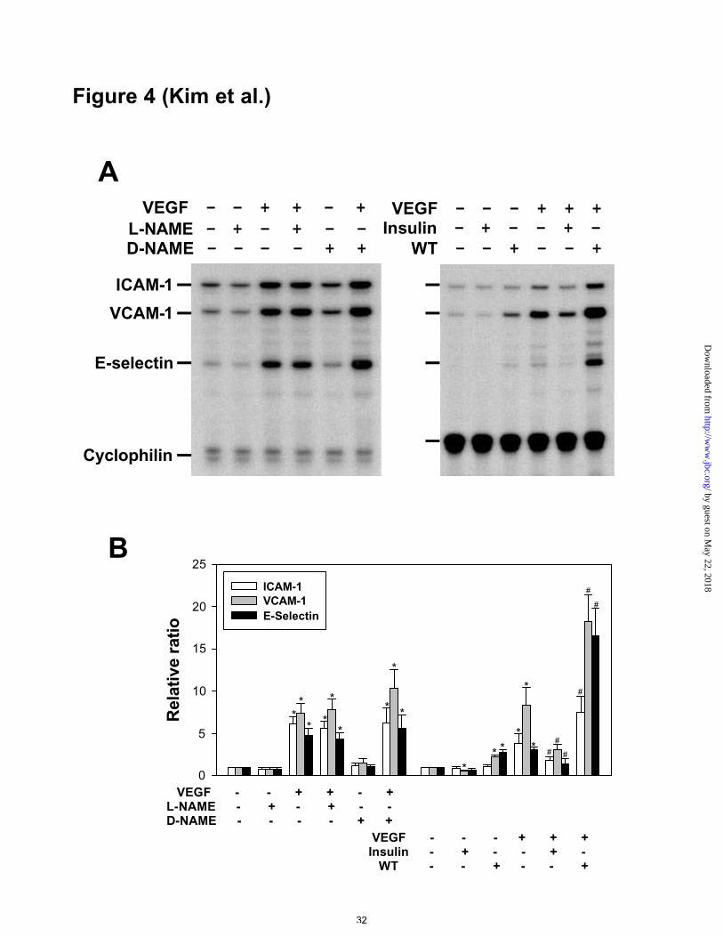
VEGF-induced expression of ICAM-1, VCAM-1, and E-selectin was independent
of NO, but was suppressed by activation of PI 3’ -kinase
Addition of NOS inhibitor L-NAME (3 mM), but not its inactive D isomer D-NAME
(3 mM), markedly suppressed basal and VEGF-stimulated NOS activity (Table 1).
Under these conditions, basal and VEGF-stimulated expression of ICAM-1, VCAM-1,
and E-selectin was not changed (Figures 4A and 4B). Addition of PI 3’ -kinase inhibitor
wortmannin (30 nM) markedly suppressed basal and VEGF-stimulated NOS activity
(Table 1). Under these conditions, both the basal and the VEGF-stimulated expression
of the three adhesion molecules was enhanced (Figures 4A and 4B). Inhibition of PI 3’-
kinase activity with LY294002 (100 nM) produced a similar effect (data not shown).
Alternatively, activation of PI 3’-kinase with insulin (50 µU) suppressed basal and
VEGF-stimulated expression of the three adhesion molecules (Figures 4A and 4B).
VEGF increased the protein levels of ICAM-1 and VCAM-1, and inhibitors
changed this effect
by guest on May 22, 2018
http://ww
w.jbc.org/
Dow
nloaded from
Kim et al., MS# M0:09705
14
Because ICAM-1 and VCAM-1 showed strongest response to VEGF among the three
molecules we examined, we looked further at the protein levels of ICAM-1 and VCAM-
1 in HUVECs treated with VEGF. Addition of VEGF (20 ng/ml) increased protein
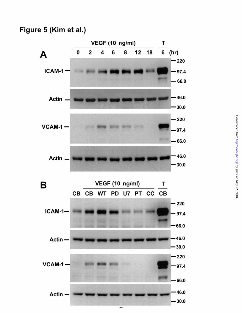
levels of ICAM-1 as early as 2 hr and produced a maximal effect at 6-12 hr (Figure 5A,
upper panels). These effects declined but continued to be higher than control levels up
to 18 hr. The maximum mean increase in ICAM-1 was 8.7-fold. Addition of VEGF (20
ng/ml) increased protein levels of VCAM-1 as early as 2 hr and produced a maximal
effect at 4-6 hr (Figure 5A, lower panels). These effects declined but continued to be
higher than control levels up to 12 hr. The maximum mean increase in VCAM-1 was
6.5-fold. TNF-α (1 ng/ml), used as a positive control, increased protein levels of ICAM-
1 and VCAM-1 markedly at 6 hr. The effect of various inhibitors on VEGF-induced
protein levels of ICAM-1 and VCAM-1 was similar to their effect on VEGF-induced
mRNA levels. MEK 1/2 inhibitor did not produce any changes, whereas inhibitors of
PLC, NF-κB and PKC suppressed VEGF-induced protein levels of ICAM-1 and
VCAM-1 (Figure 5B). PI 3’-kinase inhibitor enhanced VEGF-induced protein levels of
ICAM-1 and VCAM-1 (Figure 5B). Using flow cytometry, we also confirmed that the
protein levels of VCAM-1 and ICAM-1 on the endothelial cell surface increased after
treatment of VEGF (20 ng/ml) for 8 h (data not shown).
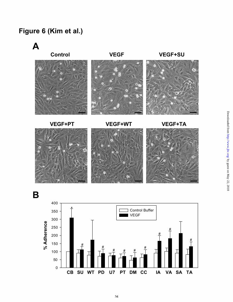
VEGF-induced leukocyte adhesiveness was correlated with VEGF-induced
expression of adhesion molecules
Because the induction of adhesion molecules in endothelial cells induces leukocyte
adhesiveness, we examined whether VEGF induces leukocyte adhesion to HUVECs.
Accordingly, addition of VEGF (20 ng/ml) produced approximately 3.1-fold increases
by guest on May 22, 2018
http://ww
w.jbc.org/
Dow
nloaded from
Kim et al., MS# M0:09705
15
in leukocyte adhesiveness after 8 hr compared to addition of control buffer (Figures 6A
and 6B). Flk-1/KDR antagonist (SU1498, 20 µM), MEK 1/2 inhibitor (PD98059, 50
µM) PLC inhibitor (U73122, 1 µM), NF-κB inhibitor (PDTC, 50 µg/ml), and PKC
inhibitor (chelerythrine chloride, 5 µM) all suppressed basal and VEGF-induced
leukocyte adhesiveness (Figures 6A and 6B). However, PI 3-kinase inhibitor
(wortmannin, 30 nM) produced a profoundly variable effect on VEGF-induced
leukocyte adhesiveness (Figures 6A and 6B). Although functional blocking antibodies
to ICAM-1, VCAM-1, and E-selectin did not produce significant changes in basal
leukocyte adhesiveness, they reduced VEGF-induced leukocyte adhesiveness (Figures
6A and 6B). A triple combination of these antibodies produced marked suppression of
VEGF-induced leukocyte adhesiveness (Figures 6A and 6B).
by guest on May 22, 2018
http://ww
w.jbc.org/
Dow
nloaded from
Kim et al., MS# M0:09705
16
Discussion
VEGF exerts its action by binding to two cell surface receptors, Flk-1/KDR and Flt-1
(25). In Flk-1/KDR null mutant mice, development of endothelial and hematopoietic
cells is impaired (26). Flt-1 null mutant mice have an apparent overgrowth of
endothelial cells, accompanied by blood vessel disorganization (27). The distinct
phenotypes of the Flk-1/KDR and Flt-1 knockout animals show that these receptors
have different biological functions. Therefore, it is likely that the two VEGF receptors
signal through different transduction pathways. Our results indicate that a specific Flk-
1/KDR antagonist completely blocked VEGF-induced expression of ICAM-1, VCAM-1,
and E-selectin and blocked VEGF-induced NF-κB activity. However, a specific Flt-1
ligand, PlGF, did not produce any effect on the expression of the adhesion molecules.
Thus, VEGF-induced expression of adhesion proteins in endothelial cells occurs
through VEGF binding to the Flk-1/KDR receptor, but not to the Flt-1 receptor (Figure
7).
Upon activation of the Flk-1/KDR receptor in endothelial cells, three major second
messenger pathways elicit cell proliferation, migration, survival, and NO production (7-
17). These pathways are: PI 3’-kinase/serine-threonine protein kinase/Akt cascade, the
tyrosine phosphorylation of PLCγ, and MEK/ERK cascade (7-17). VEGF-induced
activation of PI 3’-kinase results in phosphorylation of Akt in endothelial cells
(9,14,15,17). This phosphorylated Akt results in phosphorylation of Bad and eNOS,
resulting in cell survival, NO production and migration (9,14,15,17). Pharmacological
inhibition of PI 3’ -kinase with wortmannin and LY294002 completely inhibited these
by guest on May 22, 2018
http://ww
w.jbc.org/
Dow
nloaded from
Kim et al., MS# M0:09705
17
VEGF-induced cellular effects in endothelial cells (9,14,15). Consistent with previous
reports (14,15), we found that pharmacological inhibition of PI 3’-kinase with
wortmannin and LY294002 inhibited basal and VEGF-induced NO production.
However, unexpectedly, our data indicated that, under PI 3’-kinase inhibition, the basal
expression levels of ICAM-1, VCAM-1 and E-selectin mRNA were higher.
Furthermore, under PI 3’-kinase inhibition, VEGF-induced expression levels were
higher. Alternatively, insulin, an activator of PI 3’-kinase, decreased basal and VEGF-
induced ICAM-1, VCAM-1, and E-selectin expression. These data strongly suggest that
PI 3’-kinase could be an intracellular suppressor for the expression of ICAM-1, VCAM-
1, and E-selectin through yet unidentified signaling pathways (Figure 7). To our
knowledge, these results are the first to demonstrate an additional role of PI 3’ -kinase in
suppressing the expression of adhesion molecules. Thus, selective activation of PI 3’-
kinase suppresses the induction of ICAM-1, VCAM-1, and E-selectin in endothelial
cells. Therefore, PI 3’-kinase may decrease inflammatory responses. Therefore, a
selective activator of PI 3’-kinase could be considered as a therapeutic agent for
reducing a VEGF-induced inflammation in endothelial cells.
A previous report indicated that VEGF induces expression of monocyte
chemoattractant protein-1, a chemokine that is involved in recruiting leukocytes to sites
of inflammation, mainly through activation of NF-κB and AP-1 in retinal endothelial
cells (28). The MEK/ERK system are not involved in VEGF-induced activation of NF-
κB, but they are involved in VEGF-induced activation of AP-1 in the VEGF-induced
expression of monocyte chemoattractant protein-1 (28). VEGF/Flk-1/KDR binding
triggers a signaling cascade that results in tyrosine phosphorylation of phospholipase Cγ
by guest on May 22, 2018
http://ww
w.jbc.org/
Dow
nloaded from
Kim et al., MS# M0:09705
18
(7,11,13). Phosphorylation of phospholipase Cγ increases intracellular levels of inositol
1,4,5-triphosphate (1,4,5-IP3) and diacylglycerol. 1,4,5-IP3 elevates intracellular
calcium through an efflux from endoplasmic reticulum. The increase in intracellular
calcium also can activate sphingosine kinase to produce sphingosine-1-phosphate (29).
In turn, the increase in intracellular sphingosine-1-phosphate activates PKC. In addition,
activated phospholipase Cγ also activates PKC by increasing diacylglycerol. Activated
PKC is known to be a strong activator of NF-κB (30). There is ample evidence that
activation of NF-κB stimulates expression of ICAM-1, VCAM-1, and E-selectin
mRNAs in endothelial cells (22-24). Thus, VEGF-induced activation of PLCγ and PKC
are essential steps for induction of these adhesion molecules mRNAs in endothelial
cells, and the induction occurs through NF-κB activation (Figure 7). Upon activation of
the Flk-1/KDR receptor, increased intracellular calcium and the activation of PKC or
Akt result in activation result in activation of eNOS and, thus, increased production of
NO (11,14-16). Although previous reports (31,32) indicate that NO modulates the
protein levels of VCAM-1 or ICAM-1 differently in endothelial cells, our results
indicated that NO is not involved in VEGF-induced mRNA expression of ICAM-1,
VCAM-1, and E-selectin. Thus, NO may modulate expression of ICAM-1 and VCAM-
1 at the translational level but not at the transcriptional level. Upon activation of Flk-
1/KDR, MEK/ERK signal messenger transduction pathways are activated and lead to
cellular proliferation (10,12,13). Pharmacological inhibition of MEK/ERK pathways
with PD98059 did not have any effect on the expression of ICAM-1, VCAM-1, and E-
selectin mRNAs. Thus, NO and the MEK/ERK system are not involved in VEGF-
stimulated expression of adhesion molecules (Figure 7).
by guest on May 22, 2018
http://ww
w.jbc.org/
Dow
nloaded from
Kim et al., MS# M0:09705
19
Induction of adhesion molecules is an initial step in inflammation mediated by
leukocyte adhesion. Previous reports have shown that VEGF did not effect the
expression of ICAM-1 and VCAM-1 in human dermal microvascular endothelial cells
(33), while VEGF increased the expression of ICAM-1, but not VCAM-1 and E-selectin,
in vivo in retinal capillary endothelial cells (34). Our results indicate that VEGF
increased the expression of ICAM-1, VCAM-1, and E-selectin in HUVECs. Endothelial
cells from different areas have different characteristics and different responses to growth
factors (35,36). Thus, the expression of adhesion molecules in response to VEGF may
be different between large vessel endothelial cells and microvascular endothelial cells.
Our results clearly indicated that VEGF increased VCAM-1 and ICAM-1 protein in a
time dependent manner. Accordingly, VEGF increased leukocyte adhesion in
endothelial cells. Leukocyte adhesion to endothelial cells requires multiple cellular steps
and intracellular second messenger signaling systems. Although the kinase inhibitors
used in this study could be involved in multiple downstream effects in the endothelial
cells’ response to VEGF, there were close relationships between induction of adhesion
molecules and leukocyte adhesiveness. In addition, a combination of specific blocking
antibodies to ICAM-1, VCAM-1, and E-selectin significantly inhibited VEGF-induced
leukocyte adhesiveness to endothelial cells. Thus, VEGF-induced adhesion molecules in
endothelial cells is closely involved in VEGF-induced leukocyte adhesiveness.
In summary, the present results explain how VEGF stimulates the expression of
adhesion molecules in HUVECs. Our results show that VEGF-stimulated expression of
ICAM-1, VCAM-1, and E-selectin mRNAs was mainly through activation of PLCγ and
NF-κB. The induction was suppressed by PI 3’-kinase-mediated pathway but was
by guest on May 22, 2018
http://ww
w.jbc.org/
Dow
nloaded from
Kim et al., MS# M0:09705
20
independent of NO and MEK/ERK. Thus, VEGF simultaneously activates two signal
tranduction pathways that have opposite functions in the induction of adhesion molecule
expression.
by guest on May 22, 2018
http://ww
w.jbc.org/
Dow
nloaded from
Kim et al., MS# M0:09705
21
References
1. Gimbrone M. A. Jr. (1995) Vascular endothelium in health and disease. In Haber E.
eds. Molecular Cardiovascular Medicine, 49-61, Scientific American Medicine,
New York
2. Gimbrone M. A. Jr., Nagel, T., and Topper, J. N. (1997) J. Clin. Invest. 100, S61-
S65
3. Bischoff, J. (1997) J. Clin. Invest. 100, S37-S39
4. Ferrara, N., and Davis-Smyth, T. (1997) Endocrine Rev. 18, 4-25
5. Melder, R. J., Koenig, G. C., Witwer, B. P., Safabakhsh, N., Munn, L.L., and Jain, R.
K. (1996) Nat Med. 2, 992-997
6. Detmar, M., Brown, L. F., Schon, M. P., Elicker, B. M., Velasco, P., Richard, L.,
Fukumura, D., Monsky, W., Claffey, K. P, and Jain, R. K. (1998) J. Invest. Dermatol.
111, 1-6
7. Xia, P., Aiello, L. P., Ishii, H., Jiang, Z. Y., Park, D. J., Robinson, G. S., Takagi, H.,
Newsome, W. P., Jirousek, M. R., and King, G. L. (1996) J. Clin. Invest. 98, 2018-
2026
8. Abedi, H., and Zachary, I. (1997) J. Biol. Chem. 272, 15442-15451
9. Gerber, H. P., McMurtrey, A., Kowalski, J., Yan, M., Keyt, B. Y. , Dixit, V., and
Ferrara, N. (1998) J. Biol. Chem. 273, 30336-30343
10. Takahashi, T., Ueno, H., and Shibuya, M. (1999) Oncogene 18, 2221-2230
11. He, H., Venema, V. J., Gu, X., Venema, R. C., Marrero, M. B., and Caldwell, R. B.
(1999) J. Biol. Chem. 274, 25130-25135
12. Thakker, G. D., Hajjar, D.P., Muller, W. A., and Rosengart, T. K. (1999) J. Biol.
by guest on May 22, 2018
http://ww
w.jbc.org/
Dow
nloaded from

Kim et al., MS# M0:09705
22
Chem. 274, 10002-10007
13. Wu, L. W., Mayo, L. D., Dunbar, J. D., Kessler, K. M., Baerwald, M. R., Jaffe, E. A.,
Wang, D., Warren, R. S., and Donner, D. B. (2000) J. Biol. Chem. 275, 5096-5103
14. Fulton, D., Gratton, J.-P., McCabe, T. J., Fontana, J., Fujio, Y., Walsh, K., Franke, T.
F., Papapetropoulos, A., and Sessa, W. C. (1999) Nature 399, 597-601
15. Dimmeler, S., Fleming, I., Fisslthaler, B., Hermann, C., Busse, R., Zeiher, A. M.
(1999) Nature 399, 601-605
16. Shen, B. Q., Lee, D. Y., and Zioncheck, T. F. (1999) J. Biol. Chem. 274, 33057-
33063
17. Morales-Ruiz, M., Fulton, D., Sowa, G., Languino, L. R., Fujio, Y., Walsh, K., and
Sessa, W. C. (2000) Circ. Res. 86, 892-896
18. Kim, I., Moon, S. O., Koh, K. N., Kim, H., Uhm, C. S., Kwak, H. J., Kim, N. G.,
and Koh, G. Y. (1999) J. Biol. Chem. 274, 26523-26528
19. Neish, A. S., Williams, A. J., Palmer, H. J., Whitley, M. Z., and Collins, T. (1992) J.
Exp. Med. 176, 1583-1593
20. Akeson, A. L., and Woods, C. W. (1993) J. Immunol. Methods. 163, 181-185
21. Park, J. E., Chen, H. H., Winer, J., Houck, K. A., and Ferrara, N. (1994) J. Biol.
Chem. 269, 25646-25654
22. Ledebur, H. C., and Parks, T. P. (1995) J. Biol. Chem. 270, 933-943
23. Wrighton, C. J., Hofer-Warbinek, R., Moll, T., Eytner, R., Bach, F. H., and de Martin,
R. (1996) J. Exp. Med. 183, 1013-1022
24. Boyle, E. M. Jr., Kovacich, J. C., Canty, T. G. Jr., Morgan, E. N, Chi, E., Verrier, E.
D., and Pohlman, T. H. (1998) Circulation 98(Suppl), II282-II288
25. Petrova, T. V., Makinen, T., and Alitalo, K. (1999) Exp. Cell Res. 253,117-130
by guest on May 22, 2018
http://ww
w.jbc.org/
Dow
nloaded from
Kim et al., MS# M0:09705
23
26. Shalaby, F., Rossant, J., Yamaguchi, T. P., Gertsenstein, M., Wu, X. F., Breitman, M.
L., and Schuh, A. C. (1995) Nature 376, 62-66
27. Fong, G. H., Rossant, J., Gertsenstein, M., and Breitman, M. L. (1995) Nature 376,
66-70
28. Marumo, T., Schini-Kerth, V. B., and Busse, R. (1999) Diabetes 48, 1131-1137
29. Olivera, A., Edsall, L., Poulton, S., Kazlauskas, A., and Spiegel, S. (1999) FASEB J.
13, 1593-1600
30. Ghosh, S., and Baltimore, D. (1990) Nature 344, 678-682
31. De Caterina, R., Libby, P., Peng, H. B., Thannickal, V. J., Rajavashisth, T. B.,
Gimbrone, M. A. Jr., Shin, W. S., and Liao, J. K. (1995) J. Clin. Invest. 96, 60-68
32. Radisavljevic, Z., Avraham, H., and Avraham, S. (2000) J. Biol. Chem. 275, 20770-
20774
33. Richard, L., Velasco, P., and Detmar, M. (1998) Exp. Cell Res. 240, 1-6
34. Lu, M., Perez, V. L., Ma, N., Miyamoto, K., Peng, H. B., Liao, J. K., and Adamis, A.
P. (1999) Invest. Ophthalmol. Vis. Sci. 40, 1808-1812
35. Risau, W. (1995) FASEB J. 9, 926-933
36. Thurston, G., Baluk, P., and McDonald, D. M. (2000) Microcirculation 7, 67-80
by guest on May 22, 2018
http://ww
w.jbc.org/
Dow
nloaded from
Kim et al., MS# M0:09705
24
Footnotes
This work was supported by the Creative Research Initiatives of the Korean Ministry
of Science and Technology.
Acknowledgement - We thank Jennifer Macke for help in preparing the manuscript.
Abbreviations: VEGF, vascular endothelial growth factor; NO, nitric oxide; HUVECs,
human umbilical vein endothelial cells; ICAM-1, intracellular adhesion molecule-1;
VCAM-1, vascular cell adhesion molecule-1; NF-κB, nuclear factor-κB; PI 3’-kinase,
phosphatidylinositol 3’ -kinase; ERK, extracellular signal-regulated kinase; MEK,
mitogen-activated protein/extracellular signal-regulated kinase kinase; TNF-α , tumor
necrosis factor-α ; KDR, kinase- insert domain containing receptor; NOS, nitric oxide
synthase; L-NAME, NG-nitro-L-arginine methyl ester; D-NAME, NG-nitro-D-arginine
methyl ester; DMS, N,N-dimethylsphingosine; PDTC, pyrrolidine dithiocarbamate;
PKC, protein kinase C; RPA, RNase protection assay; FBS, fetal bovine serum
by guest on May 22, 2018
http://ww
w.jbc.org/
Dow
nloaded from
Kim et al., MS# M0:09705
25
Figure Legends
Figure 1. RPA of adhesion molecule mRNAs in VEGF-stimulated HUVECs. A,
HUVECs were incubated with VEGF165 (20 ng/ml) for the indicated times. Total RNAs
(10 µg) were subjected to multiplex RPA probed with antisense ICAM-1, antisense
VCAM-1, and antisense E-selectin RNA probes. Equivalent loading was confirmed by
probing the same reactions with an antisense cyclophilin RNA probe (105 bp). To
clarify the identity of the bands, ICAM-1 (I), VCAM-1 (V), and E-selectin (E) probes
were applied individually to the total RNA from HUVECs treated with VEGF for 4 hr
to reveal protected bands of 367, 279, and 187 bp, respectively. The positive control was
total RNA (2 µg) from HUVECs that had been incubated with TNF-α (T, 1 ng/ml) for 1
hr and subjected to the same assay conditions. B, Densitometric analyses are presented
as the relative ratio of ICAM-1, VCAM-1, or E-selectin mRNA to cyclophilin mRNA.
The relative ratio measured at time 0 hr is arbitrarily presented as 1. Results were
similar in three independent experiments. Bars represent the mean ± S.D. from three
experiments. *, p<0.05 versus time 0.
Figure 2. RPA of adhesion molecule mRNAs in VEGF-stimulated HUVECs co-treated
with inhibitors. A, HUVECs were incubated with VEGF165 (20 ng/ml) for 4 hr in the
presence of control buffer (CB), SU1498 (SU, 20 µM), wortmannin (WT, 30 nM),
PD98059 (PD, 50 µM), U73122 (U7, 1 µM), PDTC (PT, 50 µg/ml), DMS (DM, 5 µM),
or chelerythrine chloride (CC, 5 µM). Total RNAs (10 µg) isolated from the cells were
subjected to RPA as described in Figure 1. B, Densitometric analyses are presented as
the relative ratio of ICAM-1, VCAM-1, or E-selectin mRNA to cyclophilin mRNA. The
by guest on May 22, 2018
http://ww
w.jbc.org/
Dow
nloaded from
Kim et al., MS# M0:09705
26
relative ratio measured after addition of control buffer is arbitrarily presented as 1.
Results were similar in three independent experiments. Bars represent the mean ± S.D.
from three experiments. *, p<0.05 versus CB plus VEGF (20 ng/ml).
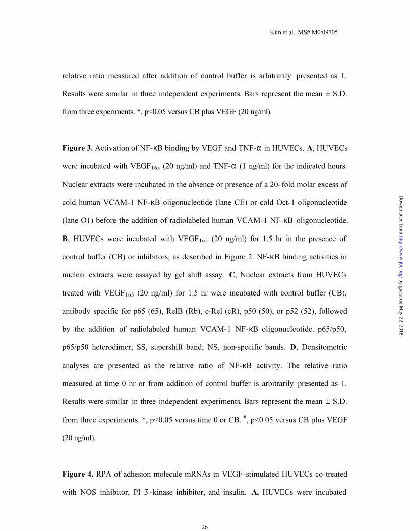
Figure 3. Activation of NF-κB binding by VEGF and TNF-α in HUVECs. A, HUVECs
were incubated with VEGF165 (20 ng/ml) and TNF-α (1 ng/ml) for the indicated hours.
Nuclear extracts were incubated in the absence or presence of a 20-fold molar excess of
cold human VCAM-1 NF-κB oligonucleotide (lane CE) or cold Oct-1 oligonucleotide
(lane O1) before the addition of radiolabeled human VCAM-1 NF-κB oligonucleotide.
B, HUVECs were incubated with VEGF165 (20 ng/ml) for 1.5 hr in the presence of
control buffer (CB) or inhibitors, as described in Figure 2. NF-κB binding activities in
nuclear extracts were assayed by gel shift assay. C, Nuclear extracts from HUVECs
treated with VEGF165 (20 ng/ml) for 1.5 hr were incubated with control buffer (CB),
antibody specific for p65 (65), RelB (Rb), c-Rel (cR), p50 (50), or p52 (52), followed
by the addition of radiolabeled human VCAM-1 NF-κB oligonucleotide. p65/p50,
p65/p50 heterodimer; SS, supershift band; NS, non-specific bands. D, Densitometric
analyses are presented as the relative ratio of NF-κB activity. The relative ratio
measured at time 0 hr or from addition of control buffer is arbitrarily presented as 1.
Results were similar in three independent experiments. Bars represent the mean ± S.D.
from three experiments. *, p<0.05 versus time 0 or CB. #, p<0.05 versus CB plus VEGF
(20 ng/ml).
Figure 4. RPA of adhesion molecule mRNAs in VEGF-stimulated HUVECs co-treated
with NOS inhibitor, PI 3’-kinase inhibitor, and insulin. A, HUVECs were incubated
by guest on May 22, 2018
http://ww
w.jbc.org/
Dow
nloaded from
Kim et al., MS# M0:09705
27
with VEGF165 (20 ng/ml) for 4 hr in the absence or presence of L-NAME (3 mM), D-
NAME (3 mM), insulin (50 µU) or wortmannin (WT, 30 nM). Total RNAs (10 µg)
isolated from the cells were subjected to RPA as described in Figure 1. B, Densitometric
analyses are presented as the relative ratio of ICAM-1, VCAM-1, or E-selectin mRNA
to cyclophilin mRNA. The relative ratio measured after addition of control buffer is
arbitrarily presented as 1. Results were similar in three independent experiments. Bars
represent the mean ± S.D. from three experiments. *, p<0.05 versus control buffer. #,
p<0.05 versus control buffer plus VEGF (20 ng/ml).
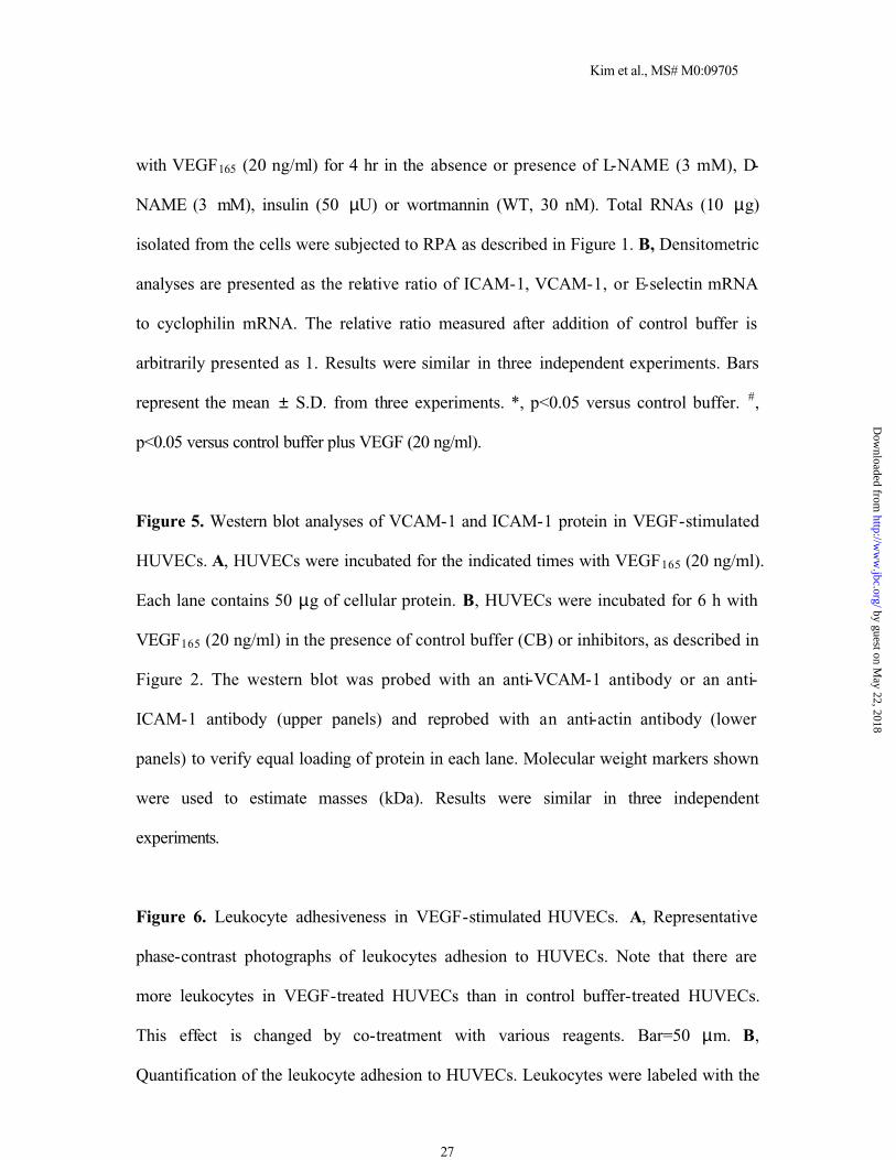
Figure 5. Western blot analyses of VCAM-1 and ICAM-1 protein in VEGF-stimulated
HUVECs. A, HUVECs were incubated for the indicated times with VEGF165 (20 ng/ml).
Each lane contains 50 µg of cellular protein. B, HUVECs were incubated for 6 h with
VEGF165 (20 ng/ml) in the presence of control buffer (CB) or inhibitors, as described in
Figure 2. The western blot was probed with an anti-VCAM-1 antibody or an anti-
ICAM-1 antibody (upper panels) and reprobed with an anti-actin antibody (lower
panels) to verify equal loading of protein in each lane. Molecular weight markers shown
were used to estimate masses (kDa). Results were similar in three independent
experiments.
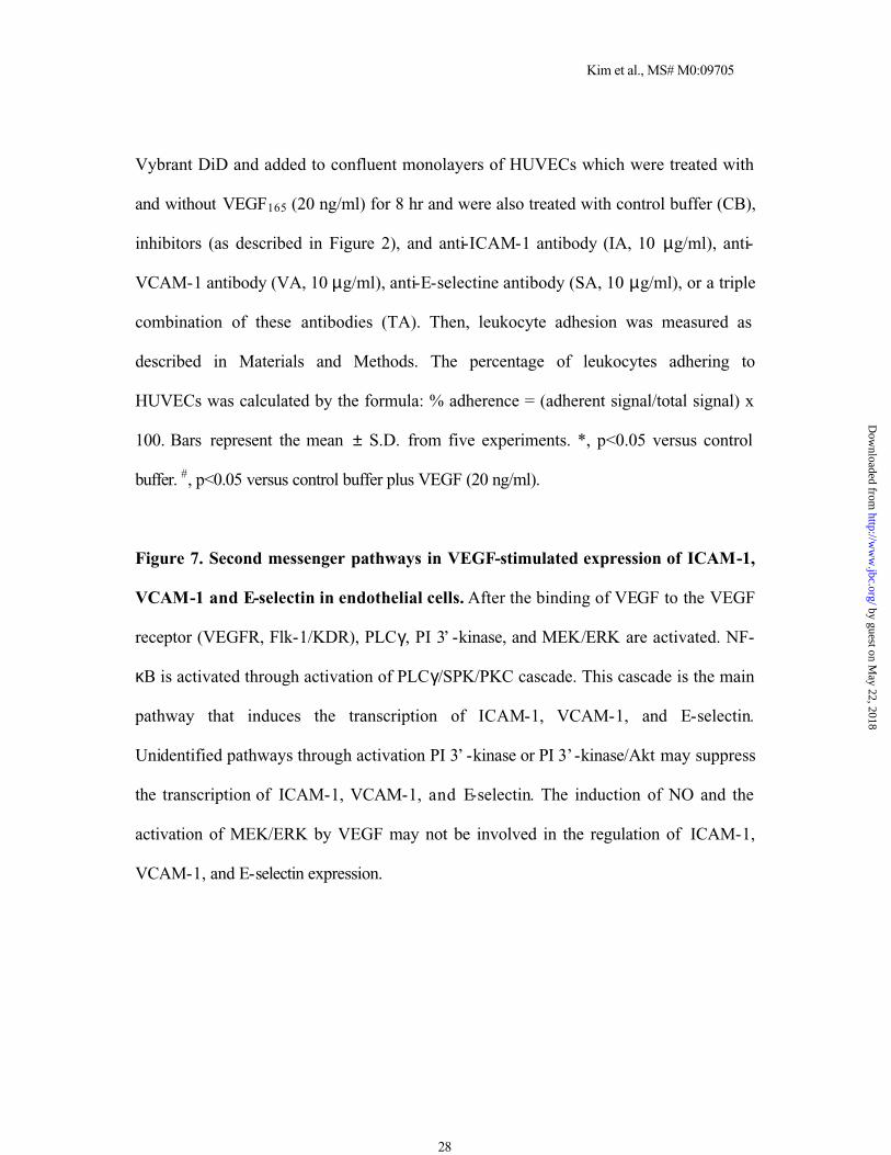
Figure 6. Leukocyte adhesiveness in VEGF-stimulated HUVECs. A, Representative
phase-contrast photographs of leukocytes adhesion to HUVECs. Note that there are
more leukocytes in VEGF-treated HUVECs than in control buffer-treated HUVECs.
This effect is changed by co-treatment with various reagents. Bar=50 µm. B,
Quantification of the leukocyte adhesion to HUVECs. Leukocytes were labeled with the
by guest on May 22, 2018
http://ww
w.jbc.org/
Dow
nloaded from
Kim et al., MS# M0:09705
28
Vybrant DiD and added to confluent monolayers of HUVECs which were treated with
and without VEGF165 (20 ng/ml) for 8 hr and were also treated with control buffer (CB),
inhibitors (as described in Figure 2), and anti-ICAM-1 antibody (IA, 10 µg/ml), anti-
VCAM-1 antibody (VA, 10 µg/ml), anti-E-selectine antibody (SA, 10 µg/ml), or a triple
combination of these antibodies (TA). Then, leukocyte adhesion was measured as
described in Materials and Methods. The percentage of leukocytes adhering to
HUVECs was calculated by the formula: % adherence = (adherent signal/total signal) x
100. Bars represent the mean ± S.D. from five experiments. *, p<0.05 versus control
buffer. #, p<0.05 versus control buffer plus VEGF (20 ng/ml).
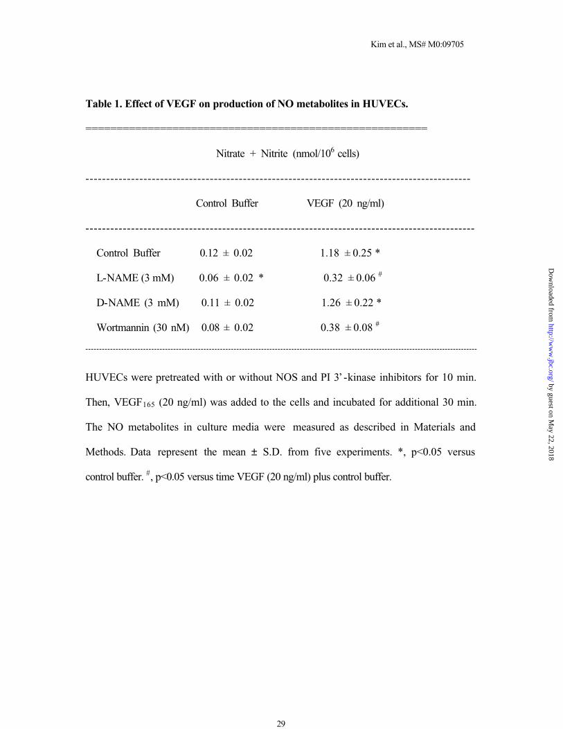
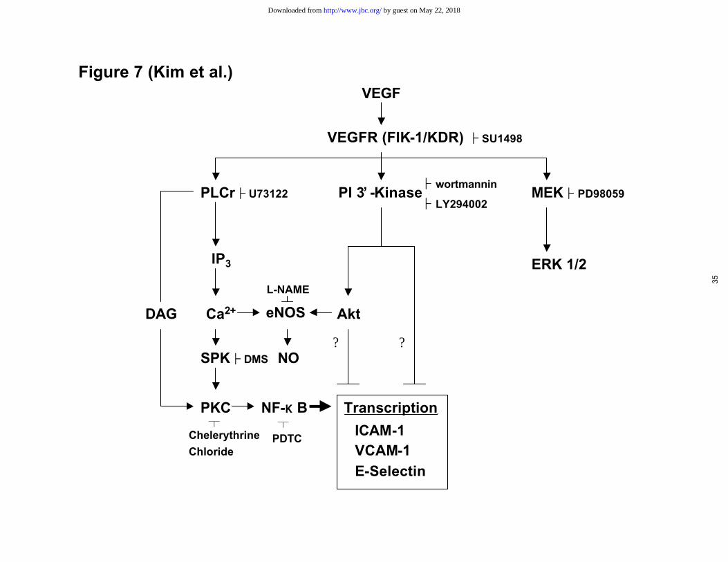
Figure 7. Second messenger pathways in VEGF-stimulated expression of ICAM-1,
VCAM-1 and E-selectin in endothelial cells. After the binding of VEGF to the VEGF
receptor (VEGFR, Flk-1/KDR), PLCγ, PI 3’ -kinase, and MEK/ERK are activated. NF-
κB is activated through activation of PLCγ/SPK/PKC cascade. This cascade is the main
pathway that induces the transcription of ICAM-1, VCAM-1, and E-selectin.
Unidentified pathways through activation PI 3’ -kinase or PI 3’-kinase/Akt may suppress
the transcription of ICAM-1, VCAM-1, and E-selectin. The induction of NO and the
activation of MEK/ERK by VEGF may not be involved in the regulation of ICAM-1,
VCAM-1, and E-selectin expression.
by guest on May 22, 2018
http://ww
w.jbc.org/
Dow
nloaded from
Kim et al., MS# M0:09705
29
Table 1. Effect of VEGF on production of NO metabolites in HUVECs.
=======================================================
Nitrate + Nitrite (nmol/106 cells)
---------------------------------------------------------------------------------------------
Control Buffer VEGF (20 ng/ml)
----------------------------------------------------------------------------------------------
Control Buffer 0.12 ± 0.02 1.18 ± 0.25 *
L-NAME (3 mM) 0.06 ± 0.02 * 0.32 ± 0.06 #
D-NAME (3 mM) 0.11 ± 0.02 1.26 ± 0.22 *
Wortmannin (30 nM) 0.08 ± 0.02 0.38 ± 0.08 #
-----------------------------------------------------------------------------------------------------------------------------------------------
HUVECs were pretreated with or without NOS and PI 3’-kinase inhibitors for 10 min.
Then, VEGF165 (20 ng/ml) was added to the cells and incubated for additional 30 min.
The NO metabolites in culture media were measured as described in Materials and
Methods. Data represent the mean ± S.D. from five experiments. *, p<0.05 versus
control buffer. #, p<0.05 versus time VEGF (20 ng/ml) plus control buffer.
by guest on May 22, 2018
http://ww
w.jbc.org/
Dow
nloaded from
ICAM-1
VCAM-1
E-selectin
Cyclophilin
VEGF(20 ng/ml)
0h 1h 2h 4h 6h 8h I V E TAR
ela
tiv
e r
ati
o
0
2
4
6
8
10
12
ICAM-1VCAM-1E-Selectin
0 1 2 4 6 8 (hr)
VEGF (20 ng/ml)
*
*
*
*
*
*
*
*
*
*
B
Figure 1 (Kim et al.)
29
by guest on May 22, 2018
http://ww
w.jbc.org/
Dow
nloaded from
E-selectin
Cyclophilin
VCAM-1
ICAM-1
CB SU CB SU CB CB WT PD U7 PT DM CC
VEGF VEGFAR
elat
ive
rati
o
0
5
10
15
20
25
ICAM-1VCAM-1E-Selectin
CB CB SU WT PD U7 PT DM CC
VEGF (20 ng/ml)
* * * * * * * ** *
*
* * * *
*
* *
Figure 2 (Kim et al.)
B
30
by guest on May 22, 2018
http://ww
w.jbc.org/
Dow
nloaded from
D
0 0.5 1 2 4 6 0 0.5 1 CE O1
VEGF TNF-αA
0 1 2 4 6 CB CB WT PD U7 PT DM CC
Rel
ativ
e ra
tio
0
2
4
6
8
10
12
14
(hr)
VEGF (20 ng/ml) VEGF (20 ng/ml)
**
*
**
**
*
*
#
#
## #
0.5
CB CB 65 Rb cR 50 52
SS
p65/p50
NSNS
p65/p50
NSNS
CB CB WT PD U7 PT DM CC
VEGFB C
Figure 3 (Kim et al.)
VEGF
p65/p50
31
by guest on May 22, 2018
http://ww
w.jbc.org/
Dow
nloaded from
ICAM-1
VCAM-1
E-selectin
Cyclophilin
AVEGF − − + + − +
L-NAME − + − + − −D-NAME − − − − + +
VEGF − − − + + +
WT − − + − − +Insulin − + − − + −
Figure 4 (Kim et al.)
Rel
ativ
e ra
tio
0
5
10
15
20
25
ICAM-1VCAM-1E-Selectin
VEGF - - + + - +L-NAME - + - + - -D-NAME - - - - + +
VEGF - - - + + + Insulin - + - - + - WT - - + - - +
**
* *
*
*
*
*
*
*
*
* *
*
* ##
#
#
#
#
B
32
by guest on May 22, 2018
http://ww
w.jbc.org/
Dow
nloaded from
0 2 4 6 8 12 18 6 (hr)
VEGF (10 ng/ml) T
ICAM-1
Actin
VCAM-1
Actin
220
97.4
66.0
46.0
30.0
220
97.4
66.0
46.0
30.0
220
97.4
66.0
46.0
30.0
220
97.4
66.0
46.0
30.0
ICAM-1
Actin
VCAM-1
Actin
VEGF (10 ng/ml) T
CB CB WT PD U7 PT CC CB
A
B
Figure 5 (Kim et al.)
33
by guest on May 22, 2018
http://ww
w.jbc.org/
Dow
nloaded from
B
A%
Ad
her
ence
0
50
100
150
200
250
300
350
400
Control BufferVEGF
CB SU WT PD U7 PT DM CC IA VA SA TA
*
##
# # ##
#
##
Control VEGF VEGF+SU
VEGF+PT VEGF+WT VEGF+TA
B
Figure 6 (Kim et al.)
34
by guest on May 22, 2018
http://ww
w.jbc.org/
Dow
nloaded from
MEK├├ PD98059
VEGF
VEGFR (FIK-1/KDR) ├├ SU1498
PI 3’ -Kinase
IP3 ERK 1/2
DAG Ca2+ eNOS Akt
SPK├├ DMS NO? ?
PLCr├├ U73122
PKC NF-κ B
├├ wortmannin
├├ LY294002
┬Chelerythrine
Chloride
┬
PDTC
Transcription
ICAM-1VCAM-1E-Selectin
L-NAME
Figure 7 (Kim et al.)
35
by guest on May 22, 2018http://www.jbc.org/Downloaded from
Young KohInjune Kim, Sang-Ok Moon, Sung Hoon Kim, Hyung Jin Kim, Young Soon Koh and Gou
factor-kappaB activation in endothelial cellsVEGF stimulates expression of ICAM-1, VCAM-1 and E-selectin through nuclear
published online December 6, 2000J. Biol. Chem.
10.1074/jbc.M009705200Access the most updated version of this article at doi:
Alerts:
When a correction for this article is posted•
When this article is cited•
to choose from all of JBC's e-mail alertsClick here
by guest on May 22, 2018
http://ww
w.jbc.org/
Dow
nloaded from


![Orthosilicic acid, Si(OH)4, stimulates osteoblast differentiation in … · 2019. 2. 13. · regulate osteoblast differentiation were summarized by Vimalraj and Selvamurugan [51].](https://static.fdocument.org/doc/165x107/5fde13c5c61ed2381970cc83/orthosilicic-acid-sioh4-stimulates-osteoblast-differentiation-in-2019-2-13.jpg)