Cytomegalovirus IE2 Protein Stimulates Interleukin 1 Gene
12
MOLECULAR AND CELLULAR BIOLOGY, 0270-7306/99/$04.0010 Oct. 1999, p. 6803–6814 Vol. 19, No. 10 Copyright © 1999, American Society for Microbiology. All Rights Reserved. Cytomegalovirus IE2 Protein Stimulates Interleukin 1b Gene Transcription via Tethering to Spi-1/PU.1 NAWARAT WARA-ASWAPATI, 1,2 ² ZHIYONG YANG, 1,3 WAYNE R. WATERMAN, 1 YOSHINOBU KOYAMA, 1,3 SOTIRIOS TETRADIS, 1 ‡ BOB K. CHOY, 1,3 ANDREW C. WEBB, 4 AND PHILIP E. AURON 1,3 * The New England Baptist Bone & Joint Institute, Beth Israel Deaconess Medical Center, 1 Department of Periodontology, Harvard School of Dental Medicine, 2 and Department of Medicine, Harvard Medical School, 3 Boston, Massachusetts 02115, and Department of Biological Sciences, Wellesley College, Wellesley, Massachusetts 02181 4 Received 23 March 1999/Returned for modification 17 May 1999/Accepted 28 July 1999 Potent induction of the gene coding for human prointerleukin 1b (il1b) normally requires a far-upstream inducible enhancer in addition to a minimal promoter located between positions 2131 and 112. The tran- scription factor Spi-1 (also called PU.1) is necessary for expression and binds to the minimal promoter, thus providing an essential transcription activation domain (TAD). In contrast, infection by human cytomegalovirus (HCMV) can strongly activate il1b via the expression of immediate early (IE) viral proteins and eliminates the requirement for the upstream enhancer. Spi-1 has been circumstantially implicated as a host factor in this process. We report here the molecular basis for the direct involvement of Spi-1 in HCMV activation of il1b. Transfection of Spi-1-deficient HeLa cells demonstrated both the requirement of Spi-1 for IE activity and the need for a shorter promoter (259 to 112) than that required in the absence of IE proteins. Furthermore, in contrast to normal, enhancer-dependent il1b expression, which absolutely requires both the Spi-1 winged helix-turn-helix (wHTH) DNA-binding domain and the majority of the Spi-1 TAD, il1b expression in the presence of IE proteins does not require the Spi-1 TAD, which plays a synergistic role. In addition, we demonstrate that a single IE protein, IE2, is critical for the induction of il1b. Protein-protein interaction experiments revealed that the wing motif within the Spi-1 wHTH domain directly recruits IE2. In turn, IE2 physically associates with the Spi-1 wing and requires the integrity of at least one region of IE2. Functional analysis demonstrates that both this region and a carboxy-terminal acidic TAD are required for IE2 function. Therefore, we propose a protein-tethered transactivation mechanism in which the il1b promoter-bound Spi-1 wHTH tethers IE2, which provides a TAD, resulting in the transactivation of il1b. The clinical importance of human cytomegalovirus (HCMV) infection has increased over the past several decades, in par- ticular as immunosuppressive posttransplantation therapies and AIDS and other immunosuppressive states have become significant medical concerns. Although a majority of the in- fected population remains subclinically infected, an active in- fection occurring in a debilitated individual usually leads to mortality. Monocytes and tissue macrophages play prominent roles in host response to HCMV infection and are also an important reservoir during latent infection. After infecting the host, HCMV stimulates the production of various monocyte/ macrophage inflammatory cytokines, such as interleukin 1b (IL-1b), IL-2, tumor necrosis factor alpha, IL-6, IL-8, and beta interferon (10, 12, 17, 18). A markedly increased level of IL-1b observed during the active phase of HCMV infection indicates a significant role for this mediator in the interaction between the host defense mechanism and disease progression (25). We have previously reported that the HCMV immediate early (IE) gene products upregulate the expression of the human prointerleukin 1b gene (il1b) in infected monocytes as well as in transient-trans- fection assays (12, 25). Interestingly, in contrast to normal expression of il1b, which requires both a far-upstream en- hancer and a promoter element (30, 48), transactivation of il1b by HCMV IE gene products eliminates the need for the es- sential upstream enhancer located between positions 23134 and 22729 and requires only the il1b promoter element lo- cated at positions 2131 to 112 (11). Within the il1b promoter, there are two binding sites for the myelomonocyte- and B-cell- specific ETS transcription factor Spi-1 (also called PU.1) which have been demonstrated to be critical for basal il1b promoter activity (30) (sites A and B in Fig. 1). Spi-1, a winged helix- turn-helix (wHTH) transcription factor, plays a pivotal role in the development of hematopoietic cell lineages and has also been implicated as an essential host factor in viral transactiva- tion of various cellular genes (17, 24, 26, 28, 54). Studies have suggested direct interactions between Spi-1 and viral proteins, such as the human T-cell leukemia virus type 1 Tax protein (54) and the Epstein-Barr virus nuclear protein-2 (EBNA-2) (26). In the case of il1b transactivation by HCMV, mutation of the Spi-1-binding site located at 250 to 239 results in a dra- matic loss of IE protein-dependent transactivation, suggesting a possible involvement of Spi-1 (24). However, this report did not demonstrate a direct role for Spi-1 protein in activation by IE protein because the Spi-1-binding site is also required in the absence of viral protein (30). Due to the fact that there are three predominant HCMV IE proteins, IE1(p72), IE2(p55), and IE2(p86), reported to acti- vate the transcription of cellular genes by considerably differ- ent mechanisms (20, 37), it is important to identify the roles of individual IE proteins in il1b transactivation. It has been shown * Corresponding author. Mailing address: Harvard Institutes of Medicine, Room 245, 77 Avenue Louis Pasteur, Boston, MA 02115- 5727. Phone: (617) 667-0741. Fax: (617) 975-5299. E-mail: pauron @caregroup.med.harvard.edu. ² Present address: Khon Kaen University, Faculty of Dentistry, Khon Kaen 40002, Thailand. ‡ Present address: UCLA School of Dentistry, Los Angeles, CA 90095. 6803 Downloaded from https://journals.asm.org/journal/mcb on 21 February 2022 by 168.205.108.252.
Transcript of Cytomegalovirus IE2 Protein Stimulates Interleukin 1 Gene
MOLECULAR AND CELLULAR BIOLOGY,0270-7306/99/$04.0010
Oct. 1999, p. 6803–6814 Vol. 19, No. 10
Copyright © 1999, American Society for Microbiology. All Rights Reserved.
Cytomegalovirus IE2 Protein Stimulates Interleukin 1b GeneTranscription via Tethering to Spi-1/PU.1
NAWARAT WARA-ASWAPATI,1,2† ZHIYONG YANG,1,3 WAYNE R. WATERMAN,1 YOSHINOBU KOYAMA,1,3
SOTIRIOS TETRADIS,1‡ BOB K. CHOY,1,3 ANDREW C. WEBB,4 AND PHILIP E. AURON1,3*
The New England Baptist Bone & Joint Institute, Beth Israel Deaconess Medical Center,1 Department of Periodontology,Harvard School of Dental Medicine,2 and Department of Medicine, Harvard Medical School,3 Boston,
Massachusetts 02115, and Department of Biological Sciences, Wellesley College,Wellesley, Massachusetts 021814
Received 23 March 1999/Returned for modification 17 May 1999/Accepted 28 July 1999
Potent induction of the gene coding for human prointerleukin 1b (il1b) normally requires a far-upstreaminducible enhancer in addition to a minimal promoter located between positions 2131 and 112. The tran-scription factor Spi-1 (also called PU.1) is necessary for expression and binds to the minimal promoter, thusproviding an essential transcription activation domain (TAD). In contrast, infection by human cytomegalovirus(HCMV) can strongly activate il1b via the expression of immediate early (IE) viral proteins and eliminates therequirement for the upstream enhancer. Spi-1 has been circumstantially implicated as a host factor in thisprocess. We report here the molecular basis for the direct involvement of Spi-1 in HCMV activation of il1b.Transfection of Spi-1-deficient HeLa cells demonstrated both the requirement of Spi-1 for IE activity and theneed for a shorter promoter (259 to 112) than that required in the absence of IE proteins. Furthermore, incontrast to normal, enhancer-dependent il1b expression, which absolutely requires both the Spi-1 wingedhelix-turn-helix (wHTH) DNA-binding domain and the majority of the Spi-1 TAD, il1b expression in thepresence of IE proteins does not require the Spi-1 TAD, which plays a synergistic role. In addition, wedemonstrate that a single IE protein, IE2, is critical for the induction of il1b. Protein-protein interactionexperiments revealed that the wing motif within the Spi-1 wHTH domain directly recruits IE2. In turn, IE2physically associates with the Spi-1 wing and requires the integrity of at least one region of IE2. Functionalanalysis demonstrates that both this region and a carboxy-terminal acidic TAD are required for IE2 function.Therefore, we propose a protein-tethered transactivation mechanism in which the il1b promoter-bound Spi-1wHTH tethers IE2, which provides a TAD, resulting in the transactivation of il1b.
The clinical importance of human cytomegalovirus (HCMV)infection has increased over the past several decades, in par-ticular as immunosuppressive posttransplantation therapiesand AIDS and other immunosuppressive states have becomesignificant medical concerns. Although a majority of the in-fected population remains subclinically infected, an active in-fection occurring in a debilitated individual usually leads tomortality. Monocytes and tissue macrophages play prominentroles in host response to HCMV infection and are also animportant reservoir during latent infection. After infecting thehost, HCMV stimulates the production of various monocyte/macrophage inflammatory cytokines, such as interleukin 1b(IL-1b), IL-2, tumor necrosis factor alpha, IL-6, IL-8, and betainterferon (10, 12, 17, 18).
A markedly increased level of IL-1b observed during theactive phase of HCMV infection indicates a significant role forthis mediator in the interaction between the host defensemechanism and disease progression (25). We have previouslyreported that the HCMV immediate early (IE) gene productsupregulate the expression of the human prointerleukin 1bgene (il1b) in infected monocytes as well as in transient-trans-
fection assays (12, 25). Interestingly, in contrast to normalexpression of il1b, which requires both a far-upstream en-hancer and a promoter element (30, 48), transactivation of il1bby HCMV IE gene products eliminates the need for the es-sential upstream enhancer located between positions 23134and 22729 and requires only the il1b promoter element lo-cated at positions 2131 to 112 (11). Within the il1b promoter,there are two binding sites for the myelomonocyte- and B-cell-specific ETS transcription factor Spi-1 (also called PU.1) whichhave been demonstrated to be critical for basal il1b promoteractivity (30) (sites A and B in Fig. 1). Spi-1, a winged helix-turn-helix (wHTH) transcription factor, plays a pivotal role inthe development of hematopoietic cell lineages and has alsobeen implicated as an essential host factor in viral transactiva-tion of various cellular genes (17, 24, 26, 28, 54). Studies havesuggested direct interactions between Spi-1 and viral proteins,such as the human T-cell leukemia virus type 1 Tax protein(54) and the Epstein-Barr virus nuclear protein-2 (EBNA-2)(26). In the case of il1b transactivation by HCMV, mutation ofthe Spi-1-binding site located at 250 to 239 results in a dra-matic loss of IE protein-dependent transactivation, suggestinga possible involvement of Spi-1 (24). However, this report didnot demonstrate a direct role for Spi-1 protein in activation byIE protein because the Spi-1-binding site is also required in theabsence of viral protein (30).
Due to the fact that there are three predominant HCMV IEproteins, IE1(p72), IE2(p55), and IE2(p86), reported to acti-vate the transcription of cellular genes by considerably differ-ent mechanisms (20, 37), it is important to identify the roles ofindividual IE proteins in il1b transactivation. It has been shown
* Corresponding author. Mailing address: Harvard Institutes ofMedicine, Room 245, 77 Avenue Louis Pasteur, Boston, MA 02115-5727. Phone: (617) 667-0741. Fax: (617) 975-5299. E-mail: [email protected].
† Present address: Khon Kaen University, Faculty of Dentistry,Khon Kaen 40002, Thailand.
‡ Present address: UCLA School of Dentistry, Los Angeles, CA90095.
6803
Dow
nloa
ded
from
http
s://j
ourn
als.
asm
.org
/jour
nal/m
cb o
n 21
Feb
ruar
y 20
22 b
y 16
8.20
5.10
8.25
2.
by many studies that IE1 and IE2, either independently orsynergistically, transactivate various promoters. These promot-ers include those regulating cellular expression of the gene forthe 70,000-molecular-weight heat shock protein (hsp70),c-myc, c-fos, and c-jun, as well as the promoter for the humanimmunodeficiency virus (HIV) long terminal repeat (LTR) (3,4, 21, 31, 55). IE1(p72) has been described as a transactivatorfor the HCMV major IE promoter (6) and for some cellulargenes via NF-kB (17, 32, 44, 57). IE2(p86) transactivates theHCMV UL 112/113 early promoter and represses the major IEpromoter via direct DNA binding (34, 46). It is also a potenttransactivator for many cellular genes (14, 33, 36, 41, 47, 56). Incontrast to that by IE1, which has not been reported to bindany DNA sequence, transactivation by IE2 appears to involveboth direct DNA binding and interaction with various cellularproteins (14). Recent studies have revealed physical associa-tions of IE2 with a number of transcription factors, includingTATA-binding protein (TBP), TFIIB (5), Sp1, Tef-1, Rb (19),p53, c-Jun, JunB (47), and p300/CREB-binding protein (45).The function of IE2(p55), an alternatively spliced version ofIE2, is unclear, but it does not appear to directly supporttransactivation (40).
The activation of gene transcription has been widely dem-onstrated to involve numerous protein-protein interactions.Many of these involve various members of the ETS transcrip-tion factor family, including Spi-1 (1, 23, 30, 51, 58). Our studywas aimed at determining whether Spi-1 protein is involved ina physical interaction with HCMV IE proteins in enhancer-independent il1b transactivation. Using transient transfectionof Spi-1-deficient HeLa cells, we directly demonstrate a re-quirement for Spi-1. Furthermore, we show that the wHTHDNA-binding domain of Spi-1 is sufficient to support signifi-cant transactivation of il1b by HCMV IE proteins. A protein-protein interaction assay was used to demonstrate that IE2, butnot IE1, interacts with Spi-1 and to map specific regions of IE2and Spi-1 which are essential for a physical association be-tween these two proteins. Functional analysis indicates that, inaddition to a centrally located region of IE2 required forstrong interaction with the Spi-1 wHTH wing motif, a carboxy-terminal region is required and an amino-proximal region issupportive for il1b transactivation. These two regions havepreviously been reported to serve as an acidic activation do-main (40) and a TBP-binding domain (52), respectively. Basedon this evidence, we propose a protein-tethered transactivation(PTT) mechanism of il1b by IE2 in which the Spi-1 wHTHdirectly binds to the il1b promoter and tethers IE2 protein,which is unable to directly bind the promoter, providing amultifunctional TAD required for gene expression.
MATERIALS AND METHODS
Cell culture. HeLa cells (strain S3) were cultured as previously described (30).The cells were grown in Dulbecco modified Eagle medium (DMEM) containing10% fetal bovine serum and 0.5% penicillin-streptomycin. Every three days, cellswere split 1:10 by adding cold phosphate-buffered saline (PBS) containing 25mM EDTA to detach the cells.
Reporter constructs and expression vectors. Plasmids 3MHT and 3MDT,previously described (48), contain the il1b promoter sequence 2131 to 112 andthe sequence 259 to 112 (Fig. 1), respectively, ligated to chloramphenicolacetyltransferase (CAT) reporter plasmid pA10CAT3M (3M).
Plasmid pGL3B-DT contains the il1b promoter sequence 255 to 112 ligatedto the luciferase reporter pGL3-Basic vector (Promega) at XbaI and BglII sites.
The sense and antisense Spi-1 pRc/CMV expression vectors contain the entirewild-type Spi-1 cDNA as previously reported (15). The series of Spi-1 mutantconstructs (see Fig. 3a) with deletions in the PEST, PN, and NN regions wereconstructed by shuttling the cDNAs (43), gifts from Richard Maki (The ScrippsResearch Institute, La Jolla, Calif.), into pRc/CMV expression vectors. The Spi-1D100 expression vector for the truncated Spi-1 protein, lacking the first 100amino acids of Spi-1 but containing amino acids 101 to 272, was constructed bydigesting the Spi-1 cDNA with NcoI followed by ligation into pRc/RSV (Invitro-gen) in the presence of a small oligonucleotide complementary to the overhangsgenerated by NcoI and HindIII and was provided by Jack Hensold (Case West-ern Reserve University, Cleveland, Ohio). The Spi-1 D8/32 pECE expressionvector, which lacks amino acids 8 to 32 and was provided by Richard Maki, hadan additional 50-bp T7 promoter sequence located upstream of the cDNA. TheSpi-1 ETS domain (wHTH) cDNA corresponding to amino acids 161 to 272 wasPCR amplified with the following primers: 59 TTG CAA GCT TCC GCC ATGCTT CTG CAC GGG GAG ACA G 39 and 59 TTG CTC TAG ATC AGT GGGGCG GGA GGC G 39. The overhangs were generated with HindIII and XbaIand ligated into the pRc/CMV expression vector.
The HCMV IE expression vectors pEQ273(IE1), pEQ326(IE2), andpEQ276(IE112), gifts from Adam Geballe (Fred Hutchinson Cancer ResearchCenter, Seattle, Wash.), contain the genomic HCMV IE DNA inserted into thepGEM1 vector. The expression vector pEQ336 contains only the HCMV IEpromoter without any IE protein coding sequence. The vector pEQ273(IE1)contains genomic DNA coding for the IE1(p72) protein. The vector pEQ326(IE2) contains genomic DNA for IE2 protein. The vector pEQ276(IE112)contains the entire genomic DNA sequence for both IE1 and IE2 via alternativesplicing.
The HCMV IE protein expression vectors IE1(p72), IE2(p86), and IE2(p55),as well as the mutated IE2 expression vectors IE2 DMX, IE2 DSN, IE2 DSX519,IE2 DXN, and IE2NheI were gifts from Deborah Spector (University of Cali-fornia, San Diego) and contained individual IE cDNAs (see Fig. 6a and b) in thepSG5 (Stratagene) expression vector.
The Stat 3 expression vector contains Stat 3 cDNA in the pRc/CMV expres-sion plasmid, a gift from Z. Zhong and J. Darnell, Jr. (Rockefeller University,New York, N.Y.).
Transfections and reporter gene assays. The CaPO4 transfection and CATassays were performed as previously described (48). CAT assays were carried outby liquid scintillation (50). In this kinetic method, which is not affected by thesaturation limitation of endpoint methods such as conventional thin-layer chro-matography, HeLa cell lysates (60 mg of total protein) were adjusted to a finalvolume of 150 ml with TT buffer (0.25 M Tris-Cl [pH 7.8], 0.5% Triton X-100],and 100 ml of 2.5 mM chloramphenicol (Sigma) solution containing 0.25 mCi of[3H]acetyl-coenzyme A (NEN) was added. The CAT activities were evaluated bycalculating the slopes within a linear range of the kinetic response generated by10 cycles of scintillation counting (total reaction time of approximately 6 h). Thefirst-order slopes were derived by a polynomial curve fit generating a coefficientof correlation which was routinely very close to unity, and only those fits gener-ating r values between 0.998 and 1.000 were used.
For luciferase reporters, liposomal transfection was performed by usingDOTAP transfection reagent (Boehringer Mannheim GmbH) according to themanufacturer’s instructions. Briefly, HeLa cells (5 3 104) were plated in 16-mm-diameter culture plates 24 h before transfection. Immediately before transfec-tion, cells (60 to 80% confluence) were washed with 500 ml of DMEM threetimes and incubated in 1 ml of DMEM containing 10% fetal bovine serum andpenicillin-streptomycin for 3 h. Plasmid DNA (2 mg of total DNA [1 mg of thereporter and 0.5 mg of each expression vector]) was gently mixed with DOTAP(1 mg of DNA per 7.5 ml of DOTAP) and HEPES buffer (20 mM, pH 7.4, cellculture grade) in a sterile reaction tube (total volume, 120 ml). The transfectionmixture was incubated for 20 min at room temperature before being added to thecells. The plates were incubated for 5 h and washed with 500 ml of DMEM. After40 h of incubation, cells were harvested and lysed in 150 ml of cell culture lysisreagent (Promega). The lysates were assayed for protein concentrations by usingCoomassie protein assay reagent (Pierce) and processed for the luciferase assayby using a Promega luciferase assay kit according to the instructions of themanufacturer. Fifty microliters of the lysate was mixed with 100 ml of luciferaseassay reagent (Promega), and the luciferase activity was measured in a luminom-eter (AutoLumat LB 953; EG&G Berthold). The values obtained were normal-ized to the protein concentration in each cell lysate.
Human THP-1 cells were transfected by the DEAE-dextran method as de-
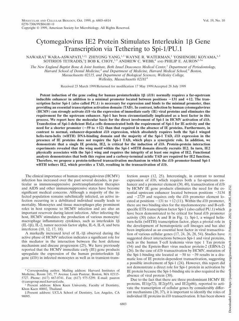
FIG. 1. Schematic representation of the il1b promoter showing two Spi-1-binding sites, one located adjacent to the transcription start site and the otherlocated further upstream. The TATA sequence (TATAAAA) and the TBP arealso illustrated. Two fragments of the il1b promoter, HT (2131 to 112) and DT(259 to 112), used in the transfection experiments in this study are shown asbars (see Kominato et al. [30] for a detailed description of these sequences).
6804 WARA-ASWAPATI ET AL. MOL. CELL. BIOL.
Dow
nloa
ded
from
http
s://j
ourn
als.
asm
.org
/jour
nal/m
cb o
n 21
Feb
ruar
y 20
22 b
y 16
8.20
5.10
8.25
2.
scribed previously (48). Briefly, 107 cells (per 100-mm-diameter plate) weretransfected with 10 mg of plasmid. After transfection, cells were treated withlipopolysaccharide (LPS) (10 ng/ml) for 16 h prior to CAT assays.
Whole-cell extraction and Western blotting. HeLa cells were plated on 100-mm-diameter plates and incubated for 24 h in order to allow growth to 60 to 80%confluence prior to transfection. Transfections were performed with DOTAPreagent as described above. After 40 h of incubation at 37°C with 5% CO2, cellswere washed with cold PBS once and detached with cold PBS containing 25 mMEDTA. The cells were collected by centrifugation and resuspended in 200 ml ofcold lysis buffer (20 mM HEPES [pH 7.9], 1 mM MgCl2, 10 mM KCl, 300 mMNaCl, 0.1% Triton X-100, 20% glycerol, 1 mM dithiothreitol, 1 mM phenylmeth-ylsulfonyl fluoride [PMSF], 10 mM NaF, 1 mM ZnCl2, 1 mM Na orthovanadate,1 protease inhibitor cocktail tablet [Boehringer Mannheim]/50 ml). After incu-bation on ice for 10 min, the lysates were cleared by centrifugation for 10 min at12,000 3 g at 4°C. Protein concentrations were measured by the Bradfordmethod (Bio-Rad). Ten micrograms of whole-cell extract for each sample wasseparated by sodium dodecyl sulfate-polyacrylamide gel electrophoresis (SDS-PAGE) and transferred to a nitrocellulose membrane. The membranes wereincubated with a 1:1,000 dilution of mouse anti-IE2 antibody (Godwin Instituteof Cancer Research, Inc.), followed by incubation with a 1:2,000 dilution ofconjugated horseradish peroxidase-labeled anti-mouse antibody. Horseradishperoxidase activity was detected with the enhanced-chemiluminescence system(Amersham).
GST fusion vectors. The glutathione S-transferase (GST)–wild-type Spi-1 fu-sion vector (WT) containing the Spi-1 coding sequence was provided by D. G.Tenen (Beth Israel Deaconess Medical Center, Harvard Medical School, Boston,Mass.).
GST fused with various motifs of Spi-1 fusion vectors, specifically, GST–Spi-1(171-272), GST–Spi-1 (171-242), and GST–Spi-1 (202-254) (the amino acid cod-ing region of each construct is indicated by the numbers after GST–Spi-1), whichcontain different motifs of the Spi-1 DNA-binding domain as shown in Fig. 6a,were constructed by PCR amplification by combining the following primers, asrequired: 59 wHTH, 59 ATG GGA TCC AAG ATT CGC CTG TAC 39; 59 b2motif, 59 ATC GGA TCC CAG TTC TCG TCC AAG C 39; 39 wHTH, 59 GATGAA TTC ATC AGT GGG GCG GGA G 39; 39 b4 motif, 59 ATG AAT TCAGAA CTG GTA GGT GAG C 39; and 39 a3 motif, 59 GAT GAA TTC CTCGCC TGT CTT GCC GTA GT 39. Each PCR was performed in a total volumeof 100 ml composed of 10 ml of 103 Pfu polymerase buffer (Stratagene), 0.8 mlof deoxynucleoside triphosphates (25 mM each deoxynucleoside triphosphate),1.0 ml of the DNA template (100 ng/ml), 2.5 ml of each primer (100 ng/ml), 2.0 mlof the cloned Pfu DNA polymerase (2.5 U/ml; Stratagene), and 81.2 ml ofdistilled water. The sample was overlaid with 70 ml of mineral oil, and amplifi-cation was performed with a program consisting of 94°C for 1 min, followed by30 cycles of 94°C for 45 s, 55°C for 45 s, and 72°C for 45 s, and a final cycle of 72°Cfor 10 min. The PCR fragments with overhangs generated with BamHI andEcoRI were purified with a Mermaid Kit (Bio 101, Inc.) and ligated into thepGEX-2T (Pharmacia) GST expression plasmid. GST–Spi-1 (243-254) was con-structed with synthetic oligonucleotide sequences 59 GAT CCG TGA AGAAAG TCA AGA AGA AGC TCA CCT ACC AGT TCG 39 and 59 AAT TCGAAC TGG TAG GTG AGC TTC TTC TTG ACT TTC TTC ACG 39, annealed,and ligated into the pGEX-2T expression vector (Pharmacia) at the BamHI andEcoRI restriction sites.
Expression and purification of GST fusion protein. Overnight culture ofEscherichia coli DH5a containing one of the GST fusion constructs was diluted1:10 in Luria broth with ampicillin (100 mg/ml) and grown for 1 h with shakingat 37°C. Isopropyl-b-D-thiogalactopyranoside (IPTG) was added to a final con-centration of 0.5 mM, and cultures were incubated at 37°C for 3 h with shaking.Bacteria (20 ml) were pelleted and suspended in 0.6 ml of cold NETN buffer (20mM Tris [pH 8.0], 1 mM EDTA [pH 8.0], 200 mM NaCl, 0.5% NP-40, 0.2 mMPMSF, 1 mM dithiothreitol, 1% Triton X-100, 1 protease inhibitor cocktail tablet[Boehringer Mannheim]/50 ml). The cells were sonicated three times for 15 s onice and then centrifuged at 10,000 3 g for 10 min. The protein concentrations ofthe lysates were measured with a Bio-Rad protein assay kit. Glutathione-Sepha-rose beads (Pharmacia) were equilibrated with NETN buffer. Equivalentamounts of GST fusion proteins (as determined by Bio-Rad and confirmed byCoomassie blue staining) were bound to 50 ml of glutathione-Sepharose beads byincubation in a total volume of 500 ml of NETN buffer for 1 h at 4°C. The beadswere washed three times in NETN buffer.
Protein-protein interaction assay. The GST fusion protein beads were incu-bated with 35S-labeled, in vitro-translated protein at 4°C for 1 h. The beads werewashed five times in NETN buffer, and the bound proteins were eluted by boilingthe beads for 5 min in SDS-PAGE loading buffer (50 mM Tris [pH 6.8], 30%glycerol, 0.4% SDS, 0.1% bromphenol blue) containing b-mercaptoethanol. Pro-teins were separated by SDS-PAGE (10% polyacrylamide gel), which was thensoaked in Amplify fluorographic reagent (Amersham Life Science) and exposedto Kodak X-Omat film.
In vitro-translated proteins. Coupled in vitro transcription-translation wascarried out to obtain recombinant proteins by using the TNT T7 Coupled Re-ticulocyte Lysate System (Promega) according to the manufacturer’s instructionsin the presence of [35S]methionine. Briefly, each reaction mixture was composedof 40 ml of TNT T7 Quick Master Mix, 2 ml of [35S]methionine (1,000 Ci/mmol)at 10 mCi/ml, 1 mg of the DNA template, and nuclease-free water to a final
volume of 50 ml. The reaction mixture was incubated at 30°C for 90 min. Thetranslated proteins were analyzed by SDS-PAGE and stored at 280°C.
GST one-hybrid DNA-binding assays. A modification of a technique previ-ously reported by Chittenden et al. (7) was used as a sensitive and versatilemethod to detect protein interaction with DNA. Equivalent amounts of GSTfusion proteins (as determined by SDS-PAGE and Coomassie blue staining)were bound to 50 ml of glutathione-Sepharose beads as described above. Afterbeing washed with NETN buffer three times, the beads were resuspended in 200ml of DNA probe-binding buffer [20 mM HEPES (pH 8.0), 1 mM EDTA, 50 mMNaCl, 3 mM MgCl2, 20 mg of poly(dA-dT)/reaction mixture, 1 mM dithiothreitol,0.2 mM PMSF] and incubated for 5 min at 4°C. The beads were then incubatedfor 20 min at 4°C with either 200,000 cpm of a 32P-labeled Spi-1 DNA probederived from the il1b promoter (sense, GCC TCC TAC TTC TGC TTT TGAAAG CTA TAA AA; antisense, CTG TTT TTA TAG CTT TCA AAA GCAGAA GTA GG) or 200,000 cpm of a 32P-labeled IE2 DNA probe (cis repressionsequence [CRS]) derived from the major IE promoter (34, 46) (sense, 59 AGCTTG AGG TCT ATA TAA GCA GAG CTC GTT TAG TGA ACC GTC AGGATC 39; antisense, GAT CCT GAC GGT TCA CTA AAC GAG CTC TGCTTA TAT AGA CCT CAA GCT 39). The beads were washed twice with thebinding buffer, and their radioactivity was counted. Specific binding was assessedby retention of the protein-DNA complex on the glutathione-Sepharose beads.
GST two-hybrid interaction assay (GHIA). In vitro-translated Spi-1 wHTHwas incubated with either GST-IE2 or GST control proteins bound to glutathi-one-Sepharose beads. The optimal condition required to obtain a GST-IE2–Spi-1 wHTH was the same as that described above for the protein-proteinbinding assay. After extensive washing, binding of the GST-IE2–Spi-1 wHTHbead complexes with radiolabeled oligonucleotides containing either the strongb-globin B1-A Spi-1-binding site (59 ACC TTC CTA TCA GAA AAA AAGGGG AAG CGA TTC T 39) or this site with a specific mutation (underlined)that results in a loss of Spi-1 binding (59 ACC TTC CTA TCA GAA AAA CCCGGG AAG CGA TTC T 39) (15, 30) was carried out in 200 ml of probe bindingbuffer at 4°C for 20 min. The probe-binding buffer [20 mM HEPES (pH 8.0), 1mM EDTA (pH 8.0), 50 mM NaCl, 3 mM MgCl2, 0.2 mg of poly(dI-dC) per ml,1 mM dithiothreitol, 0.2 mM PMSF] was modeled after that previously reportedto be suitable for the binding of Spi-1 to its recognition DNA sequence (15).Gentle washing with a probe elution buffer (same as the probe binding buffer butcontaining 0.5% NP-40) was performed. Special attention was paid to probewashing, especially during the evaluation of ternary-complex formation by Spi-1,IE2, and DNA. Although the complex formed quite readily, association with theDNA probe was not maintained by the GST–IE2–Spi-1 wHTH complex as wellas was that with the same probe by the GST–Spi-1 wHTH complex (approxi-mately 10-fold less) (data not shown). This result suggests that the protein-protein interaction between IE2 and the Spi-1 wHTH decays much more rapidlythan the interaction between the Spi-1 wHTH complex and the DNA probe. Theamount of radioactive probe bound to the GST-IE2–Spi-1 wHTH was deter-mined by Cerenkov counting of the Sepharose beads in a scintillation counter.
RESULTS
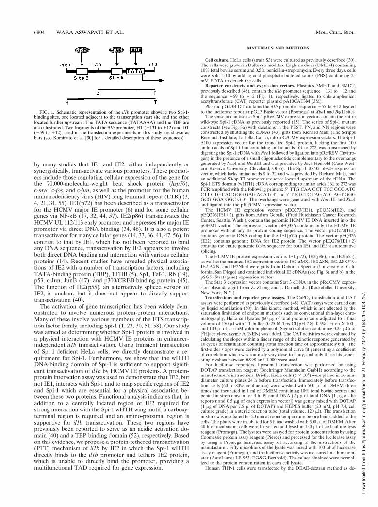
Strong transactivation of the il1b promoter by IE proteinsrequires a single Spi-1-binding site. Consistent with our pre-vious report (24), we found that, in transfected THP-1 mono-cytes, IE proteins upregulate il1b expression in the absence ofthe il1b enhancer (Fig. 2). This expression did not require anyadditional exogenous stimuli, but in combination with LPS, IEproteins supported a stronger (approximately 70-fold) totalinduction. When il1b promoter CAT reporters (HT and DT)carrying single point mutations in Spi-1-binding site A (Fig. 2)were cotransfected with a genomic IE1-IE2 expression vector[pEQ276(IE112)], a dramatic loss of IE protein-dependentactivation was observed. The upstream Spi-1-binding site B,which we previously reported to be essential for enhancer-dependent il1b expression in the absence of IE protein (30),was not required for IE protein-dependent expression (Fig. 2).Mutation at both Spi-1 sites resulted in IE protein-dependentactivity comparable to that observed for the mutation at site Aalone. The il1b promoter fragment containing only site A (DTfragment) demonstrated IE protein-dependent activity compa-rable to that of the longer sequence (HT fragment). Theseresults indicate that Spi-1-binding site A, but not site B, iscritical for IE protein-dependent induction of il1b.
Enhancer-independent il1b transactivation by HCMV IEgene products requires only the Spi-1 DNA-binding domain(wHTH). We have previously reported that potent il1b tran-scription is controlled by two independent elements, an up-
VOL. 19, 1999 IE2 INTERACTION WITH Spi-1/PU.1 ACTIVATES il1b 6805
Dow
nloa
ded
from
http
s://j
ourn
als.
asm
.org
/jour
nal/m
cb o
n 21
Feb
ruar
y 20
22 b
y 16
8.20
5.10
8.25
2.
stream inducible enhancer and a cell-type-specific promoter(48). This requires the binding of transcription factor Spi-1 totwo sites within the promoter (30) (Fig. 1). The HCMV IEproteins support strong il1b transcription in the absence of anenhancer. Mutation of the Spi-1-binding site located adjacentto the transcription start site has indicated a significant role forthis transcription factor in both the presence (24) and absenceof HCMV IE protein (30). However, the direct involvement ofSpi-1 in the HCMV transactivation of il1b has not been dem-onstrated. In order to address this, we transiently transfectedSpi-1-deficient HeLa cells (16), which do not express il1b, evenwhen they are induced by agents such as phorbol myristateacetate (PMA) (8). We have previously demonstrated thattransfection of Spi-1 into HeLa cells can support strong trans-activation of an il1b promoter sequence in the presence of anabbreviated simian virus 40 enhancer (30) but not in the pres-ence of the il1b far-upstream enhancer, as was previously re-ported for monocytes (48).
An enhancerless CAT reporter vector containing the il1bpromoter sequence 2131 to 112 (fragment HT in Fig. 1) wascotransfected into HeLa cells along with a genomic IE1-IE2vector that expresses both HCMV IE1 and IE2. Transfectionswere executed in the presence of various Spi-1 expressionvectors such as antisense Spi-1, wild-type Spi-1, and severalmutated Spi-1 expression vectors containing cDNAs coding fordeleted protein regions (Fig. 3a). As shown in Fig. 3b, theantisense Spi-1 expression vector failed to support IE protein-activated il1b promoter activity. In the absence of both anenhancer and the IE proteins, wild-type Spi-1 induced a 9-foldincrease in CAT activity over that of the control and an addi-tional 2.5-fold increase in activity when it was supplementedwith IE proteins. This confirms the absolute requirement forSpi-1 in il1b transactivation by HCMV IE proteins. Surpris-ingly, an expression vector coding only for the Spi-1 wHTH,which is inactive in the absence of IE, was able to supportsignificant il1b promoter activity when it was cotransfected withthe IE protein expression vector.
The PEST sequence and the TBP-binding domain of Spi-1have been previously shown to be differentially critical for theexpression of various Spi-1-dependent genes (2, 27, 30, 43).However, for enhancer-dependent activity of il1b in the ab-
sence of IE protein, both the TBP and Q domains are required(30). Figure 3 demonstrates that with IE proteins, deletion ofeither the PEST sequence (DPEST vector) or amino acids 8 to32 (D8/32 vector) within the TBP-binding domain of Spi-1 didnot affect the transactivation activities of IE proteins, indicat-ing the dispensability of both of these regions in il1b transac-tivation by IE proteins. Other Spi-1 deletion constructs,namely, those with deletions of the PN sequence, the NNsequence, and the first 100 amino acids of Spi-1 (DPN, DNN,and D100 vectors, respectively), all lacked the Q domain andresulted in a 50% loss of CAT activity when levels were com-pared to that of the wild-type Spi-1 expression vector. Thisresult is similar to what was observed in the absence of IEprotein (30), which suggests that the Spi-1 Q domain is essen-tial for maximal il1b transactivation both in the presence andabsence of IE proteins. Based on these data, we conclude thatHCMV IE proteins use an unique transcriptional activationpathway which partially circumvents the requirement for bothan enhancer and the Spi-1 TAD. Furthermore, the Spi-1 DNA-binding module (wHTH) is sufficient to support significant il1btransactivation by HCMV IE proteins.
The 259-to-112 il1b promoter sequence functions as an IEprotein-dependent and enhancer-independent promoter. Wehave previously reported that in the presence of IE proteinsthe il1b promoter from 2131 to 112 (region HT in Fig. 1) isable to support transactivation in the absence of the normallyrequired far-upstream enhancer. Within this promoter se-quence, there are two binding sites for Spi-1 (Fig. 1). Theproximal Spi-1-binding site is located between 250 and 239and has been shown to be crucial for il1b transactivation inboth the presence and absence of HCMV IE proteins. How-ever, the requirement for the more upstream Spi-1-binding sitelocated between 2115 and 297 has been determined only inthe absence of IE protein (30).
Transient transfections were executed with HeLa cells. Anenhancerless CAT reporter containing either the il1b promotersequence 2131 to 112 (fragment HT; which contains both ofthe Spi-1-binding sites) or the shorter sequence 259 to 112(fragment DT; which contains only the TATA-box-proximalSpi-1 site) was cotransfected with the IE1-IE2 expression vec-tor. The results shown in Fig. 4 argue that in the presence ofSpi-1, the 259-to-112 il1b promoter (DT) can function aseffectively as the 2131-to-112 promoter (HT) in IE protein(enhancer-independent) transactivation of il1b, resulting inabout a 20-fold-greater activity than that of the control. Theactivities of IE protein-induced HT and DT promoter frag-ments in the presence of the Spi-1 wHTH expression vector arealso indistinguishable from each other and are about 50% ofthe activity mediated by the full-length Spi-1 expression vector.Therefore, the il1b promoter sequence from 259 to 112 servesas a minimal IE protein-dependent (enhancer-independent)promoter requiring only one Spi-1-binding site.
Both IE1 and IE2 synergistically transactivate il1b. To de-termine the role of individual IE proteins in il1b transactiva-tion, HeLa cells were cotransfected with an il1b promoter CATreporter and Spi-1 expression vectors along with one of threedifferent IE protein genomic expression vectors: pEQ273(IE1),pEQ326(IE2), and pEQ276(IE112). Whole-cell extracts fromthe transfected HeLa cells were analyzed by SDS-PAGE andWestern blotting with an antibody directed against both IE1and IE2 in order to confirm the expression of IE proteins inHeLa cells (data not shown). Figure 5 reveals that in thepresence of the Spi-1 expression vector, IE2 alone significantlyinduced il1b promoter CAT activity and that IE1 did not.Synergy was observed when both the IE1 and IE2 expressionvectors were cotransfected along with the Spi-1 expression
FIG. 2. The il1b promoter is strongly transactivated by IE proteins in humanTHP-1 monocytes. Shown are the CAT activities of the il1b promoter CATreporters (HT and DT) containing point mutations at the distal (B) and proximal(A) Spi-1-binding sites, respectively, and cotransfected with a genomic IE1-IE2expression vector [pEQ276(IE112)] which expresses both IE1 and IE2. Theresults are expressed as average percentages of the relative CAT activity ob-served for the wild-type HT promoter when it was cotransfected with the IE1-IE2expression vector. Open bars correspond to unstimulated cells, whereas filledbars correspond to cells treated with 10 ng of LPS per ml. Standard deviationsare indicated for a minimum of three repetitions. mA, mutation at site A; mB,mutation at site B; mA,B, mutations at both sites.
6806 WARA-ASWAPATI ET AL. MOL. CELL. BIOL.
Dow
nloa
ded
from
http
s://j
ourn
als.
asm
.org
/jour
nal/m
cb o
n 21
Feb
ruar
y 20
22 b
y 16
8.20
5.10
8.25
2.
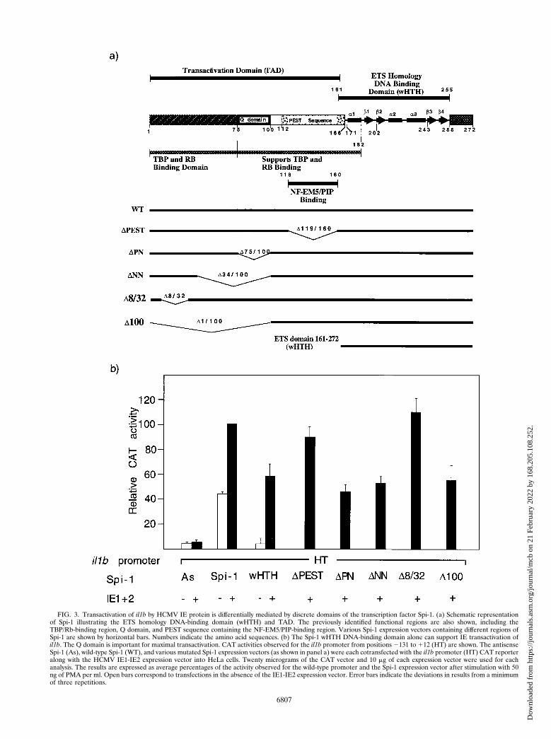
FIG. 3. Transactivation of il1b by HCMV IE protein is differentially mediated by discrete domains of the transcription factor Spi-1. (a) Schematic representationof Spi-1 illustrating the ETS homology DNA-binding domain (wHTH) and TAD. The previously identified functional regions are also shown, including theTBP/Rb-binding region, Q domain, and PEST sequence containing the NF-EM5/PIP-binding region. Various Spi-1 expression vectors containing different regions ofSpi-1 are shown by horizontal bars. Numbers indicate the amino acid sequences. (b) The Spi-1 wHTH DNA-binding domain alone can support IE transactivation ofil1b. The Q domain is important for maximal transactivation. CAT activities observed for the il1b promoter from positions 2131 to 112 (HT) are shown. The antisenseSpi-1 (As), wild-type Spi-1 (WT), and various mutated Spi-1 expression vectors (as shown in panel a) were each cotransfected with the il1b promoter (HT) CAT reporteralong with the HCMV IE1-IE2 expression vector into HeLa cells. Twenty micrograms of the CAT vector and 10 mg of each expression vector were used for eachanalysis. The results are expressed as average percentages of the activity observed for the wild-type promoter and the Spi-1 expression vector after stimulation with 50ng of PMA per ml. Open bars correspond to transfections in the absence of the IE1-IE2 expression vector. Error bars indicate the deviations in results from a minimumof three repetitions.
6807
Dow
nloa
ded
from
http
s://j
ourn
als.
asm
.org
/jour
nal/m
cb o
n 21
Feb
ruar
y 20
22 b
y 16
8.20
5.10
8.25
2.
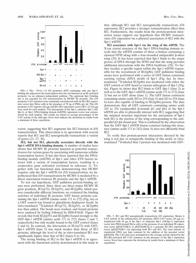
vector, suggesting that IE1 augments the IE2 function in il1btransactivation. This observation is in agreement with severalreports that IE1 and IE2 synergistically transactivate a varietyof genes (21, 37, 38).
IE2, but not IE1, physically associates directly with theSpi-1 wHTH DNA-binding domain. A number of studies haveshown that HCMV IE proteins function as powerful transac-tivators for various genes by associating with numerous cellulartranscription factors. It has also been reported that the DNA-binding module (wHTH) of Spi-1 and other ETS factors in-teract with a variety of transcription factors, resulting in acooperative gene activation (reviewed in reference 1). To-gether with our functional data demonstrating that HCMVrequires only the Spi-1 wHTH for il1b transactivation, we hy-pothesized that il1b transactivation by HCMV is mediated by adirect association between IE proteins and the Spi-1 wHTH.
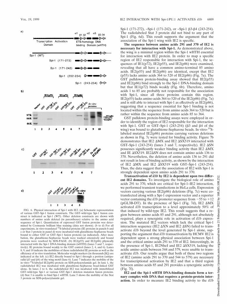
To test our hypothesis, GST pulldown protein-binding as-says were performed. Since there are three major HCMV IEgene products, IE1(p72), IE2(p55), and IE2(p86), which pos-sess considerably different functions, we evaluated the bindingactivities of individual IE proteins. GST fusion proteins con-taining the Spi-1 wHTH (amino acids 171 to 272) (Fig. 6a) ora GST control was bound to glutathione-Sepharose beads. Invitro-translated 35S-labeled IE1(p72), IE2(p55), or IE2(p86)was then added. The beads were washed, and bound proteinswere analyzed by boiling followed by SDS-PAGE. Figure 6breveals that both IE2(p55) and IE2(p86) bound strongly to theGST–Spi-1 wHTH (amino acids 171 to 272) (lanes 3 and 7,respectively) but only weakly bound to the GST control (lanes2 and 6). In contrast, the interaction of IE1(p72) with GST-Spi-1 wHTH (lane 5) was much weaker than those of IE2proteins, although the level of the in vitro-translated IE1 wassignificantly higher than that of IE2 (lanes 8 to 10).
The strong binding of IE2 to the Spi-1 wHTH is in agree-ment with the functional activity demonstrated in this study in
that, although IE1 and IE2 synergistically transactivate il1bexpression, IE2 provides a stronger transactivation effect thanIE1. Furthermore, the results from the protein-protein inter-action assays support our hypothesis that HCMV transacti-vates il1b expression via a physical association of IE2 with theSpi-1 wHTH.
IE2 associates with Spi-1 via the wing of the wHTH. TheX-ray crystal structure of the Spi-1 DNA-binding domain re-veals that the wHTH consists of three a-helices containing aclassical HTH along with a four-stranded antiparallel b-sheetwing (29). It has been reported that Spi-1 binds to the majorgroove of DNA through the HTH and that the wing providesadditional interactions with the DNA backbone (29). To fur-ther localize a specific region within the Spi-1 wHTH respon-sible for the recruitment of IE2(p86), GST pulldown bindingassays were performed with a series of GST fusion constructscarrying various cDNA motifs of Spi-1 (Fig. 6a). In vitro-translated 35S-labeled IE2(p86) was incubated with either theGST control or GST fused to various subregions of Spi-1 (Fig.6a). Figure 6c shows that IE2 binds to GST–Spi-1 (lane 2) aswell as to the GST–Spi-1 wHTH (amino acids 171 to 272) (lane3) but not to GST alone (lane 1). The GST fusion constructscontaining amino acids 202 to 254 (lane 5) and 243 to 254 (lane6) were also capable of binding to IE2(p86) protein. The datademonstrate that all GST constructs containing amino acids243 to 254 (antiparallel b-strands b3 and b4) of the Spi-1wHTH are able to physically interact with IE2, suggesting thatthe minimal structure important for the association of Spi-1with IE2 is the portion of the wing corresponding to the anti-parallel b3-b4 strand pair. This is confirmed by the observationthat the GST–Spi-1 wHTH fusion construct lacking this struc-ture (amino acids 171 to 242) (lane 4) does not efficiently bindIE2.
To verify that protein-protein interaction directed by thewing is not due to nonspecific binding of this motif, in vitro-translated 35S-labeled Stat 3 protein was incubated with GST–
FIG. 4. The 259-to-112 il1b promoter (DT) containing only one Spi-1-binding site adjacent to the transcription start site can function as an IE-activatedpromoter via an enhancer-independent pathway. The upstream Spi-1-bindingsite is not essential for il1b transactivation by HCMV IE proteins. The il1bpromoter CAT reporters were transiently cotransfected with an IE1-IE2 expres-sion vector into HeLa cells in the presence of 50 ng of PMA per ml. The il1bpromoter CAT reporter (20 mg) and IE1-IE2 expression vector (IE112) (10 mg)were used for each analysis. Ten micrograms of the Spi-1, antisense Spi-1 (AS.Spi-1), or Spi-1 DNA-binding domain (wHTH) expression vector was cotrans-fected for each analysis. The results are shown as average percentages of theCAT activity of the wild type. Error bars indicate the deviations in results froma minimum of three repetitions.
FIG. 5. IE1 and IE2 synergistically transactivate il1b expression. Shown isCAT activity of the enhancerless il1b promoter (HT) CAT vector (20 mg) co-transfected with 10 mg of the Spi-1 or antisense Spi-1 (AS.Spi-1) expressionvector into PMA-treated HeLa cells, in the presence of an individual IE expres-sion vector [pEQ273(IE1) or pEQ326(IE2)] or a genomic IE1-IE2 expressionvector [pEQ276(IE112)] expressing both IE1 and IE2. The total amount oftransfected DNA was kept constant by the addition of the parental vector[pEQ336]. The CAT data were normalized to the average activity elicited by theIE1-IE2-activated HT CAT construct in the presence of the Spi-1 expressionvector. Error bars represent the deviations in results from a minimum of threerepetitions.
6808 WARA-ASWAPATI ET AL. MOL. CELL. BIOL.
Dow
nloa
ded
from
http
s://j
ourn
als.
asm
.org
/jour
nal/m
cb o
n 21
Feb
ruar
y 20
22 b
y 16
8.20
5.10
8.25
2.
Spi-1 (171-272), –Spi-1 (171-242), or –Spi-1 b3-b4 (243-254).The radiolabeled Stat 3 protein did not bind to any part ofSpi-1 (Fig. 6d). This result supports the argument that theassociation of the Spi-1 wing with IE2 is specific.
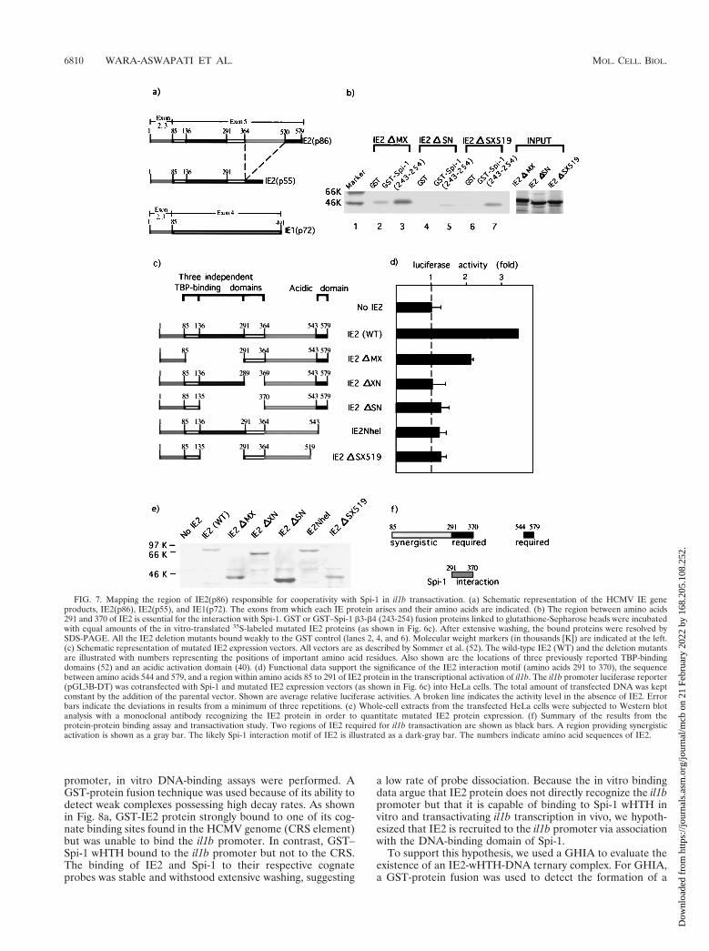
The sequence between amino acids 291 and 370 of IE2 isnecessary for interaction with Spi-1. As demonstrated above,the wing is a minimal region within the Spi-1 wHTH essentialfor interaction with IE2 protein. In order to map a specificregion of IE2 responsible for interaction with Spi-1, the se-quences of IE1(p72), IE2(p55), and IE2(p86) were examined,revealing that all have a common amino-terminal 85 aminoacids. IE2(p55) and IE2(p86) are identical, except that IE2(p55) lacks amino acids 364 to 520 of IE2(p86) (Fig. 7a). TheGST pulldown protein-binding assay showed that IE2(p55)and IE2(p86) bind strongly to the Spi-1 DNA-binding domainbut that IE1(p72) binds weakly (Fig. 6b). Therefore, aminoacids 1 to 85 are probably not responsible for the associationwith Spi-1, since all three proteins contain this region.IE2(p55) lacks amino acids 364 to 520 of the IE2(p86) (Fig. 7a)and is still able to interact with Spi-1 as effectively as IE2(p86),suggesting that a sequence essential for Spi-1 binding is notlocated within the sequence from amino acids 364 to 520 but israther within the sequence from amino acids 85 to 364.
GST pulldown protein-binding assays were employed in or-der to identify the region of IE2 responsible for the interactionwith Spi-1. GST or GST–Spi-1 (243-254) (b3 and b4 of thewing) was bound to glutathione-Sepharose beads. In vitro-35S-labeled mutated IE2(p86) proteins carrying various deletionsas shown in Fig. 7c were tested for binding activity. Figure 7bdemonstrates that IE2 DMX and IE2 DSX519 interacted withGST–Spi-1 (243-254) (lanes 3 and 7, respectively). IE2 DSNpossesses significantly weaker binding activity than IE2 DMXand IE DSX519. IE2DSN does not contain amino acids 136 to370. Nevertheless, the deletion of amino acids 136 to 291 didnot result in loss of binding activity, as shown by the interactionof IE2 DMX and IE2 DSX519 with GST–Spi-1 (243-254).Thus, the data suggest that the association of IE2 with Spi-1 isstrongly dependent upon amino acids 291 to 370.
Transactivation of il1b by IE2 is dependent upon two differ-ent IE2 domains. To investigate the biological role of aminoacids 291 to 370, which are critical for Spi-1–IE2 interaction,we performed transient transfections in HeLa cells. Expressionvectors carrying various IE2(p86) deletions (Fig. 7c) were co-transfected along with a Spi-1 expression vector and a reportervector containing the il1b promoter sequence from 255 to 112(pGL3B-DT). In the presence of Spi-1 (Fig. 7d), IE2 DMXactivated il1b transcription to a level approximately 50% ofthat induced by wild-type IE2. This result suggests that a re-gion between amino acids 85 and 291, although not absolutelyrequired, plays a synergistic role in activation of il1b expres-sion. The mutated IE2 vectors which lack the critical Spi-1interaction sequence (IE2 DXN and IE2 DSN) failed to trans-activate il1b beyond the level generated by Spi-1 alone, sup-porting the argument that il1b transactivation by HCMV IE2 isdependent upon a strong physical association between Spi-1and the critical amino acids 291 to 370 of IE2. Interestingly, inthe presence of Spi-1, IE2NheI and IE2 DSX519, lacking theacidic amino acids between 544 and 579, were unable to trans-activate il1b. Our results argue that both of these two regionsof IE2 (amino acids 291 to 370 and 544 to 579) are necessaryfor transcriptional activation by IE2 and that a third regionbetween amino acids 85 and 291 provides a synergistic activity(Fig. 7f).
IE2 and the Spi-1 wHTH DNA-binding domain form a ter-nary complex with DNA that requires a protein-protein inter-action. In order to measure IE2 binding activity to the il1b
FIG. 6. Physical interaction of Spi-1 with IE2. (a) Schematic representationof various GST–Spi-1 fusion constructs. The GST–wild-type Spi-1 fusion con-struct is indicated as Spi-1 (WT). Other deletion constructs are shown withnumbers of amino acids deleted (in parentheses) relative to the amino acidsequence of Spi-1. Quantitation of expressed GST fusion proteins was deter-mined by Coomassie brilliant blue staining (data not shown). (b to d) For allexperiments, in vitro-translated 35S-labeled proteins (IE proteins in panels b andc or Stat 3 protein in panel d) were incubated with glutathione-Sepharose beadsbound to either GST or GST–Spi-1 fusion proteins (as indicated). After incu-bation, the glutathione-Sepharose beads were washed extensively and boundproteins were resolved by SDS-PAGE. (b) IE2(p55) and IE2(p86) physicallyinteracted with the Spi-1 DNA-binding domain (wHTH) (lanes 3 and 7, respec-tively). IE proteins bound weakly to the GST control (lanes 2, 4, and 6). Lanes8, 9, and 10 indicate the mobilities of these radiolabeled IE proteins as well as theefficiency of in vitro translation. Molecular weight markers (in thousands [K]) areindicated at the left. (c) IE2 directly bound to Spi-1 through a portion (antipa-rallel b3 and b4) of the wing motif (lane 6). Lane 7 indicates the mobility of thein vitro-35S-labeled IE2(p86) protein on SDS-polyacrylamide gel. In lane 1, theradiolabeled IE2 was incubated with glutathione-Sepharose bead-linked GSTalone. In lanes 2 to 6, the radiolabeled IE2 was incubated with immobilizedGST–wild-type Spi-1 or various GST–Spi-1 deletion mutation fusion proteins.(d) Stat 3 is unable to bind Spi-1 wHTH. Lane 5 shows the mobility of the Stat3 protein on SDS-polyacrylamide gel.
VOL. 19, 1999 IE2 INTERACTION WITH Spi-1/PU.1 ACTIVATES il1b 6809
Dow
nloa
ded
from
http
s://j
ourn
als.
asm
.org
/jour
nal/m
cb o
n 21
Feb
ruar
y 20
22 b
y 16
8.20
5.10
8.25
2.
promoter, in vitro DNA-binding assays were performed. AGST-protein fusion technique was used because of its ability todetect weak complexes possessing high decay rates. As shownin Fig. 8a, GST-IE2 protein strongly bound to one of its cog-nate binding sites found in the HCMV genome (CRS element)but was unable to bind the il1b promoter. In contrast, GST–Spi-1 wHTH bound to the il1b promoter but not to the CRS.The binding of IE2 and Spi-1 to their respective cognateprobes was stable and withstood extensive washing, suggesting
a low rate of probe dissociation. Because the in vitro bindingdata argue that IE2 protein does not directly recognize the il1bpromoter but that it is capable of binding to Spi-1 wHTH invitro and transactivating il1b transcription in vivo, we hypoth-esized that IE2 is recruited to the il1b promoter via associationwith the DNA-binding domain of Spi-1.
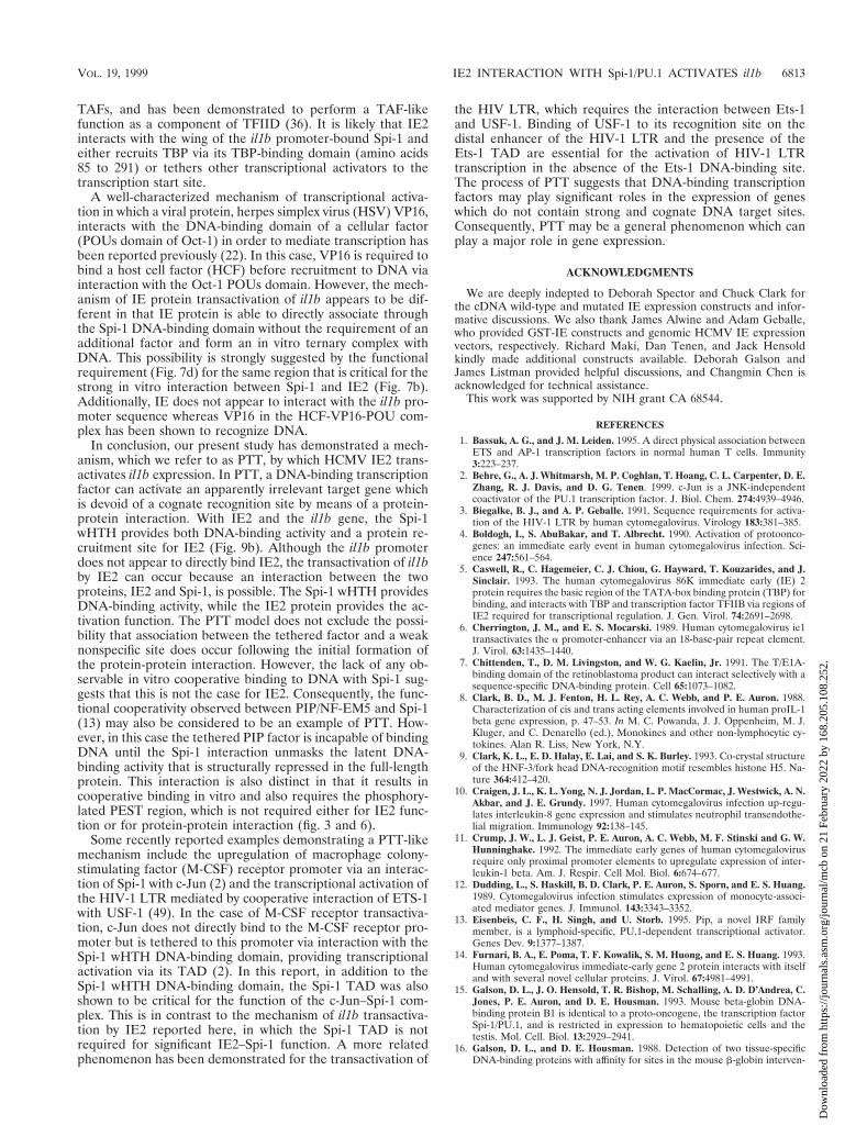
To support this hypothesis, we used a GHIA to evaluate theexistence of an IE2-wHTH-DNA ternary complex. For GHIA,a GST-protein fusion was used to detect the formation of a
FIG. 7. Mapping the region of IE2(p86) responsible for cooperativity with Spi-1 in il1b transactivation. (a) Schematic representation of the HCMV IE geneproducts, IE2(p86), IE2(p55), and IE1(p72). The exons from which each IE protein arises and their amino acids are indicated. (b) The region between amino acids291 and 370 of IE2 is essential for the interaction with Spi-1. GST or GST–Spi-1 b3-b4 (243-254) fusion proteins linked to glutathione-Sepharose beads were incubatedwith equal amounts of the in vitro-translated 35S-labeled mutated IE2 proteins (as shown in Fig. 6c). After extensive washing, the bound proteins were resolved bySDS-PAGE. All the IE2 deletion mutants bound weakly to the GST control (lanes 2, 4, and 6). Molecular weight markers (in thousands [K]) are indicated at the left.(c) Schematic representation of mutated IE2 expression vectors. All vectors are as described by Sommer et al. (52). The wild-type IE2 (WT) and the deletion mutantsare illustrated with numbers representing the positions of important amino acid residues. Also shown are the locations of three previously reported TBP-bindingdomains (52) and an acidic activation domain (40). (d) Functional data support the significance of the IE2 interaction motif (amino acids 291 to 370), the sequencebetween amino acids 544 and 579, and a region within amino acids 85 to 291 of IE2 protein in the transcriptional activation of il1b. The il1b promoter luciferase reporter(pGL3B-DT) was cotransfected with Spi-1 and mutated IE2 expression vectors (as shown in Fig. 6c) into HeLa cells. The total amount of transfected DNA was keptconstant by the addition of the parental vector. Shown are average relative luciferase activities. A broken line indicates the activity level in the absence of IE2. Errorbars indicate the deviations in results from a minimum of three repetitions. (e) Whole-cell extracts from the transfected HeLa cells were subjected to Western blotanalysis with a monoclonal antibody recognizing the IE2 protein in order to quantitate mutated IE2 protein expression. (f) Summary of the results from theprotein-protein binding assay and transactivation study. Two regions of IE2 required for il1b transactivation are shown as black bars. A region providing synergisticactivation is shown as a gray bar. The likely Spi-1 interaction motif of IE2 is illustrated as a dark-gray bar. The numbers indicate amino acid sequences of IE2.
6810 WARA-ASWAPATI ET AL. MOL. CELL. BIOL.
Dow
nloa
ded
from
http
s://j
ourn
als.
asm
.org
/jour
nal/m
cb o
n 21
Feb
ruar
y 20
22 b
y 16
8.20
5.10
8.25
2.
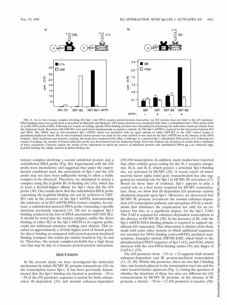
ternary complex involving a second unlabeled protein and aradiolabeled DNA probe (Fig. 8b). Experiments with the il1bprobe were inconclusive and suggested that under the experi-mental conditions used, the association of Spi-1 and the il1bprobe may not have been sufficiently strong to allow a stablecomplex to be observed. Therefore, we attempted to detect acomplex using the b-globin B1-A-binding site (16), which hasat least a fivefold-higher affinity for Spi-1 than did the il1bprobe (30). Our results show that the radiolabeled DNA probecontaining the b-globin binding site can be tethered to GST-IE2 only in the presence of the Spi-1 wHTH, demonstratingthe existence of an IE2-wHTH-DNA ternary complex. In con-trast, a radiolabeled mutated DNA probe containing a specificmutation previously reported (15, 30) not to support Spi-1binding resulted in the loss of DNA association with GST-IE2.It should be noted that the ternary complex, unlike the directbinding of either IE2 or the Spi-1 wHTH to its cognate probe,could not withstand extensive washing (not shown). This re-sulted in approximately a 10-fold higher level of bound probefor direct binding as compared with protein-protein-mediatedbinding (compare the counts per minute bound in Fig. 8a andb). Therefore, the ternary complex probably has a high decayrate that may be due to a tenuous protein-protein interaction.
DISCUSSION
In the present study, we have investigated the molecularmechanism by which HCMV IE proteins transactivate il1b viathe transcription factor Spi-1. It has been previously demon-strated that the Spi-1 binding site located at positions 250 to239 of the il1b promoter sequence is crucial for both enhanc-erless IE-dependent (24) and normal enhancer-dependent
(30) il1b transcription. In addition, many studies have reportedthat other cellular genes coding for the IL-1 receptor antago-nist, IL-6, and IL-8, which possess a potential Spi-1-bindingsite, are activated by HCMV (28). A recent report of tumornecrosis factor alpha (tnfa) gene transactivation has also sug-gested an essential role for Spi-1 in HCMV IE activation (17).Based on these lines of evidence, Spi-1 appears to play acentral role as a host factor required for HCMV transactiva-tion. Here, we show that IE-dependent il1b promoter activityabsolutely depends upon Spi-1. Moreover, we discovered thatHCMV IE proteins circumvent the normal enhancer-depen-dent il1b transcription pathway and upregulate il1b by a mech-anism that eliminates the requirement not only for an en-hancer but also, to a significant degree, for the Spi-1 TAD.This TAD is required for enhancer-dependent transcription inthe absence of HCMV IE (30). In the presence of IE, only theSpi-1 wHTH DNA-binding domain is required to support sig-nificant il1b expression. This observation is distinct from thosemade with some other systems in which additional sequencesnot essential for DNA binding (extra-wHTH) mediated tran-scription. Examples include PIP/NF-EM5, which binds to thephosphorylated PEST sequence of Spi-1 (42), and FOG, whichinteracts with the non-DNA-binding amino (N) zinc finger ofGATA-1 (53).
The il1b promoter from 2131 to 112 supports both normalenhancer-dependent and IE protein-mediated transcription(11, 24, 30). Within this promoter, there are two Spi-1-bindingsites, one located adjacent to the transcription start site and theother located further upstream (Fig. 1), raising the question ofwhether the functions of these two sites are different for il1btransactivation by HCMV IE proteins. In the absence of IEproteins, a shorter 259-to-112 il1b promoter is inactive (30).
FIG. 8. An in vitro ternary complex involving IE2–Spi-1 and DNA requires protein-protein interaction. (a) IE2 protein does not bind to the il1b promoter.DNA-binding assays were performed as described in Materials and Methods. GST fusion proteins were incubated with either a radiolabeled Spi-1 DNA probe (il1b)or an IE2 DNA probe (CRS). Following two rounds of washing, specific DNA-binding activities were determined by measuring the radioactive counts per minute fromthe Sepharose beads. Reactions with GST-IE1 were performed simultaneously as negative controls. (b) The Spi-1 wHTH is required for the interaction between IE2and DNA. The GHIA used in vitro-translated Spi-1 wHTH, which was incubated with an equal amount of either GST-IE2 or the GST control bound toglutathione-Sepharose beads. The in vitro-translated control protein was made by the same method as was used for the Spi-1 wHTH but in the absence of the DNAtemplate. After incubation and extensive washing, the beads were incubated with either a wild-type or a mutated Spi-1 radiolabeled DNA probe (15). Following tworounds of washing, the amount of bound radioactive probe was determined from the Sepharose beads. Error bars indicate the deviations in results from a minimumof three repetitions. Cartoons explain the details of the experiment in which the natures of individual proteins and radiolabeled DNAs ( ) are indicated. bgb,b-globin-binding site; mbgb, mutated b-globin-binding site.
VOL. 19, 1999 IE2 INTERACTION WITH Spi-1/PU.1 ACTIVATES il1b 6811
Dow
nloa
ded
from
http
s://j
ourn
als.
asm
.org
/jour
nal/m
cb o
n 21
Feb
ruar
y 20
22 b
y 16
8.20
5.10
8.25
2.

We now demonstrate that, in the presence of IE proteins, the259-to-112 promoter can function as effectively as the 2131-to-112 promoter, indicating that the presence of the upstreamSpi-1-binding site, which is normally required, is not impera-tive for activation by IE. In other words, this finding demon-strates that the IE proteins specifically target the proximalSpi-1-binding site in order to activate the expression of il1b.
Our study shows that IE2 protein plays a significant role inil1b transactivation. Since IE2 is able to transactivate il1b, eventhough there is no IE consensus binding site (34, 46) in the il1bpromoter and we cannot detect any in vitro binding, it is notunreasonable to speculate that IE2 activates il1b transcriptionby, at least in part, directly interacting with Spi-1 bound to il1b.Spi-1 has been reported to bind various viral proteins in theactivation of cellular genes. An interaction between Spi-1 andEBNA-2 has been shown to play an important role in latentmembrane protein 1 (LMP1) gene activation (26). Further-more, we recently showed that il1b transactivation by humanT-cell leukemia virus type 1 Tax protein likely involves thecooperation of Tax with Spi-1 and with NF-IL6 (C/EBPb) (54).None of these reports identified a specific region of Spi-1responsible for the interaction with viral proteins. The findingsin our present study have confirmed a direct binding of Spi-1with IE2 protein and indicate that a specific region betweenamino acids 291 and 370 of IE2 is important for a strongphysical association with a portion (antiparallel b3 and b4strands) of the Spi-1 wHTH wing. Our in vitro experimentsfurther reveal that IE2 is able to form a Spi-1 wHTH-depen-dent ternary complex on DNA. These data appear to supporta mechanism for IE2-dependent transcriptional activation me-diated by direct interaction with il1b promoter-bound Spi-1.Moreover, we find that both IE1 and IE2 synergistically up-regulate il1b expression, although direct IE1 interaction withSpi-1 could not be detected. The mechanism of synergy be-tween the two IE proteins is not clearly understood. It has beendemonstrated that activation by individual IE proteins is de-
pendent upon both the promoter-regulatory region of the cel-lular gene and the cell type (39). A report that IE1 does notdirectly interact with IE2 (14) argues that the cooperativeactivation of il1b is unlikely to occur via physical associationbetween IE1 and IE2. Since IE1 has been shown to transacti-vate a variety of genes, including that coding for IE2 (38), it ispossible that IE1 induces the expression of a coactivator, whichin turn is able to directly bind IE2 and upregulate il1b tran-scription.
The wing of the wHTH proteins, including Spi-1, all otherETS factors, and various non-ETS wHTH proteins such asheat shock factor (HSF), is a small b-sheet consisting of threeor four antiparallel b-strands and their interconnecting loops,which extend to one side of the triple a-helical bundle con-taining the HTH (9, 29). We have recently determined thatGATA-1 and GATA-2 are also capable of interacting withSpi-1 via the wing (59). In addition, a recent crystal structurereport reveals that HSF multimerization on DNA occurs viawing-mediated protein-protein interaction (35). Therefore, thewHTH wing may represent a site for protein-protein interac-tion among many other diverse factors that have been reportedto functionally cooperate in transcriptional activation.
In accordance with our observation that IE2 transactivationof il1b requires only the Spi-1 wHTH DNA-binding domainbut not an enhancer or the Spi-1 TAD, we hypothesize thatIE2 functions by providing a TAD normally supplied by theSpi-1 TAD in cooperation with critical factors that bind to theil1b upstream enhancer. This speculation is supported by ourfunctional data which indicate that, in addition to the putativeinteraction motif in IE2 (amino acids 291 to 370), amino acids544 to 579, which contain a potent independently acting acidicTAD (40), are also necessary for activation. In addition, theamino acid sequence between 85 and 291 of IE2, containingtwo previously reported TBP-binding domains (Fig. 7c), ap-pears to be important for maximal il1b transactivation. IE2 hasbeen reported to bind several transcription factors, including
FIG. 9. Proposed model of PTT-mediated transcriptional activation of il1b by HCMV IE2. (a) Potent il1b transcription normally requires both a far-upstreamenhancer and the 2131-to-112 il1b promoter which contains two essential Spi-1-binding sites. Two Spi-1 TAD subregions are also required. One of these is aglutamine-rich (Q) domain (hatched areas), and the other directly binds TBP (filled areas). The Spi-1 wHTH DNA-binding domain is shown as an oval, with the wingindicated by “w”. (b) IE2 replaces the functions of the Spi-1 TBP-binding domain and the il1b enhancer. Furthermore, only a minimal il1b promoter located between259 and 112 is sufficient to support IE-activated il1b expression. IE2 is tethered to the il1b promoter via a direct interaction with the Spi-1 wing and possibly interactswith TBP. Also, an acidic domain of IE2 (shown by a minus sign) provides an activation effect eliminating the absolute requirement for the Spi-1 TAD.
6812 WARA-ASWAPATI ET AL. MOL. CELL. BIOL.
Dow
nloa
ded
from
http
s://j
ourn
als.
asm
.org
/jour
nal/m
cb o
n 21
Feb
ruar
y 20
22 b
y 16
8.20
5.10
8.25
2.
TAFs, and has been demonstrated to perform a TAF-likefunction as a component of TFIID (36). It is likely that IE2interacts with the wing of the il1b promoter-bound Spi-1 andeither recruits TBP via its TBP-binding domain (amino acids85 to 291) or tethers other transcriptional activators to thetranscription start site.
A well-characterized mechanism of transcriptional activa-tion in which a viral protein, herpes simplex virus (HSV) VP16,interacts with the DNA-binding domain of a cellular factor(POUs domain of Oct-1) in order to mediate transcription hasbeen reported previously (22). In this case, VP16 is required tobind a host cell factor (HCF) before recruitment to DNA viainteraction with the Oct-1 POUs domain. However, the mech-anism of IE protein transactivation of il1b appears to be dif-ferent in that IE protein is able to directly associate throughthe Spi-1 DNA-binding domain without the requirement of anadditional factor and form an in vitro ternary complex withDNA. This possibility is strongly suggested by the functionalrequirement (Fig. 7d) for the same region that is critical for thestrong in vitro interaction between Spi-1 and IE2 (Fig. 7b).Additionally, IE does not appear to interact with the il1b pro-moter sequence whereas VP16 in the HCF-VP16-POU com-plex has been shown to recognize DNA.
In conclusion, our present study has demonstrated a mech-anism, which we refer to as PTT, by which HCMV IE2 trans-activates il1b expression. In PTT, a DNA-binding transcriptionfactor can activate an apparently irrelevant target gene whichis devoid of a cognate recognition site by means of a protein-protein interaction. With IE2 and the il1b gene, the Spi-1wHTH provides both DNA-binding activity and a protein re-cruitment site for IE2 (Fig. 9b). Although the il1b promoterdoes not appear to directly bind IE2, the transactivation of il1bby IE2 can occur because an interaction between the twoproteins, IE2 and Spi-1, is possible. The Spi-1 wHTH providesDNA-binding activity, while the IE2 protein provides the ac-tivation function. The PTT model does not exclude the possi-bility that association between the tethered factor and a weaknonspecific site does occur following the initial formation ofthe protein-protein interaction. However, the lack of any ob-servable in vitro cooperative binding to DNA with Spi-1 sug-gests that this is not the case for IE2. Consequently, the func-tional cooperativity observed between PIP/NF-EM5 and Spi-1(13) may also be considered to be an example of PTT. How-ever, in this case the tethered PIP factor is incapable of bindingDNA until the Spi-1 interaction unmasks the latent DNA-binding activity that is structurally repressed in the full-lengthprotein. This interaction is also distinct in that it results incooperative binding in vitro and also requires the phosphory-lated PEST region, which is not required either for IE2 func-tion or for protein-protein interaction (fig. 3 and 6).
Some recently reported examples demonstrating a PTT-likemechanism include the upregulation of macrophage colony-stimulating factor (M-CSF) receptor promoter via an interac-tion of Spi-1 with c-Jun (2) and the transcriptional activation ofthe HIV-1 LTR mediated by cooperative interaction of ETS-1with USF-1 (49). In the case of M-CSF receptor transactiva-tion, c-Jun does not directly bind to the M-CSF receptor pro-moter but is tethered to this promoter via interaction with theSpi-1 wHTH DNA-binding domain, providing transcriptionalactivation via its TAD (2). In this report, in addition to theSpi-1 wHTH DNA-binding domain, the Spi-1 TAD was alsoshown to be critical for the function of the c-Jun–Spi-1 com-plex. This is in contrast to the mechanism of il1b transactiva-tion by IE2 reported here, in which the Spi-1 TAD is notrequired for significant IE2–Spi-1 function. A more relatedphenomenon has been demonstrated for the transactivation of
the HIV LTR, which requires the interaction between Ets-1and USF-1. Binding of USF-1 to its recognition site on thedistal enhancer of the HIV-1 LTR and the presence of theEts-1 TAD are essential for the activation of HIV-1 LTRtranscription in the absence of the Ets-1 DNA-binding site.The process of PTT suggests that DNA-binding transcriptionfactors may play significant roles in the expression of geneswhich do not contain strong and cognate DNA target sites.Consequently, PTT may be a general phenomenon which canplay a major role in gene expression.
ACKNOWLEDGMENTS
We are deeply indepted to Deborah Spector and Chuck Clark forthe cDNA wild-type and mutated IE expression constructs and infor-mative discussions. We also thank James Alwine and Adam Geballe,who provided GST-IE constructs and genomic HCMV IE expressionvectors, respectively. Richard Maki, Dan Tenen, and Jack Hensoldkindly made additional constructs available. Deborah Galson andJames Listman provided helpful discussions, and Changmin Chen isacknowledged for technical assistance.
This work was supported by NIH grant CA 68544.
REFERENCES
1. Bassuk, A. G., and J. M. Leiden. 1995. A direct physical association betweenETS and AP-1 transcription factors in normal human T cells. Immunity3:223–237.
2. Behre, G., A. J. Whitmarsh, M. P. Coghlan, T. Hoang, C. L. Carpenter, D. E.Zhang, R. J. Davis, and D. G. Tenen. 1999. c-Jun is a JNK-independentcoactivator of the PU.1 transcription factor. J. Biol. Chem. 274:4939–4946.
3. Biegalke, B. J., and A. P. Geballe. 1991. Sequence requirements for activa-tion of the HIV-1 LTR by human cytomegalovirus. Virology 183:381–385.
4. Boldogh, I., S. AbuBakar, and T. Albrecht. 1990. Activation of protoonco-genes: an immediate early event in human cytomegalovirus infection. Sci-ence 247:561–564.
5. Caswell, R., C. Hagemeier, C. J. Chiou, G. Hayward, T. Kouzarides, and J.Sinclair. 1993. The human cytomegalovirus 86K immediate early (IE) 2protein requires the basic region of the TATA-box binding protein (TBP) forbinding, and interacts with TBP and transcription factor TFIIB via regions ofIE2 required for transcriptional regulation. J. Gen. Virol. 74:2691–2698.
6. Cherrington, J. M., and E. S. Mocarski. 1989. Human cytomegalovirus ie1transactivates the a promoter-enhancer via an 18-base-pair repeat element.J. Virol. 63:1435–1440.
7. Chittenden, T., D. M. Livingston, and W. G. Kaelin, Jr. 1991. The T/E1A-binding domain of the retinoblastoma product can interact selectively with asequence-specific DNA-binding protein. Cell 65:1073–1082.
8. Clark, B. D., M. J. Fenton, H. L. Rey, A. C. Webb, and P. E. Auron. 1988.Characterization of cis and trans acting elements involved in human proIL-1beta gene expression, p. 47–53. In M. C. Powanda, J. J. Oppenheim, M. J.Kluger, and C. Denarello (ed.), Monokines and other non-lymphocytic cy-tokines. Alan R. Liss, New York, N.Y.
9. Clark, K. L., E. D. Halay, E. Lai, and S. K. Burley. 1993. Co-crystal structureof the HNF-3/fork head DNA-recognition motif resembles histone H5. Na-ture 364:412–420.
10. Craigen, J. L., K. L. Yong, N. J. Jordan, L. P. MacCormac, J. Westwick, A. N.Akbar, and J. E. Grundy. 1997. Human cytomegalovirus infection up-regu-lates interleukin-8 gene expression and stimulates neutrophil transendothe-lial migration. Immunology 92:138–145.
11. Crump, J. W., L. J. Geist, P. E. Auron, A. C. Webb, M. F. Stinski and G. W.Hunninghake. 1992. The immediate early genes of human cytomegalovirusrequire only proximal promoter elements to upregulate expression of inter-leukin-1 beta. Am. J. Respir. Cell Mol. Biol. 6:674–677.
12. Dudding, L., S. Haskill, B. D. Clark, P. E. Auron, S. Sporn, and E. S. Huang.1989. Cytomegalovirus infection stimulates expression of monocyte-associ-ated mediator genes. J. Immunol. 143:3343–3352.
13. Eisenbeis, C. F., H. Singh, and U. Storb. 1995. Pip, a novel IRF familymember, is a lymphoid-specific, PU.1-dependent transcriptional activator.Genes Dev. 9:1377–1387.
14. Furnari, B. A., E. Poma, T. F. Kowalik, S. M. Huong, and E. S. Huang. 1993.Human cytomegalovirus immediate-early gene 2 protein interacts with itselfand with several novel cellular proteins. J. Virol. 67:4981–4991.
15. Galson, D. L., J. O. Hensold, T. R. Bishop, M. Schalling, A. D. D’Andrea, C.Jones, P. E. Auron, and D. E. Housman. 1993. Mouse beta-globin DNA-binding protein B1 is identical to a proto-oncogene, the transcription factorSpi-1/PU.1, and is restricted in expression to hematopoietic cells and thetestis. Mol. Cell. Biol. 13:2929–2941.
16. Galson, D. L., and D. E. Housman. 1988. Detection of two tissue-specificDNA-binding proteins with affinity for sites in the mouse b-globin interven-
VOL. 19, 1999 IE2 INTERACTION WITH Spi-1/PU.1 ACTIVATES il1b 6813
Dow
nloa
ded
from
http
s://j
ourn
als.
asm
.org
/jour
nal/m
cb o
n 21
Feb
ruar
y 20
22 b
y 16
8.20
5.10
8.25
2.
ing sequence 2. Mol. Cell. Biol. 8:381–392.17. Geist, L. J., H. A. Hopkins, L. Y. Dai, B. He, M. M. Monick, and G. W.
Hunninghake. 1997. Cytomegalovirus modulates transcription factors nec-essary for the activation of the tumor necrosis factor-alpha promoter. Am. J.Respir. Cell Mol. Biol. 16:31–37.
18. Geist, L. J., M. M. Monick, M. F. Stinski, and G. W. Hunninghake. 1991.The immediate early genes of human cytomegalovirus upregulate expressionof the interleukin-2 and interleukin-2 receptor genes. Am. J. Respir. CellMol. Biol. 5:292–296.
19. Hagemeier, C., R. Caswell, G. Hayhurst, J. Sinclair, and T. Kouzarides.1994. Functional interaction between the HCMV IE2 transactivator and theretinoblastoma protein. EMBO J. 13:2897–2903.
20. Hagemeier, C., S. Walker, R. Caswell, T. Kouzarides, and J. Sinclair. 1992.The human cytomegalovirus 80-kilodalton but not the 72-kilodalton imme-diate-early protein transactivates heterologous promoters in a TATA box-de-pendent mechanism and interacts directly with TFIID. J. Virol. 66:4452–4456.
21. Hagemeier, C., S. M. Walker, P. J. Sissons, and J. H. Sinclair. 1992. The 72KIE1 and 80K IE2 proteins of human cytomegalovirus independently trans-activate the c-fos, c-myc and hsp70 promoters via basal promoter elements.J. Gen. Virol. 73:2385–2393.
22. Herr, W., and M. A. Cleary. 1995. The POU domain: versatility in transcrip-tional regulation by a flexible two-in-one DNA-binding domain. Genes Dev.9:1679–1693.
23. Hohaus, S., M. S. Petrovick, M. T. Voso, Z. Sun, D. E. Zhang, and D. G.Tenen. 1995. PU.1 (Spi-1) and C/EBP alpha regulate expression of thegranulocyte-macrophage colony-stimulating factor receptor alpha gene. Mol.Cell. Biol. 15:5830–5845.
24. Hunninghake, G. W., B. G. Monks, L. J. Geist, M. M. Monick, M. A.Monroy, M. F. Stinski, A. C. Webb, J. M. Dayer, P. E. Auron, and M. J.Fenton. 1992. The functional importance of a cap site-proximal region of thehuman prointerleukin 1 beta gene is defined by viral protein trans-activation.Mol. Cell. Biol. 12:3439–3448.
25. Iwamoto, G. K., M. M. Monick, B. D. Clark, P. E. Auron, M. F. Stinski, andG. W. Hunninghake. 1990. Modulation of interleukin 1 beta gene expressionby the immediate early genes of human cytomegalovirus. J. Clin. Investig.85:1853–1857.
26. Johannsen, E., E. Koh, G. Mosialos, X. Tong, E. Kieff, and S. R. Grossman.1995. Epstein-Barr virus nuclear protein 2 transactivation of the latent mem-brane protein 1 promoter is mediated by J kappa and PU.1. J. Virol. 69:253–262.
27. Klemsz, M. J., and R. A. Maki. 1996. Activation of transcription by PU.1requires both acidic and glutamine domains. Mol. Cell. Biol. 16:390–397.
28. Kline, J. N., L. J. Geist, M. M. Monick, M. F. Stinski, and G. W. Hunning-hake. 1994. Regulation of expression of the IL-1 receptor antagonist (IL-1ra)gene by products of the human cytomegalovirus immediate early genes.J. Immunol. 152:2351–2357.
29. Kodandapani, R., F. Pio, C. Z. Ni, G. Piccialli, M. Klemsz, S. McKercher,R. A. Maki, and K. R. Ely. 1996. A new pattern for helix-turn-helix recogni-tion revealed by the PU.1 ETS-domain–DNA complex. Nature 380:456–460.
30. Kominato, Y., D. Galson, W. R. Waterman, A. C. Webb, and P. E. Auron.1995. Monocyte expression of the human prointerleukin 1 beta gene (IL1B)is dependent on promoter sequences which bind the hematopoietic tran-scription factor Spi-1/PU.1. Mol. Cell. Biol. 15:59–68.
31. Koval, V., F. M. Jault, P. G. Pal, T. N. Moreno, C. Aiken, D. Trono, S. A.Spector, and D. H. Spector. 1995. Differential effects of human cytomega-lovirus on integrated and unintegrated human immunodeficiency virus se-quences. J. Virol. 69:1645–1651.
32. Kowalik, T. F., B. Wing, J. S. Haskill, J. C. Azizkhan, A. S. Baldwin, Jr., andE. S. Huang. 1993. Multiple mechanisms are implicated in the regulation ofNF-kappa B activity during human cytomegalovirus infection. Proc. Natl.Acad. Sci. USA 90:1107–1111.
33. Lang, D., S. Gebert, H. Arlt, and T. Stamminger. 1995. Functional interac-tion between the human cytomegalovirus 86-kilodalton IE2 protein and thecellular transcription factor CREB. J. Virol. 69:6030–6037.
34. Lang, D., and T. Stamminger. 1993. The 86-kilodalton IE-2 protein ofhuman cytomegalovirus is a sequence-specific DNA-binding protein thatinteracts directly with the negative autoregulatory response element locatednear the cap site of the IE-1/2 enhancer-promoter. J. Virol. 67:323–331.
35. Littlefield, O., and H. C. Nelson. 1999. A new use for the ‘wing’ of the‘winged’ helix-turn-helix motif in the HSF-DNA cocrystal. Nat. Struct. Biol.6:464–470.
36. Lukac, D. M., N. Y. Harel, N. Tanese, and J. C. Alwine. 1997. TAF-likefunctions of human cytomegalovirus immediate-early proteins. J. Virol. 71:7227–7239.
37. Lukac, D. M., J. R. Manuppello, and J. C. Alwine. 1994. Transcriptionalactivation by the human cytomegalovirus immediate-early proteins: require-ments for simple promoter structures and interactions with multiple com-ponents of the transcription complex. J. Virol. 68:5184–5193.
38. Malone, C. L., D. H. Vesole, and M. F. Stinski. 1990. Transactivation of ahuman cytomegalovirus early promoter by gene products from the immedi-
ate-early gene IE2 and augmentation by IE1: mutational analysis of the viralproteins. J. Virol. 64:1498–1506.
39. Monick, M. M., L. J. Geist, M. F. Stinski, and G. W. Hunninghake. 1992.The immediate early genes of human cytomegalovirus upregulate expressionof the cellular genes myc and fos. Am. J. Respir. Cell Mol. Biol. 7:251–256.
40. Pizzorno, M. C., M. A. Mullen, Y. N. Chang, and G. S. Hayward. 1991. Thefunctionally active IE2 immediate-early regulatory protein of human cyto-megalovirus is an 80-kilodalton polypeptide that contains two distinct acti-vator domains and a duplicated nuclear localization signal. J. Virol. 65:3839–3852.
41. Pizzorno, M. C., P. O’Hare, L. Sha, R. L. LaFemina, and G. S. Hayward.1988. trans-Activation and autoregulation of gene expression by the imme-diate-early region 2 gene products of human cytomegalovirus. J. Virol. 62:1167–1179.
42. Pongubala, J. M., and M. L. Atchison. 1997. PU.1 can participate in an activeenhancer complex without its transcriptional activation domain. Proc. Natl.Acad. Sci. USA 94:127–132.
43. Pongubala, J. M., S. Nagulapalli, M. J. Klemsz, S. R. McKercher, R. A.Maki, and M. L. Atchison. 1992. PU.1 recruits a second nuclear factor to asite important for immunoglobulin kappa 39 enhancer activity. Mol. Cell.Biol. 12:368–378.
44. Sambucetti, L. C., J. M. Cherrington, G. W. Wilkinson, and E. S. Mocarski.1989. NK-kappa B activation of the cytomegalovirus enhancer is mediated bya viral transactivator and by T cell stimulation. EMBO J. 8:4251–4258.
45. Schwartz, R., B. Helmich, and D. H. Spector. 1996. CREB and CREB-binding proteins play an important role in the IE2 86-kilodalton protein-mediated transactivation of the human cytomegalovirus 2.2-kilobase RNApromoter. J. Virol. 70:6955–6966.
46. Schwartz, R., M. H. Sommer, A. Scully, and D. H. Spector. 1994. Site-specificbinding of the human cytomegalovirus IE2 86-kilodalton protein to an earlygene promoter. J. Virol. 68:5613–5622.
47. Scully, A. L., M. H. Sommer, R. Schwartz, and D. H. Spector. 1995. Thehuman cytomegalovirus IE2 86-kilodalton protein interacts with an earlygene promoter via site-specific DNA binding and protein-protein associa-tions. J. Virol. 69:6533–6540.
48. Shirakawa, F., K. Saito, C. A. Bonagura, D. L. Galson, M. J. Fenton, A. C.Webb, and P. E. Auron. 1993. The human prointerleukin 1 beta gene re-quires DNA sequences both proximal and distal to the transcription start sitefor tissue-specific induction. Mol. Cell. Biol. 13:1332–1344.
49. Sieweke, M. H., H. Tekotte, U. Jarosch, and T. Graf. 1998. Cooperativeinteraction of ets-1 with USF-1 required for HIV-1 enhancer activity in Tcells. EMBO J. 17:1728–1739.
50. Sleigh, M. J. 1986. A nonchromatographic assay for expression of the chlor-amphenicol acetyltransferase gene in eukaryotic cells. Anal. Biochem. 156:251–256.
51. Smith, L. T., S. Hohaus, D. A. Gonzalez, S. E. Dziennis, and D. G. Tenen.1996. PU.1 (Spi-1) and C/EBP alpha regulate the granulocyte colony-stim-ulating factor receptor promoter in myeloid cells. Blood 88:1234–1247.
52. Sommer, M. H., A. L. Scully, and D. H. Spector. 1994. Transactivation by thehuman cytomegalovirus IE2 86-kilodalton protein requires a domain thatbinds to both the TATA box-binding protein and the retinoblastoma protein.J. Virol. 68:6223–6231.
53. Tsang, A. P., J. E. Visvader, C. A. Turner, Y. Fujiwara, C. Yu, M. J. Weiss,M. Crossley, and S. H. Orkin. 1997. FOG, a multitype zinc finger protein,acts as a cofactor for transcription factor GATA-1 in erythroid andmegakaryocytic differentiation. Cell 90:109–119.
54. Tsukada, J., M. Misago, Y. Serino, R. Ogawa, S. Murakami, M. Nakanishi,S. Tonai, Y. Kominato, I. Morimoto, P. E. Auron, and S. Eto. 1997. HumanT-cell leukemia virus type I Tax transactivates the promoter of humanprointerleukin-1beta gene through association with two transcription factors,nuclear factor-interleukin-6 and Spi-1. Blood 90:3142–3153.
55. Walker, S., C. Hagemeier, J. G. Sissons, and J. H. Sinclair. 1992. A 10-base-pair element of the human immunodeficiency virus type 1 long terminalrepeat (LTR) is an absolute requirement for transactivation by the humancytomegalovirus 72-kilodalton IE1 protein but can be compensated for byother LTR regions in transactivation by the 80-kilodalton IE2 protein. J. Vi-rol. 66:1543–1550.
56. Yoo, Y. D., C.-J. Chiou, K. S. Choi, Y. Yi, S. Michelson, S. Kim, G. S.Hayward, and S.-J. Kim. 1996. The IE2 regulatory protein of human cyto-megalovirus induces expression of the human transforming growth factor b1gene through an Egr-1 binding site. J. Virol. 70:7062–7070.
57. Yurochko, A. D., T. F. Kowalik, S.-M. Huong, and E.-S. Huang. 1995. Humancytomegalovirus upregulates NF-kB activity by transactivating the NF-kBp105/p50 and p65 promoters. J. Virol. 69:5391–5400.
58. Zhang, D. E., C. J. Hetherington, H. M. Chen, and D. G. Tenen. 1994. Themacrophage transcription factor PU.1 directs tissue-specific expression ofthe macrophage colony-stimulating factor receptor. Mol. Cell. Biol. 14:373–381.
59. Zhang, P., G. Behre, J. Pan, A. Iwama, N. Wara-aswapati, H. S. Radomska,P. E. Auron, D. G. Tenen, and Z. Sun. 1999. Negative cross-talk betweenhematopoietic regulators: GATA proteins repress PU.1. Proc. Natl. Acad.Sci. USA 96:8705–8710.
6814 WARA-ASWAPATI ET AL. MOL. CELL. BIOL.
Dow
nloa
ded
from
http
s://j
ourn
als.
asm
.org
/jour
nal/m
cb o
n 21
Feb
ruar
y 20
22 b
y 16
8.20
5.10
8.25
2.


![Orthosilicic acid, Si(OH)4, stimulates osteoblast differentiation in … · 2019. 2. 13. · regulate osteoblast differentiation were summarized by Vimalraj and Selvamurugan [51].](https://static.fdocument.org/doc/165x107/5fde13c5c61ed2381970cc83/orthosilicic-acid-sioh4-stimulates-osteoblast-differentiation-in-2019-2-13.jpg)