Cytomegalovirus-inducedsalivaryglandpathology:AREG,FGF8,TNF … et al., 2013a.pdf ·...
12
Cytomegalovirus-induced salivary gland pathology: AREG, FGF8, TNF-α, and IL-6 signal dysregulation and neoplasia Michael Melnick a, ⁎, Krysta A. Deluca a , Parish P. Sedghizadeh b , Tina Jaskoll a a Laboratory for Developmental Genetics, USC, USA b Oral and Maxillofacial Pathology, Division of Diagnostic Sciences, USC, 925 W 34th Street, Los Angeles, CA, 90089-0641 USA abstract article info Article history: Received 8 November 2012 and in revised form 10 January 2013 Available online 8 February 2013 Keywords: Cytomegalovirus CMV-induced pathology Salivary gland AREG FGF8 IL-6 TNF-α Mucoepidermoid carcinoma (MEC) is the most common malignant tumor originating in major and minor sal- ivary glands (SGs). Although the precise multifactorial etiology of human SG-MEC is largely unknown, we have recently shown that cytomegalovirus (CMV) is an important component of MEC tumorigenesis. Despite the well-documented overexpression of the EGFR →ERK signaling pathway in SG-MEC, there has been limit- ed to no clinical success with inhibition of this pathway. Using our previously characterized mouse model of CMV-induced SG dysplasia/neoplasia, we report that inhibitors of the EGFR →ERK pathway do not ameliorate or rescue well-established pathology, either singly or in combination, but they do inhibit the evolution of pro- gressive pathogenesis (“disease tolerance”) in the face of mounting CMV burden. Failure to rescue SG pathol- ogy, suggested a possible increase in the ligand levels of alternative pathways that share cell proliferation and survival effectors (e.g. ERK and PI3K). Here we present evidence of a highly significant upregulation of ligands for the EGFR, FGFR, IL-6R, and TNFR signaling pathways, all of which converge upon the Raf/MEK/ERK ampli- fier module. This explains our finding that even in the presence of the highest nontoxic dose of an ERK phosphorylation inhibitor, pERK is undiminished. Given the considerable pathway crosstalk, a deep under- standing of subversion and dysregulation of the SG interactome by CMV is a priori quite daunting. Circumventing this dilemma, we present evidence that concurrent inhibition of ERK phosphorylation (U0126) and CMV replication (acyclovir) obviates progressive pathogenesis and results in complete SG res- cue (tumor regression). These findings provide a mechanistic foundation for potential clinical trials that uti- lize similar concurrent treatment with extant FDA-approved drugs. © 2013 Elsevier Inc. All rights reserved. Introduction Human cytomegalovirus (hCMV) is common, 50–90% of adults being seropositive, depending on geographical location and socioeco- nomic status (Boppana and Fowler, 2007). Like other DNA herpes vi- ruses, hCMV establishes lifelong persistence and latent infection following primary exposure; the precise triggering mechanisms that promote hCMV reactivation in immunocompetent persons are un- known (Yuan et al., 2009). Of special interest, hCMV, both active and latent, has a particular tropism for salivary glands (Nichols and Boeckh, 2000; Wagner et al., 1996). We have recently confirmed that hCMV is a principle element in the multifactorial causation of salivary gland (SG) mucoepidermoid carcinoma (MEC) (Melnick et al., 2012). Active hCMV protein expres- sion positively correlates with over 90% of SG-MEC tumor cases; active hCMV also correlates and colocalizes with upregulation and ac- tivation of an established oncogenic signaling pathway (COX/AREG/ EGFR/ERK). That human SG-MEC is a virally implicated pathology, is further supported by our finding that purified mouse CMV (mCMV) induces cellular pathology in an in vitro mouse SG organ model that displays a number of histologic and molecular characteristics similar to human MEC (Jaskoll et al., 2011; Melnick et al., 2011). Specifically, we have demonstrated that mCMV induces (1) mesenchymal-to-epithelial transformation (MET); (2) epithelial and mesenchymal hyperplasia, dysplasia and neoplasia; (3) loss of basement membrane zone com- ponents, reduced expression of epithelial-specific adherens junction proteins (E-cadherin, p120), and admixing of stromal-derived and epithelial-derived cells; (4) expression of CRTC1 protein, a protein found in SG MECs but not in normal SG tissue (Tirado et al, 2007); and (5) upregulation of the activated COX/AREG/EGFR/ERK signaling pathway. Further, the neoplastic phenotype is initially ameliorated by treatment with small molecule inhibitors of several key targets in the autocrine loop; inhibition of ERK phosphorylation completely pre- cluded pathology, but less so with inhibitors of COX or EGFR. This suggested that other receptor tyrosine kinases mediating signals Experimental and Molecular Pathology 94 (2013) 386–397 ⁎ Corresponding author at: Laboratory for Developmental Genetics, University of Southern California, 925 W 34th Street, DEN 4266, MC-0641, Los Angeles, CA 90089-0641, USA. Fax: + 1 213 740 7560. E-mail addresses: [email protected] (M. Melnick), [email protected] (K.A. Deluca), [email protected] (P.P. Sedghizadeh), [email protected] (T. Jaskoll). 0014-4800/$ – see front matter © 2013 Elsevier Inc. All rights reserved. http://dx.doi.org/10.1016/j.yexmp.2013.01.005 Contents lists available at SciVerse ScienceDirect Experimental and Molecular Pathology journal homepage: www.elsevier.com/locate/yexmp
Transcript of Cytomegalovirus-inducedsalivaryglandpathology:AREG,FGF8,TNF … et al., 2013a.pdf ·...

Experimental and Molecular Pathology 94 (2013) 386–397
Contents lists available at SciVerse ScienceDirect
Experimental and Molecular Pathology
j ourna l homepage: www.e lsev ie r .com/ locate /yexmp
Cytomegalovirus-induced salivary gland pathology: AREG, FGF8, TNF-α, and IL-6 signaldysregulation and neoplasia
Michael Melnick a,⁎, Krysta A. Deluca a, Parish P. Sedghizadeh b, Tina Jaskoll a
a Laboratory for Developmental Genetics, USC, USAb Oral and Maxillofacial Pathology, Division of Diagnostic Sciences, USC, 925 W 34th Street, Los Angeles, CA, 90089-0641 USA
⁎ Corresponding author at: Laboratory forDevelopmentalCalifornia, 925W 34th Street, DEN 4266, MC-0641, LosFax: +1 213 740 7560.
E-mail addresses: [email protected] (M. Melnick), [email protected] (P.P. Sedghizadeh), [email protected] (T
0014-4800/$ – see front matter © 2013 Elsevier Inc. Allhttp://dx.doi.org/10.1016/j.yexmp.2013.01.005
a b s t r a c t
a r t i c l e i n f oArticle history:Received 8 November 2012and in revised form 10 January 2013Available online 8 February 2013
Keywords:CytomegalovirusCMV-induced pathologySalivary glandAREGFGF8IL-6TNF-α
Mucoepidermoid carcinoma (MEC) is the most commonmalignant tumor originating in major andminor sal-ivary glands (SGs). Although the precise multifactorial etiology of human SG-MEC is largely unknown, wehave recently shown that cytomegalovirus (CMV) is an important component of MEC tumorigenesis. Despitethe well-documented overexpression of the EGFR→ERK signaling pathway in SG-MEC, there has been limit-ed to no clinical success with inhibition of this pathway. Using our previously characterized mouse model ofCMV-induced SG dysplasia/neoplasia, we report that inhibitors of the EGFR→ERK pathway do not ameliorateor rescue well-established pathology, either singly or in combination, but they do inhibit the evolution of pro-gressive pathogenesis (“disease tolerance”) in the face of mounting CMV burden. Failure to rescue SG pathol-ogy, suggested a possible increase in the ligand levels of alternative pathways that share cell proliferation andsurvival effectors (e.g. ERK and PI3K). Here we present evidence of a highly significant upregulation of ligandsfor the EGFR, FGFR, IL-6R, and TNFR signaling pathways, all of which converge upon the Raf/MEK/ERK ampli-fier module. This explains our finding that even in the presence of the highest nontoxic dose of an ERKphosphorylation inhibitor, pERK is undiminished. Given the considerable pathway crosstalk, a deep under-standing of subversion and dysregulation of the SG interactome by CMV is a priori quite daunting.Circumventing this dilemma, we present evidence that concurrent inhibition of ERK phosphorylation(U0126) and CMV replication (acyclovir) obviates progressive pathogenesis and results in complete SG res-cue (tumor regression). These findings provide a mechanistic foundation for potential clinical trials that uti-lize similar concurrent treatment with extant FDA-approved drugs.
© 2013 Elsevier Inc. All rights reserved.
Introduction
Human cytomegalovirus (hCMV) is common, 50–90% of adultsbeing seropositive, depending on geographical location and socioeco-nomic status (Boppana and Fowler, 2007). Like other DNA herpes vi-ruses, hCMV establishes lifelong persistence and latent infectionfollowing primary exposure; the precise triggering mechanisms thatpromote hCMV reactivation in immunocompetent persons are un-known (Yuan et al., 2009). Of special interest, hCMV, both activeand latent, has a particular tropism for salivary glands (Nichols andBoeckh, 2000; Wagner et al., 1996).
We have recently confirmed that hCMV is a principle element inthe multifactorial causation of salivary gland (SG) mucoepidermoidcarcinoma (MEC) (Melnick et al., 2012). Active hCMV protein expres-sion positively correlates with over 90% of SG-MEC tumor cases;
Genetics, University of SouthernAngeles, CA 90089-0641, USA.
[email protected] (K.A. Deluca),. Jaskoll).
rights reserved.
active hCMV also correlates and colocalizes with upregulation and ac-tivation of an established oncogenic signaling pathway (COX/AREG/EGFR/ERK).
That human SG-MEC is a virally implicated pathology, is furthersupported by our finding that purified mouse CMV (mCMV) inducescellular pathology in an in vitro mouse SG organ model that displaysa number of histologic andmolecular characteristics similar to humanMEC (Jaskoll et al., 2011; Melnick et al., 2011). Specifically, we havedemonstrated that mCMV induces (1) mesenchymal-to-epithelialtransformation (MET); (2) epithelial and mesenchymal hyperplasia,dysplasia and neoplasia; (3) loss of basement membrane zone com-ponents, reduced expression of epithelial-specific adherens junctionproteins (E-cadherin, p120), and admixing of stromal-derived andepithelial-derived cells; (4) expression of CRTC1 protein, a proteinfound in SG MECs but not in normal SG tissue (Tirado et al, 2007);and (5) upregulation of the activated COX/AREG/EGFR/ERK signalingpathway. Further, the neoplastic phenotype is initially ameliorated bytreatment with small molecule inhibitors of several key targets in theautocrine loop; inhibition of ERK phosphorylation completely pre-cluded pathology, but less so with inhibitors of COX or EGFR. Thissuggested that other receptor tyrosine kinases mediating signals
387M. Melnick et al. / Experimental and Molecular Pathology 94 (2013) 386–397
that converge downstream on ERK are also likely to play a key role inSG tumorigenesis (e.g. FGFR).
Since CMV and other viruses commonly hijack host cell molecularnetworks for self-propagation, eliciting concomitant cellular patholo-gy, obvious questions remain. Among these are: Given the wide-spread resistance to anticancer kinase inhibitors (Engelman andSettleman, 2008; Wilson et al., 2012), can inhibitors of EGFR andERK phosphorylation ameliorate or rescue well-established mouseSG pathology? Given that virally implicated pathology is routinely as-sociated with perturbations to multiple host signaling pathways(Gulbahce et al., 2012; Rozenblatt-Rosen et al., 2012), what additionalpathways might be dysregulated by mouse CMV proteins?
To address these questions, we have designed a series of mechanis-tic experiments and report that inhibitors of the EGFR→ERK pathwaydo not ameliorate or rescue well-established pathology, either singlyor in combination, but they do inhibit the evolution of progressive path-ogenesis, a sort of “disease tolerance” in the face of mounting CMV bur-den (Medzhitov et al., 2012). Further, complete rescuewith an antiviralnucleoside analogue, acyclovir, in combination with a kinase inhibitor,indicates that mCMV replication is necessary to maintain SG pathogen-esis. In this regard, mCMV-associated kinase inhibitor resistanceappears to be related to increased expression and activation of thetargeted kinase (ERK), along with the dysregulation of alternative sig-naling pathways that promote downstream cell proliferation and sur-vival. Transcriptional analysis identified several of these dysregulatedhost cell pathways in addition to EGFR→ERK: FGF8, IL-6, TNF-α.All four growth factors and cytokines are primarily localized to theabnormally abundant stroma and its resident dysplastic/neoplastic fi-broblasts. Targeting this enabling microenvironment may have thera-peutic importance.
Materials and methods
Animals
Timed pregnant inbred C57/BL6 female mice (Charles River,Wilmington, MA) were purchased from Charles River (Wilmington,MA) (plug day=day 0 of gestation) and newborn (NB) mice wereharvested as previously described (Melnick et al., 2006, 2009). Allprotocols involving mice were approved by the Institutional AnimalCare and Use Committee (USC, Los Angeles, CA).
Organ culture
Newborn (NB) submandibular glands (SMGs) were dissected andcultured for 6 (NB+6) or 12 days (NB+12) using a 3D organ culturesystem and BGJb medium (Invitrogen Corporation, Carlsbad, CA) aspreviously described (Melnick et al., 2006). This SMG organ culturesystem maintains the three-dimensional architecture and microenvi-ronment associations between epithelial and stromal cells seen invivo. Briefly, SMG organs were cultured on small discs of Nucleoporefilter (150 μm thick, with 0.8 μm pores) placed upon a stainless steelsupporting grid (~15–25 filters per grid). The grids were then placedon the inner ring of Grobstein culture dishes and 1 ml of mediumwasadded to the well below the grid. The SMG organs develop at the air/medium interface, with the 1×10⁵ plaque-forming units (PFU)/ml oflacZ-tagged mCMV RM427+ (Saederup et al., 1999) in BGJb mediumbeing below the grid on day 0 for 24 h and then with virus-free me-dium for the remaining culture period; controls consisted of SMGscultured in control medium for the entire period. Media was changeddaily. 3D SMG organs were not “bathed in” mCMV-infected mediumas in cell and tissue culture systems, exposed to virus-infected medi-um for the entire culture period, nor inoculated with virus. SMGswere collected and processed for hematoxylin and eosin histology,immunolocalization or Western blot analysis. For histology, andimmmunolocalization analysis, SMGs were fixed for 4 h at 4 °C in
Carnoy's fixative or 10% neutral buffered formalin, embedded in lowmelting point paraplast, serially-sectioned at 8 μm and stained as pre-viously described (Melnick et al., 2006).
Assay of mCMV infection: we assayed for β-galactosidase (lacZ)activity essentially as previously described (Melnick et al., 2006).β-Galactosidase-stained whole mounts were then dehydrated throughgraded alcohols, embedded in paraffin, serially-sectioned and counter-stained with eosin. β-Galactosidase-stained plaques appear blue andunstained tissue appears pink.
Immunolocalization
Cultured SMGs were serially-sectioned at 8 μm and immuno-stained as previously described (Melnick et al., 2006, 2009) usingthe following commercially-available polyclonal antibodies: pERK1/2 (Thr202/Tyr204); Amphiregulin; and IL-6 (M-19) (Santa Cruz Bio-technology, Inc, Santa Cruz, CA); FGF8 and TNFα (Abcam, Cambridge,MA). Nuclei were counterstained with 4′,6-Diamidino-2-phenylindole,dihydrochloride (DAPI) (Invitrogen Corporation). Negative controlswere performed in parallel under identical conditions and consistedof sections incubated without primary antibodies. For each treat-ment group, 3–6 SMGs per day were analyzed. All images were ac-quired with a Zeiss Axioplan microscope equipped with a SPOT RT3camera and processed with SPOT Advanced and Adobe PhotoshopCS2 software.
Western blot analysis
Newborn (NB) SMGs cultured for 6 (NB+6) or 12 (NB+12)uninfected (control) and mCMV-infected SMG organs were collected;each independent sample consisted of 3–4 SMGs per group. Proteins(25–35 μg) were separated by SDS-PAGE gels, transferred to a PVDFmembrane, stained with antibodies overnight and the membraneswere subjected to chemiluminescence detection (ECL) according tothe manufacturer's instructions (ThermoScientific, Rockford, IL) aspreviously described (Melnick et al., 2006) Antibodies: pERK1/2(Thr202/Tyr204) and β-actin (Santa Cruz Biotechnology). Data wasquantitated using the ImageJ image analysis software (NIH) and nor-malized to the level of β-actin expression in each sample.
Statistical analysis
Significant differences betweenmCMV-infected and control SMGs, aswell as betweenmCMVandmCMV+treatment SMGs,were determinedby student t-test, withα=0.05 and the null hypothesis of R=1. The cal-culated expression ratios (Rs) were log or arcsin transformed prior toanalysis.
Interruption studies
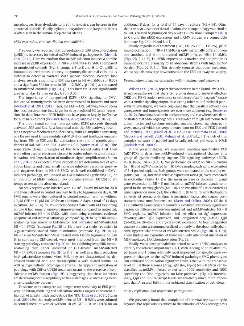
(1) Targeting of EGFR→ERK signaling: To interrupt EGFR signal-ing, we employed 10 μM gefitinib (GEF) (Selleck Chemicals LLC,Houston, TX), a small molecule inhibitor which blocks the bindingof ATP to the intracellular TK domain of EGFR to inhibit EGFR signal-ing, as described previously (Melnick et al., 2011). To interrupt ERKsignaling, we employed 10 μM U0126 (EMD Chemicals, Inc,Gibbstown, NJ), a potent and specific inhibitor of MEK-mediatedERK1 and ERK2 phosphorylation, as previously described (Melnicket al., 2011). These concentrations were previously shown to be theoptimal, nontoxic dose that substantially precludes mCMV-inducedpathology on day 6 of culture (Melnick et al., 2011). NB SMG organswere infected with 1×10⁵ PFU/ml mCMV for 24 h and in control me-dium until day 6. Beginning on day 6 (NB+6), mCMV-infected SMGorgans were cultured in the presence or absence of either 10 μMGEF or 10 μM U0126 in control medium for an additional 8 days, atotal of 14 days in culture (NB+14). (2) Co-targeted inhibition of
388 M. Melnick et al. / Experimental and Molecular Pathology 94 (2013) 386–397
the EGF→ERK pathway: NB SMG organs were infected with1×105 PFU/ml mCMV for 24 h and cultured control medium untilday 6. Beginning on day 6, explants were cultured in the presenceor absence of 10 μM GEF+10 μM U1026 (D6GEF+D6U) for an addi-tional 8 days, a total of 14 days in culture (NB+14). (3) Inhibition ofCMV replication: To inhibit CMV replication, we used 10 μg/ml acy-clovir sodium (American Pharmaceutical Partners, Inc, Schaumberg,Il), a synthetic purine nucleoside analogue which is a highly selectiveagent for CMV with low toxicity to the host cell (Burns et al., 1981)and previously shown to inhibit mCMV infection in mouse SMGs invitro (Melnick et al., 2006); this concentration was previouslyshown to be the optimal, nontoxic dose (Melnick et al., 2006). NBSMG organs were infected with 1×10⁵ PFU/ml mCMV for 24 h andcultured in control medium until day 6. Beginning on day 6,mCMV-infected SMGs were cultured in the presence 10 μg/ml acyclo-vir, 10 μMU0126+10 μg/ml acyclovir, 10 μMGEF+10 μg/ml acyclo-vir or no drugs for an additional 8 days, a total of 14 days in culture(NB+14). Controls consisted of uninfected SMGs without treatmentor with D6GEF, D6U0126, or D6Acy treatment (singly or in combina-tion) cultured for 14 days. No differences were seen betweenuntreated and inhibitor-treated uninfected SMGs; untreated controlsare presented. In all experiments, media was changed daily and treat-ments (GEF, GEF+U0126, or U0126 with/without acyclovir) wereadded daily beginning on day 6.
Quantitative RT-PCR
For analysis of gene expression, qRT-PCRwas conducted as previous-ly described (Melnick et al., 2006, 2009) on NB+6 control andmCMV-infected samples; each sample consisted of 4–5 pooled explants.RNAwas extracted and 1μg RNAwas reverse transcribed into first strandcDNA using ReactionReady™ First Strand cDNA Synthesis Kit: C-01 forreverse transcription (SABiosciences, Frederick, MD). The primer setsused were prevalidated to give single amplicons and purchased fromSABiosciences (Frederick, MD): Areg (PPM02976A); Egf (PPM03703A);Fgf8 (PM02962A); Il6 (PPM03015A); Tnfa (PPM03113A). Primerswere used at concentration of 0.4 μM. The cycling parameters were95 °C, 15 min; 40 cycles of (95 °C, 15 s; 55 °C, 30–40 s and 72 °C,30 s). Specificity of the reactionswas determined by subsequentmeltingcurve analysis. RT-PCRs of RNA (not reverse transcribed) were used asnegative controls. GAPDH was used to control for equal cDNA inputsand the levels of PCR product were expressed as a function of GAPDH.The relative fold changes of gene expression between the gene of inter-est andGAPDH, or between theNB+6 control andmCMV-infected sam-ples, were calculated by the 2−ΔΔCT method. Significant expressiondifferences between mCMV-infected and control samples were deter-mined by student t-test, with α=0.01 and the null hypothesis of R=1,where R is themean relative expression ration (mCMV/control) acrossthe entire sample. Expression ratios were log transformed prior to anal-ysis to satisfy the assumption of normality.
Probabilistic neural network (PNN) analysis
We used PNN analyses to determine the contribution of each indi-vidual gene to the discrimination between experimental groups with100% sensitivity and specificity (Melnick et al., 20011). As such, PNN
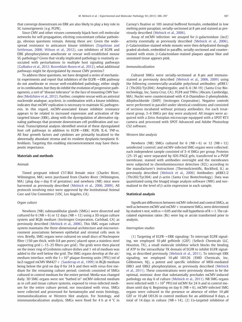
Fig. 1. mCMV induced notable changes in morphology and pERK protein expression in NB+by densely packed, branched cuboidal epithelial ducts and pro-acini (e) surrounded by fibrom(B, D) show significant viral CPE in the stroma and epithelial dysplasia consisting of a decrepithelia (double black arrows). These abnormal epithelial structures are embedded in a hypeosinophilic cells. E-H. mCMV induced pERK protein expression in NB+6 and NB+12 SMGcontrol (E, G) and mCMV-infected (F, H) SMGs. In control SMGs (E, G), little immunodetectpERK primarily in abnormal stroma (compare F to E and H to G). I–J. Western blot analysiComparisons of levels of pERK proteins show a significant increase with mCMV infection o
analyses identify the relative importance (0–1, with 0 being of no rel-ative importance and 1 being relatively most important) of specificgene expression changes that discriminate between phenotypes. Itis the contextual change in expression, not the direction of change,which is important in defining the molecular phenotype. The founda-tional algorithm we used is based upon the work of Specht and col-leagues (Chen, 1996; Specht, 1988; Specht and Shapiro, 1991). Theproprietary software designed by Ward Systems Group (Frederick,MD) formulates Specht's procedure around a genetic algorithm(Goldberg, 1989).
Results
The trajectory of this research is to identify molecular targets thatmay prove critical to a therapeutic solution. To this end, one has al-ways to discern patterns of covarying molecular and histopathologicphenotypes, relating measurements and localization of transcriptsand proteins (input) to a well-characterized pathologic phenotype(output). Here we utilize an in vitro 3D submandibular gland (SMG)organ culture strategy previously shown to induce cellular pathologyresembling secretory gland neoplasia (Jaskoll et al., 2011; Melnicket al., 2006, 2011).
Progressive histopathology
Newborn (NB) mouse SMGs are cultured in 1×10⁵ PFU/ml mCMVfor 24 h and maintained in control medium for a total of 6 or 12 days;controls consist of NB SMGs cultured for identical periods in controlme-dium. Our novel 3D SMG organ culture system maintains the morpho-logical integrity, 3D architecture and microenvironment seen in invivo SMGs. As we have previously reported (Melnick et al., 2011) andreplicate here (Fig. 1), mCMV infection induces a severe cytopathic ef-fect (CPE), one which becomes progressively more severe in the periodfrom 6 to 12 days in culture. Control SMGs on days 6 and 12 are charac-terized by densely packed, branched, cuboidal epithelia; these acini andducts are embedded in a sparse fibromyxoid stroma containing variablenumbers of stellate to ovoid fibroblasts (Figs. 1A, C). mCMV-infectedSMGs cultured for 6 days (NB+6) are characterized by a marked dim-inution of branching epithelia, acini and ducts; abnormal duct epitheliais hypoplastic and pseudo-stratified and the lumina are dilated andfilled with cellular debris (Fig. 1B). These dysplastic epithelial compo-nents are embedded in a substantial, hypercellular stroma primarilycomposed of giant basophilic round cells mixed with far fewer andsmaller eosinophilic cells (Fig. 1B).
mCMV-infected SMGs cultured for 12 days (NB+12) exhibit amoresevere viral CPE in the stroma, and abnormal parenchyma consistentwith a tumorigenic phenotype (Fig. 1D). The marked increased stromalcellularity is composed of sheets of large basophilic, pleomorphic cellsand smaller eosinophilic cells with high nuclear-to-cytoplasmratios, prominent nuclei and nucleoli, and frequent kidney-shapednuclei pathognomonic of CMV infection (Alwine, 2012). The increasing-ly sparse branched epithelia frequently display severely dilatedlumina, and individual cells with increased nuclear-to-cytoplasmic ra-tios, hyperchromatism, and visible nucleoli. Intraluminal and extrabasalproliferation of epithelial cells is often seen, imparting a multi-layered appearance to these epithelial islands. Further, a spectrum of
6 and NB+12 SMGs. A–D Histological analyses. Control SMGs (A, C) are characterizedyxoid stroma (black *) consisting of stellate to ovoid fibroblasts. mCMV-infected SMGs
ease in branched epithelia, severely dilated lumina (red *), and multi-layered cuboidalercellular stroma (black *) composed of large basophilic, pleiomorphic cells and smallers. E–J. Comparison of cell specific immunolocalization of pERK in NB+6 and NB+12
able pERK is seen. In contrast, mCMV-induced a notable increase in immunodetectables of pERK expression in NB+6 (I) and NB+12 (J) control and mCMV-infected SMGs.n both days 6 and 12 of culture. Bar: 50 μm.

389M. Melnick et al. / Experimental and Molecular Pathology 94 (2013) 386–397
390 M. Melnick et al. / Experimental and Molecular Pathology 94 (2013) 386–397
morphotypes, from dysplastic to in situ to invasive, can be seen in theabnormal epithelia. Finally, pyknotic, karyorhexic and karyolitic debrisis often seen in the lumina of epithelial islands.
pERK expression, viral distribution and inhibition
Previously we reported that upregulation of ERK phosphorylation(pERK) is necessary for initial mCMV-induced pathogenesis (Melnicket al., 2011). Here we confirm that mCMV infection induces a notableincrease in pERK expression in NB+6 and NB+12 SMGs comparedto uninfected controls (Fig. 1; compare F to E and H to G). pERK isimmunolocalized almost entirely to cytomegalic stromal cells and isdifficult to detect in controls. With mCMV infection, Western blotanalysis reveals a significant 40% increase in NB+6 SMGs (pb0.05)and a significant 50% increase in NB+12 SMGs (pb0.01) as comparedto uninfected controls (Figs. 1I, J). This increase is not significantlygreater on day 12 than on day 6 (p>0.50).
The importance of upregulated EGFR→ERK signaling to CMV-induced SG tumorigenesis has been demonstrated in humans and mice(Melnick et al., 2011, 2012). Thus, the EGF→ERK pathway would seemthe most parsimonious first target for drug-mediated SG tumor regres-sion. To date, however, EGFR inhibitors have proven largely ineffectivefor human SG tumors (Bell and Hanna, 2012; Gillespie et al., 2012).
The input signal coming from activated EGFR proceeds throughactivated SOS and Ras. As a functional unit, the ERK pathway resem-bles a negative feedback amplifier (NFA) with an amplifier consistingof a three-tiered kinase module Raf-MEK-ERK and feedbacks emanat-ing from ERK to SOS and Raf activation; the ratio of protein abun-dances of Raf, MEK and ERK is about 1:3:6 (Sturm et al., 2010). Theremarkable design principles of the NFA recapitulates that theywere often used in electronic circuits to confer robustness, output sta-bilization, and linearization of nonlinear signal amplification (Sturmet al., 2010). As expected, these properties are determinative of acti-vation kinetics and drug (small molecule inhibitor) response, positiveand negative. Here in NB+6 SMGs with well-established mCMV-induced pathology, we utilized an EGFR inhibitor (gefitinib/GEF) oran inhibitor of MEK-mediated, ERK phosphorylation (U0126) to at-tempt to rescue the abnormal gland.
NB SMG organs were infected with 1×10⁵ PFU/ml mCMV for 24 hand then cultured in control medium to day 6; beginning on day 6, NBSMG organs were then cultured in control medium with or without10 μM GEF or 10 μM U0126 for an additional 8 days, a total of 14 daysin culture (NB+14). mCMV-infected SMGs treated with GEF beginningon day 6 had some phenotypic improvement compared to untreated,mCMV-infected NB+14 SMGs, with there being continued evidenceof epithelial and stromal pathology (compare Fig. 2D to A). pERK immu-nostaining was similar in GEF-treated and untreated mCMV-infectedNB+14 SMGs (compare Fig. 2E to B); there is a slight reduction inβ-galactosidase-stained virus distribution (compare Fig. 2F to C).NB+14 mCMV-infected SMGs treated with U0126 beginning on day6, in contrast to GEF-treated, were most improved from the NB+6starting pathology (compare Fig. 2G to 1B); exhibiting less pERK immu-nostaining than either untreated or GEF-treated mCMV-infectedNB+14 SMGs (compare Fig. 2H to B, E), as well as a slight reductionin β-galactosidase-stained virus. Still, they are characterized by de-creased branched acini and ductal epithelia with dilated lumina, aswell as hypercellular, cytomegalic stroma. The apparent mitigation ofpathology with GEF or U0126 treatment occurs in the presence of con-siderable mCMV burden (Figs. 2F, I), suggesting that these inhibitorsare decreasing host susceptibility to tissue damage (i.e. increasing toler-ance to pathology burden).
To secure more complete and longer-term sensitivity to ERK path-way inhibition,modeling and cell culture studies suggest concurrent in-hibition of targets inside and outside the NFA (Little et al., 2011; Sturmet al., 2010). For this study, mCMV-infected NB+6 SMGswere culturedin control medium with or without 10 μM GEF+10 μM U0126 for an
additional 8 days, for a total of 14 days in culture (NB+14). Otherthan the near absence of ductal dilation, the histopathologywas similarto SMGs treated beginning on day 6 with U0126 alone (compare Fig. 3Jto G), and the pERK expression and mCMV burden are comparable(compare Fig. 2K to H and L to I).
Finally, regardless of treatment (GEF, U0126, GEF+U0126), pERKimunolocalization in NB+14 SMGs is only marginally different fromone another, and from untreated mCMV-infected NB+14 SMGs(Figs. 2B, E, H, K), i.e. pERK expression is marked and the protein isimmunolocalized primarily to an abnormal stroma with high mCMVburden (Figs. 2C, F, I, L). This strongly suggests that other mediatorswhose signals converge downstream on the ERK pathway are at play.
Dysregulation of ligands associated with multifunctional pathways
Wilson et al. (2012) report that an increase in the ligand levels of al-ternative pathways that share cell proliferation and survival effectors(ERK and PI3K) confers resistance to inhibitors of an “oncogenic” kinasewith a similar signaling output. In selecting other multifunctional path-ways to investigate, we were cognizant that the parallels between or-ganogenesis and tumorigenesis are ever more apparent (e.g. Becker etal., 2012). Functional studies in our laboratory and elsewhere have dem-onstrated that SMG organogenesis is regulated through interconnectedgrowth factor and cytokine mediated multifunctional signaling path-ways, many of which converge downstream on ERK and PI3K (Jaskolland Melnick, 1999; Jaskoll et al., 2002, 2004; Kashimata et al., 2000;Melnick and Jaskoll, 2000; Melnick et al., 2001a,b,c). The hub of thiscomplex network of parallel and broadly related pathways is NFκB(Melnick et al., 2001b).
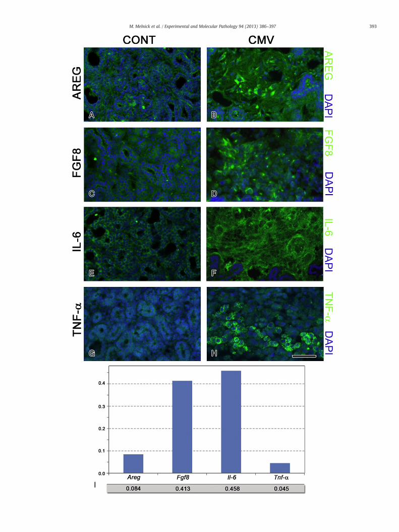
In the present studies, we employed real-time quantitative PCR(qRT-PCR) to determine mCMV-induced transcriptional changes in agroup of ligands mediating cognate ERK signaling pathways (EGFR,FGFR, IL-6R, TNGR) (Fig. 3). We performed qRT-PCR on NB+6 control(n=6) andmCMV-infected (n=6) SMGsamples; each sample consistedof 3–4 pooled explants. Both groups were compared to the starting ex-plants (NB+0), and these relative expression ratios (R) were comparedto each other (Table 1). R is the mean increase or decrease in glandgene expression in NB+6 uninfected or mCMV-infected glands com-pared to the starting glands (NB+0). The variation of R is calculated asgene expression noise (x); the value of x (0 to 1) reflects fluctuationsin levels of promotor-binding, transcription factor abundance, post-transcriptional modifications, etc. (Raser and O'Shea, 2005). Of the 6ERK pathway ligand genes measured, 5 exhibited statistically significantexpression differences between untreated and mCMV-infected NB+6SMG explants. mCMV infection had no effect on Egf expression,downregulated Tgf-a expression, and upregulated Areg (4-fold), Fgf8(5-fold), Il-6 (64-fold), and Tnf-α (2-fold) expression. All four upregulatedcognate proteins are immunolocalized primarily to the abnormally abun-dant, hypercellular stroma of mCMV-infected SMGs (Figs. 4B, D, F, H).These finding are expositive of those seen with attempted inhibition ofMEK-mediated, ERK phosphorylation (Fig. 2).
Finally, we utilized probabilistic neural network (PNN) analyses toidentify the relative importance (0–1, with 0 being of no relative im-portance and 1 being relatively most important) of specific gene ex-pression changes to the mCMV-induced pathologic SMG phenotype.Our unbiased optimization algorithm reveals that with the transcriptlevel of just these 4 genes (Areg, Fgf8, Il-6, Tnf-α) NB+6 SMGs can beclassified as mCMV-infected or not with 100% sensitivity and 100%specificity (no false negatives; no false positives) (Fig. 4I). Interest-ingly, Fgf8 and Il-6 transcript levels are relatively much more impor-tant than Areg and Tnf-α in the unbiased classification of pathology.
mCMV replication and progressive pathogenesis
We previously found that completion of the viral replication cyclebeyond DNA replication is critical to the initiation of SMG pathogenesis
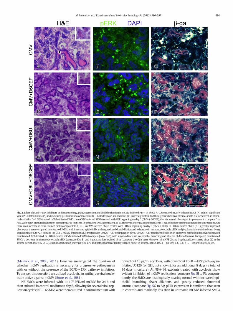
Fig. 2. Effect of EGFR→ERK inhibitors on histopathology, pERK expression and viral distribution inmCMV-infectedNB+14 SMGs. A–C. UntreatedmCMV-infected SMGs (A) exhibit significantviral CPE, dilated lumina (*) and increased pERK immunolocalization (B).β-Galactosidase-stained virus (C) is densely distributed throughout abnormal stroma, and to a lesser extent, in abnor-mal epithelia. D–F. GEF-treated,mCMV-infected SMGs. InmCMV-infected SMGs treatedwith GEF beginning on day 6 (CMV+D6GEF), there is a small phenotypic improvement (compare D toAD),with pERK immunolocalization being similar to that seen in untreated SMGs (compare E to B). However, there is a slight decrease inβ-galactosidase staining compared to untreated SMGs;there is an increase in eosin-stained pink (compare F to C). G–I. mCMV-infected SMGs treated with U0126 beginning on day 6 (CMV+D6U). In U0126-treated SMGs (G), a greatly improvedphenotype is seen compared to untreated SMGs,with increased epithelial branching, reducedductal dilation and a decrease in immunodetectable pERKandβ-galactosidase-stained virus beingseen (compareG toA,H to B and I to C). J-L.mCMV-infected SMGs treatedwithU0126+GEFbeginningonday6. U0126+GEF treatment results in an improved epithelial phenotype comparedto untreated, GEF-treated, or U0126-treatedmCMV-infected SMGs (compare J to A, D, G), with amarked increase in epithelial branching and absence of dilated lumina. Compared to untreatedSMGs, a decrease in immunodetectable pERK (compare K to B) and β-galactosidase-stained virus (compare L to C) is seen. However, viral CPE (J) and β-galactosidase-stained virus (L) in thestroma persist. Insets A, D, G, J. High magnification showing viral CPE and pathognomonic kidney-shaped nuclei in stroma. Bar: A, D.G, J— 60 μm; B, C, E, F, K–L— 50 μm; insets 30 μm.
391M. Melnick et al. / Experimental and Molecular Pathology 94 (2013) 386–397
(Melnick et al., 2006, 2011). Here we investigated the question ofwhether mCMV replication is necessary for progressive pathogenesiswith or without the presence of the EGFR→ERK pathway inhibitors.To answer this question, we utilized acyclovir, an antiherpesviral nucle-oside active against mCMV (Burns et al., 1981).
NB SMGs were infected with 1×10⁵ PFU/ml mCMV for 24 h andthen cultured in control medium to day 6, allowing for several viral rep-lication cycles;NB+6SMGswere then cultured in controlmediumwith
orwithout 10 μg/ml acyclovir, with orwithout EGFR→ERKpathway in-hibitor, U0126 (or GEF, not shown), for an additional 8 days (a total of14 days in culture). At NB+14, explants treated with acyclovir showevident inhibition of mCMV replication (compare Fig. 5I to F); concom-itantly, the SMGs are histologically nearing normal with increased epi-thelial branching, fewer dilations, and greatly reduced abnormalstroma (compare Fig. 5G to A); pERK expression is similar to that seenin controls and markedly less than in untreated mCMV-infected SMGs
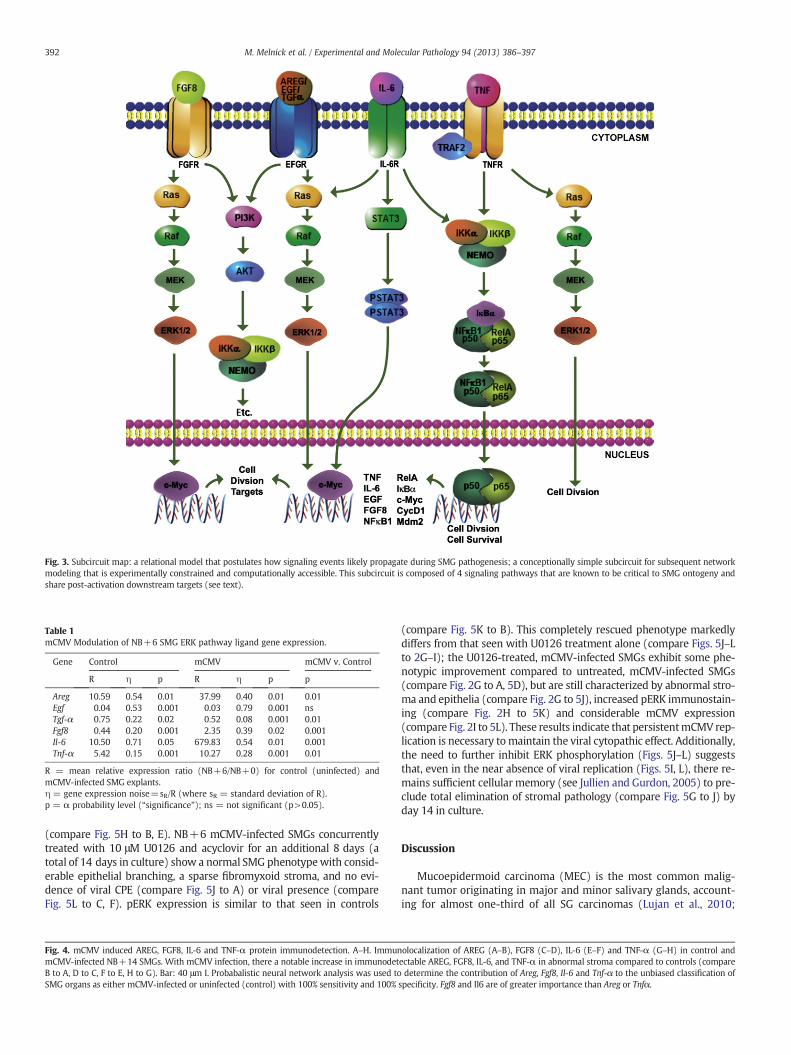
Fig. 3. Subcircuit map: a relational model that postulates how signaling events likely propagate during SMG pathogenesis; a conceptionally simple subcircuit for subsequent networkmodeling that is experimentally constrained and computationally accessible. This subcircuit is composed of 4 signaling pathways that are known to be critical to SMG ontogeny andshare post-activation downstream targets (see text).
Table 1mCMV Modulation of NB+6 SMG ERK pathway ligand gene expression.
Gene Control mCMV mCMV v. Control
R η p R η p p
Areg 10.59 0.54 0.01 37.99 0.40 0.01 0.01Egf 0.04 0.53 0.001 0.03 0.79 0.001 nsTgf-α 0.75 0.22 0.02 0.52 0.08 0.001 0.01Fgf8 0.44 0.20 0.001 2.35 0.39 0.02 0.001Il-6 10.50 0.71 0.05 679.83 0.54 0.01 0.001Tnf-α 5.42 0.15 0.001 10.27 0.28 0.001 0.01
R = mean relative expression ratio (NB+6/NB+0) for control (uninfected) andmCMV-infected SMG explants.η = gene expression noise=sR/R (where sR = standard deviation of R).p = α probability level (“significance”); ns = not significant (p>0.05).
392 M. Melnick et al. / Experimental and Molecular Pathology 94 (2013) 386–397
(compare Fig. 5H to B, E). NB+6 mCMV-infected SMGs concurrentlytreated with 10 μM U0126 and acyclovir for an additional 8 days (atotal of 14 days in culture) show a normal SMG phenotypewith consid-erable epithelial branching, a sparse fibromyxoid stroma, and no evi-dence of viral CPE (compare Fig. 5J to A) or viral presence (compareFig. 5L to C, F). pERK expression is similar to that seen in controls
Fig. 4. mCMV induced AREG, FGF8, IL-6 and TNF-α protein immunodetection. A–H. ImmunmCMV-infected NB+14 SMGs. With mCMV infection, there a notable increase in immunodetB to A, D to C, F to E, H to G). Bar: 40 μm I. Probabalistic neural network analysis was used toSMG organs as either mCMV-infected or uninfected (control) with 100% sensitivity and 100%
(compare Fig. 5K to B). This completely rescued phenotype markedlydiffers from that seen with U0126 treatment alone (compare Figs. 5J–Lto 2G–I); the U0126-treated, mCMV-infected SMGs exhibit some phe-notypic improvement compared to untreated, mCMV-infected SMGs(compare Fig. 2G to A, 5D), but are still characterized by abnormal stro-ma and epithelia (compare Fig. 2G to 5J), increased pERK immunostain-ing (compare Fig. 2H to 5K) and considerable mCMV expression(compare Fig. 2I to 5L). These results indicate that persistentmCMV rep-lication is necessary to maintain the viral cytopathic effect. Additionally,the need to further inhibit ERK phosphorylation (Figs. 5J–L) suggeststhat, even in the near absence of viral replication (Figs. 5I, L), there re-mains sufficient cellular memory (see Jullien and Gurdon, 2005) to pre-clude total elimination of stromal pathology (compare Fig. 5G to J) byday 14 in culture.
Discussion
Mucoepidermoid carcinoma (MEC) is the most common malig-nant tumor originating in major and minor salivary glands, account-ing for almost one-third of all SG carcinomas (Lujan et al., 2010;
olocalization of AREG (A–B), FGF8 (C–D), IL-6 (E–F) and TNF-α (G–H) in control andectable AREG, FGF8, IL-6, and TNF-α in abnormal stroma compared to controls (comparedetermine the contribution of Areg, Fgf8, Il-6 and Tnf-α to the unbiased classification of
specificity. Fgf8 and Il6 are of greater importance than Areg or Tnfα.
394 M. Melnick et al. / Experimental and Molecular Pathology 94 (2013) 386–397
Schwarz et al., 2011). Although the precise etiology of MEC is largelyunknown, we have recently shown that cytomegalovirus is an impor-tant component of MEC tumorigenesis (Melnick et al., 2011, 2012).Despite the well-documented overexpression of the EGFR→ERK sig-naling pathway in MEC (Akrish et al., 2009; Ito et al., 2009; Lujanet al., 2010), there has been limited to no success with inhibition ofthis pathway (Bell and Hanna, 2012; Gillespie et al., 2012), not unlikethat seen with other malignancies (Engelman and Settleman, 2008).Using our previously described mouse model of CMV-induced SGdysplasia/neoplasia (Jaskoll et al., 2011; Melnick et al., 2011), we pro-vide evidence that concurrent inhibition of ERK phosphorylation andinhibition of CMV replication obviates progressive pathogenesis andresults in complete SG rescue (regression). These findings provide amechanistic foundation for clinical trials that utilize similar concur-rent treatment with extant FDA-approved drugs.
CMV has evolved numerous strategies to co-opt and alter hostcell signaling networks for the purpose of propagating ever morevirus (Sanchez and Spector, 2008; Schleiss, 2011). Cells respondto environmental challenges such as viral infection by recon-figuring regulated gene expression either by slow transcriptionaladaptation or rapid post-transcriptional adaptation (Chalanconet al., 2012). The gene expression programs of signaling networksmaintain homeostatic function at any given moment in a cell's lifecycle. Corruption of these programs by a virus such as CMV can re-sult in the host cell and tissue chaos we call dysplasia, neoplasia,and metaplasia.
Given the considerable crosstalk and redundancy in mammalianhost cells, and the multifunctional paths mediated by single molecu-lar components, a deep understanding of the subversion anddysregulation of the SMG interactome by CMV is a priori quite daunt-ing (Gulbahce et al., 2012). For example, all the distinct ways in whichmere 10 heterogeneous proteins can interact can be calculated asBell's number (Bn), which in this case is 1.16×10⁵ (Koch, 2012).Even if we assume that only 1 per 1000 of these potential interactionsare biologically relevant, the number to be characterized would be116, a task likely to consume the lifetime of most laboratories. Inreality, of course, we must consider far more proteins and their mil-lions of potential interactions. A way to circumvent this bad news, isto study groups of proteins as interacting single pathway modules(e.g. Fig. 3); correspondingly, the number of potential interactionsprecipitously declines (Koch, 2012). In this way, systematic analysisof virus-corrupted host cell targets should more easily identifydysregulated host cell networks relevant to viral-implicated tumori-genesis (Rozenblatt-Rosen et al., 2012).
We had every reason to initially assume that either single orco-targeted small molecule inhibition of the EGFR→ERK pathwaywould result in the complete rescue of SMG pathology (Little et al.,2011; Melnick et al., 2011; Poulikakos and Solit, 2011; Sturm et al.,2010). This provednot to be the case (Fig. 2). To be sure, therewas a pre-clusion of progressive pathogenesis (stability, if you will), even in thecontinued presence of active CMV. This phenomenon has recentlybeen termed diseases tolerance, i.e. the reduction of the negative impactof an infection on host fitness without directly affecting the pathogen(Medzhitov et al., 2012). Here, specifically, small molecule inhibitors"protected" the SMGs from further destructive pathogenesis indepen-dently of CMV load (Fig. 2). All this notwithstanding, failure to rescueSMG pathology suggested to us that othermediators whose signals con-verge downstream on the ERK pathway remain functional; even in thepresence of the highest nontoxic dose of U0126 (an inhibitor ofMEK-mediated ERK phosphorylation), pERK protein was undiminished(Fig. 2).
Recently, Wilson et al. (2012) presented convincing evidence thatresistance to inhibitors of "oncogenic" kinase (e.g. gefitinib) resultsfrom an increase in the ligand levels of alternative pathways thatshare cell proliferation and survival effectors (ERK and PI3K), i.e. theyshare similar signaling outputs. Here we present data which supports
this proposition. Namely, there is a highly significant upregulation of li-gands for the FGFR, IL-6R and TNFR signaling pathways (Table 1; Fig. 4),all of which converge upon the Raf/MEK/ERK amplifier module (Fig. 3).These findings are clearly expositive of the failed attempt to simply in-hibit EGFR-initiated, MEK mediated, ERK phosphorylation and effectcomplete rescue of the SMG explants (Fig. 2). It is interesting to notethat all relevant ligands (AREG, FGF8, IL-6, TNF-α) are immunolocalizedto the abnormal stroma of the CMV-infected SMGs, as is downstreampERK (Figs. 1, 4). This highlights the importance of the stromalmicroen-vironment to progressive pathogenesis, regardless of the degree towhich stromal paracrine stimulation participates in the epithelial path-ogenesis (Hanahan andWeinberg, 2011; Pietras and Ostman, 2010). In-deed, FGF and IL-6 are both highly expressed by tumor-associatedfibroblasts in a variety of tumor types (Bhowmick et al., 2004), andhere they are relatively most important in characterizing themCMV-induced pathologic phenotype (Fig. 4I).
hCMV-mediated overexpression of IL-6 and TNF-α has previouslybeen reported (Zheng et al., 2012). Further, overexpression of theEGFR, FGFR, IL-6R, and TNFR pathways have been seen in humanSG-MEC and other solid tumors (Azevedo et al., 2011; Elo et al., 2010;Ettl et al., 2012; Leivo et al., 2005; Mocellin et al., 2005). All of thesepathways modulate cell proliferation and cell survival. Untowardupregulation of these particular multifunctional pathways mimicswhat we and others have shown in normal SG organogenesis (Jaskolland Melnick, 1999; Jaskoll et al., 2002, 2004; Kashimata et al., 2000;Koyama et al., 2003; Melnick and Jaskoll, 2000; Melnick et al., 2001a,b,c). Drawing on the evermore apparent parallels between organogenesisand tumorigenesis (e.g. Becker et al., 2012), it should be noted that theEGFR, FGFR, IL-6R and TNFR pathways are not likely to be functionallyindependent (Jaskoll et al., 2002, 2004;Melnick et al., 2001a,c), but rath-er part of a larger network whose operational hub is NFκB (Melnick andJaskoll, 2000; Melnick et al., 2001b; Fig. 3). This has been proffered forhuman tumors as well (Karin et al., 2002). Thus, it is becoming increas-ingly apparent that shared embryonic and tumor genetic signaturesmay be prognostically important (Becker et al., 2012).
Conclusion
CMV is a principle element in the multifactorial causation ofhuman salivary gland mucoepidermoid carcinoma. Despite thewell-documented overexpression of the EGFR→ERK signaling path-way in SG-MEC, there has been little clinical success with inhibitionstrategies. It has been proposed that failed inhibition results from anincrease in the ligand levels of alternative pathways that share cellproliferation and survival effectors (ERK, PI3K). In support of thisproposition, we present mouse model evidence of a highly significantupregulation of ligands for the FGFR, IL-6R and TNFR signaling path-ways, all of which converge upon the Raf/MEK/ERK amplifier module.These finding are clearly expositive of the failed attempt to simply in-hibit EGFR-initiated, MEK-mediated, ERK phosphorylation and effectcomplete rescue. Here we model a potential solution that obviatesprogressive pathogenesis and effects complete SG rescue (regres-sion), namely the concurrent inhibition of ERK phosphorylation(U0126) and CMV replication (acyclovir).
Conflict of interests
The authors declare that there are no conflicts of interest.
List of abbreviations
AREG amphiregulinCMV cytomegalovirusCONT controlCRTC1 CREB-regulated transcription coactivator 1DAPI-4′,6 Diamidino-2-phenylindole, dihydrochloride
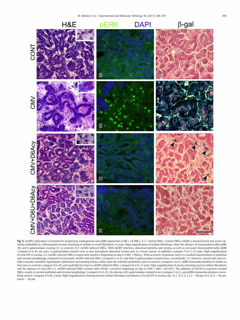
Fig. 5. mCMV-replication is essential for progressive pathogenesis and pERK expression in NB+14 SMGs. A–C. Control SMGs. Control SMGs exhibit a normal ductal and acinar ep-ithelia embedded in a fibromyxoid stroma consisting of stellate to ovoid fibroblasts. A, inset. High magnification of stellate fibroblasts. Note the absence of immunodetectable pERK(B) and β-galactosidase staining (C) in controls. D-F. mCMV-infected SMGs. With mCMV infection, abnormal epithelia and stroma, as well as increased immunodetectable pERK(compare E to B), are seen; β-galactosidase-stained virus is seen throughout abnormal stroma and, to a lesser extent, in epithelia (compare F to C). D, inset. High magnificationof viral CPE in stroma. G-I. mCMV-infected SMGs treated with acyclovir beginning on day 6 (CMV+D6Acy). With acyclovir-treatment, there is a marked improvement in epithelialand stromal morphology compared to untreated, mCMV-infected SMGs (compare G to D) and little β-galactosidase-stained virus (arrowheads) (I). However, ductal and acini ex-hibit a pseudo-stratified, hyperplastic epithelium surrounding lumina rather than the cuboidal epithelium seen in controls (compare G to A). pERK immunolocalization is similar tothat seen in controls (compare H to B) and markedly less than in mCMV-infected SMGs (compare H to E). G inset. High magnification of stroma showing normal stellate fibroblastsand the absence of viral CPE. J–L. mCMV-infected SMGs treated with U0126+acyclovir beginning on day 6 (CMV+D6U+D6 ACY). The addition of U0126 to acyclovir-treatedSMGs results in normal epithelial and stromal morphology (compare J to A, D), the absence of β-galactosidase-stained virus (compare L to C), and pERK immunolocalization resem-bling control (compare K to B). J inset. High magnification showing normal stellate fibroblast and absence of viral CPE in stroma. Bar: A, C, D, F, G, I, J, L— 60 μm; B, E, H, K — 50 μm;insets — 30 μm.
395M. Melnick et al. / Experimental and Molecular Pathology 94 (2013) 386–397
396 M. Melnick et al. / Experimental and Molecular Pathology 94 (2013) 386–397
ERK extracellular signal-regulated protein kinases 1 and 2 (ERK1/2EGFR epidermal growth factor receptorGEF gefitinibhCMV human cytomegalovirusmCMV mouse cytomegalovirusMEK mitogenactivated protein kinase kinaseNB newbornNFA negative feedback amplifierpERK phosphorylated ERK1/2PFU plaque forming unitsPNN Probabilistic neural network analysisqRT-PCR quantitative RT-PCRRaf rapidly accelerated fibrosarcoma oncogeneRas rat sarcoma viral oncogeneRTK receptor tyrosine kinaseSG salivary glandSMG salivary glandSOS guaninenucleotide exchange factors sonof sevenless oncogeneTK tyrosine kinase
Acknowledgments
Wewould like to thank Dr. EdwardMocarski for his generous gift ofmCMV. This research was supported by the Oral Biology Fund of theUniversity of Southern California.
References
Akrish, S., Peled, M., Ben-Izhak, B., Nagler, R.M., 2009. Malignant salivary gland tumorsand cyclo-oxygenase-2: a histopathological and immunohistochemical analysiswith implications on histogenesis. Oral Oncology 45, 1044–1050.
Alwine, J.C., 2012. The human cytomegalovirus assembly compartment: a masterpieceof viral manipulation of cellular processes that facilitates assembly and egress.PLoS Pathogens 8, e1002878.
Azevedo, A., Cunha, V., Teixeira, A.L., Medeiros, R., 2011. IL-6/IL-6R as a potential key sig-naling pathway in prostate cancer development. World Journal of Clinical Oncology 2,384–396.
Becker, D., Sfakianakis, I., Krupp, M., Staib, F., Gerhold-Ay, A., Victor, A., Binder, H.,Blettner, M., Maass, T., Thorgeirsson, S., Galle, P.R., Teufel, A., 2012. Genetic signaturesshared in embryonic liver development and liver cancer define prognostically rele-vant subgroups in HCC. Molecular Cancer 11, 55.
Bell, D., Hanna, E.Y., 2012. Salivary gland cancers: biology and molecular targets fortherapy. Current Oncology Reports 14, 166–174.
Bhowmick, N.A., Neilson, E.G., Moses, H.L., 2004. Stromal fibroblasts in cancer initiationand progression. Nature 432, 332–337.
Boppana, S.B., Fowler, K.B., 2007. Persistence in the Population: Epidemiology andTransmission. Cambridge University Press, Cambridge, Mass.
Burns, W.H., Wingard, J.R., Bender, W.J., Saral, R., 1981. Thymidine kinase not required forantiviral activity of acyclovir against mouse cytomegalovirus. Journal of Virology 39,889–893.
Chalancon, G., Kruse, K., Babu, M.M., 2012. Cell biology. Reconfiguring regulation. Sci-ence 335, 1050–1051.
Chen, C. (Ed.), 1996. Fuzzy Logic and Neural Network Handbook. McGraw-Hill, New York.Elo, T.D., Valve, E.M., Seppänen, J.A., Vuorikoski, H.J., Mäkelä, S.I., Poutanen, M., Kujala,
P.M., Härkönen, P.L., 2010. Stromal activation associated with development of pros-tate cancer in prostate-targeted fibroblast growth factor 8b transgenic mice. Neopla-sia 12, 915–927.
Engelman, J.A., Settleman, J., 2008. Acquired resistance to tyrosine kinase inhibitorsduring cancer therapy. Current Opinion in Genetics and Development 18, 73–79.
Ettl, T., Stiegler, C., Zeitler, K., Agaimy, A., Zenk, J., Reichert, T.E., Gosau, M., Kühnel, T.,Brockhoff, G., Schwarz, S., 2012. EGFR, HER2, survivin, and loss of pSTAT3 charac-terize high-grade malignancy in salivary gland cancer with impact on prognosis.Human Pathology 43, 921–931.
Gillespie, M.B., Albergotti, W.G., Eisele, D.W., 2012. Recurrent salivary gland cancer.Current Treatment Options in Oncology 13, 58–70.
Goldberg, D.E., 1989. Genetics Algorithms in Search, Optimization, and Machine Learn-ing. Addison-Wesley, Reading, Mass.
Gulbahce, N., Yan, H., Dricot, A., Padi, M., Byrdsong, D., Franchi, R., Lee, D.S., Rozenblatt-Rosen, O., Mar, J.C., Calderwood, M.A., Baldwin, A., Zhao, B., Santhanam, B., Braun, P.,Simonis, N., Huh, K.W., Hellner, K., Grace, M., Chen, A., Rubio, R., Marto, J.A.,Christakis, N.A., Kieff, E., Roth, F.P., Roecklein-Canfield, J., Decaprio, J.A., Cusick, M.E.,Quackenbush, J., Hill, D.E., Münger, K., Vidal, M., Barabási, A.L., 2012. Viral perturbationsof host networks reflect disease etiology. PLoS Computational Biology 8, e1002531.
Hanahan, D., Weinberg, R.A., 2011. Hallmarks of cancer: the next generation. Cell 144,646–674.
Ito, F.A., Coletta, R.D., Graner, E., de Almeida, O.P., Lopes,M.A., 2009. Salivary gland tumors:immunohistochemical study of EGF, EGFR, ERbB-2, FAS and Ki-67. Analytical andQuantitative Cytology and Histology 31, 280–287.
Jaskoll, T., Melnick, M., 1999. Submandibular gland morphogenesis: stage-specific ex-pression of TGF-alpha/EGF, IGF, TGF-beta, TNF, and IL-6 signal transduction in nor-mal embryonic mice and the phenotypic effects of TGF-beta2, TGF-beta3, and EGF-r null mutations. Anatomical Record 256, 252–268.
Jaskoll, T., Zhou, Y.M., Chai, Y., Makarenkova, H.P., Collinson, J.M., West, J.D.,Hajihosseini, M.K., Lee, J., Melnick, M., 2002. Embryonic submandibular gland mor-phogenesis: stage-specific protein localization of FGFs, BMPs, Pax6 and Pax9 innormal mice and abnormal SMG phenotypes in FgfR2-IIIc(+/Delta), BMP7(-/-)and Pax6(-/-) mice. Cells, Tissues, Organs 170, 83–98.
Jaskoll, T., Witcher, D., Toreno, L., Bringas, P., Moon, A.M., Melnick, M., 2004. FGF8 dose-dependent regulation of embryonic submandibular salivary gland morphogenesis.Developmental Biology 268, 457–469.
Jaskoll, T., Htet, K., Abichaker, G., Kaye, F.J., Melnick, M., 2011. CRTC1 expression duringnormal and abnormal salivary gland development supports a precursor cell originfor mucoepidermoid cancer. Gene Expression Patterns 11, 57–63.
Jullien, J., Gurdon, J., 2005. Morphogen gradient interpretation by a regulated traffickingstep during ligand-receptor transduction. Genes & Development 19, 2682–2694.
Karin, M., C. Y., Greten, F.R., Li, Z.W., 2002. NF-kappaB in cancer: from innocent by-stander to major culprit. Nature Reviews. Cancer 2, 301–310.
Kashimata, M., Sayeed, S., Ka, A., Onetti-Muda, A., Sakagami, H., Faraggiana, T., Gresik,E.W., 2000. The ERK-1/2 signaling pathway is involved in the stimulation ofbranching morphogenesis of fetal mouse submandibular glands by EGF. Develop-mental Biology 220, 183–196.
Koch, C., 2012. Systems biology. Modular biological complexity. Science 337, 531–532.Koyama, N., Kashimata, M., Sakashita, H., Sakagami, H., Gresik, E.W., 2003. EGF-stimu-
lated signaling by means of PI3K, PLCgamma1, and PKC isozymes regulatesbranching morphogenesis of the fetal mouse submandibular gland. DevelopmentalDynamics 227, 216–226.
Leivo, I., Jee, K.J., Heikinheimo, K., Laine, M., Ollila, J., Nagy, B., Knuutila, S., 2005. Char-acterization of gene expression in major types of salivary gland carcinomas withepithelial differentiation. Cancer Genetics and Cytogenetics 156, 104–113.
Little, A.S., Balmanno, K., Sale, M.J., Newman, S., Dry, J.R., Hampson, M., Edwards, P.A.,Smith, P.D., Cook, S.J., 2011. Amplification of the driving oncogene, KRAS or BRAF,underpins acquired resistance to MEK1/2 inhibitors in colorectal cancer cells. Sci-ence Signaling 4, ra17.
Lujan, B., Hakim, S., Moyano, S., Nadal, A., Caballero, M., Diaz, A., Valera, A., Carrera, M.,Cardesa, A., Alos, L., 2010. Activation of the EGFR/ERK pathway in high-grademucoepidermoid carcinomas of the salivary glands. British Journal of Cancer 103,510–516.
Medzhitov, R., Schneider, D.S., Soares, M.P., 2012. Disease tolerance as a defense strat-egy. Science 335, 936–941.
Melnick, M., Jaskoll, T., 2000. Mouse submandibular glandmorphogenesis: a paradigm forembryonic signal processing. Critical Reviews in Oral Biology and Medicine 11,199–215.
Melnick, M., Chen, H., Zhou, Y.M., Jaskoll, T., 2001a. Interleukin-6 signaling and embryonicmouse submandibular salivary gland morphogenesis. Cells, Tissues, Organs 168,233–245.
Melnick, M., Chen, H., Min Zhou, Y., Jaskoll, T., 2001b. The functional genomic responseof developing embryonic submandibular glands to NF-kappa B inhibition. BMC De-velopmental Biology 1, 15.
Melnick, M., Chen, H., Zhou, Y., Jaskoll, T., 2001c. Embryonic mouse submandibular sal-ivary gland morphogenesis and the TNF/TNF-R1 signal transduction pathway. An-atomical Record 262, 318–330.
Melnick, M., Mocarski, E.S., Abichaker, G., Huang, J., Jaskoll, T., 2006. Cytomegalovirus-induced embryopathology: mouse submandibular salivary gland epithelial–mes-enchymal ontogeny as a model. BMC Developmental Biology 6, 42.
Melnick, M., Phair, R.D., Lapidot, S.A., Jaskoll, T., 2009. Salivary gland branching mor-phogenesis: a quantitative systems analysis of the Eda/Edar/NFkappaB paradigm.BMC Developmental Biology 9, 32.
Melnick, M., Abichaker, G., Htet, K., Sedghizadeh, P., Jaskoll, T., 2011. Small molecule in-hibitors of the host cell COX/AREG/EGFR/ERK pathway attenuate cytomegalovirus-induced pathogenesis. Experimental and Molecular Pathology 1, 400–410.
Melnick, M., Sedghizadeh, P.P., Allen, C.M., Jaskoll, T., 2012. Human cytomegalovirusand mucoepidermoid carcinoma of salivary glands: cell-specific localization of ac-tive viral and oncogenic signaling proteins is confirmatory of a causal relationship.Experimental and Molecular Pathology 92, 118–125.
Mocellin, S., Rossi, C.R., Pilati, P., Nitti, D., 2005. Tumor necrosis factor, cancer and anti-cancer therapy. Cytokine & Growth Factor Reviews 16, 35–53.
Nichols, W.G., Boeckh, M., 2000. Recent advances in the therapy and prevention of CMVinfections. Journal of Clinical Virology 16, 25–40.
Pietras, K., Ostman, A., 2010. Hallmarks of cancer: interactions with the tumor stroma.Experimental Cell Research 316, 1324–1331.
Poulikakos, P.I., Solit, D.B., 2011. Resistance to MEK inhibitors: should we co-target up-stream? Science Signaling 4, pe16.
Raser, J.M., O'Shea, E.K., 2005. Noise in gene expression: origins, consequences, andcontrol. Science 309, 2010–2013.
Rozenblatt-Rosen, O., Deo, R.C., Padi, M., Adelmant, G., Calderwood, M.A., Rolland, T.,Grace, M., Dricot, A., Askenazi, M., Tavares, M., Pevzner, S.J., Abderazzaq, F.,Byrdsong, D., Carvunis, A.R., Chen, A.A., Cheng, J., Correll, M., Duarte, M., Fan, C.,et al., 2012. Interpreting cancer genomes using systematic host network perturba-tions by tumour virus proteins. Nature 487, 491–495.
Saederup, N., Lin, Y.C., Dairaghi, D.J., Schall, T.J., Mocarski, E.S., 1999. Cytomegalovirus-encoded beta chemokine promotes monocyte-associated viremia in the host.

397M. Melnick et al. / Experimental and Molecular Pathology 94 (2013) 386–397
Proceedings of the National Academy of Sciences of the United States of America 96,10881–10886.
Sanchez, V., Spector, D.H., 2008. Subversion of cell cycle regulatory pathways. CurrentTopics in Microbiology and Immunology 325, 243–262.
Schleiss, M.R., 2011. Congenital cytomegalovirus infection: molecular mechanisms me-diating viral pathogenesis. Infectious Disorders Drug Targets 11, 449–465.
Schwarz, S., Stiegler, C., Müller, M., Ettl, T., Brockhoff, G., Zenk, J., Agaimy, A., 2011. Sal-ivary gland mucoepidermoid carcinoma is a clinically, morphologically and genet-ically heterogeneous entity: a clinicopathological study of 40 cases with emphasison grading, histological variants and presence of the t(11;19) translocation. Histo-pathology 58, 557–570.
Specht, D., 1988. Probabilistic neural network for classification, mapping, or associativememory. Proceeding of the IEEE International Conference on Neural Networks, 1, pp.525–532.
Specht, D., Shapiro, P., 1991. Generalization accuracy of probabilistic neural networkscaptured with back-propagation networks. Proceeding of the IEEE InternationalJoint Conference on Neural Networks, 1, pp. 887–892.
Sturm, O.E., Orton, R., Grindlay, J., Birtwistle, M., Vyshemirsky, V., Gilbert, D., Calder, M.,Pitt, A., Kholodenko, B., Kolch, W., 2010. The mammalian MAPK/ERK pathway ex-hibits properties of a negative feedback amplifier. Science Signaling 3, ra90.
Tirado, Y., Williams, M.D., Hanna, E.Y., Kaye, F.J., Batsakis, J.G., El-Naggar, A.K., 2007.CRTC1/MAML2 fusion transcript in high grade mucoepidermoid carcinomas of sal-ivary and thyroid glands and Warthin's tumors: implications for histogenesis andbiologic behavior. Genes, Chromosomes & Cancer 46, 708–715.
Wagner, R.P., Tian, H., McPherson, M.J., Latham, P.S., Orenstein, J.M., 1996. AIDS-associatedinfections in salivary gland: autopsy survey of 60 cases. Clinical Infectious Diseases 22,369–371.
Wilson, T.R., Fridlyand, J., Yan, Y., Penuel, E., Burton, L., Chan, E., Peng, J., Lin, E., Wang,Y., Sosman, J., Ribas, A., Li, J., Moffat, J., Sutherlin, D.P., Koeppen, H., Merchant, M.,Neve, R., Settleman, J., 2012. Widespread potential for growth-factor-driven resis-tance to anticancer kinase inhibitors. Nature 487, 505–509.
Yuan, J., Liu, X., Wu, A., McGonagill, P., Keller, M., Galle, C., Meier, J., 2009. Breakinghuman cytomegalovirus major immediate-early gene silence by vasoactive intesti-nal peptide stimulation of the protein kinase A-CREB-TORC2 signaling cascade inhuman pluripotent embryonal NTera2 cells. Journal of Virology 83, 6391–6403.
Zheng, Q., Tao, R., Gao, H., Xu, J., Shang, S., Zhao, N., 2012. HCMV-encoded UL128 en-hances TNF-α and IL-6 expression and promotes PBMC proliferation through theMAPK/ERK pathway in vitro. Viral Immunology 25, 98–105.