UNIVERSITÀ DEGLI STUDI DI PARMA -...
107
UNIVERSITÀ DEGLI STUDI DI PARMA Faculty of Veterinary Medicine Department of Animal Health – Pathology Unit Ph.D. in Experimental and Comparative Immunology and Immunopathology XXIV Cycle HUMORAL AND CELL-MEDIATED IMMUNITY TO PORCINE CIRCOVIRUS TYPE 2 (PCV2) VACCINATION AND NATURAL INFECTION Coordinator: Prof. Attilio Corradi Tutor: Prof. Paolo Borghetti Candidate: Dr. Marina Morganti
Transcript of UNIVERSITÀ DEGLI STUDI DI PARMA -...
UNIVERSITÀ DEGLI STUDI DI PARMA Faculty of Veterinary Medicine
Department of Animal Health – Pathology Unit
Ph.D. in
Experimental and Comparative Immunology and Immunopathology
XXIV Cycle
HUMORAL AND CELL-MEDIATED IMMUNITY TO
PORCINE CIRCOVIRUS TYPE 2 (PCV2)
VACCINATION AND NATURAL INFECTION
Coordinator: Prof. Attilio Corradi
Tutor: Prof. Paolo Borghetti
Candidate: Dr. Marina Morganti

To my family,
To Dario
I
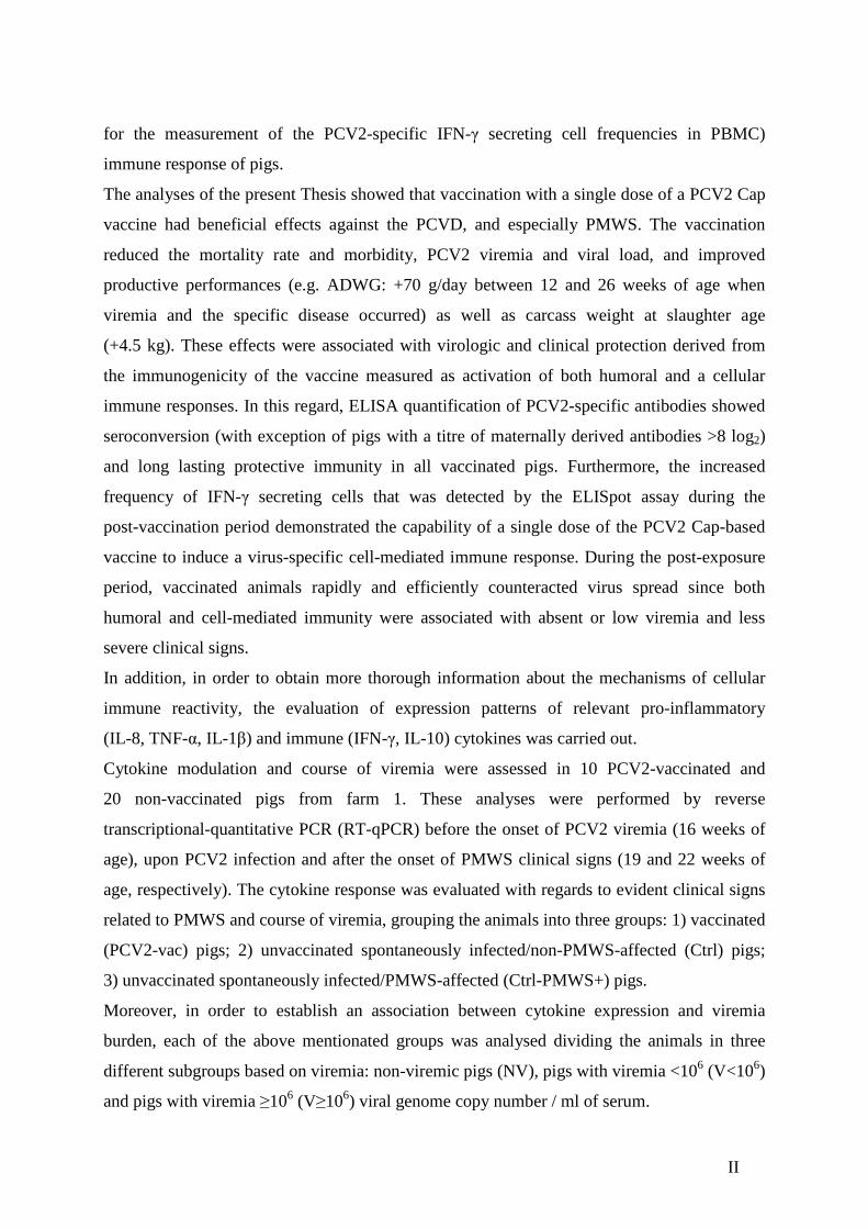
Abstract
Porcine circovirus type 2 (PCV2) has been identified as the main causative agent of the
postweaning multisystemic wasting syndrome (PMWS), one of the major swine diseases
worldwide that is commonly referred, together with other relevant porcine diseases related to
PCV2, as belonging to the porcine circovirus associated diseases (PCVD).
The most important strategy to prevent and control PCV2 associated diseases, apart from
management procedures and control of coinfections, is the vaccination of piglets or sows and
gilts. Nowadays there are three commercial PCV2 vaccines available; even if their efficacy in
reducing the viremia burden and viral-induced specific lymphoid lesions has been proved, the
mechanisms by which they are able to elicit protective immunity have not been thoroughly
clarified. Besides the development of humoral immunity that is generally characterised by the
detection of total anti-PCV2 and virus-neutralizing antibodies, the mechanisms that allow the
adaptive cell-mediated immune response to control PCV2 infection and the related diseases
have not been clearly elucidated, particularly under field conditions.
The present Thesis investigated the efficacy of a one-dose porcine circovirus 2 (PCV2)
subunit vaccine based on the PCV2 Cap protein expressed in a baculovirus system in two
different farms (farm1 and 2) at which a history of porcine circovirus-associated disease
(PCVD) was present. Morbidity, mortality, average daily weight gain, carcass weight, PCV2
load in serum and vaccine immunogenicity, in terms of PCV2-specific antibodies,
PCV2-specific IFN-γ secreting cell frequencies and mRNA expression profiles of relevant
pro-inflammatory and immune cytokines, were assessed. Serology to potential coinfections
due to porcine reproductive and respiratory syndrome virus (PRRSV) and
Mycoplasma hyopneumoniae (M. hyo.) was also carried out.
A double-blind, randomised, and controlled field trial was performed distributing 818 piglets
in two treatment groups. At inclusion (weaning at 21±3 days of age), 408 animals received a
2-ml intramuscular dose of Porcilis PCV® (vaccinated group) suspended in a tocopherol-based
adjuvant (Diluvac Forte®). Controls (410 piglets) received 2 ml of the same adjuvant alone
intramuscularly. Weights were recorded at inclusion and at 12 and 26 weeks of age, and the
average daily weight gain (ADWG) was calculated. The carcass weights of the pigs from farm
2 were recorded at slaughter (274 day-old pigs). All dead animals (died or culled) underwent
autopsy to classify them as PMWS-affected or not. At each farm, blood samples were
collected for serologic and cellular studies aimed at investigating the humoral
(ELISA determination of PCV2-antibody titres in serum) and cell-mediated (ELISpot assay
II
for the measurement of the PCV2-specific IFN-γ secreting cell frequencies in PBMC)
immune response of pigs.
The analyses of the present Thesis showed that vaccination with a single dose of a PCV2 Cap
vaccine had beneficial effects against the PCVD, and especially PMWS. The vaccination
reduced the mortality rate and morbidity, PCV2 viremia and viral load, and improved
productive performances (e.g. ADWG: +70 g/day between 12 and 26 weeks of age when
viremia and the specific disease occurred) as well as carcass weight at slaughter age
(+4.5 kg). These effects were associated with virologic and clinical protection derived from
the immunogenicity of the vaccine measured as activation of both humoral and a cellular
immune responses. In this regard, ELISA quantification of PCV2-specific antibodies showed
seroconversion (with exception of pigs with a titre of maternally derived antibodies >8 log2)
and long lasting protective immunity in all vaccinated pigs. Furthermore, the increased
frequency of IFN-γ secreting cells that was detected by the ELISpot assay during the
post-vaccination period demonstrated the capability of a single dose of the PCV2 Cap-based
vaccine to induce a virus-specific cell-mediated immune response. During the post-exposure
period, vaccinated animals rapidly and efficiently counteracted virus spread since both
humoral and cell-mediated immunity were associated with absent or low viremia and less
severe clinical signs.
In addition, in order to obtain more thorough information about the mechanisms of cellular
immune reactivity, the evaluation of expression patterns of relevant pro-inflammatory
(IL-8, TNF-α, IL-1β) and immune (IFN-γ, IL-10) cytokines was carried out.
Cytokine modulation and course of viremia were assessed in 10 PCV2-vaccinated and
20 non-vaccinated pigs from farm 1. These analyses were performed by reverse
transcriptional-quantitative PCR (RT-qPCR) before the onset of PCV2 viremia (16 weeks of
age), upon PCV2 infection and after the onset of PMWS clinical signs (19 and 22 weeks of
age, respectively). The cytokine response was evaluated with regards to evident clinical signs
related to PMWS and course of viremia, grouping the animals into three groups: 1) vaccinated
(PCV2-vac) pigs; 2) unvaccinated spontaneously infected/non-PMWS-affected (Ctrl) pigs;
3) unvaccinated spontaneously infected/PMWS-affected (Ctrl-PMWS+) pigs.
Moreover, in order to establish an association between cytokine expression and viremia
burden, each of the above mentionated groups was analysed dividing the animals in three
different subgroups based on viremia: non-viremic pigs (NV), pigs with viremia <106 (V<106)
and pigs with viremia ≥106 (V≥106) viral genome copy number / ml of serum.
III
Higher IL-8, TNF-α and IFN-γ levels were detected in the PCV2-vac group, testifying a more
efficient immune responsiveness, especially when compared to the Ctrl-PMWS+ group.
In Ctrl-PMWS+ pigs, lower IFN-γ at 19 weeks of age was associated with high IL-10 at
19 weeks of age and low levels of pro-inflammatory cytokines at 22 weeks of age, namely
IL-8 and TNF-α, a condition likely correlated with the onset of the disease.
Contrarily, at 19 weeks of age, PCV2-vac and Ctrl pigs showed lower IL-10 expression,
together with higher IFN-γ levels than the Ctrl-PMWS+ animals. At 22 weeks of age,
vaccinated animals maintained higher levels of the pro-inflammatory cytokines.
These evidences support that the outcome of PMWS could be associated with a reduction of
the innate/pro-inflammatory response. Overall, the results show a different cytokine
modulation in vaccinated and unvaccinated-infected pigs also developing PMWS. Vaccinated
pigs coped with infection showing low or absent viremia burden, absence of PMWS disease
and stronger inflammatory response and cellular IFN-γ-related reactivity.
IV
Riassunto
Il Circovirus suino di tipo 2 (PCV2) è stato identificato come il principale agente eziologico
della sindrome da deperimento post-svezzamento del suino (PMWS), una delle patologie del
suino maggiormente diffuse in tutto il mondo, comunemente indicata, insieme ad altre
patologie legate all’infezione da PCV2, come facente parte delle malattie associate al
circovirus suino (PCVD).
La strategia più importante per controllare e prevenire le malattie associate a PCV2, oltre alle
procedure di gestione e controllo delle coinfezioni, è la vaccinazione dei suinetti o delle scrofe
e delle scrofette. Tre sono i vaccini commerciali oggi disponibili; anche se è stata dimostrata
la loro efficacia nel ridurre la viremia e le lesioni presenti nei tessuti linfoidi indotte dal virus,
i meccanismi attraverso i quali questi vaccini sono in grado di indurre un’immunità protettiva
non sono ancora stati completamente chiariti.
A parte lo sviluppo di una risposta immunitaria di tipo umorale generalmente caratterizzata
dalla presenza di anticorpi totali e anticorpi virus neutralizzanti PCV2-specifici, i meccanismi
che permettono all’immunità adattativa cellulo-mediata di controllare l’infezione data da
PCV2 e le malattie ad essa associate non sono stati completamente compresi, specialmente in
condizioni di campo.
In questo lavoro di Tesi è stata valutata l’efficacia di un vaccino monodose verso PCV2,
basato sulla proteina capsidica Cap del virus espressa in un sistema baculovirus,
somministrato in due diversi allevamenti (allevamento 1 e 2) con anamnesi di malattia
associata a circovirus suino di tipo 2 (PCVD).
Sono stati considerati parametri quali morbilità, mortalità, incremento ponderale giornaliero,
peso della carcassa, titolo di PCV2 nel siero e immunogenicità del vaccino in termini di
anticorpi specifici verso PCV2, di frequenza di cellule secernenti IFN-γ PCV2-specifiche e di
livelli d’espressione genica di importanti citochine pro-infiammatorie e immunitarie. Sono
state inoltre effettuate analisi sierologiche verso potenziali coinfezioni sostenute da virus della
sindrome riproduttiva e respiratoria del suino (PRRSV) e Mycoplasma hyopneumoniae
(M. hyo.).
È stata condotta una prova di campo in doppio-cieco, randomizzata e con gruppo di controllo
distribuendo 818 suineti in due gruppi di trattamento. All’inizio della prova (giorno dello
svezzamento: 21±3 giorni di età), 408 suinetti (gruppo vaccinato) hanno ricevuto una dose di
vaccino Porcilis PCV®, risospeso in adiuvante a base di tocoferolo (Diluvac Forte®), per via
intramuscolare (2 ml). Gli animali controllo (410 suinetti) hanno ricevuto 2 ml di solo
V
adiuvante per via intramuscolare. L’aumento ponderale giornaliero (ADWG) è stato calcolato
misurando il peso degli animali all’inizio della prova, a 12 e a 16 settimane di età. I pesi delle
carcasse dei suini dell’allevamento 2 sono stati registrati al momento della macellazione
(274 giorni di vita). Tutti gli animali morti (morti o abbattuti) sono stati sottoposti ad autopsia
per essere classificati come animali affetti o non affetti da PMWS.
In ciascun allevamento sono stati prelevati campioni di sangue per effettuare indagini
sierologiche e della componente immunitaria cellulare. La risposte immunitarie umorali e
cellulo-mediate dei suini sono state valutate rispettivamente mediante ELISA, per rilevare il
titolo degli anticorpi PCV2-specifici nel siero, e tecnica ELISpot, per misurare la frequenza
delle cellule secernenti IFN-γ PCV2-specifiche nelle PBMC.
I risultati riportati nella presente Tesi suggeriscono che una singola dose di vaccino basato
sulla proteina Cap di PCV2 sia efficace contro l’insorgenza delle PCVD e in particolare della
PMWS.
La vaccinazione ha ridotto il tasso di mortalità e morbilità, la viremia specifica per PCV2 e la
carica virale, portando a un miglioramento delle performance produttive (es. ADWG:
70 g/giorno tra 12 e 26 settimane di età, quando si registra l’insorgenza di viremia e malattia
ad essa associata) e del peso della carcassa alla macellazione (+4,5 kg). Questi effetti sono
stati associati alla protezione dall’infezione e dal manifestarsi di sintomatologia clinica
determinata dall’immunogenicità del vaccino, misurata come attivazione della risposta
immunitaria umorale e cellulo-mediata. A questo proposito, la quantificazione mediante
tecnica ELISA degli anticorpi PCV2-specifici ha dimostrato sieroconversione (fatta eccezione
per i suini con titolo di anticorpi di derivazione materna >8 log2) e immunità protettiva di
lunga durata in tutti i suini vaccinati.
Inoltre, l’aumentata frequenza delle cellule secernenti IFN-γ PCV2-specifiche, quantificata
mediante tecnica ELISpot durante il periodo post-vaccinazione, ha dimostrato la capacità di
una singola dose di vaccino basato sulla proteina Cap di PCV2 di indurre una risposta
immunitaria cellulo-mediata virus-specifica.
Durante il periodo post-esposizione gli animali vaccinati hanno contrastato efficacemente e
rapidamente la replicazione virale; l’immunità umorale e cellulo-mediata sono risultate infatti
associate ad una bassa o assente viremia e segni clinici di minor gravità.
Inoltre, al fine di ottenere informazioni più approfondite sui meccanismi di reattività
immunitaria cellulare, sono stati valutati i profili d’espressione di importanti citochine
pro-infiammatorie (IL-8, TNF-α, IL-1β) e immunitarie (IFN-γ, IL-10). La modulazione

VI
dell’espressione citochinica e il corso della viremia sono state valutate in 10 animali vaccinati
contro PCV2 e 20 animali non vaccinati dell’allevamento 1. Queste analisi sono state
effettuate mediante PCR quantitativa Retro Trascizionale (RT-qPCR) prima dell'insorgenza
della viremia associata a PCV2 (16 settimane di età) e a seguito dell'infezione da PCV2 e la
comparsa di sintomatologia clinica da PMWS (19 e 22 settimane di età).
La risposta citochinica è stata valutata tenendo in considerazione gli evidenti segni clinici
relativi alla PMWS e al corso della viremia, suddividendo gli animali in tre gruppi: 1) suini
vaccinati (PCV2-vac); 2) suini non vaccinati spontaneamente infettati/non affetti da PMWS
(Ctrl); 3) suini non vaccinati spontaneamente infettati/affetti da PMWS (Ctrl-PMWS+).
Inoltre, per determinare un’associazione tra l’espressione delle citochine e l’andamento della
viremia, ciascuno dei gruppi sopracitati è stato analizzato dividendo gli animali in tre diversi
sottogruppi definiti sulla base del titolo virale (numero di copie di genoma virale/ ml di siero):
suini non viremici (NV), suini con viremia<106 (V<106) e suini con viremia ≥106 (V≥106).
Gli alti livelli di IL-8, TNF-α e IFN-γ rilevati nel gruppo PCV2-vac indicano che questi
animali hanno mostrato una responsività immunitaria più efficiente, specialmente se
confrontati al gruppo di animali Ctrl-PMWS+.
Nei suini Ctrl-PMWS+, rispetto agli altri gruppi sperimentali, sono stati osservati livelli più
ridotti di IFN-γ a 19 settimane di età, associati a più elevati livelli di IL-10 a
19 settimane di età e ad una più ridotta espressione di citochine pro-infiammatorie a 22
settimane di età, in particolare IL-8 e TNF-α, condizione probabilmente correlata
all’insorgenza della malattia. Al contrario, a 19 settimane di età, i suini dei gruppi PCV2-vac
e Ctrl hanno mostrato una più bassa espressione di IL-10 e maggiori livelli di IFN-γ rispetto
agli animali Ctrl-PMWS+. A 22 settimane di età gli animali vaccinati hanno mantenuto livelli
di citochine pro-infiammatorie più elevati.
Questi dati supportano l’ipotesi che l'esito della PMWS potrebbe essere associato ad una
riduzione della risposta immunitaria innata/pro-infiammatoria. Complessivamnete, i risultati
mostrano una diversa modulazione citochinica tra i suini vaccinati e non vaccinati infetti da
PCV2 che sviluppano PMWS. I suini vaccinati combattono l'infezione mostrando ridotta o
assente viremia, assenza di PMWS e una risposta infiammatoria e reattività cellulare associata
all’IFN-γ più intense.
VII
Table of Contents
CHAPTER 1. INTRODUCTION .....................................................................................................1
1.1 Porcine circovirus type 2 (PCV2) ...........................................................................................2
1.1.1 PCV2 history....................................................................................................2
1.1.2 Taxonomy.........................................................................................................3
1.1.3 Genotypes.........................................................................................................3
1.1.4 Molecular characteristics..................................................................................4
1.1.5 PCV2-associated diseases ................................................................................7 1.2 Postweaning multisystemic wasting syndrome (PMWS)....................................................10
1.2.1 Epidemiology .................................................................................................10
1.2.2 Clinical features..............................................................................................11
1.2.3 Diagnosis........................................................................................................12
1.2.4 Pathogenesis ...................................................................................................13
1.2.5 Intervention strategies ....................................................................................15
1.2.6 Vaccine development and vaccination...........................................................16
1.2.7 Effectiveness of vaccination...........................................................................17 1.3. PCV2 and the immune system .............................................................................................19
1.3.1 Interaction between PCV2 and immune cells ................................................19
1.3.2 Humoral response...........................................................................................22
1.3.3 Cell-mediated immune responses...................................................................23
1.3.4 PCV2 modulation of cytokine profiles...........................................................24 CHAPTER 2. OBJECTIVES OF THE RESEARCH...................................................................26 CHAPTER 3. EXPERIMENTAL STUDY UNDER FIELD CONDITION S.............................30
3.1. Materials and methods..........................................................................................................31
3.2. Results ....................................................................................................................................41 CHAPTER 4. DISCUSSION AND CONCLUSIONS...................................................................67 CHAPTER 5. REFERENCES ........................................................................................................77 CHAPTER 6. RESEARCH ACTIVITIES AND PUBLICATIONS .... .......................................92
VIII
List of abbreviations
ADV Aujeszky’s disease virus
ADWG average daily weight gain
AM alveolar macrophages
BMDC bone-marrow derived dendritic cells
Ctrl controls (unvaccinated spontaneously infected/non-PMWS-affected pigs)
Ctrl-PMWS+ controls-PMWS+ (unvaccinated spontaneously infected/PMWS-affected pigs)
DC dendritic cells
GAPDH glyceraldehyde 3-phosphate dehydrogenase
HI haemoagglutination inhibition
ICTV International Committee for the Taxonomy of Viruses
IFN interferon
Ig immunoglobulin
IL interleukin
MDA maternally derived antibodies
M. hyo Mycoplasma hyopneumoniae
MdM monocyte-derived macrophages
MOI multiplicity of infection
NA neutralising antibodies
ODN oligodeoxynucleotides
PCV porcine circovirus
PCV1 porcine circovirus type 1
PCV2 porcine circovirus type 2
PCVAD porcine circovirus associated diseases
PCVD porcine circovirus diseases
PMWS postweaning multisystemic wasting syndrome
PPV porcine parvovirus
PCV2-vac PCV2-vaccinated pigs
PRRSV porcine reproductive and respiratory syndrome virus
RT-qPCR reverse transcription-quantitative PCR (polimersase chain reaction)
SC secreting cells
SIV swine influenza virus

CHAPTER 1.
INTRODUCTION
Chapter 1 Introduction
2
1.1 Porcine circovirus type 2 (PCV2)
1.1.1 PCV2 history
Porcine circovirus (PCV) was first detected in 1974 as a virus morphologically similar to a
picornavirus, contaminating the porcine kidney cell line PK-15 (ATTC-CCL33)
(Tischer et al., 1974). This contaminant agent was subsequently demonstrated to be a circular
single-stranded DNA (ssDNA) virus that was accordingly named porcine circovirus (PCV)
(Tischer et al., 1982). Since the virus induced an antibody response but no disease in the pig
population, it was defined as non-pathogenic (Tischer et al., 1986; Dulac and Afshar, 1989;
Allan et al., 1994).
Postweaning multisystemic wasting syndrome (PMWS) is a multifactorial disease that was
first reported in North America in 1991 (Clark, 1997; Harding, 1997); since then, this disease
has affected the swine industry worldwide. The clinical signs of this syndrome include weight
loss, severe growth retardation and death in weaned piglets; PMWS is also characterized by a
multiorgan disease including lymphadenopathy, respiratory dysfunction, hepatitis,
splenomegaly and gastric ulcers (Clark, 1997; Harding, 1997), lymphocyte depletion,
monocytic infiltration in lymphoid tissues and high amounts of viruses in these lesions
(Segalés et al. 2002).
After the isolation of a PCV-like agent from tissues of PMWS-affected pigs, both in
North America and Europe (Allan et al., 1998b; Ellis et al., 1998), the non-pathogenic PK-15
cell culture-derived virus and the circovirus isolated from PMWS-affected pigs were
compared. Nucleotide sequence analyses revealed significant genetic differences between
viruses (Allan et al., 1998a), less than 80% of sequence identity (Meehan et al., 1998);
because of that they were divided into two types: the non pathogenic PCV type 1 (PCV1) and
the virus associated with clinical disease, that is PCV type 2 (PCV2) (Hamel et al., 1998;
Meehan et al., 1998).
Chapter 1 Introduction
3
1.1.2 Taxonomy
Both PCV1 and PCV2 belong to the Circoviridae family (Todd et al. 2005; Opriessnig et al.
2007) that is composed of icosahedrical, small, non-enveloped ssDNA viruses infecting
vertebrates (Lukert et al., 1995). Viruses within the Circoviridae family, based of their
morphology and genomic organization, are divided in two genera: Circovirus and Gyrovirus
(Todd et al. 2005; Opriessnig et al. 2007).
Circovirus genus includes Porcine circovirus type 1 and type 2 and, according to the
International Committee for the Taxonomy of Viruses (ICTV), other known avian viruses
such as Beak and feather disease virus (Ritchie et al., 1989), Canary circovirus (Phenix et al.,
2001), Duck circovirus (Hattermann et al., 2003), Finch circovirus (Shivaprasad et al., 2004),
Goose circovirus (Todd et al., 2001), Gull circovirus (Smyth et al., 2006), Pigeon circovirus
(Woods et al., 1993), Starling circovirus (Johne et al., 2006) and Swan circovirus
(Halami et al., 2008).
The genus Gyrovirus, that differs from circovirus for its negative sense genome and its large
virions (Gelderblom et al., 1989; Gillespie et al., 2009), contains only Chicken anaemia virus
(CAV) (Todd et al., 2005; Opriessnig et al., 2007).
1.1.3 Genotypes
Several phylogenetic analyses have demonstrated that PCV2 isolates from different
geographical origins can be divided into 2 distinctive genogroups (Larochelle et al., 2002;
Mankertz et al., 2000; Olvera et al., 2007). In some studies a stronger association of certain
PCV2 genogroups with the PCVD onset (Grau- Roma et al., 2008; Timmusk et al., 2008) has
been described whereas other reports have stated that there is no direct relationship between
the development of PMWS and the infection by a specific genogroup of PCV2
(Allan et al., 2007; Olvera et al., 2007). The difficulty to identify pathogenic differences
between genotypes has been recently attributed to the presence of a conserved decoy epitope
in the C-terminal region of the PCV2 capsid protein (Trible and Rowland, 2011).
The two phylogenetic groups, depending on the author, have been commonly referred as
PCV2a and PCV2b in North America and PCV2 group 1 (included in the PCVb group) and
PCV2 group 2 (included in the PCVa group) in Europe. In addition, some North American
laboratories, based on predicted restriction fragment length polymorphism (RFLP) patterns,
Chapter 1 Introduction
4
grouped the virus into two RFLP patterns designated as 422 and 321. Isolates with the RFLP
pattern 422 typically cluster into PCV2a (PCV2 group 2), whereas isolates with a 321 RFLP
pattern can be either PCV2a (PCV2 group 2) or PCV2b (PCV2 group 1) (Olvera et al., 2007;
Opriessnig T. et al., 2007).
Nowadays the North American nomenclature is officially accepted and the two clusters are
designated as PCV2a and PCV2b (Gagnon et al., 2007; Segalés et al., 2008); PCV2a contains
a genome of 1.767 nucleotides (nt) and can be divided into 3 clusters (1A–1C), while PCV2b
is characterised by a 1.768 nt genome and can be divided into 5 clusters (2A–2E)
(Olvera et al., 2007; Gillespie et al., 2009). The existence of discrete antigenic differences
between different PCV2 genetic clusters has been described in a recent study that performed
epitopes’ competition analysis using a panel of universal and cluster-specific mAbs
(Saha et al., 2011).
Several epidemiological studies worldwide have reported that PCV2b is becoming
predominant in many countries, underling a genotype switch of virus from PCV2a
(circulating with prevalence in the 1990’s) to this major group (Trible and Rowland, 2011). A
new PCV2 genogroup, PCV2c has been recently detected in Denmark in archived serum
samples from non-clinical pigs collected in 1980, 1987 and 1990; furthermore, it has been
demonstrated that this genogroup is more closely related to PCV2b (95%) than PCV2a (91-
93.6%) as sequence homology (Dupont et al. 2008; Opriessing et al. 2010).
1.1.4 Molecular characteristics
Porcine circoviruses (PCVs) are the smallest viruses infecting mammalian cells, being
characterised by an icosahedral, non-enveloped virion particle of 17±1.3 nm of diameter
(Tischer et al., 1982) that contains a covalently closed circular ssDNA genome with a size of
1759 bp and 1768 bp for PCV1 and PCV2 respectively (Meehan et al., 1998). PCVs, as the
other Circoviruses, replicate via rolling circle replication (RCR) so, after the infection of the
cells, their ssDNA is converted into a intermediate dsDNA, called replicative form (RT).
During this phase of the viral life cycle, the genome has an ambisense organization and genes
encoded by both the positive and negative strands (Cheung 2006; Meehan et al., 1997)
(Fig.1).
Chapter 1 Introduction
5
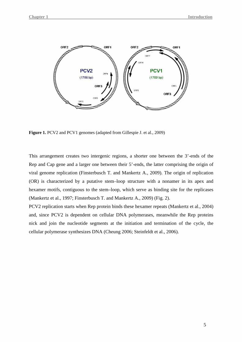
Figure 1. PCV2 and PCV1 genomes (adapted from Gillespie J. et al., 2009)
This arrangement creates two intergenic regions, a shorter one between the 3’-ends of the
Rep and Cap gene and a larger one between their 5’-ends, the latter comprising the origin of
viral genome replication (Finsterbusch T. and Mankertz A., 2009). The origin of replication
(OR) is characterized by a putative stem–loop structure with a nonamer in its apex and
hexamer motifs, contiguous to the stem–loop, which serve as binding site for the replicases
(Mankertz et al., 1997; Finsterbusch T. and Mankertz A., 2009) (Fig. 2).
PCV2 replication starts when Rep protein binds these hexamer repeats (Mankertz et al., 2004)
and, since PCV2 is dependent on cellular DNA polymerases, meanwhile the Rep proteins
nick and join the nucleotide segments at the initiation and termination of the cycle, the
cellular polymerase synthesizes DNA (Cheung 2006; Steinfeldt et al., 2006).
Chapter 1 Introduction
6
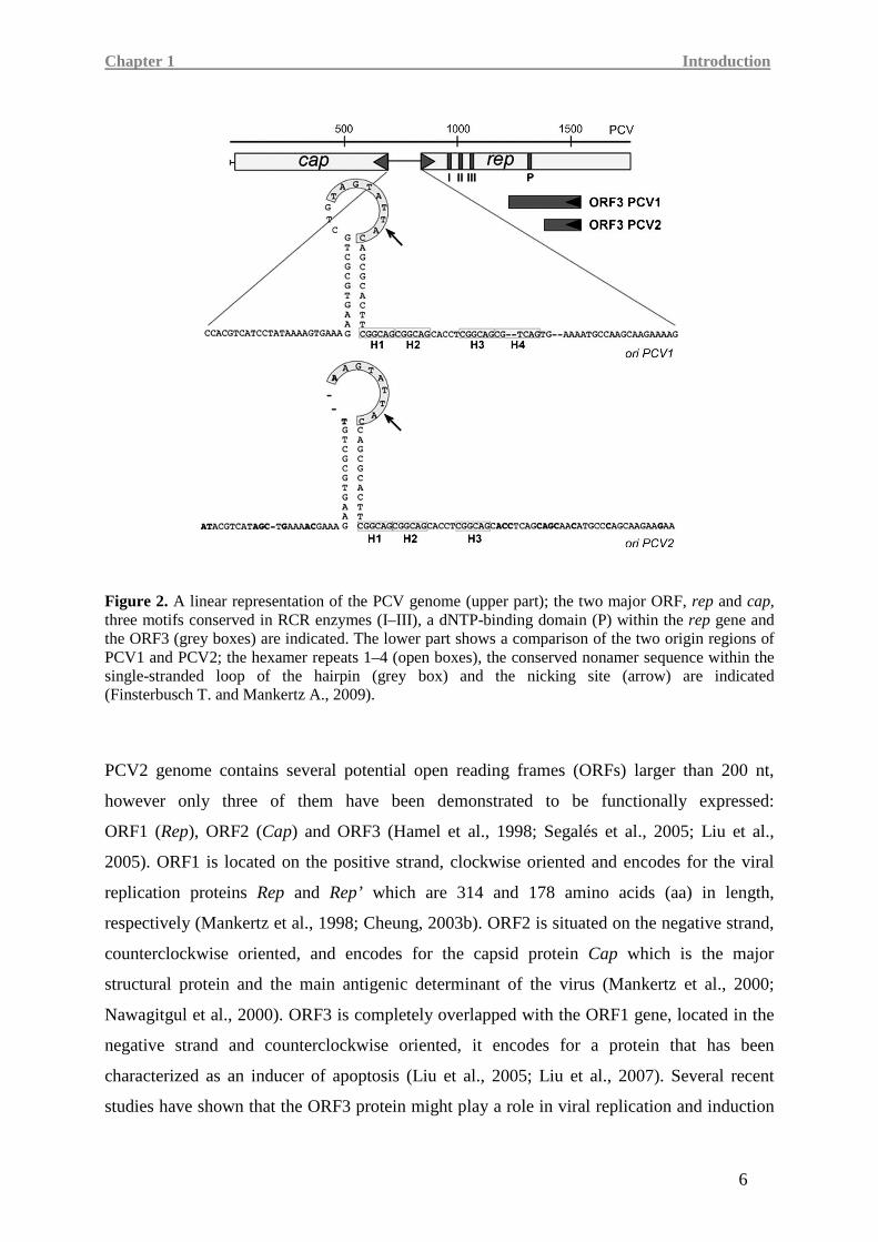
Figure 2. A linear representation of the PCV genome (upper part); the two major ORF, rep and cap, three motifs conserved in RCR enzymes (I–III), a dNTP-binding domain (P) within the rep gene and the ORF3 (grey boxes) are indicated. The lower part shows a comparison of the two origin regions of PCV1 and PCV2; the hexamer repeats 1–4 (open boxes), the conserved nonamer sequence within the single-stranded loop of the hairpin (grey box) and the nicking site (arrow) are indicated (Finsterbusch T. and Mankertz A., 2009).
PCV2 genome contains several potential open reading frames (ORFs) larger than 200 nt,
however only three of them have been demonstrated to be functionally expressed:
ORF1 (Rep), ORF2 (Cap) and ORF3 (Hamel et al., 1998; Segalés et al., 2005; Liu et al.,
2005). ORF1 is located on the positive strand, clockwise oriented and encodes for the viral
replication proteins Rep and Rep’ which are 314 and 178 amino acids (aa) in length,
respectively (Mankertz et al., 1998; Cheung, 2003b). ORF2 is situated on the negative strand,
counterclockwise oriented, and encodes for the capsid protein Cap which is the major
structural protein and the main antigenic determinant of the virus (Mankertz et al., 2000;
Nawagitgul et al., 2000). ORF3 is completely overlapped with the ORF1 gene, located in the
negative strand and counterclockwise oriented, it encodes for a protein that has been
characterized as an inducer of apoptosis (Liu et al., 2005; Liu et al., 2007). Several recent
studies have shown that the ORF3 protein might play a role in viral replication and induction
Chapter 1 Introduction
7
of immunosuppression (Karuppannan et al., 2009); indeed it has been proved, both in
BALB/c mice (Liu J. et al., 2006) and in specific-pathogen-free (SPF) piglets, that the knock-
out of ORF3 reduces PCV2 pathogenicity (Karuppannan, A. K., 2009). The non pathogenic
PCV1 also has a third open reading frame but its function has still to be characterised
(Chaiyakul M. et al., 2010).
1.1.5 PCV2-associated diseases
All the recognized syndromes associated with PCV2 infection can be nowadays designated by
two similar terms: PCVD (porcine circovirus diseases), proposed in 2002 by Allan and
co-workers and still predominantly used in Europe, and PCVAD (porcine circovirus
associated diseases), introduced by the American Association of Swine Veterinarians
(AASV) in 2006 and used mainly in North America. At present there is still no consensus
with regard to the disease nomenclature and both acronyms are accepted.
PCV2 is the primary causative agent of the syndromes included in PCVD but many other
common pathogens are involved in their onset; the different forms can be accomplished by
observation of characteristic lesions in the intestines, lungs, and lymphoid tissue
(Opriessnig et al., 2007).
PCV2 has been associated with subclinical diseases or other clinical manifestations such as
postweaning multisystemic wasting syndrome (PMWS), PCV2-Associated Enteritis, PCV2-
Associated Pneumonia, PCV2-Associated Reproductive Failure, Porcine Dermatitis and
Nephropathy Syndrome (PDNS) and PCV2-Associated Neuropathy (Gillespie et al., 2009).
The syndromes associated with PCV2, besides PMWS, are the following:
- Subclinical infections characterised by the absence of evidence of clinical disease although
PCV2 is present. In vivo study on PCV2-inoculated pigs have shown that PCV2 lesions can
be limited to 1 or 2 lymph nodes without causing any apparent clinical problems
(Opriessnig et al., 2004; Opriessnig et al., 2006a); cases of necrotising lymphadenitis
(Opriessnig et al., 2006a; Kim et al., 2005) or decrease in vaccine efficacy have also been
reported in healthy PCV2-infected pigs (Opriessnig et al., 2006b).
Chapter 1 Introduction
8
- PCV2-Associated Enteritis affects piglets from 8 to 16 weeks of age inducing increased
mortality, diarrhea and severe growth retardation: this syndrome is similar in clinical signs to
ileitis associated with Lawsonia intracellularis infection but an histopathological study can
distinguish between the two diseases; animals affected by PCV2-Associated Enteritis are
indeed characterised at necroscopy by an enlargement of mesenteric lymph nodes, thickening
of intestinal mucosa (Jensen et al., 2006), distinctive PCV2 lesions in Peyer’s patches but not
in other lymph nodes and granulomatous entheritis detectable by means of microscopical
analysis.
- PCV2-Associated Pneumonia occurs in pigs from 8 to 26 weeks of age and its
symptomatology includes reduced feed efficiency and growth rate, anorexia, fever, cough,
and dyspnea (Gillespie et al., 2009); histopathological studies on diseased pigs show
lymphohistiocytic to granulomatous bronchointerstitial pneumonia with necrotizing and
ulcerative bronchiolitis and bronchiolar fibrosis characterised by abundant PCV2 antigen in
the lesions. This evidence suggest that PCV2 may play a role in the Porcine Respiratory
Disease Complex (PRDC) in which also Porcine Reproductive and Respiratory Syndrome
Virus (PRRSV) are involved (Sorden, 1999; Sorden et al., 2000; Gillespie et al., 2009).
- PCV2-Associated Reproductive Failure damage herds of gilt startups or new populations
(Mikami et al., 2005) inducing clinical manifestations as increased abortion, still births, foetal
mummies, and pre-weaning mortalities; a non-suppurative to necrotizing or fibrosing
myocarditis has been also found in histopathological lesions of stillborn and neonatal pigs
(Mikami et al., 2005; Opriessnig et al., 2007). It has been proved that the time of infection
determines the clinical course of the disease: several studies have demonstrated that fetuses
experimentally intrauterine infected in an earlier phase of gestation (57 weeks of gestation)
present higher viral load and lesions as edema, enlarged liver and congestion, than those
infected in a later phase (75 and 92 days of gestation) (Sanchez et al., 2001); it has been also
shown that late term infections (86, 92, and 93 days of gestation) can cause an increase in
reproductive abnormalities (Johnson et al., 2002; Gillespie et al., 2009).
- Porcine Dermatitis and Nephropathy Syndrome (PDNS) was first described in the
United Kingdom in 1993 (Smith et al., 1993) and was associated with PCV2 only later, in
2000 (Rossel et al., 2000). Many pathogens including PRRSV and bacteria such as
Chapter 1 Introduction
9
Pasteurella multocida, Streptococcus suis type 1 and 2, among others, have been implicated
in the etiology of disease (Lainson et al., 2002; Thomson et al., 2002). PDNS is not always
associated with PCV2, in several studies it was indeed experimentally reproduced with
PRRSV and TTV in PCV2-free pigs (Krakowka et al., 2008).
This disease mostly affects growing pigs but can also occur in recently weaned and feeder
pigs from 1.5 to 4 months of age (Smith et al., 1993; Thibault et al., 1998); PDNS is clinically
characterized by an acute onset of multifocal and well circumscribed skin lesions
(raised purple progressing to multifocal raised red scabs with black centers most prominent on
the rear legs), fever, and lethargy and is often fatal within 3 days of development
(Done et al., 2001; Duran et al., 1997; Chae, 2005). Macroscopically, the kidney appears
enlarged and having pale cortex with multiple red circular haemorrhagic cortical foci (Ramos-
Vara et al., 1997). Microscopically, the most significant lesion is the severe, fibrinoid,
necrotizing vasculitis in the dermis, subcutis, lymph nodes, stomach, spleen, liver and kidney
which can be associated with dermal and epidermal necrosis and necrotizing and fibrinous
glomerulonephritis.
- PCV2-Associated Neuropathy causes in pigs congenital tremors and a nonsuppurative
menigoencaphalitis associated with demyelination of the brain and spinal cord
(Larochelle et al., 2002; Pensaert et al., 2004; Correa et al., 2007). PCV2 infection has also
been associated with cerebellar lymphohistiocytic vasculitis or with lymphohistiocytic
meningitis (Correa et al., 2007) but even today the role played by this virus in this kind of
diseases has still to be clarified.
Chapter 1 Introduction
10
1.2 Postweaning multisystemic wasting syndrome (PMWS)
The most significant manifestation of PCVAD is the postweaning multisystemic wasting
syndrome (PMWS). Since the 1990s, porcine circovirus type 2 (PCV2) has been considered
the causative agent of this disease, even if the majority of infections caused by PCV2 are
sub-clinical, and only a small proportion of PCV2-infected pigs develops the clinical form of
disease. PMWS is considered a multifactorial disease and the fully clinical expression of this
syndrome is indeed due to the co-presence of PCV2 and pathogens such as porcine
reproductive and respiratory syndrome virus (PRRSV), swine influenza virus (SIV), porcine
parvovirus (PPV), Haemophilus parasuis, Actinobacillus pleuropneumoniae,
Streptococcus suis and Mycoplasma hyopneumoniae (Chae, 2004).
1.2.1 Epidemiology
PMWS was described for the first time in a Canadian high-health-status herd in 1991 and was
later recognized worldwide (Allan and Ellis, 2000) being associated with major losses in
Europe (Harding et al., 2000; Opriessing et al., 2008). In 1996, both in British and French
farms, there were cases of wasting and high losses in growing pigs (Madec et al., 2004)
and PCV2 was isolated from the animals and the disease was later defined as PMWS.
Cases of PMWS were retrospectively identified in archived serum and tissues samples from
1962 in Germany (Jacobsen et al., 2009), 1969 in Belgium (Sanchez et al., 2001a), 1970
in the United Kingdom (Grierson et al., 2004), 1973 in Ireland (Walker et al., 2000) and 1985
in Canada and Spain (Magar et al., 2000; Rodríguez-Arrioja et al., 2003).
During the following decade, PMWS spread over the world becoming a considerable
economic problem in many pig-producing countries. In fact, PCV2 infection is so widespread
today that it is almost impossible to find seronegative farms in epidemiology studies
(Grau-Roma et al., 2009). PMWS morbidity is associated with the development of clinical
manifestations of disease and ranges between 4% and 30%, being (although) up to 60% in
some herds (Segalés and Domingo, 2002). PMWS prevalence generally ranges from 4% to
30% and mortality ranges from 4% to 20%, but can reach 50% (Allan and Ellis 2000;
Harding and Clark, 1997; Segalés and Domingo, 2002).
Chapter 1 Introduction
11
1.2.2 Clinical features
PCV2 infection can occur during the whole pig productive life, but PMWS usually affects
animals from 8 to 16 weeks of age (Sibila et al., 2004; Grau-Roma et al., 2009).
However, PMWS has been shown to occur at different ages in the United States
(from 7 to16 weeks of age) and Europe (from 5 to 12 weeks of age) (Allan and Ellis 2000;
Segalés and Domingo, 2002) due to different management and vaccination practices.
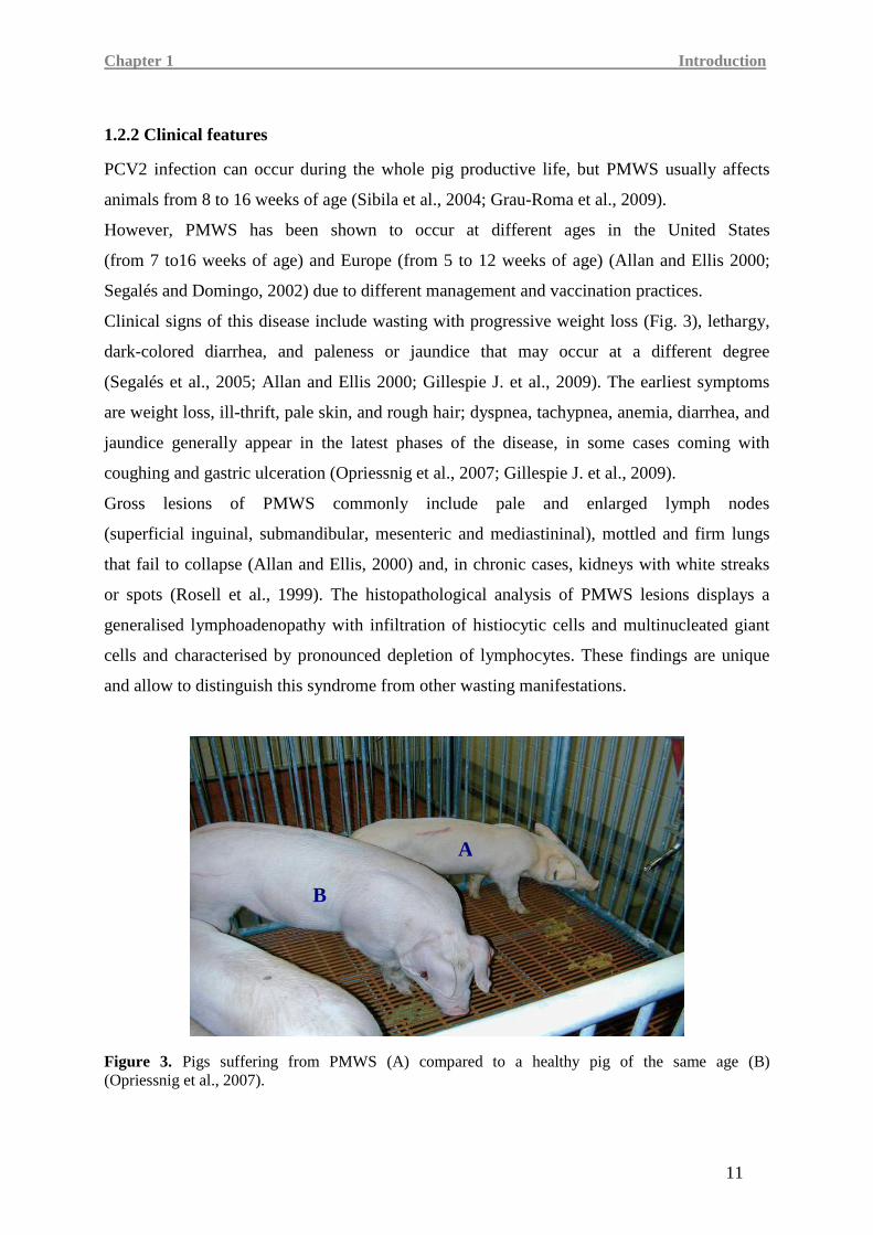
Clinical signs of this disease include wasting with progressive weight loss (Fig. 3), lethargy,
dark-colored diarrhea, and paleness or jaundice that may occur at a different degree
(Segalés et al., 2005; Allan and Ellis 2000; Gillespie J. et al., 2009). The earliest symptoms
are weight loss, ill-thrift, pale skin, and rough hair; dyspnea, tachypnea, anemia, diarrhea, and
jaundice generally appear in the latest phases of the disease, in some cases coming with
coughing and gastric ulceration (Opriessnig et al., 2007; Gillespie J. et al., 2009).
Gross lesions of PMWS commonly include pale and enlarged lymph nodes
(superficial inguinal, submandibular, mesenteric and mediastininal), mottled and firm lungs
that fail to collapse (Allan and Ellis, 2000) and, in chronic cases, kidneys with white streaks
or spots (Rosell et al., 1999). The histopathological analysis of PMWS lesions displays a
generalised lymphoadenopathy with infiltration of histiocytic cells and multinucleated giant
cells and characterised by pronounced depletion of lymphocytes. These findings are unique
and allow to distinguish this syndrome from other wasting manifestations.
Figure 3. Pigs suffering from PMWS (A) compared to a healthy pig of the same age (B) (Opriessnig et al., 2007).
A
B
Chapter 1 Introduction
12
The lymphatic system can be involved at different levels by the disease. Based on that, a
scoring system has been defined to evaluate the severity of the disease. This estimation
system allow to assign scores from 0 to 9 ranking the severity of lesions, the amount of PCV2
antigen and the distribution of lesions in seven indicative lymphoid tissues such as the
tracheobronchial lymph nodes, the mesenteric lymph nodes, the mediastinal lymph nodes, the
superficial inguinal lymph nodes, the external iliac lymph nodes, the tonsils, and the spleen
(Opriessnig at al., 2004; Gillespie et al., 2009).
In case of PMWS the immune system of pigs can be strongly compromised and this can
increase the probability to be subjected to secondary infections (Segalés et al., 2005).
1.2.3 Diagnosis
PCV2 induces several clinical signs that are also shared by other pig diseases; for this reason
a diagnosis of a specific syndrome is not easy to define. The presence of PCV2 genome in
serum and the observation of a diseased status of pigs is not enough to define a PCVD case.
It has been established that to make a diagnosis of PCVD, in addition to clinical signs, PCV2
antigen has to be necessarily found in more than one lymphoid tissue, or one lymphoid tissue
and one other organ such as the lungs, liver, kidney or intestine, or in two organs
(Gillespie et al., 2009).
More specifically, to categorise PMWS cases, Segalés and co-workers suggested in 2005
the following criteria:
1) clinical signs compatible with PMWS (growth retardation and wasting);
2) moderate to severe histopathological lesions characterized by lymphocyte depletion
together with granulomatous inflammation;
3) moderate to high amount of PCV2 genome/antigen within lesions.
Tests such as polymerase chain reaction, in situ hybridization (ISH) and
immunohistochemistry (IHC) are considered the optimal techniques to detect PCV2 antigen
or nucleic acid and make a diagnosis of PMWS (Opriessnig et al., 2007). Serological tests
such as IPMA (immunoperoxidase monolayer assay) or SN (seroneutralisation) are useful to
identify an infectious state but not enough to substitute histopathological evaluations and PCR
analysis (Allan et al., 1998b; Grierson et al., 2004; Allan et al., 2000; Gillespie et al., 2009).
Chapter 1 Introduction
13
Seropositivity to PCV2 can be found in many clinically healthy pigs and the status of
subclinical infection is commonly set when low amounts of PCV2 are detected in blood
and/or tissues, associated with no or minimal lesions. The cut-off generally considered to
distinguish between PMWS-diseased pigs and sub-clinically infected pigs is 107 PCV2 DNA
copies/ml of serum (Opriessnig et al., 2007).
Currently, there is no field test for the diagnosis of PMWS, but to identify and manage its
outbreaks within a herd it is important to determine whether the disease is a significant or a
sporadic problem. It has been defined that there is an important herd problem if the following
conditions are observed (Segales J., 2006; Gillespie et al., 2009):
1) significant increased postweaning mortality that is equal or higher than the mean
historical mortality plus 1.66 times the standard deviation. If historical data are not
available, a herd problem can be described when the postweaning mortality exceeds
the national or regional level by 50% or more;
2) confirmation of PMWS in individual cases.
1.2.4 Pathogenesis
In case of PCV2 infection there are significant differences between sub-clinical and
PMWS-affected pigs; current evidence support a central role for immunodepression in the
pathogenesis of PMWS.
In pigs that develop PMWS, the highest amount of PCV2 is found in the cytoplasm of
monocyte and machrophage lineage cells (Rosell et al., 1999; Sanchez et al., 2004).
The virus can infect these cells without an active replication for a long period of time
(Gilpin et al., 2003; Vincent et al., 2003). The capability of PCV2 to induce functional
impairment of in vitro cultured dendritic cells (DC) has been also described (Vincent et al.,
2005); this underlines the ability of the virus to interfere with innate and virus-specific
immune responses.
It has been displayed that PCV2 is not able to encode for its own polymerase and its
replication depends on host’s nuclear polymerases (Tischer et al., 1987). For this reason it is
possible to identify cells that support replication of PCV2 by evaluating the presence of Rep,
the PCV2 replication associated protein, in the nucleus of the cell (Rovira et al., 2002). Earlier
studies have demonstrated the presence of nuclear PCV2 in epithelial cells of PMWS-affected
pigs, proposing these cells as candidate for primary PCV2 infection (Rossel et al., 1999).
Chapter 1 Introduction
14
Studies on experimentally PCV2-inoculated pigs have proven viral replication in lymphocytes
from PBMC and bronchial lymph nodes by measuring Cap mRNA levels in the cells
(Yu et al., 2007a). The subpopulations of leukocytes that support PCV2 replication are
prevalently circulating T cells, both CD4+ and CD8+, and in a lower proportion
B lymphocytes; PBMC-derived monocytes do not seem to sustain viral replication
(Yu et al., 2007b; Lefebvre et al., 2008b; Lin et al., 2008).
PMWS is characterised by lymphocyte and follicular dendritic cell depletion from follicular
regions, together with increased numbers of histiocytic cells (Chianini et al., 2003).
It is still unknown whether the lymphocyte depletion is due to reduced production in the bone
marrow, reduced proliferation in secondary lymphoid tissues, or increased loss of
lymphocytes in the bone marrow, peripheral blood, or secondary lymphoid tissues via
virus-induced necrosis or apoptosis (Opriessnig et al., 2007). A reduction of B and T
lymphocytes, especially CD8+ cells, has been also reported in blood circulation; at the same
time, an increased number of circulating neutrophils and monocytes determine a reversal of
the normal ratio of lymphocytes/neutrophils (Nielsen et al., 2003; Segales et al., 2001).
Infection studies have not clarified yet this phenomenon.
An experimental PCV2 infection study showed that at 7 days post-PCV2 infection the
lymphocyte depletion has already started, whereas maximal depletion of both B and T cell
subsets, followed by a huge or total loss of NK cells, occurs later, at 21 days post-infection
(Nielsen et al., 2003).
Humoral immunity seems to play a very important role in controlling and resolving viremia
(Fort et al., 2007; Opriessnig et al., 2008b). In sub-clinical animals, an efficient humoral
response is frequently associated with long-lasting viremia, low concentration of virus at
lymphoid tissue, and no significant changes in the status of the immune system
(Allan et al., 1999a; Resendes et al., 2004a). On the contrary, weak humoral responses can be
related to increased viral replication, resulting in the severe lymphoid lesions and
immunosuppressive status characteristic of PMWS (Bolin et al., 2001).
Several experimental and field studies supported the multifactorial nature of PMWS and
highlighted that not all pigs that are infected by PCV2 develop clinical PCVAD.
The outcome of this syndrome can be influenced by several factors that can be grouped in
four main areas: virus, host, coinfections, and immune modulation (Opriessnig et al., 2007).
The accurate mechanism by which these factors cooperate determining the onset of the
Chapter 1 Introduction
15
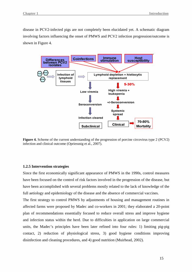
disease in PCV2-infected pigs are not completely been elucidated yet. A schematic diagram
involving factors influencing the onset of PMWS and PCV2 infection progression/outcome is
shown in Figure 4.
Figure 4. Scheme of the current understanding of the progression of porcine circovirus type 2 (PCV2) infection and clinical outcome (Opriessnig et al., 2007).
1.2.5 Intervention strategies
Since the first economically significant appearance of PMWS in the 1990s, control measures
have been focused on the control of risk factors involved in the progression of the disease, but
have been accomplished with several problems mostly related to the lack of knowledge of the
full aetiology and epidemiology of the disease and the absence of commercial vaccines.
The first strategy to control PMWS by adjustments of housing and management routines in
affected farms were proposed by Madec and co-workers in 2001; they elaborated a 20-point
plan of recommendations essentially focused to reduce overall stress and improve hygiene
and infection status within the herd. Due to difficulties in application on large commercial
units, the Madec’s principles have been later refined into four rules: 1) limiting pig-pig
contact, 2) reduction of physiological stress, 3) good hygiene conditions improving
disinfection and cleaning procedures, and 4) good nutrition (Muirhead, 2002).
5-30%
Chapter 1 Introduction
16
The implementation of Madec’s plan as a guideline for the control of postweaning mortality
in PMWS affected farms proved to be efficacious in reducing the PMWS-associated losses
(Allan and McNeilly, 2006; Segalés et al., 2005).
1.2.6 Vaccine development and vaccination
Vaccination against PCV2 represents an important strategy to control PMWS in pig herds.
For this reason there is active interest in the development of vaccines able to prevent or limit
the PCV2-associated diseases. Nowadays, several commercial products are used in the herds
and all of them are based on PCV2a genotypes that have been demonstrated to provide
cross-protection also to PCV2b (Fort et al., 2008, 2009; Segalés et al., 2009).
Nowadays, three commercially vaccines are available against PCV2:
• CIRCOVAC®
(Merial) was the first vaccine on the market; it has been extensively
used in Europe but it has been also available in Canada. It is an inactivated PCV2,
oil-adjuvanted vaccine for use in sows and gilts 2-4 weeks prior to farrowing
(Charreyre et al., 2005; Gillespie et al., 2009) that is given as two injections
IM (intramuscular administration) 3-4 weeks apart and completed at least 2 weeks
before breeding and once at each subsequent gestation (Opriessnig et al., 2007).
The active immunisation on these breeding-aged animals is used to induce passive
immunisation to the offspring by means of colostrum transfer.
The other two commercial products use different approaches for establishing protective
immunity against PCV2. They are recombinant vaccines designed for use in growing pigs,
at about 3-4 weeks of age.
• Ingelvac CircoFLEX® is a capsid-based subunit vaccine based on the product of the
ORF2 expressed in a baculovirus system; it is administered as a single dose IM in
piglets from 2 weeks of age. The immunity of the treated animals starts about 2 weeks
after vaccination remaining protective at least for further 17 weeks. A significant
decrease in mortality in vaccinated pigs compared to unvaccinated pigs was reported
on 4 different Canadian finishing sites (Desrosier et al., 2007; Gillespie et al., 2009).
Chapter 1 Introduction
17
• The vaccine from Intervet/Schering-Plough/Merck is also a capsid-based subunit
vaccine expressed in a baculovirus, it is designated as Porcilis PCV®in Europe and
Asia and Circumvent PCV ®
in the United States and Canada (Gillespie et al., 2009).
Porcilis PCV ® can be administered to 3-day-old and older piglets, following a double
dose IM protocol (at 3-5 days and 3 weeks of age) or given as a single dose IM at
3 weeks of age; Circumvent® is given to 3-week-old and older piglets and is
administered as a single dose at 3 weeks of age. Both of them induce protective
immunity that remains active from 2 to 22 weeks after vaccination; studies including
35,000 pigs on 21 different farms showed that mortality of vaccinated pigs is reduced
by 77.5% when compared to unvaccinated pigs (Grau et al., 2007; Gillespie et al.,
2009).
1.2.7 Effectiveness of vaccination
The effectiveness of the vaccines available against PCV2 has been widely evaluated in several
studies. Due to the poor clinical manifestation in piglets infected by PCV2 only, some studies
have also evaluated the responses in animals co-infected by two or three porcine pathogens.
Experimental co-infection try to reproduce herd field conditions in which numerous
pathogens, more frequently PRRSV and Mycoplasma Hyopneumoniae, contribute to PCVD
outbreak (Beach and Meng, 2011). In this regard it has been found that the presence of
PRRSV can increase the severity of PCV2-related clinical signs, inducing a wide spread of
the virus by oronasal and faecal excretions (Allan et al., 2000; Rovira et al., 2002;
Sinha et al., 2011). Contrarily, a large number of vaccines against PCV2 have demonstrated to
induce neutalising antibody (NA) secretion, reduced viral load and lymphoid lesions in cases
of PCV2 infection but also in the presence of SIV (swine influenza virus) or PRRSV
(Opriessnig et al., 2009) coinfections.
The vaccination of boars by using Suvaxyn PCV2 one dose, followed by infection with PCV2
or Mycoplasma, seems to prevent serious clinical manifestation, reduce viral titres in the
blood and virus excretion by faeces and semen with respect to an unvaccinated control group
(Opriessnig et al., 2011). Boars vaccination does not alter semen characteristics and is proved
to be a good practice to reduce vertical PCV2 transmission.
Chapter 1 Introduction
18
In addition, sow and gilt vaccination has been reported to increase the number of live born
pigs and the number of pigs per sow per year, reducing the number of mummies per sow
(Thacker et al., 2008). It is relevant to highlight that the vaccination of sows is not able to
completely eliminate viral transmission by colostrum or prevent intrauterine infection but is
however important to increase productive parameters of the herds (Beach and Meng, 2011).
Several studies have demonstrated the efficacy of PCV2 vaccination; in a infection study,
vaccine treatment of piglets from a PRDC group reduced the mean viral titre from 83% to
55% and its duration overtime, improving average daily weight gain (ADWG) (Fachinger et
al., 2008). In another study, vaccination of piglets from a PMWS/PCVD farm showed a
50% reduction of mortality and a 9.3 % increase of ADWG (Horlen et al., 2008).
The efficacy of a subunit vaccine containing PCV2 capsid protein has been also proven after
experimental infection with four different PCV2 isolates of the two genotypes (PCV2a and
PCV2b) (Fort et al., 2008).
Therefore, after vaccination of growing pigs, under experimental conditions, it has been
observed a reduction of viremia, lymphoid lesions and amount of PCV2 DNA in tissues,
oronasal and fecal PCV2 excretion and specific IgM, IgG and NA production, as well as
cross-protection to both PCV2a and PCV2b (Fenaux et al., 2004; Fort et al., 2008;
Opriessnig et al., 2008c). Under field conditions, increases of ADWG and percentage of lean
meat, improvement of feed conversion index and reductions of mortality have been observed
(Fachinger et al., 2008; Horlen et al., 2008; Kixmoller et al., 2008; Cline et al., 2008;
King et al., 2008; Opriessnig et al., 2008a, 2008b; Tacker et al., 2008; Desrosiers et al., 2009;
Segalés et al., 2009; Martelli et al., 2011), together with increased numbers of PCV2-specific
IFN-γ secreting cells (IFN-γ SC) suggesting the presence of effector and/or memory T cells
(Fort et al., 2009; Lyoo et al., 2011).
Chapter 1 Introduction
19
1.3. PCV2 and the immune system
The specific immune response that develops in pigs after infection by PCV2 is crucial and
strongly influences the outcome of infection. The animals that develop PMWS often appear to
be immunosuppressed and unable to eliminate the virus from the blood circulation.
In the final phase of the clinical manifestation, infected-diseased pigs show extensive
lymphoid lesions and altered cytokine expression patterns in PBMC and lymphoid organs due
to their ineffective immune response (Kekarainen et al., 2010).
Not always PCV2 infection determines the outbreak of clinical signs and immunological
disorders; in fact, there are asymptomatic animals that show higher virus-specific and
neutralising antibody titres than PMWS-affected animals. The mechanisms by which PCV2
can affect the immune responses have not been completely elucidated but recent studies have
pointed out virus interaction with macrophages and plasmacytoid dendritic cells and the role
of viral DNA in regulation of immune cell functions.
1.3.1 Interaction between PCV2 and immune cells
In infected animals, cells of the monocytic lineage, including monocytes, macrophages and
dendritic cells (DC), are most frequently associated to intracellular detection of PCV2 which
however does not seem to replicate in such cells (Gilpin et al., 2003; Vincent et al., 2003).
These cells accumulate viral antigen and DNA for extended periods of time, but since viral
replication is inefficient, they are thought to play a major role in viral persistence and
transmission (Gilpin et al., 2003; Vincent et al., 2003; Pérez-Martín et al., 2007).
Dendritic cells
In dendritic cells (DC) the presence of live PCV2 particles leads to different effects depending
on the cell subpopulation.
In myeloid dendritic cells (mDC) the virus does not appear to be detrimental to cell survival
and does not interfere with their maturation; in vitro studies on mDC infected with PCV2
have indeed proven that the cell expression of major histocompatibility complex (MHC) class
I and II or cluster of differentiation (CD) 80/86 is not altered by the virus, even after exposure
to IFN-α and tumor necrosis factor TNF-α (Vincent et al., 2005; Vincent et al., 2003).
Chapter 1 Introduction
20
In the same studies the antigen presenting and processing ability of mDC was reported not to
be compromised by the infection.
On the contrary, the interaction of PCV2 with plasmocitoid dendritic cells (pDC), also called
Natural Interferon Producing cells (NIPC), induces impaired responsiveness to danger signals,
inhibits interferon (IFN)-α and tumor necrosis factor (TNF)-α production and interferes with
NIPC maturation as well as paracrine maturation of mDC (Vincent et al., 2005).
The virus seems not to be transmitted from DC to lymphocytes; this may be a viral strategy to
escape the host’s immune defence and disseminate exploiting the circulation of DC
(Vincent et al., 2003). Since asymptomatic piglets produce anti-PCV2 antibodies
(Krakowka et al., 2002; Ladekjaer-Mikkelsen et al., 2002; Nielsen et al., 2003; Steiner et al.,
2009) and cytotoxic responses (Steiner et al., 2009), the presence of PCV2 in DC does not
always impair their immunobiological interaction with lymphocytes (Kekarainen et al., 2010).
Monocytes and macrophages
In vitro and ex vivo studies on the early immune responses following PCV2 infection have
determined that also monocytes, monocyte-derived macrophages (MdM) and alveolar
macrophages (AM) are able to internalize PCV2 (Gilpin et al., 2003; Kekarainen et al., 2010);
in particular, AM phagocyte the virus but, as for DC, viral replication is not strongly
detectable (Chang et al., 2006). The microbicidal and phagocytic functions of macrophages
seem to be influenced by PCV2; in addition, increased production of pro-inflammatory
cytokines such as interleukin (IL)-8 and TNF-α as well as the up-regulation of
macrophage-derived chemotactic factor-II (AMCF-II), granulocyte colony-stimulating factor
(G-CSF) and monocyte chemotactic protein-1 (MCP-1) have been reported
(Chang et al., 2006; Kekarainen et al., 2010).
It was also suggested that the PCV2-mediated alteration of AM functionality can support
opportunistic and secondary pulmonary infections.
Peripheral Blood Mononuclear Cells (PBMC)
The lymphopenia observed in PMWS-affected pigs is likely due to an indirect effect
promoted by PCV2 infection in DC and macrophages (Kekarainen et al., 2010). In response
to recall antigen (PCV2), PBMC from diseased pigs can respond by an increased production
of IL-10 and IFN-γ compared to PBMC from infected healthy pigs, and display an impaired
ability to produce IL-4, IL-2 and IFN-γ upon stimulation with antigen (porcine pseudorabies
Chapter 1 Introduction
21
virus; PRV), mitogen (phytohaemagglutinin; PHA) or superantigen (staphylococcal
enterotoxin B; SEB) (Darwich et al., 2003a; Kekarainen et al., 2008b). Moreover, Kekarainen
and co-workers (2008b) reported that PCV2 is able to modulate the specific immune
responses developed by pigs to other pathogens. They showed that IL-12, IFN-α, IFN-γ and
IL-2 recall responses of PBMC after pseudorabies virus stimulation were down-regulated by
PCV2, underling that the decreased production of IL-12, IFN-α and IFN-γ could be due to the
release of PCV2-induced IL-10 by PBMC, CD172a+ cells and bone-marrow derived DC
(BMDC).
Immune regulatory role for PCV2 DNA
An immune regulatory role for PCV2 DNA was described for the first time in a study focused
on the evaluation of the ability of various subpopulations of porcine DC to endocytose PCV2
from infected PK15A cell lysates (Vincent et al., 2003). Viral genomic ssDNA (Vincent et al.,
2005) and dsDNA replicative intermediates endocytosed from the infected cell lysates
(Vincent et al., 2007) were found in DC. Vincent et al. (2007) theorised that these DNA
elements can interact with endosomal toll-like receptors (TLR) or cytosolic helicases inducing
impairment of cytokines produced by pDC. These findings suggested the presence of DNA
sequences in the PCV2 genome capable to interfere with DC and immune defences.
Several CpG motifs have been characterised in PCV2 genome revealing synthetic
oligodeoxynucleotides (ODN) sequences able to modulate cytokine production of porcine
PBMC cultures by induction or inhibition (Hasslung Wikström et al., 2003; Kekarainen et al.,
2008a). The effect of these ODN was also analysed on porcine PBMC recall responses and
cytokine production by BMDC (Kekarainen et al., 2008a; Kekarainen et al., 2010).
It has been found that some ODN induce a decrease in the IFN-α response of BMDC upon
PRV stimulation. The majority of the inhibitory ODN is located within the Rep region of the
PCV2 genome but none of them, conversely to the whole virus, have been demonstrated to
induce IL-10 production in PBMC (Kekarainen et al., 2008b). However, the presence of ODN
in PBMC cultures appeared to decrease the IFN-γ or IL-2 responses to recall antigens
(Kekarainen et al., 2010).
Chapter 1 Introduction
22
1.3.2 Humoral response
Immunity against PCV2 in piglets can be transmitted as maternal antibodies by the sow. This
passive immunity has been demonstrated to protect from PMWS onset in a dose-dependent
manner (Rose et al., 2007). Colustrum administration is an important practice to induce
protection of the offspring but also the PCV2 infection status of the sow has been
demonstrated to be crucial. With regards to these evidence Calsamiglia et al. (2007) showed
that, from sows with low levels of PCV2 antibodies, there is a higher percentage of piglets
that result PMWS-affected. Nevertheless, it is important to remind that the presence of PCV2
antibodies is not necessarily protective because not all antibodies are neutralizing upon PCV2
infection.
In experimentally infected pigs, seroconversion commonly occurs between 14 and 28 days
post-inoculation (dpi) (Allan et al., 1999a; Balasch et al., 1999; Krakowka et al., 2001;
Meerts et al., 2005; Segalés et al., 2005). More specifically, several studies have observed that
PMWS-affected pigs seroconvert later (Fort et al., 2007; Meerts et al., 2006;
Bolin et al., 2001; Okuda et al., 2003), or show a weak response characterised by lower
antibody titres at 21 dpi than in subclinically PCV2-infected animals (Meerts et al., 2006;
Kekarainen et al., 2010).
The different immunoglobulin isotypes follow the course of total antibody titres
(Meerts et al., 2006); in PCV2-subclinically infected pigs also the titres of virus neutralising
antibodies (NA) follow this course (Meerts et al., 2006; Fort et al., 2007). Contrarily, the
impaired humoral response of diseased pigs is characterised by decreased production of total
antibodies and low levels of NA that subsequently result in a higher viral load
(Fort et al., 2007; Meerts et al., 2005).
Under field conditions, the first protection against PCV2 is represented by passive immunity
transferred by the sow; this protection lasts during the lactating and nursery periods and is
depleted by the end of nursery and the beginning of fattening periods (Rodriguez-Arrioja et
al., 2002; Rose et al., 2002; Larochelle et al., 2003). Active seroconversion to PCV2 usually
occurs between 7-12 weeks of age (Segalés et al., 2005) and anti-PCV2 antibodies generally
last until at least 28 weeks of age (Rodriguez-Arrioja et al., 2002). An impaired humoral
response may extend the period between the decline of maternal immunity and the onset of
Chapter 1 Introduction
23
active seroconversion in piglets, thus increasing the probability to develop PMWS upon
PCV2 infection.
It has also been showed that the titres of anti-PCV2 IgM overtime, in contrast with the course
of IgG1, IgG2 and IgA antibodies, remain lower in diseased pigs than in subclinical infected
animals (Meerts et al., 2006).
In case of PCV2 infection, the adaptive humoral immune responses play a crucial role in
determining whether or not the outbreak of PMWS occurs despite other immune mechanisms
are required to obtain complete viral clearance.
1.3.3 Cell-mediated immune responses
The role of adaptive cell-mediated responses in controlling PCV2 infection is still less studied
compared to humoral responses (Kekarainen et al., 2010). However, the few studies that have
already been performed provide relevant information. On gnotobiotic experimentally infected
pigs, it has been demonstrated that the treatment with cyclosporine A (CyA)
(i.e. an immunosuppressing agent), before PCV2 inoculation determines an increase of viral
replication (Krakowka et al., 2002; Meerts et al., 2005). The IFN-γ mRNA expression levels
in PBMC from these PCV2-inoculated animals resulted to be correlated with viral replication
and immunosuppressed status induced by CyA, suggesting that a higher expression of IFN-γ
can help pigs be less susceptible to PCV2 replication (Meerts et al., 2005).
Other studies have analysed the development of IFN-γ secreting cells (IFN-γ SC) in either
conventional colostrum-fed pigs infected with PCV2 alone (Fort et al., 2009a) or in
colostrum-fed specific pathogen free (SPF) pigs infected with PCV2 together with Porcine
parvovirus as a potential triggering factor for PMWS development (Steiner et al., 2009).
Caesarean-derived colostrum deprived (CD/CD) pigs infected with PCV2 along with
lipopolysaccharide (LPS) (Fort et al., 2009a) have also been analysed (Kekarainen et al.,
2010). The results of these three studies underline the key role of IFN-γ SC in developing the
anti-PCV2 adaptive cellular response.
It has been found that vaccination treatements are effective in reducing PCV2 load in the
blood, concomitantly with the development of a virus-specific humoral response, especially
mediated by NA both in field (Kixmoller et al., 2008) and experimental (Opriessnig et al.,
Chapter 1 Introduction
24
2008c) infection models. In addiction, it has been shown the onset of a cellular response in
terms of PCV2-specific IFN-γ SC in pigs vaccinated with a PCV2 sub-unit vaccine,
experimentally infected (Fort et al., 2009b). Thus, we can assume that if at least one of these
responses fails, viral clearance will be impaired, and the risk of developing PMWS can
increase. However, the cell-mediated responses of pigs to PCV2 infection, although in cases
of previous vaccination, have still to be thoroughly investigated.
1.3.4 PCV2 modulation of cytokine profiles
Cytokine mRNA expression profiles can be important to characterise the host’s immune
responses that occur upon viral infections, therefore pro-inflammatory and immune cytokine
production has been recently investigated in different PMWS and infection models (Chae et
al., 2011; Kekarainen et al., 2008a, 2010). With regards to lymphoid tissues, increased levels
of IL-10 mRNA expression were found in the thymus and decreased levels of IL-4 and IL-2
were detected in tonsils and spleen from PMWS-affected pigs (Darwich et al., 2003b).
Opposed results were described for IFN-γ: low mRNA expression levels were detected in
inguinal and tracheo-bronchial lymph nodes whereas high expression was shown in tonsils
(Darwich et al., 2003b; Zhang et al., 2010).
An increase of IL-10, together with slight increases in IL-8, IFN-γ and TNF-α and a decrease
in IL-2 and IL-4 mRNA levels, were also detected in PBMC from PCV2 naturally infected
pigs (Sipos et al., 2004).
Significantly higher expression of IL-10 was found in PCV2-infected lymphoid tissues
compared to uninfected control tissues (Doster et al., 2010); furthermore, serum IL-10 was
detected in pigs developing severe PMWS and the increased expression of this cytokine has
been reported to be correlated with viremia in subclinically PCV2-infected pigs (Stevenson et
al., 2006).
Increased serum levels of the acute phase proteins (APP) haptoglobin, pig major acute phase
protein (pig-MAP), C-reactive protein (CRP), serum amyloid A (SAA) and albumin were also
reported in PMWS-affected pigs (Parra et al., 2006; Stevenson et al., 2006; Segalés et al.,
2004).
Chapter 1 Introduction
25
The production of cytokines and the balance between pro-inflammatory, pro-immune and
regulatory cytokines play a pivotal role in eliciting the innate response as well as in priming
and coordinating the adaptive immune response.
For this reason the study of cytokines appears to be an important tool to evaluate the cellular
immune response against PCV2. The above mentioned evidence suggest a severe
immunosuppression in PMWS-affected pigs but the mechanisms determining the
immunological impairment, that is not detectable in subclinically infected animals, are still
poorly understood (Kekarainen et al., 2010).
CHAPTER 2.
OBJECTIVES OF THE RESEARCH
Chapter 2 Objectives of the Research
27
Management strategies, control of coinfections and vaccination are at present the measures by
which PMWS and Porcine Circovirus Diseases (PCVD) are controlled.
Nowadays PCV2 vaccination represents one of the major strategies to overcome PCV2
infections in the herds and therefore several commercial vaccines are available.
All PCV2 vaccines currently available on the market have been tested under field conditions
resulting to be effective and helpful in decreasing mortality and cull rates and significantly
improving the average daily weight gain (ADWG), concomitantly with decreasing the
frequency of coinfections in herds affected with PMWS (Cline et al., 2008; Desrosiers et al.,
2009; Fachinger et al., 2008; Horlen et al., 2008; King et al., 2008; Kixmoller et al., 2008;
Opriessnig et al., 2008b,c; Tacker et al., 2008; Segalés et al., 2009; Pérez-Martin, 2010).
Even if PCV2 vaccines are effective in reducing the viremia burden and viral-induced specific
lymphoid lesions, the mechanisms by which they are able to elicit protective immunity are not
thoroughly known (Fachinger et al., 2008; Fort et al., 2008; Horlen et al., 2008).
However, since PCV2 can evade immune surveillance and PCV2 infection is often associated
with co-infections in the field (e.g. porcine reproductive and respiratory syndrome virus -
PRRSV, Mycoplasma hyopenumoniae - M. hyo.), it is not easy to obtain a complete resolution
of the disease.
Therefore, the improvement of vaccine formulations and administration strategies represents
one of the main areas of interest in PCV2 vaccinology.
The activation of the host’s immune response has been proved to be one of the primary
factors modulating the progression of the disease. Several studies have shown the importance
of the antibody response, especially that mediated by neutralizing antibodies (NA), in coping
with infection. Also the cell-mediated immune response seems to play a key role in
preventing viral replication and counteract viral diffusion, despite many immune mechanisms
which sustain virus clearance and the resolution of infection are still unclear
(Fenaux et al., 2004b; Kixmoller et al., 2008; Fort et al., 2009a, 2009b; Steiner et al., 2009;
Pérez-Martin et al., 2010).
For these reasons, several studies have been performed to investigate the mechanisms by
which PCV2 can elude the immune defences in pigs and to develop new vaccination
strategies aimed at inducing efficient immune activation and immune protection upon
infection. The effects of vaccination treatments are related to total and neutralising antibody
responses as well as to cell-mediated immunity (Larochelle et al., 2003; Fort et al., 2008,
2009a; Kixmoller et al., 2008; Opriessnig et al., 2008b, 2008c).
Chapter 2 Objectives of the Research
28
The humoral immunity to PCV2 infection were characterised, by most of the published
reports, through the detection of total anti-PCV2 antibodies, showing seroconversion that
occurs either in subclinically or non-PMWS infected and PMWS-affected pigs (Rodrıiguez-
Arrioja et al., 2000; Sibila et al., 2004; Grau-Roma et al., 2009).
On the other hand, the role and mechanisms of the adaptive cell-mediated immune response
in controlling PCV2 infection and the related diseases have not been clearly elucidated,
particularly under field conditions. Previous reports based on laboratory trials describe that
viral clearance may be mediated by cell-mediated immunity, measured by the number of
PCV2-specific interferon-γ (IFN-γ) secreting cells (SC), together with neutralising antibodies
(Fort et al., 2009a, 2009b) and that the load and the extent of viral replication may influence
the intensity of the cell-mediated immune response.
Specifically PCV2 vaccines showed to induce an intense antibody and cellular responses but
data regarding the immune responses under field conditions and underlying vaccine-induced
protection are still incomplete.These aspects are worth investigating under field conditions
both in diseased pigs naturally infected by PCV2 in the presence of coinfections and in
vaccinated animals showing no or few clinical signs.
Furthermore, the modulation of cytokine patterns are important to categorize the host immune
responses that occur during viral infections; for successful resolution of infection, efficient
activation of innate/inflammatory and acquired immunity is required to block pathogen
replication and invasion, as well as to promote tissue clearance of the pathogens and/or
infected cells. The production of pro-inflammatory cytokines (IL-1β, TNF-α, IL-8) and the
balance between pro-immune (IFN-γ) and regulatory (IL-10) cytokines play a pivotal role in
eliciting the innate response as well as in priming and coordinating the adaptive immune
response. However, if production is impaired, the innate response will be delayed and
inefficient in clearing the pathogen (Borghetti et al., 2010).
Therefore, pro-inflammatory and immune cytokine production has been investigated in
different PMWS and infection models. Increased levels of IL-10 mRNA expression were
found in the thymus and decreased levels of IL-4 and IL-2 were detected in the tonsils and
spleen from PMWS-affected pigs (Sipos et al., 2004). Contrary results were described for
IFN-γ: low mRNA expression levels were detected in inguinal and tracheobronchial lymph
nodes whereas high expression was shown in the tonsils (Darwich et al., 2003b; Zhang et al.,
2010). An increase of IL-10, together with slight increases of IL-8, IFN-γ and TNF-α and a
decrease of IL-2 and IL-4 mRNA levels, were also detected in PBMC from naturally
Chapter 2 Objectives of the Research
29
PCV2-infected pigs (Sipos et al., 2004). Furthermore, serum IL-10 was associated with
subclinically PCV2-infected pigs or pigs developing severe PMWS (Stevenson et al., 2006).
A significantly higher expression of IL-10 was also found in PCV2-infected lymphoid tissues
compared to uninfected control tissues (Doster et al. 2010).
Cytokine secretion may play a key role in evaluating the cellular immune response against
PCV2, but the cytokine modulation investigated so far in different organs has not provided
univocal results likely due to different working conditions.
Taking into account the above mentioned features, the aim of the present thesis is to
investigate the efficacy of PCV2 vaccination under field conditions in vaccinated and
unvaccinated pigs upon PCV2 natural infection and associated PCVD in terms of clinical
protection and development of the humoral and cell-mediated immune response.
The specific objectives of the present study are the following:
1. the evaluation of the efficacy of a one-dose PCV2 subunit vaccine based on the
PCV2 Cap protein expressed in a baculovirus system (Porcilis PCV®) at two
different farms where PCVD was present, in terms of clinical protection;
2. the assessment of the vaccine immunogenicity in vaccinated and unvaccinated pigs
exposed to PCV2 natural infection in terms of development of humoral (total
PCV2-specific antibodies) and cell-mediated (PCV2-specific IFN-γ secreting cells)
immune responses;
3. the quantitative modulation of pro-inflammatory and immune cytokines in
vaccinated and unvaccinated pigs exposed to natural PCV2 infection, in relation to
the onset of PCV2 viremia burden and PMWS clinical signs.
The results regarding the evaluation of PCV2 vaccine efficacy as clinical protection and
humoral and cell-mediated immune responses were published in Martelli P., Ferrari L.,
Morganti M., De Angelis E., Bonilauri P., Guazzetti S., Caleffi A., Borghetti P. Veterinary
Microbiology. 2011; 149(3-4): 339-351.
The data concerning the study of cytokine immune modulation were submitted to the journal
“Comparative Immunology, Microbiology and Infectious Diseases” as Borghetti P., Morganti
M., Ferrari L., De Angelis E., Saleri R., Cavalli V., Corradi A., Martelli P. Modulation of
pro-inflammatory and immune cytokines in PBMC of vaccinated and unvaccinated pigs
exposed to porcine circovirus type 2 (PCV2) natural infection (submitted, 2011).
CHAPTER 3.
EXPERIMENTAL STUDY UNDER FIELD CONDITIONS
Chapter 3 Experimental Study
31
3.1. Materials and methods
3.1.1. Selection of farms
The study was conducted in the Northern part of Italy at two farms with history of PMWS.
In both herds, at approximately 15-20 weeks of age, clinical signs of PWMS characterised by
wasting, respiratory signs and growth retardation were mainly associated with a marked
increase in the mortality rate. The diagnosis fulfilled the internationally accepted disease case
definition, including clinical signs, gross lesions, histopathologic findings, and presence of
PCV2 in lymphoid lesions (Segale´ s et al., 2005). Seropositivity to PCV2 in all categories of
animals (replacement gilts, sows, nursery pigs, growers, and fatteners) was demonstrated.
Before the start of the study, 5 wasted pigs were autopsied at each farm and the diagnosis was
reconfirmed.
FARM 1 was a 900-sow farrow-to-finish herd that, in the previous year, experienced a
6% and an 8% mortality rate in the nursery and fattening periods, respectively. This farm was
seronegative for Aujeszky’s disease virus (ADV) and seropositive for PRRSV,
M. hyopneumoniae and A. pleuropneumoniae. Low titres of antibodies at haemagglutination
inhibition (HI) to SIV were obtained from some samples and were not informative.
FARM 2 was a three-site farm with 1850 sows experiencing a 2% and a 10% mortality rate in
the nursery and fattening periods, respectively. Seropositivity to PRRSV and
M. hyopneumoniae and seronegativity to ADV were found.
Sows of both herds were vaccinated for Aujeszky’s disease (3 times/year), porcine parvovirus
and erysipelas (both at mid-lactation). Piglets were vaccinated for Aujeszky’s disease
according to the National Control Program.
The protocols and results of the evaluation of PCV2 vaccine efficacy as clinical protection
and humoral and cell-mediated immune responses are published in Martelli P., Ferrari L.,
Morganti M., De Angelis E., Bonilauri P., Guazzetti S., Caleffi A., Borghetti P. Veterinary
Microbiology. 2011; 149(3-4): 339-351.
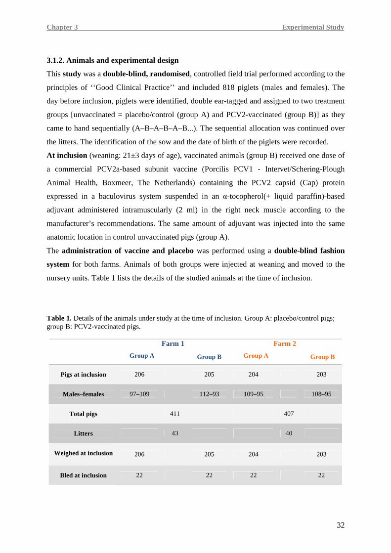
Chapter 3 Experimental Study
32
3.1.2. Animals and experimental design
This study was a double-blind, randomised, controlled field trial performed according to the
principles of ‘‘Good Clinical Practice’’ and included 818 piglets (males and females). The
day before inclusion, piglets were identified, double ear-tagged and assigned to two treatment
groups [unvaccinated = placebo/control (group A) and PCV2-vaccinated (group B)] as they
came to hand sequentially (A–B–A–B–A–B...). The sequential allocation was continued over
the litters. The identification of the sow and the date of birth of the piglets were recorded.
At inclusion (weaning: 21±3 days of age), vaccinated animals (group B) received one dose of
a commercial PCV2a-based subunit vaccine (Porcilis PCV1 - Intervet/Schering-Plough
Animal Health, Boxmeer, The Netherlands) containing the PCV2 capsid (Cap) protein
expressed in a baculovirus system suspended in an α-tocopherol(+ liquid paraffin)-based
adjuvant administered intramuscularly (2 ml) in the right neck muscle according to the
manufacturer’s recommendations. The same amount of adjuvant was injected into the same
anatomic location in control unvaccinated pigs (group A).
The administration of vaccine and placebo was performed using a double-blind fashion
system for both farms. Animals of both groups were injected at weaning and moved to the
nursery units. Table 1 lists the details of the studied animals at the time of inclusion.
Table 1. Details of the animals under study at the time of inclusion. Group A: placebo/control pigs; group B: PCV2-vaccinated pigs.
Farm 1
Group A
Group B
Farm 2
Group A
Group B
Pigs at inclusion
206 205 204 203
Males–females
97–109 112–93 109–95 108–95
Total pigs
411 407
Litters
43 40
Weighed at inclusion
206 205 204 203
Bled at inclusion
22 22 22 22
Chapter 3 Experimental Study
33
After weaning, pigs from both treatment groups were kept in mixed groups until the end of
the trial, when animals were sent to the slaughterhouse (at approximately 9 months of age).
Treatments, housing, husbandry, and feeding were conformed to the European Union
Guidelines and identical for both experimental groups at each farm. At each accommodation
change, pigs were commingled according to usual farm procedures.
Pigs were clinically monitored on a weekly basis from the administration of vaccine or
placebo until slaughter. From one week post-vaccination, pigs were monitored daily for
vaccination reactions. Individual live body weights of all animals enrolled in the study were
measured the day before inclusion and at 12 and 26 weeks of age. The average daily weight
gain (ADWG) was calculated based on the ADWG of animals being alive at the end of each
weighing period. Carcass weight was recorded at the slaughterhouse in pigs from farm 2.
If concomitant treatments (injections) were used, the number of animals treated within each
group and the duration of treatment were recorded. The number of relapsed animals
(retreatments) were also considered. The number of injections was used as a parameter to
evaluate morbidity .
Dead animals or those that had to be euthanised for reasons of animal welfare were recorded
daily.
In both herds, at inclusion (day of vaccination, 3 weeks of age), blood samples were collected
from 2 piglets of each pluriparous sow litter: one for each treatment group, up to 44
(22 piglets for each treatment group). These piglets were identified by a progressively
numbered ear tag.
Moreover, in piglets from FARM 1, blood samples were taken at 4 [+1 week post-
vaccination (PV)], 5 (+2 weeks PV), 6 (+3 weeks PV), 7 (+4 weeks PV), 9 (+6 weeks PV),
12 (+9 weeks PV), 15 (+12 weeks PV), 16 (+13 weeks PV), 17 (+14 weeks PV),
18 (+15 weeks PV), 19 (+16 weeks PV), 20 (+17 weeks PV), 22 (+19 weeks PV),
26 (+23 weeks PV), and 35 (+32 weeks PV) weeks of age.
In FARM 2, blood samples were collected at vaccination (3 weeks of age), at 4 (+1 week
PV), 6 (+3 weeks PV), 12 (+9 weeks PV), 16 (+13 weeks PV), 18 (+15 weeks PV),
20 (+17 weeks PV), 22 (+19 weeks PV), 24 (+21 weeks PV), 26 (+23 weeks PV), and
35 (+32 weeks PV) weeks of age.
Chapter 3 Experimental Study
34
3.1.3. Evaluation of gross pathology, histopathology, and immunohistochemistry
The objective of the pathologic studies was to establish the PMWS diagnosis in all pigs from
both farms that died, those needing euthanasia, and runts during the entire study period. These
pigs underwent gross pathologic examination and histopathology within 24 hours.
Samples from inguinal, mesenteric and mediastinic lymph nodes were removed from
necropsied pigs and fixed in 10% buffered formalin. Fixed samples were processed for
routine histopathology and 5-µm thick sections were stained with hematoxylin and eosin to be
examined for lesions compatible with PMWS.
The diagnosis of PMWS was made when all three criteria of the accepted international
individual case definition for the disease were present, that are: 1) clinical signs, mainly
including wasting; 2) moderate-to-severe lymphoid lesions; 3) moderate-to-high amounts of
PCV2 in those lesions.
The amount of PCV2 in tissue samples was also assessed by real-time quantitative PCR
(qPCR) using the methods reported by Olvera et al. (2004).
Immunohistochemistry for detection of PCV2-specific antigen was performed on formalin-
fixed and paraffin embedded sections of inguinal, mediastinal, and mesenteric lymph nodes
using a rabbit polyclonal antiserum (Sorden, 2000). PCV2 antigen scoring was performed by
a pathologist in a blind manner using the score range in accordance with Opriessnig et al.
(2004).
3.1.4. Extraction and qPCR detection of PCV2 DNA from tissue samples and serum
In order to detect and quantify the PCV2 DNA by PCR, DNA was firstly extracted from
200 µl of serum or 200 µl of 1:10 phosphate-buffered saline (PBS) homogenate of lymph
node tissue, by using TRIzol LS (Invitrogen, San Diego, CA,USA) following the
manufacturer’s instructions. The DNA obtained was suspended in 50 µl of
diethylpyrocarbonate (DEPC)-treated water. Real-time qPCR was carried out using a
LightCycler 1.5 (Roche, Basel-CH). Real-time qPCR was performed using primers and
probes according to Olvera et al. (2004). Results of the qPCR were expressed as number of
PCV2 genome copies per milliliter of serum or gram of tissue.
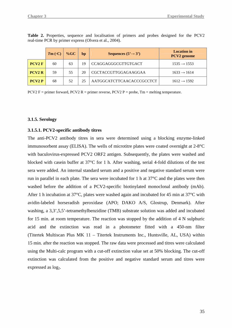
Chapter 3 Experimental Study
35
Table 2. Properties, sequence and localisation of primers and probes designed for the PCV2 real-time PCR by primer express (Olvera et al., 2004).
Tm (◦C) %GC bp Sequences (5’→ 3’) Location in PCV2 genome
PCV2 F 60 63 19 CCAGGAGGGCGTTGTGACT 1535 → 1553
PCV2 R 59 55 20 CGCTACCGTTGGAGAAGGAA 1633 → 1614
PCV2 P 68 52 25 AATGGCATCTTCAACACCCGCCTCT 1612 → 1592
PCV2 F = primer forward, PCV2 R = primer reverse, PCV2 P = probe, Tm = melting temperature.
3.1.5. Serology 3.1.5.1. PCV2-specific antibody titres
The anti-PCV2 antibody titres in sera were determined using a blocking enzyme-linked
immunosorbent assay (ELISA). The wells of microtitre plates were coated overnight at 2-8°C
with baculovirus-expressed PCV2 ORF2 antigen. Subsequently, the plates were washed and
blocked with casein buffer at 37°C for 1 h. After washing, serial 4-fold dilutions of the test
sera were added. An internal standard serum and a positive and negative standard serum were
run in parallel in each plate. The sera were incubated for 1 h at 37°C and the plates were then
washed before the addition of a PCV2-specific biotinylated monoclonal antibody (mAb).
After 1 h incubation at 37°C, plates were washed again and incubated for 45 min at 37°C with
avidin-labeled horseradish peroxidase (APO; DAKO A/S, Glostrup, Denmark). After
washing, a 3,3’,5,5’-tetramethylbenzidine (TMB) substrate solution was added and incubated
for 15 min. at room temperature. The reaction was stopped by the addition of 4 N sulphuric
acid and the extinction was read in a photometer fitted with a 450-nm filter
(Titertek Multiscan Plus MK 11 – Titertek Instruments Inc., Huntsville, AL, USA) within
15 min. after the reaction was stopped. The raw data were processed and titres were calculated
using the Multi-calc program with a cut-off extinction value set at 50% blocking. The cut-off
extinction was calculated from the positive and negative standard serum and titres were
expressed as log2.
Chapter 3 Experimental Study
36
3.1.5.2. Antibody titres to other infectious agents
The presence of antibodies to PRRSV and sample/positive (S/P) ratios were determined using
a commercially available ELISA kit (HerdChek1 Porcine Reproductive and Respiratory
Syndrome Antibody Test Kit, IDEXX Laboratories, Westbrook, ME, USA) according to the
manufacturer’s instructions. The Herd Check1 test bases the sample classification on the S/P
ratio, which is defined as (sample O.D. - negative control O.D.) / (positive control O.D.-
negative control O.D.). Sample to positive control ratios ≥ 0.4 were considered positive.
Antibodies to M. hyopneumoniae were evaluated by a commercially available ELISA test
(Herd Check1 M. hyopneumoniae, IDEXX Laboratories). The presence of antibodies to gE
glycoprotein of ADV was measured using a commercially available ELISA kit [HerdChek1
PRV g1 (gE) test kit, IDEXX Laboratories] according to the manufacturer’s instructions.
A commercial ELISA kit (CHEKIT-APP-Apx IV ELISA test kit, IDEXX Laboratories) was
used for the detection of antibodies against A. pleuropneumoniae. Serology to swine influenza
virus (SIV) was performed by using HI.
3.1.6. ELISpot for determination of PCV2-specific IFN-γ secreting cells (SC)
An IFN-γ ELISpot assay was performed according to Martelli et al. (2009) in order to
evaluate the frequencies of IFN-γ SC in the peripheral blood of pigs from farm 1.
The assay was performed in MultiScreen®HTS-IP plates (MSIPS4510 – Millipore) coated with
10 µg/ml anti-pig IFN-γ mAb (P2G10, BD, Biosciences, Franklinlakes, NJ, USA) at 4°C
overnight. After incubation, plates were washed with sterile PBS and blocked with
RPMI-1640 supplemented with 10% foetal bovine serum (FBS) for 2 h at 37°C, 5% CO2.
Peripheral blood mononuclear cells (PBMC) were isolated by density gradient using a
Histopaque-1.077® solution and plated at 2 x 10⁵ cells/well in RPMI-1640 + 10% FBS.
For the ex vivo antigen recall, a whole PCV2 strain (I12/11) at 0.1 multiplicity of infection
(MOI) was used as stimulus, in RPMI-1640 + 10% FBS, for 20 h at 37°C, 5% CO2; the SC
response was also evaluated at 0.05 and 0.25 MOI. In all samples, PBMC were > 98% viable
as confirmed by Trypan blue exclusion. Afterward, cells were removed by washing with PBS
+ 0.05% Tween-20 (PBST) and the plates were incubated for 1 h at 37°C with 0.5 µg/ml
anti-pig IFN-γ biotin-labeled mAb (P2C11, BD). After washing, plates were incubated with
1:750 alkaline phosphatise (AP)-conjugated anti-biotin mAb in PBS + 0.5% BSA for 1 h at
37°C. Plates were finally incubated for 7 min. with a BCIP/NBT solution (BioRad, Hercules,
Chapter 3 Experimental Study
37
CA, USA) in order to let spots develop and the reaction was stopped with distilled water. The
frequencies of PCV2-specific IFN-γ SC were determined using an AID® ELISpot Reader
(AID ® ELISpot Software v.3.5). As a positive control, 1 x 10⁵ PBMC/well were incubated
with phytohemagglutinin (PHA, 10 µg/ml); as a negative control, 2 x 10⁵ PBMC were
incubated in the absence of antigen (mock stimulus: supernatant of non-PCV2-infected PK-15
cells). The background values (number of spots in negative control wells) were subtracted
from the respective counts of the stimulated cells and the immune responses were expressed
as number of IFN-γ SC per million PBMC (IFN-γ SC/106 PBMC).
The IFN-γ SC responses were also classified as responsiveness categories on the basis of the
responses observed in the present study and with regards to categories reported in literature
(Fort et al, 2009b; Ferrari et al., 2011).
Specifically the responsiveness categories identified were:
• 0-40 IFN-γ SC/106 PBMC = no-poor
• 45-100 IFN-γ SC/106 PBMC = low
• 105-200 IFN-γ SC/106 PBMC = intermediate
• 205-400 IFN-γ SC/106 PBMC = high
• ≥ 405 IFN-γ SC/106 PBMC = very high
Particularly, the range for the “no-poor” category was determined based on the responses
observed in unvaccinated animals throughout the post-vaccination period and data in
literature (Pérez-Martin et al., 2010).
3.1.7. Evaluation of cytokine gene expression
Blood samples collected from 10 PCV2-vaccinated and 20 non-vaccinated pigs were used as
source of swine peripheral blood mononuclear cells (PBMC) in order to determine mRNA
expression levels of relevant pro-inflammatory and pro-immune cytokines.
At the end of the trial, taking into consideration the time of the onset of viremia and the
appearance of clinical signs, blood samples taken at 16 (before infection and the desease
onset), 19 and 22 weeks of age (during PCV2 viremia and disease outcome) were used for the
quantification of cytokine expression.
For the purposes of this study, the thirty sampled pigs were attributed to three different
groups: 1) non vaccinated and spontaneously PCV2 infected animals (N = 5) that showed
Chapter 3 Experimental Study
38
over clinical signs attributable to PMWS (Ctrl-PMWS+); 2) non-vaccinated and
spontaneously PCV2 infected animals (N = 15) that did not develop PMWS (Ctrl);
3) vaccinated pigs (N = 10) that were not infected and did not develop PMWS (PCV2-vac).
Moreover, to establish an association between cytokine expression and viremia burden, each
of the above mentionated groups was analysed dividing the animals in three different
subgroups based on viremia: non-viremic pigs (NV), pigs with viremia <106 (V<106) and pigs
with viremia ≥106 (V≥106) viral genome copy number / ml of serum.
3.1.7.1 Isolation of PBMC and extraction of total RNA
mRNA expression levels of relevant pro-inflammatory (IL-8, TNF-α, IL-1β) and immune
(IFN-γ, IL-10) cytokines were determined in swine peripheral blood mononuclear cells
(PBMC) after isolation by density gradient using Histopaque-1077® (Sigma) and total RNA
extraction by TRI-reagent® (Ambion) according to the manufacturer’s instructions; purity and
concentration were assessed by UV spectrophotometry at 260/280 and 260 nm respectively
(GeneQuant Pro®, Amersham Pharmacia Biotech-GE Healthcare Life Sciences, Little
Chalfont, UK).
3.1.7.2. RNA reverse transcription (RT)
Total RNA (1 µg) was reverse transcripted using a High-capacity cDNA Reverse
Transcription kit (Applied Biosystems). The reverse transcription was performed by using a
StepOne™ thermocycler (Applied Biosystems, StepOne software v. 2.1) and, according to the
manufacturer's established procedures, under the following thermal conditions: 10 min.
at 25°C, 120 min. at 37°C followed by 5 min. at 85°C and a pre-storage at 4°C. All cDNA
samples were stored at -20°C until PCR was performed.
3.1.7.3 Quantification of cytokine mRNA by quantitative PCR
The obtained cDNA was used (20 ng) as a template for real-time PCR (qPCR) performed by
using a StepOne™ thermocycler (Applied Biosystems, StepOne software v. 2.1). The cDNA
(20 ng/20 µl) was amplified in duplicate with Fast SYBR® Green Master Mix (Applied
Biosystems) along with specific sets of primers at optimized concentrations.
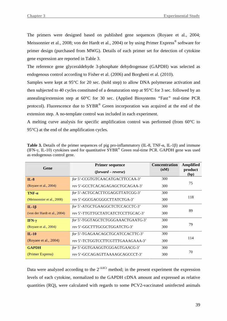
Chapter 3 Experimental Study
39
The primers were designed based on published gene sequences (Royaee et al., 2004;
Meissonnier et al., 2008; von der Hardt et al., 2004) or by using Primer Express® software for
primer design (purchased from MWG). Details of each primer set for detection of cytokine
gene expression are reported in Table 3.
The reference gene glyceraldehyde 3-phosphate dehydrogenase (GAPDH) was selected as
endogenous control according to Fisher et al. (2006) and Borghetti et al. (2010).
Samples were kept at 95°C for 20 sec. (hold step) to allow DNA polymerase activation and
then subjected to 40 cycles constituted of a denaturation step at 95°C for 3 sec. followed by an
annealing/extension step at 60°C for 30 sec. (Applied Biosystems “ Fast” real-time PCR
protocol). Fluorescence due to SYBR® Green incorporation was acquired at the end of the
extension step. A no-template control was included in each experiment.
A melting curve analysis for specific amplification control was performed (from 60°C to
95°C) at the end of the amplification cycles.
Table 3. Details of the primer sequences of pig pro-inflammatory (IL-8, TNF-α, IL-1β) and immune (IFN-γ, IL-10) cytokines used for quantitative SYBR® Green real-time PCR. GAPDH gene was used as endogenous control gene.
Gene Primer sequence
(forward – reverse)
Concentration (nM)
Amplified product
(bp) for 5’-CCGTGTCAACATGACTTCCAA-3’ 300 IL-8
(Royaee et al., 2004) rev 5’-GCCTCACAGAGAGCTGCAGAA-3’ 300
75
for 5’-ACTGCACTTCGAGGTTATCGG-3’ 300 TNF-α
(Meissonnier et al., 2008) rev 5’-GGCGACGGGCTTATCTGA-3’ 300
118
for 5’-ATGCTGAAGGCTCTCCACCTC-3’ 300 IL-1 β
(von der Hardt et al., 2004) rev 5’-TTGTTGCTATCATCTCCTTGCAC-3’ 300
89
for 5’-TGGTAGCTCTGGGAAACTGAATG-3’ 300 IFN-γ
(Royaee et al., 2004) rev 5’-GGCTTTGCGCTGGATCTG-3’ 300
79
for 5’-TGAGAACAGCTGCATCCACTTC-3’ 300 IL-10
(Royaee et al., 2004) rev 5’-TCTGGTCCTTCGTTTGAAAGAAA-3’ 300
114
for 5’-GGTGAAGGTCGGAGTGAACG-3’ 300 GAPDH
(Primer Express) rev 5’-GCCAGAGTTAAAAGCAGCCCT-3’ 300
70
Data were analyzed according to the 2-∆∆Ct method; in the present experiment the expression
levels of each cytokine, normalized to the GAPDH cDNA amount and expressed as relative
quantities (RQ), were calculated with regards to some PCV2-vaccinated uninfected animals
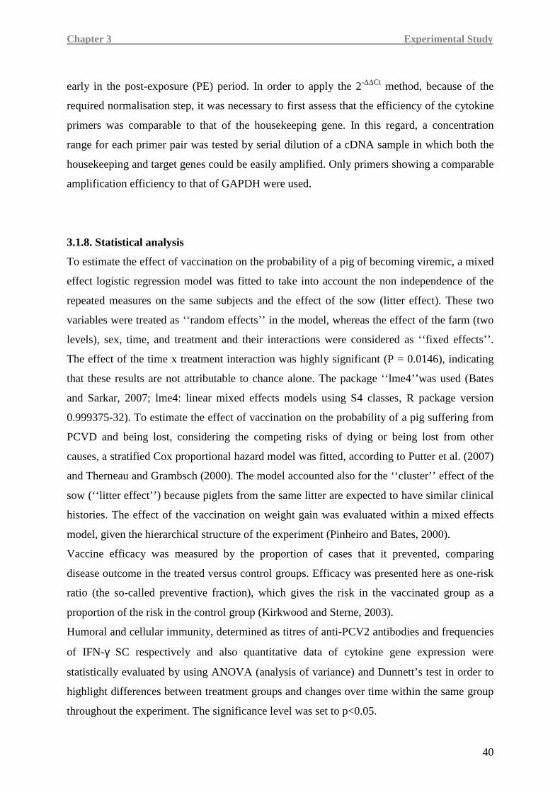
Chapter 3 Experimental Study
40
early in the post-exposure (PE) period. In order to apply the 2-∆∆Ct method, because of the
required normalisation step, it was necessary to first assess that the efficiency of the cytokine
primers was comparable to that of the housekeeping gene. In this regard, a concentration
range for each primer pair was tested by serial dilution of a cDNA sample in which both the
housekeeping and target genes could be easily amplified. Only primers showing a comparable
amplification efficiency to that of GAPDH were used.
3.1.8. Statistical analysis
To estimate the effect of vaccination on the probability of a pig of becoming viremic, a mixed
effect logistic regression model was fitted to take into account the non independence of the
repeated measures on the same subjects and the effect of the sow (litter effect). These two
variables were treated as ‘‘random effects’’ in the model, whereas the effect of the farm (two
levels), sex, time, and treatment and their interactions were considered as ‘‘fixed effects’’.
The effect of the time x treatment interaction was highly significant (P = 0.0146), indicating
that these results are not attributable to chance alone. The package ‘‘lme4’’was used (Bates
and Sarkar, 2007; lme4: linear mixed effects models using S4 classes, R package version
0.999375-32). To estimate the effect of vaccination on the probability of a pig suffering from
PCVD and being lost, considering the competing risks of dying or being lost from other
causes, a stratified Cox proportional hazard model was fitted, according to Putter et al. (2007)
and Therneau and Grambsch (2000). The model accounted also for the ‘‘cluster’’ effect of the
sow (‘‘litter effect’’) because piglets from the same litter are expected to have similar clinical
histories. The effect of the vaccination on weight gain was evaluated within a mixed effects
model, given the hierarchical structure of the experiment (Pinheiro and Bates, 2000).
Vaccine efficacy was measured by the proportion of cases that it prevented, comparing
disease outcome in the treated versus control groups. Efficacy was presented here as one-risk
ratio (the so-called preventive fraction), which gives the risk in the vaccinated group as a
proportion of the risk in the control group (Kirkwood and Sterne, 2003).
Humoral and cellular immunity, determined as titres of anti-PCV2 antibodies and frequencies
of IFN-γ SC respectively and also quantitative data of cytokine gene expression were
statistically evaluated by using ANOVA (analysis of variance) and Dunnett’s test in order to
highlight differences between treatment groups and changes over time within the same group
throughout the experiment. The significance level was set to p<0.05.
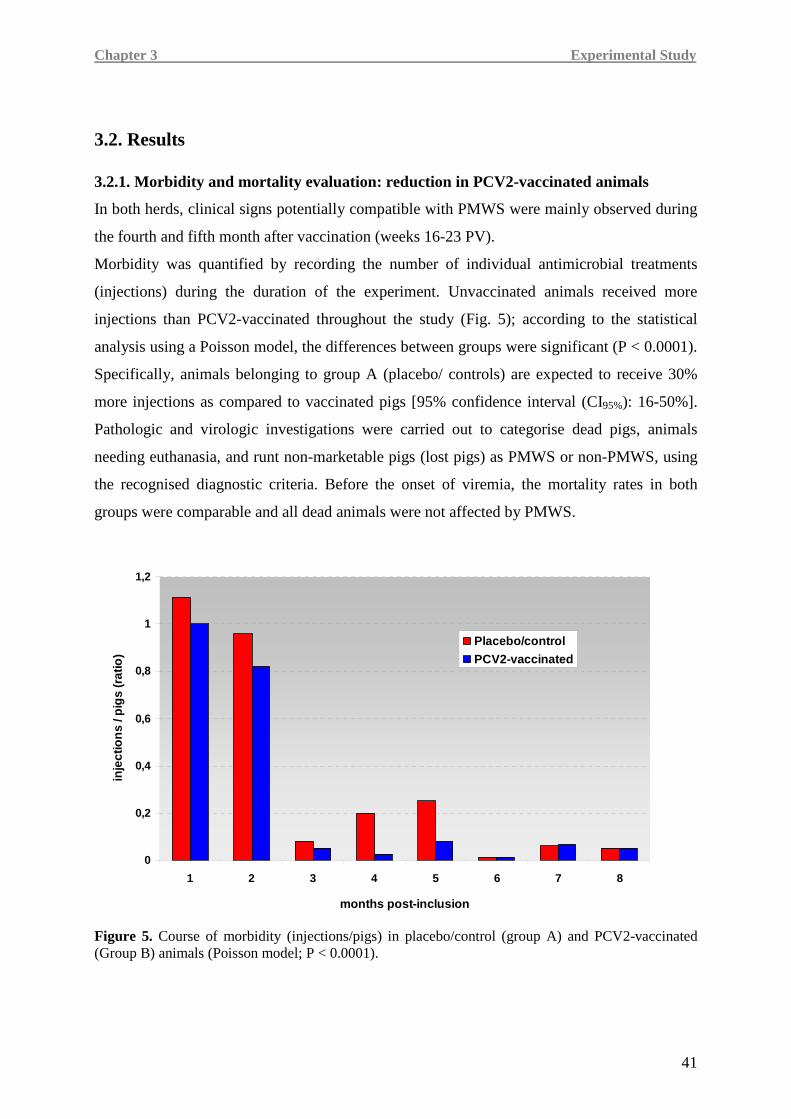
Chapter 3 Experimental Study
41
3.2. Results 3.2.1. Morbidity and mortality evaluation: reduction in PCV2-vaccinated animals
In both herds, clinical signs potentially compatible with PMWS were mainly observed during
the fourth and fifth month after vaccination (weeks 16-23 PV).
Morbidity was quantified by recording the number of individual antimicrobial treatments
(injections) during the duration of the experiment. Unvaccinated animals received more
injections than PCV2-vaccinated throughout the study (Fig. 5); according to the statistical
analysis using a Poisson model, the differences between groups were significant (P < 0.0001).
Specifically, animals belonging to group A (placebo/ controls) are expected to receive 30%
more injections as compared to vaccinated pigs [95% confidence interval (CI95%): 16-50%].
Pathologic and virologic investigations were carried out to categorise dead pigs, animals
needing euthanasia, and runt non-marketable pigs (lost pigs) as PMWS or non-PMWS, using
the recognised diagnostic criteria. Before the onset of viremia, the mortality rates in both
groups were comparable and all dead animals were not affected by PMWS.
0
0,2
0,4
0,6
0,8
1
1,2
1 2 3 4 5 6 7 8
months post-inclusion
inje
ctio
ns
/ pig
s (r
atio
)
Placebo/control
PCV2-vaccinated
Figure 5. Course of morbidity (injections/pigs) in placebo/control (group A) and PCV2-vaccinated (Group B) animals (Poisson model; P < 0.0001).
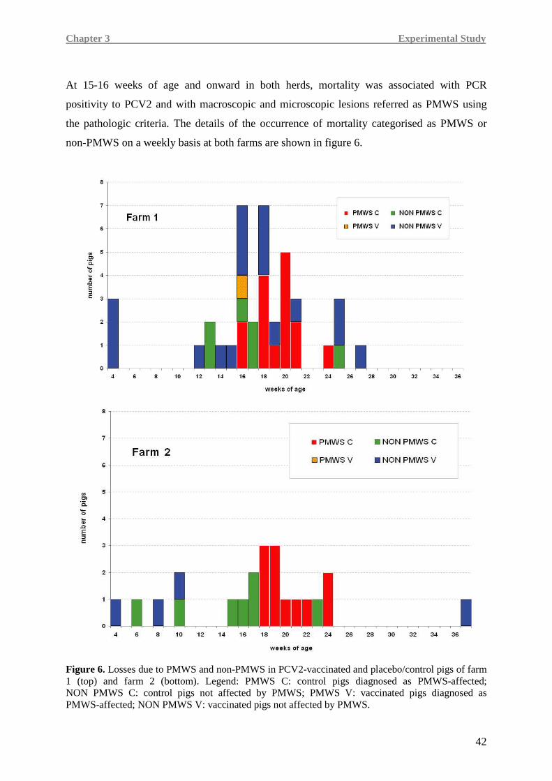
Chapter 3 Experimental Study
42
At 15-16 weeks of age and onward in both herds, mortality was associated with PCR
positivity to PCV2 and with macroscopic and microscopic lesions referred as PMWS using
the pathologic criteria. The details of the occurrence of mortality categorised as PMWS or
non-PMWS on a weekly basis at both farms are shown in figure 6.
Figure 6. Losses due to PMWS and non-PMWS in PCV2-vaccinated and placebo/control pigs of farm 1 (top) and farm 2 (bottom). Legend: PMWS C: control pigs diagnosed as PMWS-affected; NON PMWS C: control pigs not affected by PMWS; PMWS V: vaccinated pigs diagnosed as PMWS-affected; NON PMWS V: vaccinated pigs not affected by PMWS.
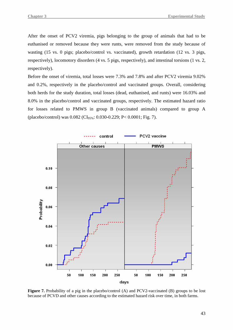
Chapter 3 Experimental Study
43
After the onset of PCV2 viremia, pigs belonging to the group of animals that had to be
euthanised or removed because they were runts, were removed from the study because of
wasting (15 vs. 0 pigs; placebo/control vs. vaccinated), growth retardation (12 vs. 3 pigs,
respectively), locomotory disorders (4 vs. 5 pigs, respectively), and intestinal torsions (1 vs. 2,
respectively).
Before the onset of viremia, total losses were 7.3% and 7.8% and after PCV2 viremia 9.02%
and 0.2%, respectively in the placebo/control and vaccinated groups. Overall, considering
both herds for the study duration, total losses (dead, euthanised, and runts) were 16.03% and
8.0% in the placebo/control and vaccinated groups, respectively. The estimated hazard ratio
for losses related to PMWS in group B (vaccinated animals) compared to group A
(placebo/control) was 0.082 (CI95%: 0.030-0.229; P< 0.0001; Fig. 7).
Figure 7. Probability of a pig in the placebo/control (A) and PCV2-vaccinated (B) groups to be lost because of PCVD and other causes according to the estimated hazard risk over time, in both farms.
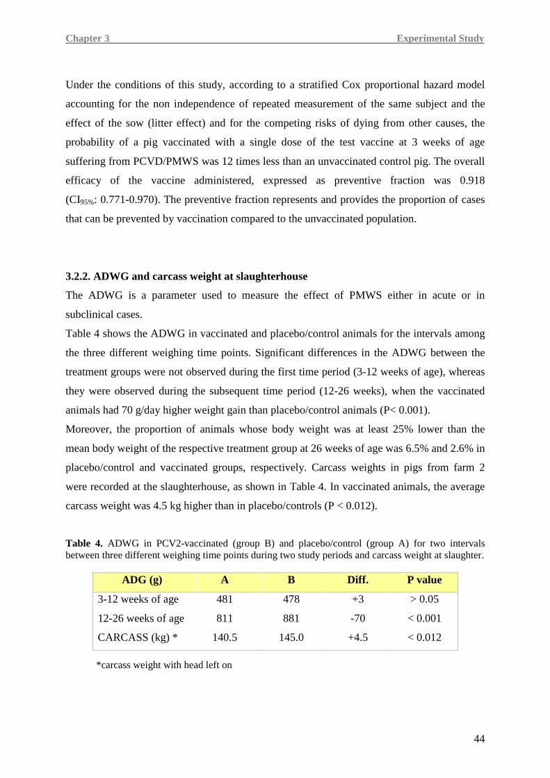
Chapter 3 Experimental Study
44
Under the conditions of this study, according to a stratified Cox proportional hazard model
accounting for the non independence of repeated measurement of the same subject and the
effect of the sow (litter effect) and for the competing risks of dying from other causes, the
probability of a pig vaccinated with a single dose of the test vaccine at 3 weeks of age
suffering from PCVD/PMWS was 12 times less than an unvaccinated control pig. The overall
efficacy of the vaccine administered, expressed as preventive fraction was 0.918
(CI95%: 0.771-0.970). The preventive fraction represents and provides the proportion of cases
that can be prevented by vaccination compared to the unvaccinated population.
3.2.2. ADWG and carcass weight at slaughterhouse
The ADWG is a parameter used to measure the effect of PMWS either in acute or in
subclinical cases.
Table 4 shows the ADWG in vaccinated and placebo/control animals for the intervals among
the three different weighing time points. Significant differences in the ADWG between the
treatment groups were not observed during the first time period (3-12 weeks of age), whereas
they were observed during the subsequent time period (12-26 weeks), when the vaccinated
animals had 70 g/day higher weight gain than placebo/control animals (P< 0.001).
Moreover, the proportion of animals whose body weight was at least 25% lower than the
mean body weight of the respective treatment group at 26 weeks of age was 6.5% and 2.6% in
placebo/control and vaccinated groups, respectively. Carcass weights in pigs from farm 2
were recorded at the slaughterhouse, as shown in Table 4. In vaccinated animals, the average
carcass weight was 4.5 kg higher than in placebo/controls (P < 0.012).
Table 4. ADWG in PCV2-vaccinated (group B) and placebo/control (group A) for two intervals between three different weighing time points during two study periods and carcass weight at slaughter.
ADG (g) A B Diff. P value
3-12 weeks of age 481 478 +3 > 0.05
12-26 weeks of age 811 881 -70 < 0.001
CARCASS (kg) * 140.5 145.0 +4.5 < 0.012
*carcass weight with head left on

Chapter 3 Experimental Study
45
3.2.3. PCV2 genome detection and viremia calculation
The course of PCV2 viremia at both farms is shown in figure 8.
A rapid onset of viremia was observed at 16-17 weeks of age at both farms.
In placebo/control animals of farm 1, peak levels of 95-100% of PCR-positive blood samples
were reached when animals were 20-22 weeks old. A decline of PCR positivity was detected
at 26 weeks. The majority of samples with high viral loads (>106 DNA copies/ml of serum)
were observed at 19-22 weeks of age (Fig. 8a), and 70% of the animals had at least one blood
sample with a viral load >107 DNA copies/ml.
Contrarily, in the vaccinated group, the proportion of viremic pigs was significantly lower
compared to the placebo/control group; between weeks 19 and 22 of age, 40% of the animals
were viremic, with a viral load never >106 DNA copies/ml serum (Fig. 8a).
At farm 2, the peak of viremia was observed at 18-20 weeks of age with 95% PCR-positive
pigs; pigs with a high viral load ranged from 55% to 60% (Fig. 8b). A viral burden >107 DNA
copies/ml was detected in 42% of blood samples from the controls.
In the vaccinated group only one blood sample was PCR positive at 18 weeks of age, with a
low viral burden (<106 DNA copies/ml; Fig. 8b).
(a)
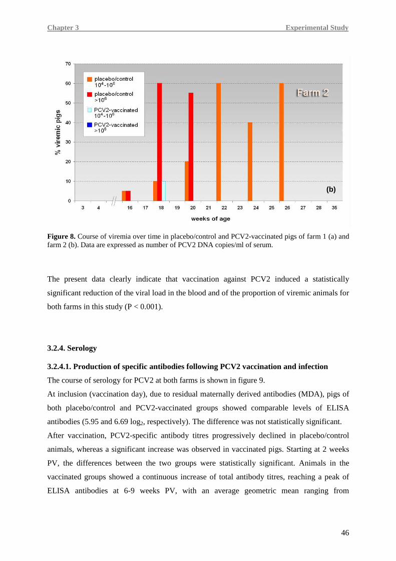
Chapter 3 Experimental Study
46
Figure 8. Course of viremia over time in placebo/control and PCV2-vaccinated pigs of farm 1 (a) and farm 2 (b). Data are expressed as number of PCV2 DNA copies/ml of serum.
The present data clearly indicate that vaccination against PCV2 induced a statistically
significant reduction of the viral load in the blood and of the proportion of viremic animals for
both farms in this study (P < 0.001).
3.2.4. Serology 3.2.4.1. Production of specific antibodies following PCV2 vaccination and infection
The course of serology for PCV2 at both farms is shown in figure 9.
At inclusion (vaccination day), due to residual maternally derived antibodies (MDA), pigs of
both placebo/control and PCV2-vaccinated groups showed comparable levels of ELISA
antibodies (5.95 and 6.69 log2, respectively). The difference was not statistically significant.
After vaccination, PCV2-specific antibody titres progressively declined in placebo/control
animals, whereas a significant increase was observed in vaccinated pigs. Starting at 2 weeks
PV, the differences between the two groups were statistically significant. Animals in the
vaccinated groups showed a continuous increase of total antibody titres, reaching a peak of
ELISA antibodies at 6-9 weeks PV, with an average geometric mean ranging from
(b)
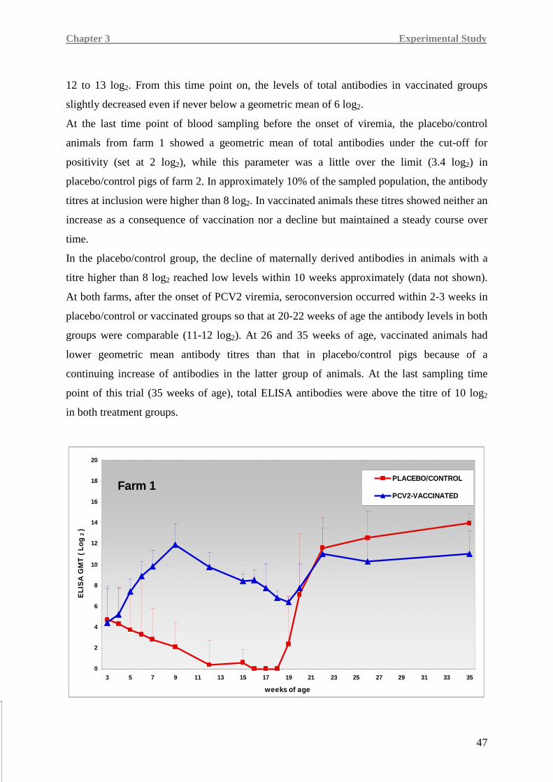
Chapter 3 Experimental Study
47
12 to 13 log2. From this time point on, the levels of total antibodies in vaccinated groups
slightly decreased even if never below a geometric mean of 6 log2.
At the last time point of blood sampling before the onset of viremia, the placebo/control
animals from farm 1 showed a geometric mean of total antibodies under the cut-off for
positivity (set at 2 log2), while this parameter was a little over the limit (3.4 log2) in
placebo/control pigs of farm 2. In approximately 10% of the sampled population, the antibody
titres at inclusion were higher than 8 log2. In vaccinated animals these titres showed neither an
increase as a consequence of vaccination nor a decline but maintained a steady course over
time.
In the placebo/control group, the decline of maternally derived antibodies in animals with a
titre higher than 8 log2 reached low levels within 10 weeks approximately (data not shown).
At both farms, after the onset of PCV2 viremia, seroconversion occurred within 2-3 weeks in
placebo/control or vaccinated groups so that at 20-22 weeks of age the antibody levels in both
groups were comparable (11-12 log2). At 26 and 35 weeks of age, vaccinated animals had
lower geometric mean antibody titres than that in placebo/control pigs because of a
continuing increase of antibodies in the latter group of animals. At the last sampling time
point of this trial (35 weeks of age), total ELISA antibodies were above the titre of 10 log2
in both treatment groups.
0
2
4
6
8
10
12
14
16
18
20
3 5 7 9 11 13 15 17 19 21 23 25 27 29 31 33 35
weeks of age
EL
ISA
GM
T (
Lo
g 2
)
PLACEBO/CONTROL
PCV2-VACCINATED Farm 1
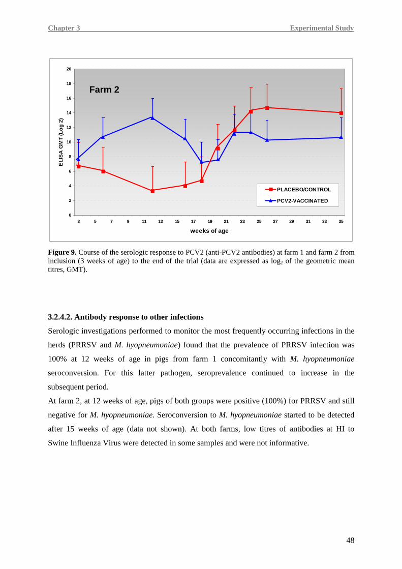
Chapter 3 Experimental Study
48
Figure 9. Course of the serologic response to PCV2 (anti-PCV2 antibodies) at farm 1 and farm 2 from inclusion (3 weeks of age) to the end of the trial (data are expressed as log2 of the geometric mean titres, GMT).
3.2.4.2. Antibody response to other infections
Serologic investigations performed to monitor the most frequently occurring infections in the
herds (PRRSV and M. hyopneumoniae) found that the prevalence of PRRSV infection was
100% at 12 weeks of age in pigs from farm 1 concomitantly with M. hyopneumoniae
seroconversion. For this latter pathogen, seroprevalence continued to increase in the
subsequent period.
At farm 2, at 12 weeks of age, pigs of both groups were positive (100%) for PRRSV and still
negative for M. hyopneumoniae. Seroconversion to M. hyopneumoniae started to be detected
after 15 weeks of age (data not shown). At both farms, low titres of antibodies at HI to
Swine Influenza Virus were detected in some samples and were not informative.
0
2
4
6
8
10
12
14
16
18
20
3 5 7 9 11 13 15 17 19 21 23 25 27 29 31 33 35
weeks of age
EL
ISA
GM
T (L
og
2)
PLACEBO/CONTROL
PCV2-VACCINATED
Farm 2
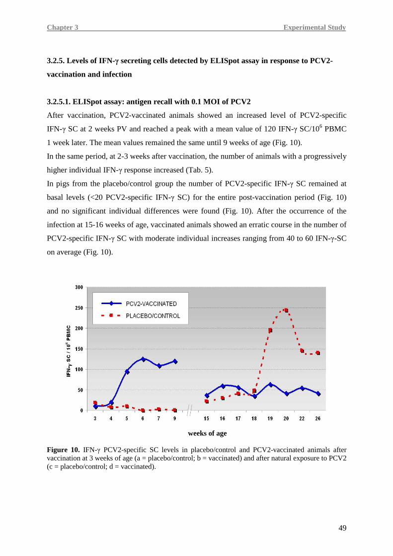
Chapter 3 Experimental Study
49
3.2.5. Levels of IFN-γ secreting cells detected by ELISpot assay in response to PCV2-
vaccination and infection
3.2.5.1. ELISpot assay: antigen recall with 0.1 MOI of PCV2
After vaccination, PCV2-vaccinated animals showed an increased level of PCV2-specific
IFN-γ SC at 2 weeks PV and reached a peak with a mean value of 120 IFN-γ SC/106 PBMC
1 week later. The mean values remained the same until 9 weeks of age (Fig. 10).
In the same period, at 2-3 weeks after vaccination, the number of animals with a progressively
higher individual IFN-γ response increased (Tab. 5).
In pigs from the placebo/control group the number of PCV2-specific IFN-γ SC remained at
basal levels (<20 PCV2-specific IFN-γ SC) for the entire post-vaccination period (Fig. 10)
and no significant individual differences were found (Fig. 10). After the occurrence of the
infection at 15-16 weeks of age, vaccinated animals showed an erratic course in the number of
PCV2-specific IFN-γ SC with moderate individual increases ranging from 40 to 60 IFN-γ-SC
on average (Fig. 10).
Figure 10. IFN-γ PCV2-specific SC levels in placebo/control and PCV2-vaccinated animals after vaccination at 3 weeks of age (a = placebo/control; b = vaccinated) and after natural exposure to PCV2 (c = placebo/control; d = vaccinated).
weeks of age
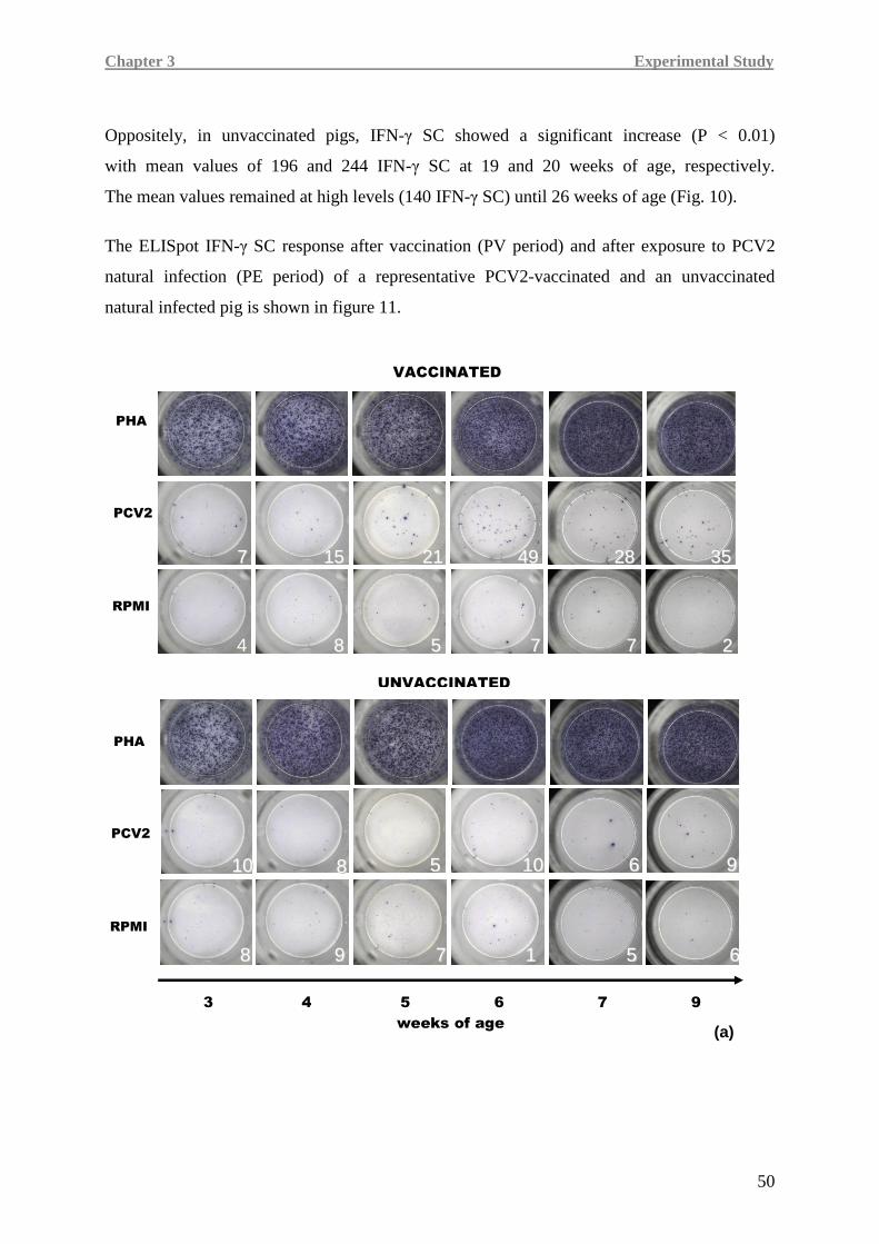
Chapter 3 Experimental Study
50
Oppositely, in unvaccinated pigs, IFN-γ SC showed a significant increase (P < 0.01)
with mean values of 196 and 244 IFN-γ SC at 19 and 20 weeks of age, respectively.
The mean values remained at high levels (140 IFN-γ SC) until 26 weeks of age (Fig. 10).
The ELISpot IFN-γ SC response after vaccination (PV period) and after exposure to PCV2
natural infection (PE period) of a representative PCV2-vaccinated and an unvaccinated
natural infected pig is shown in figure 11.
88
VACCINATED
PHA
PCV2
RPMI
77 1155 2211 4499 2288 3355
UNVACCINATED
1100 88 55 1100 99 66
88 99 77 11 55 66
PHA
PCV2
RPMI
3 4 5 6 7 9
weeks of age (a)
4 88 55 77 77 22
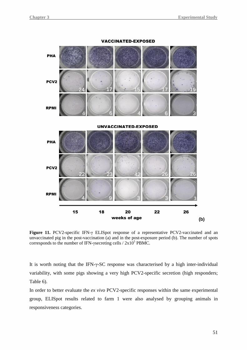
Chapter 3 Experimental Study
51
Figure 11. PCV2-specific IFN-γ ELISpot response of a representative PCV2-vaccinated and an unvaccinated pig in the post-vaccination (a) and in the post-exposure period (b). The number of spots corresponds to the number of IFN-γsecreting cells / 2x10⁵ PBMC.
It is worth noting that the IFN-γ-SC response was characterised by a high inter-individual
variability, with some pigs showing a very high PCV2-specific secretion (high responders;
Table 6).
In order to better evaluate the ex vivo PCV2-specific responses within the same experimental
group, ELISpot results related to farm 1 were also analysed by grouping animals in
responsiveness categories.
2244 1177 1155 1177 1199
66 44 66 3 4
VACCINATED-EXPOSED
PHA
PCV2
RPMI
UNVACCINATED-EXPOSED
2222 2233 4422 2266 2266
44 99 33 33 11
PHA
PCV2
RPMI
15 18 20 22 26
weeks of age (b)
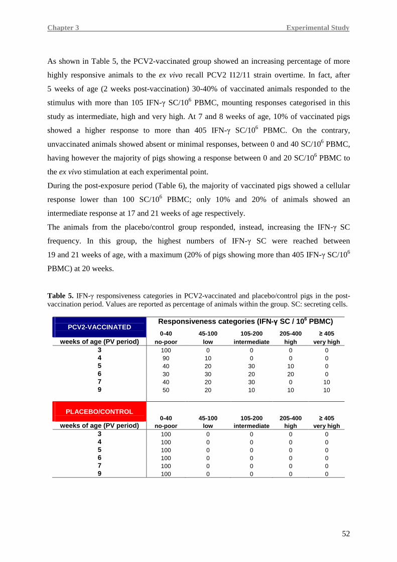
Chapter 3 Experimental Study
52
As shown in Table 5, the PCV2-vaccinated group showed an increasing percentage of more
highly responsive animals to the ex vivo recall PCV2 I12/11 strain overtime. In fact, after
5 weeks of age (2 weeks post-vaccination) 30-40% of vaccinated animals responded to the
stimulus with more than 105 IFN-γ SC/106 PBMC, mounting responses categorised in this
study as intermediate, high and very high. At 7 and 8 weeks of age, 10% of vaccinated pigs
showed a higher response to more than 405 IFN-γ SC/106 PBMC. On the contrary,
unvaccinated animals showed absent or minimal responses, between 0 and 40 SC/106 PBMC,
having however the majority of pigs showing a response between 0 and 20 SC/106 PBMC to
the ex vivo stimulation at each experimental point.
During the post-exposure period (Table 6), the majority of vaccinated pigs showed a cellular
response lower than 100 SC/106 PBMC; only 10% and 20% of animals showed an
intermediate response at 17 and 21 weeks of age respectively.
The animals from the placebo/control group responded, instead, increasing the IFN-γ SC
frequency. In this group, the highest numbers of IFN-γ SC were reached between
19 and 21 weeks of age, with a maximum (20% of pigs showing more than 405 IFN-γ SC/106
PBMC) at 20 weeks.
Table 5. IFN-γ responsiveness categories in PCV2-vaccinated and placebo/control pigs in the post-vaccination period. Values are reported as percentage of animals within the group. SC: secreting cells.
Responsiveness categories (IFN-γ SC / 106 PBMC) PCV2-VACCINATED
0-40 45-100 105-200 205-400 ≥ 405 weeks of age (PV period) no-poor low intermediate high very high
3 100 0 0 0 0 4 90 10 0 0 0 5 40 20 30 10 0 6 30 30 20 20 0 7 40 20 30 0 10 9 50 20 10 10 10
0-40 PLACEBO/CONTROL
0-40 45-100 105-200 205-400 ≥ 405 weeks of age (PV period) no-poor low intermediate high very high
3 100 0 0 0 0 4 100 0 0 0 0 5 100 0 0 0 0 6 100 0 0 0 0 7 100 0 0 0 0 9 100 0 0 0 0
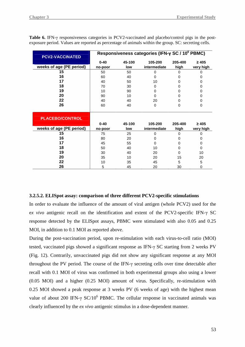
Chapter 3 Experimental Study
53
Table 6. IFN-γ responsiveness categories in PCV2-vaccinated and placebo/control pigs in the post-exposure period. Values are reported as percentage of animals within the group. SC: secreting cells.
Responsiveness categories (IFN-γ SC / 106 PBMC) PCV2-VACCINATED
0-40 45-100 105-200 205-400 ≥ 405
weeks of age (PE period) no-poor low intermediate high very high 15 50 50 0 0 0 16 60 40 0 0 0 17 40 50 10 0 0 18 70 30 0 0 0 19 10 90 0 0 0 20 90 10 0 0 0 22 40 40 20 0 0 26 60 40 0 0 0
PLACEBO/CONTROL 0-40 45-100 105-200 205-400 ≥ 405
weeks of age (PE period) no-poor low intermediate high very high 15 75 25 0 0 0 16 80 20 0 0 0 17 45 55 0 0 0 18 50 40 10 0 0 19 30 40 20 0 10 20 35 10 20 15 20 22 10 35 45 5 5 26 5 45 20 30 0
3.2.5.2. ELISpot assay: comparison of three different PCV2-specific stimulations
In order to evaluate the influence of the amount of viral antigen (whole PCV2) used for the
ex vivo antigenic recall on the identification and extent of the PCV2-specific IFN-γ SC
response detected by the ELISpot assays, PBMC were stimulated with also 0.05 and 0.25
MOI, in addition to 0.1 MOI as reported above.
During the post-vaccination period, upon re-stimulation with each virus-to-cell ratio (MOI)
tested, vaccinated pigs showed a significant response as IFN-γ SC starting from 2 weeks PV
(Fig. 12). Contrarily, unvaccinated pigs did not show any significant response at any MOI
throughout the PV period. The course of the IFN-γ secreting cells over time detectable after
recall with 0.1 MOI of virus was confirmed in both experimental groups also using a lower
(0.05 MOI) and a higher (0.25 MOI) amount of virus. Specifically, re-stimulation with
0.25 MOI showed a peak response at 3 weeks PV (6 weeks of age) with the highest mean
value of about 200 IFN-γ SC/106 PBMC. The cellular response in vaccinated animals was
clearly influenced by the ex vivo antigenic stimulus in a dose-dependent manner.
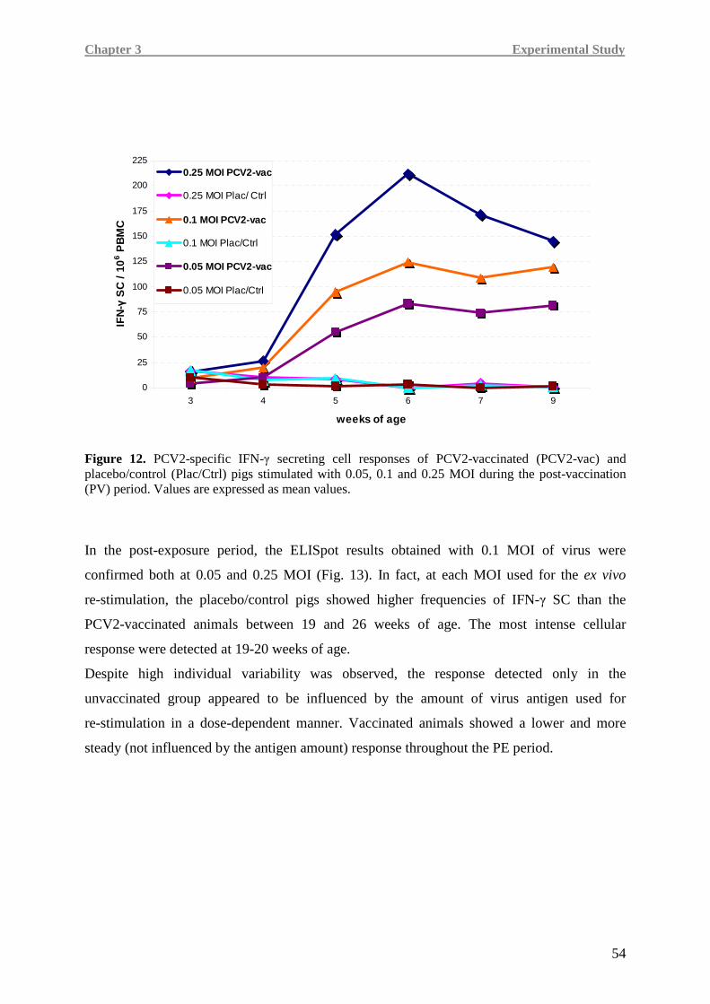
Chapter 3 Experimental Study
54
0
25
50
75
100
125
150
175
200
225
3 4 5 6 7 9
weeks of age
IFN
-γ S
C /
10
6 PB
MC
0.25 MOI PCV2-vac
0.25 MOI Plac/ Ctrl
0.1 MOI PCV2-vac
0.1 MOI Plac/Ctrl
0.05 MOI PCV2-vac
0.05 MOI Plac/Ctrl
Figure 12. PCV2-specific IFN-γ secreting cell responses of PCV2-vaccinated (PCV2-vac) and placebo/control (Plac/Ctrl) pigs stimulated with 0.05, 0.1 and 0.25 MOI during the post-vaccination (PV) period. Values are expressed as mean values.
In the post-exposure period, the ELISpot results obtained with 0.1 MOI of virus were
confirmed both at 0.05 and 0.25 MOI (Fig. 13). In fact, at each MOI used for the ex vivo
re-stimulation, the placebo/control pigs showed higher frequencies of IFN-γ SC than the
PCV2-vaccinated animals between 19 and 26 weeks of age. The most intense cellular
response were detected at 19-20 weeks of age.
Despite high individual variability was observed, the response detected only in the
unvaccinated group appeared to be influenced by the amount of virus antigen used for
re-stimulation in a dose-dependent manner. Vaccinated animals showed a lower and more
steady (not influenced by the antigen amount) response throughout the PE period.
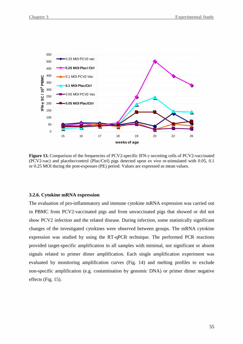
Chapter 3 Experimental Study
55
0
50
100
150
200
250
300
350
400
450
500
550
15 16 17 18 19 20 22 26
weeks of age
IFN
-γ S
C /
10
6 PB
MC
0.25 MOI PCV2-vac
0.25 MOI Plac/ Ctrl
0.1 MOI PCV2-Vac
0.1 MOI Plac/Ctrl
0.05 MOI PCV2-Vac
0.05 MOI Plac/Ctrl
Figure 13. Comparison of the frequencies of PCV2-specific IFN-γ secreting cells of PCV2-vaccinated (PCV2-vac) and placebo/control (Plac/Ctrl) pigs detected upon ex vivo re-stimulated with 0.05, 0.1 or 0.25 MOI during the post-exposure (PE) period. Values are expressed as mean values.
3.2.6. Cytokine mRNA expression
The evaluation of pro-inflammatory and immune cytokine mRNA expression was carried out
in PBMC from PCV2-vaccinated pigs and from unvaccinated pigs that showed or did not
show PCV2 infection and the related disease. During infection, some statistically significant
changes of the investigated cytokines were observed between groups. The mRNA cytokine
expression was studied by using the RT-qPCR technique. The performed PCR reactions
provided target-specific amplification in all samples with minimal, not significant or absent
signals related to primer dimer amplification. Each single amplification experiment was
evaluated by monitoring amplification curves (Fig. 14) and melting profiles to exclude
non-specific amplification (e.g. contamination by genomic DNA) or primer dimer negative
effects (Fig. 15).
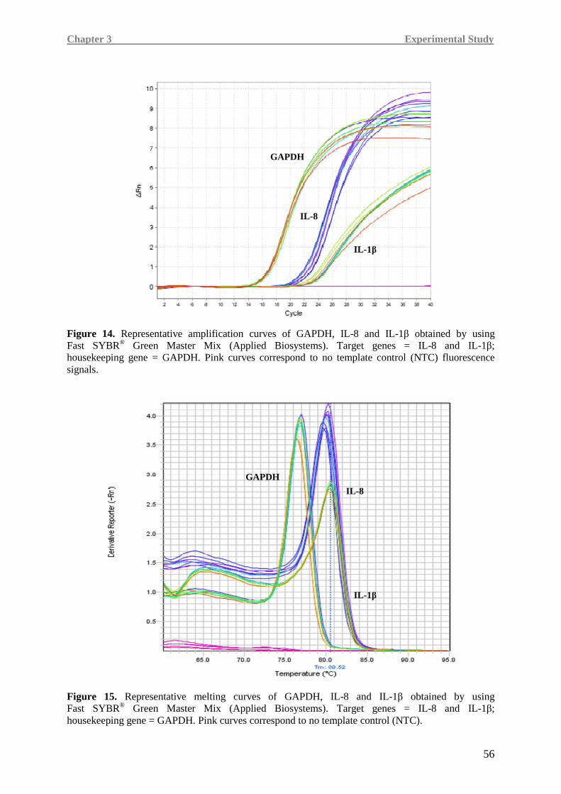
Chapter 3 Experimental Study
56
Figure 14. Representative amplification curves of GAPDH, IL-8 and IL-1β obtained by using Fast SYBR® Green Master Mix (Applied Biosystems). Target genes = IL-8 and IL-1β; housekeeping gene = GAPDH. Pink curves correspond to no template control (NTC) fluorescence signals.
Figure 15. Representative melting curves of GAPDH, IL-8 and IL-1β obtained by using Fast SYBR® Green Master Mix (Applied Biosystems). Target genes = IL-8 and IL-1β; housekeeping gene = GAPDH. Pink curves correspond to no template control (NTC).
GAPDH
IL-8
IL-1 β
GAPDH
IL-8
IL-1 β
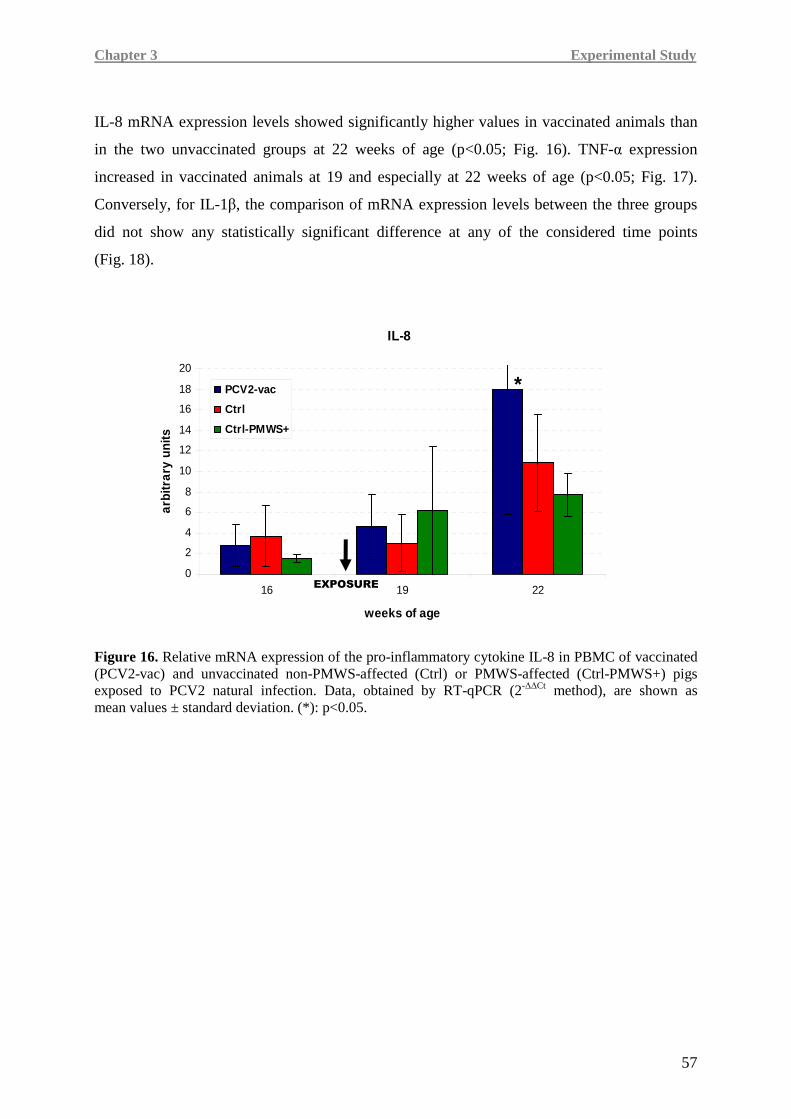
Chapter 3 Experimental Study
57
IL-8 mRNA expression levels showed significantly higher values in vaccinated animals than
in the two unvaccinated groups at 22 weeks of age (p<0.05; Fig. 16). TNF-α expression
increased in vaccinated animals at 19 and especially at 22 weeks of age (p<0.05; Fig. 17).
Conversely, for IL-1β, the comparison of mRNA expression levels between the three groups
did not show any statistically significant difference at any of the considered time points
(Fig. 18).
Figure 16. Relative mRNA expression of the pro-inflammatory cytokine IL-8 in PBMC of vaccinated (PCV2-vac) and unvaccinated non-PMWS-affected (Ctrl) or PMWS-affected (Ctrl-PMWS+) pigs exposed to PCV2 natural infection. Data, obtained by RT-qPCR (2-∆∆Ct method), are shown as mean values ± standard deviation. (*): p<0.05.
IL-8
0
2
4
6
8
10
12
14
16
18
20
16 19 22
weeks of age
arbi
trar
y un
its
PCV2-vac
Ctrl
Ctrl-PMWS+
*
EXPOSURE
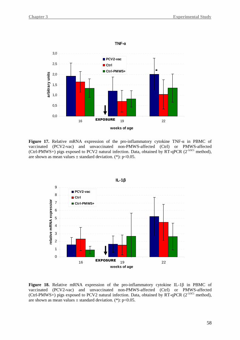
Chapter 3 Experimental Study
58
Figure 17. Relative mRNA expression of the pro-inflammatory cytokine TNF-α in PBMC of vaccinated (PCV2-vac) and unvaccinated non-PMWS-affected (Ctrl) or PMWS-affected (Ctrl-PMWS+) pigs exposed to PCV2 natural infection. Data, obtained by RT-qPCR (2-∆∆Ct method), are shown as mean values ± standard deviation. (*): p<0.05.
IL-1β
0
1
2
3
4
5
6
7
8
9
16 19 22weeks of age
rela
tive
mR
NA
exp
ress
ion
PCV2-vac
Ctrl
Ctrl-PMWS+
EXPOSURE
Figure 18. Relative mRNA expression of the pro-inflammatory cytokine IL-1β in PBMC of vaccinated (PCV2-vac) and unvaccinated non-PMWS-affected (Ctrl) or PMWS-affected (Ctrl-PMWS+) pigs exposed to PCV2 natural infection. Data, obtained by RT-qPCR (2-∆∆Ct method), are shown as mean values ± standard deviation. (*): p<0.05.
TNF-α
0,0
0,5
1,0
1,5
2,0
2,5
3,0
16 19 22
weeks of age
arbi
trar
y un
its
PCV2-vac
Ctrl
Ctrl-PMWS+
EXPOSURE
*
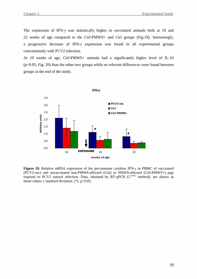
Chapter 3 Experimental Study
59
The expression of IFN-γ was statistically higher in vaccinated animals both at 19 and
22 weeks of age compared to the Ctrl-PMWS+ and Ctrl groups (Fig.19). Interestingly,
a progressive decrease of IFN-γ expression was found in all experimental groups
concomitantly with PCV2 infection.
At 19 weeks of age, Ctrl-PMWS+ animals had a significantly higher level of IL-10
(p<0.05, Fig. 20) than the other two groups while no relevant differences were found between
groups at the end of the study.
IFN-γ
0,0
0,5
1,0
1,5
2,0
2,5
3,0
3,5
16 19 22
weeks of age
arb
itra
ry u
nit
s
PCV2-vac
Ctrl
Ctrl-PMWS+
**
Figure 19. Relative mRNA expression of the pro-immune cytokine IFN-γ in PBMC of vaccinated (PCV2-vac) and unvaccinated non-PMWS-affected (Ctrl) or PMWS-affected (Ctrl-PMWS+) pigs exposed to PCV2 natural infection. Data, obtained by RT-qPCR (2-∆∆Ct method), are shown as mean values ± standard deviation. (*): p<0.05.
EXPOSURE
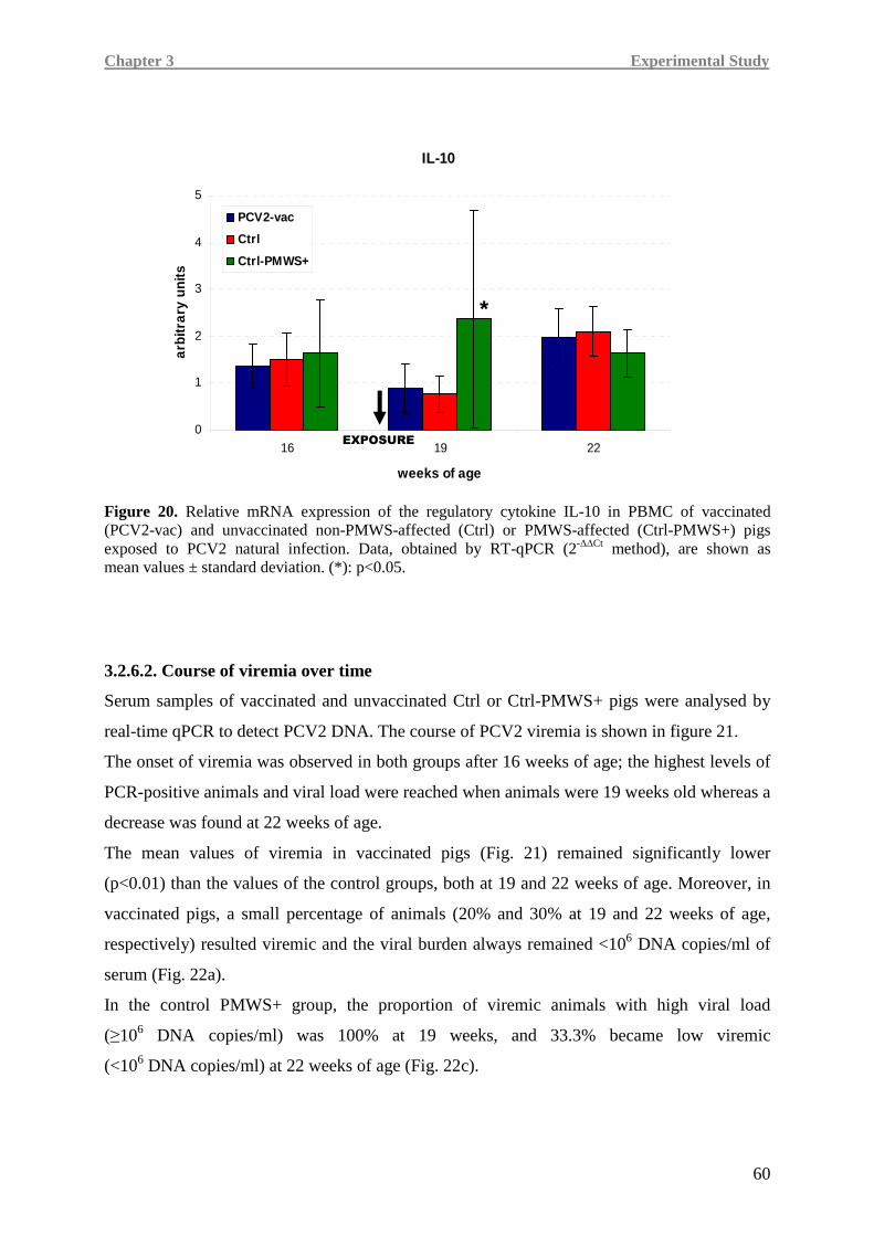
Chapter 3 Experimental Study
60
Figure 20. Relative mRNA expression of the regulatory cytokine IL-10 in PBMC of vaccinated (PCV2-vac) and unvaccinated non-PMWS-affected (Ctrl) or PMWS-affected (Ctrl-PMWS+) pigs exposed to PCV2 natural infection. Data, obtained by RT-qPCR (2-∆∆Ct method), are shown as mean values ± standard deviation. (*): p<0.05.
3.2.6.2. Course of viremia over time
Serum samples of vaccinated and unvaccinated Ctrl or Ctrl-PMWS+ pigs were analysed by
real-time qPCR to detect PCV2 DNA. The course of PCV2 viremia is shown in figure 21.
The onset of viremia was observed in both groups after 16 weeks of age; the highest levels of
PCR-positive animals and viral load were reached when animals were 19 weeks old whereas a
decrease was found at 22 weeks of age.
The mean values of viremia in vaccinated pigs (Fig. 21) remained significantly lower
(p<0.01) than the values of the control groups, both at 19 and 22 weeks of age. Moreover, in
vaccinated pigs, a small percentage of animals (20% and 30% at 19 and 22 weeks of age,
respectively) resulted viremic and the viral burden always remained <106 DNA copies/ml of
serum (Fig. 22a).
In the control PMWS+ group, the proportion of viremic animals with high viral load
(≥106 DNA copies/ml) was 100% at 19 weeks, and 33.3% became low viremic
(<106 DNA copies/ml) at 22 weeks of age (Fig. 22c).
IL-10
0
1
2
3
4
5
16 19 22
weeks of age
arbi
trar
y un
its
PCV2-vac
Ctrl
Ctrl-PMWS+
*
EXPOSURE
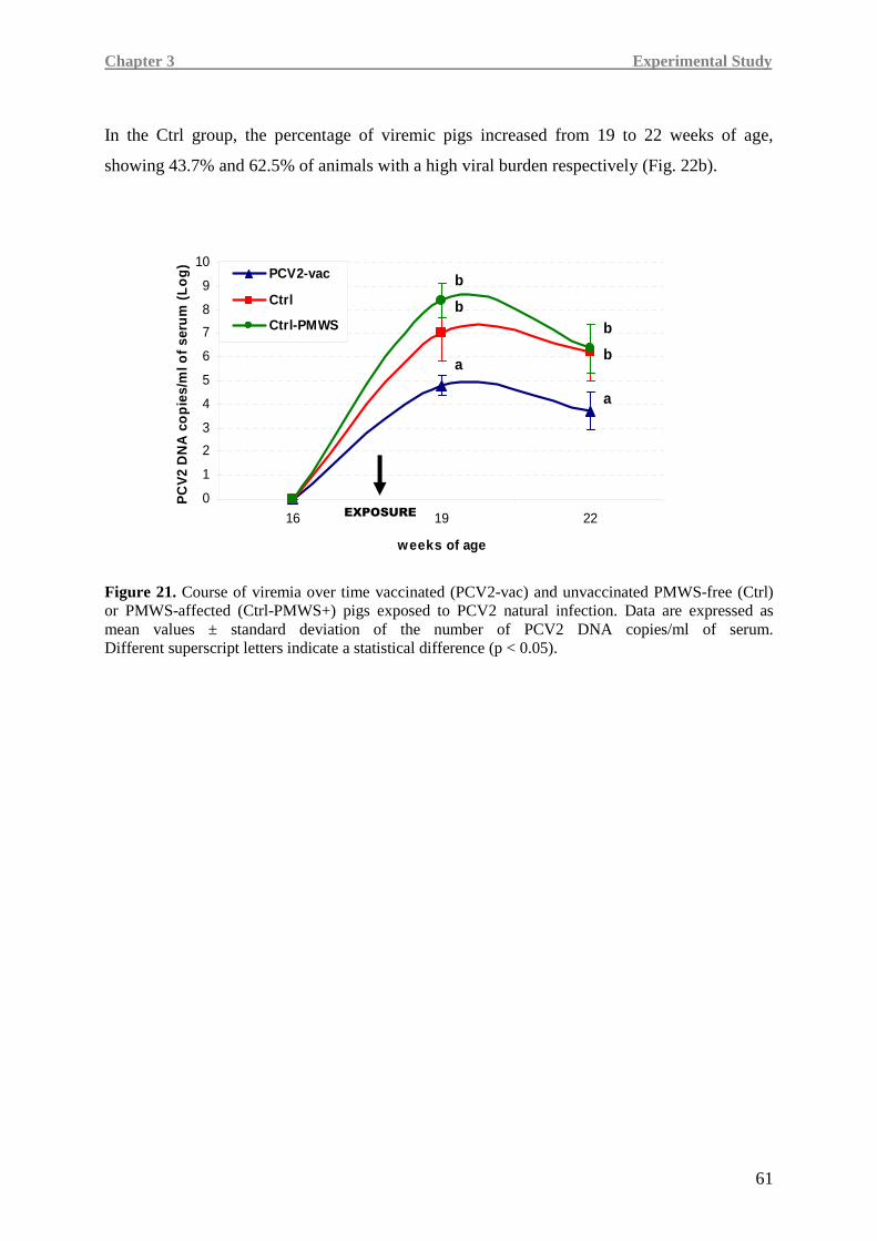
Chapter 3 Experimental Study
61
In the Ctrl group, the percentage of viremic pigs increased from 19 to 22 weeks of age,
showing 43.7% and 62.5% of animals with a high viral burden respectively (Fig. 22b).
Figure 21. Course of viremia over time vaccinated (PCV2-vac) and unvaccinated PMWS-free (Ctrl) or PMWS-affected (Ctrl-PMWS+) pigs exposed to PCV2 natural infection. Data are expressed as mean values ± standard deviation of the number of PCV2 DNA copies/ml of serum. Different superscript letters indicate a statistical difference (p < 0.05).
0
1
2
3
4
5
6
7
8
9
10
16 19 22
weeks of age
PC
V2
DN
A c
op
ies/
ml o
f se
rum
(L
og
)
PCV2-vac
Ctrl
Ctrl-PMWS
b
b
ba
b
a
EXPOSURE
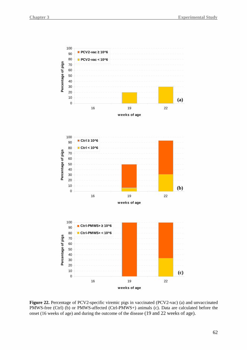
Chapter 3 Experimental Study
62
0
10
20
30
40
50
60
70
80
90
100
16 19 22
weeks of age
Per
cen
tag
e o
f p
igs
PCV2-vac ≥ 10^6
PCV2-vac < 10^6
0
10
20
30
40
50
60
70
80
90
100
16 19 22
weeks of age
Per
cen
tag
e o
f p
igs
Ctrl ≥ 10^6
Ctrl < 10^6
0
10
20
30
40
50
60
70
80
90
100
16 19 22
weeks of age
Per
cen
tag
e o
f p
igs
Ctrl-PMWS+ ≥ 10^6
Ctrl-PMWS+ < 10^6
Figure 22. Percentage of PCV2-specific viremic pigs in vaccinated (PCV2-vac) (a) and unvaccinated PMWS-free (Ctrl) (b) or PMWS-affected (Ctrl-PMWS+) animals (c). Data are calculated before the onset (16 weeks of age) and during the outcome of the disease (19 and 22 weeks of age).
(c)
(b)
(a)
Chapter 3 Experimental Study
63
3.2.6.3. Association between cytokine mRNA expression and viral load in the blood
Pro-inflammatory and immune cytokine mRNA expression was also evaluated in relation to
the viremia burden.
According to the amount of PCV2 in serum, three different subgroups were considered in
order to have further information about the cytokine kinetics both in vaccinated and
unvaccinated PCV2 groups as previoulsy stated (PCV2-vac, Ctrl-PMWS+ and Ctrl):
non-viremic pigs (NV), pigs with viremia <106 PCV2 DNA copies/ml of serum (V<106 ),
and pigs with viremia ≥106 PCV2 DNA copies/ml of serum (V≥106).
Table 7 shows the modulation of cytokine levels observed between all subgroups of each
experimental group (PCV2-vac, Ctrl, Ctrl-PMWS+) over time.
Comparing IFN-γ expression in relation to the extent of viremia between groups
(PCV2-vac; Ctrl-PMWS+ and Ctrl) over time (Table 7), despite a decreasing trend of this
cytokine from 16 to 22 weeks of age in all considered groups, in PCV2-vac animals IFN-γ did
not show differences between the newly constituted subgroups both at 19 and 22 weeks of
age. Nevertheless, these animals maintained higher expression levels compared to the
subgroups in Ctrl and Ctrl-PMWS+ animals. Particularly, PCV2-vac animals even with low
viremia (V<106) had higher IFN-γ expression than highly viremic pigs in both Ctrl groups at
22 weeks of age (p<0.05).
IL-10 mRNA expression appeared to increase in relation to the occurrence of infection and in
relation to the extent of viremia (Table 7); particularly, highly viremic (V≥106) animals of the
Ctrl-PMWS+ group showed an early increase (19 weeks of age) of this cytokine compared to
PCV2-vac animals and Ctrl groups.
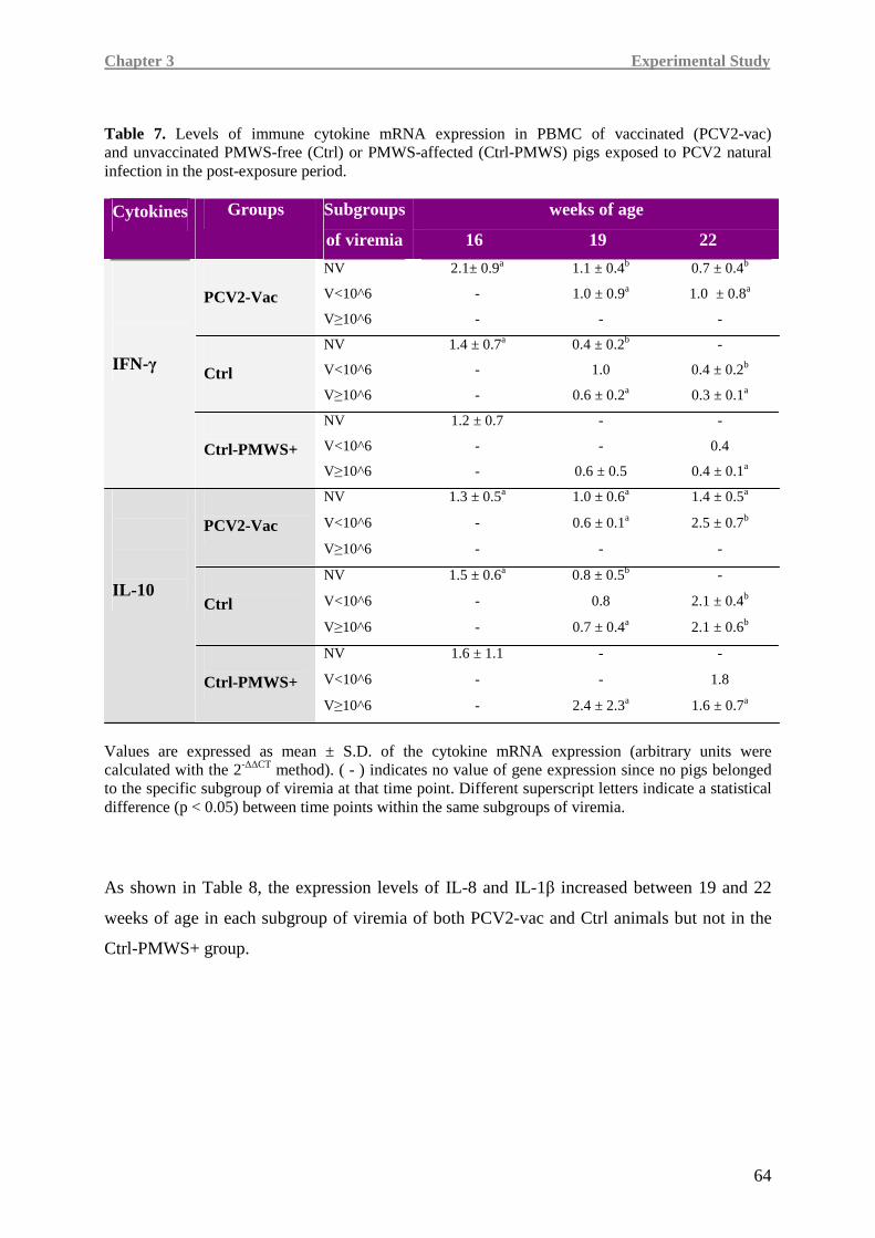
Chapter 3 Experimental Study
64
Table 7. Levels of immune cytokine mRNA expression in PBMC of vaccinated (PCV2-vac) and unvaccinated PMWS-free (Ctrl) or PMWS-affected (Ctrl-PMWS) pigs exposed to PCV2 natural infection in the post-exposure period.
weeks of age Cytokines Groups Subgroups
of viremia 16 19 22
NV 2.1± 0.9a 1.1 ± 0.4b 0.7 ± 0.4b
V<10^6 - 1.0 ± 0.9a 1.0 ± 0.8a
PCV2-Vac V≥10^6 - - -
NV 1.4 ± 0.7a 0.4 ± 0.2b -
V<10^6 - 1.0 0.4 ± 0.2b
Ctrl V≥10^6 - 0.6 ± 0.2a 0.3 ± 0.1a
NV 1.2 ± 0.7 - -
V<10^6 - - 0.4
IFN-γ
Ctrl-PMWS+ V≥10^6 - 0.6 ± 0.5 0.4 ± 0.1a
NV 1.3 ± 0.5a 1.0 ± 0.6a 1.4 ± 0.5a
V<10^6 - 0.6 ± 0.1a 2.5 ± 0.7b
PCV2-Vac V≥10^6 - - -
NV 1.5 ± 0.6a 0.8 ± 0.5b -
V<10^6 - 0.8 2.1 ± 0.4b
Ctrl V≥10^6 - 0.7 ± 0.4a 2.1 ± 0.6b
NV 1.6 ± 1.1 - -
V<10^6 - - 1.8
IL-10
Ctrl-PMWS+ V≥10^6 - 2.4 ± 2.3a 1.6 ± 0.7a
Values are expressed as mean ± S.D. of the cytokine mRNA expression (arbitrary units were calculated with the 2-∆∆CT method). ( - ) indicates no value of gene expression since no pigs belonged to the specific subgroup of viremia at that time point. Different superscript letters indicate a statistical difference (p < 0.05) between time points within the same subgroups of viremia.
As shown in Table 8, the expression levels of IL-8 and IL-1β increased between 19 and 22
weeks of age in each subgroup of viremia of both PCV2-vac and Ctrl animals but not in the
Ctrl-PMWS+ group.
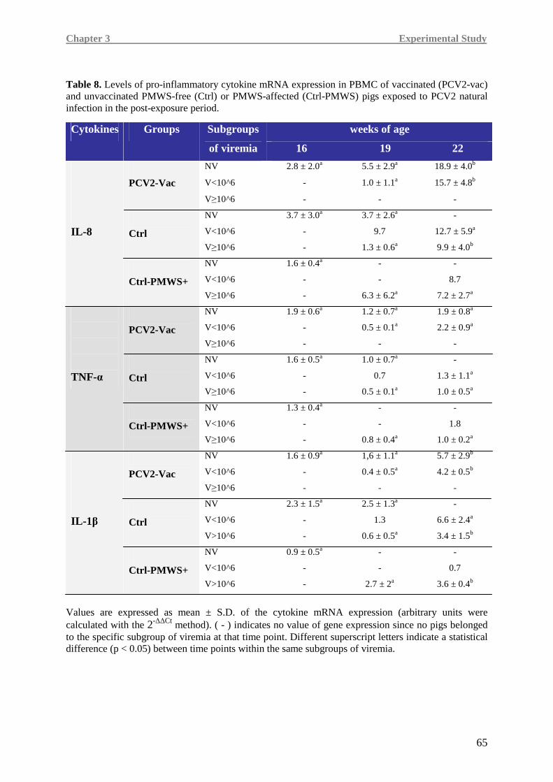
Chapter 3 Experimental Study
65
Table 8. Levels of pro-inflammatory cytokine mRNA expression in PBMC of vaccinated (PCV2-vac) and unvaccinated PMWS-free (Ctrl) or PMWS-affected (Ctrl-PMWS) pigs exposed to PCV2 natural infection in the post-exposure period.
weeks of age Cytokines Groups Subgroups
of viremia 16 19 22
NV 2.8 ± 2.0a 5.5 ± 2.9a 18.9 ± 4.0b
V<10^6 - 1.0 ± 1.1a 15.7 ± 4.8b
PCV2-Vac
V≥10^6 - - -
NV 3.7 ± 3.0a 3.7 ± 2.6a -
V<10^6 - 9.7 12.7 ± 5.9a
Ctrl V≥10^6 - 1.3 ± 0.6a 9.9 ± 4.0b
NV 1.6 ± 0.4a - -
V<10^6 - - 8.7
IL-8
Ctrl-PMWS+ V≥10^6 - 6.3 ± 6.2a 7.2 ± 2.7a
NV 1.9 ± 0.6a 1.2 ± 0.7a 1.9 ± 0.8a
V<10^6 - 0.5 ± 0.1a 2.2 ± 0.9a
PCV2-Vac V≥10^6 - - -
NV 1.6 ± 0.5a 1.0 ± 0.7a -
V<10^6 - 0.7 1.3 ± 1.1a
Ctrl V≥10^6 - 0.5 ± 0.1a 1.0 ± 0.5a
NV 1.3 ± 0.4a - -
V<10^6 - - 1.8
TNF-α
Ctrl-PMWS+ V≥10^6 - 0.8 ± 0.4a 1.0 ± 0.2a
NV 1.6 ± 0.9a 1,6 ± 1.1a 5.7 ± 2.9b
V<10^6 - 0.4 ± 0.5a 4.2 ± 0.5b
PCV2-Vac V≥10^6 - - -
NV 2.3 ± 1.5a 2.5 ± 1.3a -
V<10^6 - 1.3 6.6 ± 2.4a
Ctrl V>10^6 - 0.6 ± 0.5a 3.4 ± 1.5b
NV 0.9 ± 0.5a - -
V<10^6 - - 0.7
IL-1 β
Ctrl-PMWS+ V>10^6 - 2.7 ± 2a 3.6 ± 0.4b
Values are expressed as mean ± S.D. of the cytokine mRNA expression (arbitrary units were calculated with the 2-∆∆Ct method). ( - ) indicates no value of gene expression since no pigs belonged to the specific subgroup of viremia at that time point. Different superscript letters indicate a statistical difference (p < 0.05) between time points within the same subgroups of viremia.
Chapter 3 Experimental Study
66
In particular, between 19 and 22 weeks, a statistically significant higher levels of IL-8 were
detected in non-viremic (NV) PCV2-vaccinated compared to highly viremic (V≥106) control
pigs and control PMWS+; in the same period, and particularly at 22 weeks of age, all
subgroups of PCV2 vaccinated pigs showed higher levels of IL-1β compared to the highly
viremic (V≥106) pigs of the Ctrl groups, TNF-α showed a similar trend to increase between 19
and 22 weeks of age in PCV2 vaccinated group in respect to control viremic pigs but not if
compared to PMWS pigs at 22 weeks.
Overall, in Ctrl-PMWS+ pigs, lower IFN-γ at 19 weeks of age was associated with high IL-10
and subsequently, at 22 weeks of age, is evident a reduction of pro-inflammatory cytokines,
namely IL-8 and IL-1β (Table 7 and 8).
Contrarily, the PCV2-vaccinated pigs, despite an increase of IL-10 from 19 to 22 weeks,
showed higher levels of these pro-inflammatory cytokines at 22 weeks of age (Tables 7
and 8).

CHAPTER 4.
DISCUSSION AND CONCLUSIONS
Chapter 4 Discussion and Conclusions
68
The present Thesis reports a study performed to characterize the humoral and cell-mediated
immune response upon PCV2 vaccination and natural infection, evaluating the efficacy of a
one-dose of a commercial PCV2a sub-type based subunit vaccine containing the capsid
protein expressed in a baculovirus system (Porcilis PCV®) in vaccinated and unvaccinated
pigs subsequently exposed to PCV2 natural infection.
The trial was performed at two large farms where the exposure of the animals to various
pathogens such as PCV, PRRSV, M. hyopneumoniae, and commonly detected bacteria was
the classical predisposing condition for PCVD (Kawashima et al., 2007). Under the
conditions of this study, PCV2 infection was detected during the fifth month of age, a period
in which also the onset of the clinical signs related to PCVD occurred in both herds.
The test vaccine, administered intramuscularly with a single dose at 3 weeks of age,
consistently reduced clinical signs attributed to PCVD as well as mortality, PCV2 viral load
and viremia. Furthermore vaccinated animals showed a different response of
pro-inflammatory and immune cytokines upon PCV2-natural infection.
Despite the vaccine used in this field trial and the schedule of administration had been tested
under laboratory conditions (Fort et al., 2009a), no peer-reviewed paper has described its
efficacy under field conditions.
This study pointed out the effects of a PCV2 vaccine on farms with the presence of PCVD
during the fattening period in animals aged 4-5 months, in line with the disease history in both
farms. The effects of vaccination involved clinical signs and productivity (decreased numbers
of pigs needing intramuscular therapy, improvement of ADWG, and decreased mortality) and
PCV2 viral load in target organs of dead animals and in the blood of sampled pigs in amounts
and duration of viremia.
Serology demonstrated that PCV2 infection occurred in combination with other pathogens,
namely, PRRSV and M. hyopneumoniae, in both herds. Morbidity was measured recording,
throughout the study, the number of individual treatments in unvaccinated and vaccinated
animals and was significantly higher in the placebo/control group. According to the statistical
model applied (Poisson model), unvaccinated animals were expected to receive on average
30% more injections than vaccinated pigs.
The efficacy of the vaccine under investigation was also evaluated by the comparison of
ADWG. Before the onset of PCV2 viremia and associated diseases (from 3 to 12 weeks of
age) no differences in ADWG were recorded. Conversely, when PCV2 viremia and PCVD
occurred (from 12 to 26 weeks of age), the ADWG in vaccinated pigs was 70 g/day higher
Chapter 4 Discussion and Conclusions
69
than in controls as a result of the protective effect induced by vaccination. This result is
improved compared to evidences reported in previous studies (Fachinger et al., 2008; Horlen
et al., 2008; Kixmoller et al.,2008; Segale´ s et al., 2009). At 26 weeks of age 6.5% and 2.6%
of animals from placebo/control and vaccinated groups, respectively, had a body weight at
least 25% lower than the mean body weight of the respective treatment group.
The overall mortality at both farms was reduced by vaccination; the statistically significant
differences were related to a decreased number of pigs suffering from PMWS and showing
specific lesions in target tissues. In fact, PMWS was the cause of death only for 1 of 408
vaccinated pigs. The results show that a pig vaccinated with a single dose of Porcilis PCV®
at 3 weeks of age had a probability of dying from PMWS 12 times less than that of
an unvaccinated control pig.
Furthermore, the similar ADWG and mortality rates in both treatment groups within the first
9 weeks after vaccination indicate that the vaccine does not negatively influence the health
status of the animals and suggests that the vaccine is well tolerated. All the improved
parameters are associated with a significant reduction in the proportion of infected pigs and in
the viral load in the blood.
To categorise pigs as subclinically infected, suspected, and diseased for PCV2-associated
diseases, was used a classification based on the amount of PCV2 load in the blood, namely
<106, between 106 and 107, and >107 DNA copies/ml, respectively (Olvera et al., 2004;
Opriessniget al., 2007). During the period of PCV2 infection that occurs between 16 and 26
weeks of age, high viral loads in serum (>107 DNA copies/ml) were detected in a high
number of placebo/control pigs, whereas in vaccinated pigs the duration of viremia and the
viral load were markedly lower. In fact, in the placebo/control group the 70% and 42% of
pigs, for farms 1 and 2 respectively, showed an amount of PCV2 in the blood ≥107 DNA
copies/ml. Conversely, none of the vaccinated pigs had a viral load in the blood as high as this
level; it always was <106 DNA copies/ml. The ability to diminish both the proportion of
infected pigs as well as the viral load of infected animals is one of the better-documented
feature of PCV2 piglet vaccination under field conditions (Fachinger et al., 2008;
Horlen et al., 2008; Kixmoller et al., 2008).
The reduced number of PCV2-positive pigs and of the viral load in vaccinated pigs is
associated with the improved ADWG and reduced mortality. qPCR can be performed on
samples from live animals; for this reason it is the most practical tool to monitor the efficacy
of a vaccine treatment (Segales et al., 2010).
Chapter 4 Discussion and Conclusions
70
Quantitative PCR analysis showed that all tested animals were PCV2 negative at the time of
vaccination so that the antibody titres detected were most likely of maternal origin.
However, a prompt seroconversion is induced by the use of this PCV2-specific vaccine on
piglets, independently of the level of MDA when the titres of ELISA antibodies are below an
observed threshold of 8 log2 (approximately 90% of vaccinated pigs). Over this level,
observed in approximately 10% of the vaccinated population, the antibody titres showed
neither an increase as a consequence of vaccination nor a decline, but maintained a steady
course over time. Thus, at the time of infection (>16-17 weeks of age) all vaccinated pigs,
independently from the level of antibodies at vaccination, were protected showing an ELISA
titre >6 log2.
Conversely, placebo/control pigs presented either MDA <8 log2 or high levels (>8 log2)
at inclusion, and showed a gradually declining course that reached low levels within a few
weeks. The high proportion of viremic pigs (100% and 95%, respectively, from farms 1 and
2) and the incidence of clinical signs and mortality supported that, during the late infection
phase, none of the control animals was protected.
Using the conditions of this study, we suggest that it is possible to set two different thresholds
of ELISA antibodies. In pigs with titre of MDA is >8 log2 seroconversion to vaccination does
not occur; however, when infection occurred, these animals resulted protected and showed
ELISA titres >6 log2. Conversely, in controls, the highest titres of MDA decline similarly so
that, at later exposure to PCV2, the ELISA titres are <6 log2 and the animals are completely
susceptible to infection with a high viral load.
These results confirm that high MDA titres do not interfere with the effect of a single dose of
the vaccine under investigation (Fort et al., 2008, 2009b; Opriessnig et al., 2008b) and that the
vaccine is suitable for immunization of seropositive piglets, conferring clinical and virologic
protection even if infection occurs very late after vaccination (≥4-5 months).
In contrast to the results reported by Kixmoller et al. (2008), the significant increase of
ELISA antibody titres in vaccinated animals from 3 weeks PV allows the use of this serologic
investigation, and particularly seroconversion at two subsequent sampling time points, as a
reliable tool to evaluate vaccine compliance differentiating vaccinated and controls before
infection. However, the very low number of vaccinated animals becoming infected does not
allow to assess a correlation between ELISA titres and clinical and virological protection. It is
important to point out that under the conditions of this study, PCVD lasted at least until 26
weeks of age and accordingly, the high increase of weight gain in vaccinated animals
Chapter 4 Discussion and Conclusions
71
compared to placebo/controls indicates that: a) PCVD have devastating effects if they occur at
late time points (which is more and more often the case under field conditions), b) passively
acquired maternal derived antibodies are not protecting at this age, c) this vaccine provides a
long lasting protective immunity.
The protective immunity induced by commercial PCV2 vaccines has been already
investigated in terms of development of an effective humoral response, whereas the role of
cell-mediated immunity has not been yet clarified. Fort and co-workers demonstrated, in some
laboratory studies (2008, 2009b), that during the course of PCV2 infection pigs develop
cell-mediated immunity specific to the virus and suggested that the development of
PCV2-specific IFN-γ SC might contribute together with neutralising antibodies to viral
clearance.
In the present Thesis, we described disease under field conditions where coinfections are
present and investigated the cell-mediated immune response after PCV2 vaccination and
subsequent infection. Under laboratory conditions, experimentally challenged pigs do not
show the specific PCV2-associated diseases, whereas under field conditions coinfections as
well as other intrinsic or extrinsic factors exacerbate the disease.
In this study the cell-mediated immunity specific for PCV2 vaccination/infection was
determined as PCV2 specific IFN-γ Secereting Cells (SC) by means ELISpot assay.
ELISpot assays detect a significant increase of the frequency of IFN-γ SC already at 2 weeks
after vaccination, demonstrating that a single dose of the PCV2 Cap-based vaccine induces a
virus-specific cell-mediated immune response.
This evidence also suggests that cell-mediated responses play an important role in
vaccine-induced protection, since the protective effect of PCV2 antibodies is titre dependent
and the induction of a humoral response alone might not assure full protection against PCV2
infection (Blanchard et al., 2003; Opriessnig et al., 2009; Fort et al.,2009b).
A marked increase of IFN-γ SC was shown by control pigs after the onset of infection, and at
the end of the observation period, their high levels of IFN-γ SC were also associated with a
reduction of viremia. This demonstrates that cell-mediated responses is involved in the
adaptive immunity that pigs develop over the course of the spontaneous PCV2 infection.
According to Fort et al. (2009b), our result suggest that the IFN-γ SC response may be related
to viral replication. Thus, in vaccinated pigs, as a consequence of the primary activation by
vaccination, the number of these cells was rather low or at residual levels (ranging from
Chapter 4 Discussion and Conclusions
72
40 to 60 IFN-γ SC/106 PBMC on average) and the PCV2 viral load remained low or absent
overtime.
Conversely, controls pigs had a higher frequency of IFN-γ SC, concomitantly with the
increase of PCV2 antibodies and the exhibition of high levels of viremia and disease
consistent with PCV2 replication. During the post-exposure period, the animals of the
placebo/control group that showed the highest numbers of IFN-γ secreting cells (≥405 IFN-γ
SC/ 106 PBMC at 20 weeks of age) were the ones that showed the highest levels of viremia.
This association was observed in the majority of unvaccinated animals; an exception was
shown by unvaccinated animals that died in which the constantly low levels of IFN-γ SC
testify that the cell-mediated response was not efficiently triggered by antigen recognition and
PCV2 replication (together with secondary co-infections) was not efficiently counteracted by
the IFN-γ response.
Contrarily, vaccinated pigs presented low levels of IFN-γ SC paralleled by absent or low
viremia; these results suggest that the IFN-γ SC responses of these animals may act more
efficiently and/or other efficient immune mechanisms, including virus-neutralizing antibodies,
fast counteract virus spread before the need of further clonal proliferation of virus-specific
IFN-γ secreting memory cells.
The observed high response in controls with high viral load can be explained by the fact that
the IFN-γ SC response develops against both Cap and Rep proteins, in fact, the intensity of
the generated response differs using either the Cap protein or the whole PCV2 as ex vivo
stimulus. The stimulation of PBMC with the whole virus can induce higher IFN-γ responses
than with the Cap protein, suggesting that infected animals in which PCV2 is replicating
might respond strongly to other viral components different from the Cap protein (Fort et al.,
2009a). It is worth noting that the cellular responses detected by using the ELISpot assay
proved to be increased by using increasing amounts of virus for the ex vivo stimulation.
Under the conditions of the present study, we cannot exclude that the interaction of PCV2 and
the adaptive immunity might be modulated by coinfections, with effects on viral replication
and load in vivo and on the onset of the clinical evidence in vivo.
Based on these observations, even if further studies are required to elucidate the inner
mechanisms used by cell-mediated immunity to complete viral clearance and its major
antigenic target proteins, it seems that the development of the PCV2-specific cell-mediated
response might help contrasting progression of PCV2 infection.
Chapter 4 Discussion and Conclusions
73
The knowledge on the activity of immune mediators such as cytokines and of lymphocyte cell
subpopulations is the basis of vaccine strategies and protocols aimed at controlling the related
diseases. The production of pro-inflammatory cytokines and the balance between pro-immune
and regulatory cytokines play a pivotal role in stimulating the innate response and priming
and substaining the adaptive immune response.
This study investigated also the changes in the expression of pro-inflammatory (IL-8,
TNF-α, IL-1β) and immune (IFN-γ, IL-10) cytokines in vaccinated and unvaccinated pigs, at
specific time points of the post-exposure period. The obtained results were analysed in
relation to the appearance of PMWS clinical signs and PCV2 specific viremia.
As it is a field study and there are other infections, namely PRRS, it is worth to note that
PRRV infection has occurred before the onset of PCV2 viremia as demonstrated by the high
seroprevalence (100%) of PRRSV antibodies observed at 12 weeks of age and by the negative
results obtained by PCR performed on the frozen samples at 16 and 19 weeks of age.
Consequently, it’s rational to consider that between 19 and 22 weeks of age the probability to
have a PRRSV infected animal is very reduced and that the major/unique infecting agent able
to modulate the cytokine expression is PCV2.
IL-8 is a chemokine involved in early inflammation and its primary function is the
recruitment of inflammatory cells, especially neutrophils, by chemotaxis.
At the early phase of infection, the mRNA expression of this pro-inflammatory cytokine was
not significantly different between animals that subsequently developed PMWS
(Ctrl-PMWS+) and PCV2-vac or Ctrl group, whereas its up-regulation was observed in
PCV2-vaccinated pigs later after PCV2 natural exposure (22 weeks of age). This could
sustain a more efficient innate response against infection in immunized animals compared to
controls: in fact, the IL-8 higher levels were associated with absent or minimal viral load.
Conversely, in unvaccinated animals IL-8 expression showed a lower increase during
infection, particularly in high viremic and diseased pigs. The values obtained in the late phase
of infection (22 weeks of age) could parallel with the findings of lower IL-8 levels in
lymphoid tissues at more advanced stages of the disease and increased lesions (Darwich et al.,
2003a, 2003b).
TNF-α is a crucial pro-inflammatory cytokine that mediate local and systemic effetcs and play
a crucial role in the innate immune response.
Chapter 4 Discussion and Conclusions
74
The level of TNF-α remained lower in the two unvaccinated groups (Ctrl and Ctrl-PMWS+)
especially at 22 weeks, thus supporting a reduced efficiency of the inflammatory/innate
immune response.
With regards to the pro-inflammatory cytokine IL-1β, both the results in the vaccinated group
over time and in animals categorized on the basis of PCV2 viremia burden, showed a trend to
increase. As previously demonstrated by in vitro stimulation experiments (Darwich et al.,
2003a), the exposure to PCV2 can stimulate the secretion of IL-1β in vaccinated and healthy
control pigs confirming their capability to react against PCV2 infection through effective
inflammatory mechanisms.
IFN-γ is a pro-immune cytokine mainly produced by Th1, cytotoxic T lymphocytes (CTL)
and natural killer (NK) cells which plays a key role in the immune response against virus
infections involving both innate and specific mechanisms; this cytokine sustains the activation
of macrophages and stimulation of MHC type I and co-stimulatory molecule production in the
differentiation of CD4+ naïve T helper cells into Th1 cells and also in the inhibition of
Th2 cell proliferation.
The evaluation of IFN-γ expression plays a very important role in the study of the immune
response because it is responsible for the regulation and amplification of the antiviral
response. The higher levels of IFN-γ in the vaccinated animals could support a higher
immune reactivity to the infection able to rapidly and efficiently contrast viremia and the
related occurrence of the disease.
Indeed, the association between viral titre in serum samples and IFN-γ increase, as well as
IFN-γ secreting cell frequency, underlined the relevance of cellular activation and antibody
humoral reaction against PCV2 replication (Fort et al., 2009a, 2009b; Martelli et al., 2011;
Meerts et al., 2005). Contrarely, reduced IFN-γ expression levels were found both in PBMC
(Sipos et al., 2004; Shi et al., 2010; Darwich et al., 2003a) and in lymphoid tissues
(Darwich et al., 2003b) of PMWS-affected animals.
IL-10 is a regulatory cytokine, produced primarily by monocytes and to a lesser extent by
lymphocytes, as immunosuppressive and anti-inflammatory molecule. It negatively inhibits
macrophages and can inhibit the synthesis of pro-inflammatory cytokines such as IFN-γ,
IL-2, TNF-α and GM-CSF (Meerts et al., 2005; Crisci et al., 2010). The antagonist immune
cytokines, IFN-γ and IL-10, showed opposite levels in the early phase of PCV2 infection.
In accordance with Darwich et al. (2008), control PMWS-affected animals (Ctrl-PMWS+)
showed the highest IL-10 levels at the peak of viremia (19 weeks of age), likely supporting a
Chapter 4 Discussion and Conclusions
75
potential inibithory effect on the production of Th1 cytokines, such as IFN-γ
(Shi et al., 2010). Indeed, the increased expression and production of IL-10 in tissues and
PBMC were found both in the study of single infection and in PCV2-PRRSV coinfection
cases (Doster et al., 2010; Shi et al., 2010; Darwich et al., 2003a). During PCV2 infection and
the PCVD, IL-10 is mainly secreted by macrophage-monocyte and dendritic cell lineage but
also by T cell clones, which have been impaired by a prolonged antigen stimulation; indeed
PCV2 is able to stimulate the IL-10 release from PBMC and tissues (Darwich et al., 2003b;
Kekarainen et al., 2008b; Crisci et al., 2010).
In our study, Ctrl-PMWS+ animals showed significantly higher IL-10 expression levels
compared to the other two groups at 19 weeks. On the contrary, in vaccinated-exposed
animals, IFN-γ expression remained at higher levels at both post-exposure time points
(19 and 22 weeks of age), and this could testify a protective effect of vaccination. Because of
the present data, is not possible to define a statistical correlation beteween IL-10 and IFN-γ
changes, we cannot assume that IL-10 had a clear immunosuppressive role influencing the
pathogenesis of PCV2 infection and the PMWS outcome.
Anyway, taken together, all these information highlight that a different modulation of the
cytokine profiles between animals vaccined with a PCV2 Cap protein-based vaccine and
unvaccinated animals occurs. Particularly, in the case of these study, under natural conditions
of PCV2 infection and PMWS outcome, cytokine modulation can indicate some
immunoregulatory mechanism involving a reduction of early proinflammatory response as a
condition that could influence the outcome of the PMWS disease.
Besides, vaccinated pigs, in addition to low viremia burden and absence of PMWS disease,
showed stronger cellular IFN-γ-related reactivity as well as an effective expression of
pro-inflammatory/innate cytokines (namely IL-8 and IL-1β) after the occurrence of the
natural infection (week 19-20 to 22) contrarily to what is shown in PMWS-affected animals.
In conclusion, the study reported in the present thesis demonstrates that the vaccination with
a single dose of a PCV2 Cap vaccine against PCVD induces beneficial effect under field
conditions.
- The similarity between ADWG and mortality rates observed in both experimental
groups within the first 9 weeks after vaccination, considered together with the absence
of local reactions and the reduced number of injections in vaccinated animals as
compared to placebo/controls in the same time period, support the hypothesis that the
Chapter 4 Discussion and Conclusions
76
vaccine does not negatively influence the health status of the animals and is well
tolerated.
- The vaccination reduces the mortality rate, morbidity, PCV2 viremia and viral load,
and improves productive performances, namely, daily weight gain as well as carcass
weight at the time of slaughter.
- The immunogenicity of the tested vaccine is mainly determined by induction of either
humoral (PCV2-specific total antibodies) or cellular immune response (PCV2-specific
IFN-γ SC and some pro-inflammatory and immune cytokines patterns in PBMC) and
results in virologic and clinical protection.
Particularly, the evaluation of time-related expression changes of proinflammatory and
immune cytokines showed some interesting results but their assessment as markers of
infection and outcome of the PCV2 related diseases needs further studies.
Marina Morganti

CHAPTER 5.
REFERENCES
Chapter 5 References
78
1) Allan G. M., Phenix K.V., Todd D., McNulty M.S., 1994. Some biological and physico-chemical properties of porcine circovirus. Zentralbl Veterinarmed B. 41, 17-26.
2) Allan G., Meehan B., Todd D., Kennedy S., McNeilly F., Ellis J., Clark E. G., Harding J., Espuna
E., Botner A., Charreyre C.,1998a. Novel porcine circoviruses from pigs with wasting disease syndromes. Vet Rec. 142, 467-468.
3) Allan G. M., McNeilly F., Kennedy S., Daft B., Clarke E. G., Ellis J. A., Haines D. M., Meehan B.
M., Adair B. M., 1998b. Isolation of porcine circovirus-like viruses from pigs with a wasting disease in the USA and Europe. J Vet Diagn Invest. 10, 3-10.
4) Allan G.M., Kennedy S., McNeilly F., Foster J.C., Ellis J.A., Krakowka S.J., Meehan B.M., Adair
B.M., 1999a. Experimental reproduction of severe wasting disease by co-infection of pigs with porcine circovirus and porcine parvovirus. J Comp Pathol. 121, 1-11.
5) Allan G.M., McNeilly F., Meehan B.M., Kennedy S., Mackie D.P., Ellis J.A, Clark EG, Espuna E., Saubi N., Riera P., Bøtner A., Charreyre C.E., 1999b. Isolation and characterisation of circoviruses from pigs with wasting syndromes in Spain Denmark and Northern Ireland. Vet. Microbiol. 66, 115–123.
6) Allan G.M., Ellis J.A., 2000. Porcine circoviruses: a review. J Vet Diagn Invest. 12 (1), 3-14. 7) Allan G.M., McNeilly F., Meehan B.M., Ellis J.A., Connor T.J., McNair I., Krakowka S., Kennedy
S., 2000. A sequential study of experimental infection of pigs with porcine circovirus and porcine parvovirus: Immunostaining of cryostat sections and virus isolation. J Vet Med B Infect Dis Vet Public Health. 47, 81-94.
8) Allan G., Krakowka S., Ellis J., 2002. PCV2: Ticking time bomb?. Pig Prog. 18, 14-15. 9) Allan G.M. and McNeilly F., 2006. PMWS/PCVD: Diagnosis, disease, and control: What do we
know? .In Proceedings of the19th IPVS Congress, Copenhagen, Denmark. pp. 1-9. 10) Allan G.M., McNeilly F., McMenamy M., McNair I., Krakowka S.G., Timmusk S., Walls D.,
Donnelly M., Minahin D., Ellis J., Wallgren P., Fossum C., 2007. Temporal distribution of porcine circovirus 2 genogroups recovered from postweaning multisystemic wasting syndrome affected and nonaffected farms in Ireland and Northern Ireland. J Vet Diagn Invest. 19, 668-673.
11) American Association of Swine Veterinarians (AASV). Porcine circovirus associated disease
(PCVAD) case definition, 2006. Available at: http://www.aasp.org/aasv/position-PCVAD. htm. 12) Balasch M., Segalés J., Rosell C., Domingo M., Mankertz A., Urniza A., Plana-Duran J., 1999.
Experimental inoculation of conventional pigs with tissue homogenates from pigs with post-weaning multisystemic wasting syndrome. J. Comp. Pathol. 121, 139-148.
13) Bates D.M., Sarkar D., 2007. lme4: Linear mixed-effects models using S4 classes. R package version 0.999375-32.
14) Beach N.M and Mang X.J., 2011. Efficacy and future prospects of commercially available and
experimental vaccines against porcine circovirus type 2 (PCV2). Virus Res. Ahead of print.
15) Blanchard P., Mahe D., Cariolet R., Keranflec’h A., Baudouard M.A., Cordioli P., Albina E., Jestin A., 2003. Protection of swine against post-weaning multisystemic wasting syndrome (PMWS) by porcine circovirus type 2 (PCV2) proteins. Vaccine. 21, 4565-4575.
Chapter 5 References
79
16) Bolin S.R., Stoffregen W.C., Nayar G.P., Hamel A.L., 2001. Postweaning multisystemic wasting syndrome induced after experimental inoculation of cesarean-derived, colostrum-deprived piglets with type 2 porcine circovirus. J Vet Diagn Invest. 13, 185-194.
17) Borghetti P., Saleri R., Ferrari L., Morganti M., De Angelis E., Franceschi V., Bottarelli E., Martelli P., 2010. Cytokine expression, glucocorticoid and growth hormone changes after porcine reproductive and respiratory syndrome virus (PRRSV-1) infection in vaccinated and unvaccinated naturally exposed pigs. Comp Immunol Microbiol Infect Dis. 34(2), 143-155.
18) Calsamiglia M., Fraile L., Espinal A., Cuxart A., Seminati C., Martín M., Mateu E., Domingo M.,
Segalés J., 2007. Sow porcine circovirus type 2 (PCV2) status effect on litter mortality in postweaning multisystemic wasting syndrome (PMWS). Res. Vet. Sci. 82, 299-304.
19) Chae C., 2004. Postweaning multisystemic wasting syndrome: a review of aetiology, diagnosis and pathology. Vet. J. 168, 41-49.
20) Chae C., 2005. A review of porcine circovirus 2-associated syndromes and diseases. Vet J. 169(3),
326-336.
21) Chae J.S. and Choi K.S., 2011. Proinflammatory cytokine expression in the lung of pigs with porcine circovirus type 2-associated respiratory disease. Res Vet Sci. 90(2), 321-323.
22) Chaiyakul M., Hsu K., Dardari R., Marshall F., Czub M., 2010. Cytotoxicity of ORF3 Proteins from
a Nonpathogenic and a Pathogenic Porcine Circovirus. Journal of Virology. 84 (21), 11440-11447. 23) Chang H.W., Jeng C.R., Lin T.L., Liu J.J., Chiou M.T., Tsai Y.C., Chia M.Y., Jan, T.R., Pang V.F.,
2006. Immunopathological effects of porcine circovirus type 2 (PCV2) on swine alveolar macrophages by in vitro inoculation. Vet Immunol Immunopathol. 110, 207-19.
24) Charreyre C., Beseme S., Brun A., Bublot M., Joisel F., Lapostolle B., Sierra P., Vaganay A., 2005
Vaccination strategies for the control of circoviral diseases in pigs. Proc Intern Conf Anim. Circoviruses Assoc. Dis. European Society for Veterinary Virology 26–30.
25) Cheung A.K., 2003. Transcriptional analysis of porcine circovirus type 2. Virology. 305, 168-180. 26) Cheung A.K., 2006. Rolling-circle replication of an animal circovirus genome in a theta-replicating
bacterial plasmid in Escherichia coli. J Virol. Sep. 80(17), 8686-8694. 27) Cheung A.K., Lager K.M., Kohutyuk O.I., Vincent A.L., Henry S.C., Baker R.B., Rowland R.R.,
Dunham A.G., 2007. Detection of two porcine circovirus type 2 genotypic groups in United States swine herds. Arch Virol. 152, 1035-1044.
28) Cheung A.K., 2003. The essential and nonessential transcription units for viral protein synthesis and
DNA replication of porcine circovirus type 2. Virology. 313, 452-459. 29) Chianini F., Majó N., Segalés J., Domínguez J., Domingo M., 2003. Immunohistochemical
characterisation of PCV2 associate lesions in lymphoid and non-lymphoid tissues of pigs with natural postweaning multisystemic wasting syndrome (PMWS). Vet Immunol Immunopathol. 94(1-2), 63-75.
30) Clark E. 1997. Postweaning multisystemic wasting syndrome. American association of Swine
Practitioners. 499-501.
Chapter 5 References
80
31) Cline G., Wilt V., Diaz E., Edler R., 2008. Efficacy of immunising pigs against porcine circovirus type 2 at three or six weeks of age. Vet Rec. 163(25), 737-740.
32) Correa A.M., Zlotowski P., de Barcellos D.E., da Cruz C.E., Driemeier D., 2007. Brain lesions in
pigs affected with postweaning multisystemic wasting syndrome. J Vet Diagn Invest. 19, 109-112.
33) Crisci E., Ballester M., Domínguez J., Segalés J., Montoya M., 2010. Increased numbers of myeloid and lymphoid IL-10 producing cells in spleen of pigs with naturally occurring postweaning multisystemic wasting syndrome. Vet Immunol Immunopathol. 136, 305-310.
34) Darwich L., Balasch M., Plana-Duran J., Segalés J., Domingo M., Mateu E., 2003a. Cytokine
profiles of peripheral blood mononuclear cells from pigs with postweaning multisystemic wasting syndrome in response to mitogen, superantigen or recall viral antigens. J Gen Virol. 84, 3453-3457.
35) Darwich L., Pie S., Rovira A., Segalés J., Domingo M., Oswald I. P., Mateu E., 2003b. Cytokine
mRNA expression profiles in lymphoid tissues of pigs naturally affected by postweaning multisystemic wasting syndrome. J Gen Virol. 84, 2117-2125.
36) Darwich L., Segalés J., Mateu E., 2004. Pathogenesis of postweaning multisystemic wasting syndrome caused by porcine circovirus 2: an immune riddle. Arch. Virol. 149, 857-874.
37) Darwich L., Segalés J., Resendes A., Balasch M., Plana-Durán J., Mateu E., 2008. Transient correlation between viremia levels and IL-10 expression in pigs subclinically infected with porcine circovirus type 2 (PCV2). Res Vet Sci. 84(2), 194-198.
38) Desrosier R., Clark E., Tremblay D., 2007. Pulmonary results with Ingelvacs Circo FLEXa¨ to
protect multiple ages of Quebec pigs against PCVAD. Proc Am Assoc Swine Pract. 38,143-145.
39) Desrosiers R., Clark E., Tremblay D., Tremblay R., Polson D., 2009. Use of a one-dose subunit vaccine to prevent losses associated with porcine circovirus type 2. J. Swine Health Prod. 17, 148-154.
40) Doster A.R., Subramaniam S., Yhee J.Y., Kwon B.J., Yu C.H., Kwon S.Y., Osorio F.A., Sur J.H.,
2010. Distribution and characterization of IL-10-secreting cells in lymphoid tissues of PCV2-infected pigs. J Vet Sci. 11(3), 177-183.
41) Dulac G.C. and Afshar A., 1989. Porcine circovirus antigens in PK-15 cell line (ATCC CCL-33)
and evidence of antibodies to circovirus in Canadian pigs. Can J Vet Res. 53, 431-433. 42) Dupont K., Nielsen E.O., Baekbo P., Larsen L.E., 2008. Genomic analysis of PCV2 isolates from
Danish archives and a current PMWS case-control study supports a shift in genotypes with time. Vet Microbiol. 128, 56-64.
43) Ellis J., Hassard L., Clark E., Harding J., Allan G., Willson P., Strokappe J., Martín K., McNeilly F.,
Meehan B., Todd D., Haines D., 1998. Isolation of circovirus from lesions of pigs with postweaning multisystemic wasting syndrome. Can Vet J. 39, 44-51.
44) Fachinger V., Bischoff R., Jedidia S.B., Saalmüller A. Elbers K., 2008. The effect of vaccination
against porcine circovirus type 2 in pigs suffering from porcine respiratory disease complex. Vaccine. 26 (11), 1488-1499.
Chapter 5 References
81
45) Fenaux M., Opriessnig T., Halbur P.G., Meng X.J., 2003. Immunogenicity and pathogenicity of chimeric infectious DNA clones of pathogenic porcine circovirus type 2 (PCV2) and nonpathogenic PCV1 in weanling pigs. J Virol. 77, 11232-11243.
46) Fenaux M., Opriessnig T., Halbur P.G. Elvinger F., Meng X.J., 2004a. Two amino acid mutations in
the capsid protein of type 2 porcine circovirus (PCV2) enhanced PCV2 replication in vitro and attenuated the virus in vivo.J Virol. 78, 13440-13446.
47) Fenaux M., Opriessnig T., Halbur P.G, Elvinger F, Meng X.J , 2004b. A chimeric porcine circovirus
(PCV) with the immunogenic capsid gene of the pathogenic PCV type 2 (PCV2) cloned into the genomic backbone of the nonpathogenic PCV1 induces protective immunity against PCV2 infection in pigs. J Virol. 78, 6297-6303.
48) Ferrari L., Borghetti P., Gozio S., De Angelis E., Ballotta L., Smeets J., Blanchaert A., Martelli P., 2011. Evaluation of the immune response induced by intradermal vaccination by using a needle-less system in comparison with the intramuscular route in conventional pigs. Res Vet Sci. 90, 64-71.
49) Finsterbusch T. and Mankertz A., 2009. Porcine circoviruses-Small but powerful. Virus Research.
143, 177-183.
50) Fisher T., Buttner M., Rziha H.J., 2006. T helper 1-type cytokine transcription in peripheral blood mononuclear cells of pseudorabies virus (Suid herpesvirus 1)-primed swine indicates efficient immunization. Immunol. 101, 378-387.
51) Fort M., Olvera A., Sibila M., Segalés J., Mateu E., 2007. Detection of neutralizing antibodies in
postweaning multisystemic wasting syndrome (PMWS)-affected and non-PMWS-affected pigs. Vet Microbiol. 125, 244-255.
52) Fort M., Sibila M., Allepuz A., Mateu E., Roerink F., Segalés J., 2008. Porcine circovirus type 2
(PCV2) vaccination of conventional pigs prevents viremia against PCV2 isolates of different genotypes and geographic origins. Vaccine. 26(8), 1063-1071.
53) Fort M., Fernandes L. T., Nofrarias M., Díaz I., Sibila M., Pujols J., Mateu E., Segalés J., 2009a.
Development of cell-mediated immunity to porcine circovirus type 2 (PCV2) in caesarean-derived, colostrum-deprived piglets. Vet. Immunol. Immunopathol. 129, 101-107.
54) Fort M., Sibila M., Pérez-Martín E., Nofrarías M., Mateu E., Segalés J., 2009b. One dose of a
porcine circovirus 2 (PCV2) sub-unit vaccine administered to 3-week-old conventional piglets elicits cell-mediated immunity and significantly reduces PCV2 viremia in an experimental model. Vaccine. 27(30), 4031-4037.
55) Gelderblom H., Kling S., Lurz R., Tischer I., von Bülow V., 1989. Morphological characterization
of chicken anaemia agent (CAA). Arch Virol. 109, 115-120. 56) Gillespie J., Opriessnig T., Meng X.J., Pelzer K., Buechner-Maxwell V., 2009. Porcine Circovirus
Type 2 and Porcine Circovirus-Associated Disease. J Vet Intern Med. 23, 1151-1163. 57) Gilpin D.F., McCullough K., Meehan B.M., McNeilly F., McNair I., Stevenson L.S., Foster J.C.,
Ellis J.A., Krakowka S., Adair B.M., Allan G.M., 2003. In vitro studies on the infection and replication of porcine circovirus type 2 in cells of the porcine immune system. Vet Immunol Immunopathol. 94, 149-61.
58) Grau A.F., Jorgensen J., Thacker B., et al., 2007. Field performance of a conditionally licensed
vaccine: The US experience. Proc Am Assoc Swine Vet.: PCV2/PMWS Sem. 38, 159-161.
Chapter 5 References
82
59) Grau-Roma L., Crisci E., Sibila M., Lopez-Soria S., Nofrarias M., Cortey M., Fraile L., Olvera, A.,
Segalés J., 2008. A proposal on porcine circovirus type 2 (PCV2) genotype definition and their relation with postweaning multisystemic wasting syndrome (PMWS) occurrence. Vet Microbiol. 128, 23-35.
60) Grau-Roma L., Hjulsager C.K., Sibila M., Kristensen C. S., López-Soria S., Enoe, C., Casal J.,
Botner A., Nofrarías M., Bille-Hansen V., Fraile L., Baekbo P., Segalés J., Larsen L.E., 2009. Infection, excretion and seroconversion dynamics of porcine circovirus type 2 (PCV2) in pigs from post-weaning multisystemic wasting syndrome (PMWS) affected farms in Spain and Denmark. Vet Microbiol. 135, 272-282.
61) Grierson S.S., King D.P., Sandvik T., Hicks D., Spencer Y., Drew T.W., Banks M., 2004. Detection
and genetic typing of type 2 porcine circoviruses in archived pig tissues from the UK. Arch Virol. 149, 1171-1183.
62) Halami M., Nieper H., Muller H., Johne R., 2008. Detection of a novel circovirus in mute swans
(Cygnus olor) by using nested broad-spectrum PCR. Virus Res. 132, 208-212. 63) Hamel A. L., Lin L.L., Nayar G.P. 1998. Nucleotide sequence of porcine circovirus associated with
postweaning multisystemic wasting syndrome in pigs. J Virol. 72, 5262-7. 64) Harding J., Clark E. 1997. Recognizing and diagnosing postweaning multisystemic wasting
syndrome (PMWS). J Swine Health Prod. 5, 201-203. 65) Harding J., Clark E.G; Strokappe J.H., Willson P.I., Ellis J.A.,1998. Postweaning multisystemic
wasting syndrome: Epidemiology and clinical presentation. Swine Health and Production. 6 (6), 249-254.
66) Hasslung Wikström F., Berg M., Allan G., Meehan B., Mc Neilly F., Fossum C., 2003.
Identification of a sequence from the genome of porcine circovirus type 2 with an inhibitory effect on IFN-a production by porcine PBMCs. J. Gen. Virol. 84, 2937-2945.
67) Hattermann K., Schmitt C., Soike D., Mankertz A., 2003. Cloning and sequencing of Duck
circovirus (DuCV). Arch Virol. 148, 2471-2480. 68) Horlen K.P., Dritz S.S., Nietfeld J.C., Henry S.C., Hesse R.A., Oberst R., Hays M., Anderson J.,
Rowland R.R., 2008. A field evaluation of mortality rate and growth performance in pigs vaccinated against porcine circovirus type 2. J Am Vet Med Assoc. 232(6), 906-912.
69) Jacobsen B., Krueger L., Seeliger F., Bruegmann M., Segalés J., Baumgaertner W., 2009.
Retrospective study on the occurrence of porcine circovirus 2 infection and associated entities in Northern Germany. Vet Microbiol. 138(1-2), 27-33.
70) Jensen T., Vigre H., Svensmark B., Bille-Hansen V., 2006. Distinction between porcine circovirus
type 2 enteritis and porcine proliferative enteropathy caused by Lawsonia intracellularis. J Comp Pathol. 135, 176-182.
71) Johne R., Fernández-de-Luco D., Hofle U., Muller H., 2006. Genome of a novel circovirus of
starlings, amplified by multiply primed rolling-circle amplification. J Gen Virol. 87, 1189-95. 72) Johnson C.S., Joo H.S , Direksin K., Yoon K.J., Choi Y.K., .2002. Experimental in utero inoculation
of late-term swine fetuses with porcine circovirus type 2. J Vet Diagn Invest. 14, 507-512.
Chapter 5 References
83
73) Karuppannan A.K., Jong M.H., Lee S.H., Zhu Y., Selvaraj M., Lau J., Jia Q., Kwang J., 2009. Attenuation of porcine circovirus 2 in SPF piglets by abrogation of ORF3 function. Virology. 383, 338-47.
74) Kawashima K., Katsuda K., Tsunemitsu H., 2007. Epidemiological investigation of the prevalence and features of postweaning multisystemic wasting syndrome in Japan. J. Vet. Diagn. Invest. 19, 60-68.
75) Kekarainen T., Montoya M., Domínguez J., Mateu E., Segalés J., 2008a. Porcine circovirus type 2
(PCV2) viral components immunomodulate recall antigen responses. Vet Immunol Immunopathol. 124, 41-49.
76) Kekarainen T., Montoya M., Mateu E., Segalés J., 2008b. Porcine circovirus type 2-induced
interleukin-10 modulates recall antigen responses. J Gen Virol. 89, 760-765. 77) Kekarainen T., McCullough K., Fort M., Fossum C., Segalés J., Allan G.M., 2010. Immune
responses and vaccine-induced immunity against Porcine circovirus type 2. Veterinary Immunology and Immunopathology. 136, 185-193.
78) Kim J., Chae C., 2005. Necrotising lymphadenitis associated with porcine circovirus type 2 in pigs.
Vet Rec. 156, 177-178.
79) King D., DuBois P., Painter T., Holck T., Edler R., Johnson C., Diaz E., 2008. Biologic and economic benefits of controlling subclinical PCVAD with vaccination. In Proceedings of the AASV Congress. pp. 159-162.
80) Kirkwood B.R., Stern, J.A.C., 2003. Medical Statistics, 2nd ed. Blackwell Science, Oxford, UK, pp. 454-455.
81) Kixmöller M., Ritzmann M., Eddicks M., Saalmüller A., Elbers K., Fachinger V., 2008. Reduction
of PMWS-associated clinical signs and co-infections by vaccination against PCV2. Vaccine. 26.(27-28), 3443-3451.
82) Krakowka S., Ellis J.A., McNeilly F., Ringler S., Rings D.M., Allan G., 2001. Activation of the
immunesystem is the pivotal event in the production of wasting disease in pigs infected with porcine circovirus-2 (PCV-2). Vet. Pathol. 38, 31-42.
83) Krakowka S., Ellis J. A., McNeilly F., Gilpin D., Meehan B., McCullough K., Allan G., 2002.
Immunologic features of porcine circovirus type 2 infection. Viral Immunol. 15, 567-82. 84) Krakowka S., Hartunian C., Hamberg A., Shoup D., Rings M., Zhang Y., Allan G., Ellis J.A., 2008.
Evaluation of induction of porcine dermatitis and nephropathy syndrome in gnotobiotic pigs with negative results for porcine circovirus type 2. Am J Vet Res. 69, 1615-1622.
85) Ladekjaer-Mikkelsen A.S., Nielsen J., Stadejek T., Storgaard T., Krakowka S., Ellis J., McNeilly F.,
Allan G., Botner A., 2002. Reproduction of postweaning multisystemic wasting syndrome (PMWS) in immunostimulated and non-immunostimulated 3-week-old piglets experimentally infected with porcine circovirus type 2 (PCV2). Vet Microbiol. 89, 97-114.
86) Ladinig A., Von Rueden S., Ritzmann M., Fiebig K., Sondermeijer P., Eggen A., Palzer A., Kuehn
T., 2011. Use of the new BacuChechTM ELISA to differentiated between PCV2-vaccinated and unvaccinated pigs of different ages. 6th International Symposium on Emerging and Re-emerging Pig Diseases that currently takes place. Barcelona, Spain. P.41
Chapter 5 References
84
87) Lainson F.A., Aitchison K.D., Donachie W., Thomson J.R., 2002. Typing of Pasteurella multocida isolated from pigs with and without porcine dermatitis and nephropathy syndrome. J Clin Microbiol. 40, 588-593.
88) Larochelle R., Magar R., D’Allaire S., 2002. Genetic characterization and phylogenetic analysis of
porcine circovirus type 2 (PCV2) strains from cases presenting various clinical conditions. Virus Res. 90, 101-112.
89) Larochelle R., Magar R., D’Allaire S., 2003. Comparative serologic and virologic study of
commercial swine herds with and without postweaning multisystemic wasting syndrome. Can. J. Vet. Res. 67, 114-20.
90) Lefebvre D.J., Costers S., Van Doorsselaere J., Misinzo G., Delputte P.L., Nauwynck H.J., 2008a.
Antigenic differences among porcine circovirus type 2 strains, as demonstrated by the use of monoclonal antibodies. J Gen Virol. 89, 177-187.
91) Lefebvre D.J., Meerts P., Costers S., Misinzo G., Barbe F., Van Reeth K., Nauwynck H.J., 2008b.
Increased porcine circovirus type 2 replication in porcine leukocytes in vitro and in vivo by concanavalin A stimulation. Vet Microbiol. 132, 74-86.
92) Lin C.M., Jeng C.R., Chang H.W., Guo I.C., Huang Y.L., Tsai Y.C., Chia M.Y., Pang V.F., 2008.
Characterization of porcine circovirus type 2 (PCV2) infection in swine lymphocytes using mitogen-stimulated peripheral blood lymphocytes from healthy PCV2-carrier pigs. Vet Immunol Immunopathol. 124, 355-366.
93) Liu J., Chen I., Kwang J., 2005. Characterization of a previously unidentified viral protein in
porcine circovirus type 2-infected cells and its role in virus-induced apoptosis. J. Virol. 79, 8262-8274.
94) Liu J., Chen I., Du Q., Chua H., Kwang J., 2006. The ORF3 protein of porcine circovirus type 2 is
involved in viral pathogenesis in vivo. J. Virol. 80, 5065-5073. 95) Liu J., Zhu Y., Chen I., Lau J., He F., Lau A., Wang Z., Karuppannan A.K., Kwang J., (2007). The
ORF3 protein of porcine circovirus type 2 interacts with porcine ubiquitin E3 ligase Pirh2 and facilitates p53 expression in viral infection. J Virol. 81, 9560-9567.
96) Lukert P., de Boer G. F., Dale J. L., Keese P., McNulty M. S., Tischer I., 1995. The Circoviridae.
Virus Taxonomy. Sixth Report of the International Committee on Taxonomy of Viruses, 166-168. 97) Lyoo K., Joo H., Caldwell B., Kim H., Davies P.R., Torrison J., 2011. Comparative efficacy of
three commercial PCV2 vaccines in conventionally reared pigs. Vet J. 189 (1), 58-62. 98) Magar R., Muller P., Larochelle R., 2000. Retrospective serological survey of antibodies to porcine
circovirus type 1 and type 2. Can J Vet Res. 64, 184-186. 99) Madec F., Rose N., Eveno E., Morvan P., Larour G., Jolly J. P., Le Diguerher G., Cariolet R., Le
Dimna M., Blanchard P., Jestin A., 2001. PMWS: on-farm observations and preliminary analytic epidemiology. In Proceedings ssDNA Viruses Plants, Birds, Pigs and Primates (ESVV), p. 86-87.
100) Mankertz A., Mankertz J., Wolf K., Buhk H.J., 1998. Identification of a protein essential for
replication of porcine circovirus. J Gen Virol. 79(2), 381-384.
Chapter 5 References
85
101) Mankertz A., Domingo M., Folch J.M., LeCann P., Jestin A., Segale´ s J., Chmielewicz B., Plana-Dura´ n J., Soike D., 2000. Characterisation of PCV-2 isolates from Spain, Germany and France. Virus Res. 66, 65-77.
102) Martelli P., Gozio S., Ferrari L., Rosina S., DeAngelis E., Quintavalla C., Bottarelli E., Borghetti
P., 2009. Efficacy of a modified live porcine reproductive and respiratory syndrome virus (PRRSV) vaccine in pigs naturally exposed to a heterologous European (Italian cluster) field strain: clinical protection and cell-mediated immunity. Vaccine. 27, 3788-3799.
103) Martelli P., Ferrari L., Morganti M., De Angelis E., Bonilauri P., Guazzetti S., Caleffi A.,
Borghetti P., 2011. One dose of a porcine circovirus 2 subunit vaccine induces humoral and cell-mediated immunity and protects against porcine circovirus-associated disease under field conditions. Vet Microbiol. 149(3-4), 339-351.
104) Meehan B.M., Creelan J.L., McNulty M.S., Todd D., 1997. Sequence of porcine circovirus DNA:
Affinities with plant circoviruses. J Gen Virol. 78 (1), 221-227. 105) Meehan B.M., McNeilly F., Todd D., Kennedy S., Jewhurst V.A., Ellis J.A., Hassard L.E., Clark
E.G., Haines D.M., Allan G. M., 1998. Characterization of novel circovirus DNAs associated with wasting syndromes in pigs. J Gen Virol. 79 (9), 2171-2179.
106) Meerts P., Van Gucht S., Cox E., Vandebosch A., Nauwynck H., 2005. Correlation between type
of adaptive immune response against porcine circovirus type 2 and level of virus replication. Viral Immunol. 18, 333-341.
107) Meerts P., Misinzo G., Lefebvre D., Nielsen J., Botner A., Kristensen C.S., Nauwynck H.J., 2006.
Correlation between the presence of neutralizing antibodies against porcine circovirus 2 (PCV2) and protection against replication of the virus and development of PCV2-associated disease. BMC Vet. Res. 30, 2-6.
108) Meissonnier G.M., Pinton P., Laffitte J., Cossalter A.M., Gong Y.Y., Wild C.P., Bertin G., Galtier
P., Oswald I.P., 2008. Immunotoxicity of aflatoxin B1: impairment of the cell-mediated response to vaccine antigen and modulation of cytokine expression. Toxicol Appl Pharmacol. 231, 142-149.
109) Mikami O., Nakajima H., Kawashima K., Yoshii M., Nakajima Y., 2005. Nonsuppurative
myocarditis caused by porcine circovirus type 2 in a weakborn piglet. J Vet Med Sci. 67, 735-738. 110) Muirhead M., 2002. The control of PMWS and PDNS. Meat and Livestock Commission: Milton
Keynes, UK. 111) Nawagitgul P., Harms P.A., Morozov I., Thacker B.J., Sorden S.D., Lekcharoensuk C, Paul P.S.,
2002. Modified indirect porcine circovirus (PCV) type 2-based and recombinant capsid protein (ORF2)-based enzymelinked immunosorbent assays for detection of antibodies to PCV. Clin. Diagn. Lab. Immunol. 9, 33-40.
112) Nielsen J., Vincent I.E., Botner A., Ladekaer-Mikkelsen A.S., Allan G., Summerfield A.,
McCullough K.C., 2003. Association of lymphopenia with porcine circovirus type 2 induced postweaning multisystemic wasting syndrome (PMWS). Vet Immunol Immunopathol. 92, 97-111.
113) Okuda Y., Ono M., Yazawa S., Shibata I., 2003. Experimental reproduction of postweaning
multisystemic wasting syndrome in cesarean-derived, colostrum-deprived piglets inoculated with porcine circovirus type 2 (PCV2): investigation of quantitative PCV2 distribution and antibody responses. J. Vet. Diagn. Invest. 15, 107-114.
Chapter 5 References
86
114) Olvera A., Sibila M., Calsamiglia M., Segalés J., Domingo M., 2004. Comparison of porcine
circovirus type 2 load in serum quantified by a real time PCR in postweaning multisystemic wasting syndrome and porcine dermatitis and nephropathy syndrome naturally affected pigs. J Virol Methods. 117, 75-80.
115) Olvera A., Cortey M., Segales J., 2007. Molecular evolution of porcine circovirus type 2
genomes: phylogeny and clonality. Virology. 357, 175-85. 116) Opriessnig T., Thacker E.L., Yu S., Fenaux M., Meng X.J., Halbur P.G., 2004. Experimental
reproduction of postweaning multisystemic wasting syndrome in pigs by dual infection with Mycoplasma hyopneumoniae and porcine circovirus type 2. Vet Pathol. 41, 624-640.
117) Opriessnig T., Janke B.H., Halbur P.G., 2006a. Cardiovascular lesions in pigs naturally or
experimentally infected with porcine circovirus type 2. J Comp Pathol. 134, 105-110. 118) Opriessnig T., McKeown N.E., Harmon K.L., Meng X.J., Halbur P.G., 2006b. Porcine circovirus
type 2 infection decreases the efficacy of a modified live porcine reproductive and respiratory syndrome virus vaccine. Clin Vaccine Immunol. 13, 923-929.
119) Opriessnig T., Meng X., Halbur P. G., 2007. Porcine circovirus type 2–associated disease: Update
on current terminology, clinical manifestations, pathogenesis, diagnosis, and intervention strategies. J Vet Diagn Invest. 19, 591-615.
120) Opriessnig T., Ramamoorthy S., Madson D.M., Patterson A.R., Pal N., Carman S., Meng X.J.,
Halbur P.G., 2008a. Differences in virulence among porcine circovirus type 2 isolates are unrelated to cluster type 2a or 2b and prior infection provides heterologous protection Journal of General Virology. 89, 2482-2491.
121) Opriessnig T., Patterson A.R., Elsener J., Meng X.J., Halbur P.G., 2008b. Influence of maternal
antibodies on efficacy of porcine circovirus type 2 (PCV2) vaccination to protect pigs from experimental infection with PCV2. Clin Vaccine Immunol. 15, 397-401.
122) Opriessnig T., Madson D.M., Prickett J.R., Kuhar D., Lunney J.K., Elsener J., Halbur P.G., 2008c.
Effect of porcine circovirus type 2 (PCV2) vaccination on porcine reproductive and respiratory syndrome virus (PRRSV) and PCV2 coinfection. Veterinary Microbiology. 13, 103–114.
123) Opriessnig T., Patterson A.R., Madson D.M., Pal N., Halbur P.G., 2009. Comparison of efficacy
of commercial one dose and two dose PCV2 vaccines using a mixed PRRSV-PCV2-SIV clinical infection model 2–3- months post vaccination. Vaccine. 27, 1002-1007.
124) Opriessnig T., Madson D.M., Schalk S., Brockmeier S., Shen H.G., Beach N.M., Meng X.J.,
Baker R.B., Zanella E.L., Halbur P.G., 2011. Porcine circovirus type 2 (PCV2) vaccination is effective in reducing disease and PCV2 shedding in semen of boars concurrently infected with PCV2 and Mycoplasma hyopneumoniae. Theriogenology. 76(2), 351-360.
125) Parra M.D., Fuentes P., Tecles F., Martinez-Subiela S., Martinez J.S., Munoz A., Ceron J.J., 2006.
Porcine acute phase protein concentrations in different diseases in field conditions. J Vet Med B Infect Dis Vet Public Health. 53, 488-493.
126) Pensaert M.B., Sanchez R.E. Jr., Ladekjaer-Mikkelsen A.S Allan G.M., Nauwynck H.J., 2004.
Viremia and effect of fetal infection with porcine viruses with special reference to porcine circovirus 2 infection. Vet Microbiol. 98, 175-183.
Chapter 5 References
87
127) Pérez-Martín E., Rovira, A. Calsamiglia M., Mankertz A., Rodríguez F., Segalés J., 2007. A new method to identify cell types that support porcine circovirus type 2 replication in formalin-fixed, paraffin-embedded swine tissues. J Virol Methods. 146(1-2), 86-95.
128) Pérez-Martin E., Gómez-Sebastián S., Argilaguet J.M., Sibila M., Fort M., Nofrarías M., Kurtz S.,
Escribano J.M., Segalés J., Rodríguez F., 2010. Immunity conferred by an experimental vaccine based on the recombinant PCV2 Cap protein expressed in Trichoplusia ni-larvae. Vaccine. 28, 2340-2349.
129) Phenix K.V., Weston J.H., Ypelaar I., Lavazza A., Smyth J.A., Todd D., Wilcox G.E., Raidal
S.R., 2001. Nucleotide sequence analysis of a novel circovirus of canaries and its relationship to other members of the genus Circovirus of the family Circoviridae. J Gen Virol. 82, 2805-2809.
130) Pinheiro J.C., Bates D.M., 2000. Mixed-Effects Models in S and S-PLUS. Springer, NY. 131) Putter H., Fiocco M., Geskus R.B., 2007. Tutorial in biostatistics: competing risks and multi-state
models. Stat. Med. 26, 2389-2430. 132) Resendes A., Segalés J., Balasch M., Calsamiglia M., Sibila M., Ellerbrok H., Mateu E., Plana-
Duran J., Mankertz A., Domingo M., 2004. Lack of an effect of a commercial vaccine adjuvant on the development of postweaning multisystemic wasting syndrome (PMWS) in porcine circovirus type 2 (PCV2) experimentally infected conventional pigs. Vet Res. 35, 83-90.
133) Reynaud G., Brun A., Charreyre C., Desgouilles S., Jeannin P., 2004. Safety of a repeated
overdose of an inactivated adjuvanted PCV2 vaccine in conventional pregnant gilts and sows. In Proceedings of the 18th Congress International Pig Veterinary Society (IPVS). p. 87.
134) Ritchie B.W., Niagro F.D., Lukert P.D., Steffens W.L., 3rd, Latimer K.S., 1989. Characterization
of a new virus from cockatoos with psittacine beak and feather disease. Virology. 171, 83-88. 135) Rodríguez -Arrioja G.M., Segalés J., Balasch M., Rosell C., Quintant J., Folch J.M., Plana-Durán
J., Mankertz A., Domingo M., 2000. Serum antibodies to porcine circovirus type 1 (PCV- 1) and type 2 (PCV-2) in pigs with and without postweaning multisystemic wasting syndrome (PMWS). Vet. Rec. 146, 762-764.
136) Rodríguez-Arrioja G.M., Segalés J., Calsamiglia M., Resendes A.R., Balasch M., Plana-Duran J.,
Casal J., Domingo M., 2002. Dynamics of porcine circovirus type 2 infection in a herd of pigs with postweaning multisystemic wasting syndrome. Am J Vet Res. 63, 354-357.
137) Rose N., Blanchard P., Larour G., Le Diguerher G., Eveno E., Jolly J., Oler A., Le Dimna M.,
Jestin A., Madec F., 2002. Post-weaning multisystemic wasting syndrome (PMWS) in France: serological profiles of affected versus non-affected herds and preliminary analytical epidemiology. Pig J. 50, 124-134.
138) Rosell C., Segales J., Plana-Duran J., Balasch M., Rodríguez-Arrioja G.M., Kennedy S., Allan
G.M., McNeilly F., Latimer K.S., Domingo M., 1999. Pathological, immunohistochemical, and in-situ hybridization studies of natural cases of postweaning multisystemic wasting syndrome (PMWS) in pigs. J Comp Pathol. 120, 59-78.
139) Rosell C., Segales J., Ramos-Vara J.A., Folch J.M., Rodríguez-Arrioja G.M., Duran C.O., Balasch
M., Plana-Durán J., Domingo M., 2000. Identification of porcine circovirus in tissues of pigs with porcine dermatitis and nephropathy syndrome. Vet Rec. 146, 40-43.
Chapter 5 References
88
140) Rovira A., 2002. Determination of cell types that support PCV2 replication in pigs with PMWS. in 17th IPVS. Ames, Iowa.
141) Royaee A.R., Husmann R.J., Dawson H.D., Calzada-Nova G., Schnitzlein W.M., Zuckermann
F.A., Lunney J.K., 2004. Deciphering the involvement of innate immune factors in the development of the host response to PRRSV vaccination. Vet Immunol Immunopathol. 102, 199-216.
142) Sanchez A., Nauwynck H., Pensaert M., 2001a. Serological suervey of porcine cirvovirus type 2
antibodies in domestic and feral pig populations in Belgium. In: Proceedings of the ssDNA Viruses of Plants, Birds, Pigs and Primates, p. 122.
143) Sanchez R.E. Jr, Nauwynck H.J., McNeilly F., Allan G.M., Pensaert M.B., 2001b. Porcine
circovirus 2 infection in swine foetuses inoculated at different stages of gestation. Vet Microbiol. 83, 169-176.
144) Saha D., Huang L., Bussalleu E., Lefebvre D.J., Fort M., Van Doorsselaere J., Nauwynck H.J.,
2011. Antigenic subtyping and epitopes' competition analysis of porcine circovirus type 2 using monoclonal antibodies. Vet Microbiol. In press.
145) Segales J., Alonso F., Rosell C., Pastor J., Chianini F., Campos E., López-Fuertes L., Quintana J.,
Rodríguez-Arrioja G., Calsamiglia M., Pujols J., Domínguez J., Domingo M., 2001. Changes in peripheral blood leukocyte populations in pigs with natural postweaning multisystemic wasting syndrome (PMWS). Vet Immunol Immunopathol. 81(1-2), 37-44.
146) Segalés J. And Domingo M. 2002. Postweaning multisystemic wasting syndrome (PMWS) in
pigs. A review. Vet Q. 24, 109-24. 147) Segalés J., Pineiro C., Lampreave F., Nofrarias M., Mateu E., Calsamiglia M., Andres M.,
Morales J., Pineiro M. And Domingo M., 2004. Haptoglobin and pig-major acute protein are increased in pigs with postweaning multisystemic wasting syndrome (PMWS). Vet Res. 35, 275-82.
148) Segalés J., Allan G. M., Domingo M.. 2005. Porcine circovirus diseases. Anim Health Res Rev. 6,
119-42. 149) Segalés J., 2006. Proc Am Assoc Swine Veterinarians: PCV2/PMWS Seminar. 12, 1-7. 150) Segalés J., Olvera A., Grau-Roma L., Charreyre C., Nauwynck H., Larsen L., Dupont K.,
McCullough K., Ellis J., Krakowka S., Mankertz A., Fredholm M., Fossum C., Timmusk S., Stockhofe-Zurwieden N., Beattie V., Armstrong D., Grassland B., Baekbo P., Allan G., 2008. PCV-2 genotype definition and nomenclature. Vet Rec. 162, 867-868.
151) Segalés J., Urniza A., Alegere A., Bru T., Crisci E., Nofrarias M., Lopez-Soria S., Balasch M.,
Sibila M., Xu Z., Chu H.J., Fraile L., Plana-Duran J., 2009. A genetically engineered chimeric vaccine against porcine circovirus type 2 (PCV2) improves clinical, pathological and virological outcomes in postweaning multisystemic wasting syndrome affected farms. Vaccine. 27(52), 7313-7121.
152) Shang S.B., Jin Y.L., Jiang X.T., Zhou J.Y., Zhang X., Xing G., He J.L., Yan Y., 2009. Fine
mapping of antigenic epitopes on capsid proteins of porcine circovirus, and antigenic phenotype of porcine circovirus type 2. Mol Immunol. 46 (3), 327-334.
Chapter 5 References
89
153) Shi K.C., Guo X., Ge X.N., Liu Q., Yang H.C., 2010. Cytokine mRNA expression profiles in peripheral blood mononuclear cells from piglets experimentally co-infected with porcine reproductive and respiratory syndrome virus and porcine circovirus type 2. Vet Microbiol. 140, 155-160.
154) Shivaprasad H.L., Hill D., Todd D., Smyth J.A., 2004. Circovirus infection in a Gouldian finch
(Chloebia gouldiae). Avian Pathol. 33, 525-529. 155) Sibila M., Calsamiglia M., Segale´ s J., Blanchard P., Badiella L., Le Dimna M., Jestin A.,
Domingo M., 2004. Use of a polymerase chain reaction assay and an ELISA to monitor porcine circovirus type 2 infection in pigs from farms with and without postweaning multisystemic wasting syndrome. Am. J. Vet. Res. 65, 88-92.
156) Sinha A., Shen H.G., Schalk S., Beach N.M., Huang Y.W., Meng X.J., Halbur P.G., Opriessnig
T., 2011. Porcine reproductive and respiratory syndrome virus (PRRSV) influences infection dynamics of porcine circovirus type 2 (PCV2) subtypes PCV2a and PCV2b by prolonging PCV2 viremia and shedding. Vet Microbiol. 152 (3-4), 235-246.
157) Sipos W., Duvigneau J.C., Willheim M., Schilcher F., Hartl R.T., Hofbauer G., Exel B.,
Pietschmann P., Schmoll F., 2004. Systemic cytokine profile in feeder pigs suffering from natural postweaning multisystemic wasting syndrome (PMWS) as determined by semiquantitative RT-PCR and flow cytometric intracellular cytokine detection. Vet Immunol Immunopathol. 99(1-2), 63-71.
158) Smith W.J., Thomson J.R., Done S., 1993. Dermatitis/nephropathy syndrome of pigs. Vet Rec.
132, 47. 159) Smyth J.A., Todd D., Scott A., Beckett A., Twentyman C.M., Brojer C., Uhlhorn H., Gavier-
Widen D., 2006. Identification of circovirus infection in three species of gull. Vet Rec. 159, 212-214.
160) Sorden S.D., Harms P.A., Nawagitgul P., Cavanaugh D., Paul P.S., 1999. Development of a
polyclonal-antibody-based immunohistochemical method for the detection of type 2 porcine circovirus in formalin-fixed, paraffin-embedded tissue. J Vet Diagn Invest. 11, 528-530.
161) Sorden S.D., 2000, Update on porcine circovirus and postweaning multisystemic wasting
syndrome (PMWS). Swine Health Prod. 8, 133-136. 162) Steiner E., Balmelli C., Gerber H., Summerfield A., McCullough K.C., 2009. Cellular adaptive
immune response against porcine circovirus type 2 in subclinically infected pigs. BMC Vet. Res. 5, 45.
163) Steinfeldt T., Finsterbusch T., Mankertz A., 2006. Demonstration of nicking/joining activity at the
origin of DNA replication associated with the rep and rep' proteins of porcine circovirus type 1. J Virol. Jul. 80(13), 6225-6234.
164) Stevenson L.S., McCullough K., Vincent I., Gilpin D.F., Summerfield A., Nielsen J., McNeilly F.,
Adair B.M., Allan G.M., 2006. Cytokine and C-reactive protein profiles induced by porcine circovirus type 2 experimental infection in 3-week-old piglets. Viral Immunol. 19, 189-195.
165) Thacker B., Wilson W., Francisco C., Schlueter R., 2008. Circumvent PCV vaccine: Performance
evaluation and serological studies update. In: Proceedings of the AASV Congress, p. 153-156.
Chapter 5 References
90
166) Therneau T.M., Grambsch P.M., 2000. Modeling Survival Data: Extending the Cox Model. Springer, New York.
167) Thomson J.R., Higgins R.J., Smith W.J., Done S.H., 2002. Porcine dermatitis and nephropathy
syndrome. Clinical and pathological features of cases in the United Kingdom (1993–1998). J Vet Med A Physiol Pathol Clin Med. 49, 430-437.
168) Timmusk S., Wallgren P., Brunborg I. M., Wikstrom F. H., Allan G., Meehan B., McMenamy M.,
McNeilly F., Fuxler L., Belak K., Podersoo D., Saar T., Berg M., Fossum C., 2008. Phylogenetic analysis of porcine circovirus type 2 (PCV2) pre- and post-epizootic postweaning multisystemic wasting syndrome (PMWS). Virus Genes. 36 (3), 509-520.
169) Tischer I., Peters D., Rasch R., 1987. Replication of porcine circovirus: induction by glucosamine
and cell cycle dependence. Arch Virol. 96, 39-57. 170) Tischer I., Rasch R., Tochtermann G., 1974. Characterization of papovavirus-and picornavirus-
like particles in permanent pig kidney cell lines. Zentralbl Bakteriol. 226, 153-167. 171) Tischer I., Gelderblom H., Vettermann W., Koch M.A., 1982. A very small porcine virus with
circular single-stranded DNA. Nature. 295, 64-66. 172) Tischer I., Mields W., Wolff D., Vagt M., Griem W., 1986. Studies on epidemiology and
pathogenicity of porcine circovirus. Arch Virol. 91, 271-276. 173) Todd D., Weston J.H., Soike D., Smyth J.A., 2001. Genome sequence determinations and
analyses of novel circoviruses from goose and pigeon. Virology. 286, 354-362. 174) Todd D., Bendinelli M., Biagini P., Hino S., Mankertz A., Mishiro S., Nieel C., Okamoto H.,
Raidal S., Ritchie B., Teo G., 2005. Circoviridae. In: Ball virus taxonomy, Eighth report of the international committee on taxonomy of viruses, ed. Fauquet M.A., Mayo J., Maniloff U., Desselberger L.A., pp. 327-334.
175) Trible B.R., Rowland R.R., 2011. Genetic variation of porcine circovirus type 2 (PCV2) and its
relevance to vaccination, pathogenesis and diagnosis.Virus Res. In press. 176) Villa T., 2008. PCV2 and reproductive performance: facts and figures for the disbelievers. Merial
Symposium. 18th IPVS, Durban, South Africa. 177) Vincent I.E., Carrasco C.P., Herrmann B., Meehan B.M., Allan G.M., Summerfield A.,
McCullough K.C., 2003. Dendritic cells harbor infectious porcine circovirus type 2 in the absence of apparent cell modulation or replication of the virus. J Virol. 77, 13288-13300.
178) Vincent I.E., Carrasco C.P., Guzylack-Piriou L., Herrmann B., McNeilly F., Allan G.M.,
Summerfield A., McCullough K.C., 2005. Subset-dependent modulation of dendritic cell activity by circovirus type 2. Immunology. 115, 388-398.
179) Von der Hardt K., Kandler M.A., Popp K., Schoof E., Chada M., Rascher W., Dotsch J., 2002.
Aerosolized adrenomedullin suppresses pulmonary transforming growth factor-beta 1 and interleukin-1 beta gene expression in vivo. Eur J Pharmacol. 457, 71-76.
180) Walker I.W., Konoby C.A., Jewhurst V.A., McNair I., McNeilly F., Meehan B.M., Cottrell T.S.,
Ellis J.A., Allan, G.M., 2000. Development and application of a competitive enzyme-linked
Chapter 5 References
91
immunosorbent assay for the detection of serum antibodies to porcine circovirus type 2. J Vet Diagn Invest. 12, 400-405.
181) Wang X. Jiang P. Li Y. Jang W., Dong X., 2007. Protection of pigs against post-weaning
multisystemic wasting syndrome by a recombinant adenovirus expressing the capsid protein of porcine circovirus type 2. Vet Microbiol. 121, 215-224.
182) Woods L.W., Latimer K.S., Barr B.C., Niagro F.D., Campagnoli R.P., Nordhausen R.W., Castro
A.E., 1993. Circovirus-like infection in a pigeon. J Vet Diagn Invest. 5, 609-612, 183) Yu S., Opriessnig T., Kitikoon P., Nilubol D., Halbur P. G., Thacker E., 2007a. Porcine circovirus
type 2 (PCV2) distribution and replication in tissues and immune cells in early infected pigs. Vet Immunol Immunopathol. 115, 261-272.
184) Yu S., Vincent A., Opriessnig T., Carpenter S., Kitikoon P., Halbur P. G., Thacker E., 2007b.
Quantification of PCV2 capsid transcript in peripheral blood mononuclear cells (PBMCs) in vitro. Vet Microbiol. 123, 34-42.
185) Zhang H., Lunney J.K., Baker R.B., Opriessnig T., 2010. Cytokine and chemokine mRNA
expression profiles in tracheobronchial lymph nodes from pigs singularly infected or coinfected with porcine circovirus type 2 (PCV2) and Mycoplasma hyopneumoniae (MHYO). Vet Immunol Immunopathol. 140(1-2), 152-158.
CHAPTER 6.
RESEARCH ACTIVITIES AND PUBLICATIONS
Chapter 6 Research activity and Pubblications
93
In 2009-2011, Dr. MARINA MORGANTI attended the doctoral course in
“Experimental and Comparative Immunology and Immunopathology” of University of Parma
(Parma, Italy), performing research activities at the Unit of General Pathology and Veterinary
Pathological Anatomy of the Department of Animal Health, Faculty of Veterinary Medicine,
coordinated by Prof. Attilio Corradi.
She worked as a Ph.D. student in the group headed by Prof. Paolo Borghetti, supervised by
Dr. Luca Ferrari and Dr. Elena de Angelis, in collaboration with Prof. Paolo Martelli,
Coordinator of the Unit of Internal Medicine, Department of Animal Health, especially with
regards to the evaluation of vaccine efficacy and clinical signs upon PCV2 natural infection.
In 2009 (first year of the Ph.D. course), Dr. Morganti dealt with the isolation,
immunophenotyping and study of the cell-mediated immune response (after in vitro
stimulation) of lymphocytes from peripheral blood of pigs PCV2-vaccinated.
She also collaborated with the research group headed by Prof. Angelo Borghetti
(Unit of Molecular Pathology and Immunology, Department of Experimental Medicine,
University of Parma), dealing with the following topics:
- development of chitosan tubular prosthesis and evaluation of their potential application in
replacement surgery of the biliary tract;
- study of the pro-apoptotic activity of bisphosphonate drugs on cells of cholangiocarcinoma;
- biological characterization of ozonated oils in models derived from epithelial and
mesenchymal cells.
In 2010 (second year), Dr. Morganti worked especially on the cell-mediated immune
response in PCV2-vaccinated and unvaccinated pigs, subsequently exposed to PCV2 natural
infection. The evaluation of pro-inflammatory and immune cytokine expression levels was
performed in mononuclear cells isolated from peripheral blood (PBMC) by using quantitative
RT-PCR. Intracellular staining coupled with flow cytometry was performed to determinate
the fraction of IFN-γ-positive cells in PBMC, after in vitro stimulation with PCV2.
From 01/08/2010 to 31/01/2011 Dr. Morganti attended the Laboratory of Immunology,
Faculty of Veterinary Medicine of University of Ghent (Ghent, Belgium), coordinated by
Prof. Eric Cox whose work has long been focused on the study of the immune response to
infection by enterotoxigenic Escherichia coli F4+ (ETEC F4+) in swine.
Chapter 6 Research activity and Pubblications
94
During this period of internship she was mainly involved in:
- Isolation of pigs immune cells from blood and jejunal lamina propria followed analysis of
aminopeptidase-N (receptor for ETEC F4 fimbriae) expression and activation status of
antigen-presenting cells by flow cytometry directly or after in vitro stimulation.
- Study of the endocytosis mechanisms of aminopeptidase N in an experimental model using
ST (Swine Testis) cells grown in trans-well systems by ELISA and immunocytochemistry.
- Transfection experiments to obtain a porcine cell line stably expressing aminopeptidase-N
to use as experimental model.
In 2011 (third year ) Dr. Marina Morganti worked on the following research projects:
• Study of the humoral and cell-mediated immune response in pigs simultaneously
vaccinated against PCV2 and PRRSV and subsequently exposed to natural infection by
both viruses. Comparison of double vaccination with single vaccinations (PCV2 or
PRRSV alone) and unvaccinated animals.
- ELISPOT assays were performed to evaluate the number of virus-specific IFN-γ
secreting cells in PBMC;
- quantitative RT-PCR experiments were performed to study the expression levels of
pro-inflammatory and immune cytokines such as TNF-α and IL-10, respectively.
- ELISA were performed to evaluate seroconversion to PCV2 and PRRSV and the
levels of virus-specific IgG and IgM antibodies over time.
- Viremia burden was determined by quantitative real-time PCR in serum and fecal
samples.
• Study of the cell-mediated immune response in PCV2-vaccinated and unvaccinated pigs,
subsequently exposed to PCV2 natural infection. The expression levels of several
pro-inflammatory and immune cytokines were evaluated by using quantitative RT-PCR;
the number of PCV2-specific IFN-γ producing cells in PBMC was determined after
in vitro-ex vivo stimulation by intracellular staining and ELISpot assay.
• Assessment of cytokine patterns in PBMC of pigs vaccinated and unvaccinated against
PRRSV and subsequently exposed to natural infection by a heterologous strain, after
Chapter 6 Research activity and Pubblications
95
in vitro stimulation with different PRRSV isolates. Following the in vitro stimulation,
cytokine expression was assessed in PBMC by semi-quantitative RT-PCR and cytokine
release was quantified by ELISA in the supernatants of cultured cells. The number of
IFN-γ secreting cells in PBMC ex vivo re-stimulated with PRRSV strains was also
measured.
Congresses
1) “ Modulazione di citochine pro-infiammatorie e immunitarie in PBMC di suini vaccinati e
non vaccinati esposti a infezione naturale da circovirus tipo 2 del suino (PCV2)” .
Ferrari L., Morganti M., Borghetti P., De Angelis E., Martelli P. Proceedings of the
Società Italiana di Patologia ed Allevamento dei Suini (SIPAS), XXXVII Annual Meeting
– Piacenza, 24-25 March 2011, 266-276 (ISBN 978-88-903311-3-8).
2) “ Pro-inflammatory and immune cytokines in PBMC of vaccinated and unvaccinated pigs
exposed to porcine circovirus type 2 (PCV2) natural infection” . Ferrari L., Morganti M.,
Borghetti P., De Angelis E., Martelli P. Proceedings of the 3rd European Symposium on
Porcine Health Management (ESPHM) – Espoo, Finland, 25-27 May 2011, 21.
3) “ Evaluation of cytokines and immunomodulatory hormones in pigs vaccinated against
PRRSV and naturally exposed to a heterologous field isolate” . Ferrari L., Borghetti P.,
Morganti M., Martelli P. International PRRS Symposium (IPRRS Symposium) – Chicago,
USA, 2-3 December 2011, 31.
4) “ Immune reactivity is associated with clinical protection upon vaccination with a
modified-live PRRSV-1 vaccine and subsequent exposure to natural infection by a field
strain” . Ferrari L., Borghetti P., Morganti M., Martelli P. International PRRS Symposium
(IPRRS Symposium) – Chicago, USA, 2-3 December 2011, 32.
5) “ Different cytokine patterns in ex vivo stimulated PBMC are related to the PRRSV
isolate” . Ferrari L., Borghetti P., De Angelis E., Morganti M., Saleri R., Martelli P. 2011
International PRRS Symposium (IPRRS Symposium) – Chicago, USA, 2-3 December
2011, 33.
Chapter 6 Research activity and Pubblications
96
Publications
1) Romani A.A., Desenzani S., Morganti M.M. , La Monica S., Borghetti A.F.,
Soliani P., 2009. Zoledronic acid determines S-phase arrest but fails to induce
apoptosis in cholangiocarcinoma cells. Biochem Pharmacol. 78(2): 133-141.
2) Romani A.A., Desenzani S., Morganti M.M. , Baroni M.C., Borghetti A.F., Soliani P.,
2010. The BH3-mimetic ABT-737 targets the apoptotic machinery in
cholangiocarcinoma cell lines resulting in synergistic interactions with zoledronic
acid. Cancer Chemotherapy and Pharmacology. 67(3): 557-567.
3) Borghetti P., Saleri R., Ferrari L., Morganti M. , De Angelis E., Franceschi V.,
Bottarelli E., Martelli P., 2010. Cytokine expression, glucocorticoid and growth
hormone changes after porcine reproductive and respiratory syndrome virus
(PRRSV-1) infection in vaccinated and unvaccinated naturally exposed pigs.
Comparative immunology, microbiology and infectious diseases. 34(2): 143-155.
4) Martelli P., Ferrari L., Morganti M., De Angelis E., Bonilauri P., Guazzetti S., Caleffi
A., Borghetti P., 2011. One dose of a porcine circovirus 2 subunit vaccine induces
humoral and cell-mediated immunity and protects against porcine circovirus-
associated disease under field conditions. Vet. Microbiol. 149(3-4): 339-351.
Articles on revision
1) Borghetti P., Morganti M ., Ferrari L., De Angelis E., Saleri R., Cavalli V.,
Corradi A., Martelli P., 2011. Modulation of pro-inflammatory and immune cytokines
in PBMC of vaccinated and unvaccinated pigs exposed to porcine circovirus type 2
(PCV2) natural infection. Comparative Immunology, Microbiology and Infectious
Diseases (CIMID) (2011, submitted).

Acknowledgements
Giunta alla conclusione di questi tre anni di dottorato, è davvero importante per me
ringraziare tutte le persone che hanno contribuito allo svolgimento del mio progetto di ricerca
e alla stesura di questa tesi.
Un sincero ringraziamento va al Prof. Paolo Borghetti per avermi accompagnata lungo questo
percorso fomativo con estrema professionalità e disponibilità. Ringrazio il Prof. Attilio
Corradi per aver supervisionato alle mie attività di ricerca ed il Prof. Paolo Martelli la cui
collaborazione è risultata fondamentale per la realizzazione di questo lavoro di tesi.
Un ringraziamento va a tutti i componenti della sezione di Patologia Generale e Anatomia
Patologica Veterinaria dell’Università di Parma per aver creato un ambiente lavorativo
positivo e collaborativo. Ringrazio inoltre il Prof. Eric Cox eper avermi ospitato nel suo
laboratorio e per tutti i preziosi insegnamenti.
Uno sentito ringraziamento al Prof. Angelo Borghetti per aver posto le basi della mia
formazione e ad Antonello e Silvia per aver sempre creduto in me.
Ringrazio con tanto affetto la Dott.ssa Elena Deangelis che è stata per me un punto di
riferimento molto importante, sia professionalmente che umanamente.
Un grande grazie va al Dott. Ferrari, Luca, che ha seguito il mio lavoro durante tutti questi
anni dimostrando di essere un valido collega ma soprattutto un amico prezioso…grazie per
avermi sopportato nei momenti di crisi e garzie mille per il grande aiuto che mi hai dato con
la tesi…!
…Uno speciale ringraziamento a Cecila e Valentina, sempre pronte a correre in mio aiuto e ad
esorcizzare le mie paure con un sorriso.
…Grazie di cuore alla mia famiglia per il sostegno a il grande affetto che mi danno ogni
giorno.
Infine…un immenso grazie a Dario…sei la mia forza!