Symmetric Behavior of Hemoglobin α- and β- Subunits during Acid-Induced Denaturation Observed by...
10
Symmetric Behavior of Hemoglobin R- and - Subunits during Acid-Induced Denaturation Observed by Electrospray Mass Spectrometry ² Brian L. Boys, Mark C. Kuprowski, and Lars Konermann* Department of Chemistry, The UniVersity of Western Ontario, London, Ontario N6A 5B7, Canada ReceiVed June 2, 2007; ReVised Manuscript ReceiVed July 10, 2007 ABSTRACT: This work employs electrospray mass spectrometry (ESI-MS) and UV-vis spectroscopy for monitoring the mechanism of acid-induced hemoglobin (Hb) denaturation. The protein for these experiments has been freshly prepared from bovine blood. All three Hb derivatives studied ( oxy Hb, met Hb, and cyanomet Hb) respond to gradual changes from pH 6.8 to 2.1 in a manner that can be described by a stepwise sequential unfolding mechanism: (R h h ) 2 f 2 R h h f 2 R h folded + 2 h folded f 2 R a unfolded + 2 a unfolded + 4 heme (superscripts “h” and “a” refer to holo- and apo-forms, respectively). The results obtained on these freshly prepared samples are significantly different from those of similar experiments previously conducted on met Hb obtained commercially as lyophilized powder. Those earlier experiments suggested a highly asymmetric behavior of the two globin chains, involving a heme-deficient dimer (R h a ) as a mechanistically important intermediate on the (dis)assembly pathway. Importantly, heme-deficient dimers are virtually undetectable for the freshly prepared Hb derivatives studied herein at any pH. This apparent discrepancy is attributed to the occurrence of oxidative modifications in the commercial protein. Liquid chromatography and tandem mass spectrometry reveal significant levels of sulfoxide formation for all four methionine residues in commercially obtained met Hb. The extent of these modifications for freshly prepared protein is lower by at least a factor of 10. It is concluded that the acid-induced denaturation of Hb follows a highly symmetric mechanism. The occurrence of other mechanisms (possibly involving asymmetric elements) under different solvent conditions cannot be ruled out. Hemoglobin A (Hb 1 ) is a highly conserved oxygen transport protein found in mammalian red blood cells (RBCs) at a tetramer concentration around 5 mM (1). Hb is composed of two heme-containing R- and -globin dimers (R) arranged in a tetrahedral dimer of dimers fashion (2, 3). Vertebrate R- and -globins share roughly 50% sequence homology (4). The R- and -subunits are equally divergent from the oxygen storage protein myoglobin, with a globin fold comprising seven and eight helices, respectively (4, 5). The noncovalent contacts that stabilize tetrameric Hb are located along two distinct interfaces and encompass nonpolar and van der Waals interactions, hydrogen bonds, and salt bridges (5). R- and -subunits interacting along the R 1 1 (R 2 2 ) packing interface form contacts that involve some 34 residues (6-8). The R 1 2 (R 2 1 ) sliding interface is charac- terized by somewhat weaker contacts, involving only about 19 residues (2, 8). The specific architecture of this R 1 2 (R 2 1 ) interface provides the structural basis for cooperative oxygen binding. More extensive salt bridges are present in the T (tense) or deoxy state than in the R (relaxed) or oxy state (3). This structural change leads to a remarkable increase in the tetramer-dimer dissociation constant upon oxygenation, from ca. 10 -11 M in the T state to ca. 10 -6 M in the R state (1, 9). Hb also serves as an important model system for exploring the mechanisms by which multi-subunit complexes assemble from their monomeric constituents. Studies of this kind have been carried out in Vitro (10-12) and in cell-free systems (13) as well as in ViVo (14). A total of eight moieties, namely, two apo-R-chains (R a ), two apo--chains ( a ), and four heme groups are required for the formation of the intact (R h h ) 2 tetramer (superscripts a and h refer to apo- and heme-bound holo states, respectively). For many previous in Vitro experiments, the assembly of Hb has been triggered by mixing initially separated R h and h subunits under native solvent conditions. Prior to mixing, the isolated subunits form noncovalent (R h ) 2 and ( h ) 4 complexes, respectively. The dissociation of these species into monomers allows the formation of R heterodimers to occur, which can then form the native (R h h ) 2 structure (10, 15-19). Electrostatic interactions play an important role in guiding these assembly processes (20-22). The in ViVo mechanism of Hb formation likely involves co-translational folding and heme binding of the individual subunits. A chaperone (AHSP, R-hemoglobin- stabilizing protein) binds to nascent monomeric R h , thereby preventing the formation of cytotoxic R-globin aggregates (8, 23-28). Interestingly, the assembly of intact met Hb from extensively unfolded monomeric subunits and free heme can ² This work was supported by the Natural Sciences and Engineering Research Council of Canada (NSERC), the Canada Foundation for Innovation (CFI), and the Canada Research Chairs Program. * To whom correspondence should be addressed. Tel: (519) 661- 2111 ext. 86313. Fax: (519) 661-3022. E-mail: [email protected]. 1 Abbreviations: R a , apo-R-globin; a , apo--globin; a ox, oxidatively damaged apo--globin; R h , holo-R-globin; h , holo--globin; DPG, 2-3- diphosphoglycerate; ESI-MS, electrospray ionization mass spectrometry; Hb, hemoglobin; cyanomet Hb, ferri (Fe 3+ ) hemoglobin with heme iron ligated by CN - in the distal position; met Hb, ferri (Fe 3+ ) hemoglobin; oxy Hb, ferro (Fe 2+ ) hemoglobin with heme iron ligated by O2 in the distal position; LC, liquid chromatography; RBC, red blood cell. 10675 Biochemistry 2007, 46, 10675-10684 10.1021/bi701076q CCC: $37.00 © 2007 American Chemical Society Published on Web 08/24/2007
Transcript of Symmetric Behavior of Hemoglobin α- and β- Subunits during Acid-Induced Denaturation Observed by...
Symmetric Behavior of HemoglobinR- andâ- Subunits during Acid-InducedDenaturation Observed by Electrospray Mass Spectrometry†
Brian L. Boys, Mark C. Kuprowski, and Lars Konermann*
Department of Chemistry, The UniVersity of Western Ontario, London, Ontario N6A 5B7, Canada
ReceiVed June 2, 2007; ReVised Manuscript ReceiVed July 10, 2007
ABSTRACT: This work employs electrospray mass spectrometry (ESI-MS) and UV-vis spectroscopy formonitoring the mechanism of acid-induced hemoglobin (Hb) denaturation. The protein for these experimentshas been freshly prepared from bovine blood. All three Hb derivatives studied (oxyHb, metHb, andcyanometHb) respond to gradual changes from pH 6.8 to 2.1 in a manner that can be described by a stepwisesequential unfolding mechanism: (Rhâh)2 f 2 Rhâh f 2 Rh
folded + 2 âhfolded f 2 Ra
unfolded + 2 âaunfolded
+ 4 heme (superscripts “h” and “a” refer to holo- and apo-forms, respectively). The results obtained onthese freshly prepared samples are significantly different from those of similar experiments previouslyconducted onmetHb obtained commercially as lyophilized powder. Those earlier experiments suggesteda highly asymmetric behavior of the two globin chains, involving a heme-deficient dimer (Rhâa) as amechanistically important intermediate on the (dis)assembly pathway. Importantly, heme-deficient dimersare virtually undetectable for the freshly prepared Hb derivatives studied herein at any pH. This apparentdiscrepancy is attributed to the occurrence of oxidative modifications in the commercial protein. Liquidchromatography and tandem mass spectrometry reveal significant levels of sulfoxide formation for allfour methionine residues in commercially obtainedmetHb. The extent of these modifications for freshlyprepared protein is lower by at least a factor of 10. It is concluded that the acid-induced denaturation ofHb follows a highly symmetric mechanism. The occurrence of other mechanisms (possibly involvingasymmetric elements) under different solvent conditions cannot be ruled out.
Hemoglobin A (Hb1) is a highly conserved oxygentransport protein found in mammalian red blood cells (RBCs)at a tetramer concentration around 5 mM (1). Hb is composedof two heme-containingR- and â-globin dimers (Râ)arranged in a tetrahedral dimer of dimers fashion (2, 3).VertebrateR- and â-globins share roughly 50% sequencehomology (4). TheR- andâ-subunits are equally divergentfrom the oxygen storage protein myoglobin, with a globinfold comprising seven and eight helices, respectively (4, 5).The noncovalent contacts that stabilize tetrameric Hb arelocated along two distinct interfaces and encompass nonpolarand van der Waals interactions, hydrogen bonds, and saltbridges (5). R- and â-subunits interacting along theR1â1
(R2â2) packing interface form contacts that involve some 34residues (6-8). TheR1â2 (R2â1) sliding interface is charac-terized by somewhat weaker contacts, involving only about19 residues (2, 8). The specific architecture of thisR1â2
(R2â1) interface provides the structural basis for cooperativeoxygen binding. More extensive salt bridges are present in
the T (tense) or deoxy state than in the R (relaxed) or oxystate (3). This structural change leads to a remarkableincrease in the tetramer-dimer dissociation constant uponoxygenation, from ca. 10-11 M in the T state to ca. 10-6 Min the R state (1, 9).
Hb also serves as an important model system for exploringthe mechanisms by which multi-subunit complexes assemblefrom their monomeric constituents. Studies of this kind havebeen carried outin Vitro (10-12) and in cell-free systems(13) as well asin ViVo (14). A total of eight moieties, namely,two apo-R-chains (Ra), two apo-â-chains (âa), and four hemegroups are required for the formation of the intact (Rhâh)2
tetramer (superscripts a and h refer to apo- and heme-boundholo states, respectively). For many previousin Vitroexperiments, the assembly of Hb has been triggered bymixing initially separatedRh and âh subunits under nativesolvent conditions. Prior to mixing, the isolated subunits formnoncovalent (Rh)2 and (âh)4 complexes, respectively. Thedissociation of these species into monomers allows theformation ofRâ heterodimers to occur, which can then formthe native (Rhâh)2 structure (10, 15-19). Electrostaticinteractions play an important role in guiding these assemblyprocesses (20-22). Thein ViVo mechanism of Hb formationlikely involves co-translational folding and heme binding ofthe individual subunits. A chaperone (AHSP,R-hemoglobin-stabilizing protein) binds to nascent monomericRh, therebypreventing the formation of cytotoxicR-globin aggregates(8, 23-28). Interestingly, the assembly of intactmetHb fromextensively unfolded monomeric subunits and free heme can
† This work was supported by the Natural Sciences and EngineeringResearch Council of Canada (NSERC), the Canada Foundation forInnovation (CFI), and the Canada Research Chairs Program.
* To whom correspondence should be addressed. Tel: (519) 661-2111 ext. 86313. Fax: (519) 661-3022. E-mail: [email protected].
1 Abbreviations:Ra, apo-R-globin;âa, apo-â-globin;âaox, oxidatively
damaged apo-â-globin;Rh, holo-R-globin;âh, holo-â-globin; DPG, 2-3-diphosphoglycerate; ESI-MS, electrospray ionization mass spectrometry;Hb, hemoglobin;cyanometHb, ferri (Fe3+) hemoglobin with heme ironligated by CN- in the distal position;metHb, ferri (Fe3+) hemoglobin;oxyHb, ferro (Fe2+) hemoglobin with heme iron ligated by O2 in thedistal position; LC, liquid chromatography; RBC, red blood cell.
10675Biochemistry2007,46, 10675-10684
10.1021/bi701076q CCC: $37.00 © 2007 American Chemical SocietyPublished on Web 08/24/2007

proceedin Vitro even in the absence of AHSP (29). Theoverall picture that emerges from these previous studies aswell as from equilibrium experiments (7) is that the Hbassembly mechanism follows the general sequence 2R + 2â f 2 Râ f (Râ)2 (23, 30, 31). It is undisputed that native(Rhâh)2 is generated by the binding of twoRhâh heterodimersto each other. However, the question whetherRhâh ispreceded by another heterodimeric species is still a matterof debate (32). Soret absorption measurements and otherspectroscopic experiments suggest that the assembly processat near-neutral pH may involve semi-R-hemoglobin (Rhâa)as a major intermediate for both human and bovine Hb (13,32-37).
In principle, bothR- and â-globin can exist in variousconformations, heme binding states, and quaternary struc-tures, thus resulting in a vast number of possible species thatcould potentially be involved in the assembly process. Onthe basis of conventional spectroscopic measurements, it ischallenging to obtain clear-cut evidence for the existence ofspecific reaction pathways and to prove or disprove theexistence of certain intermediates. Electrospray ionization(ESI) mass spectrometry (MS), however, is a highly selectivetechnique that allows the detection of multiple coexistingprotein species (38). The gentle nature of the ESI processimplies that intact ligand-protein and protein-proteincomplexes can be transferred into the gas phase such thatthe composition of these assemblies can be deduced fromtheir mass (38-43). At the same time, the charge-statedistributions of the observed protein ions provide informationon the overall compactness of the corresponding solution-phase conformations. Unfolded proteins generally result inhigher protonation states than tightly folded conformers (38,44, 45).
In an interesting study, Griffith and Kaltashov (46) recentlyemployed ESI-MS for monitoring the response ofmetHb toincreasing acid concentrations. A stepwise decrease in pHfrom 8 to 3 resulted in a gradual decay of the (Rhâh)2 signal.Rh andRhâh were the major species observed around pH 5.Below pH 4, unfoldedRa andâa chains were dominant. Theseexperiments suggested characteristic differences in thebehavior ofR- andâ-globin. The former was able to bindheme independent of its association state, apparent by theobservation of monomericRh. In contrast, monomericâ-globin was seen exclusively in its apo-state. On the basisof these observations, it was proposed thatâ-globin exhibitsheme-binding competency only while in association withRh
chains, in either a dimeric or tetrameric complex. Anothernotable result was the observation of a heme-deficient dimer(Rhâa), which was interpreted as a key intermediate of the(dis)assembly process. It was suggested that the apparentasymmetric behavior of the two subunits during acid-inducedunfolding might have general implications for the assemblyof Hb under nondenaturing conditions bothin Vitro and inViVo (35, 46, 47). A subsequent study from our laboratorycame to somewhat different conclusions. Upon monitoringthe refolding ofmetHb at slightly basic pH, it was found thatboth monomericR- and â-globin were capable of bindingheme. Moreover, these refolding data did not support thenotion thatRhâa is an obligatory intermediate en route towardthe native (Rhâh)2 complex (29).
Most biochemical experiments on Hb that have beenperformed since the 1950s employed protein that had been
freshly prepared from RBCs. This is in contrast to quite anumber of more recent ESI-MS studies that were based onHb obtained commercially as lyophilized powder (29, 47-52). In particular, experiments on commercial proteinprovided the basis for the proposed asymmetric unfoldingmechanism of Hb at acidic pH (35, 46). It has been notedearlier that commercial Hb contains significant levels ofchemical modifications (52, 53). While studies on thesesamples can undoubtedly provide interesting information, itis imperative to explore how far the history of the proteinaffects its biochemical properties. This work revisits themechanism of acid-induced Hb denaturation. Using ESI-MS,we show that the behavior of freshly prepared Hb isdramatically different from that of protein from commercialsources. We study the response to changes in pH for threeforms, oxyHb (oxygen-bound with heme iron in the Fe2+
oxidation state),metHb (physiologically inactive, with hemeiron in the Fe3+ oxidation state), andcyanometHb (metHb withheme iron distally ligated by CN-) (5). The pH profilesobtained in this way reveal a remarkably symmetricalbehavior for bothR- andâ-globin, with Rhâa dimers beingvirtually undetectable under any conditions.
EXPERIMENTAL PROCEDURES
Materials.BovineoxyHb was prepared from fresh hemoly-sate via standard procedures (5). Briefly, fresh blood wascollected from a Holstein heifer (54) using sterile intravenoustechniques into a chilled glass vial containing sodium citrateas an anticoagulant to a final concentration of 0.3% (w/v).Centrifugation at 5,500g for 20 min gave a RBC pellet.Plasma and buffy coat (containing white blood cells andplatelets) were removed with suction. Isolated RBCs wereresuspended and washed in isotonic 0.9% (w/v) sodiumchloride and then centrifuged again at 5,500g for 20 min.This washing step was repeated four times to rid the samplesof plasma proteins and cellular debris. Hemolysate wasobtained through osmotic shock, and stromal impurities wereextracted into an organic phase. This was achieved by mixingthe packed RBCs with an equal volume of distilled watercontaining 10% (v/v) toluene. Centrifugation at 15,000g for30 min yielded an aqueous layer of purified hemolysate,which was then dialyzed at 4°C against 10 mM ammoniumacetate over a period of 36 h with multiple buffer exchanges.The total concentration of the purifiedoxyHb stock solutionobtained in this way was determined via the pyridinehemochromogen method to be 1.7 mM (as tetramer) (55).Analyses employing UV-vis spectroscopy oncyanometHb with,ε540 ) 44 mM-1 cm-1 (as tetramer), were in agreement withthis result (56, 57). In contrast to human Hb, the bovineprotein interacts only weakly with 2-3-diphosphoglycerate(DPG), and the 2-3-DPG levels in bovine RBCs arerelatively low (58, 59). Inductively coupled plasma MSrevealed that the dialysis procedure outlined above furtherlowered the concentration of organophosphates in thehemolysate by roughly 1 order of magnitude. In order tominimize auto-oxidation ofoxyHb to metHb, the protein stocksolutions were flash frozen in liquid nitrogen in 10 mMammonium acetate. Rapid thawing was practiced in ac-cordance with previous studies (60). The percentage ofmetHbin the thawed oxyHb stock solutions was ca. 2%, asdetermined by measuring the absorbance decrease at 620 nmupon addition of KCN (61-63). Aliquots of freshly prepared
10676 Biochemistry, Vol. 46, No. 37, 2007 Boys et al.
oxyHb were purposely converted tometHb by oxidation witha 1.2-fold stoichiometric excess of potassium ferricyanidefor 5 min at 25°C. A fraction of themetHb was then furtherderivatized tocyanometHb by exposing it to a 1.2-fold sto-ichiometric excess of neutralized potassium cyanide for 2min at 25°C. All derivatized Hb species were desalted on a3 × 25 cm G-25 Sephadex column prior to analysis.
Commercial bovinemetHb was purchased from Sigma (St.Louis, MO). The purification procedure used by thismanufacturer (64) is similar to that outlined above, exceptthat the protein was lyophilized after purification. Stocksolutions were generated from the lyophilized powder bydissolving it in 10 mM ammonium acetate at a concentration1 mM (as tetramer). Subsequently, these solutions weredialyzed against 10 mM ammonium acetate. Small amountsof insoluble debris were removed by centrifugation. Post-dialysis samples were flash frozen in liquid nitrogen andstored at-80 °C. The percentage ofmetHb in the com-mercially obtained stock was found to be around 99%. Allexperiments in this work were carried out on Hb solutionscontaining ammonium acetate at a final concentration of 10mM. Absorption measurements were performed on a Carry-300 Varian UV-vis spectrophotometer (Palo Alto, CA),using a protein concentration of 15µM (as tetramer) and acuvette with a 1 mmoptical path length.
Mass Spectrometry.Mass spectra were recorded on aQ-TOF Ultima API instrument (Waters/Micromass, Manches-ter, UK) utilizing a standard Z-spray ESI source operatingin positive ion mode. All MS parameters were optimized togive the highest Hb tetramer signal at pH 6.8. A capillaryvoltage of 3.5 kV, a cone voltage of 45 V, and an RF lens1 voltage of 20 V were found to be optimal. The desolvationand source temperatures were kept low (30 and 80°C,respectively) to minimize tetramer dissociation. Sampleswere introduced into the mass spectrometer at a flow rate of5 µL min-1 via a syringe pump. Hb samples analyzed byESI-MS were of high concentration (60µM as tetramer) tominimize the dissociation of noncovalent complexes insolution. All Hb samples were diluted from the correspondingstock solution, and the pH was adjusted with formic acid,allowing an equilibration period of 1 h prior to analysis. Themass spectrometer was calibrated with CsI. The ion opticswere adjusted to provide uniform transmission in them/zrange of interest. All data were acquired and analyzed usingMassLynx software provided by the instrument manufacturer.Consistent with previous reports, the subunit masses of theprotein were found to be 15,053 and 15,954 Da forRa andâa, respectively (50, 65). The heme prosthetic group accountsfor an additional 616 Da for each subunit.
The locations of oxidative modification sites in commercialmetHb were determined by tryptic peptide mapping and LC-ESI-MS/MS. Fifty micromolarmetHb was digested withtrypsin for 4 h at 37°C using a 10:1metHb/trypsin ratio byweight. The digest was loaded into a 25µL sample loopand separated on a Symmetry 300 C18 column (2.1 100 mm,Waters). The 1525µm pump (Waters) employed for theseHPLC experiments was operated at a flow rate of 100µLmin-1, with a linear water/acetonitrile gradient in the presenceof 0.05% trifluoroacetic acid. The acetonitrile content of themobile phase was ramped from 2% to 60% within 60 min.Peptides of interest were analyzed by ESI-MS/MS as they
eluted from the column by operating the Q-TOF in data-dependent acquisition mode.
RESULTS AND DISCUSSION
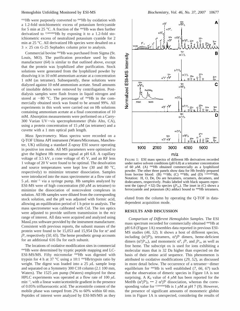
Comparison of Different Hemoglobin Samples.The ESImass spectrum recorded for commercially obtainedmetHb atpH 6.8 (Figure 1A) resembles data reported in previous ESI-MS studies (46, 52). It shows a host of different species,including (Rhâh)2 tetramers,Rhâh dimers, heme-deficientdimers (Rhâa
ox), and monomericRh, âh, andâaox as well as
free heme. The subscript ox is used for ions exhibiting amolecular mass that is 32 Da higher than expected on thebasis of their amino acid sequence. This phenomenon isattributed to oxidative modifications (29, 52), as discussedin more detail below. The occurrence of a tetramer-dimerequilibrium for metHb is well established (7, 66, 67) suchthat the observation of dimeric species in Figure 1A is notsurprising. AKd value of 4µM has been reported for theMetHb (Rhâh)2 T 2 Rhâh dissociation, whereas the corre-sponding value forcyanometHb is 1µM at pH 7 (9). However,the presence of significant amounts of monomeric globinions in Figure 1A is unexpected, considering the results of
FIGURE 1: ESI mass spectra of different Hb derivatives recordedunder native solvent conditions (pH 6.8) at a tetramer concentrationof 60 µM. (A) metHb obtained commercially as a lyophilizedpowder. The other three panels show data for Hb freshly preparedfrom bovine blood: (B)oxyHb; (C) metHb; and (D) cyanometHb.Notation: H, O, De, Do are hexamers, octamers, decamers, anddodecamers, respectively. Peaks labeled with black squares repre-sent the (apo-â +32) Da species (âa
ox). The inset in (C) shows aferrocyanide and potassium (K) adduct bound tometHb tetramers.
Hemoglobin Unfolding Monitored by ESI-MS Biochemistry, Vol. 46, No. 37, 200710677
previous experiments carried out in bulk solution (7, 66, 67).It might be suspected that some of the monomeric anddimeric species in the spectrum are fragmentation productsgenerated during ESI, but recent hydrogen/deuterium ex-change studies have shown this not to be the case. Instead,all of the ionic signals mentioned so far correspond to proteinspecies that exist in solution (53). Figure 1A also revealsthe presence of hexamers (Rhâh)3, octamers (Rhâh)4, decamers(Rhâh)5, and dodecamers (Rhâh)6. Although there is evidencefor the possible formation of higher order Hb assembliesunder some conditions (1), (Rhâh)n species withn > 2observed here likely represent clusters generated duringionization, a common occurrence in ESI-MS (53, 68, 69).Somewhat surprisingly, the relative contributions of theselarge assemblies were not strongly dependent on the proteinconcentration used. Decreasing the concentration by a factorof 10, or increasing it 2-fold, resulted in hexamer tododecamer intensities similar to those in Figure 1A (datanot shown).
Compared to the data for commercialmetHb, the ESI massspectrum of freshly preparedoxyHb has a much simplerappearance (Figure 1B), being strongly dominated by (Rhâh)2
ions. The contributions ofRhâh and larger aggregates aregreatly reduced. Monomeric species as well as heme-deficient dimers are virtually undetectable. The measuredmass corresponds to that of de-oxygenated Hb, that is, O2
binding is not maintained during the ESI process, althoughthe protein is infused into the ion source in its fullyoxygenated form. The peak width is significantly reducedbecause of vast improvements in desolvation behavior. Signalbroadening caused by the presence of residual solventadducts is frequently observed in ESI mass spectra of largenoncovalent complexes, as exemplified in Figure 1A (40,70, 71). The observation of much more favorable desolvationproperties for freshly preparedoxyHb (Figure 1B) is remark-able. To investigate whether the different ESI-MS behaviorof the two samples is related to the oxidation state of theheme iron, freshly preparedoxyHb was converted tometHbby exposure of the protein to potassium ferricyanide. Theresulting spectrum (Figure 1C) is very similar to that obtainedfor oxyHb, albeit with slightly elevated intensities for dimersand larger aggregates (hexamer to dodecamer). Close inspec-tion of the tetramer signals reveals peak splitting due to aresidual Fe(CN)6 adduct (Figure 1C, inset). This behavior isconsistent with the known tendency of Hb to accommodateferrocyanide in its organophosphate binding site, which liesbetween the twoâ-subunits in the tetrameric complex (5,72). Dimers do not possess this binding site, consistent withthe observation thatRhâh does not form a ferrocyanideadduct. For completeness, we also investigated the propertiesof cyanometHb, which was obtained by incubation ofmetHb witha slight molar excess of neutralized KCN (see the Experi-mental Procedures section). The overall differences betweenthe ESI mass spectra of freshly preparedcyanometHb (Figure1D) andmetHb (Figure 1C) are relatively small. However,Figure 1D shows a lower contribution ofRhâh, consistentwith the fact that heme ligation with CN- reduces theKd of(Rhâh)2 by a factor of 4, as mentioned above (9).
In additional experiments, freshly preparedoxyHb waslyophilized, followed by redissolution and extended storageof the resulting samples. Mass spectra obtained for proteintreated in this way resembled that of commercialmetHb
(Figure 1A), with prominent monomer, dimer, andRhâaox
signals (data not shown).Acid-Induced Denaturation.Figure 2 depicts the changes
in the ESI mass spectrum upon acidification of freshlypreparedoxyHb. Lowering the pH from 6.8 to 4.4 (Figure2A and B) results in a dramatic shift of the tetramer-dimerequilibrium such thatRhâh becomes the dominant species.In addition, ionic signals corresponding to monomericRh
andâh are observed. The low charge states (around 7+) ofthese monomer ions indicate that they represent highlycompact protein conformers in solution (45, 73). Upondecreasing the pH to 4.0, the relative signal intensities ofRh
and âh increase further. At the same time, highly chargedRa andâa ions appear in the spectrum, indicating the presenceof significantly unfolded apo-globin chains in solution(Figure 2C). This trend continues upon further acidification(Figure 2D). Figure 2E represents the end point of thetitration. The spectrum under these conditions (pH 2.1)exclusively showsRa andâa ions, with charge-state distribu-tions that have their maxima around 17+. These data reflectthe fact that at pH 2.1 all quaternary contacts and heme-protein interactions have been disrupted and that the mon-omeric subunits are extensively unfolded in solution (45, 73).The higher signal intensities forRa than for âa can beattributed to ion suppression effects as well as to differencesin the desolvation behavior of the two globin chains (74).The heme released during denaturation is not detectable inFigure 2C-E because of the rapid aggregation of the freeporphyrin under acidic conditions (75, 76). ESI mass spectravery similar to those depicted in Figure 2 were obtainedduring the acid-induced denaturation of freshly preparedmetHb andcyanometHb, demonstrating that the overall denatur-ation mechanism of the three forms studied here is largelyinsensitive to changes in heme oxidation and ligation state.Detailed ESI-MS titration data for the two ferri Hb deriva-tives are not shown because of space limitations; however,selected spectra formetHb will be discussed below (Figure7).
Complementary information on the acid-induced denatur-ation of the three freshly prepared Hb derivatives can beobtained by UV-vis spectroscopy. The absorbance changesaccompanying the titration ofoxyHb are depicted in Figure3. At neutral pH, the protein shows bands at 577 and 541nm and a dominant Soret signal at 415 nm. The presence ofthese so-calledR, â, andγ bands confirms that the proteinis indeed in its fully oxygenated low-spin Fe2+ state (5).Acidification leads to a gradual disappearance of the 577/541 nm doublet, and the Soret peak shifts to 405 nm. Thisspectral transition reflects a change in formal heme oxidationstate to Fe3+, corresponding to that ofmetHb. The onset ofthis process occurs around pH 4, that is, in the range wheremonomericRh, âh, and free apoprotein start to occur in theESI mass spectrum (Figure 2C). These changes reflect thefact that the highly specific structural environment of thenative heme binding pocket inoxyHb protects the heme ironfrom autoxidation (77). Disruption of the binding pocketduring denaturation allows the Fe2+ f Fe3+ transition tooccur under the aerobic solution conditions used here. Furtheracidification leads to a broad spectrum that is blue shiftedeven further, characteristic of heme that has been releasedfrom the protein and has undergone acid aggregation (78-80).
10678 Biochemistry, Vol. 46, No. 37, 2007 Boys et al.

The spectral changes accompanying the acid-induceddenaturation of freshly preparedmetHb exhibit an isosbesticpoint at 380 nm (Figure 4). This suggests that the ferri-hemechromophore experiences a two-state transition, from protein-bound to aggregated in acidic solution. Since these spectralchanges only probe the heme environment, the apparent two-state nature of the optical transition does not contradict theobservation of various protein oligomerization states (mono-mers, dimers, and tetramers) by ESI-MS. In the range
between pH 4 and pH 2.1, the data forcyanometHb (Figure 5)are very similar to those formetHb (Figure 4), reflecting thetransition from protein-bound ferri-heme to the acid-denatured state of Hb. In the case ofcyanometHb, however,this process is preceded by the loss of cyanide, apparent froman absorbance decrease around 540 nm, as well as a shift ofthe Soret band from 419 to 405 nm (Figure 5) (5).Presumably, the driving force for this reaction is theprotonation of cyanide at low pH. This cyanide loss is a
FIGURE 2: ESI mass spectra of freshly preparedoxyHb recorded at different pH (as indicated). Notation: the same as that in Figure 1; thered and blue symbols representRa andâa ions, respectively. The charge states of selected ions are indicated.
Hemoglobin Unfolding Monitored by ESI-MS Biochemistry, Vol. 46, No. 37, 200710679

gradual process that can be monitored directly by ESI-MS(Figure 6). In the near-neutral range, the mass spectra show(Rhâh)2 bound to up to four CN- groups. Acidificationinduces a gradual shift of the distribution, until at pH 3.5the base peak corresponds to the mass of unligated (Rhâh)2.Because protonation is an integral part of the ionizationprocess, it is possible that some cyanide is released in theform of HCN during ESI. Thus, the distributions in Figure
6 likely under-represent the actual cyanide binding levels inbulk solution. No cyanide binding was observed for dimericor monomeric globins.
A key conclusion from the acid-induced denaturation datapresented so far (especially those in Figures 1 and 2) is thattheR- andâ-subunits of freshly prepared Hb exhibit a highlysymmetric behavior: changes in association state, hemebinding, and conformation exhibited byR-globin are closelymirrored by theâ-subunit. On the basis of Figure 2, thedenaturation process can be summarized by the followingsimplified scheme (ignoring the possible involvement ofhigher order aggregates):
This result contrasts the data previously obtained on com-mercial metHb, which indicate highly asymmetric behaviorof the two subunits, as discussed in the Introduction section(46).
A particularly striking result in previous studies oncommercialmetHb was the observation of anRhâa dimerduring acid-induced denaturation (46). It was proposed thatthis species represents a mechanistically important intermedi-ate during the (dis)assembly process of the native tetramer(35, 46, 47). The spectrum depicted in Figure 1A confirmsthe existence of a heme-deficient heterodimer for thecommercial protein at pH 6.8, albeit with a+32 Da
FIGURE 3: UV-vis spectral changes accompanying the acid-induced denaturation of freshly preparedoxyHb. The pH values forthe individual spectra are indicated. Also shown are the character-istic absorption maxima foroxyHb andmetHb (5).
FIGURE 4: UV-vis spectral changes accompanying the acid-induced denaturation of freshly preparedmetHb.
FIGURE 5: UV-vis spectral changes accompanying the acid-induced denaturation of freshly preparedcyanometHb. Also shownare the characteristic absorption maxima formetHb andcyanometHb(5).
FIGURE 6: Partial ESI mass spectra showing the (Rhâh)2 18+ regionof freshly preparedcyanometHb at different pH values. The dottedlines numbered 0 through 4 indicate the number of cyano groupsbound to the protein tetramer. The changes in the overall spectrumas a function of pH are very similar to those depicted in Figure 2.
(Rhâh)2 f 2 Rhâh f
2 Rhfolded + 2 âh
foldedf (Scheme1)
2 Raunfolded+ 2 âa
unfolded+ 4 heme
10680 Biochemistry, Vol. 46, No. 37, 2007 Boys et al.

modification. MS/MS experiments verified the compositionof this species asRhâa
ox, ruling out other possible combina-tions that would result in the same mass such asRa
oxâh (81).Importantly, none of the freshly prepared Hb speciesinvestigated here exhibits significant levels of heme-deficientdimers when studied under native solvent conditions (Figure1B-D). The same result is obtained when exposing thefreshly prepared protein to semi-denaturing conditions. Inthe case ofoxyHb, this has already been demonstrated by thedata in Figure 2. A direct comparison of ESI mass spectrafor commercial and freshly preparedmetHb (i.e., using thesame iron oxidation state in both cases) confirms the lackof Rhâa and Rhâa
ox for the freshly prepared protein, asexemplified for pH 5.8 (Figure 7A and B) and pH 4.2 (Figure7C and D). Moreover, these data demonstrate that thecommercial protein is less resistant to acid denaturation, asseen from the much higher intensity of monomeric globinchains in Figure 7C than in Figure 7D.
It is concluded that the asymmetric behavior (46) of theR- and â-subunits during acid-induced denaturation ofcommercialmetHb represents an artifact, likely caused bystructural damage of the protein during isolation and/or
storage. This apparent asymmetry seems to be largely causedby substantial levels of chemically alteredâ-globin (termedâox). The existence ofRhâa
ox complexes implies thatâox isstill able to form noncovalent contacts withRh, whileexhibiting a greatly reduced heme affinity. However, thepresence of a considerable fraction of freeâa
ox under nativeconditions (Figure 1A) suggests that the interaction betweenRh and âa
ox is relatively weak. Since the overallR/âstoichiometry in the sample is unity (74), the existence offree âa
ox necessarily leads to the presence of unboundR-globin. The latter maintains a compact heme-boundconformation, as seen by the low ESI charge states ofRh. Incontrast, the lack of heme inâa
ox traps modifiedâ chains ina non-native conformation (12) that is characterized by highESI charge states (labeled with black squares in Figure 1A).
Identification of Oxidation Sites in CommercialmetHb.Deconvoluted mass distributions of acid-denaturedmetHbprovide an estimate of the overall level of covalent modifica-tions. As expected from the data discussed above, theâ-subunit of the commercial protein exhibits a dominantsatellite peak that is shifted by+32 Da (Figure 8A). Inaddition, theâ-globin mass distribution in Figure 8A exhibitsless intense signals at+16 and+48 Da. These observationsare attributed to the incorporation of one, two, or threeoxygen atoms (82). Signals corresponding to up to threeoxidation events also appear in the mass distribution ofR-globin, albeit at lower levels. In contrast, the massdistributions of freshly preparedmetHb subunits recordedunder identical conditions show only trace levels of oxidativemodifications for both subunits (Figure 8B).
Tryptic peptide mapping was carried out in order toidentify the locations of oxidation sites in commercialmetHb.Shown below are the sequences of bovineR- andâ-globin,with trypsin cleavage sites indicated as spaces (65).
FIGURE 7: ESI mass spectra of commercially obtainedmetHb (A)and freshly preparedmetHb (B) recorded at pH 5.8. Also shownare the corresponding data for pH 4.2 (C, commercialmetHb; D,freshly preparedmetHb). The insets focus on them/z rangecorresponding to the 13+ heterodimer. The expected peak positionsfor Rhâa
ox13+ andRhâa 13+ are indicated as vertical dashed lines in
the inset of panel (A). Arrows marked with “X” in (B) and (D)highlight the lack ofRhâa and/orRhâa
ox in the spectra of freshlypreparedmetHb.
FIGURE 8: Deconvoluted mass distributions ofR- and â-globin,obtained for acid-denaturedmetHb at pH 2.1. (A) Commercialprotein; (B) freshly prepared protein. The main peaks observed foreach distribution correspond to the masses of unmodifiedR- andâ-globin. Mass shifts of the observed covalent modifications areindicated. All four mass distributions have been normalized suchthat the base peak has roughly the same height in each case.
Hemoglobin Unfolding Monitored by ESI-MS Biochemistry, Vol. 46, No. 37, 200710681
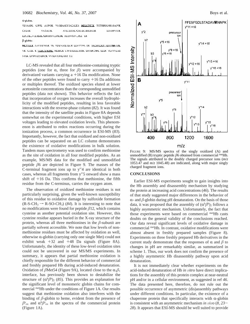
LC-MS revealed that all four methionine-containing trypticpeptides (one forR, three for â) were accompanied byderivatized variants carrying a+16 Da modification. Noneof the other peptides were found to carry+16 Da additionsor multiples thereof. The oxidized species eluted at loweracetonitrile concentrations than the corresponding unmodifiedpeptides (data not shown). This behavior reflects the factthat incorporation of oxygen increases the overall hydrophi-licity of the modified peptides, resulting in less favorableinteractions with the reverse-phase column (82). It was foundthat the intensity of the satellite peaks in Figure 8A dependssomewhat on the experimental conditions, with higher ESIvoltages leading to elevated oxidation levels. This phenom-enon is attributed to redox reactions occurring during theionization process, a common occurrence in ESI-MS (83).Importantly, however, the fact that oxidized and non-oxidizedpeptides can be separated on an LC column demonstratesthe existence of oxidative modifications in bulk solution.Tandem mass spectrometry was used to confirm methionineas the site of oxidation in all four modified peptides. As anexample, MS/MS data for the modified and unmodifiedpeptide â6 are depicted in Figure 9. The masses of theC-terminal fragment ions up to y′′4 are identical in bothcases, whereas all fragments from y′′5 onward show a massshift of +16 Da. This confirms that methionine, the fifthresidue from the C-terminus, carries the oxygen atom.
The observation of oxidized methionine residues is notparticularly surprising, given the well-known susceptibilityof this residue to oxidative damage by sulfoxide formation(R-S-CH3 f R-SO-CH3) (84). It is interesting to note thatno modifications were found for peptideâ12, which containscysteine as another potential oxidation site. However, thiscysteine residue appears buried in the X-ray structure of theprotein, whereas all three methionines in theâ-subunit arepartially solvent accessible. We note that low levels of non-methionine residues must be affected by oxidation as well,otherwiseR-globin (carrying only one single Met) could notexhibit weak +32 and +48 Da signals (Figure 8A).Unfortunately, the identity of these low-level oxidation sitescould not be uncovered in our MS/MS experiments. Insummary, it appears that partial methionine oxidation ischiefly responsible for the different behavior of commercialand freshly prepared Hb during acid-induced denaturation.Oxidation ofâMet54 (Figure 9A), located close to theR1â1
interface, has previously been shown to destabilize thestructure of (Rhâh)2 (85). This provides an explanation forthe significant level of monomeric globin chains for com-mercialmetHb under the conditions of Figure 1A. Our resultssuggest that methionine oxidation also interferes with thebinding of â-globin to heme, evident from the presence ofâa
ox and Rhâaox in the spectra of the commercial protein
(Figure 1A).
CONCLUSIONS
Earlier ESI-MS experiments sought to gain insights intothe Hb assembly and disassembly mechanism by studyingthe protein at increasing acid concentrations (46). The resultsof that study suggested major differences in the behavior ofR- andâ-globin during pH denaturation. On the basis of thosedata, it was proposed that the assembly of (Rhâh)2 follows ahighly asymmetric mechanism. Unfortunately, the fact thatthose experiments were based on commercialmetHb castsdoubts on the general validity of the conclusions reached.Our data reveal significant levels of oxidative damage incommercialmetHb. In contrast, oxidative modifications werealmost absent in freshly prepared samples (Figure 8).Experiments on three freshly prepared Hb derivatives in thecurrent study demonstrate that the responses ofR andâ tochanges in pH are remarkably similar, as summarized inscheme 1. Thus, our work does not confirm the presence ofa highly asymmetric Hb disassembly pathway upon aciddenaturation.
It is not immediately clear whether experiments on theacid-induced denaturation of Hbin Vitro have direct implica-tions for the assembly of this protein complex at near-neutralpH and/or in a cellular environment, as suggested in ref46.The data presented here, therefore, do not rule out thepossible occurrence of asymmetric (dis)assembly pathwaysunder different conditions. In particular, the existence of achaperone protein that specifically interacts withR-globinis consistent with an asymmetric mechanismin ViVo (8, 23-28). It appears that ESI-MS should be well suited to provide
FIGURE 9: MS/MS spectra of the singly oxidized (A) andunmodified (B) tryptic peptideâ6 obtained from commercialmetHb.The signals attributed to the doubly charged precursor ions (m/z1053.47 andm/z 1045.48) are indicated, along with major singlycharged fragment ions.
10682 Biochemistry, Vol. 46, No. 37, 2007 Boys et al.
detailed insights into the exact way in which this chaperoneassists the assembly process of Hb in RBC precursors.
ACKNOWLEDGMENT
We thank Chris Buchner and family and Terry Boys forhelp with blood acquisition.
REFERENCES
1. Riggs, A. F. (1998) Self-association, cooperativity and superco-operativity of oxygen binding by hemoglobins,J. Exp. Biol. 201,1073-1084.
2. Eaton, W. A., Henry, E. R., Hofrichter, J., and Mozzarelli, A.(1999) Is cooperative oxygen binding by hemoglobin reallyunderstood?Nat. Struct. Biol. 6, 351-358.
3. Perutz, M. F. (1970) Stereochemistry of cooperative effects inhaemoglobin,Nature 228, 726-739.
4. Hardison, R. C. (1996) A brief history of hemoglobins: plant,animal, protist, and bacteria,Proc. Nat. Acad. Sci. U.S.A. 93,5675-5679.
5. Antonini, E., and Brunori, M. (1971)Hemoglobin and Myoglobinin Their Reactions With Ligands, Vol. 21, North-Holland Publish-ing Company, Amsterdam.
6. Edelstein, S. J., Rehmar, M. J., Olson, J. S., and Gibson, Q. H.(1970) Functional aspects of the subunit association-dissociationequilibria of hemoglobin,J. Biol. Chem. 245, 4372-4381.
7. Schaeffer, J. R., McDonald, M. J., Turci, S. M., Dinda, D. M.,and Bunn, H. F. (1984) Dimer-monomer dissociation of humanhemoglobin A,J. Biol. Chem. 259, 14544-14547.
8. Santiveri, C. M., Perez-Canadillas, J. M., Viadivelu, M. K., Allen,M. D., Rutherford, T. J., Watkins, N. A., and Bycroft, M. (2004)NMR structure of theR-hemoglobin stabilizing protein,J. Biol.Chem. 279, 34963-34970.
9. White, S. L. (1975) The molecular dissociation of ferrihemoglobinderivatives,J. Biol. Chem. 250, 1263-1268.
10. Shaeffer, J. R., McDonald, M. J., and Bunn, H. F. (1981) Assemblyof normal and abnormal human hemoglobins,Trends Biochem.Sci. 6, 158-161.
11. Rose, M. Y., and Olson, J. S. (1983) The kinetic mechanism ofheme binding to human apohemoglobin,J. Biol. Chem. 258,4298-4303.
12. Leutzinger, Y., and Beychok, S. (1981) Kinetics and mechanismof heme-induced refolding of humanR-globin,Proc. Natl. Acad.Sci. U.S.A. 78, 780-784.
13. Adachi, K., Zhao, Y., and Surrey, S. (2003) Effects of hemeaddition on formation of stable human globin chains andhemoglobin subunit assembly in a cell-free system,Arch. Biochem.Biophys. 413, 99-106.
14. Hernan, R. A., and Sligar, S. G. (1995) Tetrameric hemoglobinexpressed inEscherichia coli, J. Biol. Chem. 270, 26257-26264.
15. Joshi, A. A., and McDonald, M. J. (1994) Role ofR and âcarboxyl-terminal residues in the kinetics of human oxyhemo-globin dimer assembly,J. Biol. Chem. 269, 8549-8553.
16. McGovern, P., Reisberg, P., and Olson, J. S. (1976) Aggregationof deoxyhemoglobin subunits,J. Biol. Chem. 251, 7871-7879.
17. McDonald, M. J., Turci, S. M., Mrabet, N. T., Himelstein, B. P.,and Bunn, H. F. (1987) The kinetics of assembly of normal andvariant human oxyhemoglobins,J. Biol. Chem. 262, 5951-5956.
18. Kawamura, Y., Hasumi, H., and Nakamura, S. (1982) Kineticstudies on the reconstitution of deoxyhemoglobin from isolatedR andâ chains,J. Biochem. 92, 1227-1233.
19. Adachi, K., Yang, Y., Joshi, A. A., Vasudevan, G., Morris, A.,and McDonald, M. J. (2001) Consequences ofâ16 andâ112replacements on the kinetics of hemoglobin assembly,Biochem.Biophys. Res. Commun. 289, 75-79.
20. Bunn, H. F., and McDonald, M. J. (1983) Electrostatic interactionsin the assembly of haemoglobin,Nature 306, 498-500.
21. Mrabet, N. T., McDonald, M. J., Turci, S., Sarkar, R., Szabo, A.,and Bunn, H. F. (1986) Electrostatic attraction governs the dimerassembly of human hemoglobin,J. Biol. Chem. 261, 5222-5228.
22. Yamaguchi, T., Yang, Y., McDonald, M. J., and Adachi, K. (2000)Surface and interfaceâ-chain residues synergistically affecthemoglobin assembly,Biochem. Biophys. Res. Commun. 270,683-687.
23. Luzzatto, L., and Notaro, R. (2002) Haemoglobin’s chaperone,Nature 417, 703-704.
24. Kihm, A. J., Kong, Y., Hong, W., Russel, J. E., Rouda, S., Adachi,K., Simon, M. C., Blobel, G. A., and Weiss, M. J. (2002) Anabundant erythroid protein that stabilizes freeR-haemoglobin,Nature 417, 758-763.
25. Baudin-Creuza, V., Vasseur-Godbillon, C., Pato, C., Prehu, C.,Wajcman, H., and Marden, M. C. (2004) Transfer of humanR-to â-hemoglobin via its chaperone protein,J. Biol. Chem. 279,36530-36533.
26. Hodge, D., Coghill, E., Keys, J., Maguire, T., Hartman, B.,McDowall, A., Weiss, M., Grimmond, S., and Perkins, A. (2006)A global role for EKLF in definitive and primitive erythropoiesis,Blood 15, 3359-3370.
27. Feng, L., A., G. D., Zhou, S., Gu, L., Kong, Y., Li, J., Hu, M.,Yan, N., Lee, C., Rich, A. M., Armstrong, R. S., Lay, P. A., Gow,A. J., Weiss, M. J., Mackay, J. P., and Shi, Y. (2004) Molecularmechanism of AHSP-mediated stabilization ofR-hemoglobin,Cell119, 629-640.
28. Feng, L., Zhou, S., Gu, L., Gell, D. A., Mackay, J. P., Weiss, M.J., Gow, A. J., and Shi, Y. (2005) Structure of oxidizedR-haemoglobin bound to AHSP reveals a protective mechanismfor haem,Nature 435, 697-701.
29. Boys, B. L., and Konermann, L. (2007) Folding and assembly ofhemoglobin monitored by electrospray mass spectrometry usingan on-line dialysis system,J. Am. Soc. Mass Spectrom. 18, 8-16.
30. Komar, A. A., Kommer, A., Krasheninnikov, I. A., and Spirin,A. S. (1997) Cotranslational folding of globin,J. Biol. Chem. 272,10646-10651.
31. Komar, A. A., Kommer, A., Krasheninnikov, I. A., and Spirin,A. S. (1993) Cotranslational heme binding to nascent globinchains,FEBS Lett. 326, 261-263.
32. Vasudevan, G., and McDonald, M. J. (2002) Ordered heme bindingensures the assembly of fully functional hemoglobin: a hypothesis,Curr. Protein Pept. Sci. 3, 461-466.
33. Vasudevan, G., and McDonald, M. J. (1997) Spectral demonstra-tion of semihemoglobin formation during CN-hemin incorporationinto human apohemoglobin,J. Biol. Chem. 272, 517-524.
34. Vasudevan, G., and McDonald, M. J. (2006) Soret spectral andbioinformatic approaches provide evidence for a critical role ofthe R-subunit in assembly of tetrameric hemoglobin,Protein J.25, 45-56.
35. Griffith, W. P., and Kaltashov, I. A. (2007) Protein conformationalheterogeneity as a binding catalyst: ESI-MS study of hemoglobinH formation,Biochemistry 46, 2020-2026.
36. Kawamura-Konishi, Y., and Suzuki, H. (1985) Binding reactionof hemin to globin,J. Biochem. 98, 1181-1190.
37. Kawamura-Konishi, Y., Chiba, K., Kihara, H., and Suzuki, H.(1992) Kinetics of the reconstitution of hemoglobin from semi-hemoglobinsR andâ with heme,Eur. Biophys. J. 21, 85-92.
38. Kaltashov, I. A., and Eyles, S. J. (2002) Studies of biomolecularconformations and conformational dynamics by mass spectrom-etry, Mass Spectrom. ReV. 21, 37-71.
39. Heck, A. J. R., and Van den Heuvel, R. H. H. (2004) Investigationof intact protein complexes by mass spectrometry,Mass Spectrom.ReV. 23, 368-389.
40. Loo, J. A. (2005) inThe Encyclopedia of Mass Spectrometry(Gross, M. L., and Caprioli, R. M., Eds.) pp 289-299, Elsevier,Amsterdam.
41. Ruotolo, B. T., and Robinson, C. V. (2006) Aspects of nativeproteins are retained in vacuum,Curr. Opin. Chem. Biol. 10, 402-408.
42. Smith, A. M., Jahn, T. R., Ashcroft, A. E., and Radford, S. E.(2006) Direct observation of oligomeric species formed in the earlystatges of amyloid formation using electrospray ionization massspectrometry,J. Mol. Biol. 364, 9-19.
43. Potier, N., Donald, L. J., Chernushevich, I., Ayed, A., Ens, W.,Arrowsmith, C. H., Standing, K. G., and Duckworth, H. W. (1998)Study of a noncovalent trp repressor: DNA operator complex byelectrospray ionization time-of-flight mass spectrometry,ProteinSci. 7, 1388-1395.
44. Grandori, R., Matecko, I., and Muller, N. (2002) Uncoupledanalysis of secondary and tertiary protein structure by circulardichroism and electrospray ionization mass spectrometry,J. MassSpectrom. 37, 191-196.
45. Konermann, L. (2007) A minimalist model for exploring confor-mational effects on the electrospray charge state distribution ofproteins,J. Phys. Chem. B 111, 6534-6543.
46. Griffith, W. P., and Kaltashov, I. A. (2003) Highly asymmetricinteractions between globin chains during hemoglobin assembly
Hemoglobin Unfolding Monitored by ESI-MS Biochemistry, Vol. 46, No. 37, 200710683

revealed by electrospray ionization mass spectrometry,Biochem-istry 42, 10024-10033.
47. Griffith, W. P., and Kaltashov, I. A. (2006) Mass spectrometry inthe study of hemoglobin: from covalent structure to higher orderassembly,Curr. Org. Chem. 10, 535-553.
48. Light-Wahl, K. J., Schwartz, B. L., and Smith, R. D. (1994)Observation of the noncovalent quaternary association of proteinsby electrospray ionization mass spectrometry,J. Am. Chem. Soc.116, 5271-5278.
49. Schmidt, A., and Karas, M. (2001) The influence of electrostaticinteractions on the detection of heme-globin complexes in ESI-MS, J. Am. Soc. Mass Spectrom. 12, 1092-1098.
50. Versluis, C., and Heck, A. J. R. (2001) Gas-phase dissociation ofhemoglobin,Int. J. Mass Spectrom. 210/211, 637-649.
51. Mekecha, T. T., Amunugama, R., and McLuckey, S. A. (2006)Ion trap collision-induced dissociation of human hemoglobinR-chain cations,J. Am. Soc. Mass Spectrom. 17, 923-931.
52. Simmons, D. A., Wilson, D. J., Lajoie, G. A., Doherty-Kirby, A.,and Konermann, L. (2004) Subunit disassembly and unfoldingkinetics of hemoglobin studied by time-resolved electrospray massspectrometry,Biochemistry 43, 14792-14801.
53. Hossain, B. M., and Konermann, L. (2006) Pulsed hydrogen/deuterium exchange MS/MS for studying the relationship betweennoncovalent protein complexes in solution and in the gas phaseafter electrospray ionization,Anal. Chem. 78, 1613-1619.
54. Shreffler, D. C., and Salisbury, G. W. (1959) Distribution ofinheritance of hemoglobin variants in american cattle,J. DairySci. 42, 1147-1156.
55. Paul, K. G., Theorell, H., and Akeson, A. (1953) The molar lightabsorption of pyridine ferroprotoporphyrin (pyridine haemochro-mogen),Acta Chem. Scand. 7, 1284-1287.
56. Crosby, W. H., and Houchin, D. N. (1957) Preparing standardsolutions of cyanmethemoglobin,Blood 12, 1132-1136.
57. International Committee for Standardization in Haematology.(1978) Recommendations for reference method for haemoglobi-nometry in human blood (ICSH Standard EP 6/2:1977) andspecifications for international haemiglobincyanide referencepreparation (ICSH Standard EP 6/3:1977),J. Clin. Pathol. 31,139-143.
58. Breepoel, P. M., Kreuzer, F., and Hazevoet, M. (1981) Interactionof Organic phosphates with bovine hemoglobin,Pflugers Arch.389, 219-225.
59. Bunn, H. F. (1971) Differences in the interaction of 2,3-diphosphoglycerate with certain mammalian hemoglobins,Science172, 1049-1050.
60. Moore, G. L., and et_al. (1992) Evaluation of methemoglobinformation during the storage of various hemoglobin solutions,Artif. Organs 16, 513-518.
61. Evelyn, K. A., and Malloy, H. T. Microdetermination of oxyhe-moglobin, methemoglobin, and sulfhemoglobin in a single sampleof blood,J. Biol. Chem. 126, 655-662.
62. Leahy, T., and Smith, R. (1960) Notes on methemoglobindetermination,Clin. Chem. 6, 148-152.
63. Cruz-Laneira, A., Bal, B. J., Quintela, O., and Lopez-Rivadulla,M. (2002) Determination of methemoglobin and total hemoglobinin toxicological studies by derivative spectrophotometry,J. Anal.Toxicol. 26, 67-72.
64. Budaveri, S., Ed. (2006)The Merck Index, pp 4648, Merck &Co. Inc., Whitehouse station, NJ.
65. Mueser, T. C., Rogers, P. H., and Arnone, A. (2000) Interfacesliding as illustrated by the multiple quaternary structures ofliganded hemoglobin,Biochemistry 39, 15353-15364.
66. Ackers, G. K., and Halvorson, H. R. (1974) The linkage betweenoxygenation and subunit dissociation in human hemoglobin,Proc.Natl. Acad. Sci. U.S.A. 71, 4312-4316.
67. Lunelli, L., Paola, Z., and Baldini, G. (1994) Evidence ofhemoglobin dissociation,Biopolymers 34, 747-757.
68. Peschke, M., Verkerk, U. H., and Kebarle, P. (2004) Features ofthe ESI mechanism that affect the observation of multiply charged
noncovalent protein complexes and the determination of theassociation constant by the titration method,J. Am. Soc. MassSpectrom. 15, 1424-1434.
69. Sun, J., Kitova, E. N., Wang, W., and Klassen, J. S. (2006) Methodfor distinguishing specific from nonspecific protein-ligand com-plexes in nanoelectrospray ionization mass spectrometry,Anal.Chem. 78, 3010-3018.
70. Green, B. N., and Vinogradov, S. N. (2004) An electrosprayionization mass spectrometric study of the subunit structure ofthe giant hemoglobin from the leechNephelopsis oscura, J. Am.Soc. Mass Spectrom. 15, 22-27.
71. McKay, A. R., Ruotolo, B. T., Ilag, L. L., and Robinson, C. V.(2006) Mass measurements of increased accuracy resolve hetero-geneous populations of intact ribosomes,J. Am. Chem. Soc. 128,11433-11442.
72. Sen, U., Dasgupta, J., Choudhury, D., Datta, P., Chakrabarti, A.,Chakrabarty, S. B., Chakrabarty, A., and Dattagupta, J. K. (2004)Crystal structure of HbA2 and HbE and modeling of hemoglobinδ4: interpretation of the thermal stability and the antisickling effectof HbA2 and identification of the ferrocyanide binding site in Hb,Biochemistry 43, 12477-12488.
73. Dobo, A., and Kaltashov, I. A. (2001) Detection of multiple proteinconformational ensembles in solution via deconvolution of charge-state distributions in ESI MS,Anal. Chem. 73, 4763-4773.
74. Kuprowski, M. C., Boys, B. L., and Konermann, L. (2007)Analysis of protein mixtures by electrospray mass spectrometry:effects of conformation and desolvation behavior on the signalintensities of hemoglobin subunits,J. Am. Soc. Mass Spectrom.18, 1279-1285.
75. Sogbein, O. O., Simmons, D. A., and Konermann, L. (2000) Theeffects of pH on the kinetic reaction mechanism of myoglobinunfolding studied by time-resolved electrospray ionization massspectrometry,J. Am. Soc. Mass Spectrom. 11, 312-319.
76. Adams, P. A. (1976) The kinetics and mechanism of therecombination reaction between apomyoglobin and haemin,Bio-chem. J. 159, 371-376.
77. Shikama, K. (2006) Nature of FeO2 bonding in myoglobin andhemoglobin: a new molecular paradigm,Prog. Biophys. Mol. Biol.91, 83-162.
78. Sage, J. T., Morikis, D., and Champion, P. M. (1991) Spectroscopicstudies of myoglobin at low pH: heme structure and ligation,Biochemistry 30, 1227-1237.
79. Sage, J. T., Li, P., and Champion, P. M. (1991) Spectroscopicstudies of myoglobin at low pH: heme ligation kinetics,Bio-chemistry 30, 1237-1247.
80. Konermann, L., Rosell, F. I., Mauk, A. G., and Douglas, D. J.(1997) Acid-induced denaturation of myoglobin studied by time-resolved electrospray ionization mass spectrometry,Biochemistry36, 6448-6454.
81. Simmons, D. A., Dunn, S. D., and Konermann, L. (2003)Conformational dynamics of partially denatured myoglobin studiedby time-resolved electrospray mass spectrometry with onlinehydrogen-deuterium exchange,Biochemistry 42, 5896-5905.
82. Takamono, K., and Chance, M. R. (2006) Radiolytic proteinfootprinting with mass spectrometry to probe the strucuture ofmacromolecular complexes,Annu. ReV. Biophys. Biomol. Struct.35, 251-276.
83. Chen, M., and Cook, K. D. (2007) Oxidation artifacts in theelectrospray mass spectrometry of Aâ peptide,Anal. Chem. 79,2031-2036.
84. Creighton, T. E. (1993)Proteins, W. H. Freeman & Co., NewYork.
85. Amiconi, G., Ascoli, F., Barra, D., Bertollini, A., Matarese, R.M., Verzili, D., and Brunori, M. (1989) Selective oxidation ofmethionineâ(55)D6 at theR1â1 interface in hemoglobin com-pletely destabilizes the T-state,J. Biol. Chem. 264, 17745-17749.
BI701076Q
10684 Biochemistry, Vol. 46, No. 37, 2007 Boys et al.