Unlocking the Mystery of Cancer: Do the Mutant p110α Subunits of PI3-Kinase Hold the Key?
5
Unlocking the Mystery of Cancer: Do the Mutant p110α Subunits of PI3-Kinase Hold the Key? Dr. Peter K. Vogt The Scripps Research Institute Molecular and Experimental Medicine Division of Oncovirology La Jolla, California ADV ANCING LIFE SCIENCE TOGETHER™ Research. Development. Production.
-
Upload
emd-millipore-bioscience -
Category
Documents
-
view
230 -
download
0
Transcript of Unlocking the Mystery of Cancer: Do the Mutant p110α Subunits of PI3-Kinase Hold the Key?
8/7/2019 Unlocking the Mystery of Cancer: Do the Mutant p110α Subunits of PI3-Kinase Hold the Key?
http://slidepdf.com/reader/full/unlocking-the-mystery-of-cancer-do-the-mutant-p110-subunits-of-pi3-kinase 1/5
Unlocking the Mysteryof Cancer:
Do the Mutant p110α Subunits of PI3-KinaseHold the Key?
Dr. Peter K. Vogt
The Scripps Research InstituteMolecular and Experimental MedicineDivision of OncovirologyLa Jolla, California
ADVANCING LIFE SCIENCE TOGETHER™Research. Development. Production.
8/7/2019 Unlocking the Mystery of Cancer: Do the Mutant p110α Subunits of PI3-Kinase Hold the Key?
http://slidepdf.com/reader/full/unlocking-the-mystery-of-cancer-do-the-mutant-p110-subunits-of-pi3-kinase 2/5
• Ras-binding domain mediating
interaction between p110 and
Ras-GTP and contributing to the
stimulation of the PI3K and the
Ras-driven signaling pathways
• C2 domain with affinity for lipid
membranes
• Helical domain of still undeter-
mined function
• Carboxy-terminal kinase domain
[Figure 1](4)
The standard regulatory subunit
of class I PI3Ks is p85, a protein that
contains several modular protein-
protein interaction domains:
• Two Src-homology 2 (SH2) domains
• Src-homology 3 (SH3) domain
• Poly-proline stretches (PP)
• BCR homology domain (BH)
• Inter-SH2 domain [Figure 3](8, 9)
The latter is the primary p110-
interacting surface. The regulatory
subunit links p110 to upstream
signals, interacting with tyrosine
receptor kinases and G protein-coupled
receptors(2, 4). A mutant version of
the p85 regulatory subunit, p65, has
been shown to induce the constitutive
activation of PI3K and contribute to
cellular transformation(5).
CELLULAR ACTIVITIESCONTROLLED BYCLASS I PI3KClass I PI3K controls numerous cellular
activities that are connected to
growth, replication and survival,
differentiation, movement andinvasiveness, immune signaling and
metabolism(2, 6, 7). Figure 4 shows
three important signaling chains
that originate in PI3K. All three are
mediated by the downstream target
Akt, which together with its main
activating kinase, PDK1, is recruited
to the plasma membrane through its
affinity for the product of class I
PI3K, PIP3. Akt serves as a branching
point for these signals that are
INTRODUCTIONThe discovery of cancer-specific
mutations in PIK3CA, the gene coding
for the catalytic subunit p110α of
PI3-kinase, has transformed the field
of lipid kinases from an area of basic
biochemistry into a focus of intense
interest for drug development, yet
the new opportunities depend
largely on the availability of purified,
mutant p110α.
This paper provides an overview
of PI3-kinases and what makes them
unique, their implications for cancer,
their roles in signal transduction and
the challenges researchers face
today with advancing this research.
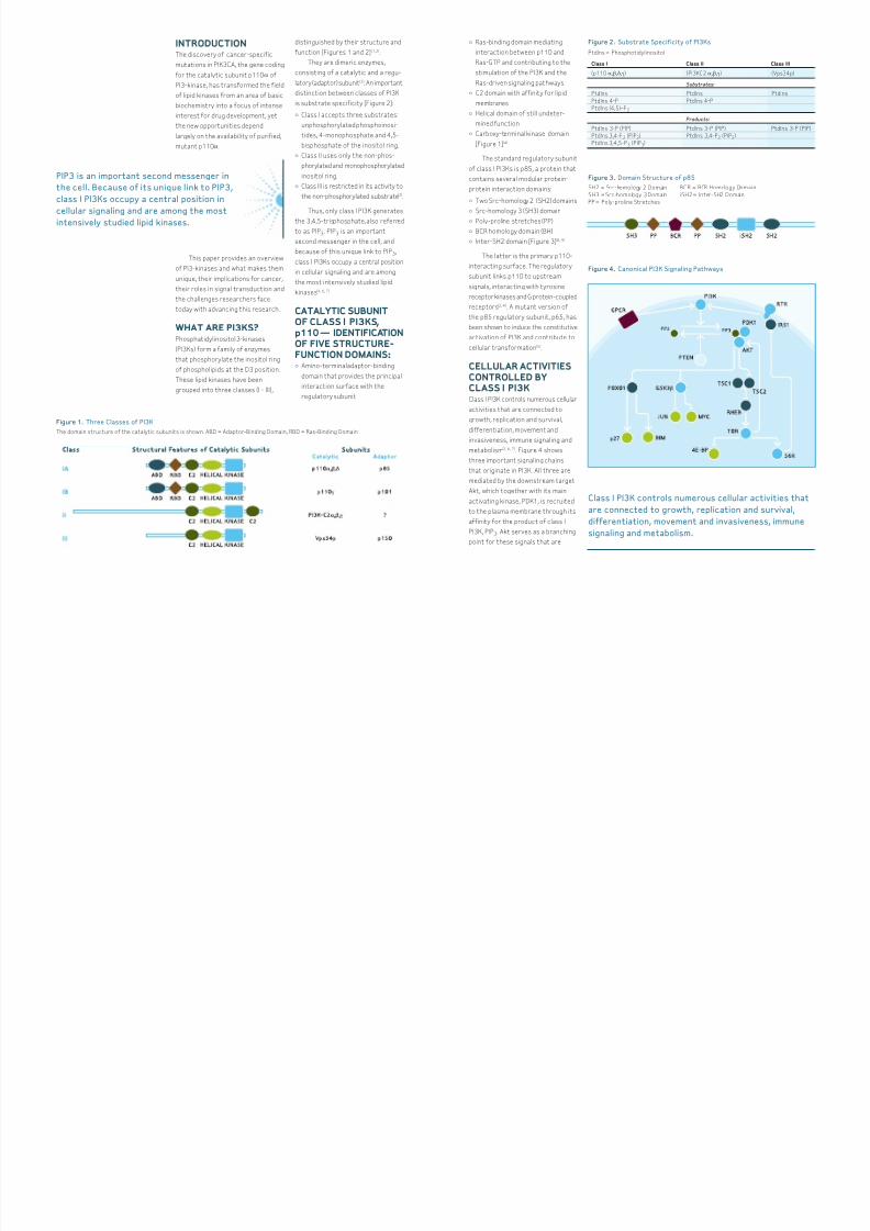
WHAT ARE PI3KS?Phosphatidylinositol 3-kinases
(PI3Ks) form a family of enzymes
that phosphorylate the inositol ring
of phospholipids at the D3 position.
These lipid kinases have been
grouped into three classes (I - III),
distinguished by their structure and
function [Figures 1 and 2] (1,2).
They are dimeric enzymes,
consisting of a catalytic and a regu-
latory (adaptor) subunit(3). An important
distinction between classes of PI3K
is substrate specificity [Figure 2]:
• Class I accepts three substrates:
unphosphorylated phosphoinosi-
tides, 4-monophosphate and 4,5-
bisphosphate of the inositol ring.
• Class II uses only the non-phos-
phorylated and monophosphorylated
inositol ring.
• Class III is restricted in its activity to
the non-phosphorylated substrate(2).
Thus, only class I PI3K generates
the 3,4,5-trisphosphate, also referred
to as PIP3. PIP3 is an important
second messenger in the cell, and
because of this unique link to PIP 3,
class I PI3Ks occupy a central position
in cellular signaling and are among
the most intensively studied lipid
kinases(4, 6, 7).
CATALYTIC SUBUNITOF CLASS I PI3KS,p110 — IDENTIFICATIONOF FIVE STRUCTURE-FUNCTION DOMAINS:• Amino-terminal adaptor-binding
domain that provides the principal
interaction surface with the
regulatory subunit
PIP3 is an important second messenger in
the cell. Because of its unique link to PIP3,
class I PI3Ks occupy a central position in
cellular signaling and are among the mostintensively studied lipid kinases.
Class I Class II Class III
(p110 α,β,δ,γ) (PI3KC2 α,β,γ) (Vps34p)
Substrates:
PtdIns PtdIns PtdIns
PtdIns 4-P PtdIns 4-P
PtdIns (4,5)–P2
Products:
PtdIns 3-P (PIP) PtdIns 3-P (PIP) PtdIns 3-P (PIP)
PtdIns 3,4-P2 (PIP2) PtdIns 3,4-P2 (PIP2)
PtdIns 3,4,5-P3 (PIP3)
Class I PI3K controls numerous cellular activities that
are connected to growth, replication and survival,
differentiation, movement and invasiveness, immune
signaling and metabolism.
Figure 1. Three Classes of PI3K
The domain structure of the catalytic subunits is shown. ABD = Adaptor-Binding Domain, RBD = Ras-Binding Domain
Figure 2. Substrate Specificity of PI3Ks
PtdIns = Phosphotidylinositol
Figure 3. Domain Structure of p85
SH2 = Src-homology 2 Domain BCR = BCR Homology Domain
SH3 = Src-homology 3 Domain iSH2 = Inter-SH2 Domain
PP = Poly-proline Stretches
Figure 4. Canonical PI3K Signaling Pathways
8/7/2019 Unlocking the Mystery of Cancer: Do the Mutant p110α Subunits of PI3-Kinase Hold the Key?
http://slidepdf.com/reader/full/unlocking-the-mystery-of-cancer-do-the-mutant-p110-subunits-of-pi3-kinase 3/5
directed toward FOXO1, GSK3β and
mTOR(10). Akt-mediated phosphoryla-
tion of FOXO1 leads to the inactivation
of this proapoptotic protein and a
downregulation of its growth-
attenuating targets, p27 and BIM.(11, 12)
Akt-dependent phosphorylation also
inhibits GSK3β(13) and lifts the GSK3β-
mediated inhibition of Jun(14) and of
Myc(15). In a third signaling chain, Akt
phosphorylates and thereby inhibits
the TSC2 protein downregulating its
GTPase-activating functions that
negatively regulate the Ras-like
protein Rheb. The result is activation
of Rheb and of the downstream kinase
mTOR(16). mTOR has multiple func-
tions; among them is the enhancementof protein synthesis.(17) This is
accomplished by activating the posi-
tive regulator of protein synthesis,
p70S6K, and interfering with the
negative regulator, 4E-BP(10, 16, 17). The
mTOR-mediated stimulation of protein
synthesis preferentially affects the
translation of mRNAs that contain
complex 5’ untranslated regions. The
mRNAs of many growth-promoting
proteins belong in this category. The
overall effect of these PI3K signals is
an upregulation of growth-stimulatory
and a reduction of growth-inhibitory
cellular functions. Figure 4 presents
selected examples of the multiple
cellular activities controlled by
PI3Ks. There are numerous others,
notably metabolic activities that are
not considered here.
PI3K SUBUNIT p110 ANDITS LINK TO CANCERPI3K has been tied to cancer for a
long time. The first sign of an
involvement of PI3K in cancer was
an association of lipid kinase activity
with the viral oncoproteins, Src and
middle T. In these complexes the PI3K
regulatory subunit p85 functions as
a connector, binding phosphotyrosine
residues on the oncoproteins and
recruiting the catalytic PI3K subunit
p110 (18, 19). The oncogenic potential of
p110α was then clearly demonstrated
by an avian retrovirus, ASV16, that
carries a copy of the cellular gene
coding for p110α as an oncogene. This
viral version of p110α is fused to viral
sequences that direct the protein to
the cell membrane, making it inde-
pendent of upstream signaling and
hence constitutively active(20). Mem-
brane-localized p110α is oncogenic.
Most human cancers show a
gain of function in PI3K signaling (10).
The increased activity can result from
changes in PI3K or in its downstream
target Akt or in the PI3K antagonist
PTEN. Akt can be overexpressed,
amplified, or its level of activating
phosphorylation can be increased.
PTEN shows frequent loss of function
in cancer, either by mutation or by
transcriptional silencing. The regula-
tory or catalytic subunits of PI3K
are often differentially expressed or
mutated in cancer(10). Of particular
interest are specific mutations in
PIK3CA, the gene coding for the
catalytic subunit p110α of PI3K(21).
These mutations occur in many
cancers [Table 1] and together with
other diverse experimental data they
single out the α isoform of class I
PI3K as a particularly important
contributor to oncogenesis(22-26, 27).
However, the non-α isoforms of class
I PI3K may have relevance for cancer
as well(28). Although no cancer-specific
mutations have been identified in
the non-α isoforms, they show
oncogenic potential when overex-
pressed in avian cells(28), and may also
become contributing factors in human
cancer by differential expression.
IDENTIFYING “HOTSPOT” MUTATIONSSome 20 to 30 percent of cancers
of the breast, colon, prostate, and
endometrium contain a mutated
PIK3CA [Table 1](29). Strikingly, about
80 percent of the mutations occur
in one of three “hot spots” in the
coding sequence of PIK3CA. Each of
the "hot spot" mutations is a single
nucleotide substitution leading to a
single amino acid substitution(21). The
overwhelming prevalence of these
three distinct mutations in cancer
strongly suggests that p110α carrying
one of these mutations bestows the
cell with a strong selective advantage
in growth and survival. Indeed, p110α
with a "hot spot" mutation shows gain
of function in enzymatic activity(30)
and signals constitutively through
Akt and mTOR. It can also induce
oncogenic transformation of cultured
cells and tumors in animals(22, 23). The
mutations in PIK3CA have thus all
the attributes of “driver” mutations,
responsible for neoplastic properties
of the cancer cell that harbors them.
Mutated p110α has emerged as
arguably the most promising cancer
target currently available.
5 Salient Features of the
"Hot Spot" Mutations in
PIK3CA:
• Cancer-specific; not found in
normal tissue
• Occurring at high frequencies
• Show a gain of function, which is
far easier to correct than loss
of function
• They are "driver" mutations
• Mutant protein is a kinase, readily
targetable by small molecules
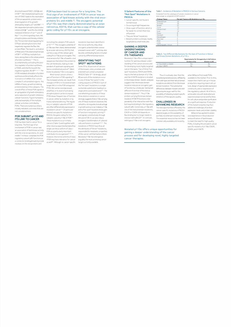
GAINING A DEEPERUNDERSTANDINGOF CANCER ANDITS THERAPIESMutated p110α offers unique oppor-
tunities for gaining a deeper under-
standing of the cancer process and
for developing novel, highly targeted
cancer therapies. Two of three "hot
spot" mutations, E542K and E545K,
map to the helical domain of p110α,
and the H1047R mutation is located
in the kinase domain. Genetic analyses
suggest that the kinase domain
mutation induces an oncogenic gain
of function by a molecular mechanism
different from that of the helical
domain mutations(31). The p110α
protein carrying the kinase domain
mutation H1047R functions inde-
pendently of an interaction with Ras
but requires binding to the regulatory
subunit p85. Conversely, p110α with
one of the helical domain mutations,
E542K or E545K still depends on
Ras binding but no longer needs to
interact with p85 [2](31). In contrast,
wild-type p110α is not oncogenic.
Thus it is already clear that the
mutantenzyme behavesvery differently
from wild-type and that helical domain
mutations are distinct from the kinase
domain mutation. The substantial
differences between mutant and wild-
type enzyme augur well for the
possibility of obtaining mutant-specific
inhibitors of therapeutic quality.
CHALLENGES INADVANCING RESEARCHThe new opportunities offered by the
cancer-specific mutations in PIK3CA
depend largely on the availability of
purified, recombinant mutant p110α.
This essential resource has not been
commercially available until recently,
when Millipore first made PI3Ks
available to the market. Prior to this,
researchers had to set up in-house
production, requiring insect cell cul-
ture, design of appropriate expression
constructs, and co-expression of
the regulatory subunit. All of this is
achievable only with dedicated and
experienced personnel and facilities
for cell culture and protein purification
at a significant expense. Production
of the mutant enzymes may face
additional challenges of protein
expression levels and protein stability.
Millipore has applied its exten-
sive experience in the production
and purification of lipid kinases
to meet the need for high-quality
PI3Ks, including the prevalent cancer
specific mutants of p110α: E542K,
E545K, and H1047R.
PI3K has been tied to cancer for a long time. The
first sign of an involvement of PI3K in cancer was an
association of lipid kinase activity with the viral onco-
proteins Src and middle T. The oncogenic potential
of p110α was then clearly demonstrated by an avian
retrovirus, ASV16, that carries a copy of the cellular
gene coding for p110α as an oncogene.
Mutated p110α offers unique opportunities for
gaining a deeper understanding of the cancer
process and for developing novel, highly targeted
cancer therapies.
Primary Tissue Percent Tumors with Mutations in p110α
Endometrium 23
Prostate 29
Breast 27
Colon 23
Urinary tract 17
Liver 12
Ovary 8
Stomach 8
Oesophagus 7
Pancreas 6
Brain 5
Lung 3
H ematop oietic t issue 4
Table 1. Incidence of Mutation in PIK3CA in Various Cancers.
(Compiled from the catalogue of somatic mutations in cancer
www.sanger.ac.uk/genetics/CGP/cosmic/)
Requirements for Oncogenicity in Cell Culture
B in di ng t o R as B in di ng t o p 85
p110α E542K or E545K Yes No
p110α H1047R No Yes
Table 2. Two Different Mechanisms for the Gain of Function in Helical
and Kinase Domain Mutations of p110α
.
8/7/2019 Unlocking the Mystery of Cancer: Do the Mutant p110α Subunits of PI3-Kinase Hold the Key?
http://slidepdf.com/reader/full/unlocking-the-mystery-of-cancer-do-the-mutant-p110-subunits-of-pi3-kinase 4/5
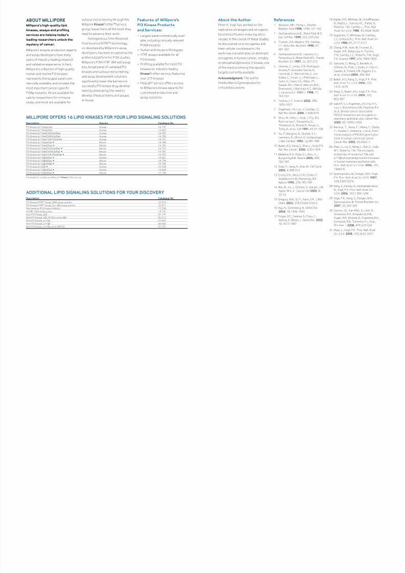
ABOUT MILLIPOREMillipore’s high-quality lipid
kinases, assays and profiling
services are helping today’s
leading researchers unlock the
mystery of cancer.
Millipore’s enzyme production experts
and assay developers have many
years of industry-leading research
and validation experience. In fact,
Millipore’s collection of high-quality
human and murine PI3 kinases
represents the largest panel com-
mercially available, and includes the
most important cancer-specific
PI3Kα mutants. All are available for
sale to researchers for in-house
study, and most are available for
outsourced screening through the
Millipore KinaseProfiler™ service,
giving researchers all the tools they
need to advance their work.
Homogeneous Time-Resolved
Fluorescence (HTRF®) technology,
co-developed by Millipore’s assay
developers, has been accepted as the
preferred platform for PI3K studies.
Millipore’s PI3K HTRF 384 well assay
kits, broad panel of validated PI3
kinases and outsourced screening
and assay development solutions
significantly lower the barriers to
successful PI3 kinase drug develop-
ment by eliminating the need to
develop these proteins and assays
in-house.
Features of Millipore’s
PI3 Kinase Products
and Services:
• Largest panel commercially avail-
able, including clinically relevant
PI3KA mutants
• Human and mouse orthologues
• HTRF assays available for all
PI3 kinases
• Profiling available for most PI3
kinases on industry-leading
KinaseProfiler service, featuring
over 275 kinases
• FlexLabSM service offers access
to Millipore’s kinase experts for
customized production and
assay solutions
MILLIPORE OFFERS 16 LIPID KINASES FOR YOUR LIPID SIGNALING SOLUTIONSDescription Species Catalogue No.
PI3 Kinase (p110α/p65α) Human 14-790
PI3 Kinase (p110α/p85α) Human 14-602
PI3 Kinase (p110α(E542K)/p85α) Human 14-782
PI3 Kinase (p110α(E545K)/p85α) Human 14-783
PI3 Kinase (p110α(H1047R)/p85α) Human 14-792
PI3 Kinase (p110α/p65α) • Mouse 14-786
PI3 Kinase (p110α/p85α) • Mouse 14-785
PI3 Kinase (p110α(E542K)/p85α) • Mouse 14-791
PI3 Kinase (p110α(E545K)/p85α) • Mouse 14-781
PI3 Kinase (p110α(H1047R)/p85α) • Mouse 14-787
PI3 Kinase (p110β/p85α) • Human 14-603
PI3 Kinase (p110β/p85α) • Mouse 14-794
PI3 Kinase (p110β/p85β) • Mouse 14-788
PI3 Kinase (p120δ) • Human 14-558
PI3 Kinase (p110δ/p85α) • Human 14-604
PI3 Kinase (p110δ/p85α) • Mouse 14-789
• Available for screening in Millipore’s KinaseProfiler service
ADDITIONAL LIPID SIGNALING SOLUTIONS FOR YOUR DISCOVERYDescription Catalogue No.
PI3 Kinase HTRF® Assay (384 assay points) 33-016
PI3 Kinase HTRF® Assay (5 x 384 assay points) 33-017
Wortmannin (PI3 Kinase inhibitor) 12-338
mTOR (1362-end), active 14-770
Anti-PI3 Kinase, p85 06-195
Anti-PI3 Kinase, p85, N-SH3, clone AB6 05-212
Anti-PI3 Kinase, p110α 07-658
Anti-PI3 Kinase, p110β 06-568
Anti-PI3 Kinase, p110δ, clone AW103 05-703
About the Author
Peter K. Vogt has worked on the
replicative strategies and oncogenic
functions of tumor-inducing retro-
viruses. In the course of these studies,
he discovered viral oncogenes and
their cellular counterparts. His
work now concentrates on dominant
oncogenes in human cancer, notably
on phosphatidylinositol 3-kinase, one
of the most promising therapeutic
targets currently available.
Acknowledgment: The author
thanks Marco Gymnopoulos for
critical discussions.
References
1. Wymann, M.P.; Pirola, L. Biochim
Biophys. Acta 1998, 1436, 127-150
2. Vanhaesebroeck, B.; Waterfield, M.D.
Exp. Cell Res. 1999, 253, 239-254
3. Fruman, D.A.; Meyers, R.E.; Cantley,
L.C. Annu. Rev. Biochem. 1998, 67,
481-507
4. Vanhaesebroeck, B.; Leevers, S.J.;
Panayotou, G.; Waterfield, M.D. Trends
Biochem. Sci. 1997, 22, 267-272
5. Jimenez, C.; Jones, D.R.; Rodríguez-
Viciana, P.; Gonzalez-García, A.;
Leonardo, E.; Wennström, S.; von
Kobbe, C.; Toran, J.L.; R-Borlado, L.;
Calvo, V.; Copin, S.G.; Albar, J.P.;
Gaspar, M.L.; Diez, E.; Marcos, M.A.;
Downward, J.; Martinez-A, C.; Mérida,
I.; Carrera, A.C. EMBO J. 1998, 17,
743-753
6. Cantley, L.C. Science 2002, 296,
1655-1657
7. Engelman, J.A.; Luo, J.; Cantley, L.C.
Nat. Rev. Genet. 2006, 7, 606-619
8. Otsu, M.; Hiles, I.; Gout, I.; Fry, M.J.;
Ruiz-Larrea, F.; Panayotou, G.;
Thompson, A.; Dhand, R.; Hsuan, J.;
Totty, N., et al., Cell 1991, 65 , 91-104
9. Hu, P.; Margolis, B.; Skolnik, E.Y.;
Lammers, R.; Ullrich, A.; Schlessinger,
J. Mol. Cell Biol. 1992, 12, 981-990
10. Bader, A.G.; Kang, S.; Zhao, L.; Vogt, P.K.
Nat. Rev. Cancer, 2005, 5 , 921-929
11. Medema, R.H.; Kops, G.J.; Bos, J.L.;
Burgering, B.M. Nature 2005, 404,
782-787
12. Vogt, P.; Jiang, H.; Aoki, M. Cell Cycle
2005, 4, 908-913
13. Cross, D.A.; Alessi, D.R.; Cohen, P.;
Andjelkovich, M.; Hemmings, B.A.
Nature 1995, 378, 785-789
14. Wei, W.; Jin, J.; Schlisio, S.; Harper, J.W;
Kaelin, W.G. Jr. Cancer Cell 2005, 8,
25-33
15. Gregory, M.A.; Qi, Y.; Hann, S.R. J. Biol.Chem. 2003, 278, 51606-51612
16. Hay, N.; Sonenberg, N. Genes Dev.
2004, 18, 1926-1945
17. Fingar, D.C.; Salama, S.; Tsou, C.;
Harlow, E.; Blenis, J. Genes Dev. 2002,
16, 1472-1487
18. Kaplan, D.R.; Whitman, M.; Schaffhausen,
B.; Raptis, L.; Garcea, R.L.; Pallas, D.;
Roberts, T.M.; Cantley, L. Proc. Natl.
Acad. Sci. U.S.A. 1986, 83, 3624-3628
19. Sugimoto, Y.; Whitman, M.; Cantley,
L.C.; Erikson, R.L. Proc. Natl. Acad. Sci.
U.S.A. 1984, 81, 2117-2121
20. Chang, H.W.; Aoki, M.; Fruman, D.;
Auger, K.R.; Bellacosa, A.; Tsichlis,
P.N.; Cantley, L.C.; Roberts, T.M.; Vogt,
P.K. Science 1997, 276, 1848-1850
21. Samuels, Y.; Wang, Z.; Bardelli, A.;
Silliman, N.; Ptak, J.; Szabo, S.; Yan, H.;
Gazdar, A.; Powell, S.M.; Riggins, G.J.,
et al., Science 2004, 304, 554
22. Bader, A.G.; Kang, S.; Vogt, P.K. Proc.
Natl. Acad. Sci. U.S.A. 2006, 103,
1475-1479
23. Kang, S.; Bader, A.G.; Vogt, P.K. Proc.
Natl. Acad. Sci. U.S.A. 2005, 102,
802-80724. Isakoff, S.J.; Engelman, J.A.; Irie, H.Y.;
Luo, J.; Brachmann, S.M.; Pearline, R.V.
et al., Breast cancer-associated
PIK3CA mutations are oncogenic in
mammary epithelial cells. Cancer Res.
2005, 65 , 10992-1000.
25. Ikenoue, T.; Kanai, F.; Hikiba, Y.; Obata,
T.; Tanaka, Y.; Imamura, J.; et al., Func-
tional analysis of PIK3CA gene muta-
tions in human colorectal cancer.
Cancer Res. 2005, 65 , 4562-7.
26. Zhao, J.J.; Liu, Z.; Wang, L.; Shin, E.; Loda,
M.F.; Roberts, T.M. The oncogenic
properties of mutant p110α and
p110β phosphatidylinositol 3-kinases
in human mammary epithelial cells.
Proc. Natl. Acad. Sci. U.S.A. 2005, 102,
18443-8.
27. Gymnopoulos, M.; Elsliger, M.A.; Vogt,
P.K. Proc. Natl. Acad. Sci. U.S.A. 2007,
104, 5569-5574
28. Kang, S.; Denley, A.; Vanhaesebroeck,
B.; Vogt, P.K. Proc. Natl. Acad. Sci.
U.S.A. 2006, 103, 1289-1294
29. Vogt, P.K.; Kang, S.; Elsliger, M.A.;
Gymnopoulos, M. Trends Biochem. Sci.
2007, 32, 342-349
30. Carson, J.D.; Van Aller, G.; Lehr, R.;
Sinnamon, R.H.; Kirkpatrick, R.B.;
Auger, K.R.; Dhanak, D.; Copeland, R.A.;
Gontarek, R.R.; Tummino, P.J., et al.,
Biochem. J. 2008, 409, 519-524
31. Zhao, L.; Vogt, P.K. Proc. Natl. Acad.
Sci. U.S.A. 2008, 105 , 2652-2657

8/7/2019 Unlocking the Mystery of Cancer: Do the Mutant p110α Subunits of PI3-Kinase Hold the Key?
http://slidepdf.com/reader/full/unlocking-the-mystery-of-cancer-do-the-mutant-p110-subunits-of-pi3-kinase 5/5
www.millipore.com/offices
Millipore and Upstate are registered trademarks of Millipore Corporation.
KinaseProfiler and the M mark and Advancing Life Science Together are
trademarks of Millipore Corporation.
FlexLab is a service mark of Millipore Corporation.
HTRF is a registered trademark of CIS BIO International Corp.
TB1096ENUS 08DD20 Printed in U.S.A.
© 2008 Millipore Corporation, Billerica, MA 01821 All rights reserved.