Supporting Information - PNAS · or mutant F1L genes in frame with a pE/L-Cherry selection cas- ......
8
Supporting Information Gerlic et al. 10.1073/pnas.1215995110 SI Materials and Methods Reagents, Chemicals, and Antibodies. Cell-culture supplies were ob- tained from Gibco. The following antibodies were used for im- munoblotting according to standard protocols: anti-Flag M2 (Sigma), anti-c-Myc (Roche), anti-human IL-1β (R&D Systems). The anti-GST, anti-GFP (B-2), anti-mouse caspase-1 protein (p)10 (M-20), anti-human caspase-1 p10 (C-20) and p20 (C-15) antibodies were purchased from Santa Cruz Biotechnology. An- tibodies against the viral proteins F1L and N1L were produced in rabbits by immunization with F1L peptide as described (1), or with purified N1L protein produced in Escherichia coli. Muramyl dipeptide (MDP) was purchased from InvivoGen or Alexis for either cell-based or in vitro studies. Lipopolysaccharide (LPS) was obtained from Alexis, and N-acetyl-Trp-Glu-His-Asp-AMC (7-amino-4-methylcoumarin) (Ac-WEHD-AMC), N-acetyl-Trp- Glu-His-Asp-rhodamine-D112 (Dansyl ethylenediamine) (Ac- WEHD-RhoD112), and Ac-WEHD-CHO were obtained from Calbiochem. Plasmids. Plasmids encoding Flag-Caspase-1, ProIL-1β, Myc- apoptosis-associated speck-like protein containing a caspase re- cruitment domain (ASC), Flag/Myc-pyrin domain containing protein (NLRP) 1, Myc-nucleotide–binding oligomerization domain-containing protein 2 (NOD2), GFP-F1L, -Δ44 and -Δ57–78 were described (1, 2). Recombinant baculovirus transfer vectors encoding human NLRP1, pro-Caspase-1 (p45) or NLRP1Δleucine- rich repeat (LRR) mutant (retains 1–723/1236–1473 amino acids) were described (3). GST Pull-Down. Recombinant proteins were produced by baculo- virus infection of nucleotide-binding oligomerization domain, leu- cine-rich repeat and Spodoptera frugiperda (Sf9) insect cells (GST- NLRP1, GST-NLRP1ΔLRR) or in bacteria [GST–BCL2-related protein A1 (Bfl-1), GST-F1L, or GST] as described (3, 4). Various proteins were mixed (5 μg) with or without purified F1L protein (10 μg) in 20 mM Hepes-KOH at pH 7.5, 10 mM KCl, and 1 mM DTT in a final volume of 50 μL for 30 min on ice, followed by incubation overnight with glutathione-sepharose at 4 °C. GST- purified complexes were isolated after centrifugation, washed, and eluted with SDS-loading buffer, then analyzed by SDS/PAGE and stained by Sypro Ruby. In Vitro Reconstituted NLRP1 Inflammasome. The in vitro reconsti- tution of the NLRP1 inflammasome using proteins expressed in Sf9 cells from recombinant baculoviruses has been described (3). Briefly, reactions contained 8.5 nM His 6 -NLRP1, 8.5 nM procas- pase-1, 0.25 mM ATP, 0.5 mM Mg 2+ , 0.1 μg/mL MDP and GST- B-cell CLL/lymphoma 2 (Bcl-2) or N1L, wild-type or various mutants of recombinant 17 nM GST-F1L, or various synthetic peptides. Caspase-1 activity was measured after 60 min by hy- drolysis of Ac-WEHD-AMC substrate (20 μM), expressing data as mean ± SD, n = 3. Caspase-1 Processing in the Reconstituted NLRP1 Inflammasome. Reac- tions contained 0.1 μM His 6 -NLRP1 or GST-NLRP1ΔLRR, 0.1 μM procaspase-1, 1 mM ATP, 1 mM Mg 2+ , 10 μg/mL MDP and re- combinant GST-F1L 1–47 protein or synthetic F1L 22–47 peptide (F1Lp), or 0.4 μM recombinant N1L and were incubated 30 min at 37 °C. Proteins were separated by SDS/PAGE and analyzed by immunoblotting using anti-p10 caspase-1 antibodies in conjunc- tion with secondary IRDye 800/680CW-conjugated antibodies (LI-COR Biosciences). Results were imaged by using Li-COR Odyssey v3.0 software. Analysis of ATP Binding to NLRP1 by fluorescence polarization assay. His 6 -NLRP1 (0.125 μM) was incubated for 5 min on ice in the presence of F1L 1–47 (2 μM), 0.5 or 2 μM F1Lp 22–47, N1L, F1Lp 32–37, or 2 μM Bcl-2p 41–60. The mixture was then incubated for an additional 5 min in ice with 1 μM MDP-LD, 10 nM fluorescein- conjugated ATP analog and 0.5 mM Mg 2+ . ATP binding was analyzed by fluorescence polarization assay (FPA) [measuring milliPolars (mP)] using an Analyst AD Assay Detection System (LJL Biosystem), and the percentage of inhibition was deter- mined compared with NLRP1 incubated only with MDP-LD (mean ± SD). Peptide Synthesis and Purification. Peptides were synthesized by using Rink Amide ChemMatrix resin (Matrix Innovation) using Fmoc synthesis and diisopropylcarbodiimide/1-hydroxybenotiazde (DIC/HOBt) coupling with an Advanced ChemTech Apex 396 multiple peptide synthesizer. Peptides were cleaved from the resin by using standard cleavage and deprotection conditions and puri- fied by HPLC on a Phenomenex Luna C18 column (250 × 21.2 mm, 5 μm). Purified peptides were all single peaks (>99% purity) by analytical HPLC, and their mass was confirmed by MALDI mass spectroscopy. Cell Culture. HeLa and 293T cells were cultured in DMEM sup- plemented with 10% FBS and antibiotics. Human acute monocytic leukemia cell line (THP-1), THP-1-Blue, and PBMC were cultured in complete RPMI medium 1640 10% FBS and antibiotics. For differentiation, THP-1 cells were stimulated for 18 h with 50 ng/ mL phorbol-12-myristate-13-acetate. PBMC used in infection studies were obtained from healthy donors (San Diego Blood Bank) and isolated by density-gradient centrifugation using Ficol. Vaccinia Virus Production. The revertant F1L viruses (Flag-F1L and Flag-F1L(N32A, H35A) were generated by introducing wild-type or mutant F1L genes in frame with a pE/L-Cherry selection cas- sette. The ΔN1L virus was generated by replacing nucleotides 1–275 of the N1L open reading frame 63 (Orf63) with a pE/L-gpt- Cherry selection cassette. The fidelity of the resulting recombinant viruses was verified by DNA sequencing. Expression of the Flag- F1L proteins and loss of expression of N1L was documented by immunoblot analysis of infected cells. The Vaccinia virus (VACV) strains were grown in HeLa cells and titered on VeroE6 cells. VACV Infection of Cultured Cells. 12-D-tetradecanoylphorbol-13- acetate (TPA)-differentiated THP-1 cells were infected at various multiplicities of infection (MOI) with VACV-Western Reserve (WR), N1L deleted virus (ΔN1L), or F1L deleted virus (ΔF1L). Intracellular Staining for E3L Viral Antigen. VACV-infected THP-1 cells (as above) were collected 18 h after infection. Cells were fixed with cytofix-cytosperm (BD Biosciences) for 20 min at 4 °C. Fixed cells were subjected to intracellular E3L staining in Perm/Wash buffer (BD Biosciences) for 30 min at 4 °C. Anti-E3L Ab was kindly provided by Shane Crotty (La Jolla Institute for Allergy and Immunology) and used at a 1:100 vol/vol dilution. Cells were washed twice with BD Perm/Wash buffer and stained with goat anti-mouse IgG biotin-conjugated antibody at 4 °C followed by addition of PE-conjugated streptavidin (BD Biosciences). Sam- ples were analyzed for proportion of intracellular E3L staining Gerlic et al. www.pnas.org/cgi/content/short/1215995110 1 of 8
Transcript of Supporting Information - PNAS · or mutant F1L genes in frame with a pE/L-Cherry selection cas- ......
Supporting InformationGerlic et al. 10.1073/pnas.1215995110SI Materials and MethodsReagents, Chemicals, and Antibodies. Cell-culture supplies were ob-tained from Gibco. The following antibodies were used for im-munoblotting according to standard protocols: anti-Flag M2(Sigma), anti-c-Myc (Roche), anti-human IL-1β (R&D Systems).The anti-GST, anti-GFP (B-2), anti-mouse caspase-1 protein(p)10 (M-20), anti-human caspase-1 p10 (C-20) and p20 (C-15)antibodies were purchased from Santa Cruz Biotechnology. An-tibodies against the viral proteins F1L and N1L were produced inrabbits by immunization with F1L peptide as described (1), orwith purified N1L protein produced in Escherichia coli. Muramyldipeptide (MDP) was purchased from InvivoGen or Alexis foreither cell-based or in vitro studies. Lipopolysaccharide (LPS)was obtained from Alexis, and N-acetyl-Trp-Glu-His-Asp-AMC(7-amino-4-methylcoumarin) (Ac-WEHD-AMC), N-acetyl-Trp-Glu-His-Asp-rhodamine-D112 (Dansyl ethylenediamine) (Ac-WEHD-RhoD112), and Ac-WEHD-CHO were obtained fromCalbiochem.
Plasmids. Plasmids encoding Flag-Caspase-1, ProIL-1β, Myc-apoptosis-associated speck-like protein containing a caspase re-cruitment domain (ASC), Flag/Myc-pyrin domain containingprotein (NLRP) 1, Myc-nucleotide–binding oligomerizationdomain-containing protein 2 (NOD2), GFP-F1L, -Δ44 and -Δ57–78were described (1, 2). Recombinant baculovirus transfer vectorsencoding human NLRP1, pro-Caspase-1 (p45) or NLRP1Δleucine-rich repeat (LRR) mutant (retains 1–723/1236–1473 amino acids)were described (3).
GST Pull-Down. Recombinant proteins were produced by baculo-virus infection of nucleotide-binding oligomerization domain, leu-cine-rich repeat and Spodoptera frugiperda (Sf9) insect cells (GST-NLRP1, GST-NLRP1ΔLRR) or in bacteria [GST–BCL2-relatedprotein A1 (Bfl-1), GST-F1L, or GST] as described (3, 4). Variousproteins were mixed (5 μg) with or without purified F1L protein(10 μg) in 20 mM Hepes-KOH at pH 7.5, 10 mM KCl, and 1 mMDTT in a final volume of 50 μL for 30 min on ice, followed byincubation overnight with glutathione-sepharose at 4 °C. GST-purified complexes were isolated after centrifugation, washed, andeluted with SDS-loading buffer, then analyzed by SDS/PAGE andstained by Sypro Ruby.
In Vitro Reconstituted NLRP1 Inflammasome. The in vitro reconsti-tution of theNLRP1 inflammasome using proteins expressed in Sf9cells from recombinant baculoviruses has been described (3).Briefly, reactions contained 8.5 nM His6-NLRP1, 8.5 nM procas-pase-1, 0.25 mM ATP, 0.5 mM Mg2+, 0.1 μg/mL MDP and GST-B-cell CLL/lymphoma 2 (Bcl-2) or N1L, wild-type or variousmutants of recombinant 17 nM GST-F1L, or various syntheticpeptides. Caspase-1 activity was measured after 60 min by hy-drolysis of Ac-WEHD-AMC substrate (20 μM), expressing dataas mean ± SD, n = 3.
Caspase-1 Processing in the Reconstituted NLRP1 Inflammasome. Reac-tions contained 0.1 μMHis6-NLRP1orGST-NLRP1ΔLRR, 0.1 μMprocaspase-1, 1 mM ATP, 1 mM Mg2+, 10 μg/mL MDP and re-combinant GST-F1L 1–47 protein or synthetic F1L 22–47 peptide(F1Lp), or 0.4 μM recombinant N1L and were incubated 30min at37 °C. Proteins were separated by SDS/PAGE and analyzed byimmunoblotting using anti-p10 caspase-1 antibodies in conjunc-tion with secondary IRDye 800/680CW-conjugated antibodies
(LI-COR Biosciences). Results were imaged by using Li-COROdyssey v3.0 software.
Analysis of ATP Binding to NLRP1 by fluorescence polarization assay.His6-NLRP1 (0.125 μM) was incubated for 5 min on ice in thepresence of F1L 1–47 (2 μM), 0.5 or 2 μMF1Lp 22–47, N1L, F1Lp32–37, or 2 μMBcl-2p 41–60. The mixture was then incubated foran additional 5 min in ice with 1 μMMDP-LD, 10 nM fluorescein-conjugated ATP analog and 0.5 mM Mg2+. ATP binding wasanalyzed by fluorescence polarization assay (FPA) [measuringmilliPolars (mP)] using an Analyst AD Assay Detection System(LJL Biosystem), and the percentage of inhibition was deter-mined compared with NLRP1 incubated only with MDP-LD(mean ± SD).
Peptide Synthesis and Purification. Peptides were synthesized byusing Rink Amide ChemMatrix resin (Matrix Innovation) usingFmoc synthesis and diisopropylcarbodiimide/1-hydroxybenotiazde(DIC/HOBt) coupling with an Advanced ChemTech Apex 396multiple peptide synthesizer. Peptides were cleaved from the resinby using standard cleavage and deprotection conditions and puri-fied by HPLC on a Phenomenex Luna C18 column (250 × 21.2 mm,5 μm). Purified peptides were all single peaks (>99% purity) byanalytical HPLC, and their mass was confirmed by MALDI massspectroscopy.
Cell Culture. HeLa and 293T cells were cultured in DMEM sup-plemented with 10%FBS and antibiotics. Human acutemonocyticleukemia cell line (THP-1), THP-1-Blue, and PBMCwere culturedin complete RPMI medium 1640 10% FBS and antibiotics. Fordifferentiation, THP-1 cells were stimulated for 18 h with 50 ng/mL phorbol-12-myristate-13-acetate. PBMC used in infectionstudies were obtained from healthy donors (San Diego BloodBank) and isolated by density-gradient centrifugation using Ficol.
Vaccinia Virus Production. The revertant F1L viruses (Flag-F1L andFlag-F1L(N32A, H35A) were generated by introducing wild-typeor mutant F1L genes in frame with a pE/L-Cherry selection cas-sette. The ΔN1L virus was generated by replacing nucleotides1–275 of the N1L open reading frame 63 (Orf63) with a pE/L-gpt-Cherry selection cassette. The fidelity of the resulting recombinantviruses was verified by DNA sequencing. Expression of the Flag-F1L proteins and loss of expression of N1L was documented byimmunoblot analysis of infected cells. The Vaccinia virus (VACV)strains were grown in HeLa cells and titered on VeroE6 cells.
VACV Infection of Cultured Cells. 12-D-tetradecanoylphorbol-13-acetate (TPA)-differentiated THP-1 cells were infected at variousmultiplicities of infection (MOI) with VACV-Western Reserve(WR), N1L deleted virus (ΔN1L), or F1L deleted virus (ΔF1L).
Intracellular Staining for E3L Viral Antigen. VACV-infected THP-1cells (as above) were collected 18 h after infection. Cells were fixedwith cytofix-cytosperm (BD Biosciences) for 20 min at 4 °C. Fixedcells were subjected to intracellular E3L staining in Perm/Washbuffer (BD Biosciences) for 30 min at 4 °C. Anti-E3L Ab waskindly provided by Shane Crotty (La Jolla Institute for Allergy andImmunology) and used at a 1:100 vol/vol dilution. Cells werewashed twice with BD Perm/Wash buffer and stained with goatanti-mouse IgG biotin-conjugated antibody at 4 °C followed byaddition of PE-conjugated streptavidin (BD Biosciences). Sam-ples were analyzed for proportion of intracellular E3L staining
Gerlic et al. www.pnas.org/cgi/content/short/1215995110 1 of 8
after gating on live cells using a FACSCalibur flow cytometer withCellQuest (BD Biosciences) and FlowJo software (Tree Star).
NF-κB Activation in THP-1-Blue Cells.Using THP-1-Blue cells (ATCC;InvivoGen), NF-κB activation was evaluated by measuring alka-line phospatase activity in the supernatant for 180 min by hy-drolysis of QUANTI-Blue substrate (InvivoGen). As a positivecontrol, THP-1 macrophages were stimulated for 18 h with MDP(5 μg/mL). Reported results are mean ± SD; n = 3.
Immunoblot Analysis. Immunoblot analysis of cell extracts wasperformed by standard methods. For immunoblot analysis ofculture supernatants, the recovered fluids were filtered by using anAmicon Ultra (100 kDa cutoff). Proteins contained in eluateswere precipitated by trichloroacetic acid and washed twice withacetone. Dry pellets were resuspended in 2× Laemmli buffer,normalized by the amount of total protein, separated by SDS/PAGE, and analyzed by immunoblotting using various antibodiesin conjunction with secondary IRDye 800/680CW-conjugatedantibodies (LI-COR Biosciences). Results were imaged by usingLi-COR Odyssey v3.0 software and quantified by integratedfluorescence intensity.
Coimmunoprecipitations. HEK293T cells were transiently cotrans-fected with plasmids encoding Myc or flag-tagged NLR full-lengthproteins and either GFP-tagged N1L or GFP-tagged F1L. Forcoimmunoprecipitations, 5 × 10⁵ to 1 × 10⁶ cells were lysed inisotonic, Nonidet P-40–containing lysis buffer (150 mM NaCl, 20mM Hepes at pH 7.4, 0.2% Nonidet P-40, 5 mM MgCl2, 2 mMEDTA, 2 mM DTT, 1 mM PMSF, and 1× protease inhibitor mix(Roche). Clarified lysates were subjected to immunoprecipitation(IP) by using Dynabeads proteinG (Invitrogen) conjugated withanti-GFP, -Myc, or -flag antibodies. After overnight incubation at4 °C, immune complexes were washed three times in lysis buffer,separated by SDS/PAGE, and analyzed by immunoblotting usingvarious antibodies in conjunction with secondary IRDye 800/680CW-conjugated antibodies (LI-COR Biosciences). Resultswere imaged by using Li-COR Odyssey v3.0 software and quan-tified by integrated fluorescence intensity. Where indicated, celllysates (10% volume) were run directly in SDS gels.
Confocal Microscopy.HeLa cells were grown overnight on Lab-TekII coverslip chambers and cotransfected with plasmids encodingF1L-GFP and Myc-NLRP1 by using jetPRIME. After 24 h, cellswere fixed with 4% (kg/dL) formaldehyde, permeabilized withPBS + 0.3% (vol/vol) Triton X-100 for 10 min, and blocked withPBS + 3% BSA for 30 min. Cells were immunostained with anti-Myc antibody (Sigma; 1:200) overnight, then washed with PBS andincubated with Alexa Fluor 594-conjugated secondary antibody(1:200 vol/vol) for 1 h. Cells were washed with PBS, and coverslipswere mounted by using Vectashield. Images were acquired byconfocal laser-scanning microscopy using a LSM 710 NLO ZeissMultiphoton Laser Point Scanning Confocal Microscope equip-ped with a 63× objecting using inverted-based microscopy.
Mice. The studies reported here conform to the animal WelfareAct and followed National Institutes of Health guidelines for the
care and use of animals in biomedical research. All experimentswere performed in compliance with the regulations of the La JollaInstitute Animal care committee in accordance with the guide-lines set by the Association for assessment and Accreditation oflaboratory Animal Care. Eight- to 12-wk-old female BALB/c micewere purchased from the Jackson Laboratory. Animal experi-ments were approved by Sanford-Burnham Medical ResearchInstitutional Animal Care and Use Committee (IACUC).
VACV Intranasal Challenge.Eight- to 12-wk-old femaleBALB/cwereanesthetized by inhalation of isoflurane and inoculated by theintranasal route with VACV,Western Reserve (VACV-WR), anda mutant of WR in which only the F1L gene was deleted, VACV-F1L (104 or 105 pfu per mice). Body weight and rectal temperatureof mice were measured daily before and after infection. At varioustimes after infection, four mice from each group were killed andlungs and BAL fluids were collected. Mice were euthanized whenthey lost 30% of their initial body weight or were severely hypo-thermic (<32 °C) on two consecutive days.
Bronchoalveolar Lavage. Lungs were lavaged with 0.5 mL of PBScontaining 2%BSA. The recovered bronchoalveolar lavage (BAL)fluids were centrifuged to pellet cells. The supernatants (500 μL)were concentrated 10× by using an Amicon Ultra (3-kDa cutoff).Proteins contained in eluates were mixed with 5× Laemmli buffer,separated by SDS/PAGE, and analyzed by immunoblotting usinganti-mouse caspase-1 p10 (M-20) in conjunction with secondaryIRDye 800/680CW-conjugated antibodies (LI-COR Biosciences).Results were imaged by using Li-COR Odyssey v3.0 software andquantified by the integrated fluorescence intensity.
Characterization of Lung Histology.Lungswere removed after killinganimals, fixed in 10% zinc-buffered formalin (Protocal; FisherDiagnostics), and embedded in paraffin. Deparaffinized 5-μm-thick sections were stained with hematoxylin and eosin for histo-pathological assessment. All slides were scanned at an absolutemagnification of 400× (resolution of 0.25 μm per pixel) by usingthe Aperio ScanScope CS system (Aperio Technologies). Theacquired digital images representing whole-tissue sections wereviewed and analyzed by using the ImageScope viewer. To quantifyfoci of inflammation in lung sections, focal areas of peribronchialand perivascular inflammation were selected with the pen tool.Mean and SEM values for numbers of foci were determined. Lungsections were analyzed by the terminal deoxynucleotidyl trans-ferase end-labeling (TUNEL) method (Chemicon International)to identify the DNA fragmentation of apoptotic cells. The per-centage of TUNEL-positive cells was evaluated by a morphometricmethod using an automated image analysis system (Aperio Tech-nologies) and applying a nuclear scoring algorithm (5).
Statistics. In vivo data are presented asmean± SEM.All other datawere presented as the mean ± SD from at least three independentexperiments. Statistical comparisons between different treatmentswere performed by two-tailed Student’s t test, two-way ANOVA,log-rank analysis, or Bonferroni’s test, using PRIZM software.
1. Postigo A, Cross JR, Downward J, WayM (2006) Interaction of F1L with the BH3 domainof Bak is responsible for inhibiting vaccinia-induced apoptosis. Cell Death Differ 13(10):1651–1662.
2. Bruey JM, et al. (2007) Bcl-2 and Bcl-XL regulate proinflammatory caspase-1 activationby interaction with NALP1. Cell 129(1):45–56.
3. Faustin B, et al. (2007) Reconstituted NALP1 inflammasome reveals two-step mechanismof caspase-1 activation. Mol Cell 25(5):713–724.
4. Yu E, et al. (2011) Structural determinants of caspase-9 inhibition by the vaccinia virusprotein, F1L. J Biol Chem 286(35):30748–30758.
5. Krajewska M, et al. (2009) Image analysis algorithms for immunohistochemical assess-ment of cell death events and fibrosis in tissue sections. J Histochem Cytochem 57(7):649–663.
Gerlic et al. www.pnas.org/cgi/content/short/1215995110 2 of 8
C
NLRP1 GFP-F1L MERGE
NLRP1 GFP-N1L MERGE
A B
IP: MycWB:GFP(F1L-GFP)
WB: Myc
Lysate
WB:GFP
55kDa
55kDa
55kDa
36kDa
36kDa
36kDa250kDa130kDa78kDa
pcD
NA
3Myc
Aim
2-M
yc
NLR
P1-M
yc
NLR
P3-M
yc
ASC
-Myc
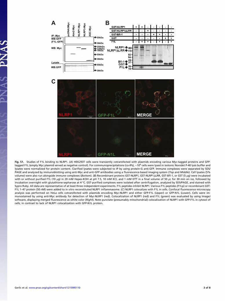
Fig. S1. Studies of F1L binding to NLRP1. (A) HEK293T cells were transiently cotransfected with plasmids encoding various Myc-tagged proteins and GFP-tagged F1L (empty Myc plasmid served as negative control). For coimmunoprecipitations (co-IPs), ∼106 cells were lysed in isotonic Nonidet P-40 lysis buffer andlysates were normalized for protein content. Clarified lysates were subjected to IP by using protein-G anti-GFP. Immune complexes were separated by SDS/PAGE and analyzed by immunoblotting using anti-Myc and anti-GFP antibodies using a fluorescence-based imaging system (Top and Middle). Cell lysates (5%volume) were also run alongside immune complexes (Bottom). (B) Recombinant proteins GST-NLRP1, GST-NLRP1ΔLRR, GST-Bfl-1, or GST (5 μg) were incubatedwith or without purified F1L (10 μg) in 20 mM Hepes-KOH at pH 7.5, 10 mM KCl, and 1 mM DTT in a final volume of 50 μL for 30 min on ice, followed byincubation overnight with glutathione-sepharose at 4 °C. GST-purified complexes were isolated after centrifugation, analyzed by SDS/PAGE, and stained withSypro Ruby. All data are representative of at least three independent experiments. F1L peptides inhibit NLRP1. Various F1L peptides (F1Lp) or recombinant GST-F1L 1–47 protein (50 nM) were added to in vitro reconstituted NLRP1 inflammasome. (C) NLRP1 colocalizes with F1L in cells. Confocal fluorescence microscopyanalysis was performed on HeLa cells cotransfected with plasmids encoding Myc-NLRP1 and either GFP-F1L (Upper) or GFP-N1L (Lower). Cells were im-munostained by using anti-Myc antibody for detection of Myc-NLRP1 (red). Colocalization of NLRP1 (red) and F1L (green) was evaluated by using ImageJsoftware, displaying merged fluorescence as white color (Right). Note punctate (presumably mitochondrial) colocalization of NLRP1 with GFP-F1L in cytosol ofcells, in contrast to lack of NLRP1 colocalization with GFP-N1L protein.
Gerlic et al. www.pnas.org/cgi/content/short/1215995110 3 of 8
MDP+ATP - + + + + + + His-NLRP1 + + + + + - - His-NLRP1 LRR - - - - - + + F1L(1-47) - - - + - - - F1Lp(22-47) - - - - + - + N1L - - + - - - -
Caspase-1 P10
No Ligand
No peptide
F1Lp (17-41)
F1Lp (22-47)
F1L (1-47) 0
20 40 60 80
100 120
Cas
pase
-1 a
ctiv
ity
(RFU
/min
)
+ MDP/ATP
F1Lp 32-37, IC50=18.8 ± 6.7 nM F1Lp 22-37, IC50=15.0 ± 4.0 nM F1Lp 22-32
0 10 20 30 40 500
40
80
120
160
200
[Peptide] (nM)
Cas
pase
-1 a
ctiv
ity
(RFU
/min
)A B C
D E F
G H I
+Caspase-1 p20/p10
F1L
(1-2
06)
F1L
(24-
206)
F1L
(48-
206)
F1L
(74-
206)
F1L
(90-
206)
IP: FLAG (NLRP1)
LOADING CONTROL
WB:GST
WB: FLAG
WB: GST
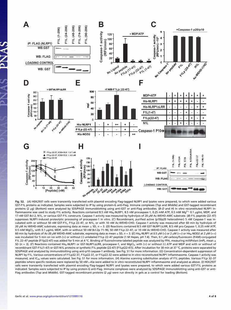
Fig. S2. (A) HEK293T cells were transiently transfected with plasmid encoding Flag-tagged NLRP1 and lysates were prepared, to which were added variousGST-F1L proteins as indicated. Samples were subjected to IP by using protein-G anti-Flag. Immune complexes (Top and Middle) and GST-tagged recombinantproteins (2 μg) (Bottom) were analyzed by SDS/PAGE immunoblotting using anti-GST or anti-Flag antibodies. (B–D and H) In vitro reconstituted NLRP1 in-flammasome was used to study F1L activity. Reactions contained 8.5 nM His6-NLRP1, 8.5 nM procaspase-1, 0.25 mM ATP, 0.5 mM Mg2+, 0.1 μg/mL MDP, and17 nM GST-Bcl-2, N1L, or various GST-F1L constructs. Caspase-1 activity was measured by hydrolysis of 20 μM Ac-WEHD-AMC substrate. (B) F1L peptide (22–47)suppresses NLRP1-induced proteolytic processing of procaspase-1 in vitro. (C) Recombinant, purified active (p10/p20 heterodimer) 5 nM Caspase-1 was in-cubated with or without 50 nM GST-F1L, F1Lp 22–47, or N1L, or with 10 nM Ac-WEHD-CHO. Caspase-1 activity was measured after 60 min by hydrolysis of20 μM Ac-WEHD-AMC substrate, expressing data as mean ± SD, n = 3. (D) Reactions contained 8.5 nM GST-NLRP1ΔLRR, 8.5 nM pro-Caspase-1, 0.25 mM ATP,0.5 mM MgCl2, with 0.1 μg/mL MDP, with or without 50 nM Bcl-2p 71–90, 50 nM F1Lp 22–47, or 10 nM Ac-WEHD-CHO. Caspase-1 activity was measured after60 min by hydrolysis of Ac-20 μM WEHD-AMC substrate, expressing data as mean ± SD, n = 3. (E) His6-NLRP1 at 0.5 μM (+) or 2 μM (++) or His6-NOD2 at 2 μM (++)was incubated for 5 min on ice with (+) or without (-) unlabeled F1Lp 22–47 peptide (1 M Hepes, pH 7.4). Then, 0.1 μM carboxyfluorescein (FAM)-conjugatedF1L 22–47 peptide (F1lp22-47) was added for 5 min at 4 °C. Binding of fluorochrome-labeled peptide was analyzed by FPA, measuring milliPolars (mP), mean ±SD (n = 3). (F) Reactions contained His6-NLRP1 or GST-NLRP1ΔLRR, procaspase-1, and MgCl2, with (+) or without (-) ATP and MDP and with or without ofrecombinant GST-F1L(1-47) or GST-N1L proteins or synthetic F1L peptide (22-47) [F1Lp(22-47)]. After incubation for 30 min at 37 °C, proteins were separated bySDS/PAGE and analyzed by immunoblotting using anti-p10 caspase-1 antibody. See Fig. S1 for more information. (G) Concentration-dependent suppression ofNLRP1 by F1L. Various concentrations of F1Lp32-37, F1Lp22-37, or F1Lp22-32 were added to in vitro reconstituted NLRP1 inflammasome. Caspase-1 activity wasmeasured, and IC50 values were calculated. See Fig. S1 for more information. (H) Alanine scanning substitution analysis of F1L peptides. Various F1Lp 32–37peptides where specific residues were replaced by 50 nM L-Ala were added to in vitro reconstituted NLRP1 inflammasome and analyzed as above. (I) HEK293Tcells were transiently transfected with plasmid encoding Flag-tagged NLRP1 and lysates were prepared, to which were added various GST-F1L proteins asindicated. Samples were subjected to IP by using protein-G anti-Flag. Immune complexes were analyzed by SDS/PAGE immunoblotting using anti-GST or anti-Flag antibodies (Top and Middle). GST-tagged recombinant proteins (2 μg) were run directly in gels as a control for loading (Bottom).
Gerlic et al. www.pnas.org/cgi/content/short/1215995110 4 of 8
WT WT
WTWTNone ΔF1LNoneΔF1L
WT
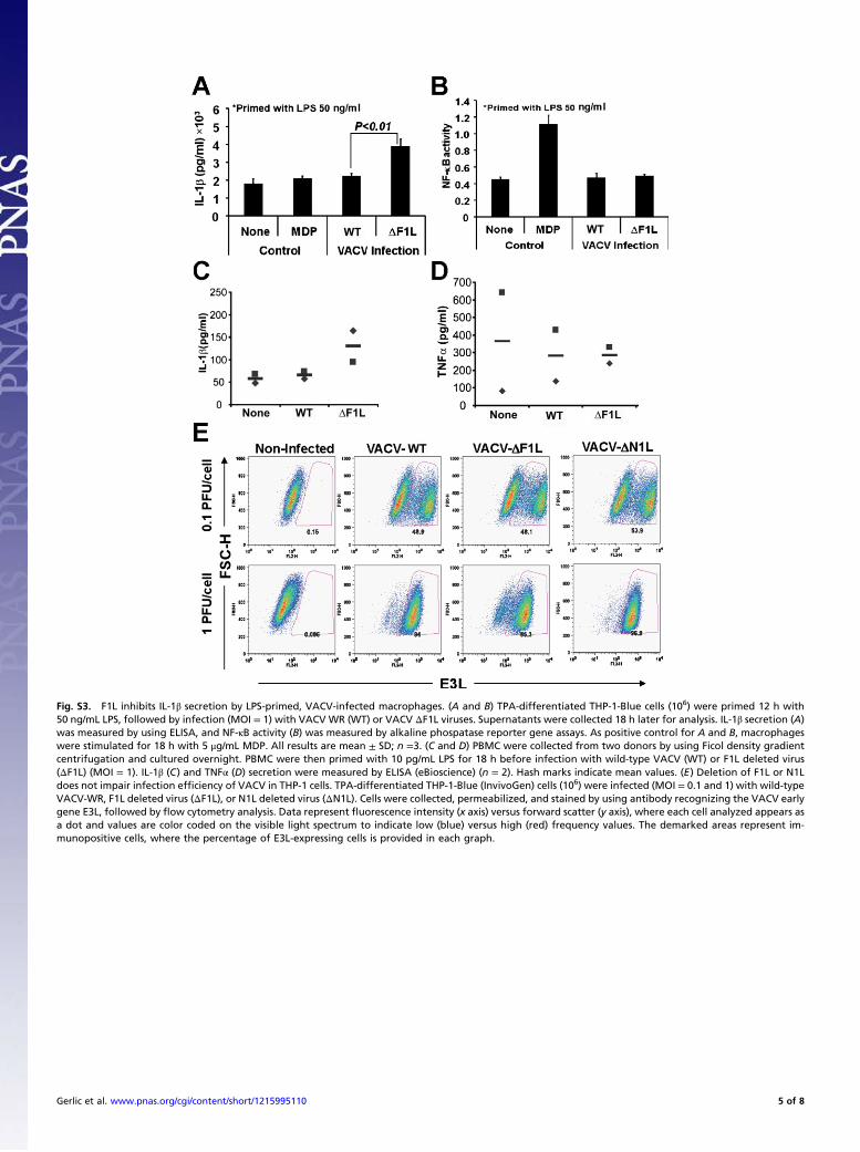
Fig. S3. F1L inhibits IL-1β secretion by LPS-primed, VACV-infected macrophages. (A and B) TPA-differentiated THP-1-Blue cells (106) were primed 12 h with50 ng/mL LPS, followed by infection (MOI = 1) with VACV WR (WT) or VACV ΔF1L viruses. Supernatants were collected 18 h later for analysis. IL-1β secretion (A)was measured by using ELISA, and NF-κB activity (B) was measured by alkaline phospatase reporter gene assays. As positive control for A and B, macrophageswere stimulated for 18 h with 5 μg/mL MDP. All results are mean ± SD; n =3. (C and D) PBMC were collected from two donors by using Ficol density gradientcentrifugation and cultured overnight. PBMC were then primed with 10 pg/mL LPS for 18 h before infection with wild-type VACV (WT) or F1L deleted virus(ΔF1L) (MOI = 1). IL-1β (C) and TNFα (D) secretion were measured by ELISA (eBioscience) (n = 2). Hash marks indicate mean values. (E) Deletion of F1L or N1Ldoes not impair infection efficiency of VACV in THP-1 cells. TPA-differentiated THP-1-Blue (InvivoGen) cells (106) were infected (MOI = 0.1 and 1) with wild-typeVACV-WR, F1L deleted virus (ΔF1L), or N1L deleted virus (ΔN1L). Cells were collected, permeabilized, and stained by using antibody recognizing the VACV earlygene E3L, followed by flow cytometry analysis. Data represent fluorescence intensity (x axis) versus forward scatter (y axis), where each cell analyzed appears asa dot and values are color coded on the visible light spectrum to indicate low (blue) versus high (red) frequency values. The demarked areas represent im-munopositive cells, where the percentage of E3L-expressing cells is provided in each graph.
Gerlic et al. www.pnas.org/cgi/content/short/1215995110 5 of 8
Legend continued on following page
Control NLRP1
1
2
3
4
5
0
TN
F (n
g/m
l)
Flag-F1L
N32AH35A
None
WT
F1L
*
F1L
Fla
g-F
1L
N32A,H
35A
1
0.75
0.5
0.25
Rela
tiv
e N
F-
B a
ctiv
ity
(fo
ld in
du
ctio
n)
WR-F1L Locus
F1L
Flag-F1L(N32A,H35A)
Flag-F1L
WT
N1L
Fla
g-F
1L
F1L
Fla
g-F
1L
(N32A,H
35A)
(N
F1
Flag
N1
Cherry
A36
WT
F1L
Fla
g-F
1L(N
32A,H
35A)
Pla
qu
e d
ia
me
te
r (m
m)
THP-1-NLRP1
THP-1-wt
THP-1-NLRP1
THP-1-wt
F1L
WT
Lip
ofe
cta
min
e
A
B C D
E F
G H
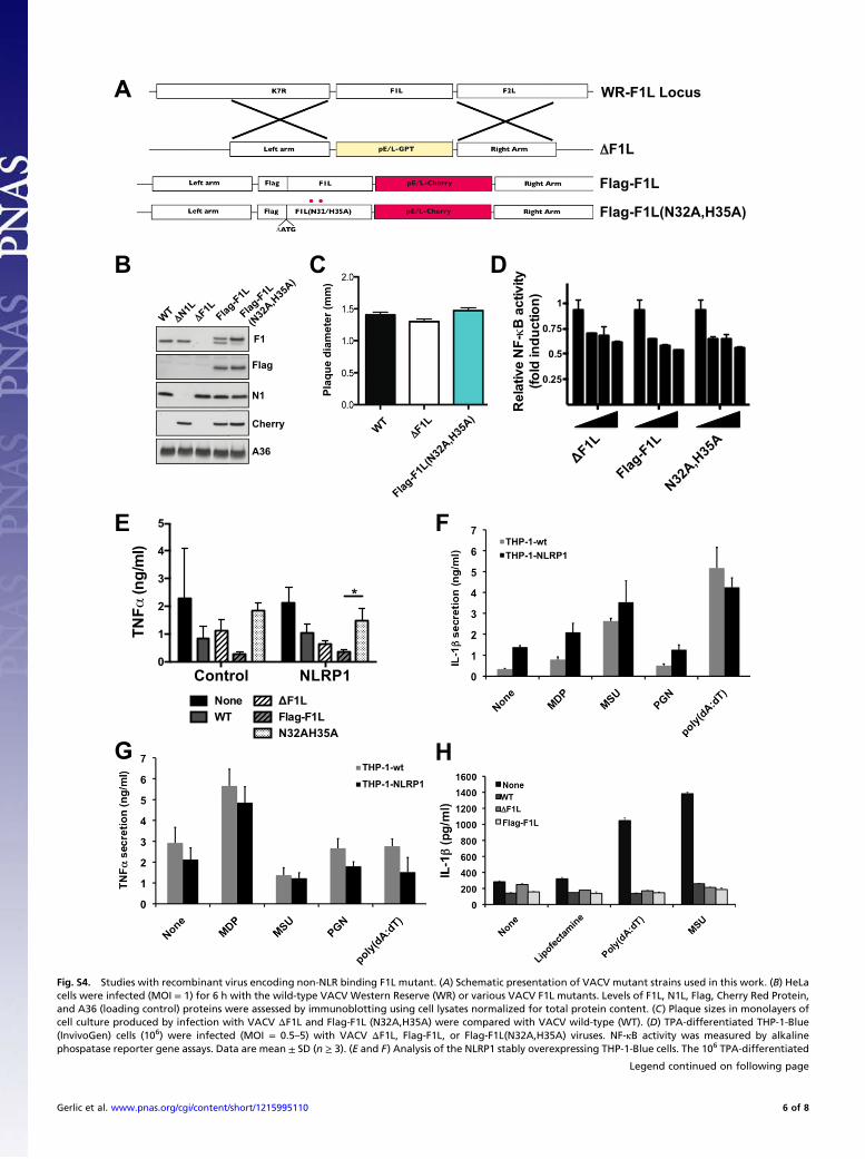
Fig. S4. Studies with recombinant virus encoding non-NLR binding F1L mutant. (A) Schematic presentation of VACV mutant strains used in this work. (B) HeLacells were infected (MOI = 1) for 6 h with the wild-type VACV Western Reserve (WR) or various VACV F1L mutants. Levels of F1L, N1L, Flag, Cherry Red Protein,and A36 (loading control) proteins were assessed by immunoblotting using cell lysates normalized for total protein content. (C) Plaque sizes in monolayers ofcell culture produced by infection with VACV ΔF1L and Flag-F1L (N32A,H35A) were compared with VACV wild-type (WT). (D) TPA-differentiated THP-1-Blue(InvivoGen) cells (106) were infected (MOI = 0.5–5) with VACV ΔF1L, Flag-F1L, or Flag-F1L(N32A,H35A) viruses. NF-κB activity was measured by alkalinephospatase reporter gene assays. Data are mean ± SD (n ≥ 3). (E and F) Analysis of the NLRP1 stably overexpressing THP-1-Blue cells. The 106 TPA-differentiated
Gerlic et al. www.pnas.org/cgi/content/short/1215995110 6 of 8
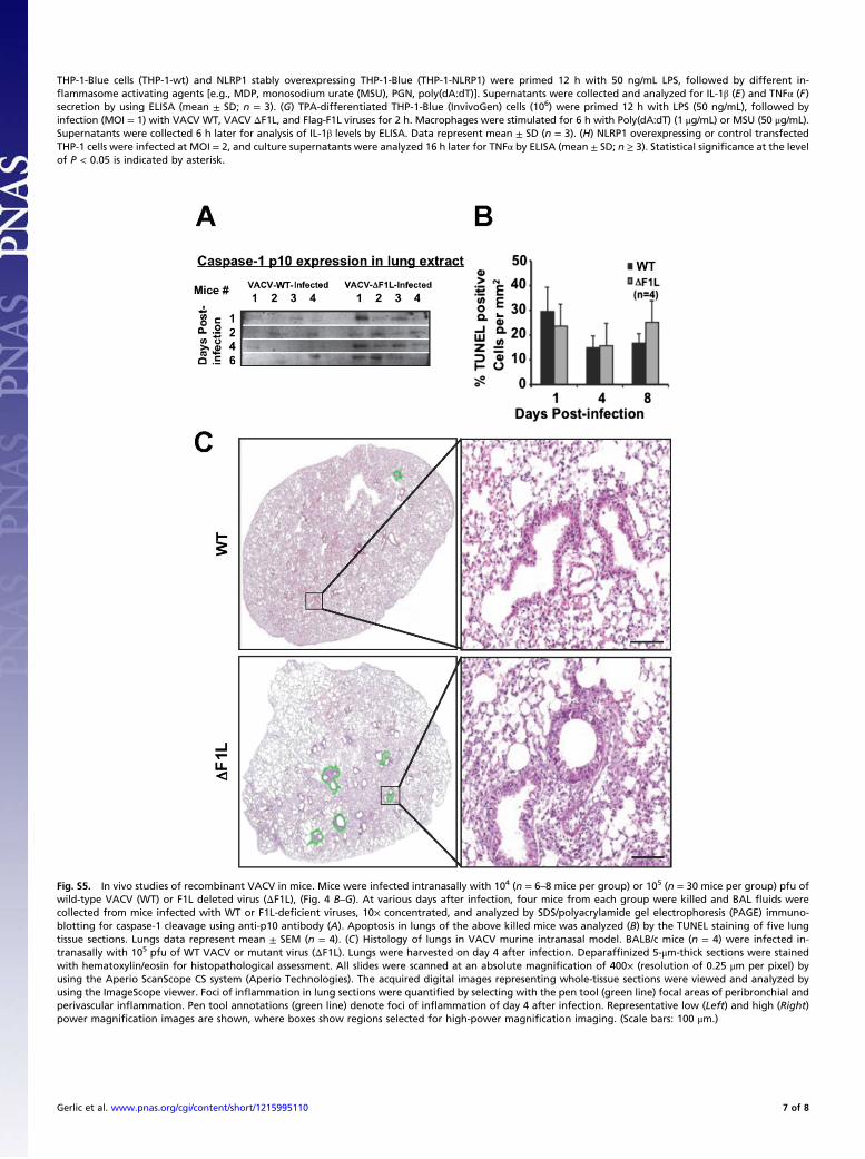
Fig. S5. In vivo studies of recombinant VACV in mice. Mice were infected intranasally with 104 (n = 6–8 mice per group) or 105 (n = 30 mice per group) pfu ofwild-type VACV (WT) or F1L deleted virus (ΔF1L), (Fig. 4 B–G). At various days after infection, four mice from each group were killed and BAL fluids werecollected from mice infected with WT or F1L-deficient viruses, 10× concentrated, and analyzed by SDS/polyacrylamide gel electrophoresis (PAGE) immuno-blotting for caspase-1 cleavage using anti-p10 antibody (A). Apoptosis in lungs of the above killed mice was analyzed (B) by the TUNEL staining of five lungtissue sections. Lungs data represent mean ± SEM (n = 4). (C) Histology of lungs in VACV murine intranasal model. BALB/c mice (n = 4) were infected in-tranasally with 105 pfu of WT VACV or mutant virus (ΔF1L). Lungs were harvested on day 4 after infection. Deparaffinized 5-μm-thick sections were stainedwith hematoxylin/eosin for histopathological assessment. All slides were scanned at an absolute magnification of 400× (resolution of 0.25 μm per pixel) byusing the Aperio ScanScope CS system (Aperio Technologies). The acquired digital images representing whole-tissue sections were viewed and analyzed byusing the ImageScope viewer. Foci of inflammation in lung sections were quantified by selecting with the pen tool (green line) focal areas of peribronchial andperivascular inflammation. Pen tool annotations (green line) denote foci of inflammation of day 4 after infection. Representative low (Left) and high (Right)power magnification images are shown, where boxes show regions selected for high-power magnification imaging. (Scale bars: 100 μm.)
THP-1-Blue cells (THP-1-wt) and NLRP1 stably overexpressing THP-1-Blue (THP-1-NLRP1) were primed 12 h with 50 ng/mL LPS, followed by different in-flammasome activating agents [e.g., MDP, monosodium urate (MSU), PGN, poly(dA:dT)]. Supernatants were collected and analyzed for IL-1β (E) and TNFα (F)secretion by using ELISA (mean ± SD; n = 3). (G) TPA-differentiated THP-1-Blue (InvivoGen) cells (106) were primed 12 h with LPS (50 ng/mL), followed byinfection (MOI = 1) with VACV WT, VACV ΔF1L, and Flag-F1L viruses for 2 h. Macrophages were stimulated for 6 h with Poly(dA:dT) (1 μg/mL) or MSU (50 μg/mL).Supernatants were collected 6 h later for analysis of IL-1β levels by ELISA. Data represent mean ± SD (n = 3). (H) NLRP1 overexpressing or control transfectedTHP-1 cells were infected at MOI = 2, and culture supernatants were analyzed 16 h later for TNFα by ELISA (mean ± SD; n ≥ 3). Statistical significance at the levelof P < 0.05 is indicated by asterisk.
Gerlic et al. www.pnas.org/cgi/content/short/1215995110 7 of 8
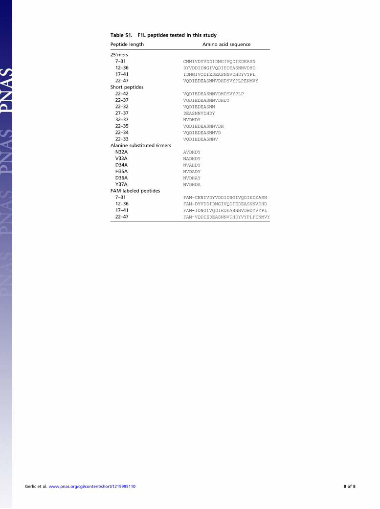
Table S1. F1L peptides tested in this study
Peptide length Amino acid sequence
25′mers7–31 CNNIVDYVDDIDNGIVQDIEDEASN
12–36 DYVDDIDNGIVQDIEDEASNNVDHD
17–41 IDNGIVQDIEDEASNNVDHDYVYPL
22–47 VQDIEDEASNNVDHDYVYPLPENMVY
Short peptides22–42 VQDIEDEASNNVDHDYVYPLP
22–37 VQDIEDEASNNVDHDY
22–32 VQDIEDEASNN
27–37 DEASNNVDHDY
32–37 NVDHDY
22–35 VQDIEDEASNNVDH
22–34 VQDIEDEASNNVD
22–33 VQDIEDEASNNV
Alanine substituted 6′mersN32A AVDHDY
V33A NADHDY
D34A NVAHDY
H35A NVDADY
D36A NVDHAY
Y37A NVDHDA
FAM labeled peptides7–31 FAM-CNNIVDYVDDIDNGIVQDIEDEASN
12–36 FAM-DYVDDIDNGIVQDIEDEASNNVDHD
17–41 FAM-IDNGIVQDIEDEASNNVDHDYVYPL
22–47 FAM-VQDIEDEASNNVDHDYVYPLPENMVY
Gerlic et al. www.pnas.org/cgi/content/short/1215995110 8 of 8