A RecA Mutant, RecA730, Suppresses the
12
A RecA Mutant, RecA 730 , Suppresses the Recombination Deficiency of the RecBC 1004 D–χ* Interaction in Vitro and in Vivo Naofumi Handa and Stephen C. Kowalczykowski⁎ Sections of Microbiology and of Molecular and Cellular Biology , Center for Genetics and Development, University of California, Davis, CA 95616, USA In Escherichia coli, homologous recombination initiated at double-stranded DNA breaks requires the RecBCD enzyme, a multifunctional heterotrimeric complex that possesses processive helicase and exonuclease activities. Upon encountering the DNA regulatory sequence, χ, the enzymatic properties of RecBCD enzyme are altered. Its helicase activity is reduced, the 3′→5′ nuclease activity is attenuated, the 5′→3′ nuclease activity is up-regulated, and it manifests an ability to load RecA protein onto single-stranded DNA. The net result of these changes is the production of a highly recombinogenic structure known as the presynaptic filament. Previously, we found that the recC1004 mutation alters χ-recognition so that this mutant enzyme recognizes an altered χ sequence, χ*, which comprises seven of the original nucleotides in χ, plus four novel nucleotides. Although some consequences of this mutant enzyme–mutant χ interaction could be detected in vivo and in vitro, stimulation of recombination in vivo could not. To resolve this seemingly contradictory observation, we examined the behavior of a RecA mutant, RecA 730 , that displays enhanced biochemical activity in vitro and possesses suppressor function in vivo. We show that the recombination deficiency of the RecBC 1004 D–χ* interaction can be overcome by the enhanced ability of RecA 730 to assemble on single-stranded DNA in vitro and in vivo. These data are consistent with findings showing that the loading of RecA protein by RecBCD is necessary in vivo, and they show that RecA proteins with enhanced single-stranded DNA-binding capacity can partially bypass the need for RecBCD-mediated loading. © 2006 Elsevier Ltd. All rights reserved. *Corresponding author Keywords: RecA; RecBCD; recombination hotspot; DNA repair; RecA-loading Introduction RecBCD enzyme is a helicase/nuclease that functions at the initiation step of recombinational DNA repair in Escherichia coli. 1,2 From a nearly blunt double-stranded DNA (dsDNA) end, RecBCD enzyme unwinds and degrades the dsDNA, pow- ered by the two motor subunits RecB and RecD. 3 When the enzyme encounters the recombination hotspot sequence, χ (5′-GCTGGTGG), from the 3′ side on one of the DNA strands, 4 its 5′ to 3′ nuclease activity is up-regulated and its 3′ to 5′ nuclease activity is down-regulated. 5–7 The consequence of these changes is the production of single-stranded DNA (ssDNA) with the χ sequence at the 3′ terminus; this product is referred to as the χ- containing ssDNA. In addition, RecBCD enzyme pauses at χ for approximately 5 s, and then resumes unwinding at a rate that is about twofold slower than the rate before χ recognition. 8,9 Perhaps the most biologically important property of the χ-modified RecBCD enzyme is its ability to load RecA onto ssDNA. 10,11 Although the molecular details of this regulatory process are not fully understood, the changes elicited by χ-recognition are not the result of RecD-ejection at χ because, even though the RecD subunit regulates RecBCD en- zyme activities, 12–14 recent single-molecule studies Present address: N. Handa, Department of Medical Genome Sciences, Graduate School of Frontier Science and Institute of Medical Science, University of Tokyo, Shirokanedai, Minato-ku, Tokyo 108-8639 Japan. Abbreviations used: ds, double-stranded; ss, single-stranded. E-mail address of the corresponding author: [email protected] doi:10.1016/j.jmb.2006.10.090 J. Mol. Biol. (2007) 365, 1314–1325 0022-2836/$ - see front matter © 2006 Elsevier Ltd. All rights reserved.
Transcript of A RecA Mutant, RecA730, Suppresses the
doi:10.1016/j.jmb.2006.10.090 J. Mol. Biol. (2007) 365, 1314–1325
A RecA Mutant, RecA730, Suppresses theRecombination Deficiency of the RecBC1004D–χ*Interaction in Vitro and in Vivo
Naofumi Handa and Stephen C. Kowalczykowski⁎
Sections of Microbiology and ofMolecular and Cellular Biology,Center for Genetics andDevelopment, University ofCalifornia, Davis, CA 95616,USA
Present address: N. Handa, DepaGenome Sciences, Graduate School oInstitute of Medical Science, UniverShirokanedai, Minato-ku, Tokyo 108Abbreviations used: ds, double-st
single-stranded.E-mail address of the correspondi
0022-2836/$ - see front matter © 2006 E
In Escherichia coli, homologous recombination initiated at double-strandedDNA breaks requires the RecBCD enzyme, a multifunctional heterotrimericcomplex that possesses processive helicase and exonuclease activities. Uponencountering the DNA regulatory sequence, χ, the enzymatic properties ofRecBCD enzyme are altered. Its helicase activity is reduced, the 3′→5′nuclease activity is attenuated, the 5′→3′ nuclease activity is up-regulated,and it manifests an ability to load RecA protein onto single-stranded DNA.The net result of these changes is the production of a highly recombinogenicstructure known as the presynaptic filament. Previously, we found that therecC1004 mutation alters χ-recognition so that this mutant enzymerecognizes an altered χ sequence, χ*, which comprises seven of the originalnucleotides in χ, plus four novel nucleotides. Although some consequencesof this mutant enzyme–mutantχ interaction could be detected in vivo and invitro, stimulation of recombination in vivo could not. To resolve thisseemingly contradictory observation, we examined the behavior of a RecAmutant, RecA730, that displays enhanced biochemical activity in vitro andpossesses suppressor function in vivo. We show that the recombinationdeficiency of the RecBC1004D–χ* interaction can be overcome by theenhanced ability of RecA730 to assemble on single-stranded DNA in vitroand in vivo. These data are consistent with findings showing that the loadingof RecA protein by RecBCD is necessary in vivo, and they show that RecAproteins with enhanced single-stranded DNA-binding capacity canpartially bypass the need for RecBCD-mediated loading.
© 2006 Elsevier Ltd. All rights reserved.
*Corresponding author
Keywords: RecA; RecBCD; recombination hotspot; DNA repair; RecA-loadingIntroduction
RecBCD enzyme is a helicase/nuclease thatfunctions at the initiation step of recombinationalDNA repair in Escherichia coli.1,2 From a nearly bluntdouble-stranded DNA (dsDNA) end, RecBCDenzyme unwinds and degrades the dsDNA, pow-ered by the two motor subunits RecB and RecD.3
When the enzyme encounters the recombination
rtment of Medicalf Frontier Science andsity of Tokyo,-8639 Japan.randed; ss,
ng author:
lsevier Ltd. All rights reserve
hotspot sequence, χ (5′-GCTGGTGG), from the 3′side on one of the DNA strands,4 its 5′ to 3′ nucleaseactivity is up-regulated and its 3′ to 5′ nucleaseactivity is down-regulated.5–7 The consequence ofthese changes is the production of single-strandedDNA (ssDNA) with the χ sequence at the 3′terminus; this product is referred to as the χ-containing ssDNA. In addition, RecBCD enzymepauses at χ for approximately 5 s, and then resumesunwinding at a rate that is about twofold slowerthan the rate before χ recognition.8,9 Perhaps themost biologically important property of theχ-modified RecBCD enzyme is its ability to loadRecA onto ssDNA.10,11 Although the moleculardetails of this regulatory process are not fullyunderstood, the changes elicited by χ-recognitionare not the result of RecD-ejection at χ because, eventhough the RecD subunit regulates RecBCD en-zyme activities,12–14 recent single-molecule studies
d.
1315RecA730 Suppresses the Deficiencies of RecBC1004D
showed that the RecD subunit continues to translo-cate with the holoenzyme after χ recognition.9
Rather, single-molecule studies,8,9 the X-ray crystal-lographic structure of the enzyme–DNA complex,15
and analysis of a RecBCD enzyme homolog (theBacillus subtilis AddAB enzyme)16 suggest thatbinding of the χ sequence itself to RecBCD enzymeelicits the allosteric changes that underlie theenzymatic alterations.Many studies suggest that the degradative ca-
pacity of RecBCD enzyme is part of an antiviralsystem of E. coli that works in conjunction withthe restriction-modification system;17,18 the χsequence allows the RecBCD enzyme to recognizechromosomal DNA and to thereby protect it fromdegradation. Consequently, in recBC-deficient cells,DNA double-strand breaks accumulate.19–21 Theχ sequence is over-represented in the E. coli ge-nome,22 whereas it is absent from or under-represented in bacteriophages (N. H. and IchizoKobayashi, unpublished results). Therefore, the χsequence is an identification marker of the E. coligenome. Interestingly, this relationship between anuclease and its cognate attenuation sequence isconserved in many prokaryotes.23 In Salmonellatyphimurium, the same eight-nucleotide sequencefunctions as χ,24 whereas in Lactococcus lactis,Bacillus subtilis and Haemophilus influenzae, thecognate χ sequences are 5′-GCGCGTG, 5′-AGCGG, and 5′-GNTGGTGG, respectively.25–27
Mutant alleles of recBCD that recovered all theactivities of RecBCD enzyme, except for χ recogni-tion, were isolated originally as pseudo-revertants ofa mutant with a recC null phenotype. This class ofmutants, called RecC*, showed almost the samebasal level of recombination as wild-type, butthis recombination was not stimulated by χ.28,29
Sequencing revealed that, due to a frameshift, all ofthe analyzed alleles possess substitutions of aminoacid residues between positions 647 and 655 in therecC gene.30 It was found that a novel sequence,different from the canonical χ sequence, wasrecognized specifically by one of these mutants,recC1004, in vivo.29 Resembling the original χsequence in its genetic behavior,31 this novelsequence conferred an increased growth rate tored– gam– bacteriophage λ on the recC1004 strain.This novel sequence, which was named χ*, is the 11nt sequence, 5′-GCTGGTGCTCG,29 and comprisesthe first seven bases of the E. coli χ, plus four novelbases. As for the wild-type pair, the nuclease activityof the recC1004 mutant protein was attenuatedin vivo by the χ* sequence.29 Purified RecBC1004Denzyme possessed wild-type levels of dsDNAexonuclease and helicase activities, but displayedreduced recognition of wild-type χ in vitro.30
However, this mutant RecBCD enzyme recognizedthe mutant χ sequence more efficiently than wild-type χ, albeit with a lower efficiency than the wild-type enzyme recognized wild-type χ. Furthermore,χ*-dependent joint molecule formation was stimu-lated by the RecBC1004D enzyme, demonstratingthat RecA-loading activity was preserved but, again,
the yield was lower than for the fully wild-typereaction. Despite these biochemical results, stimulationof recombination in vivo using bacteriophage λ crossescouldnotbedetected for thisRecC1004–χ* interaction.29
This inconsistency was explained by a greater detec-tion sensitivity of the in vitro assays relative to the invivo assay.30
E. coli possesses a second recombination pathway,called the RecF pathway.2 In the absence of RecBCDfunction, the RecF pathway can be activated tofunction at dsDNA breaks by mutation of sbcB,which is in the gene encoding exonuclease I.32 RecFprotein works together with RecO and RecRproteins to load RecA protein onto ssDNA com-plexed with ssDNA-binding (SSB) protein at the 5′end of dsDNA gaps.33 Mutations in the recF genecan be suppressed by alleles of recA that displayenhanced functionality.34–36 These mutant RecAproteins (e.g. RecA803 and RecA730) nucleate ontossDNA more rapidly than wild-type RecAprotein.37,38 Biochemical analysis further establishedthat these enhanced RecA proteins displaced SSBprotein from ssDNA faster and more completelythan wild-type protein, and the resulting filamentswere kinetically more resistant to subsequentdisplacement by SSB protein.The relationship between RecA-loading in vitro
and recombination activity in vivo is relativelyunstudied. Toward this end, we examined the effectof the recA730 allele on the recombination phenotypeof cells defective in recBCD function. We found that,in response to χ* recognition, RecBC1004D enzymeloaded RecA730 protein onto ssDNA to formnucleoprotein filaments that were more stable thanthose formed by wild-type RecA protein. Further-more, we found that recA730 suppressed the recom-bination deficiency of the mutant RecBC1004D–χ*interaction in vivo, showing that an intrinsic in-creased propensity to nucleate on ssDNA and toform more stable nucleoprotein filaments can com-pensate for the lower yield of χ*-containing ssDNAproduced by RecBC1004D enzyme processing.
Results
RecA protein is loaded onto c*-containingssDNA by RecBC1004D enzyme
Previously, it was shown that the recC1004mutation changed the specificity of χ recognitionfrom the canonical sequence to a novel sequence,χ*.29 The purified RecBC1004D enzyme producedχ*-dependent joint molecules in response to χ*recognition, but with a lower yield than the wild-type reaction.30 However, the interaction betweenRecBC1004D enzyme and χ* did not result in anincreased frequency of recombination as mea-sured by bacteriophage λ crosses,29 suggestingthat the in vitro assay was more sensitive than thein vivo assay. To analyze the mutant interactionin more detail, RecA-loading assays were per-formed as described,10 but with one significant
1316 RecA730 Suppresses the Deficiencies of RecBC1004D
difference: ATPγS was not added to stabilize theRecA nucleoprotein filaments. By omitting theATPγS, the resulting ATP-RecA nucleoprotein fila-ments were kinetically less stable, permitting experi-mental distinction between wild-type and RecA730
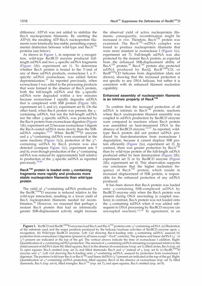
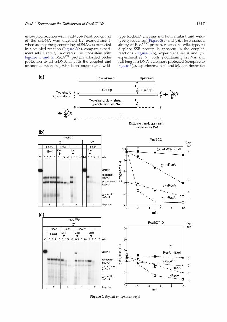
proteins (see below).As shown in Figure 1, in response to χ-recogni-
tion, wild-type RecBCD enzyme produced full-length ssDNA and two χ-specific ssDNA fragments(Figure 1(b), experiment set 1). To determinewhether RecA protein is bound to the 3′-end ofany of these ssDNA products, exonuclease I, a 3′-specific ssDNA exonuclease, was added beforedeproteinization.10 As reported previously, whenexonuclease I was added to the processing productsthat were formed in the absence of RecA protein,both the full-length ssDNA and the χ-specificssDNA were degraded within a few minutesbecause exonuclease I rapidly degrades ssDNAthat is complexed with SSB protein (Figure 1(b),experiment set 3, and (c), experiment set 8). On theother hand, when RecA protein was present, the χ-containing ssDNA, but not the full-length ssDNAnor the other χ-specific ssDNA, was protected bythe RecA protein from exonuclease digestion (Figure1(b), experiment set 2) because exonuclease I digeststhe RecA-coated ssDNA more slowly than the SSB-ssDNA complex.10,39 When RecBC1004D enzymeand a χ*-containing dsDNA were examined in thesame reactions (Figure 1(c)), protection of the χ*-containing ssDNA by RecA protein was alsodetected (compare Figure 1(c), experiment sets 5and 6), even though production of the χ*-containingssDNAwas reduced by approximately half relativeto production of the χ-specific ssDNA as reportedpreviously.30,40
RecA730 protein is loaded onto c-containingfragments more rapidly and produces morestable nucleoprotein filaments than wild-typeprotein
The yield of χ*-containing ssDNA produced bythe RecBC1004D enzyme is reduced relative to thewild-type interaction, resulting in a lower yield ofRecA nucleoprotein filaments needed for recom-bination.30 However, we reasoned that perhaps amutant RecA protein that had an intrinsicallygreater SSB-displacement activity might increase
Figure 1. RecBCDandRecBC1004D enzymes loadRecA andof the substrate used and the major products produced by threcognition. (b) Wild-type RecBCD enzyme. Left: Gel showiprotection from exonuclease I digestion (present in all lanes exceχ*) present are indicated at the top of the gel. The vertical aQuantification ofχ-containing ssDNAprotection. The amount oinitial amount of dsDNA (laneM): filled squares, RecA in the abs2); open squares: RecA omitted (exp. set 3), and filled diamonenzyme and χ*. Left: Gel showing RecA-loading onto χ*-condigestion. The proteins (wild-type RecA or RecA730) and linear dQuantification of χ*-containing ssDNA protection: filled squadiamonds, RecA (exp. set 6), filled triangles: RecA730 (exp. set 7
the observed yield of active nucleoprotein fila-ments; consequently, recombination might beincreased in vivo. Therefore, RecA730 protein wasexamined. The RecA730–ssDNA complex wasfound to produce nucleoprotein filaments thatwere more resistant to exonuclease I (Figure 1(c),experiment set 7). Full-length ssDNA was alsoprotected by the mutant RecA protein, as expectedfrom the enhanced SSB-displacement ability ofRecA730 protein.38 RecA730 protein also protectedssDNA produced by RecQ, RecB1080CD, orRecB2109CD helicases from degradation (data notshown), showing that the increased protection isnot specific to any DNA helicase, but rather it isconsistent with its enhanced filament nucleationcapability.
Enhanced assembly of nucleoprotein filamentsis an intrinsic property of RecA730
To confirm that the increased protection of allssDNA is intrinsic to RecA730 protein, reactionswhere RecA nucleoprotein filament assembly wascoupled to ssDNA production by RecBCD enzymewere compared to reactions where RecA proteinwas assembled on heat-denatured DNA in theabsence of RecBCD enzyme.10,39 As reported, wild-type RecA protein did not protect ssDNA pro-duced by heat-denaturation from exonuclease Idegradation, because it cannot displace SSB pro-tein efficiently (Figure 2(a), experiment set 2). Incontrast, there was greater protection by RecA730
than by wild-type protein of the full-length ssDNAproduced either by heat denaturation (Figure 2(b),experiment set 5) or by RecBCD enzyme (Figure2(b), experiment set 4). This observation supportsour conclusion that the higher nucleation fre-quency of RecA730 protein, which results inincreased displacement of SSB protein, is respon-sible for the enhanced protection of any ssDNAproduced.It has been shown that RecA protein was loaded
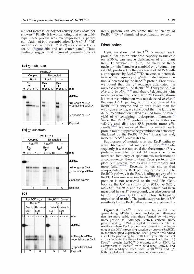
onto χ-containing SSB-complexed ssDNA byRecBCD enzyme only when the RecA protein waspresent during DNA unwinding (a coupled reac-tion); in contrast, RecA protein was not loaded ontothe χ-containing ssDNA when it was added sub-sequent to DNA processing by RecBCD enzyme (anuncoupled reaction).10,39 In agreement, in an
RecA730 proteins ontoχ*-containing ssDNA. (a) Illustratione helicase/nuclease activities of RecBCD enzyme upon χng RecA-loading onto χ-containing ssDNA, assayed bypt “-ExoI” controls). The proteins and linear dsDNA (χ+ orrrows indicate the time of exonuclease I addition. Right:fχ-containing ssDNAremaining is expressed relative to theence of exonuclease I (exp. set 1); filled circles, RecA (exp. setds: RecA and χ* instead of χ (exp. set 4). (c) RecBC1004Dtaining ssDNA, assayed by protection from exonuclease IsDNA (χ*) present are indicated at the top of the gel. Right:res, RecA in the absence of exonuclease (exp. set 5); filled); and open squares, RecA omitted (exp. set 8).
1317RecA730 Suppresses the Deficiencies of RecBC1004D
uncoupled reaction with wild-type RecA protein, allof the ssDNA was digested by exonuclease I,whereas only theχ-containing ssDNAwas protectedin a coupled reaction (Figure 3(a), compare experi-ment sets 1 and 2). In contrast, but consistent withFigures 1 and 2, RecA730 protein afforded betterprotection to all ssDNA in both the coupled anduncoupled reactions, with both mutant and wild-
Figure 1 (legend o
type RecBCD enzyme and both mutant and wild-typeχ sequences (Figure 3(b) and (c)). The enhancedability of RecA730 protein, relative to wild-type, todisplace SSB protein is apparent in the coupledreactions (Figure 3(b), experiment set 4 and (c),experiment set 7): both χ-containing ssDNA andfull-length ssDNAwere more protected (compare toFigure 3(a), experimental set 1 and (c), experiment set
n opposite page)
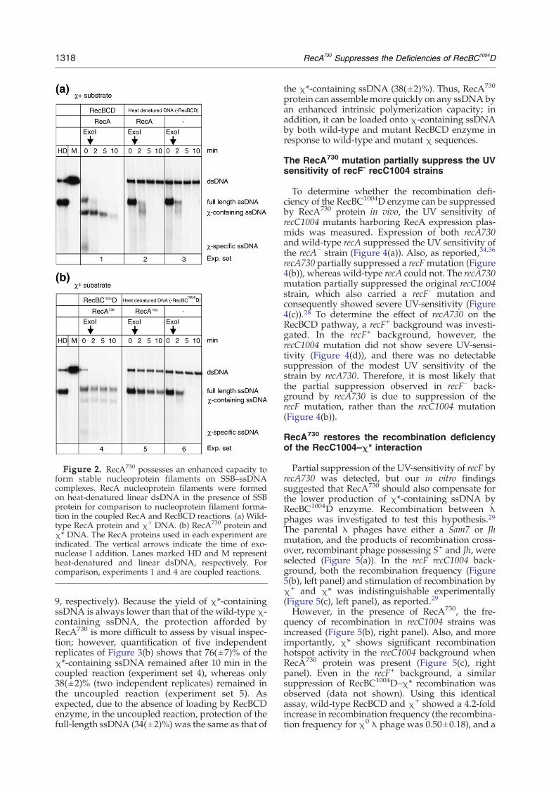
Figure 2. RecA730 possesses an enhanced capacity toform stable nucleoprotein filaments on SSB–ssDNAcomplexes. RecA nucleoprotein filaments were formedon heat-denatured linear dsDNA in the presence of SSBprotein for comparison to nucleoprotein filament forma-tion in the coupled RecA and RecBCD reactions. (a) Wild-type RecA protein and χ+ DNA. (b) RecA730 protein andχ* DNA. The RecA proteins used in each experiment areindicated. The vertical arrows indicate the time of exo-nuclease I addition. Lanes marked HD and M representheat-denatured and linear dsDNA, respectively. Forcomparison, experiments 1 and 4 are coupled reactions.
1318 RecA730 Suppresses the Deficiencies of RecBC1004D
9, respectively). Because the yield of χ*-containingssDNA is always lower than that of the wild-type χ-containing ssDNA, the protection afforded byRecA730 is more difficult to assess by visual inspec-tion; however, quantification of five independentreplicates of Figure 3(b) shows that 76(±7)% of theχ*-containing ssDNA remained after 10 min in thecoupled reaction (experiment set 4), whereas only38(±2)% (two independent replicates) remained inthe uncoupled reaction (experiment set 5). Asexpected, due to the absence of loading by RecBCDenzyme, in the uncoupled reaction, protection of thefull-length ssDNA (34(±2)%) was the same as that of
the χ*-containing ssDNA (38(±2)%). Thus, RecA730
protein can assemblemore quickly on any ssDNA byan enhanced intrinsic polymerization capacity; inaddition, it can be loaded onto χ-containing ssDNAby both wild-type and mutant RecBCD enzyme inresponse to wild-type and mutant χ sequences.
The RecA730 mutation partially suppress the UVsensitivity of recF- recC1004 strains
To determine whether the recombination defi-ciency of the RecBC1004D enzyme can be suppressedby RecA730 protein in vivo, the UV sensitivity ofrecC1004 mutants harboring RecA expression plas-mids was measured. Expression of both recA730and wild-type recA suppressed the UV sensitivity ofthe recA− strain (Figure 4(a)). Also, as reported,34,36
recA730 partially suppressed a recFmutation (Figure4(b)), whereas wild-type recA could not. The recA730mutation partially suppressed the original recC1004strain, which also carried a recF- mutation andconsequently showed severe UV-sensitivity (Figure4(c)).28 To determine the effect of recA730 on theRecBCD pathway, a recF+ background was investi-gated. In the recF+ background, however, therecC1004 mutation did not show severe UV-sensi-tivity (Figure 4(d)), and there was no detectablesuppression of the modest UV sensitivity of thestrain by recA730. Therefore, it is most likely thatthe partial suppression observed in recF– back-ground by recA730 is due to suppression of therecF mutation, rather than the recC1004 mutation(Figure 4(b)).
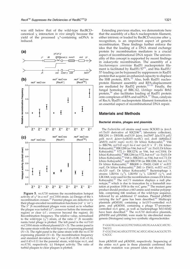
RecA730 restores the recombination deficiencyof the RecC1004–c* interaction
Partial suppression of the UV-sensitivity of recF byrecA730 was detected, but our in vitro findingssuggested that RecA730 should also compensate forthe lower production of χ*-containing ssDNA byRecBC1004D enzyme. Recombination between λphages was investigated to test this hypothesis.29The parental λ phages have either a Sam7 or Jhmutation, and the products of recombination cross-over, recombinant phage possessing S+ and Jh, wereselected (Figure 5(a)). In the recF recC1004 back-ground, both the recombination frequency (Figure5(b), left panel) and stimulation of recombination byχ+ and χ* was indistinguishable experimentally(Figure 5(c), left panel), as reported.29
However, in the presence of RecA730, the fre-quency of recombination in recC1004 strains wasincreased (Figure 5(b), right panel). Also, and moreimportantly, χ* shows significant recombinationhotspot activity in the recC1004 background whenRecA730 protein was present (Figure 5(c), rightpanel). Even in the recF+ background, a similarsuppression of RecBC1004D–χ* recombination wasobserved (data not shown). Using this identicalassay, wild-type RecBCD and χ+ showed a 4.2-foldincrease in recombination frequency (the recombina-tion frequency for χ0 λ phage was 0.50±0.18), and a
1319RecA730 Suppresses the Deficiencies of RecBC1004D
6.5-fold increase for hotspot activity assay (data notshown).29 Finally, it is worth noting that when wild-type RecA protein was over-expressed, a partialstimulation of both recombination (1.46(±0.13)-fold)and hotspot activity (1.87±0.22) was observed onlyfor χ* (Figure 5(b) and (c), center panel). Thesefindings suggest that increased concentrations of
RecA protein can overcome the deficiency ofRecBC1004D–χ* stimulated recombination in vivo.
Discussion
Here, we show that RecA730, a mutant RecAprotein that has an enhanced capacity to nucleateon ssDNA, can rescue deficiencies of a mutantRecBCD enzyme. In vitro, the yield of RecAnucleoprotein filaments assembled on χ*-containingssDNA, produced by the processing of dsDNAwitha χ* sequence by RecBC1004D enzyme, is increased.In vivo, the frequency of χ*-stimulated recombina-tion is increased by the RecA730 protein. Previously,we found that the χ* sequence attenuated thenuclease activity of the RecBC1004D enzyme both invivo and in vitro,29,30 and that χ*-dependent jointmolecules were produced in vitro.30 However, stimu-lation of recombination was not detected in vivo.29
Because DNA pairing in vitro coordinated byRecBC1004D enzyme and χ* was lower than forwild-type enzyme, we concluded that the failure todetect recombination in vivo resulted from the loweryield of χ*-containing nucleoprotein filaments.30
Since the RecA730 protein nucleates faster onssDNA and displaces SSB protein more effi-ciently,38,41 we reasoned that this mutant RecAproteinmight suppress the recombination deficiencydisplayed by the RecBC1004D–χ* interaction and,indeed, RecA730 protein did so.Suppressors of mutations in the RecF pathway
were discovered that mapped in recA.34–36 Sub-sequently, it was established that these mutant RecAproteins assembled on ssDNA faster due to anincreased frequency of spontaneous nucleation; asa consequence, these mutant RecA proteins dis-place SSB protein from ssDNA more rapidly andmore fully.38,42–47 Recently, it was shown thatcomponents of the RecF pathway can contribute toRecBCD pathway if the RecA-loading activity of theRecBCD enzyme was inactivated.11,48–50 This sup-pression is not restricted to the recB1080 allele,because the UV sensitivity of recB2154, recB2155,recC2145, recC1002, and recC1004, which had beenmeasured in a recF– background, was also correctedby recF+ (Figure 4; N.H. and Ichizo Kobayashi,unpublished results). The partial suppression of UVsensitivity by the RecF pathway can be explained by
Figure 3. RecA730 protein can be loaded ontoχ-containing ssDNA to form nucleoprotein filamentsthat are more stable than those formed by wild-typeRecA protein. (a) Wild-type RecBCD enzyme, RecAprotein and χ+ DNA. Coupled experiments refer toreactions where RecA protein was present at the begin-ning of the DNA processing reaction by enzyme RecBCD.In the uncoupled experiment, RecA protein was addedafter DNA processing by RecBCD enzyme. The verticalarrows indicate the time of exonuclease I addition. (b)RecA730 protein, RecBC1004D enzyme, and χ* DNA. (c)Comparison of RecA730 with wild-type RecBCD andχ versus wild-type RecA with RecBC1004D and χ*;both coupled and uncoupled reactions are shown.
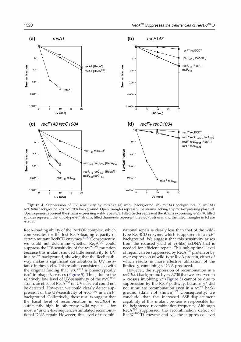
Figure 4. Suppression of UV sensitivity by recA730. (a) recA1 background. (b) recF143 background. (c) recF143recC1004 background. (d) recC1004 background. Open triangles represent the strains lacking any recA-expressing plasmid.Open squares represent the strains expressing wild-type recA. Filled circles represent the strains expressing recA730; filledsquares represent the wild-type rec+ strains; filled diamonds represent the recC73 strains; and the filled triangles in (c) arerecF143.
1320 RecA730 Suppresses the Deficiencies of RecBC1004D
RecA-loading ability of the RecFOR complex, whichcompensates for the lost RecA-loading capacity ofcertain mutant RecBCD enzymes.11,50 Consequently,we could not determine whether RecA730 couldsuppress the UV-sensitivity of the recC1004 mutationbecause this mutant showed little sensitivity to UVin a recF+ background, showing that the RecF path-way makes a significant contribution to UV resis-tance in these cells. This result is consistent also withthe original finding that recC1004 is phenotypicallyRec+ in phage λ crosses (Figure 5). Thus, due to therelatively low level of UV-sensitivity of the recC1004
strain, an effect of RecA730 on UV survival could notbe detected. However, we could clearly detect sup-pression of the UV-sensitivity of recC1004 in a recF–
background. Collectively, these results suggest thatthe basal level of recombination in recC1004 issufficiently high in otherwise wild-type cells formost χ* and χ-like sequence-stimulated recombina-tional DNA repair. However, this level of recombi-
national repair is clearly less than that of the wild-type RecBCD enzyme, which is apparent in a recF–
background. We suggest that this sensitivity arisesfrom the reduced yield of χ(-like) ssDNA that isneeded for efficient repair. This sub-optimal levelof repair can be suppressed by RecA730 protein or byover-expression of wild-type RecA protein, either ofwhich results in more effective utilization of thelimited χ-containing ssDNA produced.However, the suppression of recombination in a
recC1004 background by recA730 thatwe observed inλ crosses involving χ* (Figure 5) cannot be due tosuppression by the RecF pathway, because χ* didnot stimulate recombination even in a recF+ back-ground (data not shown).29 Consequently, weconclude that the increased SSB-displacementcapability of this mutant protein is responsible forthe heightened recombination frequency. AlthoughRecA730 suppressed the recombination defect ofRecBC1004D enzyme and χ*, the suppressed level
1321RecA730 Suppresses the Deficiencies of RecBC1004D
was still below that of the wild-type RecBCD–canonical χ interaction in vivo simply because theyield of the processed χ*-containing ssDNA isreduced.
Figure 5. recA730 restores the recombination hotspotactivity of χ* in a recF- recC1004 strain. (a) Design of the λrecombination crosses.29 Parental phages are defective fortheir phage-encoded recombination functions (red– γ– int–).The S+ Jh recombinant phages were scored as to whetherthe plaque was turbid (cI+; crossover before the immunityregion) or clear (cI–; crossover beyond the region). (b)Recombination frequency. The relative value, normalizedto the wild-type (χ0) strain, of the ratio S+ Jh recombi-nants/total phage is plotted. The left panel is the recF143recC1004 strain without plasmid (N=3). The center panel isthe same strainwith thewild-type recA expressing plasmid(N=3). The right panel is the same strain with the recA730expressing plasmid (N=4). The recombination frequencyand standard deviation for χ0 was 0.43±0.08, 0.41±0.14,and 0.43±0.11 for the parental strain, wild-type recA, andrecA730, respectively. (c) Hotspot activity. The ratio ofturbid plaques to clear plaques is plotted.
Extending previous studies, we demonstrate herethat the assembly of a RecA nucleoprotein filament,either intrinsic or loaded by RecBCD enzyme after χrecognition, is an important aspect of geneticrecombination. These findings further enforce theidea that the loading of a DNA strand exchangeprotein by recombination mediators is a crucialaspect of recombinational DNA repair. The univers-ality of this concept is supported by recent findingsin eukaryotic recombination. The assembly of aSaccharomyces cerevisiae Rad51 nucleoprotein fila-ment is facilitated by Rad55/5751, and this Rad55/57-loading can be bypassed by suppressors in Rad51protein that acquire an enhanced capacity to displacethe SSB protein, RPA.52 Also, both Rad51 nucleo-protein filament assembly and RPA-displacementare mediated by Rad52 protein.53–55 Finally, thefungal homolog of BRCA2, Ustilago maydis Brh2protein,56 also facilitates loading of Rad51 proteinonto complexes of RPA and ssDNA.57 Thus, catalysisof RecA/Rad51 nucleoprotein filament formation isan essential aspect of recombinational DNA repair.
Materials and Methods
Bacterial strains, phages and plasmids
The Escherichia coli strains used were: SCK303 (a ΔrecAsrl::Tn10 derivative of KK218658; laboratory collection),BIK1291 (= DH10B; araD139 Δ(ara, leu)7697 ΔlacX74 galUgalK mcrA Δ(mrr-hsdRMS-mcrBC) rpsL deoR (ϕ80dlacZΔM15) endA1 nupG recA1; Dr Ichizo Kobayashi),59 V66(= BIK796; recF143 argA his-4 met rpsL31 λ− F−; Dr. IchizoKobayashi),28 BIK1288 (asV66, but recF+ zic::Tn10; Dr IchizoKobayashi),29 V72 (= BIK1274; as V66, but recC1004; DrIchizoKobayashi),28 BIK1284 (as V72, but recF+ zic::Tn10; DrIchizo Kobayashi),29 V68 (= BIK2411; as V66, but recC73; DrIchizo Kobayashi),28 and BIK3738 (as BIK1288, but recC73;Dr Ichizo Kobayashi),29 BIK808 (= FS620; C600 λr recB21supE; Dr Ichizo Kobayashi),29 JM1 (= FS611; recB21 recC22sbcA20 supF; Dr Ichizo Kobayashi).29 Bacteriophage λstrains LIK916 (χ0), LIK950 (χ+), LIK907 (χ*), andLIK1068,were used for the recombination crosses (Dr IchizoKobayashi).29 The recC73 mutation displays a null phe-notype,28 which is due to truncation by a frameshift mu-tation at position 1938 in the recC gene.30 The mutant geneproduct should produce a 663 amino acid residue polypep-tide, comprising 646 residues of the wild-type sequence,followed by an additional 17 residues. Plasmid pMS421carrying the lacIq gene has been described.60 Multicopyplasmids pKM100, containing a lacUV5-controlled recAgene, and pKM300, containing a phage T7-promoter-controlled recA gene, as well as bacteriophage M13-KM2have been described.61 The recA730 derivative plasmids,pSNH50 and pSNH60, were made by site-directed muta-genesis (Stratagene) using two synthetic oligonucleotides:
5′-GGGTGAAGACCGTTCTATGGATGTGAAAACCATCTC-TACCG5′-CGGTAGAGATGGTTTTCACATCCATAGAACGGTCTT-CACCC
from pKM100 and pKM300, respectively. Sequencing ofthe entire recA gene in these plasmids confirmed thatno other mutation was present (data not shown).
1322 RecA730 Suppresses the Deficiencies of RecBC1004D
Media
E. coli cells were grown in L broth (1.0% (w/v) Bacto-tryptone, 0.5% (w/v) yeast extract and 1.0% (w/v) NaCl),or Tryptone broth (1.0% Bacto-tryptone, 0.5% NaCl)supplemented with 0.2% (w/v) maltose, 10 mM MgSO4,and 10 μg/ml of vitamin B1. Antibiotics were added at thefollowing concentrations when required: ampicillin (amp)100 μg/ml, chloramphenicol (cam) 25 μg/ml, tetracycline(tet) 10 μg/ml, and spectinomycin (spc) 30 μg/ml.
†http://www.graphpad.com
Proteins and reagents
RecBCD, RecBC1004D, SSB, and wild-type RecA pro-teins were purified as described.40,62–64 RecA730 proteinwas purified as described.65 Plasmid pSNH60, whichcarries the recA730 gene downstream of the T7 promoter,was introduced into SCK303. The transformant wascultured at 37 °C in L broth containing amp to mid-logphase (A600=0.3). RecA protein synthesis was induced byadding M13 phage (multiplicity of infection (moi)=10)that expressed T7 RNA polymerase, M13-KM2, togetherwith 1 mM isopropyl-β-d-thiogalactopyranoside (IPTG)for 3 h. Cells were harvested, re-suspended in ice-coldbuffer (50 mM Tris–HCl (pH 8), 5 mM EDTA, 25% (w/v)sucrose and 5 mM β-mercaptoethanol) and frozen at−80 °C. After thawing, cells were lysed with 0.5 mg/mlof lysozyme followed by sonication. The lysate wasmixed with 0.31% (w/v) Brij-58 and centrifuged at25,000 rpm in a JA-25 rotor (Beckman-Coulter) for45 min. The cleared lysate was diluted with the samebuffer to adjust the A260 to 160. Polyethyleneimine (pH 8)was added to 0.5% (w/v) to precipitate the nucleic acids,and then centrifuged at 10,000 rpm in a JA-25 rotor for20 min. The pellet was suspended in 50 mM Tris–HCl(pH 7.5), 1 mM EDTA, 0.3 M (NH4)2SO4 and 5 mM β-mercaptoethanol, stirred for 2 h, and then centrifuged.After the centrifugation, the supernatant was made 60%saturated by adding solid (NH4)2SO4 and centrifuged.The pellet was suspended in a dialysis buffer (20 mMTris–HCl (pH 7.5), 20 mM MgCl2, 5 mM β-mercaptoetha-nol, 10% (v/v) glycerol) and dialyzed overnight againstthe same buffer at 40 °C. The precipitate was dissolved in20 mM Tris–HCl (pH 7.5), 5 mM EDTA, 1 M NaCl, 5 mMβ-mercaptoethanol, 10% glycerol and centrifuged. Thesupernatant was loaded onto an S-300HR column(Pharmacia Biotech; 300 ml; flow-rate of 3 ml/min)equilibrated with TEM+1 M NaCl buffer (20 mM Tris–HCl (pH 7.5), 1 mM EDTA, 5 mM β-mercaptoethanol,10% glycerol and 1 M NaCl). RecA730 protein, identifiedby ssDNA-dependent ATPase activity and SDS-PAGEanalysis, eluted as a single peak. The pooled peakfractions from the S-300HR column were loaded ontoan EconoPack Q column (Bio-Rad; 5 ml; flow-rate of0.5 ml/min) after dialysis against TEM+50 mM NaCl; theRecA730 protein eluted in the flow-through. The pool wasdialyzed against 50 mM Tris–HCl (pH 8), 1 mM EDTA,5 mM β-mercaptoethanol, solid (NH4)2SO4 was added to70% saturation, and the solution was centrifuged. Thepellet was suspended in TEDS+100 mM NaCl (20 mMTris–HCl (pH 7.5), 0.1 mM EDTA, 0.1 mM dithiothreitol(DTT) and 100 mM NaCl) and dialyzed against the samebuffer. The solution was filtered through a 0.2 μm poresize filter, and then loaded onto a MonoQ HR10/10column (Pharmacia Biotech; 8 ml; flow-rate of 3.0 ml/min). RecA730 protein eluted at approximately 480 mMNaCl in a 360 ml linear gradient of 100 mM–1 M NaCl).The pooled protein was concentrated by dialysis against
storage buffer (50 mM Tris–HCl (pH 7.5), 0.1 mM EDTA,1 mM DTT, 150 mM NaCl, 10% glycerol). Theconcentration of RecA730 protein was determined spec-trophotometrically using an extinction coefficient of2.15×104 M−1 cm−1 at 278 nm.Exonuclease I, restriction endonucleases, and phage T4
polynucleotide kinase were products of New EnglandBiolabs. Shrimp alkaline phosphatase was purchased fromUnited States Biochemical Corp. Proteinase K waspurchased from Roche Molecular Biochemicals. ATP(Sigma) was dissolved in water at pH 7.5 and theconcentration was determined spectrophotometricallyusing an extinction coefficient of 1.54×10⁴ M−1 cm−1 at260 nm. All chemicals were reagent grade and solutionswere prepared with NanoPure water.
Substrate DNA for biochemical analysis
Plasmids pBR322, pNH92 and pNH9430 were purifiedusing a Qiagen kit and digested by the restriction endo-nuclease AvaI, following a reaction with shrimp alkalinephosphatase for removal of phosphoryl groups. After the5′-end of the linear dsDNA was labeled by phage T4polynucleotide kinase with 32P, unincorporated [γ-32P]ATP was removed by passage through a MicroSpin S-200HR column (Amersham Pharmacia Biotech).
Exonuclease I protection assay and quantification ofc-specific fragment production
The procedure was as described,10,39 except that ATPγSwas omitted. Reactions contained 25 mM Tris–acetate(pH 7.5), 8 mMmagnesium acetate, 5 mMATP, 1mMDTT,10 μM nucleotide linear dsDNA, 5 μM either wild-typeRecA or RecA730 protein, 4 μM SSB protein, and either0.1 nM RecBCD or 0.2 nM RecBC1004D enzyme. Reactions(37 °C) were started by addition of RecBCD enzyme. After3min, poly(dT) (50 μMnucleotide) was added to sequesterthe free RecA protein. After 2 min of further incubation, asample was taken (representing time zero) and thenexonuclease I was added to a final concentration of100 U/ml and incubated at room temperature for 10 min.Control reactions contained heat-denatured DNA insteadof dsDNA processed by RecBCD enzyme or, in the case ofthe uncoupled reactions, RecA protein was added 3 minafter addition of RecBCD enzyme. Samples were added tostop solution (40 mM EDTA, 0.8% (w/v) SDS, 1.5 μg/μl ofproteinase K and 0.04% bromophenol blue) at theindicated times after the addition of exonuclease I, andwere analyzed by 1.0% (w/v) agarose gel electrophoresis.Production of χ-specific fragments was quantified byusing aMolecular Dynamics STORM 870 PhosphorImagerand ImageQuant software (Molecular Dynamics). Thepercentages were calculated relative to the initial amountof the substrate. Standard deviations (√[Σ(yi–ymean)
2/N–1]) were calculated using GraphPad Prism version 4.02 forWindows, GraphPad Software, San Diego, CA†. In allgraphs, points represent the mean and the error bars arethe standard deviations.
UV-sensitivity measurement
Exponentially growing cultures (in L broth with ampand spc for selection of plasmid and IPTG to express the
1323RecA730 Suppresses the Deficiencies of RecBC1004D
recA gene) were diluted into M9 medium,66 and spread onL agar plates. The plates were irradiated with UV light(254 nm) for various doses (times). Colonies were scoredafter incubation at 37 °C for 20 h in the dark.
Lambda phage recombination assay
The experimental design is shown in Figure 5(a). Theprocedure was as described.29 Parental phages (bothLIK916, 950 or 907 and LIK1068) were mixed togetherbefore infection of warmed E. coli host cells. Infection wascarried out at moi=5 for each phage. After a cycle, S+-Jhrecombinant phages were counted by plating on BIK808,and total phages were measured by plating on JM1. Therecombination frequency (%) was calculated as:
ðrecombinant phage titer=total phage titerÞ � 100
and the hotspot activity was assessed by the ratio, turbidplaque number/clear plaque number, for the recombinantphages plated on BIK808.
Acknowledgements
We thank Dr Katsumi Morimatsu for the RecAexpression plasmids, advice regarding purificationof the mutant RecA protein, and helpful discussion,and Dr Ichizo Kobayashi for bacteria and phagestrains. We are grateful to the members of theKowalczykowski laboratory, Andrei Alexeev, AuraCarreira, Clarke Conant, Roberto Galletto, KatsumiMorimatsu, Amitabh Nimonkar, Behzad Rad, EdgarValencia-Morales, Jason Wong, and Liang Yang fortheir critical reading of the manuscript. This workwas supported by grants from the National Instituteof Health GM-41347 (to S. C. K.), the Japan Societyfor the Promotion of Science Postdoctoral Fellowshipfor Research Abroad, and Grants-in-Aid for Scien-tific Research from the Japan Society for the Promo-tion of Science (17049008, and 1770001 to N. H.).
References
1. Kowalczykowski, S. C., Dixon, D. A., Eggleston, A. K.,Lauder, S. D. & Rehrauer, W. M. (1994). Biochemistryof homologous recombination in Escherichia coli.Microbiol. Rev. 58, 401–465.
2. Spies, M. & Kowalczykowski, S. C. (2005). Homo-logous recombination by RecBCD and RecF path-ways. In The Bacterial Chromosome (Higgins, N. P., ed),pp. 389–403, ASM Press, Washington, DC.
3. Dillingham, M. S., Spies, M. & Kowalczykowski, S. C.(2003). RecBCD enzyme is a bipolar DNA helicase.Nature, 423, 893–897.
4. Bianco, P. R. & Kowalczykowski, S. C. (1997). Therecombination hotspot Chi is recognized by thetranslocating RecBCD enzyme as the single strand ofDNA containing the sequence 5′-GCTGGTGG-3′.Proc. Natl Acad. Sci. USA, 94, 6706–6711.
5. Dixon, D. A. & Kowalczykowski, S. C. (1991).Homologous pairing in vitro stimulated by therecombination hotspot. Chi. Cell, 66, 361–371.
6. Dixon, D. A. & Kowalczykowski, S. C. (1993). Therecombination hotspot c is a regulatory sequence thatacts by attenuating the nuclease activity of the E. coliRecBCD enzyme. Cell, 73, 87–96.
7. Anderson, D. G. & Kowalczykowski, S. C. (1997). Therecombination hot spot c is a regulatory element thatswitches the polarity of DNA degradation by theRecBCD enzyme. Genes Dev. 11, 571–581.
8. Spies, M., Bianco, P. R., Dillingham, M. S., Handa, N.,Baskin, R. J. & Kowalczykowski, S. C. (2003). Amolecular throttle: the recombination hotspot χcontrols DNA translocation by the RecBCD helicase.Cell, 114, 647–654.
9. Handa, N., Bianco, P. R., Baskin, R. J. & Kowalczy-kowski, S. C. (2005). Direct visualization of RecBCDmovement reveals cotranslocation of the RecD motorafter χ recognition. Mol. Cell, 17, 745–750.
10. Anderson, D. G. & Kowalczykowski, S. C. (1997). Thetranslocating RecBCD enzyme stimulates recombi-nation by directing RecA protein onto ssDNA in aχ-regulated manner. Cell, 90, 77–86.
11. Arnold, D. A. & Kowalczykowski, S. C. (2000).Facilitated loading of RecA protein is essential torecombination by RecBCD enzyme. J. Biol. Chem. 275,12261–12265.
12. Amundsen, S. K., Taylor, A. F. & Smith, G. R. (2000).The RecD subunit of the Escherichia coli RecBCDenzyme inhibits RecA loading, homologous recombi-nation, and DNA repair. Proc. Natl Acad. Sci. USA, 97,7399–7404.
13. Churchill, J. J. & Kowalczykowski, S. C. (2000).Identification of the RecA protein-loading domain ofRecBCD enzyme. J. Mol. Biol. 297, 537–542.
14. Spies, M. & Kowalczykowski, S. C. (2006). The RecAbinding locus of RecBCD is a general domain forrecruitment of DNA strand exchange proteins. Mol.Cell, 21, 573–580.
15. Singleton, M. R., Dillingham, M. S., Gaudier, M.,Kowalczykowski, S. C. & Wigley, D. B. (2004). Crystalstructure of RecBCD enzyme reveals a machine forprocessing DNA breaks. Nature, 432, 187–193.
16. Chédin, F., Handa, N., Dillingham, M. S. &Kowalczykowski, S. C. (2006). The AddAB helicase/nuclease forms a stable complex with its cognate χsequence during translocation. J. Biol. Chem. 281,18610–18617.
17. Handa, N., Ichige, A., Kusano, K. & Kobayashi, I.(2000). Cellular responses to postsegregational killingby restriction-modification genes. J. Bacteriol. 182,2218–2229.
18. Arnold, D. A. & Kowalczykowski, S. C. (1999).RecBCD helicase/nuclease. In Encyclopedia of LifeSciences, Nature Publishing Group, London (http://www.els.net).
19. Handa, N. & Kobayashi, I. (2003). Accumulation oflarge non-circular forms of the chromosome inrecombination-defective mutants of Escherichia coli.BMC Mol. Biol. 4, 5.
20. Seigneur, M., Bidnenko, V., Ehrlich, S. D. & Michel, B.(1998). RuvAB acts at arrested replication forks. Cell,95, 419–430.
21. Miranda, A. & Kuzminov, A. (2003). Chromosomallesion suppression and removal in Escherichia coli vialinear DNA degradation. Genetics, 163, 1255–1271.
22. Blattner, F. R., Plunkett, G., 3rd, Bloch, C. A., Perna,N.T., Burland, V., Riley, M. et al. (1997). The completegenome sequence of Escherichia coli K-12. Science, 277,1453–1474.
23. Chédin, F. & Kowalczykowski, S. C. (2002). A novel
1324 RecA730 Suppresses the Deficiencies of RecBC1004D
family of regulated helicases/nucleases from Gram-positive bacteria: insights into the initiation of DNArecombination. Mol. Microbiol. 43, 823–834.
24. Smith, G. R., Roberts, C. M. & Schultz, D. W. (1986).Activity of Chi recombinational hot spots in Salmonellatyphimurium. Genetics, 112, 429–439.
25. Biswas, I., Maguin, E., Ehrlich, S. D. & Gruss, A.(1995). A 7-base-pair sequence protects DNA fromexonucleolytic degradation in Lactococcus lactis. Proc.Natl Acad. Sci. USA, 92, 2244–2248.
26. Chédin, F., Noirot, P., Biaudet, V. & Ehrlich, S. D.(1998). A five-nucleotide sequence protects DNA fromexonucleolytic degradation by AddAB, the RecBCDanalogue of Bacillus subtilis. Mol. Microbiol. 29,1369–1377.
27. Sourice, S., Biaudet, V., El Karoui, M., Ehrlich, S. D. &Gruss, A. (1998). Identification of the Chi site of Hae-mophilus influenzae as several sequences related to theEscherichia coli Chi site. Mol. Microbiol. 27, 1021–1029.
28. Schultz, D. W., Taylor, A. F. & Smith, G. R. (1983).Escherichia coli RecBC pseudorevertants lacking Chirecombinational hotspot activity. J. Bacteriol. 155,664–680.
29. Handa, N., Ohashi, S., Kusano, K. & Kobayashi, I.(1997). Chi-star, a chi-related 11-mer sequence par-tially active in an E. coli recC1004 strain. Genes Cells, 2,525–536.
30. Arnold, D. A., Handa, N., Kobayashi, I. &Kowalczykowski, S. C. (2000). A novel, 11 nucleo-tide variant of χ, χ*: one of a class of sequencesdefining the Escherichia coli recombination hotspotχ. J. Mol. Biol. 300, 469–479.
31. Lam, S. T., Stahl, M. M., McMilin, K. D. & Stahl, F. W.(1974). Rec-mediated recombinational hot spot activ-ity in bacteriophage lambda. II. A mutation whichcauses hot spot activity. Genetics, 77, 425–433.
32. Kushner, S. R., Nagaishi, H. & Clark, A. J. (1972).Indirect suppression of recB and recC mutations byexonuclease I deficiency. Proc. Natl Acad. Sci. USA, 69,1366–1370.
33. Morimatsu, K. & Kowalczykowski, S. C. (2003).RecFOR proteins load RecA protein onto gappedDNA to accelerate DNA strand exchange: auniversal step of recombinational repair. Mol. Cell,11, 1337–1347.
34. Volkert, M. R., Margossian, L. J. & Clark, A. J. (1984).Two component suppression of recF143 by recA441 inEscherichia coli K-12. J. Bacteriol. 160, 702–705.
35. Wang, T. C. & Smith, K. C. (1986). recA (Srf)suppression of recF deficiency in the postreplicationrepair of UV-irradiated Escherichia coli K-12.J. Bacteriol. 168, 940–946.
36. Wang, T.-C. V., Chang, H. Y. & Hung, J. L. (1993).Cosuppression of recF, recR and recO mutations bymutant recA alleles in Escherichia coli cells. Mutat. Res.294, 157–166.
37. Madiraju, M. V., Templin, A. & Clark, A. J. (1988).Properties of a mutant recA-encoded protein reveal apossible role for Escherichia coli recF-encoded proteinin genetic recombination. Proc. Natl. Acad. Sci. USA,85, 6592–6596.
38. Lavery, P. E. & Kowalczykowski, S. C. (1992).Biochemical basis of the constitutive repressor clea-vage activity of recA730 protein. A comparison torecA441 and recA803 proteins. J. Biol. Chem. 267,20648–20658.
39. Churchill, J. J., Anderson, D. G. & Kowalczykowski,S. C. (1999). The RecBC enzyme loads RecA proteinonto ssDNA asymmetrically and independently of
chi, resulting in constitutive recombination activation.Genes Dev. 13, 901–911.
40. Arnold, D. A., Bianco, P. R. & Kowalczykowski, S. C.(1998). The reduced levels of χ recognition exhibitedby the RecBC1004D enzyme reflect its recombinationdefect in vivo. J. Biol. Chem. 273, 16476–16486.
41. Madiraju, M. V. V. S., Lavery, P. E., Kowalczykowski,S. C. & Clark, A. J. (1992). Enzymatic properties of theRecA803 protein, a partial suppressor of recF muta-tions. Biochemistry, 31, 10529–10535.
42. Lavery, P. E. & Kowalczykowski, S. C. (1988).Biochemical basis of the temperature-inducible con-stitutive protease activity of the RecA441 protein ofEscherichia coli. J. Mol. Biol. 203, 861–874.
43. Lavery, P. E. &Kowalczykowski, S. C. (1990). Propertiesof recA441 protein-catalyzedDNA strand exchange canbe attributed to an enhanced ability to compete withSSB protein. J. Biol. Chem. 265, 4004–4010.
44. Madiraju, M. V. V. S. & Clark, A. J. (1990). Use ofrecA803, a partial suppressor of recF, to analyze theeffects of the mutant Ssb (single-stranded DNA-binding) proteins in vivo and in vitro. Mol. Gen.Genet. 224, 129–135.
45. Madiraju, M. V. V. S. & Clark, A. J. (1992). Evidencefor ATP binding and double-stranded DNA bindingby Escherichia coli RecF protein. J. Bacteriol. 174,7705–7710.
46. Kowalczykowski, S. C. (1991). Biochemical and biolo-gical function of Escherichia coli RecA protein: behaviorof mutant RecA proteins. Biochimie, 73, 289–304.
47. Kowalczykowski, S. C. (1991). Biochemistry of geneticrecombination: energetics and mechanism of DNAstrand exchange. Annu. Rev. Biophys. Biophys. Chem.20, 539–575.
48. Ivancic-Bace, I., Peharec, P., Moslavac, S., Skrobot, N.,Salaj-Smic, E. & Brcic-Kostic, K. (2003). RecFORfunction is required for DNA repair and recombina-tion in a RecA loading-deficient recB mutant ofEscherichia coli. Genetics, 163, 485–494.
49. Amundsen, S. K. & Smith, G. R. (2003). Interchange-able parts of the Escherichia coli recombinationmachinery. Cell, 112, 741–744.
50. Anderson, D. G., Churchill, J. J. & Kowalczykowski,S. C. (1999). A single mutation, RecB(D1080A), elim-inates RecA protein loading but not Chi recognitionby RecBCD enzyme. J. Biol. Chem. 274, 27139–27144.
51. Sung, P. (1997). Yeast Rad55 and Rad57 proteins forma heterodimer that functions with replication proteinA to promote DNA strand exchange by Rad51recombinase. Genes Dev. 11, 1111–1121.
52. Fortin, G. S. & Symington, L. S. (2002). Mutations inyeast Rad51 that partially bypass the requirementfor Rad55 and Rad57 in DNA repair by increasingthe stability of Rad51-DNA complexes. EMBO J. 21,3160–3170.
53. Sung, P. (1997). Function of yeast Rad52 protein as amediator between replication protein A and theRad51 recombinase. J. Biol. Chem. 272, 28194–28197.
54. New, J. H., Sugiyama, T., Zaitseva, E. &Kowalczykowski, S. C. (1998). Rad52 proteinstimulates DNA strand exchange by Rad51 andreplication protein A. Nature, 391, 407–410.
55. Shinohara, A. & Ogawa, T. (1998). Stimulation byRad52 of yeast Rad51-mediated recombination.Nature, 391, 404–407.
56. Kojic, M., Kostrub, C. F., Buchman, A. R. & Holloman,W. K. (2002). BRCA2 homolog required for proficiencyin DNA repair, recombination, and genome stabilityin Ustilago maydis. Mol. Cell, 10, 683–691.
1325RecA730 Suppresses the Deficiencies of RecBC1004D
57. Yang, H., Li, Q., Fan, J., Holloman, W. K. & Pavletich,N. P. (2005). The BRCA2 homologue Brh2 nucleatesRAD51 filament formation at a dsDNA-ssDNAjunction. Nature, 433, 653–657.
58. Zagursky, R. J. & Berman, M. L. (1984). Cloningvectors that yield high levels of single-stranded DNAfor rapid DNA sequencing. Gene, 27, 183–191.
59. Grant, S.G., Jessee, J., Bloom, F.R.&Hanahan,D. (1990).Differential plasmid rescue from transgenic mouseDNAs into Escherichia coli methylation-restrictionmutants. Proc. Natl Acad. Sci. USA, 87, 4645–4649.
60. Heath, J. D. & Weinstock, G. M. (1991). Tandemduplications of the lac region of the Escherichia colichromosome. Biochimie, 73, 343–352.
61. Morimatsu, K. & Horii, T. (1995). Analysis of the DNAbinding site of Escherichia coli RecA protein. Advan.Biophys. 31, 23–48.
62. Roman, L. J. & Kowalczykowski, S. C. (1989).Characterization of the helicase activity of theEscherichia coli RecBCD enzyme using a novel helicaseassay. Biochemistry, 28, 2863–2873.
63. LeBowitz, J. (1985). Biochemical mechanism of strandinitiation in bacteriophage lambda DNA replication.PhD thesis, Johns Hopkins University, Baltimore, MD.
64. Mirshad, J. K. & Kowalczykowski, S. C. (2003).Biochemical basis of the constitutive coprotease activityof RecA P67W protein. Biochemistry, 42, 5937–5944.
65. Morimatsu, K. &Horii, T. (1995). The DNA-binding siteof the RecA protein. Photochemical cross-linking ofTyr103 to single-stranded DNA. Eur. J. Biochem. 228,772–778.
66. Sambrook, J., Fritsch, E. F. & Maniatis, T. (1989). Mole-cularCloning:ALaboratoryManual, 2nd edit. ColdSpringHarbor Laboratory Press, Cold Spring Harbor, NY.
Edited by J. Karn
(Received 1 September 2006; received in revised form 20 October 2006; accepted 25 October 2006)Available online 1 November 2006