Panaxanthone Isolated from Pericarp of Garcinia...
11
Abstract. Background: The antitumor growth and antimetastatic activity of panaxanthone (approximately 80% α-mangostin and 20% γ-mangostin) were studied in a mouse metastatic mammary cancer model that produces a metastatic spectrum similar to that seen in human breast cancer. Materials and Methods: Mammary tumors, induced by inoculation of syngeneic BALB/c mice with BJMC3879 cells, were subsequently treated with panaxanthone at 0, 2,500, or 5,000 ppm in their diet. In vitro studies were also conducted to evaluate the effects of α-mangostin, the main component of panaxanthone, on BJMC3879 cells. Results: In the in vivo study, tumor volumes were significantly suppressed in mice treated with 2,500 and 5,000 ppm panaxanthone in their diet. The multiplicity of lung metastasis was significantly lower in the 5,000 ppm group. Lymph node metastasis also tended to decrease in the 5,000 ppm group but not significantly. The antitumor effects of panaxanthone were associated with elevation of apoptotic cell death, antiproliferation (inhibition of PCNA) and antiangiogenesis (inhibition of microvessel density). The in vitro study demonstrated that α-mangostin induced apoptosis, as evidenced by increased numbers of TUNEL-positive cells, elevated activities of caspases and a decrease in mitochondrial membrane potential, cell cycle arrest in the G 1 -phase and decreases in the cell population in the S- and G 2 /M-phases. Conclusion: These results suggest that the observed antimetastatic activity of panaxanthone may be of clinical significance as adjuvant therapy in metastatic human breast cancer, and may also be useful as a chemopreventative of breast cancer development. Recently, many tropical plants have been shown to have interesting biological activities with potential therapeutic applications (1). The fruit hull of the mangosteen ( Garcinia mangostana L.), which is a tree found in Southeast Asia, has been used as a traditional medicine for the treatment of skin infection, wounds and diarrhea for many years (2). The fruit hull contains various xanthone derivatives including α-mangostin and γ-mangostin. Three xanthones, α- mangostin, β-mangostin and γ-mangostin, are found in the pericarp. Recent studies have revealed that these xanthones exhibit a variety of biological activities including antibacterial (3), anti-inflammatory (4) and anticancer (5-9) effects. Among these compounds, α-mangostin and γ- mangostin have the most potent effects on cancer cells. Breast cancer is the most common malignancy among women in the Western hemisphere. Whereas a series of consensus statements have established neoadjuvant and adjuvant treatment as well as surgery as state-of-the-art treatment in patients with early breast cancer, only marginal understanding has been reached concerning an internationally accepted consensus on the therapy of metastatic or advanced breast cancer (10). In Japan, 35,000 women develop breast cancer annually and there are 10,000 deaths from the disease (11). In recent years, improvements in chemotherapy and radiotherapy have prolonged life, however, the recovery from recurrent breast cancer is very 2485 Abbreviations: DMSO, Dimethyl sulfoxide; H&E, hematoxylin and eosin; LSC, laser scanning cytometer; ΔΨ, membrane potential; MMTV, mouse mammary tumor virus; PBS, phosphate-buffered saline; TUNEL, terminal deoxynucleotidyl transferase-mediated dUTP-FITC nick end-labeling; vWF, von Willebrand factor. Correspondence to: Professor Yoshinori Otsuki, Department of Anatomy and Cell Biology, Division of Life Sciences, Osaka Medical College, 2-7, Daigaku-machi, Takatsuki, Osaka 569-8686, Japan. e-mail: [email protected] Key Words: α-Mangostin, panaxanthone, antitumor, chemoprevention, mammary cancer. ANTICANCER RESEARCH 29: 2485-2496 (2009) Panaxanthone Isolated from Pericarp of Garcinia mangostana L. Suppresses Tumor Growth and Metastasis of a Mouse Model of Mammary Cancer HITOSHI DOI 1,2 , MASA-AKI SHIBATA 1,3 , EIKO SHIBATA 3 , JUNJI MORIMOTO 4 , YUKIHIRO AKAO 5 , MUNEKAZU IINUMA 6 , NOBUHIKO TANIGAWA 2 and YOSHINORI OTSUKI 1,3 1 Department of Anatomy and Cell Biology, 2 Department of General and Gastroenterological Surgery, 3 High-Tech Research Center and 4 Laboratory Animal Center, Osaka Medical College, Osaka 569-8686; 5 United Graduate School of Drug Discovery and Medical Information Science, Gifu University, Gifu 501-1193; 6 Gifu Pharmaceutical University, Gifu 502-8585, Japan 0250-7005/2009 $2.00+.40
Transcript of Panaxanthone Isolated from Pericarp of Garcinia...
Abstract. Background: The antitumor growth andantimetastatic activity of panaxanthone (approximately 80%α-mangostin and 20% γ-mangostin) were studied in a mousemetastatic mammary cancer model that produces a metastaticspectrum similar to that seen in human breast cancer.Materials and Methods: Mammary tumors, induced byinoculation of syngeneic BALB/c mice with BJMC3879 cells,were subsequently treated with panaxanthone at 0, 2,500, or5,000 ppm in their diet. In vitro studies were also conductedto evaluate the effects of α-mangostin, the main componentof panaxanthone, on BJMC3879 cells. Results: In the in vivostudy, tumor volumes were significantly suppressed in micetreated with 2,500 and 5,000 ppm panaxanthone in their diet.The multiplicity of lung metastasis was significantly lower inthe 5,000 ppm group. Lymph node metastasis also tended todecrease in the 5,000 ppm group but not significantly. Theantitumor effects of panaxanthone were associated withelevation of apoptotic cell death, antiproliferation (inhibitionof PCNA) and antiangiogenesis (inhibition of microvesseldensity). The in vitro study demonstrated that α-mangostininduced apoptosis, as evidenced by increased numbers of
TUNEL-positive cells, elevated activities of caspases and adecrease in mitochondrial membrane potential, cell cyclearrest in the G1-phase and decreases in the cell populationin the S- and G2/M-phases. Conclusion: These results suggestthat the observed antimetastatic activity of panaxanthone maybe of clinical significance as adjuvant therapy in metastatichuman breast cancer, and may also be useful as achemopreventative of breast cancer development.
Recently, many tropical plants have been shown to haveinteresting biological activities with potential therapeuticapplications (1). The fruit hull of the mangosteen (Garciniamangostana L.), which is a tree found in Southeast Asia, hasbeen used as a traditional medicine for the treatment of skininfection, wounds and diarrhea for many years (2). The fruithull contains various xanthone derivatives including α-mangostin and γ-mangostin. Three xanthones, α-mangostin, β-mangostin and γ-mangostin, are found in thepericarp. Recent studies have revealed that these xanthonesexhibit a variety of biological activities includingantibacterial (3), anti-inflammatory (4) and anticancer (5-9)effects. Among these compounds, α-mangostin and γ-mangostin have the most potent effects on cancer cells.
Breast cancer is the most common malignancy amongwomen in the Western hemisphere. Whereas a series ofconsensus statements have established neoadjuvant andadjuvant treatment as well as surgery as state-of-the-arttreatment in patients with early breast cancer, only marginalunderstanding has been reached concerning aninternationally accepted consensus on the therapy ofmetastatic or advanced breast cancer (10). In Japan, 35,000women develop breast cancer annually and there are 10,000deaths from the disease (11). In recent years, improvementsin chemotherapy and radiotherapy have prolonged life,however, the recovery from recurrent breast cancer is very
2485
Abbreviations: DMSO, Dimethyl sulfoxide; H&E, hematoxylin andeosin; LSC, laser scanning cytometer; ΔΨ, membrane potential;MMTV, mouse mammary tumor virus; PBS, phosphate-bufferedsaline; TUNEL, terminal deoxynucleotidyl transferase-mediateddUTP-FITC nick end-labeling; vWF, von Willebrand factor.
Correspondence to: Professor Yoshinori Otsuki, Department ofAnatomy and Cell Biology, Division of Life Sciences, OsakaMedical College, 2-7, Daigaku-machi, Takatsuki, Osaka 569-8686,Japan. e-mail: [email protected]
Key Words: α-Mangostin, panaxanthone, antitumor, chemoprevention,mammary cancer.
ANTICANCER RESEARCH 29: 2485-2496 (2009)
Panaxanthone Isolated from Pericarp of Garcinia mangostana L.Suppresses Tumor Growth and Metastasis
of a Mouse Model of Mammary CancerHITOSHI DOI1,2, MASA-AKI SHIBATA1,3, EIKO SHIBATA3, JUNJI MORIMOTO4, YUKIHIRO AKAO5,
MUNEKAZU IINUMA6, NOBUHIKO TANIGAWA2 and YOSHINORI OTSUKI1,3
1Department of Anatomy and Cell Biology, 2Department of General and Gastroenterological Surgery, 3High-Tech Research Center and 4Laboratory Animal Center, Osaka Medical College, Osaka 569-8686;
5United Graduate School of Drug Discovery and Medical Information Science, Gifu University, Gifu 501-1193;6Gifu Pharmaceutical University, Gifu 502-8585, Japan
0250-7005/2009 $2.00+.40
poor, and the percentage of patients who go into remissionand who eventually heal is also low (12).
The development of a new therapeutic approach to breastcancer remains one of the most challenging areas in cancerresearch. Panaxanthone isolated from pericarp of mangosteencontains 75-85% α-mangostin and 5-15% γ-mangostin. Inthe present study, we investigated panaxanthone suppressionof tumor growth and metastasis in vivo in a mouse mammarycancer model. In addition, data produced from our in vitrostudies of α-mangostin (the main component ofpanaxanthone) allowed us to identify at least part of thesuppression mechanism.
Materials and Methods
Reagents. Panaxanthone (75-85% α-mangostin and 5-15% γ-mangostin) and its main component α-mangostin were obtainedfrom Gifu Pharmaceutical University, Gifu, Japan.
Cells and animals. We established the BJMC3879 mammaryadenocarcinoma cell line which shows a high metastatic propensityto lungs and lymph nodes (13), a trait retained through culture.BJMC3879 cells are known to feature a p53 mutation (14) and werehere maintained in Dulbecco’s modified Eagle’s medium or RPMI-1640 containing 10% fetal bovine serum with streptomycin/penicillinin an incubator under 5% CO2.
Animal experiment. A total of 42 female 5-week-old BALB/c micewere used in this study (Japan SLC, Hamamatsu, Japan). Theanimals were housed at 6 per plastic cage in the tumor growth phaseon wood chip bedding with free access to water and food underconditions of controlled temperature (21±2˚C), humidity (50±10% )and lighting (12/12-h light-dark cycle). Panaxanthone was groundand mixed with a commercial powdered diet (Oriental MF; OrientalYeast Co., Tokyo, Japan) at appropriate concentrations. All animalswere held for a 2-week acclimatization period before studycommencement. All manipulations of mice were performed inaccordance with the procedures outlined in the Guide for the Careand Use of Laboratory Animals of Osaka Medical College.
Tumor growth study. Based on the results of a separate study (datanot shown), the dietary dosages of panaxanthone were chosen as5,000 ppm for the high-dose diet and 2,500 ppm for theintermediate-dose diet. BJMC3879 cells (5×10⁶ cells/0.3 mlphosphate-buffered saline (PBS)) were inoculated s.c. into the rightinguinal region of the 42 female BALB/c mice. Two weeks later,when tumors had grown to ~0.8 cm in diameter, groups of 14 micewere administered 0, 2,500, or 5,000 ppm of panaxanthone in theirdiet for 8 weeks. Individual body weights were recorded weekly.Using calipers, each mammary tumor was also measured weeklyand tumor volumes calculated using the following formula:maximum diameter × (minimum diameter)2 × 0.4 (15). All animalswere killed under anesthesia by exsanguination.
Histopathological analysis. At necropsy, tumors and lymph nodes,routinely those from the axillary and femoral regions and in additionthose appearing abnormal, were removed, fixed in 10%formaldehyde solution in phosphate buffer and routinely processed
through to paraffin embedding. Lungs were inflated withformaldehyde solution prior to excision and immersion in fixative.The individual lobes were subsequently removed from the bronchialtree, trimmed into seven pieces and examined for metastatic focibefore being similarly processed to paraffin embedding. Allparaffin-embedded tissues were cut into 4 μm sections, with
ANTICANCER RESEARCH 29: 2485-2496 (2009)
2486
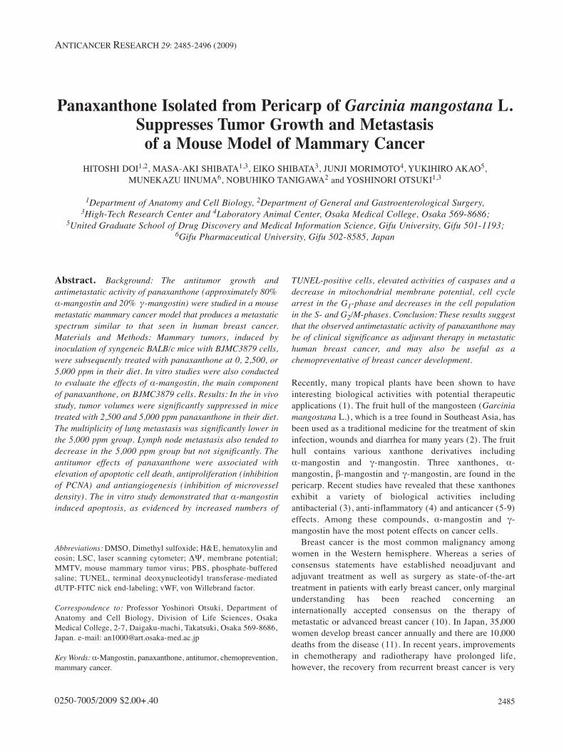
Figure 1. Body weights (a) and tumor volumes (b) in mammarycarcinomas from female BALB/c controls and mice treated with 2,500 or5,000 ppm panaxanthone. Control, 2,500 and 5,000 ppm treated groupsconsisted of 14 mice each. Data represent mean±SD. (a) There were nosignificant differences in body weights among the panaxanthone-treatedgroups and the control group. (b) Tumor growth, as assessed bycomputed volume, was significantly inhibited in mice receivingpanaxanthone at 2,500 and 5,000 pm from 3 and 2 weeks respectivelycompared with the control group (p<0.01). Data represent mean±SD.
Table I. Average food consumption and panaxanthone intake.
Panaxanthone Average food Average(ppm in diet) consumption panaxanthone
(mg/day/mouse) (mg/kg/day/mouse)
0 338 02,500 292 5435,000 290 1078
Groups of 14 mice were given 0, 2500 ppm, or 5000 ppm panaxanthonein their food for 8 weeks.The panaxanthone concentration wascalculated based on food consumption and body weight data.
sequential sections stained with hematoxylin and eosin (H&E) forhistopathological examination and the remainder reserved unstainedfor immunohistochemistry.
Cell proliferation in mammary tumors. Immunohistochemistry wasconducted using the avidin-biotin immunohistochemical complexmethod (LSAB kit; Dako Co., Carpinteria, CA, USA). Anti-PCNAmouse monoclonal Ab (PC-10; Dako Co.) was used at a dilution of
1:400. To evaluate cell kinetics, the number of PCNA-immunoreactivecells in late G1- to S-phase, and G2-phase per 1,000-5,000 cells wascounted in the mammary glands and expressed as a percentage of allmammary tumor cells.
Apoptosis in mammary tumors. For quantitative analysis ofapoptosis, sections from paraffin-embedded tumors were assayedusing the terminal deoxynucleotidyl transferase-mediated dUTP-
Doi et al: Panaxanthone Suppresses Mammary Cancer Growth and Metastasis
2487
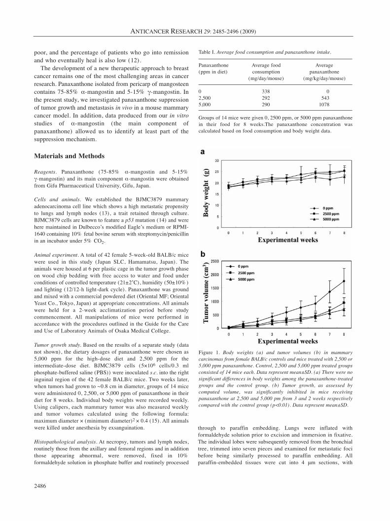
Figure 2. Histopathological analysis of mammary tumors in mice treated with panaxanthone. a, The implanted mammary tumors in a control mouse,×400. b, Metastatic foci in the lung of a control mouse. Many metastatic foci and nodules were seen in the control mice (×40). Metastatic foci (anarrow) in the lungs of mice given 2,500 (c) and 5,000 ppm (d) panaxanthone. Metastatic lung foci were much smaller in the treated groups than inthe control group (×40). e, Metastasis to a lymph node occurred 57% of control mice, ×100. Metastatic carcinoma cells (arrows) were filled withsinusoidal space (×100). f, Lymph node metastasis occurred in 29% of mice given 5,000 ppm panaxanthone. Metastatic tumor cells (arrows) werealso observed in sinusoidal space (×100). (a)-(f), H&E stain.
FITC nick end-labeling (TUNEL) method using an apoptosis in situdetection kit (Wako Pure Chemical Industries, Osaka, Japan).TUNEL-positive cells were counted in viable regions peripheral toareas of necrosis in 5,000 cells within five randomly selected highpower (×200) fields and the data expressed as percentages.
Microvessel densities in mammary tumors. To quantitatively assessblood microvessel density in the primary mammary carcinomas,immunohistochemistry was performed as described above. A rabbitpolyclonal antibody against the von Willebrand factor (vWF; DakoCo.) was used. The number of immunopositive microvessels wascounted as described elsewhere (16). Briefly, the slides were scannedat low-power (×100) magnification to identify those areas of highestmicrovascular density and these areas were then selected and countedat higher (×200-400) magnification to obtain mean±SD values.
In vitro study of the effects of α-mangostin on BJMC3879 cells. Cellviability: BJMC3879 cells were grown in RPMI-1640 mediumsupplemented with 10% (v/v) heat-inactivated fetal bovine serumand 2 mM L-glutamine under an atmosphere of 95% air and 5%CO2 at 37˚C. BJMC3879 cells were plated one day before α-mangostin treatment at 1×10³ cells/well in 96-well plates. Theywere subsequently incubated for 24 h with culture mediumcontaining vehicle (DMSO) alone or with medium containing α-mangostin at different concentrations up to 18 μM. Cell viabilitywas determined using a CellTiter-Blue Cell Viability Assay(Promega Co., Madison, WI, USA).
Cell-cycle distribution: BJMC3879 cells were grown in 2-wellchamber slides (Lab-TekII: Nalgen Nunc International, Naperville,IL, USA), treated with 8 μM α-mangostin for 24 h and fixed in cold70% ethanol. Nuclear DNA was stained with a 50 μg/ml propidiumiodide solution containing 100 μg/ml RNase A for 30 min at 37˚Cfor cell cycle analysis. Cell cycle phases were determined with amicroscope-based multiparameter laser scanning cytometer (LSC2;Olympus Optical Co., Tokyo, Japan) and the resulting data analyzedwith WinCyte software (Compucyte Co., MA, USA).
TUNEL assay and caspase activity: BJMC3879 cells, grown in 2-well chamber slides and treated with 8 μM α-mangostin for 24h, were fixed in 4% formaldehyde solution in phosphate bufferand the TUNEL staining procedure performed as described for thein vivo study. The numbers of TUNEL-positive cells per 1,000cells counted in four random high power (×400) fields byconventional light microscopy were expressed as a percentage ofthe total cells counted.
The activities of caspase-8, caspase-9 and caspase-3 weremeasured in cells treated with 8 μM α-mangostin for 24 h using afluorometric protease assay kit (MBL, Inc., Nagoya, Japan) inwhich cells were lysed with 0.1% Triton® X-100 lysis buffer andthe protein concentration adjusted to 25 μg in each sample. Caspaseactivity was measured in terms of fluorescence intensity using aVersaFluor fluorometer (Bio-Rad, Hercules, CA, USA).
Mitochondrial membrane potential (ΔΨ): Values formitochondrial ΔΨ in α-mangostin-treated and control cells weremeasured using a fluorescent cationic dye, 5,5’,6,6’-tetrachloro-1,1’,3,3’-tetraethyl-benzamidazolocarbocyanin iodide (JC-1)(Mitochondrial Membrane Potential Detection Kit; Cell TechnologyInc, Mountain View, CA, USA) 3 h after α-mangostin treatment.Mitochondrial ΔΨ was determined in terms of relative fluorescenceunits (RFU) using a VersaFluor fluorometer (Bio-Rad) with a 485to 495 nm excitation filter and a 585 to 595 nm emission filter.
Caspase inhibitor experiment: For the caspase inhibitor experiments,2 hours prior to 8 μM α-mangostin exposure, the cells were treatedwith the caspase inhibitors as follows: 10 μM and 100 μM z-DEVD-fmk against caspase-3, z-IETD-fmk against caspase-8 and z-LEHD-fmk against caspase-9 for 48 h. Cell viability was measured using afluorescent assay kit (CellTiter-Blue Cell Viability Assay; Promega)and then the activities of caspase-3, caspase-8 and caspase-9 weremeasured using a luminescent assay kit (Promega). The caspase activitydata was then adjusted to account for the corresponding cell viability aspreviously reported (17).
Statistical analysis. Data for the dose–response effects weresubjected to analysis of variance, while Tukey’s test was employedfor assessment of differences between means. Data from thecytometric studies, mitochondrial ΔΨ analyses, caspase activitystudies and caspase inhibition studies were compiled and comparedbetween control and α-mangostin-treated groups using a two-sidedStudent’s t-test.
Results
Food consumption and panaxanthone intake. The averagefood consumption was 6.0 g/day/mouse in the control group,5.2 g/day/mouse in the 2,500 ppm group and 5.1 g/day/mousein the 5,000 ppm group. The average panaxanthone intakewas 543 mg/kg/day/mouse in the 2,500 ppm group and 1078mg/kg/day/mouse in the 5,000 ppm group (Table I).
Body weights and general condition of mice. The bodyweights of control and panaxanthone-treated mice bearingmammary tumors did not differ statistically and aresummarized in Figure 1a. A total of 2 mice (one mouse fromeach of the control and 5,000 ppm groups) died duringweeks 3-4 as a result of carcinoma growth in the abdomendue to implantation failure. The general condition of all theother animals remained good throughout the study. The micewere terminated 8 weeks after the start of panaxanthonetreatment, when the largest tumor in the control group was2.0 cm in diameter.
Tumor growth. Tumor volumes are presented in Figure 1b.Significant reductions in tumor volume were evident in micereceiving 2,500 ppm panaxanthone from week 3, and in micereceiving 5,000 ppm panaxanthone from week 2. By the endof the experiment, the average tumor volume in the controlanimals was 1,749±469 mm3, while that in the 2,500 and5,000 ppm panaxanthone-treated groups was 939±197 mm3
and 698±243 mm3, respectively.
Lung and lymph node metastasis. Histopathologically, themammary carcinomas induced by BJMC3879 cellinoculation proved to be moderately differentiatedadenocarcinomas (Figure 2a). Lung metastasis occurredin 93% of controls, in 86% of the animals receiving2,500 ppm panaxanthone and in 75% of the animals given
ANTICANCER RESEARCH 29: 2485-2496 (2009)
2488
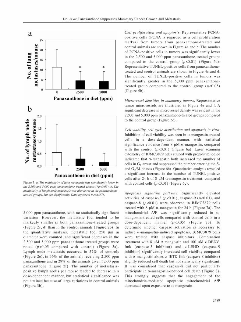
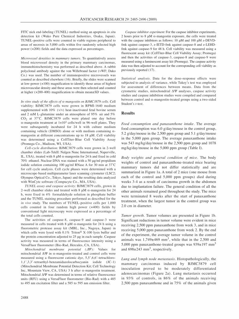
5,000 ppm panaxanthone, with no statistically significantvariation. However, the metastatic foci tended to bemarkedly smaller in both panaxanthone-treated groups(Figure 2c, d) than in the control animals (Figure 2b). Inthe quantitative analysis, metastatic foci 250 μm indiameter were counted, and significant decreases in the2,500 and 5,000 ppm panaxanthone-treated groups werenoted (p<0.05 compared with control) (Figure 3a) .Lymph node metastasis occurred in 57% of controls(Figure 2e), in 36% of the animals receiving 2,500 ppmpanaxanthone and in 29% of the animals given 5,000 ppmpanaxanthone (Figure 2f) . The number of metastasis-positive lymph nodes per mouse tended to decrease in adose-dependent manner, but statistical significance wasnot attained because of large variations in control animals(Figure 3b).
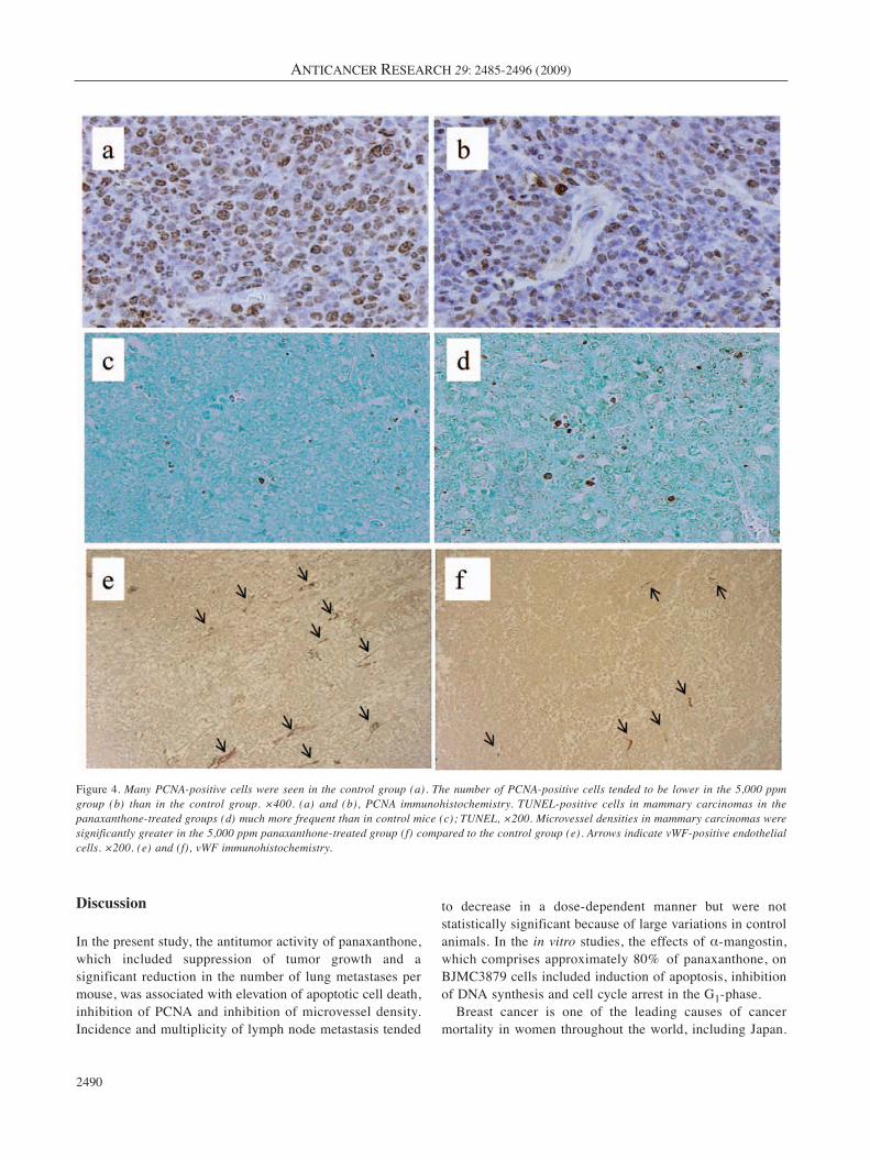
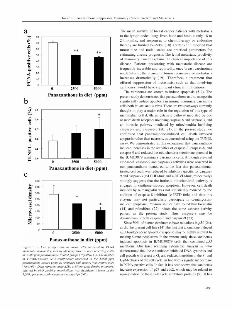
Cell proliferation and apoptosis. Representative PCNA-positive cells (PCNA is regarded as a cell proliferationmarker) from tumors from panaxanthone-treated andcontrol animals are shown in Figure 4a and b. The numberof PCNA-positive cells in tumors was significantly lowerin the 2,500 and 5,000 ppm panaxanthone-treated groupscompared to the control group (p<0.01) (Figure 5a).Representative TUNEL-positive cells from panaxanthone-treated and control animals are shown in Figure 4c and d.The number of TUNEL-positive cells in tumors wassignificantly greater in the 5,000 ppm panaxanthone-treated group compared to the control group (p<0.05)(Figure 5b).
Microvessel densities in mammary tumors. Representativetumor microvessels are illustrated in Figure 4e and f. Asignificant decrease in microvessel density was evident in the2,500 and 5,000 ppm panaxanthone-treated groups comparedto the control group (Figure 5c).
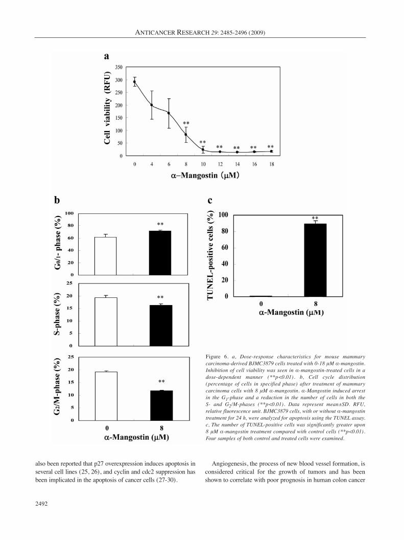
Cell viability, cell-cycle distribution and apoptosis in vitro.Inhibition of cell viability was seen in α-mangostin-treatedcells in a dose-dependent manner, with statisticalsignificance evidence from 8 μM α-mangostin, comparedwith the control (p<0.01) (Figure 6a). Laser scanningcytometry of BJMC3879 cells stained with propidium iodideindicated that α-mangostin both increased the number ofcells in G1 arrest and suppressed the number entering the S-and G2/M-phases (Figure 6b). Quantitative analysis revealeda significant increase in the number of TUNEL-positivecells after 24 h of 8 μM α-mangostin treatment, comparedwith control cells (p<0.01) (Figure 6c).
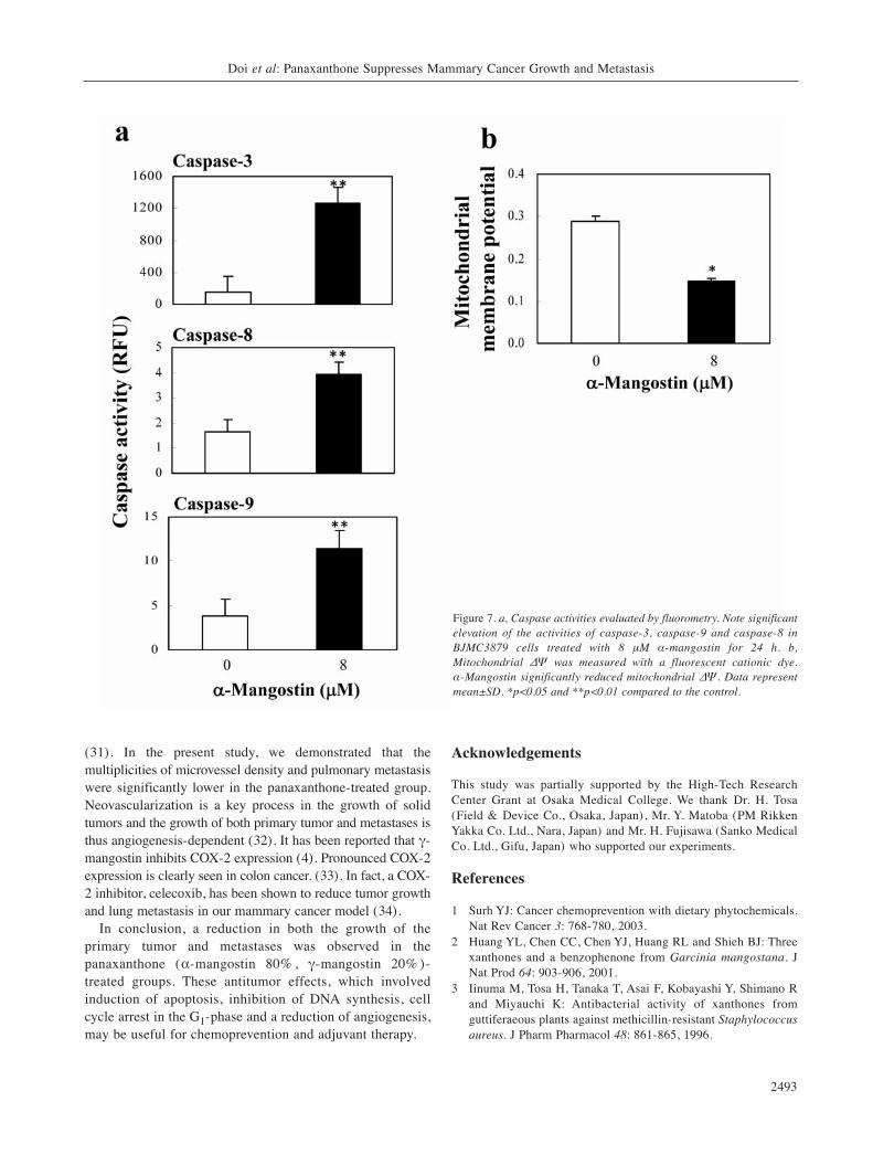
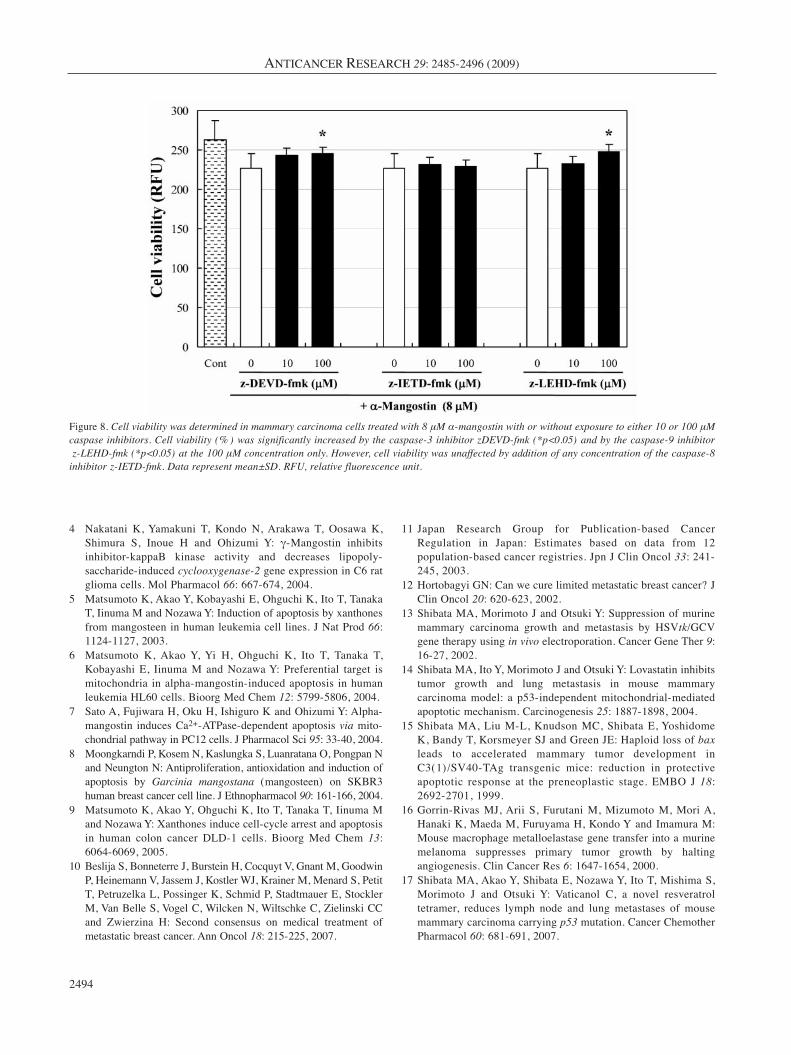
Apoptosis signaling pathway. Significantly elevatedactivities of caspase-3 (p<0.01), caspase-9 (p<0.01), andcaspase-8 (p<0.01) were observed in BJMC3879 cellstreated with 8 μM α-mangostin for 24 h (Figure 7a). Themitochondrial ΔΨ was significantly reduced in α-mangostin-treated cells compared with control cells in adose-dependent manner (p<0.05) (Figure 7b). Todetermine whether caspase activation is necessary toinduce α-mangostin-induced apoptosis, BJMC3879 cellswere treated with caspase inhibitors. Combinationtreatment with 8 μM α-mangostin and 100 μM z-DEDV-fmk (caspase-3 inhibitor) and z-LEHD (caspase-9inhibitor) significantly increased cell viability comparedwith α-mangostin alone. z-IETD-fmk (caspase-8 inhibitor)slightly reduced cell death but not statistically significant.It was considered that caspase-8 did not particularlyparticipate in α-mangostin-induced cell death (Figure 8).This strongly suggests that the engagement of themitochondria-mediated apoptotic mitochondrial ΔΨdecreased upon exposure to α-mangostin.
Doi et al: Panaxanthone Suppresses Mammary Cancer Growth and Metastasis
2489
Figure 3. a, The multiplicity of lung metastasis was significantly lower inthe 2,500 and 5,000 ppm panaxanthone-treated groups (*p<0.05). b, Themultiplicity of lymph node metastasis was also lower in the panaxanthone-treated groups, but not significantly. Data represent mean±SD.
Discussion
In the present study, the antitumor activity of panaxanthone,which included suppression of tumor growth and asignificant reduction in the number of lung metastases permouse, was associated with elevation of apoptotic cell death,inhibition of PCNA and inhibition of microvessel density.Incidence and multiplicity of lymph node metastasis tended
to decrease in a dose-dependent manner but were notstatistically significant because of large variations in controlanimals. In the in vitro studies, the effects of α-mangostin,which comprises approximately 80% of panaxanthone, onBJMC3879 cells included induction of apoptosis, inhibitionof DNA synthesis and cell cycle arrest in the G1-phase.
Breast cancer is one of the leading causes of cancermortality in women throughout the world, including Japan.
ANTICANCER RESEARCH 29: 2485-2496 (2009)
2490
Figure 4. Many PCNA-positive cells were seen in the control group (a). The number of PCNA-positive cells tended to be lower in the 5,000 ppmgroup (b) than in the control group. ×400. (a) and (b), PCNA immunohistochemistry. TUNEL-positive cells in mammary carcinomas in thepanaxanthone-treated groups (d) much more frequent than in control mice (c); TUNEL, ×200. Microvessel densities in mammary carcinomas weresignificantly greater in the 5,000 ppm panaxanthone-treated group (f) compared to the control group (e). Arrows indicate vWF-positive endothelialcells. ×200. (e) and (f), vWF immunohistochemistry.
The mean survival of breast cancer patients with metastasisto the lymph nodes, lung, liver, bone and brain is only 18 to24 months, and responses to chemotherapy or endocrinetherapy are limited to ~50% (18). Carter et al. reported thattumor size and nodal status are practical parameters forestimating disease prognosis. The lethal metastatic proclivityof mammary cancer explains the clinical importance of thisdisease. Patients presenting with metastatic disease arefrequently incurable and reportedly, once breast carcinomasreach >4 cm, the chance of tumor recurrence or metastasisincreases dramatically (19). Therefore, a treatment thatoffered suppression of metastasis, such as that involvingxanthones, would have significant clinical implications.
The xanthones are known to induce apoptosis (5-9). Thepresent study demonstrates that panaxanthone and α-mangostinsignificantly induce apoptosis in murine mammary carcinomacells both in vivo and in vitro. There are two pathways currentlythought to play a major role in the regulation of this type ofmammalian cell death: an extrinsic pathway mediated by oneor more death receptors involving caspase-8 and caspase-3, andan intrinsic pathway mediated by mitochondria involvingcaspase-9 and caspase-3 (20, 21). In the present study, weconfirmed that panaxanthone-induced cell death involvedapoptosis rather than necrosis, as determined using the TUNELassay. We demonstrated in this experiment that panaxanthoneinduced increases in the activities of caspase-3, caspase-8, andcasapse-9 and reduced the mitochondria-membrane potential inthe BJMC3879 mammary carcinoma cells. Although elevatedcaspase-8, caspase-9 and caspase-3 activities were observed inour panaxanthone-treated cells, the fact that panaxanthone-treated cell death was reduced by inhibitors specific for caspase-9 and caspase-3 (z-LEHD-fmk and z-DEVD-fmk, respectively)strongly suggests that the intrinsic mitochondrial pathway isengaged in xanthone-induced apoptosis. However, cell deathinduced by α-mangostin was not statistically reduced by theaddition of caspase-8 inhibitor (z-IETD-fmk) and thus thisenzyme may not particularly participate in α-mangostin-induced apoptosis. Previous studies have found that lovastatin(14) and raloxifene (22) induce the same caspase activitypattern as the present study. Thus, caspase-8 may bedownstream of both caspase-3 and caspase-9 (23).
Since 50% of human carcinomas have mutations in p53 (24),as did the present cell line (14), the fact that a xanthone induceda p53-independent apoptotic response may be highly relevant totreating human neoplasms. In the present study, these xanthonesinduced apoptosis in BJMC39879 cells that contained p53mutations. Our laser scanning cytometric analysis in vitrodemonstrated that these xanthones inhibited DNA synthesis andcell growth with arrest at G1 and reduced transition to the S- andG2/M-phases of the cell cycle, in line with a significant decreasein PCNA-positive cells. In fact, it has been shown that xanthonesincrease expression of p27 and cdc2, which may be related toup-regulation of these cell cycle inhibitory proteins (9). It has
Doi et al: Panaxanthone Suppresses Mammary Cancer Growth and Metastasis
2491
Figure 5. a, Cell proliferation in tumor cells, assessed by PCNAimmunohistochemistry, was significantly lower in mice receiving 2,500or 5,000 ppm panaxanthone-treated groups (**p<0.01). b, The numberof TUNEL-positive cells significantly increased in the 5,000 ppmpanaxanthone-treated group as compared with tumors from control mice(*p<0.05). Data represent mean±SD. c, Microvessel density in tumors,inferred by vWF-positive endothelium, was significantly lower in the5,000 ppm panaxanthone-treated group (*p<0.05).
also been reported that p27 overexpression induces apoptosis inseveral cell lines (25, 26), and cyclin and cdc2 suppression hasbeen implicated in the apoptosis of cancer cells (27-30).
Angiogenesis, the process of new blood vessel formation, isconsidered critical for the growth of tumors and has beenshown to correlate with poor prognosis in human colon cancer
ANTICANCER RESEARCH 29: 2485-2496 (2009)
2492
Figure 6. a, Dose-response characteristics for mouse mammarycarcinoma-derived BJMC3879 cells treated with 0-18 μM α-mangostin.Inhibition of cell viability was seen in α-mangostin-treated cells in adose-dependent manner (**p<0.01). b, Cell cycle distribution(percentage of cells in specified phase) after treatment of mammarycarcinoma cells with 8 μM α-mangostin. α-Mangostin induced arrestin the G1-phase and a reduction in the number of cells in both the S- and G2/M-phases (**p<0.01). Data represent mean±SD. RFU,relative fluorescence unit. BJMC3879 cells, with or without α-mangostintreatment for 24 h, were analyzed for apoptosis using the TUNEL assay.c, The number of TUNEL-positive cells was significantly greater upon 8 μM α-mangostin treatment compared with control cells (**p<0.01).Four samples of both control and treated cells were examined.
(31). In the present study, we demonstrated that themultiplicities of microvessel density and pulmonary metastasiswere significantly lower in the panaxanthone-treated group.Neovascularization is a key process in the growth of solidtumors and the growth of both primary tumor and metastases isthus angiogenesis-dependent (32). It has been reported that γ-mangostin inhibits COX-2 expression (4). Pronounced COX-2expression is clearly seen in colon cancer. (33). In fact, a COX-2 inhibitor, celecoxib, has been shown to reduce tumor growthand lung metastasis in our mammary cancer model (34).
In conclusion, a reduction in both the growth of theprimary tumor and metastases was observed in thepanaxanthone (α-mangostin 80% , γ-mangostin 20% )-treated groups. These antitumor effects, which involvedinduction of apoptosis, inhibition of DNA synthesis, cellcycle arrest in the G1-phase and a reduction of angiogenesis,may be useful for chemoprevention and adjuvant therapy.
Acknowledgements
This study was partially supported by the High-Tech ResearchCenter Grant at Osaka Medical College. We thank Dr. H. Tosa(Field & Device Co., Osaka, Japan), Mr. Y. Matoba (PM RikkenYakka Co. Ltd., Nara, Japan) and Mr. H. Fujisawa (Sanko MedicalCo. Ltd., Gifu, Japan) who supported our experiments.
References
1 Surh YJ: Cancer chemoprevention with dietary phytochemicals.Nat Rev Cancer 3: 768-780, 2003.
2 Huang YL, Chen CC, Chen YJ, Huang RL and Shieh BJ: Threexanthones and a benzophenone from Garcinia mangostana. JNat Prod 64: 903-906, 2001.
3 Iinuma M, Tosa H, Tanaka T, Asai F, Kobayashi Y, Shimano Rand Miyauchi K: Antibacterial activity of xanthones fromguttiferaeous plants against methicillin-resistant Staphylococcusaureus. J Pharm Pharmacol 48: 861-865, 1996.
Doi et al: Panaxanthone Suppresses Mammary Cancer Growth and Metastasis
2493
Figure 7. a, Caspase activities evaluated by fluorometry. Note significantelevation of the activities of caspase-3, caspase-9 and caspase-8 inBJMC3879 cells treated with 8 μM α-mangostin for 24 h. b,Mitochondrial ΔΨ was measured with a fluorescent cationic dye. α-Mangostin significantly reduced mitochondrial ΔΨ. Data representmean±SD. *p<0.05 and **p<0.01 compared to the control.
4 Nakatani K, Yamakuni T, Kondo N, Arakawa T, Oosawa K,Shimura S, Inoue H and Ohizumi Y: γ-Mangostin inhibitsinhibitor-kappaB kinase activity and decreases lipopoly-saccharide-induced cyclooxygenase-2 gene expression in C6 ratglioma cells. Mol Pharmacol 66: 667-674, 2004.
5 Matsumoto K, Akao Y, Kobayashi E, Ohguchi K, Ito T, TanakaT, Iinuma M and Nozawa Y: Induction of apoptosis by xanthonesfrom mangosteen in human leukemia cell lines. J Nat Prod 66:1124-1127, 2003.
6 Matsumoto K, Akao Y, Yi H, Ohguchi K, Ito T, Tanaka T,Kobayashi E, Iinuma M and Nozawa Y: Preferential target ismitochondria in alpha-mangostin-induced apoptosis in humanleukemia HL60 cells. Bioorg Med Chem 12: 5799-5806, 2004.
7 Sato A, Fujiwara H, Oku H, Ishiguro K and Ohizumi Y: Alpha-mangostin induces Ca2+-ATPase-dependent apoptosis via mito-chondrial pathway in PC12 cells. J Pharmacol Sci 95: 33-40, 2004.
8 Moongkarndi P, Kosem N, Kaslungka S, Luanratana O, Pongpan Nand Neungton N: Antiproliferation, antioxidation and induction ofapoptosis by Garcinia mangostana (mangosteen) on SKBR3human breast cancer cell line. J Ethnopharmacol 90: 161-166, 2004.
9 Matsumoto K, Akao Y, Ohguchi K, Ito T, Tanaka T, Iinuma Mand Nozawa Y: Xanthones induce cell-cycle arrest and apoptosisin human colon cancer DLD-1 cells. Bioorg Med Chem 13:6064-6069, 2005.
10 Beslija S, Bonneterre J, Burstein H, Cocquyt V, Gnant M, GoodwinP, Heinemann V, Jassem J, Kostler WJ, Krainer M, Menard S, PetitT, Petruzelka L, Possinger K, Schmid P, Stadtmauer E, StocklerM, Van Belle S, Vogel C, Wilcken N, Wiltschke C, Zielinski CCand Zwierzina H: Second consensus on medical treatment ofmetastatic breast cancer. Ann Oncol 18: 215-225, 2007.
11 Japan Research Group for Publication-based CancerRegulation in Japan: Estimates based on data from 12population-based cancer registries. Jpn J Clin Oncol 33: 241-245, 2003.
12 Hortobagyi GN: Can we cure limited metastatic breast cancer? JClin Oncol 20: 620-623, 2002.
13 Shibata MA, Morimoto J and Otsuki Y: Suppression of murinemammary carcinoma growth and metastasis by HSVtk/GCVgene therapy using in vivo electroporation. Cancer Gene Ther 9:16-27, 2002.
14 Shibata MA, Ito Y, Morimoto J and Otsuki Y: Lovastatin inhibitstumor growth and lung metastasis in mouse mammarycarcinoma model: a p53-independent mitochondrial-mediatedapoptotic mechanism. Carcinogenesis 25: 1887-1898, 2004.
15 Shibata MA, Liu M-L, Knudson MC, Shibata E, YoshidomeK, Bandy T, Korsmeyer SJ and Green JE: Haploid loss of baxleads to accelerated mammary tumor development inC3(1)/SV40-TAg transgenic mice: reduction in protectiveapoptotic response at the preneoplastic stage. EMBO J 18:2692-2701, 1999.
16 Gorrin-Rivas MJ, Arii S, Furutani M, Mizumoto M, Mori A,Hanaki K, Maeda M, Furuyama H, Kondo Y and Imamura M:Mouse macrophage metalloelastase gene transfer into a murinemelanoma suppresses primary tumor growth by haltingangiogenesis. Clin Cancer Res 6: 1647-1654, 2000.
17 Shibata MA, Akao Y, Shibata E, Nozawa Y, Ito T, Mishima S,Morimoto J and Otsuki Y: Vaticanol C, a novel resveratroltetramer, reduces lymph node and lung metastases of mousemammary carcinoma carrying p53 mutation. Cancer ChemotherPharmacol 60: 681-691, 2007.
ANTICANCER RESEARCH 29: 2485-2496 (2009)
2494
Figure 8. Cell viability was determined in mammary carcinoma cells treated with 8 μM α-mangostin with or without exposure to either 10 or 100 μMcaspase inhibitors. Cell viability (% ) was significantly increased by the caspase-3 inhibitor zDEVD-fmk (*p<0.05) and by the caspase-9 inhibitorz-LEHD-fmk (*p<0.05) at the 100 μM concentration only. However, cell viability was unaffected by addition of any concentration of the caspase-8
inhibitor z-IETD-fmk. Data represent mean±SD. RFU, relative fluorescence unit.
18 Ellis M, Hayes D and Lippman M: Treatment of metastaticbreast cancer. In: Disease of the Breast. Harris JR, Lippman ME,Morrow M and Osborne CK (eds.). Lippincott Williams &Wilkins, Philadelphia, PA, USA pp. 794-797, 2000.
19 Carter CL, Allen C and Henson DE: Relation of tumor size,lymph node status, and survival in 24,740 breast cancer cases.Cancer 63: 181-187, 1989.
20 Hengartner MO: The biochemistry of apoptosis. Nature 407:770-776, 2000.
21 Otsuki Y: Tissue specificity of apoptotic signal transduction.Med Electron Microsc 37: 163-169, 2004.
22 Morishima S, Shibata MA, Ohmichi M and Otsuki Y: Raloxifene,a selective estrogen receptor modulator, induces mitochondria-mediated apoptosis in human endometrial carcinoma cells. MedMol Morphol 41: 132-138, 2008.
23 Cha YJ, Kim HS, Rhim H, Kim BE, Jeong SW and Kim IK:Activation of caspase-8 in 3-deazaadenosine-induced apoptosisof U-937 cells occurs downstream of caspase-3 and caspase-9without Fas receptor-ligand interaction. Exp Mol Med 33: 284-292, 2001.
24 Greenblatt MS, Bennett WP, Hollstein M and Harris CC:Mutations in the p53 tumor suppressor gene: clues to canceretiology and molecular pathogenesis. Cancer Res 54: 4855-4878,1994.
25 Wang X, Gorospe M, Huang Y and Holbrook NJ: p27Kip1
overexpression causes apoptotic death of mammalian cells.Oncogene 15: 2991-2997, 1997.
26 Katayose Y, Kim M, Rakkar AN, Li Z, Cowan KH and Seth P:Promoting apoptosis: a novel activity associated with the cyclin-dependent kinase inhibitor p27. Cancer Res 57: 5441-5445,1997.
27 Yuan J, Yan R, Kramer A, Eckerdt F, Roller M, Kaufmann Mand Strebhardt K: Cyclin B1 depletion inhibits proliferation andinduces apoptosis in human tumor cells. Oncogene 23: 5843-5852, 2004.
28 Kamasani U, Huang M, Duhadaway JB, Prochownik EV,Donover PS and Prendergast GC: Cyclin B1 is a critical target ofRhoB in the cell suicide program triggered by farnesyltransferase inhibition. Cancer Res 64: 8389-8396, 2004.
29 Lai D, Weng S, Wang C, Qi L, Yu C, Fu L and Chen W: Smallantisense RNA to cyclin D1 generated by pre-tRNA splicinginhibits growth of human hepatoma cells. FEBS Lett 576: 481-486, 2004.
30 O’Connor DS, Wall NR, Porter AC and Altieri DC: A p34(cdc2)survival checkpoint in cancer. Cancer Cell 2: 43-54, 2002.
31 Takahashi Y, Kitadai Y, Bucana CD, Cleary KR and Ellis LM:Expression of vascular endothelial growth factor and its receptor,KDR, correlates with vascularity, metastasis, and proliferationof human colon cancer. Cancer Res 55: 3964-3968, 1995.
32 Folkman J: Angiogenesis-dependent diseases. Semin Oncol 28:536-542, 2001.
33 Eberhart CE, Coffey RJ, Radhika A, Giardiello FM, FerrenbachS and DuBois RN: Up-regulation of cyclooxygenase 2 geneexpression in human colorectal adenomas and adenocarcinomas.Gastroenterology 107: 1183-1188, 1994.
34 Yoshinaka R, Shibata MA, Morimoto J, Tanigawa N and OtsukiY: COX-2 inhibitor celecoxib suppresses tumor growth and lungmetastasis of a murine mammary cancer. Anticancer Res 26:4245-4254, 2006.
Received February 2, 2009Revised May 6, 2009
Accepted May 12, 2009
Doi et al: Panaxanthone Suppresses Mammary Cancer Growth and Metastasis
2495