Maintenance of adenomatous polyposis coli (APC)-mutant ...
6
Maintenance of adenomatous polyposis coli (APC)-mutant colorectal cancer is dependent on Wnt/β-catenin signaling Alix Scholer-Dahirel 1 , Michael R. Schlabach, Alice Loo, Linda Bagdasarian, Ronald Meyer, Ribo Guo, Steve Woolfenden, Kristine K. Yu, Judit Markovits, Karen Killary, Dmitry Sonkin, Yung-Mae Yao, Markus Warmuth, William R. Sellers, Robert Schlegel, Frank Stegmeier, Rebecca E. Mosher, and Margaret E. McLaughlin 2 Novartis Institutes for Biomedical Research, Cambridge, MA 02139 Edited by Bert Vogelstein, Johns Hopkins University, Baltimore, MD, and approved August 23, 2011 (received for review March 18, 2011) Persistent expression of certain oncogenes is required for tumor maintenance. This phenotype is referred to as oncogene addiction and has been clinically validated by anticancer therapies that specifically inhibit oncoproteins such as BCR-ABL, c-Kit, HER2, PDGFR, and EGFR. Identifying additional genes that are required for tumor maintenance may lead to new targets for anticancer drugs. Although the role of aberrant Wnt pathway activation in the initiation of colorectal cancer has been clearly established, it remains unclear whether sustained Wnt pathway activation is required for colorectal tumor maintenance. To address this question, we used inducible β-catenin shRNAs to temporally con- trol Wnt pathway activation in vivo. Here, we show that active Wnt/β-catenin signaling is required for maintenance of colorectal tumor xenografts harboring APC mutations. Reduced tumor growth upon β-catenin inhibition was due to cell cycle arrest and differentiation. Upon reactivation of the Wnt/β-catenin pathway colorectal cancer cells resumed proliferation and reacquired a crypt progenitor phenotype. In human colonic adenocarcinomas, high levels of nuclear β-catenin correlated with crypt progenitor but not differentiation markers, suggesting that the Wnt/β-catenin pathway may also control colorectal tumor cell fate during the maintenance phase of tumors in patients. These results support efforts to treat human colorectal cancer by pharmacological inhi- bition of the Wnt/β-catenin pathway. M utational activation of the Wnt pathway occurs in the vast majority of colorectal cancers through truncating muta- tions in adenomatous polyposis coli (APC) or AXIN2 or muta- tions in GSK3-target residues in β-catenin (1–5). In normal intestinal cells, APC associates with axin, glycogen synthase ki- nase-3β (GSK-3β), and casein kinase 1 (CK1) to form a β-catenin destruction complex. This complex phosphorylates β-catenin, resulting in its ubiquitylation and subsequent degradation by the proteasome (6). In contrast, in cells harboring mutations in APC, AXIN2, or β-catenin, β-catenin accumulates and upon its trans- location to the nucleus, interacts with the T-cell factor/lymphoid enhancer factor (TCF/LEF) family of transcription factors to activate specific Wnt target genes (7, 8). Considerable evidence from human genetic studies and mouse genetic models suggests that mutational activation of the Wnt pathway initiates the process of colon tumorigenesis. Inherited APC mutations cause familial adenomatous polyposis, and ac- quired APC mutations represent the earliest genetic alteration so far detected in the genesis of sporadic colorectal cancer (9). Rare mutations in AXIN2 or β-catenin can also be present in small neoplastic lesions (5, 10). In experimental mouse models, loss of APC (11, 12) or expression of constitutively active β-catenin (13) is sufficient to drive polyp formation. Inhibition of the Wnt pathway in colorectal cancer cells in vitro by overexpression of dominant-negative TCF4 or in- ducible β-catenin siRNA results in rapid cell cycle arrest and blocks a genetic program that is physiologically active in crypt progenitors. Consequently, colorectal cancer cells undergo dif- ferentiation (7, 14, 15). By imposing a proliferative crypt pro- genitor phenotype, aberrant Wnt pathway activation may allow benign tumors (polyps) to persist for many years, providing an opportunity for the acquisition of further mutations (e.g., in KRAS, SMAD-4, and P53 genes) required for the development of malignant colorectal tumors (16). Although the role of Wnt pathway activation in the initiation of colon tumorigenesis has been well established, it is unclear whether tumors that have acquired additional mutations in oncogenes or tumor suppressor genes remain dependent on Wnt pathway activation. Although β-catenin siRNA inhibits engraft- ment of colorectal cancer cells (17), a recent study reports that inhibition of Wnt signaling in established colorectal xenografts (mutant for the β-catenin gene) by inducible β-catenin shRNA had no significant effect on tumor growth (18). Human co- lorectal tumors with mutations in β-catenin are usually less ag- gressive and smaller than those with APC mutations (19), suggesting that APC and β-catenin mutations are not functionally equivalent. Consistently, in addition to its function in the β-cat- enin degradation complex, APC can also directly contribute to the regulation of mitosis and apoptosis (20). Such β-catenin– independent APC functions may influence the degree of depen- dency on Wnt pathway activation for colorectal tumor mainte- nance. Given the large preponderance of APC mutations in human colorectal cancer, it is crucial to determine whether sustained Wnt pathway activation is required for maintenance of APC-mutant colorectal tumors. In this study, we provide evidence that Wnt/β-catenin pathway activation is required for maintenance of colorectal tumors harboring APC mutations. We show that β-catenin inhibition in vivo strongly inhibited the growth of established APC-mutant colorectal tumor xenografts. Upon β-catenin inhibition, co- lorectal cancer cells underwent cell cycle arrest and rapidly started to differentiate into polarized intestinal epithelial cells, producing mucins and displaying microvilli. Such differentiated cancer cells, when relieved from β-catenin inhibition, could de- differentiate and regain their proliferative potential in vitro. In Author contributions: A.S.-D., M.R.S., A.L., Y.-M.Y., M.W., W.R.S., R.S., F.S., R.E.M., and M.E.M. designed research; A.S.-D., M.R.S., A.L., L.B., R.M., R.G., S.W., K.K.Y., J.M., K.K., D.S., and M.E.M. performed research; A.S.-D., M.R.S., and D.S. contributed new reagents/ analytic tools; A.S.-D., M.R.S., A.L., L.B., R.M., R.G., J.M., D.S., and M.E.M. analyzed data; and A.S.-D., M.R.S., A.L., M.W., W.R.S., R.S., F.S., R.E.M., and M.E.M. wrote the paper. The authors declare no conflict of interest. This article is a PNAS Direct Submission. 1 Present address: Institut National de la Santé et de la Recherche Médicale, U830, Genetic and Biology of Cancers, Institut Curie, F-75248 Paris Cedex 05, France. 2 To whom correspondence should be addressed. E-mail: margaret.mclaughlin@novartis. com. This article contains supporting information online at www.pnas.org/lookup/suppl/doi:10. 1073/pnas.1104182108/-/DCSupplemental. www.pnas.org/cgi/doi/10.1073/pnas.1104182108 PNAS | October 11, 2011 | vol. 108 | no. 41 | 17135–17140 MEDICAL SCIENCES
Transcript of Maintenance of adenomatous polyposis coli (APC)-mutant ...
Maintenance of adenomatous polyposis coli(APC)-mutant colorectal cancer is dependenton Wnt/β-catenin signalingAlix Scholer-Dahirel1, Michael R. Schlabach, Alice Loo, Linda Bagdasarian, Ronald Meyer, Ribo Guo, Steve Woolfenden,Kristine K. Yu, Judit Markovits, Karen Killary, Dmitry Sonkin, Yung-Mae Yao, Markus Warmuth, William R. Sellers,Robert Schlegel, Frank Stegmeier, Rebecca E. Mosher, and Margaret E. McLaughlin2
Novartis Institutes for Biomedical Research, Cambridge, MA 02139
Edited by Bert Vogelstein, Johns Hopkins University, Baltimore, MD, and approved August 23, 2011 (received for review March 18, 2011)
Persistent expression of certain oncogenes is required for tumormaintenance. This phenotype is referred to as oncogene addictionand has been clinically validated by anticancer therapies thatspecifically inhibit oncoproteins such as BCR-ABL, c-Kit, HER2,PDGFR, and EGFR. Identifying additional genes that are requiredfor tumor maintenance may lead to new targets for anticancerdrugs. Although the role of aberrant Wnt pathway activation inthe initiation of colorectal cancer has been clearly established,it remains unclear whether sustained Wnt pathway activationis required for colorectal tumor maintenance. To address thisquestion, we used inducible β-catenin shRNAs to temporally con-trol Wnt pathway activation in vivo. Here, we show that activeWnt/β-catenin signaling is required for maintenance of colorectaltumor xenografts harboring APC mutations. Reduced tumorgrowth upon β-catenin inhibition was due to cell cycle arrest anddifferentiation. Upon reactivation of the Wnt/β-catenin pathwaycolorectal cancer cells resumed proliferation and reacquired a cryptprogenitor phenotype. In human colonic adenocarcinomas, highlevels of nuclear β-catenin correlated with crypt progenitor butnot differentiation markers, suggesting that the Wnt/β-cateninpathway may also control colorectal tumor cell fate during themaintenance phase of tumors in patients. These results supportefforts to treat human colorectal cancer by pharmacological inhi-bition of the Wnt/β-catenin pathway.
Mutational activation of the Wnt pathway occurs in the vastmajority of colorectal cancers through truncating muta-
tions in adenomatous polyposis coli (APC) or AXIN2 or muta-tions in GSK3-target residues in β-catenin (1–5). In normalintestinal cells, APC associates with axin, glycogen synthase ki-nase-3β (GSK-3β), and casein kinase 1 (CK1) to form a β-catenindestruction complex. This complex phosphorylates β-catenin,resulting in its ubiquitylation and subsequent degradation by theproteasome (6). In contrast, in cells harboring mutations in APC,AXIN2, or β-catenin, β-catenin accumulates and upon its trans-location to the nucleus, interacts with the T-cell factor/lymphoidenhancer factor (TCF/LEF) family of transcription factors toactivate specific Wnt target genes (7, 8).Considerable evidence from human genetic studies and mouse
genetic models suggests that mutational activation of the Wntpathway initiates the process of colon tumorigenesis. InheritedAPC mutations cause familial adenomatous polyposis, and ac-quired APCmutations represent the earliest genetic alteration sofar detected in the genesis of sporadic colorectal cancer (9). Raremutations in AXIN2 or β-catenin can also be present in smallneoplastic lesions (5, 10). In experimental mouse models, loss ofAPC (11, 12) or expression of constitutively active β-catenin (13)is sufficient to drive polyp formation.Inhibition of the Wnt pathway in colorectal cancer cells
in vitro by overexpression of dominant-negative TCF4 or in-ducible β-catenin siRNA results in rapid cell cycle arrest andblocks a genetic program that is physiologically active in crypt
progenitors. Consequently, colorectal cancer cells undergo dif-ferentiation (7, 14, 15). By imposing a proliferative crypt pro-genitor phenotype, aberrant Wnt pathway activation may allowbenign tumors (polyps) to persist for many years, providing anopportunity for the acquisition of further mutations (e.g., inKRAS, SMAD-4, and P53 genes) required for the development ofmalignant colorectal tumors (16).Although the role of Wnt pathway activation in the initiation
of colon tumorigenesis has been well established, it is unclearwhether tumors that have acquired additional mutations inoncogenes or tumor suppressor genes remain dependent on Wntpathway activation. Although β-catenin siRNA inhibits engraft-ment of colorectal cancer cells (17), a recent study reports thatinhibition of Wnt signaling in established colorectal xenografts(mutant for the β-catenin gene) by inducible β-catenin shRNAhad no significant effect on tumor growth (18). Human co-lorectal tumors with mutations in β-catenin are usually less ag-gressive and smaller than those with APC mutations (19),suggesting that APC and β-catenin mutations are not functionallyequivalent. Consistently, in addition to its function in the β-cat-enin degradation complex, APC can also directly contribute tothe regulation of mitosis and apoptosis (20). Such β-catenin–independent APC functions may influence the degree of depen-dency on Wnt pathway activation for colorectal tumor mainte-nance. Given the large preponderance of APC mutations inhuman colorectal cancer, it is crucial to determine whethersustained Wnt pathway activation is required for maintenance ofAPC-mutant colorectal tumors.In this study, we provide evidence that Wnt/β-catenin pathway
activation is required for maintenance of colorectal tumorsharboring APC mutations. We show that β-catenin inhibitionin vivo strongly inhibited the growth of established APC-mutantcolorectal tumor xenografts. Upon β-catenin inhibition, co-lorectal cancer cells underwent cell cycle arrest and rapidlystarted to differentiate into polarized intestinal epithelial cells,producing mucins and displaying microvilli. Such differentiatedcancer cells, when relieved from β-catenin inhibition, could de-differentiate and regain their proliferative potential in vitro. In
Author contributions: A.S.-D., M.R.S., A.L., Y.-M.Y., M.W., W.R.S., R.S., F.S., R.E.M., andM.E.M. designed research; A.S.-D., M.R.S., A.L., L.B., R.M., R.G., S.W., K.K.Y., J.M., K.K.,D.S., and M.E.M. performed research; A.S.-D., M.R.S., and D.S. contributed new reagents/analytic tools; A.S.-D., M.R.S., A.L., L.B., R.M., R.G., J.M., D.S., and M.E.M. analyzed data;and A.S.-D., M.R.S., A.L., M.W., W.R.S., R.S., F.S., R.E.M., and M.E.M. wrote the paper.
The authors declare no conflict of interest.
This article is a PNAS Direct Submission.1Present address: Institut National de la Santé et de la Recherche Médicale, U830, Geneticand Biology of Cancers, Institut Curie, F-75248 Paris Cedex 05, France.
2To whom correspondence should be addressed. E-mail: [email protected].
This article contains supporting information online at www.pnas.org/lookup/suppl/doi:10.1073/pnas.1104182108/-/DCSupplemental.
www.pnas.org/cgi/doi/10.1073/pnas.1104182108 PNAS | October 11, 2011 | vol. 108 | no. 41 | 17135–17140
MED
ICALSC
IENCE
S
addition, we report that in human colonic adenocarcinomas highlevels of nuclear β-catenin correlated with crypt progenitor butnot differentiation markers, suggesting that the Wnt/β-cateninpathway may also control colorectal tumor cell fate during themaintenance phase of tumors in patients.
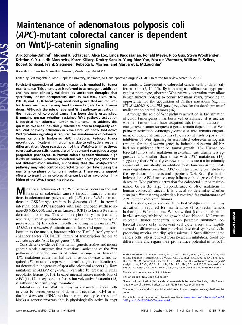
ResultsSustained Wnt/β-Catenin Pathway Activation Is Required forMaintenance of Colorectal Tumors Harboring APC Mutations. Toanalyze the role of sustained Wnt pathway activation in main-tenance of colorectal tumors, we used doxycycline-inducibleβ-catenin shRNAs to temporally control Wnt pathway activationin vivo. We infected LS411N and SW403 colorectal cancer cells(both APC mutant) with a robust inducible single-lentiviralvector pLKO-Tet-On (21), containing either control nontarget-ing (NTC) shRNA or two distinct β-catenin–targeting shRNAsequences (shRNA-35 or shRNA-36). Polyclonal stable lineswere obtained after puromycin selection and inoculated s.c. inNu/Nu mice. To confirm that the doxycycline treatment led toinhibition of the Wnt/β-catenin pathway, we treated mice bearingestablished tumors (100–300 mm3) with doxycycline for 3 d andanalyzed various molecular components of the Wnt/β-cateninpathway. Upon doxycycline treatment, β-catenin expression wasdramatically reduced at the level of both mRNA (knockdownefficiency >95%, Fig. 1A) and protein in the β-catenin shRNAtumors but not in the NTC shRNA tumors (Fig. 1 B and C andFig. S1 A and B). As expected, silencing of β-catenin causeda concomitant reduction of β-catenin target genes AXIN2 and c-MYC at the mRNA and protein levels (Fig. 1A and Fig. S1 E–H).We noted that β-catenin inhibition had a more dramatic effecton c-MYC expression in SW403 versus LS411N cells (up to 99%and 50% reduction of nuclear c-MYC staining intensity, re-spectively). Specificity of the β-catenin shRNAs was confirmedin vitro: Decreased cell viability was noted only in LS411N andSW403 colorectal cancer cell lines, not in RKO colorectal cancercells that are wild type for β-catenin and APC (Fig. S1 I–N).Taken together, these results indicate that the β-catenin shRNAsefficiently and specifically inhibit the Wnt/β-catenin pathway.We next investigated the effect of β-catenin shRNAs on tumor
growth and treated mice bearing established tumors with doxy-cycline for 14 d. Such treatment induced long-term silencing ofβ-catenin as demonstrated by down-regulation of β-catenin atday 14 (Fig. S1 C and D). Both LS411N and SW403 β-cateninshRNA tumors showed a significant inhibition of tumor growthfollowing doxycycline treatment (Fig. 1 D and E). This effect wasdue to inhibition of the Wnt/β-catenin pathway rather thandoxycycline treatment alone, as NTC shRNA tumors progressedrapidly despite treatment. Interestingly, nuclear β-catenin de-pletion by β-catenin shRNAs was more complete after shortdoxycycline treatment in LS411N tumors (60–75% and 45%reduction of nuclear β-catenin at days 3 and 14, respectively).This observation suggests that there might be some selectivepressure favoring cancer cells that either had mild initial β-cat-enin depletion or managed to escape silencing by β-cateninshRNAs. We conclude that sustained Wnt/β-catenin pathwayactivation is required for the maintenance of colorectal tumorxenografts harboring APC mutations.
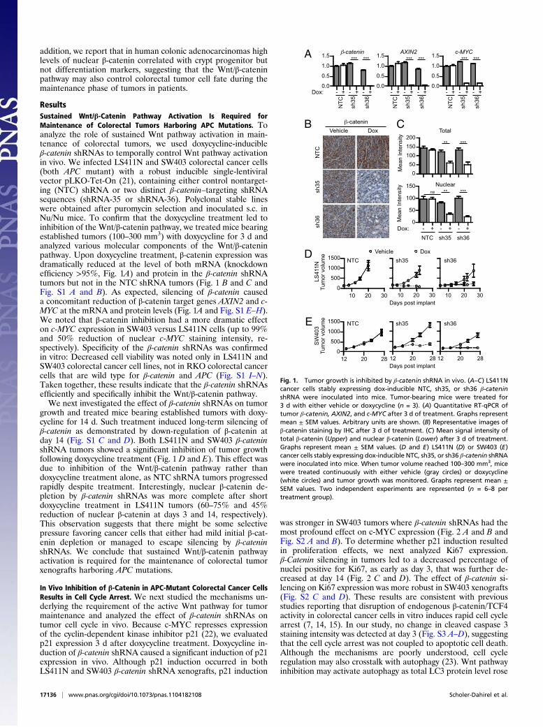
In Vivo Inhibition of β-Catenin in APC-Mutant Colorectal Cancer CellsResults in Cell Cycle Arrest. We next studied the mechanisms un-derlying the requirement of the active Wnt pathway for tumormaintenance and analyzed the effect of β-catenin shRNAs ontumor cell cycle in vivo. Because c-MYC represses expressionof the cyclin-dependent kinase inhibitor p21 (22), we evaluatedp21 expression 3 d after doxycycline treatment. Doxycycline in-duction of β-catenin shRNA caused a significant induction of p21expression in vivo. Although p21 induction occurred in bothLS411N and SW403 β-catenin shRNA xenografts, p21 induction
was stronger in SW403 tumors where β-catenin shRNAs had themost profound effect on c-MYC expression (Fig. 2 A and B andFig. S2 A and B). To determine whether p21 induction resultedin proliferation effects, we next analyzed Ki67 expression.β-Catenin silencing in tumors led to a decreased percentage ofnuclei positive for Ki67, as early as day 3, that was further de-creased at day 14 (Fig. 2 C and D). The effect of β-catenin si-lencing on Ki67 expression was more robust in SW403 xenografts(Fig. S2 C and D). These results are consistent with previousstudies reporting that disruption of endogenous β-catenin/TCF4activity in colorectal cancer cells in vitro induces rapid cell cyclearrest (7, 14, 15). In our study, no change in cleaved caspase 3staining intensity was detected at day 3 (Fig. S3 A–D), suggestingthat the cell cycle arrest was not coupled to apoptotic cell death.Although the mechanisms are poorly understood, cell cycleregulation may also crosstalk with autophagy (23). Wnt pathwayinhibition may activate autophagy as total LC3 protein level rose
0.0
0.5
1.0
1.5
0.0
0.5
1.0
1.5
0
50
100
150
A
B C
Nuclear
NTC
sh
35
sh36
Vehicle Dox
050
100150200
Dox:
Mea
n In
tens
ity
- + - + - + NTC sh35 sh36
D
E
10 20 300
500
1000
1500
10 20 30 10 20 30
12 20 280
500
1000
1500
12 20 28 12 20 28
Tum
or v
olum
e
NTC sh35 sh36
Days post implant
Days post implant
Tum
or v
olum
e
0.0
0.5
1.0
1.5-catenin AXIN2 c-MYC
Mea
n In
tens
ity
Dox: - + - + - +
NTC
sh35
sh36
Total
Vehicle Dox
NTC sh35 sh36
- + - + - +
NTC
sh35
sh36
- + - + - +
NTC
sh35
sh36
*** *** *** *** *** ***
** ***
** *** ns
-catenin
LS41
1N
SW
403
Fig. 1. Tumor growth is inhibited by β-catenin shRNA in vivo. (A–C) LS411Ncancer cells stably expressing dox-inducible NTC, sh35, or sh36 β-cateninshRNA were inoculated into mice. Tumor-bearing mice were treated for3 d with either vehicle or doxycycline (n = 3). (A) Quantitative RT-qPCR oftumor β-catenin, AXIN2, and c-MYC after 3 d of treatment. Graphs representmean ± SEM values. Arbitrary units are shown. (B) Representative images ofβ-catenin staining by IHC after 3 d of treatment. (C) Mean signal intensity oftotal β-catenin (Upper) and nuclear β-catenin (Lower) after 3 d of treatment.Graphs represent mean ± SEM values. (D and E) LS411N (D) or SW403 (E)cancer cells stably expressing dox-inducible NTC, sh35, or sh36 β-catenin shRNAwere inoculated into mice. When tumor volume reached 100–300 mm3, micewere treated continuously with either vehicle (gray circles) or doxycycline(white circles) and tumor growth was monitored. Graphs represent mean ±SEM values. Two independent experiments are represented (n = 6–8 pertreatment group).
17136 | www.pnas.org/cgi/doi/10.1073/pnas.1104182108 Scholer-Dahirel et al.
at day 3 of β-catenin silencing. However, no change in the LC3-II/LC3-I ratio or in p62 expression was observed (Fig. S3 E–G).Our results demonstrate that a major consequence of inhibitionof β-catenin in vivo is cell cycle arrest of APC-mutant colorectalcancer cells.
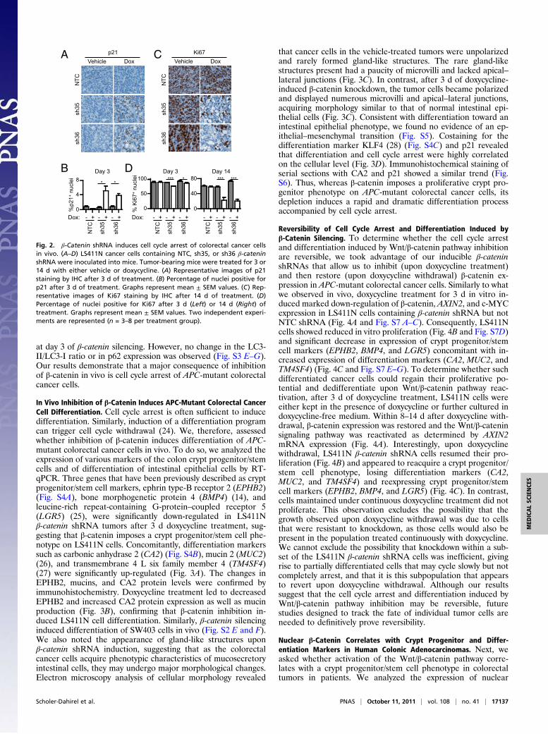
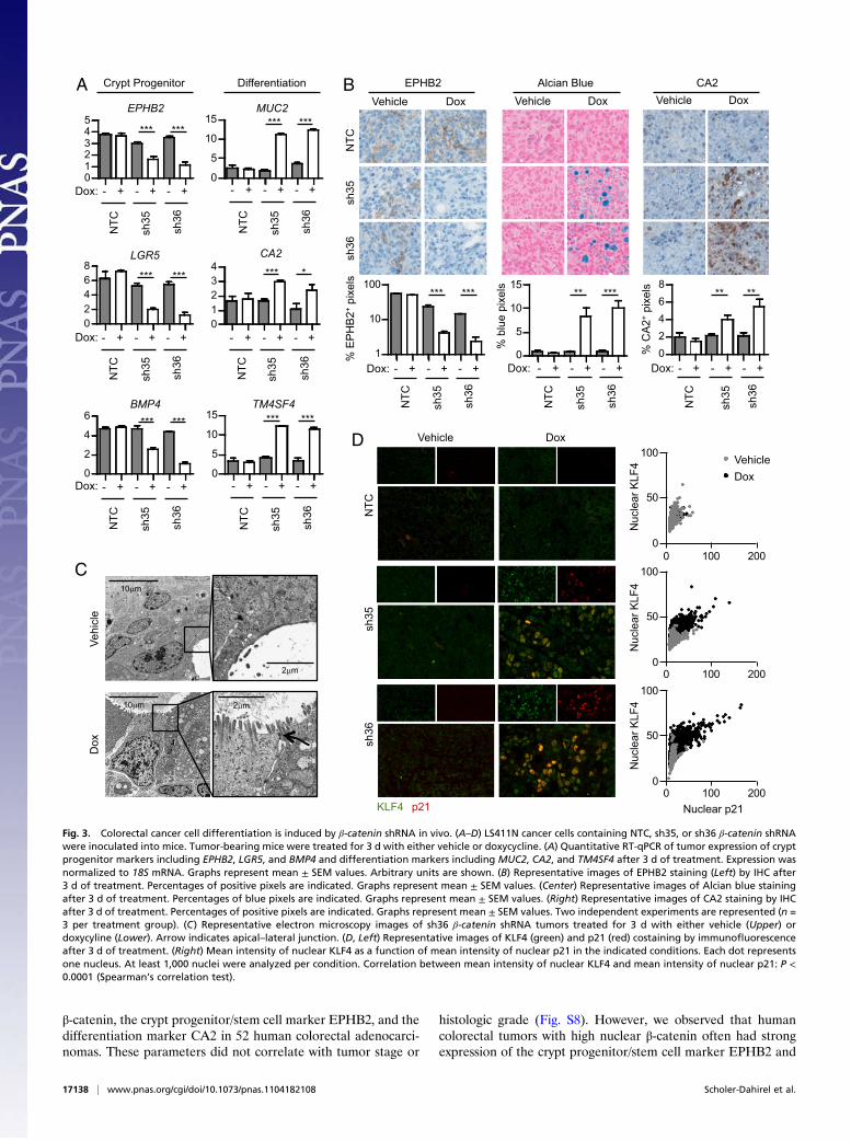
In Vivo Inhibition of β-Catenin Induces APC-Mutant Colorectal CancerCell Differentiation. Cell cycle arrest is often sufficient to inducedifferentiation. Similarly, induction of a differentiation programcan trigger cell cycle withdrawal (24). We, therefore, assessedwhether inhibition of β-catenin induces differentiation of APC-mutant colorectal cancer cells in vivo. To do so, we analyzed theexpression of various markers of the colon crypt progenitor/stemcells and of differentiation of intestinal epithelial cells by RT-qPCR. Three genes that have been previously described as cryptprogenitor/stem cell markers, ephrin type-B receptor 2 (EPHB2)(Fig. S4A), bone morphogenetic protein 4 (BMP4) (14), andleucine-rich repeat-containing G-protein–coupled receptor 5(LGR5) (25), were significantly down-regulated in LS411Nβ-catenin shRNA tumors after 3 d doxycycline treatment, sug-gesting that β-catenin imposes a crypt progenitor/stem cell phe-notype on LS411N cells. Concomitantly, differentiation markerssuch as carbonic anhydrase 2 (CA2) (Fig. S4B), mucin 2 (MUC2)(26), and transmembrane 4 L six family member 4 (TM4SF4)(27) were significantly up-regulated (Fig. 3A). The changes inEPHB2, mucins, and CA2 protein levels were confirmed byimmunohistochemistry. Doxycycline treatment led to decreasedEPHB2 and increased CA2 protein expression as well as mucinproduction (Fig. 3B), confirming that β-catenin inhibition in-duced LS411N cell differentiation. Similarly, β-catenin silencinginduced differentiation of SW403 cells in vivo (Fig. S2 E and F).We also noted the appearance of gland-like structures uponβ-catenin shRNA induction, suggesting that as the colorectalcancer cells acquire phenotypic characteristics of mucosecretoryintestinal cells, they may undergo major morphological changes.Electron microscopy analysis of cellular morphology revealed
that cancer cells in the vehicle-treated tumors were unpolarizedand rarely formed gland-like structures. The rare gland-likestructures present had a paucity of microvilli and lacked apical–lateral junctions (Fig. 3C). In contrast, after 3 d of doxycycline-induced β-catenin knockdown, the tumor cells became polarizedand displayed numerous microvilli and apical–lateral junctions,acquiring morphology similar to that of normal intestinal epi-thelial cells (Fig. 3C). Consistent with differentiation toward anintestinal epithelial phenotype, we found no evidence of an ep-ithelial–mesenchymal transition (Fig. S5). Costaining for thedifferentiation marker KLF4 (28) (Fig. S4C) and p21 revealedthat differentiation and cell cycle arrest were highly correlatedon the cellular level (Fig. 3D). Immunohistochemical staining ofserial sections with CA2 and p21 showed a similar trend (Fig.S6). Thus, whereas β-catenin imposes a proliferative crypt pro-genitor phenotype on APC-mutant colorectal cancer cells, itsdepletion induces a rapid and dramatic differentiation processaccompanied by cell cycle arrest.
Reversibility of Cell Cycle Arrest and Differentiation Induced byβ-Catenin Silencing. To determine whether the cell cycle arrestand differentiation induced by Wnt/β-catenin pathway inhibitionare reversible, we took advantage of our inducible β-cateninshRNAs that allow us to inhibit (upon doxycycline treatment)and then restore (upon doxycycline withdrawal) β-catenin ex-pression in APC-mutant colorectal cancer cells. Similarly to whatwe observed in vivo, doxycycline treatment for 3 d in vitro in-duced marked down-regulation of β-catenin, AXIN2, and c-MYCexpression in LS411N cells containing β-catenin shRNA but notNTC shRNA (Fig. 4A and Fig. S7 A–C). Consequently, LS411Ncells showed reduced in vitro proliferation (Fig. 4B and Fig. S7D)and significant decrease in expression of crypt progenitor/stemcell markers (EPHB2, BMP4, and LGR5) concomitant with in-creased expression of differentiation markers (CA2, MUC2, andTM4SF4) (Fig. 4C and Fig. S7 E–G). To determine whether suchdifferentiated cancer cells could regain their proliferative po-tential and dedifferentiate upon Wnt/β-catenin pathway reac-tivation, after 3 d of doxycycline treatment, LS411N cells wereeither kept in the presence of doxycycline or further cultured indoxycycline-free medium. Within 8–14 d after doxycycline with-drawal, β-catenin expression was restored and the Wnt/β-cateninsignaling pathway was reactivated as determined by AXIN2mRNA expression (Fig. 4A). Interestingly, upon doxycyclinewithdrawal, LS411N β-catenin shRNA cells resumed their pro-liferation (Fig. 4B) and appeared to reacquire a crypt progenitor/stem cell phenotype, losing differentiation markers (CA2,MUC2, and TM4SF4) and reexpressing crypt progenitor/stemcell markers (EPHB2, BMP4, and LGR5) (Fig. 4C). In contrast,cells maintained under continuous doxycycline treatment did notproliferate. This observation excludes the possibility that thegrowth observed upon doxycycline withdrawal was due to cellsthat were resistant to knockdown, as those cells would also bepresent in the population treated continuously with doxycycline.We cannot exclude the possibility that knockdown within a sub-set of the LS411N β-catenin shRNA cells was inefficient, givingrise to partially differentiated cells that may cycle slowly but notcompletely arrest, and that it is this subpopulation that appearsto revert upon doxycycline withdrawal. Although our resultssuggest that the cell cycle arrest and differentiation induced byWnt/β-catenin pathway inhibition may be reversible, futurestudies designed to track the fate of individual tumor cells areneeded to definitively prove reversibility.
Nuclear β-Catenin Correlates with Crypt Progenitor and Differ-entiation Markers in Human Colonic Adenocarcinomas. Next, weasked whether activation of the Wnt/β-catenin pathway corre-lates with a crypt progenitor/stem cell phenotype in colorectaltumors in patients. We analyzed the expression of nuclear
A
0
40
80D
0
50
100B
0
4
8
%p2
1+ n
ucle
i
Day 3
NTC
sh
35
sh36
Vehicle Dox C
NTC
sh
35
sh36
Vehicle Dox
% K
i67+
nuc
lei
Dox: - + - + - +
NTC
sh35
sh36
* *
- + - + - +
NTC
sh35
sh36
- + - + - +
NTC
sh35
sh36
Dox:
Day 3 Day 14 *** * *** ***
p21 Ki67
Fig. 2. β-Catenin shRNA induces cell cycle arrest of colorectal cancer cellsin vivo. (A–D) LS411N cancer cells containing NTC, sh35, or sh36 β-cateninshRNA were inoculated into mice. Tumor-bearing mice were treated for 3 or14 d with either vehicle or doxycycline. (A) Representative images of p21staining by IHC after 3 d of treatment. (B) Percentage of nuclei positive forp21 after 3 d of treatment. Graphs represent mean ± SEM values. (C) Rep-resentative images of Ki67 staining by IHC after 14 d of treatment. (D)Percentage of nuclei positive for Ki67 after 3 d (Left) or 14 d (Right) oftreatment. Graphs represent mean ± SEM values. Two independent experi-ments are represented (n = 3–8 per treatment group).
Scholer-Dahirel et al. PNAS | October 11, 2011 | vol. 108 | no. 41 | 17137
MED
ICALSC
IENCE
S
β-catenin, the crypt progenitor/stem cell marker EPHB2, and thedifferentiation marker CA2 in 52 human colorectal adenocarci-nomas. These parameters did not correlate with tumor stage or
histologic grade (Fig. S8). However, we observed that humancolorectal tumors with high nuclear β-catenin often had strongexpression of the crypt progenitor/stem cell marker EPHB2 and
NTC
sh
35
sh36
Vehicle Dox
1
10
100
% E
PH
B2+
pix
els
02468
0
5
10
15
01234
0
5
10
15
0
2
4
6
A B Vehicle Dox
02468
% C
A2+
pix
els
012345
- + - + - +
NTC
sh35
sh36
Dox:
Vehicle Dox
0
5
10
15
% b
lue
pixe
ls
- + - + - +
NTC
sh35
sh36
- + - + - +
NTC
sh35
sh36
- + - + - +
NTC
sh35
sh36
- + - + - +
NTC
sh35
sh36
- + - + - +
NTC
sh35
sh36
EPHB2
BMP4
LGR5
TM4SF4
CA2
MUC2
*** ***
*** ***
*** ***
*** ***
*** *
*** ***
C
- + - + - +
NTC
sh35
sh36
Dox: - + - + - +
NTC
sh35
sh36
Dox: - + - + - +
NTC
sh35
sh36
Dox:
*** *** ** *** ** **
EPHB2 CA2 Alcian Blue
Dox:
Dox:
D
NTC
sh
35
sh36
Vehicle Dox
Vehi
cle
2 m
10 m
Dox
2 m 10 m
Nuclear p21
Nuc
lear
KLF
4 N
ucle
ar K
LF4
Nuc
lear
KLF
4
Vehicle Dox
p21 KLF4
0 100 2000
50
100
0 100 2000
50
100
0 100 2000
50
100
Crypt Progenitor Differentiation
Fig. 3. Colorectal cancer cell differentiation is induced by β-catenin shRNA in vivo. (A–D) LS411N cancer cells containing NTC, sh35, or sh36 β-catenin shRNAwere inoculated into mice. Tumor-bearing mice were treated for 3 d with either vehicle or doxycycline. (A) Quantitative RT-qPCR of tumor expression of cryptprogenitor markers including EPHB2, LGR5, and BMP4 and differentiation markers including MUC2, CA2, and TM4SF4 after 3 d of treatment. Expression wasnormalized to 18S mRNA. Graphs represent mean ± SEM values. Arbitrary units are shown. (B) Representative images of EPHB2 staining (Left) by IHC after3 d of treatment. Percentages of positive pixels are indicated. Graphs represent mean ± SEM values. (Center) Representative images of Alcian blue stainingafter 3 d of treatment. Percentages of blue pixels are indicated. Graphs represent mean ± SEM values. (Right) Representative images of CA2 staining by IHCafter 3 d of treatment. Percentages of positive pixels are indicated. Graphs represent mean ± SEM values. Two independent experiments are represented (n =3 per treatment group). (C) Representative electron microscopy images of sh36 β-catenin shRNA tumors treated for 3 d with either vehicle (Upper) ordoxycyline (Lower). Arrow indicates apical–lateral junction. (D, Left) Representative images of KLF4 (green) and p21 (red) costaining by immunofluorescenceafter 3 d of treatment. (Right) Mean intensity of nuclear KLF4 as a function of mean intensity of nuclear p21 in the indicated conditions. Each dot representsone nucleus. At least 1,000 nuclei were analyzed per condition. Correlation between mean intensity of nuclear KLF4 and mean intensity of nuclear p21: P <0.0001 (Spearman’s correlation test).
17138 | www.pnas.org/cgi/doi/10.1073/pnas.1104182108 Scholer-Dahirel et al.
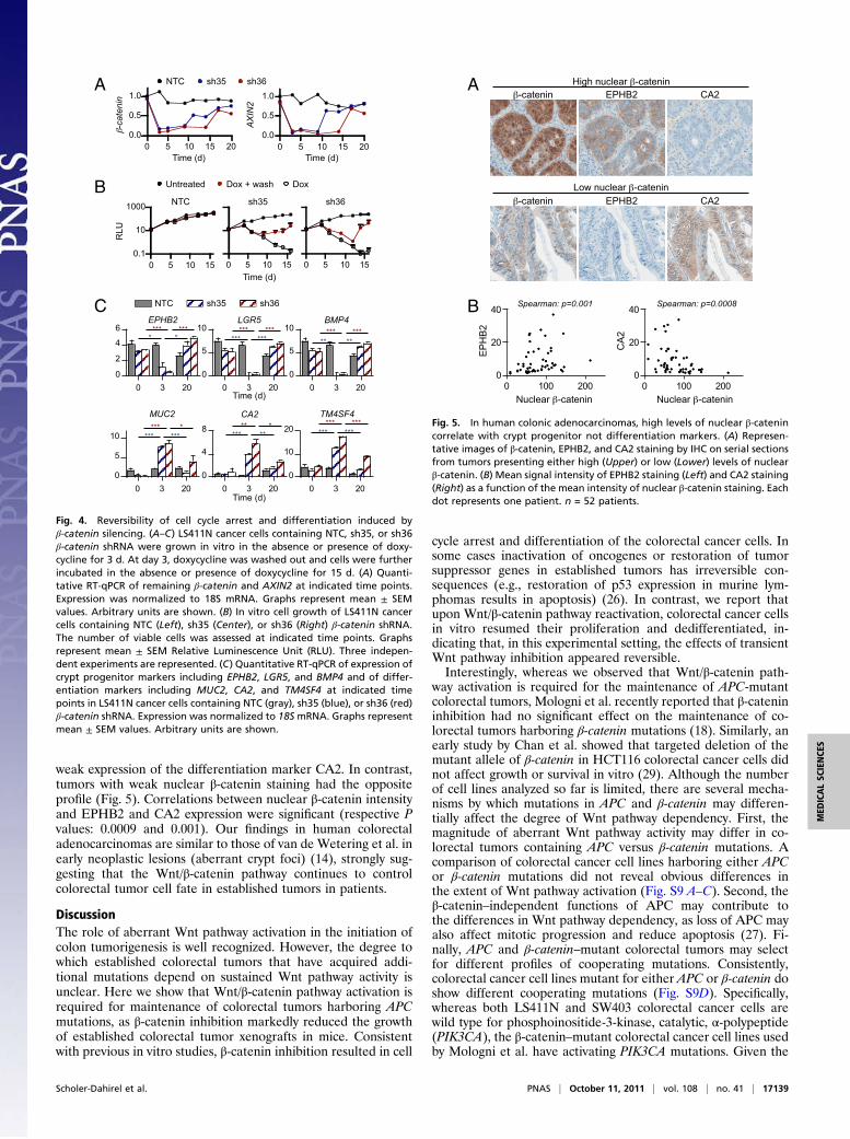
weak expression of the differentiation marker CA2. In contrast,tumors with weak nuclear β-catenin staining had the oppositeprofile (Fig. 5). Correlations between nuclear β-catenin intensityand EPHB2 and CA2 expression were significant (respective Pvalues: 0.0009 and 0.001). Our findings in human colorectaladenocarcinomas are similar to those of van de Wetering et al. inearly neoplastic lesions (aberrant crypt foci) (14), strongly sug-gesting that the Wnt/β-catenin pathway continues to controlcolorectal tumor cell fate in established tumors in patients.
DiscussionThe role of aberrant Wnt pathway activation in the initiation ofcolon tumorigenesis is well recognized. However, the degree towhich established colorectal tumors that have acquired addi-tional mutations depend on sustained Wnt pathway activity isunclear. Here we show that Wnt/β-catenin pathway activation isrequired for maintenance of colorectal tumors harboring APCmutations, as β-catenin inhibition markedly reduced the growthof established colorectal tumor xenografts in mice. Consistentwith previous in vitro studies, β-catenin inhibition resulted in cell
cycle arrest and differentiation of the colorectal cancer cells. Insome cases inactivation of oncogenes or restoration of tumorsuppressor genes in established tumors has irreversible con-sequences (e.g., restoration of p53 expression in murine lym-phomas results in apoptosis) (26). In contrast, we report thatupon Wnt/β-catenin pathway reactivation, colorectal cancer cellsin vitro resumed their proliferation and dedifferentiated, in-dicating that, in this experimental setting, the effects of transientWnt pathway inhibition appeared reversible.Interestingly, whereas we observed that Wnt/β-catenin path-
way activation is required for the maintenance of APC-mutantcolorectal tumors, Mologni et al. recently reported that β-catenininhibition had no significant effect on the maintenance of co-lorectal tumors harboring β-catenin mutations (18). Similarly, anearly study by Chan et al. showed that targeted deletion of themutant allele of β-catenin in HCT116 colorectal cancer cells didnot affect growth or survival in vitro (29). Although the numberof cell lines analyzed so far is limited, there are several mecha-nisms by which mutations in APC and β-catenin may differen-tially affect the degree of Wnt pathway dependency. First, themagnitude of aberrant Wnt pathway activity may differ in co-lorectal tumors containing APC versus β-catenin mutations. Acomparison of colorectal cancer cell lines harboring either APCor β-catenin mutations did not reveal obvious differences inthe extent of Wnt pathway activation (Fig. S9 A–C). Second, theβ-catenin–independent functions of APC may contribute tothe differences in Wnt pathway dependency, as loss of APC mayalso affect mitotic progression and reduce apoptosis (27). Fi-nally, APC and β-catenin–mutant colorectal tumors may selectfor different profiles of cooperating mutations. Consistently,colorectal cancer cell lines mutant for either APC or β-catenin doshow different cooperating mutations (Fig. S9D). Specifically,whereas both LS411N and SW403 colorectal cancer cells arewild type for phosphoinositide-3-kinase, catalytic, α-polypeptide(PIK3CA), the β-catenin–mutant colorectal cancer cell lines usedby Mologni et al. have activating PIK3CA mutations. Given the
0 3 200
5
10LGR5
*** *** *** ***
0 3 200
4
8
CA2
*** ** ** *
0 3 200
5
10
MUC2
*** *** *** *
0 3 200
2
4
6
0 3 200
5
10
0 3 200
10
20
A
B
C
0 5 10 15 200.0
0.5
1.0
0 5 10 15 200.0
0.5
1.0
TM4SF4
Time (d)
Time (d)
Time (d)
-catenin
AXIN2
Time (d)
0 5 10 150.1
10
1000
RLU
Time (d) 0 5 10 15 0 5 10 15
NTC sh35 sh36
Untreated Dox + wash Dox
* *
*** ***
*** ***
*** ***
NTC sh35 sh36
NTC sh35 sh36
BMP4
** ** *** ***
EPHB2
Fig. 4. Reversibility of cell cycle arrest and differentiation induced byβ-catenin silencing. (A–C) LS411N cancer cells containing NTC, sh35, or sh36β-catenin shRNA were grown in vitro in the absence or presence of doxy-cycline for 3 d. At day 3, doxycycline was washed out and cells were furtherincubated in the absence or presence of doxycycline for 15 d. (A) Quanti-tative RT-qPCR of remaining β-catenin and AXIN2 at indicated time points.Expression was normalized to 18S mRNA. Graphs represent mean ± SEMvalues. Arbitrary units are shown. (B) In vitro cell growth of LS411N cancercells containing NTC (Left), sh35 (Center), or sh36 (Right) β-catenin shRNA.The number of viable cells was assessed at indicated time points. Graphsrepresent mean ± SEM Relative Luminescence Unit (RLU). Three indepen-dent experiments are represented. (C) Quantitative RT-qPCR of expression ofcrypt progenitor markers including EPHB2, LGR5, and BMP4 and of differ-entiation markers including MUC2, CA2, and TM4SF4 at indicated timepoints in LS411N cancer cells containing NTC (gray), sh35 (blue), or sh36 (red)β-catenin shRNA. Expression was normalized to 18S mRNA. Graphs representmean ± SEM values. Arbitrary units are shown.
0 100 2000
20
40
0 100 2000
20
40
A
Nuclear -catenin Nuclear -catenin
EP
HB
2
CA
2
-catenin EPHB2 CA2
-catenin EPHB2 CA2 Low nuclear -catenin
High nuclear -catenin
B Spearman: p=0.0008 Spearman: p=0.001
Fig. 5. In human colonic adenocarcinomas, high levels of nuclear β-catenincorrelate with crypt progenitor not differentiation markers. (A) Represen-tative images of β-catenin, EPHB2, and CA2 staining by IHC on serial sectionsfrom tumors presenting either high (Upper) or low (Lower) levels of nuclearβ-catenin. (B) Mean signal intensity of EPHB2 staining (Left) and CA2 staining(Right) as a function of the mean intensity of nuclear β-catenin staining. Eachdot represents one patient. n = 52 patients.
Scholer-Dahirel et al. PNAS | October 11, 2011 | vol. 108 | no. 41 | 17139
MED
ICALSC
IENCE
S
important role of phosphoinositide-3-kinase (PI3K) in the reg-ulation of cell proliferation and survival (30), activating muta-tions of PIK3CA may represent an alternative driver of tumorgrowth in colorectal cancer cells harboring β-catenin mutations,thereby leading to Wnt pathway independence for tumormaintenance. Further studies will be required to test these hy-potheses. Other potential differences between the colorectaltumor xenografts used by Mologni et al. and those in the presentstudy may include growth factor expression or tumor–stromalinteractions.Mirroring findings in early neoplastic lesions (14), we report
that high levels of nuclear β-catenin in human colorectal ade-nocarcinomas correlate with crypt progenitor but not differen-tiation markers, suggesting that the Wnt/β-catenin pathwaycontinues to control colorectal tumor cell fate in establishedtumors in patients. Together with the requirement of sustainedWnt/β-catenin pathway activation for the maintenance of APC-mutant colorectal tumor xenografts, these observations supportefforts to treat APC-mutant colorectal cancer by pharmacologi-cal inhibition of the Wnt/β-catenin pathway.
Materials and MethodsCell Culture. LS411N, SW403, and RKO cancer cells were obtained from theAmerican Type Culture Collection. Cells were cultured as indicated inSI Methods.
Lentivirus and Infection. Lentiviral supernatant production and cell infectionwere performed as described in SI Methods.
RNA Extraction and Quantitative Reverse Transcription–PCR. Total RNA wasisolated as described in SI Methods. An ABI taqman gene expression assaywas performed as indicated in SI Methods.
Growth Assay. Cell proliferation assays upon shRNA-mediated knockdown ofβ-catenin were performed by seeding 1,000 cells/well in a 96-well plate
(triplicate) in the absence and presence of doxycycline. Cell Titer Glo meas-urements were taken at several time points according to the manufacturer’s(Promega) instruction to track cell proliferation.
Immunohistochemistry, Immunofluorescence, and Image Analysis. Antibodiesused are described in SI Methods. Xenograft tumor samples were fixed in10% neutral-buffered formalin for 6–24 h, processed, and paraffin embed-ded. Archival formalin-fixed, paraffin-embedded human colorectal adeno-carcinoma specimens were procured by Maine Medical Center Tissue Bank,Maine Medical Center Pathology Department, Portland, ME. Immunohisto-chemical and immunofluorescent staining was performed on the VentanaDiscovery System. Images were captured using an Aperio Scanscope or anAperio Scanscope FL (Aperio Technologies) and analyzed as described in SIMethods. The necrotic area and stromal tissue portions were manually ex-cluded, using the tools provided by the imaging systems.
In Vivo Efficacy Study of Doxycycline-Inducible β-Catenin shRNA. All animalswere handled in accordance with Novartis Institutes for Biomedical ResearchAnimal Care and Use Committee protocols and regulations. Mice werehoused in a temperature- and humidity-controlled animal facility with adlibitum access to food and water and acclimated for at least 3 d beforeexperimental manipulation. Female Nu/Nu mice (6–8 wk old; Taconic) wereinoculated s.c. with 5 × 10⁶ cells in the right dorsal axillary region. Tumordimensions were measured with calipers, and tumor volume was calculatedas (length × width2)/2. Mice bearing 100–300 mm3 established tumors wererandomized and enrolled into treatment groups (n = 6–8 per group). Micereceived either vehicle (5% dextrose in water) or doxycycline hyclate (25 mg/kg, once a day) via oral gavage for the duration of the study.
Statistical Analysis. Unpaired t tests and Spearman’s correlation tests wereused to determine statistical significance. Symbols used: *P < 0.05; **P <0.01; ***P < 0.001; ns, not significant.
ACKNOWLEDGMENTS. We thank Humphrey Gardner and the MaineMedical Center Tissue Bank for tissue collaborations, and Yeonju Shim forconstructing the Tissue MicroArray.
1. Jin LH, et al. (2003) Detection of point mutations of the Axin1 gene in colorectal
cancers. Int J Cancer 107:696–699.2. Korinek V, et al. (1997) Constitutive transcriptional activation by a beta-catenin-Tcf
complex in APC-/- colon carcinoma. Science 275:1784–1787.3. Morin PJ, et al. (1997) Activation of beta-catenin-Tcf signaling in colon cancer by
mutations in beta-catenin or APC. Science 275:1787–1790.4. Rubinfeld B, et al. (1997) Stabilization of beta-catenin by genetic defects in melanoma
cell lines. Science 275:1790–1792.5. Liu W, et al. (2000) Mutations in AXIN2 cause colorectal cancer with defective mis-
match repair by activating beta-catenin/TCF signalling. Nat Genet 26:146–147.6. Polakis P (2002) Casein kinase 1: A Wnt’er of disconnect. Curr Biol 12:R499–R501.7. Tetsu O, McCormick F (1999) Beta-catenin regulates expression of cyclin D1 in colon
carcinoma cells. Nature 398:422–426.8. He TC, et al. (1998) Identification of c-MYC as a target of the APC pathway. Science
281:1509–1512.9. Powell SM, et al. (1992) APC mutations occur early during colorectal tumorigenesis.
Nature 359:235–237.10. Sparks AB, Morin PJ, Vogelstein B, Kinzler KW (1998) Mutational analysis of the APC/
beta-catenin/Tcf pathway in colorectal cancer. Cancer Res 58:1130–1134.11. Moser AR, Pitot HC, Dove WF (1990) A dominant mutation that predisposes to mul-
tiple intestinal neoplasia in the mouse. Science 247:322–324.12. Shibata H, et al. (1997) Rapid colorectal adenoma formation initiated by conditional
targeting of the Apc gene. Science 278:120–123.13. Harada N, et al. (1999) Intestinal polyposis in mice with a dominant stable mutation of
the beta-catenin gene. EMBO J 18:5931–5942.14. van de Wetering M, et al. (2002) The beta-catenin/TCF-4 complex imposes a crypt
progenitor phenotype on colorectal cancer cells. Cell 111:241–250.15. van de Wetering M, et al. (2003) Specific inhibition of gene expression using a stably
integrated, inducible small-interfering-RNA vector. EMBO Rep 4:609–615.16. Fearon ER, Vogelstein B (1990) A genetic model for colorectal tumorigenesis. Cell 61:
759–767.
17. Verma UN, Surabhi RM, Schmaltieg A, Becerra C, Gaynor RB (2003) Small interferingRNAs directed against beta-catenin inhibit the in vitro and in vivo growth of coloncancer cells. Clin Cancer Res 9:1291–1300.
18. Mologni L, et al. (2010) Colorectal tumors are effectively eradicated by combinedinhibition of beta-catenin, KRAS, and the oncogenic transcription factor ITF2. CancerRes 70:7253–7263.
19. Samowitz WS, et al. (1999) Beta-catenin mutations are more frequent in small co-lorectal adenomas than in larger adenomas and invasive carcinomas. Cancer Res 59:1442–1444.
20. Dikovskaya D, et al. (2007) Loss of APC induces polyploidy as a result of a combinationof defects in mitosis and apoptosis. J Cell Biol 176:183–195.
21. Wiederschain D, et al. (2009) Single-vector inducible lentiviral RNAi system for on-cology target validation. Cell Cycle 8:498–504.
22. Wu S, et al. (2003) Myc represses differentiation-induced p21CIP1 expression via Miz-1-dependent interaction with the p21 core promoter. Oncogene 22:351–360.
23. Cecconi F, Levine B (2008) The role of autophagy in mammalian development: Cellmakeover rather than cell death. Dev Cell 15:344–357.
24. Zhu L, Skoultchi AI (2001) Coordinating cell proliferation and differentiation. CurrOpin Genet Dev 11:91–97.
25. Barker N, et al. (2007) Identification of stem cells in small intestine and colon bymarker gene Lgr5. Nature 449:1003–1007.
26. Carrato C, et al. (1994) Differential apomucin expression in normal and neoplastichuman gastrointestinal tissues. Gastroenterology 107:160–172.
27. Wice BM, Gordon JI (1995) A tetraspan membrane glycoprotein produced in thehuman intestinal epithelium and liver that can regulate cell density-dependent pro-liferation. J Biol Chem 270:21907–21918.
28. Shie JL, et al. (2000) Role of gut-enriched Krüppel-like factor in colonic cell growthand differentiation. Am J Physiol Gastrointest Liver Physiol 279:G806–G814.
29. Chan TA, Wang Z, Dang LH, Vogelstein B, Kinzler KW (2002) Targeted inactivation ofCTNNB1 reveals unexpected effects of beta-catenin mutation. Proc Natl Acad Sci USA99:8265–8270.
30. Engelman JA (2009) Targeting PI3K signalling in cancer: Opportunities, challengesand limitations. Nat Rev Cancer 9:550–562.
17140 | www.pnas.org/cgi/doi/10.1073/pnas.1104182108 Scholer-Dahirel et al.