Residues within Transmembrane Domains 4 and 6 of the Na,K-ATPase α Subunit Are Important for Na ...
14
Residues within Transmembrane Domains 4 and 6 of the Na,K-ATPase R Subunit Are Important for Na + Selectivity ² Gladis Sa ´nchez and Gustavo Blanco* Department of Molecular and IntegratiVe Physiology, UniVersity of Kansas Medical Center, Kansas City, Kansas 66160 ReceiVed March 15, 2004; ReVised Manuscript ReceiVed May 6, 2004 ABSTRACT: The Na,K- and H,K-ATPases are plasma membrane enzymes responsible for the active exchange of extracellular K + for cytoplasmic Na + or H + , respectively. At present, the structural determinants for the specific function of these ATPases remain poorly understood. To investigate the cation selectivity of these ATPases, we constructed a series of Na,K-ATPase mutants in which residues in the membrane spanning segments of the R subunit were changed to the corresponding residues common to gastric H,K- ATPases. Thus, mutants were created with substitutions in transmembrane domains TM1, TM4, TM5, TM6, TM7, and TM8 independently or together (designated TMAll). The function of each mutant was assessed after coexpression with the subunit in Sf-9 cells using baculoviruses. The enzymatic properties of TM1, TM7, and TM8 mutants were similar to the wild-type Na,K-ATPase, and while TM5 showed modest changes in apparent affinity for Na + , TM4, TM6, and TMAll displayed an abnormal activity. This resulted in a Na + -independent hydrolysis of ATP, a 2-fold higher K 0.5 for Na + activation, and the ability to function at low pH. These results suggest a loss of discrimination for Na + over H + for the enzymes. In addition, TM4, TM6, and TMAll mutants exhibited a 1.5-fold lower affinity for K + and a 4-5-fold decreased sensitivity to vanadate. Altogether, these results provide evidence that residues in transmembrane domains 4 and 6 of the R subunit of the Na,K-ATPase play an important role in determining the specific cation selectivity of the enzyme and also its E1/E2 conformational equilibrium. P-type ATPases form a group of enzymes that use the energy from the hydrolysis of ATP to drive the transport of a variety of cations against their transmembrane electro- chemical gradients (reviewed in refs 1 and 2). Within the members of this family of ATPases, two of them, the Na,K- and the H,K-ATPase, are the only ones capable of transport- ing one type of cation (extracellular K + ) in exchange for another (cytoplasmic Na + or H + , respectively). The Na,K- ATPase or Na pump is ubiquitously expressed, and its function is critical for the control of osmotic cell balance, cell pH, and the resting membrane potential of most tissues. In addition, the enzyme plays a primary role in driving secondary Na + -coupled transport systems and the reabsorp- tion of salt and water in many epithelia (3, 4). The H,K- ATPase on the other hand is essential for acid secretion by the gastric mucosa, kidney, and colon (5-8). Both the Na,K- and H,K-ATPase share common biochemical reaction mech- anisms and undergo E1/E2 conformational changes coupled to the binding, occlusion, and translocation of ions (reviewed in refs 9 and 10). Structurally, both transporters are het- erodimers composed of stoichiometric amounts of a catalytic R and a glycosylated subunit. The R subunit is a multispanning membrane protein with a molecular mass of approximately 110 kDa. It is composed of an N-terminal segment with four transmembrane spanning domains, a large cytoplasmic region and a carboxy-terminal segment contain- ing another six transmembrane domains (reviewed in refs 11-13). In contrast, the subunit is a 40-60 kDa polypep- tide that crosses the plasma membrane just once (reviewed in refs 14-16). The R subunit is responsible for the catalytic and transport properties of the Na,K- and H,K-ATPases, while in vertebrates, the subunit is important for the proper folding and delivery of the holoenzyme to the plasma membrane (11-15). The R polypeptide contains the binding sites for the respective cations, ATP, and the preferred inhibitors, ouabain for the Na,K-ATPase and omeprazole or Scheering 28080 for the H,K-ATPase (reviewed in refs 3, 4, 10, and 17). The Na,K- and H,K-ATPase R subunits share approximately 60% amino acid sequence identity. The major similarities between these P-type ATPases occur around the phosphorylated aspartate, the TGES/A motif between trans- membrane domains 2 and 3, and several cytoplasmic regions involved in ATP binding (1). Most likely, these conserved regions participate in the common mechanism of operation of the transporters. On the other hand, the differences may contain the structural determinants that give each ATPase the ability to select the type of cation preferentially trans- located out of the cell. A considerable amount of work has been devoted to identifying the amino acids in the Na,K- and H,K-ATPase R subunits involved in the binding occlu- sion and transport of the cations. Several different approaches have been utilized. These include the use of carboxyl- modifying agents (18), site-directed mutagenesis of specific ² This work was supported by National Science Foundation Grant MCB-0196493 and National Institutes of Health Grant HD043044- 01A1. * To whom correspondence should be addressed. Mailing address: Department of Molecular and Integrative Physiology, University of Kansas Medical Center, 3901 Rainbow Boulevard, Kansas City, KS 66160. Phone (913) 588-7405. Fax: (913) 588-7430. E-mail: gblanco@ kumc.edu. 9061 Biochemistry 2004, 43, 9061-9074 10.1021/bi049484s CCC: $27.50 © 2004 American Chemical Society Published on Web 06/19/2004
Transcript of Residues within Transmembrane Domains 4 and 6 of the Na,K-ATPase α Subunit Are Important for Na ...

Residues within Transmembrane Domains 4 and 6 of the Na,K-ATPaseR SubunitAre Important for Na+ Selectivity†
Gladis Sa´nchez and Gustavo Blanco*
Department of Molecular and IntegratiVe Physiology, UniVersity of Kansas Medical Center, Kansas City, Kansas 66160
ReceiVed March 15, 2004; ReVised Manuscript ReceiVed May 6, 2004
ABSTRACT: The Na,K- and H,K-ATPases are plasma membrane enzymes responsible for the active exchangeof extracellular K+ for cytoplasmic Na+ or H+, respectively. At present, the structural determinants forthe specific function of these ATPases remain poorly understood. To investigate the cation selectivity ofthese ATPases, we constructed a series of Na,K-ATPase mutants in which residues in the membranespanning segments of theR subunit were changed to the corresponding residues common to gastric H,K-ATPases. Thus, mutants were created with substitutions in transmembrane domains TM1, TM4, TM5,TM6, TM7, and TM8 independently or together (designated TMAll). The function of each mutant wasassessed after coexpression with theâ subunit inSf-9 cells using baculoviruses. The enzymatic propertiesof TM1, TM7, and TM8 mutants were similar to the wild-type Na,K-ATPase, and while TM5 showedmodest changes in apparent affinity for Na+, TM4, TM6, and TMAll displayed an abnormal activity.This resulted in a Na+-independent hydrolysis of ATP, a 2-fold higherK0.5 for Na+ activation, and theability to function at low pH. These results suggest a loss of discrimination for Na+ over H+ for theenzymes. In addition, TM4, TM6, and TMAll mutants exhibited a 1.5-fold lower affinity for K+ and a4-5-fold decreased sensitivity to vanadate. Altogether, these results provide evidence that residues intransmembrane domains 4 and 6 of theR subunit of the Na,K-ATPase play an important role in determiningthe specific cation selectivity of the enzyme and also its E1/E2 conformational equilibrium.
P-type ATPases form a group of enzymes that use theenergy from the hydrolysis of ATP to drive the transport ofa variety of cations against their transmembrane electro-chemical gradients (reviewed in refs1 and 2). Within themembers of this family of ATPases, two of them, the Na,K-and the H,K-ATPase, are the only ones capable of transport-ing one type of cation (extracellular K+) in exchange foranother (cytoplasmic Na+ or H+, respectively). The Na,K-ATPase or Na pump is ubiquitously expressed, and itsfunction is critical for the control of osmotic cell balance,cell pH, and the resting membrane potential of most tissues.In addition, the enzyme plays a primary role in drivingsecondary Na+-coupled transport systems and the reabsorp-tion of salt and water in many epithelia (3, 4). The H,K-ATPase on the other hand is essential for acid secretion bythe gastric mucosa, kidney, and colon (5-8). Both the Na,K-and H,K-ATPase share common biochemical reaction mech-anisms and undergo E1/E2 conformational changes coupledto the binding, occlusion, and translocation of ions (reviewedin refs 9 and 10). Structurally, both transporters are het-erodimers composed of stoichiometric amounts of a catalyticR and a glycosylatedâ subunit. The R subunit is amultispanning membrane protein with a molecular mass of
approximately 110 kDa. It is composed of an N-terminalsegment with four transmembrane spanning domains, a largecytoplasmic region and a carboxy-terminal segment contain-ing another six transmembrane domains (reviewed in refs11-13). In contrast, theâ subunit is a 40-60 kDa polypep-tide that crosses the plasma membrane just once (reviewedin refs14-16). TheR subunit is responsible for the catalyticand transport properties of the Na,K- and H,K-ATPases,while in vertebrates, theâ subunit is important for the properfolding and delivery of the holoenzyme to the plasmamembrane (11-15). TheR polypeptide contains the bindingsites for the respective cations, ATP, and the preferredinhibitors, ouabain for the Na,K-ATPase and omeprazole orScheering 28080 for the H,K-ATPase (reviewed in refs3,4, 10, and17). The Na,K- and H,K-ATPaseR subunits shareapproximately 60% amino acid sequence identity. The majorsimilarities between these P-type ATPases occur around thephosphorylated aspartate, the TGES/A motif between trans-membrane domains 2 and 3, and several cytoplasmic regionsinvolved in ATP binding (1). Most likely, these conservedregions participate in the common mechanism of operationof the transporters. On the other hand, the differences maycontain the structural determinants that give each ATPasethe ability to select the type of cation preferentially trans-located out of the cell. A considerable amount of work hasbeen devoted to identifying the amino acids in the Na,K-and H,K-ATPaseR subunits involved in the binding occlu-sion and transport of the cations. Several different approacheshave been utilized. These include the use of carboxyl-modifying agents (18), site-directed mutagenesis of specific
† This work was supported by National Science Foundation GrantMCB-0196493 and National Institutes of Health Grant HD043044-01A1.
* To whom correspondence should be addressed. Mailing address:Department of Molecular and Integrative Physiology, University ofKansas Medical Center, 3901 Rainbow Boulevard, Kansas City, KS66160. Phone (913) 588-7405. Fax: (913) 588-7430. E-mail: [email protected].
9061Biochemistry2004,43, 9061-9074
10.1021/bi049484s CCC: $27.50 © 2004 American Chemical SocietyPublished on Web 06/19/2004
residues (19-29), and extensive trypsin digestion of theenzyme (30). Another essential piece of information was theelucidation of the structure of the Ca-ATPase of skeletalmuscle sarcoplasmic reticulum (SERCA1a) through crystal-lization of the enzyme in its Ca2+-bound (31) and unbound(32) states. Homology modeling of other P-type ATPasesto the Ca-ATPase has aided in the identification of theputative cation binding sites in the Na,K-ATPase (33, 34).
Despite all the progress made in discerning the structureand function of the H,K- and the Na,K-ATPase, the precisestructural differences within the individualR subunits thatgovern their specific functional properties are not completelyunderstood. To gain insight into the structural determinantsthat specify the activity of the Na,K-ATPase, Canfield andLevenson (35) studied the function of chimeric transportersin which segments of the ouabain-resistant Na,K-ATPaseRsubunit from rat were replaced by the corresponding regionsof the gastric H,K-ATPase. The ability of the resultingchimeras to sustain Na,K-ATPase function were assayed bythe ability of the polypeptides to prevent ouabain from killingtransfected, ouabain-sensitive cells. With the use of thisapproach, it was found that 75% of the segments within theNa pumpR polypeptide could be interchanged with protonpump sequences without loss of activity. Within the remain-ing 25% of the Na,K-ATPase however, four regions werefound that, when substituted with the corresponding regionsof the H,K-ATPase, could not confer ouabain resistance tothe cells. These segments span residues 63-117, 320-413,736-861, and 899-952 and include nonmembrane regions,as well as transmembrane domains 1, 4, 5, 6, 7, and 8 (TM1,TM4, TM5, TM6, TM7, and TM8) of theR polypeptide. Inthis manner, residues contained within these regions maybe responsible for the cation selectivity exhibited by eachtransporter. The work by Blostein et al. confirmed that oneof those domains, the N-terminal half of the M4-M5 loopof the R subunit has an important role in cation selectivity(36). Subsequent studies by the same group showed that threeamino acids within TM4 (Leu319, Asn326, and Thr340), aswell as the ectodomain contained between transmembranedomains 3 and 4, are important for Na+ recognition by theNa,K-ATPase (37, 38).
To further investigate the structural determinants involvedin the cation selectivity of the Na,K-ATPase, we exploredthe role of several residues contained within theR subunittransmembrane domains. For this, we engineered a series ofNa,K-ATPase mutants targeting residues at transmembraneregions contained within the protein segments found to befunctionally noninterchangeable between the Na,K- and H,K-ATPases. In this manner, we substituted residues in the TM1,TM4, TM5, TM6, TM7, and TM8 domains of the Na,K-ATPaseR subunit with the corresponding residues of theH,K-ATPase, choosing only different amino acids that arehighly conserved for the respective transporters acrossspecies. The functional properties of the resulting Na,K-ATPaseR mutants were then studied after coexpression withthe â subunit inSf-91 insect cells using baculoviruses. Aspreviously reported, this system has been a valuable tool for
the analysis of the Na,K- and H,K-ATPases since it allowsfor expression of catalytically competent enzymes in a cellenvironment almost free of these ATPases (39, 40). Ourresults show that residues contained within spanning mem-brane segments 4 and 6 of the Na,K-ATPaseR subunit playan important role in cation selectivity of the enzyme, as wellas in the conformational equilibrium of the transporter.
EXPERIMENTAL PROCEDURES
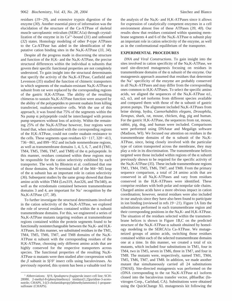
DNA and Viral Constructions. To gain insight into thesites involved in cation specificity of the Na,K-ATPase, weused site-directed mutagenesis focusing on residues intransmembrane domains of theR subunit of the enzyme. Ourmutagenesis approach assumed that residues that determinethe Na+ specificity of the enzyme are probably conservedin all Na,K-ATPases and may differ from the correspondingones common to H,K-ATPases. To select the specific aminoacids, we aligned the sequences of the Na,K-ATPaseR1,R2, R3, andR4 isoforms from different species availableand compared them with those of theR subunit of gastricproton pumps. The alignment included Na,K-ATPases frombrine shrimp, hydra,Caenorhabditis elegans, Drosophila,Xenopus, shark, rat, mouse, chicken, dog, pig and human.For the gastric H,K-ATPase, the sequences from rat, mouse,rabbit, pig, dog, and human were included. Comparisonswere performed using DNAstar and Megalign software(Madison, WI). We focused our attention on residues in thetransmembrane domains of theR subunit of the Na,K-ATPase, since, being closely involved with the particulartype of cation transported across the membrane, they mayplay a role in its discrimination. The transmembrane helicestargeted were those included within regions of theR subunitpreviously shown to be required for the specific activity ofthe Na,K-ATPase (35). These include transmembrane regionsTM1, TM4, TM5, TM6, TM7, and TM8. From the primarysequence comparison, a total of 24 amino acids that areconserved in all Na,K-ATPases and vary from residuesconserved in the H,K-ATPases were identified. Thesecomprise residues with both polar and nonpolar side chains.Charged amino acids have a more obvious impact in cationcoordination; however, neutral residues were also includedin our analysis since they have also been found to participatein ion binding (reviewed in refs19-21). Figure 1A lists thesubstitutions performed in each transmembrane region andtheir corresponding positions in the Na,K- and H,K-ATPase.The situation of the residues selected within the transmem-brane helices is shown in Figure 1B,C on the predictedstructure of the Na,K-ATPaseR subunit obtained by homol-ogy modeling to the SERCA1a Ca-ATPase. We mutage-neized groups of amino acids, switching those residuescontained within each of the selected transmembrane domainsone at a time. In this manner, we created a total of sixmutants, which included four substitutions in TM1, four inTM4, two in TM5, seven in TM6, three in TM7, and four inTM8. The mutants were, respectively, named TM1, TM4,TM5, TM6, TM7, and TM8. In addition, we made anothermutant that simultaneously carried all 24 substitutions(TMAll). Site-directed mutagenesis was performed on thecDNA corresponding to the rat Na,K-ATPaseR1 isoformcloned into the baculovirus transfer vector, pBlueBac (In-vitrogen Corp., Carlsbad, CA). Substitutions were obtainedusing the QuickChange XL mutagenesis kit following the
1 Abbreviations:Sf-9, Spodoptera frugiperdainsect cell line; SCH-28080, 2-methyl-8-(phenylmethoxy) imidazo[1,2]pyridine-3-aceto-notrile; CHAPS, 3-[(3-cholamidopropyl)dimethylammonio]-1-propane-sulfonate (CHAPS).
9062 Biochemistry, Vol. 43, No. 28, 2004 Sanchez and Blanco

protocols suggested by the supplier (Stratagene, Cedar Creek,TX). After confirming the substitutions by sequencing, weused the obtained constructs to make the correspondingrecombinant baculoviruses. Virus preparation and selectionwas performed according to the procedures recommendedby the supplier (Invitrogen Corp., Carlsbad, CA). Both wild-type and mutated forms of theR subunit of the Na,K-ATPasewere coexpressed with the ratâ1 subunit, using for this abaculovirus described previously (39). Also, baculovirusesfor the R andâ subunits of the rabbit gastric H,K-ATPasewere made and used as a control.
Cells and Viral Infections. Sf-9 cells were grown in TNM/FH medium (JRH Biosciences, Lenexa, KS, defined in ref41) supplemented with 10% (v/v) fetal bovine serum, 100units/mL penicillin, 100 mg/mL streptomycin, and 0.25 mg/mL Fungizone (complete medium). Infections were per-formed in 150 mm Petri dishes in serum-free medium for 1h. After addition of complete medium, cultures weremaintained for 72 h. Cells were harvested and centrifuged(5000× g) for 10 min and resuspended at a concentrationof 5 × 10⁶ cells/ml in 0.32 M sucrose, 1 mM EDTA, 30mM imidazole HCl, pH 7.4. Then, total cell membranepreparations were obtained as described previously (42).Briefly, Sf-9 cells were homogenized on ice using a glasshomogenizer, and the lysate was centrifuged for 10 min at1000× g. The supernatant was removed, and the pellet washomogenized and centrifuged as before. Both supernatants
of the first and second centrifugation steps were combinedand centrifuged for 30 min at 100 000× g. Final pellet wasresuspended in the homogenization buffer and used for assaysafter permeabilization with the ionophore alamethicin at aconcentration of 0.01 mg/mg of protein (43).
PAGE and Immunoblot Analysis. Protein expression wasanalyzed by SDS-polyacrylamide gel electrophoresis (SDS-PAGE) and immunoblotting. Proteins were separated in a7.5% gel (44), transferred to nitrocellulose, and immuno-blotted as described previously (42). The wild-type andmutated Na,K-ATPaseR subunits were detected with amonoclonal antibody hybridoma supernatant specific for theR polypeptide, C464/6B, provided by Michael Caplan (YaleUniversity School of Medicine). Theâ1 polypeptide wasidentified using an anti-Râ antiserum raised against rat kidneyNa,K-ATPase (42, 45). Primary antibodies were detectedusing horseradish peroxidase-conjugated secondary antibod-ies and chemiluminescence.
Immunoprecipitations. Uninfected and 72 h infectedSf-9cells grown in 6-well tissue culture plates were lysed with1% 3-[(3-cholamidopropyl)dimethylammonio]-1-propane-sulfonate (CHAPS) in 150 mM NaCl, 25 mM HEPES (pH7.4). After removal of the insoluble material (10 min at15 000× g), samples were subjected to immunoprecipitation.To precipitate the wild-type and mutantR polypeptides, 50µL of monoclonal antibody C464/6B and 100µL (1 mg/mL) of goat anti-mouse coated magnetic beads (BioMag;
FIGURE 1: Amino acid substitutions in the Na,K-ATPase. Panel A shows mutations performed in the Na,K-ATPase and the correspondingreplacements from the H,K-ATPase. Residues are listed according to the transmembrane spanning domain in which they are contained.This generated the TM1, TM4, TM5, TM6, TM7, and TM8 mutants. An additional mutant, TMAll, that simultaneously contained allmutations listed under panel A was prepared. Amino acid positions for the Na,K-ATPase correspond to those of theR1 isoform of the ratenzyme. For the H,K-ATPase, amino acid position is that of the gastric form of the rabbit enzyme. Panels B and C show homology modelsof Na,K-ATPase transmembrane helices from a lateral and cytoplasmic view, respectively. The model was obtained based on the high-resolution structure of the sarcoplasmic reticulum Ca-ATPase as described under Experimental Procedures. Colored residues depict themutations performed: red (TM1), green (TM4), blue (TM5), yellow (TM6), orange (TM7), and magenta (TM8).
Na+ Selectivity of the Na,K-ATPase Biochemistry, Vol. 43, No. 28, 20049063
Qiagen, Inc., Stanford, CA) were used. After overnightincubation on a rocking table at 4°C, beads were isolatedby holding the microcentrifuge tube to a magnet andaspirating the supernatant. The beads were washed 3 timesin the lysis buffer. The precipitated protein was eluted byresuspending the beads in sample buffer (100 mM Tris HClpH 6.8, 2% SDS, 33% glycerol, 100 mM DTT) andincubating for 15 min at 65°C. Eluted proteins wereseparated by SDS-PAGE (7.5% gel), transferred to nitrocel-lulose, and probed with the polyclonal antibody PolyRAthat recognizes theâ subunit.
Immunocytochemistry and Confocal Microscopy. Sf-9 cellswere plated in 24-well culture plates on 11 mm glass coverslips and infected with the wild-type or mutant rat Na,K-ATPaseR and theâ baculoviruses. Forty-eight hours afterinfection, cells were treated with 100µg/mL cycloheximide,an inhibitor of protein synthesis. This treatment clears thebiosynthetic pathway of newly synthesized proteins, allowingthe detection of the expressed polypeptides at their finalcellular destination. Samples were then processed for im-munocytochemistry as described (46). As the primaryantibodies, the monoclonal C464/6B hybridoma supernatantwas used to detect the wild-type and mutatedR subunits anda polyclonal affinity-purified antiserum (Upstate Biotech-nology, Lake Placid, NY) was employed to identify theâpolypeptide. A rhodamine (Rho)-conjugated goat anti-mouseand a fluorescein isothiocyanate (FITC)-conjugated goat anti-rabbit were used as the secondary antibodies. Fluorescentdigital images were obtained using a Zeiss LSM510 confocalmicroscope. Images were acquired in Multitrack channelmode (sequential excitation/emmision) with LSM510 (v 3.0)software and a Plan-NEOFLUAR 40×/1.3 oil DIC objectivewith a zoom factor of 3 (field size of 0.487 mm× 0.487mm) and frame size of 1024 pixels× 1024 pixels.
Biochemical Assays. Protein assays were performed usingthe dye-binding assay based on the method of Bradford fromBio-Rad (Hercules, CA). Na,K-ATPase activity was assayedthrough determination of the initial rate of release of32Pi
from γ[32P]-ATP as described previously (45). The ATPaseactivity of 30-µg total protein samples was measured in afinal volume of 0.25 mL. Maximal Na,K-ATPase activitywas determined in medium containing 120 mM NaCl, 30mM KCl, 3 mM MgCl2, 0.2 mM EGTA, 30 mM Tris-HCl(pH 7.4) with or without 1 mM ouabain. For maximal H,K-ATPase activity, the medium consisted of 20 mM KCl, 3mM MgCl2, 0.2 mM EGTA, 100 mM Tris-HCl (pH 7.0).The assay was started by the addition of ATP with 0.2µCiof [γ-32P]ATP (3 mM final concentration). Following a 30min incubation at 37°C, the tubes were placed on ice, andthe reaction was terminated by the addition of 25µL of 55%trichloroacetic acid. Released [32P]-Pi was converted tophosphomolybdate and extracted with isobutanol. Radioac-tivity of 190 µL of the organic phase was measured by liquidscintillation counting. The ATP hydrolyzed never exceeded15% of the total ATP present in the sample, and hydrolysiswas linear over the incubation time. Specific ATPase activitywas determined as the difference in ATP hydrolysis in theabsence and presence of 1 mM ouabain. Activity of the wild-type H,K-ATPase was determined in the presence andabsence of SCH-28080 at a concentration of 100µM. Thisinhibitor was also used to determine whether the introducedmutations in the Na,K-ATPaseR subunit had made the
resulting enzymes sensitive to the compound. In all cases,SCH-28080 did not affect the catalytic function of themutants (data not shown). For the analysis of activation byNa+ and K+, incubation media was that used for Na,K-ATPase activity except that for Na+ dependency, the Na+
concentration was varied from 0 to 120 mM. For K+
stimulation, the K+ concentration was varied from 0 to 30mM. Choline chloride was added so that the final concentra-tion of Na+ or K+ plus choline totaled 150 mM.
Data Analysis.Curve fitting of the experimental data wasperformed using a Marquardt least-squares nonlinear regres-sion computing program (Sigma Plot, Jandel Scientific, SanRafael, CA). Na+ and K+ activation curves were best fittedaccording to a cooperative model for ligand binding,represented by the equation
where [S] is the concentration of the activating cation (Na+
or K+) andn is the Hill coefficient (nH). The apparent affinity,K0.5 ) K1/n.
Dose-response relations for the inhibition of Na,K-ATPase by orthovanadate were fitted by
where V is the Na,K-ATPase corresponding to a certainconcentration of orthovanadate, [O], expressed as a fractionof activity in the absence of the inhibitor, andKi is theconcentration of orthovanadate that gives the half-maximalinhibition.
Statistical analysis of the concentration-response curvesfor each enzyme was done applying Snedecor’sF testdescribed previously (45).
Molecular Modeling.The homology modeling of wild-type and mutant versions of theR1-subunit of Na,K-ATPasefrom rat was generated using the DeepView/Swiss-Pdb-Viewer program, version 3.7 (SP5) (47). We used thecrystallographic structure of rabbit sarcoplasmic reticulumCa-ATPase, SERCA1a (PDB ID 1EUL) as a template (31,32) and a sequence alignment between the rat Na,K-ATPase(GenBank accession number AAA40775) and the rabbitSERCA1a identical to that used by Sweadner and Donnet(48). Based on the initial model of the wild-type Na,K-ATPase, we built the models of the different mutants byapplying themutatetool of DeepView and selecting the bestavailable rotamer for the side chain of each substitutedresidue. Finally, we refined the initial models by energyminimizations consisting of 1000 steps of steepest descent,followed by conjugate gradient minimization until the energydifference between two consecutive steps was below 0.05kJ/mol.
RESULTS
Recombinant baculoviruses encoding Na,K-ATPaseRsubunit mutants containing substitutions in each individualtransmembrane domain, TM1, TM4, TM5, TM6, TM7, andTM8 (Figure 1), or simultaneously in all (TMAll) were usedto infectSf-9 insect cells. These viruses, as well as the wild-type Na,K-ATPaseR subunit, were applied in combinationwith that of the rat Na,K-ATPaseâ1 subunit. To determineexpression of the corresponding virally induced polypeptides,
V ) (Vm[S]n)/(K + [S]n) (1)
V ) 100[1/(1+ [O]/Ki)] (2)
9064 Biochemistry, Vol. 43, No. 28, 2004 Sanchez and Blanco
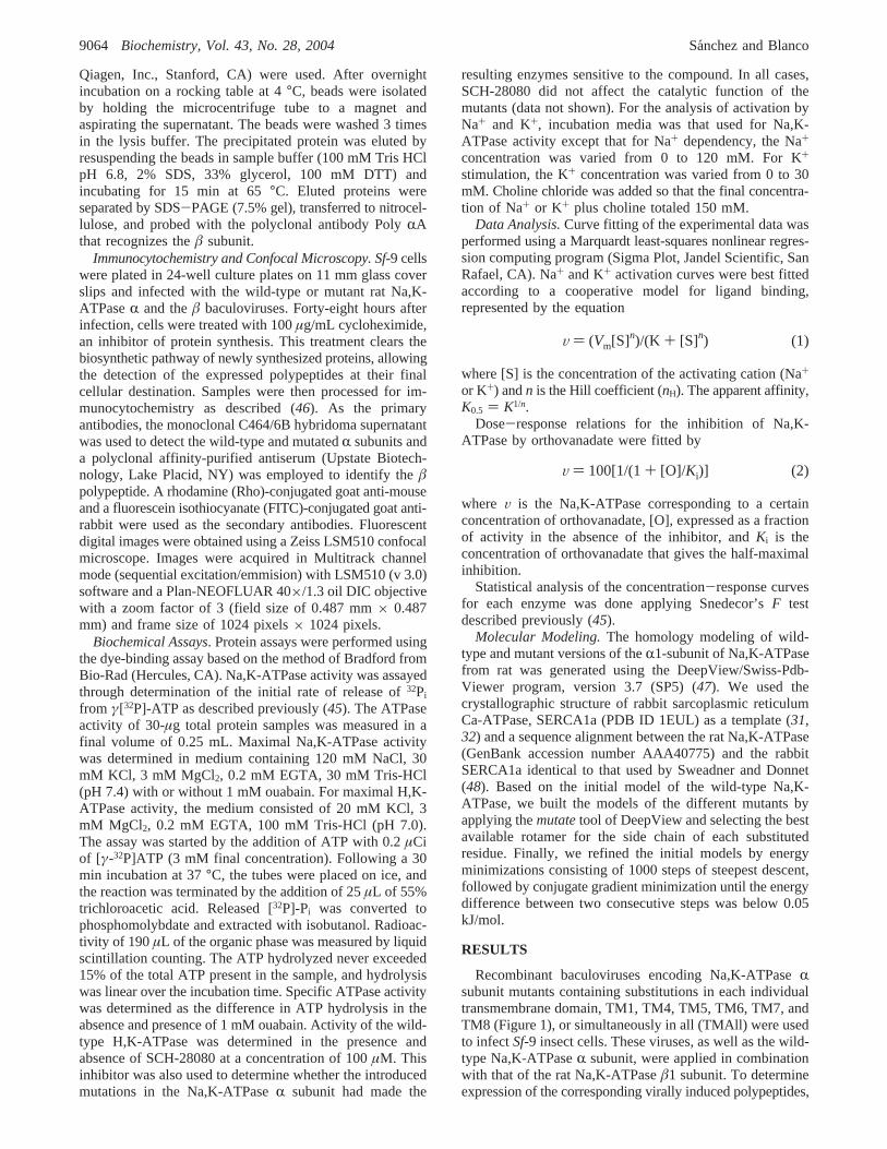
72 h after infection cells were harvested and proteins weresubjected to SDS-PAGE and immunoblotting. As shownin Figure 2A,B, antibodies specific to the Na,K-ATPaseRandâ polypeptides detected high amounts of the correspond-ing proteins only in the infected cells. All Na,K-ATPaseRmutants comigrated with the wild-type subunit at theexpected relative molecular weight. This indicates that theinsect cells are able to express the Na,K-ATPase mutants atlevels that are approximately the same as those of the wild-type enzyme.
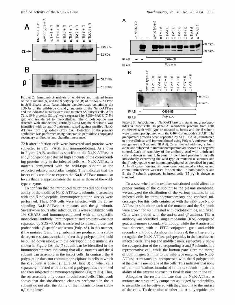
To confirm that the introduced mutations did not alter theability of the modified Na,K-ATPaseR subunits to associatewith the â polypeptide, immunoprecipitation assays wereperformed. Thus,Sf-9 cells were infected with the corre-sponding Na,K-ATPaseR mutants and theâ subunit.Seventy-two hours after infection, cells were solubilized with1% CHAPS and immunoprecipitated with anR-specificmonoclonal antibody. Immunoprecipitated proteins were thenseparated by SDS-PAGE, transferred to nitrocellulose, andprobed with aâ-specific antiserum (PolyRA). In this manner,if the mutatedR and theâ subunits are produced in a stabledetergent-resistant association, then theâ polypeptide shouldbe pulled down along with the correspondingR mutant. Asshown in Figure 3A, theâ subunit can be identified in theimmunoprecipitates indicating that allR mutants and theâsubunit can assemble in the insect cells. In contrast, theâpolypeptide does not coimmunoprecipitate in cells in whichthe R subunit is absent (Figure 3A, lane 3) or when cellsseparately infected with theR andâ polypeptides are mixedand then subjected to immunoprecipitation (Figure 3B). Thus,theRâ assembly only occurs in coinfected cells. This resultshows that the site-directed changes performed in theRsubunit do not alter the ability of the mutants to form stableRâ complexes.
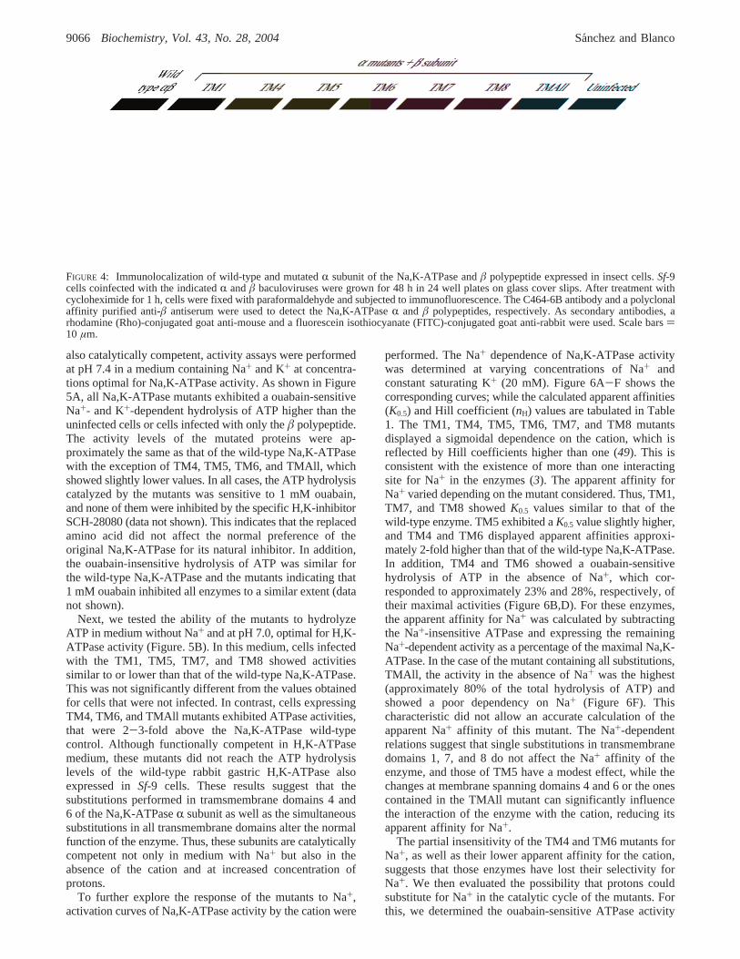
To assess whether the residues substituted could affect theproper routing of theR subunit to the plasma membrane,we analyzed the distribution of the various mutants in theinfected cells by immunocytochemistry and confocal mi-croscopy. For this, cells coinfected with the wild-type Na,K-ATPaseR subunit or each of the mutants and theâ subunitwere grown for 48 h, treated with cycloheximide, and fixed.Cells were probed with the anti-R and -â antisera. TheRantibody was identified using a rhodamine (Rho)-conjugatedgoat anti-mouse secondary antibody, while theâ antiserumwas detected with a FITC-conjugated goat anti-rabbitsecondary antibody. As shown in Figure 4, the antisera onlyrecognize the Na,K-ATPase polypeptides in the baculovirusinfected cells. The top and middle panels, respectively, showthe coexpression of the correspondingR andâ subunits in arepresentative cell, while the bottom panels are the mergeof both images. Similar to the wild-type enzyme, the Na,K-ATPaseR mutants are coexpressed with theâ polypeptideat the plasma membrane of the cells. This indicates that noneof the modifications introduced in theR subunit impair theability of the enzyme to reach its final destination in the cell.
Altogether the results indicate that the Na,K-ATPaseRmutants are structurally competent as judged by their abilityto assemble and be delivered with theâ subunit to the surfaceof the cells. To determine whether theR polypeptides are
FIGURE 2: Immunoblot analysis of wild-type and mutated formsof theR subunit (A) and theâ polypeptide (B) of the Na,K-ATPasein Sf-9 insect cells. Recombinant baculoviruses containing thecDNAs of the wild-typeR and â subunits of the Na,K-ATPaseand the indicated mutants were used to infectSf-9 insect cells. After72 h,Sf-9 proteins (30µg) were separated by SDS-PAGE (7.5%gel) and transferred to nitrocellulose. TheR polypeptide wasdetected with monoclonal antibody C464-6B; theâ subunit wasidentified with an anti-â antiserum raised against purified Na,K-ATPase from dog kidney (PolyRA). Detection of the primaryantibodies was performed using horseradish peroxidase conjugatedsecondary antibodies and chemiluminescence.
FIGURE 3: Association of Na,K-ATPaseR mutants andâ polypep-tides in insect cells. In panel A, membrane proteins from cellscoinfected with wild-type or mutatedR forms and theâ subunitwere immunoprecipitated with the C464-6B antibody (IP AB). Theprecipitated proteins were separated by SDS-PAGE, transferredto nitrocellulose, and immunoblotted using PolyRA antiserum thatrecognizes theâ subunit (IB AB). Cells infected with theâ subunitalone and subjected to immunoprecipitation are shown as a negativecontrol. Lack of reactivity of the antibody used with uninfectedcells is shown in lane 1. In panel B, combined proteins from cellsindividually expressing the wild-type or mutatedR subunits andthe â polypeptide were immunoprecipitated as described in panelA. In all cases, horseradish peroxidase conjugated antibodies andchemiluminescence was used for detection. In both panels A andB, the â subunit expressed in insect cells (15µg) is shown asstandard.
Na+ Selectivity of the Na,K-ATPase Biochemistry, Vol. 43, No. 28, 20049065
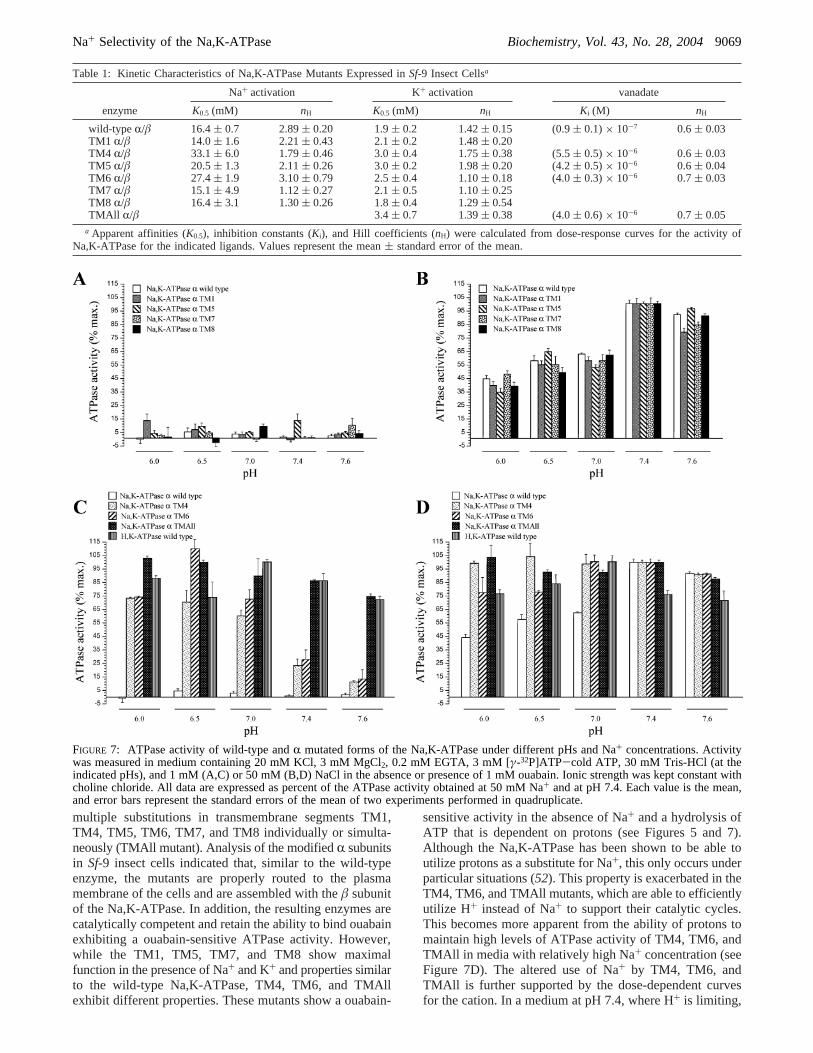
also catalytically competent, activity assays were performedat pH 7.4 in a medium containing Na+ and K+ at concentra-tions optimal for Na,K-ATPase activity. As shown in Figure5A, all Na,K-ATPase mutants exhibited a ouabain-sensitiveNa+- and K+-dependent hydrolysis of ATP higher than theuninfected cells or cells infected with only theâ polypeptide.The activity levels of the mutated proteins were ap-proximately the same as that of the wild-type Na,K-ATPasewith the exception of TM4, TM5, TM6, and TMAll, whichshowed slightly lower values. In all cases, the ATP hydrolysiscatalyzed by the mutants was sensitive to 1 mM ouabain,and none of them were inhibited by the specific H,K-inhibitorSCH-28080 (data not shown). This indicates that the replacedamino acid did not affect the normal preference of theoriginal Na,K-ATPase for its natural inhibitor. In addition,the ouabain-insensitive hydrolysis of ATP was similar forthe wild-type Na,K-ATPase and the mutants indicating that1 mM ouabain inhibited all enzymes to a similar extent (datanot shown).
Next, we tested the ability of the mutants to hydrolyzeATP in medium without Na+ and at pH 7.0, optimal for H,K-ATPase activity (Figure. 5B). In this medium, cells infectedwith the TM1, TM5, TM7, and TM8 showed activitiessimilar to or lower than that of the wild-type Na,K-ATPase.This was not significantly different from the values obtainedfor cells that were not infected. In contrast, cells expressingTM4, TM6, and TMAll mutants exhibited ATPase activities,that were 2-3-fold above the Na,K-ATPase wild-typecontrol. Although functionally competent in H,K-ATPasemedium, these mutants did not reach the ATP hydrolysislevels of the wild-type rabbit gastric H,K-ATPase alsoexpressed inSf-9 cells. These results suggest that thesubstitutions performed in tramsmembrane domains 4 and6 of the Na,K-ATPaseR subunit as well as the simultaneoussubstitutions in all transmembrane domains alter the normalfunction of the enzyme. Thus, these subunits are catalyticallycompetent not only in medium with Na+ but also in theabsence of the cation and at increased concentration ofprotons.
To further explore the response of the mutants to Na+,activation curves of Na,K-ATPase activity by the cation were
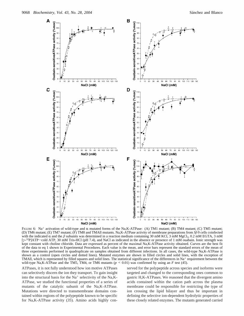
performed. The Na+ dependence of Na,K-ATPase activitywas determined at varying concentrations of Na+ andconstant saturating K+ (20 mM). Figure 6A-F shows thecorresponding curves; while the calculated apparent affinities(K0.5) and Hill coefficient (nH) values are tabulated in Table1. The TM1, TM4, TM5, TM6, TM7, and TM8 mutantsdisplayed a sigmoidal dependence on the cation, which isreflected by Hill coefficients higher than one (49). This isconsistent with the existence of more than one interactingsite for Na+ in the enzymes (3). The apparent affinity forNa+ varied depending on the mutant considered. Thus, TM1,TM7, and TM8 showedK0.5 values similar to that of thewild-type enzyme. TM5 exhibited aK0.5 value slightly higher,and TM4 and TM6 displayed apparent affinities approxi-mately 2-fold higher than that of the wild-type Na,K-ATPase.In addition, TM4 and TM6 showed a ouabain-sensitivehydrolysis of ATP in the absence of Na+, which cor-responded to approximately 23% and 28%, respectively, oftheir maximal activities (Figure 6B,D). For these enzymes,the apparent affinity for Na+ was calculated by subtractingthe Na+-insensitive ATPase and expressing the remainingNa+-dependent activity as a percentage of the maximal Na,K-ATPase. In the case of the mutant containing all substitutions,TMAll, the activity in the absence of Na+ was the highest(approximately 80% of the total hydrolysis of ATP) andshowed a poor dependency on Na+ (Figure 6F). Thischaracteristic did not allow an accurate calculation of theapparent Na+ affinity of this mutant. The Na+-dependentrelations suggest that single substitutions in transmembranedomains 1, 7, and 8 do not affect the Na+ affinity of theenzyme, and those of TM5 have a modest effect, while thechanges at membrane spanning domains 4 and 6 or the onescontained in the TMAll mutant can significantly influencethe interaction of the enzyme with the cation, reducing itsapparent affinity for Na+.
The partial insensitivity of the TM4 and TM6 mutants forNa+, as well as their lower apparent affinity for the cation,suggests that those enzymes have lost their selectivity forNa+. We then evaluated the possibility that protons couldsubstitute for Na+ in the catalytic cycle of the mutants. Forthis, we determined the ouabain-sensitive ATPase activity
FIGURE 4: Immunolocalization of wild-type and mutatedR subunit of the Na,K-ATPase andâ polypeptide expressed in insect cells.Sf-9cells coinfected with the indicatedR andâ baculoviruses were grown for 48 h in 24 well plates on glass cover slips. After treatment withcycloheximide for 1 h, cells were fixed with paraformaldehyde and subjected to immunofluorescence. The C464-6B antibody and a polyclonalaffinity purified anti-â antiserum were used to detect the Na,K-ATPaseR and â polypeptides, respectively. As secondary antibodies, arhodamine (Rho)-conjugated goat anti-mouse and a fluorescein isothiocyanate (FITC)-conjugated goat anti-rabbit were used. Scale bars)10 µm.
9066 Biochemistry, Vol. 43, No. 28, 2004 Sanchez and Blanco

of all mutants at different pHs and under two concentrationsof Na+, 1 and 50 mM. Because both these concentrations ofNa+ are not saturating for the ATPase reaction (see Figure6), this allowed the determination of the competition betweenNa+ and protons. Figure 7A,B shows the activities of theTM1, TM5, TM7, and TM8 mutants at both concentrationsof Na+, while those of TM4, TM6, and TMAll are depictedin Figure 7C,D. The wild-type Na,K- and H,K-ATPases have
been included for comparison. As shown, TM1, TM5, TM7,and TM8 have a behavior similar to that of the wild-typeNa,K-ATPase with negligible levels of ATP hydrolysis at 1mM Na+ (Figure 7A) and a Na+ activation that reachesmaximal activity at pH 7.4 (Figure 7B). In contrast, the TM4,TM6, and TMAll mutants exhibited a ouabain-sensitivehydrolysis of ATP that was independent of Na+ and isstimulated by a decrease in pH (Figure 7C). The propertiesof the TM4, TM6, and TMAll mutants are significantlydifferent from those of the wild-type Na,K-ATPase andresemble more those of the wild-type H,K-ATPase (Figure7C). In addition, at 50 mM Na+, these mutants display highactivity over the pH range tested, and they are not limitedby the decrease in proton concentration seen at 1 mM ofNa+ (Figure 7D). These observations support the idea thatthe mutated residues in TM4, TM6, and TMAll impair thecapacity of the enzyme to recognize Na+ over H+, makingit able to use both cations for ATP hydrolysis.
To characterize the kinetics of the mutants to K+, Na,K-ATPase activity was measured at varying concentrations ofK+ (0-30 mM) with Na+ fixed at 120 mM. The obtainedcurves are presented in Figure 8A-F, and values describingthe kinetic parameters are depicted in Table 1. As shown,the half-activation constant for K+ of the TM1, TM7, andTM8 is similar to that of the wild-type enzyme, while TM4,TM5, TM6, and TMAll exhibited a modest decrease in theapparent affinity for the cation withK0.5 values approximately1.5-fold higher. The computed Hill coefficients for K+ reflectpositive cooperativity at more than one ligand binding sitefor all enzymes. Different from the dependency curves toNa+, none of the mutants showed an ATP hydrolysisindependent of K+, demonstrating that the selectivity of theenzymes for this cation is retained.
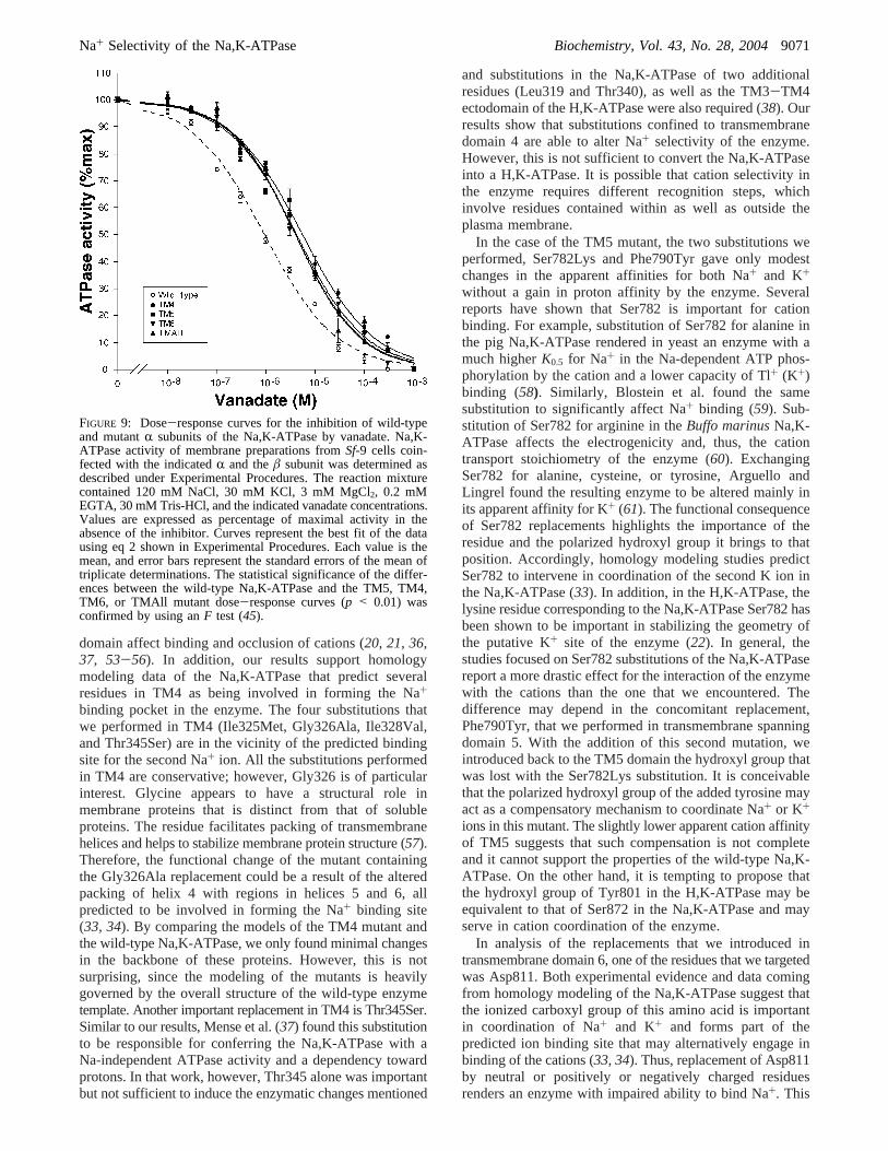
Shifts in apparent affinity for K+ in the Na,K-ATPase arecommonly associated with changes in the steady-state E1/E2 conformational equilibrium of the enzyme in favor ofthe E1 conformation (3, 4). To explore this possibility, wemeasured the sensitivity of the Na,K-ATPase activity ofTM4, TM5, TM6, and TMAll mutants to orthovanadate.Orthovanadate acts as a Na,K-ATPase inhibitor competingwith inorganic phosphate and binding to the E2 conformationof the enzyme (50). Thus, vanadate can be used as a tool toobtain information on the E1/E2 conformational state of theNa,K-ATPase (36, 51). A displacement of the conformationalequilibrium in favor of E1 is expected to result in a reducedsensitivity of the Na,K-ATPase to orthovanadate. Figure 9shows the dose response curves of Na,K-ATPase activity toorthovanadate, while the calculatedKi and Hill coefficientvalues are depicted in Table 1. As shown, the TM4, TM5,TM6, and TMAll mutants displayed affinities that are 4-5-fold lower than that of the wild-type enzyme. Also, in allcases, and consistent with the existence of a single bindingsite in the enzyme (3), the Hill coefficients indicated valueslower than 1. This indicates that the TM4, TM5, TM6, andTMAll mutants have become more resistant to orthovanadate,suggesting they exist mainly in the E1 state.
DISCUSSION
Although experimental evidence and homology modelingbased on the crystal structure of the Ca-ATPase is providingnew insights into the structure and function of P-type
FIGURE 5: ATPase activity of wild-type and mutated forms of theR subunit of the Na,K-ATPase. Panel A shows activity levels inmedium optimal for Na,K-ATPase activity. Ouabain-sensitivehydrolysis of ATP was measured as described in ExperimentalProcedures in medium containing 120 mM NaCl, 30 mM KCl, 3mM MgCl2, 0.2 mM EGTA, 3 mM [γ-32P]ATP-cold ATP, and30 mM Tris-HCl (pH 7.4) in the absence or presence of 1 mMouabain. Panel B shows ATPase activity in medium optimal forH,K-ATPase. The incubation medium contained 20 mM KCl, 3mM MgCl2, 0.2 mM EGTA, and 100 mM Tris-HCl (pH 7.0).Activity of the wild-type and mutant forms of the Na,K-ATPasewas sensitive to 1 mM ouabain, whereas activity of the H,K-ATPasewas sensitive to SCH-28080. Each value is the mean, and errorbars represent the standard errors of the mean of three to sevenexperiments performed in triplicate on samples obtained fromdifferent infections.
Na+ Selectivity of the Na,K-ATPase Biochemistry, Vol. 43, No. 28, 20049067
ATPases, it is not fully understood how ion motive ATPasescan selectively discern the ion they transport. To gain insightinto the structural basis for the Na+ selectivity of the Na,K-ATPase, we studied the functional properties of a series ofmutants of the catalytic subunit of the Na,K-ATPase.Mutations were directed to transmembrane domains con-tained within regions of the polypeptide known to be specificfor Na,K-ATPase activity (35). Amino acids highly con-
served for the polypeptide across species and isoforms weretargeted and changed to the corresponding ones common togastric H,K-ATPases. We reasoned that the divergent aminoacids contained within the cation path across the plasmamembrane could be responsible for restricting the type ofion crossing the lipid bilayer and thus be important indefining the selective ion-dependent hydrolytic properties ofthese closely related enzymes. The mutants generated carried
FIGURE 6: Na+ activation of wild-type andR mutated forms of the Na,K-ATPase: (A) TM1 mutant; (B) TM4 mutant; (C) TM5 mutant;(D) TM6 mutant; (E) TM7 mutant; (F) TM8 and TMAll mutants. Na,K-ATPase activity of membrane preparations fromSf-9 cells coinfectedwith the indicatedR and theâ subunits was determined in a reaction medium containing 30 mM KCl, 3 mM MgCl2, 0.2 mM EGTA, 3 mM[γ-32P]ATP-cold ATP, 30 mM Tris-HCl (pH 7.4), and NaCl as indicated in the absence or presence of 1 mM ouabain. Ionic strength waskept constant with choline chloride. Data are expressed as percent of the maximal Na,K-ATPase activity obtained. Curves are the best fitof the data to eq 1 shown in Experimental Procedures. Each value is the mean, and error bars represent the standard errors of the mean ofthree experiments performed in quadruplicate on samples obtained from different infections. In all cases, the wild-type Na,K-ATPase isshown as a control (open circles and dotted lines). Mutated enzymes are shown in filled circles and solid lines, with the exception ofTMAll, which is represented by filled squares and solid lines. The statistical significance of the differences in Na+ requirement between thewild-type Na,K-ATPase and the TM5, TM4, or TM6 mutants (p < 0.01) was confirmed by using anF test (45).
9068 Biochemistry, Vol. 43, No. 28, 2004 Sanchez and Blanco
multiple substitutions in transmembrane segments TM1,TM4, TM5, TM6, TM7, and TM8 individually or simulta-neously (TMAll mutant). Analysis of the modifiedR subunitsin Sf-9 insect cells indicated that, similar to the wild-typeenzyme, the mutants are properly routed to the plasmamembrane of the cells and are assembled with theâ subunitof the Na,K-ATPase. In addition, the resulting enzymes arecatalytically competent and retain the ability to bind ouabainexhibiting a ouabain-sensitive ATPase activity. However,while the TM1, TM5, TM7, and TM8 show maximalfunction in the presence of Na+ and K+ and properties similarto the wild-type Na,K-ATPase, TM4, TM6, and TMAllexhibit different properties. These mutants show a ouabain-
sensitive activity in the absence of Na+ and a hydrolysis ofATP that is dependent on protons (see Figures 5 and 7).Although the Na,K-ATPase has been shown to be able toutilize protons as a substitute for Na+, this only occurs underparticular situations (52). This property is exacerbated in theTM4, TM6, and TMAll mutants, which are able to efficientlyutilize H+ instead of Na+ to support their catalytic cycles.This becomes more apparent from the ability of protons tomaintain high levels of ATPase activity of TM4, TM6, andTMAll in media with relatively high Na+ concentration (seeFigure 7D). The altered use of Na+ by TM4, TM6, andTMAll is further supported by the dose-dependent curvesfor the cation. In a medium at pH 7.4, where H+ is limiting,
Table 1: Kinetic Characteristics of Na,K-ATPase Mutants Expressed inSf-9 Insect Cellsa
Na+ activation K+ activation vanadate
enzyme K0.5 (mM) nH K0.5 (mM) nH Ki (M) nH
wild-type R/â 16.4( 0.7 2.89( 0.20 1.9( 0.2 1.42( 0.15 (0.9( 0.1)× 10-7 0.6( 0.03TM1 R/â 14.0( 1.6 2.21( 0.43 2.1( 0.2 1.48( 0.20TM4 R/â 33.1( 6.0 1.79( 0.46 3.0( 0.4 1.75( 0.38 (5.5( 0.5)× 10-6 0.6( 0.03TM5 R/â 20.5( 1.3 2.11( 0.26 3.0( 0.2 1.98( 0.20 (4.2( 0.5)× 10-6 0.6( 0.04TM6 R/â 27.4( 1.9 3.10( 0.79 2.5( 0.4 1.10( 0.18 (4.0( 0.3)× 10-6 0.7( 0.03TM7 R/â 15.1( 4.9 1.12( 0.27 2.1( 0.5 1.10( 0.25TM8 R/â 16.4( 3.1 1.30( 0.26 1.8( 0.4 1.29( 0.54TMAll R/â 3.4( 0.7 1.39( 0.38 (4.0( 0.6)× 10-6 0.7( 0.05
a Apparent affinities (K0.5), inhibition constants (Ki), and Hill coefficients (nH) were calculated from dose-response curves for the activity ofNa,K-ATPase for the indicated ligands. Values represent the mean( standard error of the mean.
FIGURE 7: ATPase activity of wild-type andR mutated forms of the Na,K-ATPase under different pHs and Na+ concentrations. Activitywas measured in medium containing 20 mM KCl, 3 mM MgCl2, 0.2 mM EGTA, 3 mM [γ-32P]ATP-cold ATP, 30 mM Tris-HCl (at theindicated pHs), and 1 mM (A,C) or 50 mM (B,D) NaCl in the absence or presence of 1 mM ouabain. Ionic strength was kept constant withcholine chloride. All data are expressed as percent of the ATPase activity obtained at 50 mM Na+ and at pH 7.4. Each value is the mean,and error bars represent the standard errors of the mean of two experiments performed in quadruplicate.
Na+ Selectivity of the Na,K-ATPase Biochemistry, Vol. 43, No. 28, 20049069

the mutants exhibit a slower activation by Na+ with anapproximate 2-fold increase in theK0.5 values for the cation.The possibility that K+ is also replacing Na+ in the catalyticcycle of these enzymes is unlikely, because at low Na+ andat constant saturating K+ (20 mM), activity of the TM4,TM6, and TMAll mutants diminishes with the increase ofpH (see Figure 7C). Altogether these results indicate that
the modified enzymes can drive a ouabain-sensitive Na+-as well as a H+-dependent ATPase activity. This can beinterpreted as a loss of selectivity of the TM4, TM6, andTMAll mutants for Na+ over H+.
Our results indicating that residues in TM4 are importantfor Na+ selectivity of the enzyme agree with other studiesshowing that point mutations within this transmembrane
FIGURE 8: K+ dependence of wild-type andR mutated forms of the Na,K-ATPase: (A) TM1 mutant; (B) TM4 mutant; (C) TM5 mutant;(D) TM6 mutant; (E) TM7 mutant; (F) TM8 and TMAll mutants. Na,K-ATPase activity of membrane preparations fromSf-9 cells coinfectedwith the indicatedR and theâ subunit was determined as described under Experimental Procedures. The reaction medium contained 120mM NaCl, 3 mM MgCl2, 0.2 mM EGTA, 3 mM [γ-32P]ATP-cold ATP, 30 mM Tris-HCl (pH 7.4), and KCl (from 0 to 30 mM) with orwithout 1 mM ouabain. Ionic strength was kept constant with choline chloride. Data are expressed as percent of the maximal Na,K-ATPaseactivity obtained. Curves are the best fit of the data to eq 1 shown in Experimental Procedures. Each value is the mean, and error barsrepresent the standard errors of the mean of three experiments performed in quadruplicate on samples obtained from different infections.In all cases, the wild-type Na,K-ATPase is shown as a control (open circles and dotted lines). Mutated enzymes are shown in filled circlesand solid lines, with the exception of TMAll, which is represented by filled squares and solid lines. The statistical significance of thedifferences in K+ requirement between the wild-type Na,K-ATPase and the TM5, TM4, TM6, or TMAll mutants (p < 0.01) was confirmedby using anF test (45).
9070 Biochemistry, Vol. 43, No. 28, 2004 Sanchez and Blanco
domain affect binding and occlusion of cations (20, 21, 36,37, 53-56). In addition, our results support homologymodeling data of the Na,K-ATPase that predict severalresidues in TM4 as being involved in forming the Na+
binding pocket in the enzyme. The four substitutions thatwe performed in TM4 (Ile325Met, Gly326Ala, Ile328Val,and Thr345Ser) are in the vicinity of the predicted bindingsite for the second Na+ ion. All the substitutions performedin TM4 are conservative; however, Gly326 is of particularinterest. Glycine appears to have a structural role inmembrane proteins that is distinct from that of solubleproteins. The residue facilitates packing of transmembranehelices and helps to stabilize membrane protein structure (57).Therefore, the functional change of the mutant containingthe Gly326Ala replacement could be a result of the alteredpacking of helix 4 with regions in helices 5 and 6, allpredicted to be involved in forming the Na+ binding site(33, 34). By comparing the models of the TM4 mutant andthe wild-type Na,K-ATPase, we only found minimal changesin the backbone of these proteins. However, this is notsurprising, since the modeling of the mutants is heavilygoverned by the overall structure of the wild-type enzymetemplate. Another important replacement in TM4 is Thr345Ser.Similar to our results, Mense et al. (37) found this substitutionto be responsible for conferring the Na,K-ATPase with aNa-independent ATPase activity and a dependency towardprotons. In that work, however, Thr345 alone was importantbut not sufficient to induce the enzymatic changes mentioned
and substitutions in the Na,K-ATPase of two additionalresidues (Leu319 and Thr340), as well as the TM3-TM4ectodomain of the H,K-ATPase were also required (38). Ourresults show that substitutions confined to transmembranedomain 4 are able to alter Na+ selectivity of the enzyme.However, this is not sufficient to convert the Na,K-ATPaseinto a H,K-ATPase. It is possible that cation selectivity inthe enzyme requires different recognition steps, whichinvolve residues contained within as well as outside theplasma membrane.
In the case of the TM5 mutant, the two substitutions weperformed, Ser782Lys and Phe790Tyr gave only modestchanges in the apparent affinities for both Na+ and K+
without a gain in proton affinity by the enzyme. Severalreports have shown that Ser782 is important for cationbinding. For example, substitution of Ser782 for alanine inthe pig Na,K-ATPase rendered in yeast an enzyme with amuch higherK0.5 for Na+ in the Na-dependent ATP phos-phorylation by the cation and a lower capacity of Tl+ (K+)binding (58). Similarly, Blostein et al. found the samesubstitution to significantly affect Na+ binding (59). Sub-stitution of Ser782 for arginine in theBuffo marinusNa,K-ATPase affects the electrogenicity and, thus, the cationtransport stoichiometry of the enzyme (60). ExchangingSer782 for alanine, cysteine, or tyrosine, Arguello andLingrel found the resulting enzyme to be altered mainly inits apparent affinity for K+ (61). The functional consequenceof Ser782 replacements highlights the importance of theresidue and the polarized hydroxyl group it brings to thatposition. Accordingly, homology modeling studies predictSer782 to intervene in coordination of the second K ion inthe Na,K-ATPase (33). In addition, in the H,K-ATPase, thelysine residue corresponding to the Na,K-ATPase Ser782 hasbeen shown to be important in stabilizing the geometry ofthe putative K+ site of the enzyme (22). In general, thestudies focused on Ser782 substitutions of the Na,K-ATPasereport a more drastic effect for the interaction of the enzymewith the cations than the one that we encountered. Thedifference may depend in the concomitant replacement,Phe790Tyr, that we performed in transmembrane spanningdomain 5. With the addition of this second mutation, weintroduced back to the TM5 domain the hydroxyl group thatwas lost with the Ser782Lys substitution. It is conceivablethat the polarized hydroxyl group of the added tyrosine mayact as a compensatory mechanism to coordinate Na+ or K+
ions in this mutant. The slightly lower apparent cation affinityof TM5 suggests that such compensation is not completeand it cannot support the properties of the wild-type Na,K-ATPase. On the other hand, it is tempting to propose thatthe hydroxyl group of Tyr801 in the H,K-ATPase may beequivalent to that of Ser872 in the Na,K-ATPase and mayserve in cation coordination of the enzyme.
In analysis of the replacements that we introduced intransmembrane domain 6, one of the residues that we targetedwas Asp811. Both experimental evidence and data comingfrom homology modeling of the Na,K-ATPase suggest thatthe ionized carboxyl group of this amino acid is importantin coordination of Na+ and K+ and forms part of thepredicted ion binding site that may alternatively engage inbinding of the cations (33, 34). Thus, replacement of Asp811by neutral or positively or negatively charged residuesrenders an enzyme with impaired ability to bind Na+. This
FIGURE 9: Dose-response curves for the inhibition of wild-typeand mutantR subunits of the Na,K-ATPase by vanadate. Na,K-ATPase activity of membrane preparations fromSf-9 cells coin-fected with the indicatedR and theâ subunit was determined asdescribed under Experimental Procedures. The reaction mixturecontained 120 mM NaCl, 30 mM KCl, 3 mM MgCl2, 0.2 mMEGTA, 30 mM Tris-HCl, and the indicated vanadate concentrations.Values are expressed as percentage of maximal activity in theabsence of the inhibitor. Curves represent the best fit of the datausing eq 2 shown in Experimental Procedures. Each value is themean, and error bars represent the standard errors of the mean oftriplicate determinations. The statistical significance of the differ-ences between the wild-type Na,K-ATPase and the TM5, TM4,TM6, or TMAll mutant dose-response curves (p < 0.01) wasconfirmed by using anF test (45).
Na+ Selectivity of the Na,K-ATPase Biochemistry, Vol. 43, No. 28, 20049071
is reflected by a reduction in Na+ affinity for phosphorylationof the enzyme by ATP (62, 63). The decrease in the apparentNa+ affinity that we observe in the TM6 mutant agrees wellwith those observations. The homology modeling that weperformed on the TM6 mutant also supports the role ofAsp811 in Na+ binding. The carboxylic group of theglutamate that we introduced at that position is displacedapproximately 2 Å away from the predicted binding site forNa+. This would be expected to affect Na+ coordinationwithin the cation binding pocket. Therefore, Asp811 repre-sents an essential residue for Na+ coordination and recogni-tion by the Na,K-ATPase. In addition, Asp811 has beenshown to be involved in interaction with K+. This isevidenced by a lower IC50 value for the antagonism ofouabain binding by K+ (64, 65), by a decrease in K+ affinityfor occlusion (63), and by a reduction in K+ affinity fordephosphorylation of the enzyme (63). Replacement of thecorresponding amino acid in the H,K-ATPase (Glu820) alsoresults in a drastic loss of K+ affinity (66). The changes thatwe find in theK0.5 for K+ of the TM6 mutant are much moremodest. This reflects the influence of the additional substitu-tions performed. Among these, the polarized hydroxyl groupprovided by Ser819 in place of alanine could play a role infacilitating coordination of the cation. This suggests that therole of Asp811 in the Na,K-ATPase in K+ coordinationdepends on the neighboring residues. It is conceivable thatthe replacement of Asp811Glu naturally occurring in theH,K-ATPase may provide cation specificity to the enzymedisfavoring Na+ binding, while concomitant changes, suchas that of Ser819 may help maintain the interaction with K+.The other substitutions that we performed in TM6 that mostprobably affect the interaction of the enzyme with Na+ areGly813Cys and Val817Phe. Regarding Gly813, homologymodeling studies predict the residue as being involved inthe formation of the site for the third Na+ ion (33, 34). Forthe phenylalanine substituting for Val817, the side chaingeometry of phenylalanine may impose local steric effectsthat could affect ion coordination through the neighboringGly813, Thr814, and Asp815, all of which form part of thepredicted Na+ binding pocket (33).
The enzymatic changes described for the substitutions atTM4 and TM6 appear to be exacerbated in the TMAllmutant. This mutant displays the poorest dependency on Na+
and the highest Na-independent ATPase activity underdifferent pHs (see Figures 6F and 7C). This suggests asynergistic effect of both transmembrane domains in thecation selectivity of the TMAll enzyme. However, we cannotcompletely discard that the concurrent presence of substitu-tions in other transmembrane domains may be contributingto the larger effect. In this respect, transmembrane domain5, along with domains 6 and 7, is thought to shape the cationbinding pocket (34). Although the isolated mutations in TM5show only a minor change in apparent affinity for Na+ andin the E1/E2 conformational state with no activation ofATPase activity by H+, they may play a more relevant rolewhen combined with those of TM4 and TM6. Interestingly,TMAll shares some of the characteristics of the H,K-ATPase,such as its stimulation by protons. However, TMAll differsfrom the H,K-ATPase in its activation by Na+ at pH 7.4and 7.6. Thus, the substitutions in TMAll are not sufficientto convert the function of the enzyme to a H,K-ATPase, andinstead, it behaves as a hybrid Na/H,K-ATPase. Coinciden-
tally with this observation, some of the residues that wemutated (Thr345Ser in TM4 and Cys809Phe and Ala819Serin TM6) naturally occur in the distal colon ATPase from ratand guinea pig. This may explain the ability of these enzymesto transport both Na+ and H+ and function as Na/H,K-ATPases (67, 68). The function of the TMAll mutantindicates that transmembrane domains are not sufficient toconfer Na+ selectivity to the Na,K-ATPase and otherdomains are involved. Our observation confirms previousresults by Mense at al., who show that cytoplasmic, as wellas extracellular, domains interact to create the particularproperties of P-type ATPases (38).
Our results also indicate that changes in certain trans-membrane domains do not alter the enzymatic properties ofthe Na,K-ATPase. The normal behavior of TM1 and TM7mutants suggests that the residues targeted in those helicesare not involved in cation selectivity. This agrees with modelspredicting the absence of sites for cation coordination in thosepositions (33, 34). Similarly, the TM8 mutant preserved theproperties of the original Na,K-ATPase. In this spanningmembrane domain, homology modeling predicts Gly920 asa putative cation binding site (34). This residue was notchanged in the TM8 mutant, and comparison of the modelsof the TM8 and wild-type enzymes showed that the substitu-tions performed do not alter the backbone structure of thehelices in the region. Therefore, the changes introduced inTM8 are not affecting the ability of Gly920 to coordinatethe cations.
Besides the differences in the apparent affinity for Na+
and the gain in reactivity to H+, another consequence ofmutations in TM4, TM5, TM6, and TMAll is a slight changein the K0.5 for K+. In this case however, a significant K+-independent ATPase activity was not detected suggesting thatthe selectivity of the mutants to K+ is preserved. The changesin apparent affinity for K+ are accompanied by a decreasein sensitivity to vanadate. Differences in vanadate bindingare commonly used as an indicator of Na,K-ATPase con-formational equilibrium (37, 38, 50, 51). Our finding thatthe mutants are 4-5-fold more resistant to vanadate suggeststhat the substitutions introduced affect the conformationalequilibrium of the enzyme causing it to favor the E1conformation. The possibility that the vanadate binding sitehas been altered with the substitutions introduced is unlikely.The proposed vanadate binding site is located within thecytoplasmic loop between domains TM4 and TM5, in aregion highly conserved for all P-type ATPases and distantfrom the residues that we targeted (1-3). On the other hand,we cannot completely rule out the possibility that the sitesthat bind K+ in the mutants have been secondarily affectedby the changes introduced. However, the concomitant changein K+ and vanadate affinities supports the idea of adisplacement in the conformational equilibrium of theenzyme (50). Interestingly, it is known that in the H,K-ATPase, the E2K form of the enzyme is less stable than thatof the Na,K-ATPase, that the E2K to E1K transition is faster,and that it can deocclude K+ more rapidly (69). This is inagreement with the concept that the TM4, TM5, TM6, andTMAll mutants have adopted some of the properties of theH,K-ATPase. Supporting our data are the results from Menseet al., indicating that replacement of residues at Na,K-ATPaseTM4 by the corresponding ones of the H,K-ATPase stabilizesthe K+-induced conformation and renders an enzyme with
9072 Biochemistry, Vol. 43, No. 28, 2004 Sanchez and Blanco

properties of those of the H,K-ATPase (38). Likewise theH,K/Na,K chimeras engineered by Koenderink et al. showthat introduction of H,K-ATPase sequences to the Na,K-ATPase results in a shift of the enzyme toward the E1conformation (70).
In conclusion, our study shows that residues within TM4and TM6 in the Na,K-ATPase play an important role in thecation selectivity of the Na,K-ATPase and participate in theconformational transitions associated with the binding ofions. Our results also indicate that the differences betweenthe Na,K- and H,K-ATPase in the transmembrane domainsare not sufficient to account for the ability of the enzyme tofully discriminate between cations. Future studies of thefunction of other regions along the structure of these ionpumps will provide additional information in understandingion selectivity of P-type ATPases.
ACKNOWLEDGMENT
We thank Dr. Edd Rabon for providing the cDNAs of theR andâ subunits of the rabbit gastric H,K-ATPase, Drs. JianXie and Michael Caplan for supplying antibodies, Dr. JuanCodina for generously providing the H,K-inhibitor SCH-28080, Dr. Adrian Arakaki for his help in the homologymodeling analysis of the enzymes studied in this work, Dr.Elizabeth Petroske for her assistance on the confocal imagingof the cells, and Dr. Robert W. Mercer for helpful advice.
REFERENCES
1. Lutsenko, S., and Kaplan, J. H. (1995) Organization of P-typeATPases: significance of structural diversity,Biochemistry 34,15607-15613.
2. Apell, H. J. (2003) Structure-function relationship in P-typeATPases- a biophysical approach,ReV. Physiol. Biochem.Pharmacol. 150, 1-35.
3. Skou, J. C., and Esmann, M. (1992) The Na,K-ATPase,J.Bioenerg. Biomembr. 24, 249-261.
4. Glynn, I. M. (1993) Annual review prize lecture. ‘All hands tothe sodium pump’,J. Physiol. London 462, 1-30.
5. Hershey, S. J., and Sachs, G. (1995) Gastric acid secretion,Phys.ReV. 75, 155-189.
6. Doucet, A. (1997) H,K-ATPase in the kidney: localization andfunction in the nephron,Exp. Nephrol. 5, 271-276.
7. Kone, B. C. (1996) Renal H,K-ATPase: Structure, function, andregulation,Miner. Electrolyte Metab. 22, 349-365.
8. Wingo, C. S., and Smolka, A. J. (1995) Function and structure ofH,K-ATPase in the kidney,Am. J. Physiol. 269, F1-F16.
9. Jørgensen, P. L. (1990)Structure and molecular mechanisms ofthe Na,K-pump, pp 117-154, CRC Press, Boca Raton, FL.
10. Rabon, E. C., and Reuben, M. A. (1990) The mechanism andstructure of the gastric H,K-ATPase,Annu. ReV. Phys. 52, 321-344.
11. Kuhlbrandt, W., Auer, M., and Scarborough, G. A. (1998) Structureof the P-type ATPases,Curr. Opin. Struct. Biol. 8, 510-516.
12. Pressley, T. A. (1996) Structure and function of the Na,K pump:ten years of molecular biology,Miner. Electrolyte Metab. 22,264-271.
13. Lingrel, J. B., and Kuntzweiler, T. (1994) Na+,K+-ATPase.J.Biol. Chem. 269, 19659-19662.
14. McDonough, A. A., Geering, K., and Farley, R. A. (1990) Thesodium pump needs its beta subunit.FASEB J. 4, 1598-1605.
15. Geering, K. (2001) The functional role of beta subunits inoligomeric P-type ATPases,J. Bioenerg. Biomembr. 33, 425-438.
16. Chow, D. C., and Forte, J. G. (1995) Functional significance oftheâ subunit for heterodimeric P-type ATPases,J. Exp. Biol. 198,1-17.
17. Mears, J. M., and Kaplan, B. (1994) Proton pump inhibitors: newdrugs and indications,J. Biol. Chem. 269, 28249-28258.
18. Arguello, J. M., and Kaplan, J. H. (1991) Evidence for essentialcarboxyls in the cation-binding domain of the Na,K-ATPase,J.Biol. Chem. 266, 14627-14635.
19. Blostein, R. (1999) Structure-function studies of the sodium pump,Biochem. Cell. Biol. 77, 1-10.
20. Jorgensen, P. L., Hakansson, K. O., and Karlish, J. D. (2003)Structure and mechanism of Na,K-ATPase,Annu. ReV. Physiol.65, 817-849.
21. Kaplan, J. H. (2002) Biochemistry of Na,K-ATPase,Annu. ReV.Biochem. 71, 511-535.
22. Rulli, J. S., Louneva, N. M., Scripnikova, E. V., and Rabon, E.C. (2001) Site-directed mutagenesis of cation coordinating residuesin the gastric H,K-ATPase,Arch. Biochem. Biophys. 387, 27-34.
23. Asano, S., Tega, Y., Konishi, K., Fujioka, M., and Taneguchi, N.(1996) Functional expression of gastric H,K-ATPase and sitedirected mutagenesis of the putative cation binding site andcatalytic center,J. Biol. Chem. 271, 2740-2745.
24. Asano, S., Kimura, T., Sakamoto, S., and Taneguchi, N. (2001)Alanine-scanning mutagenesis of the sixth transmembrane segmentof gastric H, K.-ATPase alpha-subunit,J. Biol. Chem. 276,31265-31273.
25. Swarts H. G. P., Klaassen, C. H. W., de Boer, M., Fransen, J. M.A., and De Pont, J. J. H. H. M. (1996) Role of negatively chargedresidues in the fifth and sixth transmembrane domains of thecatalytic subunit of gastric H,K-ATPase,J. Biol. Chem. 271,29764-29772.
26. Swarts, H. G. P., Hermsen, H. P. H., Koenderink, J. B, SchuurmansStekhoven, F. M. A. H., and De Pont, J. J. H. H. M. (1998)Constitutive activation of gastric H,K-ATPase by a single muta-tion, EMBO J. 17, 3029-3035.
27. Hermsen, H. P. H., Swarts H. G. P., Wassink L., Koenderink, J.B, Willems, P. H. G. M., and De Pont, J. J. H. H. M. (2001)Mimicking of K+ activation by double mutation of Glutamate 795and Glutamate 820 of gastric H,K-ATPase,Biochemistry 40,6527-6533.
28. Swarts H. G. P., Willems, P. H. G. M., Koenderink, J. B, and DePont, J. J. H. H. M. (2003) The role of Lys791 and Asn792 ingastric H,K-ATPase,Ann. N. Y. Acad. Sci. 986, 308-309.
29. Koenderink, J. B., Swarts H. G. P., Willems, P. H. G. M., and DePont, J. J. H. H. M. (2004) A conformation-specific interhelicalsalt bridge in the K+-binding site of gastric H, K.-ATPase,J. Biol.Chem., in press.
30. Lutsenko, S., Anderko, R., and Kaplan, J. H. (1995) Membranedisposition of the M5-M6 hairpin of Na+,K+-ATPase alpha subunitis ligand dependent,Proc. Natl. Acad. Sci. U.S.A. 92, 7936-7940.
31. Toyoshima, C., Nakasako, M., Nomura, H. and Ogawa, H. (2000)Crystal structure of the calcium pump of sarcoplasmic reticulumat 2.6 Å resolution,Nature 405, 647-655.
32. Toyoshima, C., and Nomura, H. (2002) Structural changes in thecalcium pump accompanying the dissociation of calcium,Nature418, 605-611.
33. Ogawa, H., and Toyoshima, C. (2002) Homology modeling ofthe cation binding sites of Na,K-ATPase,Proc. Natl. Acad. Sci.U.S.A. 99, 15977-15982.
34. Rakowski, R. F., and Sagar, S. (2003) Found: Na+ and K+ bindingsites of the sodium pump,News Physiol. Sci. 18, 164-168.
35. Canfield, V. A., and Levenson, R. (1998) Domain swappingbetween Na,K- and H,K-ATPase identifies regions that specifyNa,K-ATPase activity,Biochemistry 37, 7509-7516.
36. Blostein, R., Dunbar, L., Mense, M., Scanzano, R., Wilczynska,A., and Caplan., M. J. (1999) Cation selectivity of gastric H,K-ATPase and Na,K-ATPase chimeras,J. Biol. Chem. 274, 18374-18381.
37. Mense, M., Dunbar, L. A., Blostein, R., and Caplan, M. J. (2000)Residues of the fourth transmembrane segments of the Na,K-ATPase and the gastric H,K-ATPase contribute to cation selectiv-ity, J. Biol. Chem. 275, 1749-1756.
38. Mense, M., Rajendran, V., Blostein, R., and Caplan, M. J. (2002)Extracellular domains, transmembrane segments, and intracellulardomains interact to determine the cation selectivity of Na,K- andgastric H,K-ATPase,Biochemistry 41, 9803-9812.
39. Blanco, G., and Mercer, R. W. (1998) Isozymes of the Na,K-ATPase: Heterogeneity in structure, diversity in function,Am. J.Physiol. 275, F633-F650.
40. Klaasen, C. H. W., Van Uem, T. J. F., De Moel, M. P., De Caluwe,G. L. J., Swarts H. G. P., and De Pont, J. J. H. H. M. (1993)Functional expression of gastric H,K-ATPase using the baculovirusexpression system,FEBS Lett. 329, 277-282.
Na+ Selectivity of the Na,K-ATPase Biochemistry, Vol. 43, No. 28, 20049073
41. O’Reilly, D. R., Miller, L. K., and Luckow, V. A. (1992)BaculoVirus Expression Vectors. A laboratory Manual, W. H.Freeman and Company, New York.
42. Koster, J. C., Blanco, G., Mills, P. B., and Mercer, R. W. (1994)Substitutions of glutamate 781 in the Na,K-ATPaseR subunitdemonstrate reduced cation selectivity and an increased affinityfor ATP, J. Biol. Chem. 271, 2413-1421.
43. Xie, Z. J., Wang, Y., Ganjeizadeh, M. McGee, R., Jr., and Askari,A. (1989) Determination of total (Na+ + K+)-ATPase activity ofisolated or cultured cells,Anal. Biochem. 183, 215-219.
44. Laemmli, U. K. (1970) Cleavage of structural proteins during theassembly of the head of bacteriophage T4,Nature 227, 680-685.
45. Blanco, G., Sanchez, G., and Mercer, R. W. (1995) Comparisonof the enzymatic properties of the Na,K-ATPaseR3â1 andR3â2isozymes,Biochemistry 34, 9897-9903.
46. Blanco, G., Melton, R., Sanchez, G., and Mercer, R. W. (1999)Functional characterization of a testes-specificR subunit isoformof the Na,K-ATPase,Biochemistry 41, 13661-13669.
47. Guex, N., and Peitsch, M. C. (1997) SWISS-MODEL and theSwiss-PdbViewer: an environment for comparative protein mod-eling, Electrophoresis 18, 2714-2723.
48. Sweadner, K. J., and Donnet, C. (2001) Structural similarities ofNa,K-ATPase and SERCA, the Ca(2+)-ATPase of the sarcoplasmicreticulum,Biochem. J. 356, 685-704.
49. Dixon, M., and Webb, E. C. (1979)Enzymes, Academic Press,New York.
50. Cantley, L. C., Jr., Cantley, L. G., and Josephson, L. (1978) Acharacterization of vanadate interactions with the (Na,K)-ATPase.Mechanistic and regulatory implications,J. Biol. Chem. 253,7361-7368.
51. Toustrup-Jensen, M., Hauge, M., and Vilsen, B. (2001) Mutationaleffects on conformational changes of the dephospho- and phosphor-forms of the Na,K-ATPase,Biochemistry 40, 5521-5532.
52. Polvani, C., and Blostein, R. (1988) Protons as substitutes forsodium and potassium in the sodium pump reaction,J. Biol. Chem.263, 16757-16763.
53. Vilsen, B. (1995) Functional consequences of mutation Asn326-Leu in the 4th transmembrane segment of theR subunit of the ratkidney Na,K-ATPase,FEBS Lett. 363, 179-183.
54. Vilsen, B. (1997) Leucine 332 at the boundary between the fourthtransmembrane segment and the cytoplasmic domain of the Na,K-ATPase plays a pivotal role in the ion translocating conformationalchanges,Biochemistry 36, 13312-13324.
55. Vilsen, B., and Andersen, P. (1998) Mutation to the glutamate inthe fourth membrane segment of Na,K-ATPase and Ca-ATPaseaffects cation binding from both sides of the membrane anddestabilizes the occluded enzyme forms,Biochemistry 37, 10961-10971.
56. Vilsen, B. (1999) Mutant Phe788-Leu of the Na,K-ATPase isinhibited by micromolar concentrations of potassium and exhibitshigh Na,ATPase activity at low sodium concentrations,Biochem-istry 38, 11389-11400.
57. Javadpour, M. M., Eilers, M., Groesbeek, M., and Smith, S. O.(1999) Helix packing in polytopic membrane proteins: Role ofglycine in transmembrane helix association,Biophys. J. 77, 1609-1618.
58. Pedersen, P. A., Nielsen, J. M., Rasmussen, J. H., and Jorgensen,P. L. (1998) Contribution to Tl+, K+, and Na+ binding of Asn776,Ser775, Thr774, Thr772, and Tyr771 in cytoplasmic part of fifthtransmembrane segment inR subunit of renal Na,K-ATPase,Biochemistry 37, 17818-17827.
59. Blostein, R., Wilczynska, A., Karlish, S. J. D., Arguello, J., andLingrel, J. B. (1997) Evidence that Ser775 in the alpha subunitof the Na,K-ATPase is a residue in the cation binding pocket,J.Biol. Chem. 272, 24987-24993.
60. Burnay, M., Crambert, G., Kharoubi-Hess, S., Geering, K., andHorisberger, J.-D. (2003) Electrogenicity of Na,K- and H,K-ATPase activity and presence of a positively charged amino acidin the fifth transmembrane segment,J. Biol. Chem. 278, 19237-19244.
61. Arguello, J. M., and Lingrel, J. B. (1995) Substitutions of serine775 in the alpha subunit of the Na,K-ATPase selectively disruptK high affinity activation without affecting Na interaction,J. Biol.Chem. 270, 22765-22771.
62. Pedersen, P. A., Rasmussen, J. H., Nielsen, J. M., and Jorgensen,P. L. (1997) Identification of Asp804 and Asp808 as Na+ andK+ coordinating residues in alpha-subunit of renal Na,K-ATPase,FEBS Lett. 400, 206-210.
63. Jorgensen, P. L., Nielsen, J. M., Rasmussen, J. H., and Pedersen,P. A. (1998) Structure-function relationships based on ATPbinding and cation occlusion at equilibrium in Na,K-ATPase,ActaPhysiol. Scand. 163, 79-87.
64. Kuntzweiler, T. A., Arguello, J. M., and Lingrel, J. B. (1996)Asp804 and Asp808 in the transmembrane domain of the Na,K-ATPaseR subunit are cation coordinating residues,J. Biol. Chem.271, 29682-29687.
65. Lingrel, J. B., Croyle, M. L., Woo, A. L., and Arguello, J. M.(1998) Ligand binding sites of Na,K-ATPase,Acta Physiol. Scand.163, 69-77.
66. Hermsen, H. P. H., Swarts, H. G. P., Koenderink, J. B., and DePont, J. J. H. H. M. (1998) The negative charge of glutamic acid-820 in the gastric H,K-ATPaseR subunit is essential for K+activation of the enzyme activity,Biochem. J. 331, 465-472.
67. Cougnon, M., Planelles, G., Crowson, M. S., Shull, G. E., Rossier,B. C., and Jaisser, F. (1996) The rat distal colon P-ATPaseRsubunit encodes a ouabain-sensitive H,K-ATPase,J. Biol. Chem.271, 7277-7280.
68. Cougnon, M., Bouyer, P., Planelles, G., and Jaisser, F. (1998) Doesthe colonic H,K-ATPase also act as an Na,K-ATPase?Proc. Natl.Acad. Sci. U.S.A. 95, 6516-6520.
69. Rabon, E. C., Bassilian, S., Sachs, G., and Karlish, S. J. (1990)Conformational transitions of the H,K-ATPase studied withsodium ions as surrogates for protons,J. Biol. Chem. 265, 19594-19599.
70. Koenderink, J. B., Swarts, H. G. P., Stronks, H. G., Hermsen, H.P. H., Willems, P. H. G. M., and De Pont, J. J. H. H. M. (2001)Chimeras of X+,K+-ATPases,J. Biol. Chem. 276, 11705-11711.
BI049484S
9074 Biochemistry, Vol. 43, No. 28, 2004 Sanchez and Blanco