RESEARCH Open Access Effect and safety of treatment with ...
13
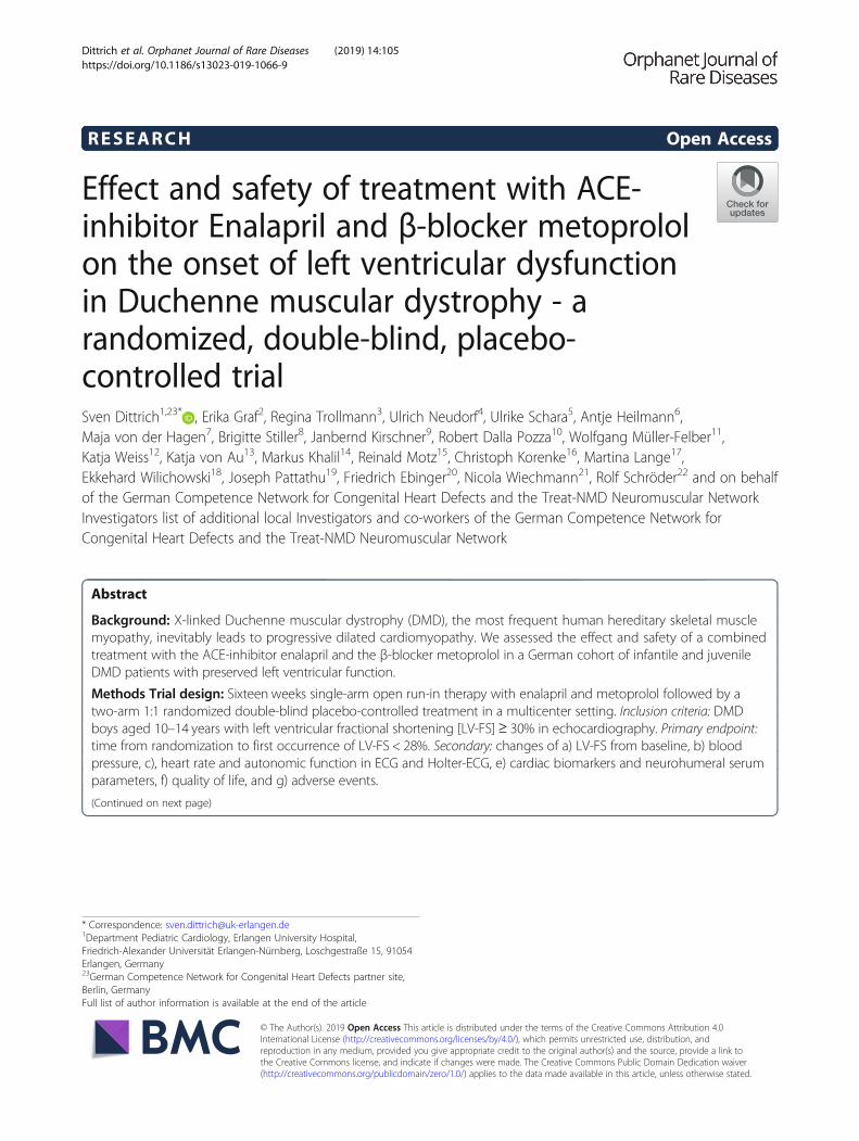
RESEARCH Open Access Effect and safety of treatment with ACE- inhibitor Enalapril and β-blocker metoprolol on the onset of left ventricular dysfunction in Duchenne muscular dystrophy - a randomized, double-blind, placebo- controlled trial Sven Dittrich 1,23* , Erika Graf 2 , Regina Trollmann 3 , Ulrich Neudorf 4 , Ulrike Schara 5 , Antje Heilmann 6 , Maja von der Hagen 7 , Brigitte Stiller 8 , Janbernd Kirschner 9 , Robert Dalla Pozza 10 , Wolfgang Müller-Felber 11 , Katja Weiss 12 , Katja von Au 13 , Markus Khalil 14 , Reinald Motz 15 , Christoph Korenke 16 , Martina Lange 17 , Ekkehard Wilichowski 18 , Joseph Pattathu 19 , Friedrich Ebinger 20 , Nicola Wiechmann 21 , Rolf Schröder 22 and on behalf of the German Competence Network for Congenital Heart Defects and the Treat-NMD Neuromuscular Network Investigators list of additional local Investigators and co-workers of the German Competence Network for Congenital Heart Defects and the Treat-NMD Neuromuscular Network Abstract Background: X-linked Duchenne muscular dystrophy (DMD), the most frequent human hereditary skeletal muscle myopathy, inevitably leads to progressive dilated cardiomyopathy. We assessed the effect and safety of a combined treatment with the ACE-inhibitor enalapril and the β-blocker metoprolol in a German cohort of infantile and juvenile DMD patients with preserved left ventricular function. Methods Trial design: Sixteen weeks single-arm open run-in therapy with enalapril and metoprolol followed by a two-arm 1:1 randomized double-blind placebo-controlled treatment in a multicenter setting. Inclusion criteria: DMD boys aged 10–14 years with left ventricular fractional shortening [LV-FS] ≥ 30% in echocardiography. Primary endpoint: time from randomization to first occurrence of LV-FS < 28%. Secondary: changes of a) LV-FS from baseline, b) blood pressure, c), heart rate and autonomic function in ECG and Holter-ECG, e) cardiac biomarkers and neurohumeral serum parameters, f) quality of life, and g) adverse events. (Continued on next page) © The Author(s). 2019 Open Access This article is distributed under the terms of the Creative Commons Attribution 4.0 International License (http://creativecommons.org/licenses/by/4.0/), which permits unrestricted use, distribution, and reproduction in any medium, provided you give appropriate credit to the original author(s) and the source, provide a link to the Creative Commons license, and indicate if changes were made. The Creative Commons Public Domain Dedication waiver (http://creativecommons.org/publicdomain/zero/1.0/) applies to the data made available in this article, unless otherwise stated. * Correspondence: [email protected] 1 Department Pediatric Cardiology, Erlangen University Hospital, Friedrich-Alexander Universität Erlangen-Nürnberg, Loschgestraße 15, 91054 Erlangen, Germany 23 German Competence Network for Congenital Heart Defects partner site, Berlin, Germany Full list of author information is available at the end of the article Dittrich et al. Orphanet Journal of Rare Diseases (2019) 14:105 https://doi.org/10.1186/s13023-019-1066-9
Transcript of RESEARCH Open Access Effect and safety of treatment with ...
RESEARCH Open Access
Effect and safety of treatment with ACE-inhibitor Enalapril and β-blocker metoprololon the onset of left ventricular dysfunctionin Duchenne muscular dystrophy - arandomized, double-blind, placebo-controlled trialSven Dittrich1,23* , Erika Graf2, Regina Trollmann3, Ulrich Neudorf4, Ulrike Schara5, Antje Heilmann6,Maja von der Hagen7, Brigitte Stiller8, Janbernd Kirschner9, Robert Dalla Pozza10, Wolfgang Müller-Felber11,Katja Weiss12, Katja von Au13, Markus Khalil14, Reinald Motz15, Christoph Korenke16, Martina Lange17,Ekkehard Wilichowski18, Joseph Pattathu19, Friedrich Ebinger20, Nicola Wiechmann21, Rolf Schröder22 and on behalfof the German Competence Network for Congenital Heart Defects and the Treat-NMD Neuromuscular NetworkInvestigators list of additional local Investigators and co-workers of the German Competence Network forCongenital Heart Defects and the Treat-NMD Neuromuscular Network
Abstract
Background: X-linked Duchenne muscular dystrophy (DMD), the most frequent human hereditary skeletal musclemyopathy, inevitably leads to progressive dilated cardiomyopathy. We assessed the effect and safety of a combinedtreatment with the ACE-inhibitor enalapril and the β-blocker metoprolol in a German cohort of infantile and juvenileDMD patients with preserved left ventricular function.
Methods Trial design: Sixteen weeks single-arm open run-in therapy with enalapril and metoprolol followed by atwo-arm 1:1 randomized double-blind placebo-controlled treatment in a multicenter setting. Inclusion criteria: DMDboys aged 10–14 years with left ventricular fractional shortening [LV-FS]≥ 30% in echocardiography. Primary endpoint:time from randomization to first occurrence of LV-FS < 28%. Secondary: changes of a) LV-FS from baseline, b) bloodpressure, c), heart rate and autonomic function in ECG and Holter-ECG, e) cardiac biomarkers and neurohumeral serumparameters, f) quality of life, and g) adverse events.
(Continued on next page)
© The Author(s). 2019 Open Access This article is distributed under the terms of the Creative Commons Attribution 4.0International License (http://creativecommons.org/licenses/by/4.0/), which permits unrestricted use, distribution, andreproduction in any medium, provided you give appropriate credit to the original author(s) and the source, provide a link tothe Creative Commons license, and indicate if changes were made. The Creative Commons Public Domain Dedication waiver(http://creativecommons.org/publicdomain/zero/1.0/) applies to the data made available in this article, unless otherwise stated.
* Correspondence: [email protected] Pediatric Cardiology, Erlangen University Hospital,Friedrich-Alexander Universität Erlangen-Nürnberg, Loschgestraße 15, 91054Erlangen, Germany23German Competence Network for Congenital Heart Defects partner site,Berlin, GermanyFull list of author information is available at the end of the article
Dittrich et al. Orphanet Journal of Rare Diseases (2019) 14:105 https://doi.org/10.1186/s13023-019-1066-9
(Continued from previous page)
Results: From 3/2010 to 12/2013, 38 patients from 10 sites were centrally randomized after run-in, with 21 patientscontinuing enalapril and metoprolol medication and 17 patients receiving placebo. Until end of study 12/2015, LV-FS< 28% was reached in 6/21 versus 7/17 patients. Cox regression adjusted for LV-FS after run-in showed a statisticallynon-significant benefit for medication over placebo (hazard ratio: 0.38; 95% confidence interval: 0.12 to 1.22; p = 0.10).Analysis of secondary outcome measures revealed a time-dependent deterioration of LV-FS with no statisticallysignificant differences between the two study arms. Blood pressure, maximal heart rate and mean-NN values weresignificantly lower at the end of open run-in treatment compared to baseline. Outcome analysis 19months afterrandomization displayed significantly lower maximum heart rate and higher noradrenalin and renin values in theintervention group. No difference between treatments was seen for quality of life. As a single, yet important adverseevent, the reversible deterioration of walking abilities of one DMD patient during the run-in period was observed.
Conclusions: Our analysis of enalapril and metoprolol treatment in DMD patients with preserved left ventricularfunction is suggestive to delay the progression of the intrinsic cardiomyopathy to left ventricular failure, but did notreach statistical significance, probably due to insufficient sample size.
Clinical trial registration: DRKS-number 00000115, EudraCT-number 2009–009871-36.
Keywords: Duchenne muscular dystrophy, Cardiomyopathy, ACE-inhibitors, ß-blockers
BackgroundMutations of the human dystrophin gene on chromosomeXp21 cause Duchenne muscular dystrophy (DMD) [1],which is the most frequently occurring muscular dys-trophy in humans with an incidence of 1 in 3600–6000male births [2]. In addition to early onset and progressivemuscular weakness and wasting, which inevitably leads toloss of ambulation of boys between 9 and 13 years of age[3], nearly all DMD patients develop dilated cardiomyop-athy with impaired systolic function in their second dec-ade of life [4–8]. Although promising therapeutic optionssuch as ataluren for stop codon read-through are availablefor eligible (< 10%) of the patients [9], to date, no curativetherapy is available for DMD. Though multidisciplinarycare, comprising early treatment with corticosteroids,physiotherapy, early antibiotic treatment of pulmonarychest infections, scoliosis surgery with insertion of spinalrods, implementation of respiratory support and drugtreatment of heart failure, has substantially improved lifeexpectancy and quality of life for DMD patients, mostpatients die in the second to the fourth decade of life dueto combined respiratory and cardiac failure [2, 4, 10, 11].Thus, regular cardiological and pulmonary diagnosticwork-up of all DMD patients is mandatory to assess indi-vidual heart and respiratory function and to adapt thera-peutic strategies [12].In general, the medical treatment of cardiomyopathy
in pediatric patients is still an open debate [13]. Whileevidence based studies and guidelines providing treat-ment recommendations for adult cardiomyopathy withimpaired left ventricular function, including the use ofthe angiotensin converting enzyme inhibitor enalapriland the beta receptor blocker metoprolol [14, 15] exists,corresponding data for pediatric patients is vastly lack-ing. Thus, the rationale for the use of most heart failure
medications in pediatric patients is mostly extrapolatedfrom studies in adult heart failure [16]. In the context ofDMD a number of open studies indicated that ACE in-hibitors, angiotensin receptor blockers, beta-blockersand/or aldosterone antagonists might improve or pre-serve left ventricular systolic function and may delay theprogression of cardiomyopathy [4, 17–21]. Moreover,one study demonstrated that the early intervention withperindopril led to a significantly higher overall survivalin DMD patients with preserved left ventricular ejectionfraction at baseline [18]. Though the comparison andinterpretation of the later studies is generally hamperedby their individual methodological design and the use ofdifferent outcome measurements [19], the available datasupports the use of heart failure medication in DMDpatients but provides no conclusive evidence regardingthe optimal timing of therapy initiation [4, 19, 21, 22].In the present multicenter study we assessed the
effects of a combined therapy of the angiotensin con-verting enzyme inhibitor enalapril and the β-receptorblocker metoprolol on the onset of significant left ven-tricular dysfunction in 10–14 year old DMD boys withpreserved left ventricular function.
MethodsPatientsPatients for this investigator-initiated, double-blind,randomized, placebo-controlled multicenter study were re-cruited at 10 German study sites (Berlin, Dresden, Erlangen,Essen, Freiburg, Giessen, Göttingen, Heidelberg, Munich,Oldenburg) from March, 2010 to December, 2013. Inclu-sion criteria for boys suffering from Duchenne musculardystrophy were: 1) the diagnosis was based on a geneticallyconfirmed disease causing mutation or report of negativedystrophin immunostaining in a diagnostic muscle biopsy,
Dittrich et al. Orphanet Journal of Rare Diseases (2019) 14:105 Page 2 of 13
2) age of 10 to 14 years, 3) preserved left ventricular func-tion as defined by echocardiography with left ventricularfractional shortening ≥30% in the long-axis motion-mode,4) normal renal function with glomerular filtration rate >30ml/min/1.73m2, and 5) ability to participate in theassessment of primary and secondary outcome measures.Exclusion criteria were i) any contraindication for treatmentwith angiotensin converting enzyme inhibitors or β-blockers, ii) previous treatment with those drugs in the pastthree months, iii) abnormal liver function defined by eleva-tion (≥2x) of gamma-glutamyltranspeptidase and bilirubine,iv) left ventricular dilation above the 97th percentile as de-fined by echocardiography in the long-axis motion-mode,and v) participation in other clinical trials. This clinical trialwas approved by the regulatory authorities and ethics com-mittees at each study site and performed in accordancewith good clinical practice guidelines. The objectives, studydesign, risks, and benefits of participation were explained toall participants, and written informed consent was obtainedfrom patients and parents before enrolment.
Open run-in, randomization and maskingThe principle of anti-congestive medications requiresup-titration of dosages to the individually maximum tol-erated level within a safety range [14, 15]. To define theindividual drug tolerance in all of the patients screenedfor eligibility in this study, we opted for a preceding 16weeks open run-in period with enalapril (enalapril-ma-leat) and metoprolol (metoprolol-succinat). Drug dos-ages of enalapril and metoprolol were increased step-by-step in 3 weight classes in 4 timely shifted steps for eachof the drugs up to the maximum final daily dosage of 10mg enalapril / 47.5mg metoprolol (patient weight < 45 kg),10mg enalapril / 71.25mg metoprolol (patient weight 45 -< 60 kg) and 20mg enalapril / 95mg metoprolol for pa-tients with a body weight > 60 kg. After 16 weeks openrun-in period, patients were randomly assigned at a 1:1ratio to receive either the combination of enalapril andmetoprolol without interruption or placebo with a 4 weeksstepwise wash-out protocol to disguise potential reboundeffects in the placebo group. A stratified block randomiza-tion with randomly varying block sizes of two or six partici-pants and stratification for trial site was used. Allocation ofpatients was performed centrally by the pharmacy of theUniversity Hospital Erlangen based on computer-generatedlists. Both active drugs and placebo were supplied by HexalAG (Holzkirchen, Germany) as identically appearing tab-lets. Active drugs and placebo were identically prepacked tomaintain the masking for the patient and investigator bythe certified pharmacy of the University Hospital Erlangenaccording to good manufacturing practice for pharmaceuti-cals. Dose levels of study medication were generally keptconstant but adapted to changes in body weight classes.The use of steroids or a history of the use of steroids was
recorded at baseline. During the study period start of ster-oid therapy was not permissible but occurred in single in-stances. Patients who had reached the primary endpoint orthe end of the study received 4 weeks of blinded wash-outmedication. Thereafter, guideline-conform treatment was atthe investigator’s discretion.
Outcome measuresThe primary outcome was the time from randomizationto the first occurrence of a left ventricular fractionalshortening < 28% in the long-axis motion-mode of echo-cardiography. Corresponding analyses were performedbiannually at the individual study sites. Visits continuedto end of study after the primary endpoint was reached.Secondary outcome measurements were 1) echocardio-
graphic changes of left ventricular fractional shorteningfrom the end of the run-in period, 2) echocardiographicchanges of left ventricular diastolic diameter and systolicventricular septum thickness measurements by motion-mode, 3) echocardiographic tissue-Doppler analyses (seebelow), 4) blood pressure values, 5) electrocardiograms andHolter-electrocardiograms (see below), 6) laboratory tests(see below), 7) quality of life rating (see below), and 8) ad-verse events.Tissue Doppler data comprised assessment of septal, left
ventricular and right ventricular longitudinal function byanalysis of systolic strain in the basal, mid and apical region,respectively. Recording of tissue-Doppler data wasrestricted to the availability of a GE-echo-machine at thestudy site. All echocardiographic and tissue-Doppler datawere collected in a standardized way in four-chamber-viewas established by the German competence network forCongenital Heart Disease (http://www.kinderkardiologie.org/fileadmin/user_upload/Stellungnahmen/Qualitaetsstan-dardsEcho.pdf). Tissue Doppler data were centrally ana-lyzed by the same investigator in the tissue Dopplerreference center of the German competence network forCongenital Heart Disease in Freiburg.Electrocardiograms and Holter-electrocardiograms were
centrally analyzed by a blinded investigator in Erlangen.Holter-ECG analyses included heart frequency analysesand heart rate variability measures (mean NN: averagenormal R to R interval; SDNN: Standard deviation of R toR intervals; SDANN: Standard deviation of the means foreach R to R segment; ASDNN: average standard deviationof all 5-min R to R- intervals; rMSSD: Root-mean-Squareof successive differences of NN [normal R to R intervals];pNN50: fraction of NN intervals that differ by more than50ms from the previous NN interval).Laboratory tests comprised the neurohumoral markers
renin, angiotensin II, aldosterone and norepinephrineand the biomarker NT-pro-BNP.The German Kiddo-KINDL questionnaire for adoles-
cents aged 12–16 years [23] was used as a generic quality
Dittrich et al. Orphanet Journal of Rare Diseases (2019) 14:105 Page 3 of 13
of life rating measure. According to the study protocol,quality of life-questionnaire was first requested at thescreening visit. A complete survey of all patients was re-peated one year after randomization and then annually.The safety of enalapril and metoprolol administration
was monitored from the run-in period until 30 days afterdiscontinuation of the study drugs by adverse event re-ports and biannual physical examination, assessment ofblood pressure, and local safety laboratory tests (includingcreatinine, potassium, sodium, urea, glutamate oxalacetatetransaminase [GOT], glutamate pyruvate transaminase[GPT], γ-glutamyl transpeptidase [γ-GT] and bilirubin).As serum creatinine titer is not a reliable biomarker forrenal function in patients with Duchenne muscular dys-trophy because of their low muscle mass [24], cystatin Cwas measured when creatinine titers were elevated. Safetylaboratory values were directly assessed by local investiga-tors. Abnormal values considered to yield clinical signifi-cance were reported as adverse events.
Statistical analysisInitially, the target was 130 patients randomized withinthree years, plus three years additional follow-up, due tofeasibility constraints. We anticipated that 50% of patientson placebo would suffer from an LV-FS < 28% after 4 yearsof individual follow-up7. With a cumulative drop-out rateof 5% up to year 4.5 (median follow-up time), a log-ranktest with two-sided significance level 5% of time fromrandomization to first occurrence of LV-FS < 28% wouldhave 80% power if the hazard ratio for enalapril and meto-prolol versus placebo was 0.46 (Lakatos approximation, 58events required), corresponding to an improvement to72.7% free of left-ventricular dysfunction (LV-FS < 28%)after 4 years. Given previous results [17], a hazard ratio of0.46 seemed achievable, but smaller treatment benefitswould also be clinically relevant. Due to difficulties in re-cruitment, the target number was reduced to 55 patientsin December 2012. This would still yield 80% power to de-tect a difference between treatments with respect tochange in LV-FS from end of run-in to the visit scheduled19months after randomization (visit 4), which was consid-ered the most relevant secondary outcome. Assuming astandard deviation of 4% at visit 4 [17], a t-test withtwo-sided significance level 5% would achieve this powerif the mean difference 19months after randomization was3.1%. By December 2013, 42 patients had given informedconsent, and it was decided to stop recruitment and con-tinue follow-up until end of December 2015.The analysis of treatment effects was done by intention-
to-treat in all 38 patients who were randomized after therun-in period. In the primary analysis, time fromrandomization to first occurrence of an LV-FS < 28% wasanalyzed with the proportional hazards model, censoring atthe last visit for those patients in whom no LV-FS < 28%
was observed. The treatment effect was tested using theWald-test at two-sided significance level of 5%, and was es-timated as a hazard ratio with two-sided 95% confidenceinterval. Due to the insufficient recruitment, covariate ad-justment for study site originally planned in the studyprotocol was replaced by adjustment for LV-FS measuredafter run-in in the statistical analysis plan before the blindwas broken. A planned sensitivity analysis to explore apossible confounding effect of concomitant treatment withsteroids was done by additional inclusion treatment withsteroids as a time-dependent covariate in the primaryproportional hazards model.Secondary efficacy outcomes were analyzed in a mixed
model for repeated measures including outcomes afterrandomization and 19months later as endpoints andoutcome after run-in, treatment, and the interaction be-tween measurement time and treatment as covariates;subjects were modelled as random effects. Linear regres-sion originally planned in the protocol was replaced bythis longitudinal model in the statistical analysis plan toallow inclusion of all randomized patients under a miss-ing at random assumption even if they dropped out afterrandomization. Changes from screening to end of run-inwere summarized by means with 95% confidence inter-vals. Entries to the KINDL questionnaires were evalu-ated in accordance with the corresponding manual.Adverse events were coded by the Medical dictionary forregulatory activities (MedDRA version 19.1) and summa-rized single-armed (verum) for those events with onsetfrom run-in to four weeks after randomization,two-armed (verum versus placebo) for those events withonset after that, restricting the analysis sets to those pa-tients who received at least one dose of study medicationin the corresponding period.All p-values were two-sided and considered explora-
tory except for the primary analysis, programming wasdone with SAS (version 9.2) in UNIX. An independentdata monitoring committee reviewed safety data on ayearly basis. An interim analysis of efficacy data, whichhad been planned initially, was cancelled because of thereduced target number of patients.
ResultsStudy populationBetween March 2010 and December 2013, 42 boys gaveinformed consent, 41 started open run-in medicationand 38 patients were randomized after a run-in (Fig. 1).The study was concluded with the last patient visit inDecember 2015.
Outcome after open run-in phase (all patients)During the open run-in medication period, two protocoldeviations were noted: in one patient the run-in had tobe repeated (due to a bone fracture) and was delayed in
Dittrich et al. Orphanet Journal of Rare Diseases (2019) 14:105 Page 4 of 13
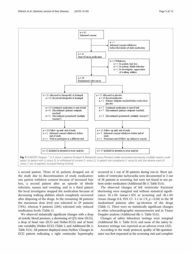
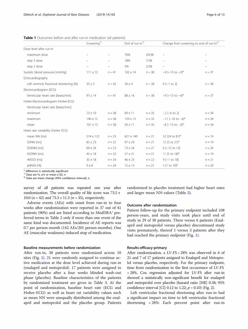
a second patient. Three of 41 patients dropped out ofthe study due to discontinuation of study medication:one patient withdrew consent because of increased hairloss, a second patient after an episode of febrileinfection, nausea and vomiting, and in a third patientthe local investigator stopped the medication because ofdecreasing walking abilities which completely recoveredafter disposing of the drugs. In the remaining 38 patientsthe maximum dose level was tolerated in 29 patients(76%), whereas 9 patients (24%) tolerated only reducedmedication levels (Table 1).We observed statistically significant changes with a drop
of systolic blood pressure, a shortening of QTc-time (ECG),a drop of heart rate (ECG and Holter-ECG) and of heartrate variability (Holter-ECG) (Table 1, and Additional file 1:Table S1A). All patients displayed sinus rhythm. Changes inECG pattern indicating a right ventricular hypertrophy
occurred in 1 out of 38 patients during run-in. Short epi-sodes of ventricular tachycardia were documented in 2 outof 38 patients at screening, but were not found in any pa-tient under medication (Additional file 1: Table S1A).The observed changes of left ventricular fractional
shortening were marginal and without statistical signifi-cance: 35 ± 4% (mean ± SD) at screening and 36 ± 4%(mean change 0.4, 95% CI -1.1 to 1.9, p = 0.58) in the 38randomized patients after up-titration of the drugs(Table 1). There were no statistically significant changesin other echocardiographic measurements and in TissueDoppler analysis (Additional file 1: Table S1A).Changes of safety laboratory testings were marginal
(Additional file 1: Table S1A) and none of the safety la-boratory testings was reported as an adverse event (AE).According to the study protocol, quality of life-question-
naire was first requested at the screening visit and complete
Fig. 1 CONSORT Diagram. 1) In 2 versus 3 patients (Enalapril & Metoprolol versus Placebo), intake terminated prematurely (multiple reasons couldapply): 5x patient wish (2 versus 3), 3x withdrawal of consent (1 versus 2), 1x patient non-compliance (1 versus 0), and one adverse event (0versus 1: loss of appetite, increased feeling of thirst)
Dittrich et al. Orphanet Journal of Rare Diseases (2019) 14:105 Page 5 of 13
survey of all patients was repeated one year afterrandomization. The overall quality of life score was 73.5 ±10.0 (n = 42) and 73.3 ± 11.3 (n = 35), respectively.Adverse events (AEs) with onset from run-in to four
weeks after randomization were reported in 37 out of 41patients (90%) and are listed according to MedDRA® pre-ferred terms in Table 2 only if more than one event of thesame kind was documented. Incidence of AE reports was0.7 per person-month (142 AEs/201 person-months). OneAE (muscular weakness) induced stop of medication.
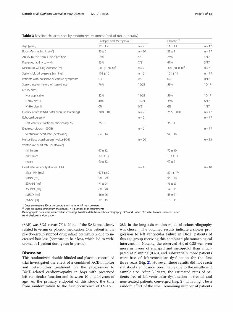
Baseline measurements before randomizationAfter run-in, 38 patients were randomized across 10sites (Fig. 1). 21 were randomly assigned to continue ac-tive medication at the dose level achieved during run-in(enalapril and metoprolol). 17 patients were assigned toreceive placebo after a four weeks blinded wash-outphase (placebo). Baseline characteristics of the patientsby randomized treatment are given in Table 3. At thepoint of randomization, baseline heart rate (ECG andHolter-ECG) as well as heart rat variability values suchas mean NN were unequally distributed among the enal-april and metoprolol and the placebo group. Patients
randomized to placebo treatment had higher heart ratesand larger mean NN-values (Table 3).
Outcome after randomizationPatient follow-up for the primary endpoint included 108person-years, and study visits took place until end ofstudy in 29 of 38 patients. Three versus 6 patients (Enal-april and metoprolol versus placebo) discontinued studyvisits prematurely, thereof 1 versus 3 patients after theyhad reached the primary endpoint (Fig. 1).
Results-efficacy-primaryAfter randomization, a LV-FS < 28% was observed in 6 of21 and 7 of 17 patients assigned to Enalapril and Metopro-lol versus placebo, respectively. For the primary endpoint,time from randomization to the first occurrence of LV-FS< 28%, Cox regression adjusted for LV-FS after run-inshowed a statistically non-significant benefit for enalapriland metoprolol over placebo (hazard ratio [HR] 0.38; 95%confidence interval [CI] 0.12 to 1.22; p = 0.10) (Fig. 2).Left ventricular fractional shortening after run-in had
a significant impact on time to left ventricular fractionalshortening < 28%: Each percent point after run-in
Table 1 Outcomes before and after run-in medication (all patients)
Screening1) End of run-in1) Change from screening to end of run-in2)
Dose level after run-in
maximum dose – – 76% 29/38 – –
step 3 dose – – 18% 7/38 – –
step 2 dose – – 5% 2/38 – –
Systolic blood pressure [mmHg] 111 ± 13 n = 41 102 ± 14 n = 38 −9 [−13 to −5]* n = 37
Echocardiography
Left ventricle fractional shortening [%] 35 ± 3 n = 42 36 ± 4 n = 38 0 [−1 to 2] n = 38
Electrocardiogram (ECG)
Ventricular heart rate [beats/min] 97 ± 14 n = 41 88 ± 16 n = 38 −9 [−13 to −4]* n = 37
Holter-Electrocardiogram (Holter-ECG)
Ventricular heart rate [beats/min]:
minimum 73 ± 10 n = 38 69 ± 11 n = 35 −2 [−6 to 2] n = 34
maximum 140 ± 15 n = 38 129 ± 15 n = 35 −11 [−16 to −6]* n = 34
mean 101 ± 13 n = 38 93 ± 11 n = 35 −8 [−13 to −3]* n = 34
Heart rate variability (Holter ECG)
mean NN [ms] 574 ± 122 n = 23 627 ± 140 n = 21 52 [24 to 81]* n = 19
SDNN [ms] 85 ± 23 n = 22 97 ± 29 n = 21 12 [3 to 21]* n = 19
SDANN [ms] 69 ± 24 n = 23 73 ± 24 n = 21 0 [−12 to 13] n = 20
ASDNN [ms] 45 ± 18 n = 22 57 ± 21 n = 21 11 [5 to 18]* n = 19
rMSSD [ms] 35 ± 18 n = 24 46 ± 23 n = 22 9 [−1 to 18] n = 21
pNN50 [%] 9 ± 8 n = 24 15 ± 13 n = 21 5 [1 to 10]* n = 20
* difference is statistically significant1) Data are %, x/n or mean ± SD, n2) Data are mean change [95% confidence interval], n
Dittrich et al. Orphanet Journal of Rare Diseases (2019) 14:105 Page 6 of 13
lowered the hazard of left ventricular dysfunction by afactor (HR) of 0.72 (95%CI 0.55 to 0.93, p = 0.011).Concomitant steroid treatment was given at least once
after randomization in 10 of 21 patients on enalapril andmetoprolol versus 11 of 17 patients on placebo. Sensitiv-ity analysis to investigate a potential confounding impactby inclusion of a time-dependent indicator of steroid in-take did not alter the estimated effect of enalapril andmetoprolol versus placebo (HR 0.32; 95% CI 0.09 to1.13; p = 0.076). The effect of steroid intake on time tofirst occurrence of LV-FS < 28% was estimated as a HRof 0.61 (95%CI 0.16 to 2.37; p = 0.47).
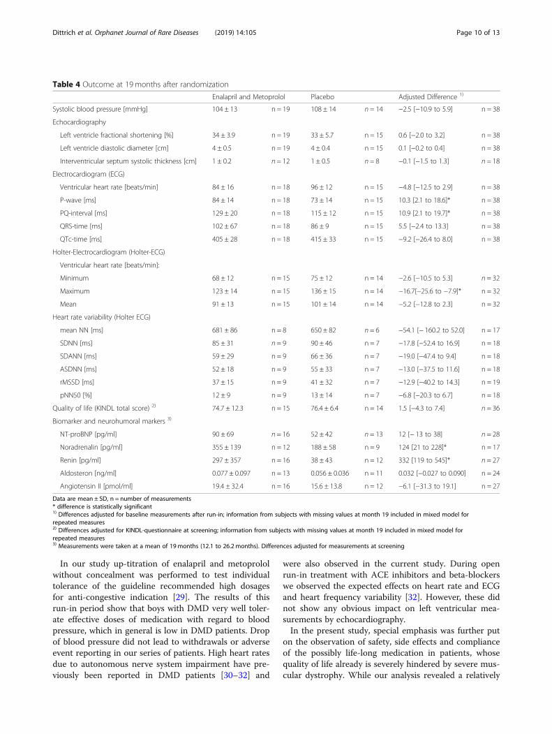
Results-efficacy-secondaryChange of left ventricular fractional shortening was con-sidered the most relevant secondary efficacy endpoint.The difference between treatments at month 19, esti-mated as 0.62% in favor of enalapril and metoprolol(Table 4), was not statistically significant (95%CI − 1.98to 3.22%, p = 0.63). Adjusted analysis for LV-FS afterrun-in showed that LV-FS decreased by − 0.10% permonth in the enalapril and metoprolol -group (95%CI −0.21 to 0.02%, p = 0.10) compared to − 0.13% per monthwith placebo (95%CI − 0.25 to 0.00%, p = 0.042). We ob-served no effect on left ventricular diameter or ventricu-lar thickness (Table 4).Adjusted differences between treatments were not sta-
tistically significant for systolic blood pressure (Table 4).
All patients had sinus rhythm during whole studyperiod. No episodes of supraventricular or ventriculartachycardias had been registered in any Holter-ECGrecordings.Baseline distribution of heart frequencies after run-in
in ECG and Holter-ECG was asymmetric (Table 3). Ad-justed differences showed significantly lower maximalventricular heart rate in Holter-ECG in the enalapril andmetoprolol group compared to placebo (Table 4).Changes of heart rate variability parameters were sta-
tistically significant as analyzed for all patients duringopen run-in medication for an increase of meanNN, anincrease of SDNN, an increase of ASDNN and an in-crease of pNN50 (Table 1). The values were asymmetric-ally distributed at randomization baseline (Table 3).Adjusted differences between randomized treatmentsafter 19 months were not significant (Table 4).NT-pro-BNP values were within a low range at screen-
ing (see Additional file 1: Table S2A) and after 19months of randomized treatment (Table 4). This also ap-plies for values of the renin–angiotensin–aldosteronesystem (RAAS) (Table 4, Additional file 1: Table S2A).However, we observed significant adjusted differenceswith an increase of noradrenalin and renin values in theenalapril and metoprolol group (Table 4).The KINDL total quality of life score did not deterior-
ate with time and showed no difference between treat-ments at month 19 (Table 4). Pooled data for subscalesare visualized in the Additional file 1: Fig. S1A).
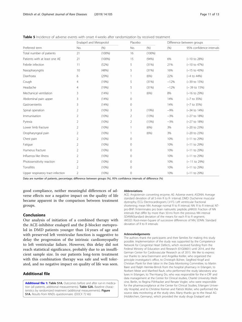
Results-safety/tolerabilityAfter randomization, the majority of patients (33 of 38)continued intake of study medication either up to the endof the trial (14 versus 7, enalapril and metoprolol versusplacebo) or until the primary endpoint was reached (Fig.2). In 2 versus 3 patients, intake terminated prematurely.Reasons (multiple reasons could apply) included 5xpatient wish (2 versus 3), 3x withdrawal of consent (1 ver-sus 2), 1x patient non-compliance (1 versus 0) and one ad-verse event (0 versus 1: loss of appetite, increased feelingof thirst). We noticed 13 protocol deviations: Adaptationof dose level to increased body weight was delayed in 11patients (4 enalapril and metoprolol, 7 placebo), not donein one patient and prematurely done in another patient(both enalapril and metoprolol). No unblinding occurred.Adverse events (AEs) with onset after randomization and
the four weeks wash-out period of the placebo arm were re-ported in 21/21 versus 15/16 (enalapril and metoprolol ver-sus placebo) of the patients. Table 5 shows AEs that weredocumented in more than one patient per arm. Incidenceof AE reports was 0.24 versus 0.26 per person-month onstudy medication (enalapril and metoprolol: 181 AEs/739person-months, placebo: 129 AEs/490 person-months).The total number of patients with at least one serious AE
Table 2 Incidence of adverse events with onset from start ofrun-in medication to 4 weeks after randomization (all patients)
Preferred term No. % 95% confidence intervals
Total number of patients 41 100%;
Patients with at least one AE 37 90% (77–97%)
Headache 11 27% (14–43%)
Nasopharyngitis 11 27% (14–43%)
Cough 8 20% (9–35%)
Nausea 8 20% (9–35%)
Febrile infection 6 15% (6–29%)
Diarrhoea 5 12% (4–26%)
Dizziness 4 10% (3–23%)
Fall 3 7% (2–20%)
Fatigue 3 7% (2–20%)
Pyrexia 3 7% (2–20%)
Abdominal pain 2 5% (0.6–17%)
Back pain 2 5% (0.6–17%)
Chest pain 2 5% (0.6–17%)
Decreased appetite 2 5% (0.6–17%)
Muscular weakness 2 5% (0.6–17%)
Rash 2 5% (0.6–17%)
Data are number of patients, percentage; 95% confidence interval
Dittrich et al. Orphanet Journal of Rare Diseases (2019) 14:105 Page 7 of 13
(SAE) was 8/21 versus 7/16. None of the SAEs was clearlyrelated to verum or placebo medication. One patient in theplacebo-group stopped drug intake prematurely due to in-creased hair loss (compare to hair loss, which led to with-drawal in 1 patient during run-in-period).
DiscussionThis randomized, double-blinded and placebo-controlledtrial investigated the effect of a combined ACE-inhibitorand beta-blocker treatment on the progression toDMD-related cardiomyopathy in boys with preservedleft ventricular function and between 10 and 14 years ofage. As the primary endpoint of this study, the timefrom randomization to the first occurrence of LV-FS <
28% in the long-axis motion-mode of echocardiographywas chosen. The obtained results indicate a slower pro-gression to left ventricular failure in DMD patients ofthis age group receiving this combined pharamacologicalintervention. Notably, the observed HR of 0.38 was evenmore in favour of enalapril and metoprolol than antici-pated at planning (0.46), and substantially more patientswere free of left-ventricular dysfunction for the firstthree years (Fig. 2). However, these results did not reachstatistical significance, presumably due to the insufficientsample size. After 3.5 years, the estimated rates of pa-tients free of left-ventricular dysfunction in treated andnon-treated patients converged (Fig. 2). This might be arandom effect of the small remaining number of patients
Table 3 Baseline characteristics by randomized treatment (end of run-in therapy)
Enalapril and Metoprolol 1) Placebo 1)
Age [years] 12 ± 1.2 n = 21 11 ± 1.1 n = 17
Body Mass Index [kg/m2] 23 ± 6 n = 20 21 ± 5 n = 17
Ability to rise from supine position 24% 5/21 24% 4/17
Preserved ability to walk 33% 7/21 41% 5/17
Maximum walking distance [m] 200 (3–6000)2) n = 7 300 (30–800)2) n = 5
Systolic blood pressure [mmHg] 103 ± 16 n = 21 101 ± 11 n = 17
Patients with presence of cardiac symptoms 0% 0/21 0% 0/17
Steroid use or history of steroid use 76% 16/21 59% 10/17
NYHA class
Not applicable 52% 11/21 59% 10/17
NYHA class I 48% 10/21 35% 6/17
NYHA class II 0% 0/21 6% 1/17
Quality of life (KINDL total score at screening) 70.8 ± 10.1 n = 21 75.6 ± 10.0 n = 17
Echocardiography n = 21 n = 17
Left ventricle fractional shortening [%] 35 ± 3 36 ± 4
Electrocardiogram (ECG) n = 21 n = 17
Ventricular heart rate [beats/min] 84 ± 14 94 ± 16
Holter-Electrocardiogram (Holter-ECG) n = 20 n = 15
Ventricular heart rate [beats/min]
minimum 67 ± 12 72 ± 10
maximum 126 ± 17 133 ± 11
mean 90 ± 12 97 ± 9
Heart rate variability (Holter ECG) n = 11 n = 10
Mean NN [ms] 678 ± 80 571 ± 174
SDNN [ms] 98 ± 29 96 ± 30
SDANN [ms] 71 ± 24 75 ± 25
ASDNN [ms] 60 ± 20 54 ± 21
rMSSD [ms] 46 ± 26 45 ± 21
pNN50 [%] 17 ± 15 13 ± 111) Data are mean ± SD or percentage, n = number of measurements2) Data are mean, (minimum-maximum), n = number of measurementsDemographic data were collected at screening, baseline data from echocardiography, ECG and Holter-ECG refer to measurements afterrun-in/before randomization
Dittrich et al. Orphanet Journal of Rare Diseases (2019) 14:105 Page 8 of 13
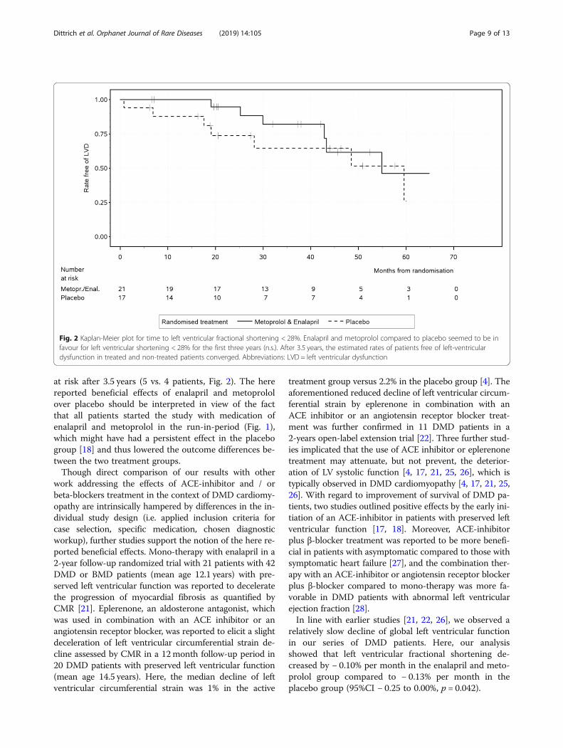
at risk after 3.5 years (5 vs. 4 patients, Fig. 2). The herereported beneficial effects of enalapril and metoprololover placebo should be interpreted in view of the factthat all patients started the study with medication ofenalapril and metoprolol in the run-in-period (Fig. 1),which might have had a persistent effect in the placebogroup [18] and thus lowered the outcome differences be-tween the two treatment groups.Though direct comparison of our results with other
work addressing the effects of ACE-inhibitor and / orbeta-blockers treatment in the context of DMD cardiomy-opathy are intrinsically hampered by differences in the in-dividual study design (i.e. applied inclusion criteria forcase selection, specific medication, chosen diagnosticworkup), further studies support the notion of the here re-ported beneficial effects. Mono-therapy with enalapril in a2-year follow-up randomized trial with 21 patients with 42DMD or BMD patients (mean age 12.1 years) with pre-served left ventricular function was reported to deceleratethe progression of myocardial fibrosis as quantified byCMR [21]. Eplerenone, an aldosterone antagonist, whichwas used in combination with an ACE inhibitor or anangiotensin receptor blocker, was reported to elicit a slightdeceleration of left ventricular circumferential strain de-cline assessed by CMR in a 12month follow-up period in20 DMD patients with preserved left ventricular function(mean age 14.5 years). Here, the median decline of leftventricular circumferential strain was 1% in the active
treatment group versus 2.2% in the placebo group [4]. Theaforementioned reduced decline of left ventricular circum-ferential strain by eplerenone in combination with anACE inhibitor or an angiotensin receptor blocker treat-ment was further confirmed in 11 DMD patients in a2-years open-label extension trial [22]. Three further stud-ies implicated that the use of ACE inhibitor or eplerenonetreatment may attenuate, but not prevent, the deterior-ation of LV systolic function [4, 17, 21, 25, 26], which istypically observed in DMD cardiomyopathy [4, 17, 21, 25,26]. With regard to improvement of survival of DMD pa-tients, two studies outlined positive effects by the early ini-tiation of an ACE-inhibitor in patients with preserved leftventricular function [17, 18]. Moreover, ACE-inhibitorplus β-blocker treatment was reported to be more benefi-cial in patients with asymptomatic compared to those withsymptomatic heart failure [27], and the combination ther-apy with an ACE-inhibitor or angiotensin receptor blockerplus β-blocker compared to mono-therapy was more fa-vorable in DMD patients with abnormal left ventricularejection fraction [28].In line with earlier studies [21, 22, 26], we observed a
relatively slow decline of global left ventricular functionin our series of DMD patients. Here, our analysisshowed that left ventricular fractional shortening de-creased by − 0.10% per month in the enalapril and meto-prolol group compared to − 0.13% per month in theplacebo group (95%CI − 0.25 to 0.00%, p = 0.042).
Fig. 2 Kaplan-Meier plot for time to left ventricular fractional shortening < 28%. Enalapril and metoprolol compared to placebo seemed to be infavour for left ventricular shortening < 28% for the first three years (n.s.). After 3.5 years, the estimated rates of patients free of left-ventriculardysfunction in treated and non-treated patients converged. Abbreviations: LVD = left ventricular dysfunction
Dittrich et al. Orphanet Journal of Rare Diseases (2019) 14:105 Page 9 of 13
In our study up-titration of enalapril and metoprololwithout concealment was performed to test individualtolerance of the guideline recommended high dosagesfor anti-congestive indication [29]. The results of thisrun-in period show that boys with DMD very well toler-ate effective doses of medication with regard to bloodpressure, which in general is low in DMD patients. Dropof blood pressure did not lead to withdrawals or adverseevent reporting in our series of patients. High heart ratesdue to autonomous nerve system impairment have pre-viously been reported in DMD patients [30–32] and
were also observed in the current study. During openrun-in treatment with ACE inhibitors and beta-blockerswe observed the expected effects on heart rate and ECGand heart frequency variability [32]. However, these didnot show any obvious impact on left ventricular mea-surements by echocardiography.In the present study, special emphasis was further put
on the observation of safety, side effects and complianceof the possibly life-long medication in patients, whosequality of life already is severely hindered by severe mus-cular dystrophy. While our analysis revealed a relatively
Table 4 Outcome at 19 months after randomization
Enalapril and Metoprolol Placebo Adjusted Difference 1)
Systolic blood pressure [mmHg] 104 ± 13 n = 19 108 ± 14 n = 14 −2.5 [−10.9 to 5.9] n = 38
Echocardiography
Left ventricle fractional shortening [%] 34 ± 3.9 n = 19 33 ± 5.7 n = 15 0.6 [−2.0 to 3.2] n = 38
Left ventricle diastolic diameter [cm] 4 ± 0.5 n = 19 4 ± 0.4 n = 15 0.1 [−0.2 to 0.4] n = 38
Interventricular septum systolic thickness [cm] 1 ± 0.2 n = 12 1 ± 0.5 n = 8 −0.1 [−1.5 to 1.3] n = 18
Electrocardiogram (ECG)
Ventricular heart rate [beats/min] 84 ± 16 n = 18 96 ± 12 n = 15 −4.8 [−12.5 to 2.9] n = 38
P-wave [ms] 84 ± 14 n = 18 73 ± 14 n = 15 10.3 [2.1 to 18.6]* n = 38
PQ-interval [ms] 129 ± 20 n = 18 115 ± 12 n = 15 10.9 [2.1 to 19.7]* n = 38
QRS-time [ms] 102 ± 67 n = 18 86 ± 9 n = 15 5.5 [−2.4 to 13.3] n = 38
QTc-time [ms] 405 ± 28 n = 18 415 ± 33 n = 15 −9.2 [−26.4 to 8.0] n = 38
Holter-Electrocardiogram (Holter-ECG)
Ventricular heart rate [beats/min]:
Minimum 68 ± 12 n = 15 75 ± 12 n = 14 −2.6 [−10.5 to 5.3] n = 32
Maximum 123 ± 14 n = 15 136 ± 15 n = 14 −16.7[−25.6 to −7.9]* n = 32
Mean 91 ± 13 n = 15 101 ± 14 n = 14 −5.2 [−12.8 to 2.3] n = 32
Heart rate variability (Holter ECG)
mean NN [ms] 681 ± 86 n = 8 650 ± 82 n = 6 −54.1 [− 160.2 to 52.0] n = 17
SDNN [ms] 85 ± 31 n = 9 90 ± 46 n = 7 −17.8 [−52.4 to 16.9] n = 18
SDANN [ms] 59 ± 29 n = 9 66 ± 36 n = 7 −19.0 [−47.4 to 9.4] n = 18
ASDNN [ms] 52 ± 18 n = 9 55 ± 33 n = 7 −13.0 [−37.5 to 11.6] n = 18
rMSSD [ms] 37 ± 15 n = 9 41 ± 32 n = 7 −12.9 [−40.2 to 14.3] n = 19
pNN50 [%] 12 ± 9 n = 9 13 ± 14 n = 7 −6.8 [−20.3 to 6.7] n = 18
Quality of life (KINDL total score) 2) 74.7 ± 12.3 n = 15 76.4 ± 6.4 n = 14 1.5 [−4.3 to 7.4] n = 36
Biomarker and neurohumoral markers 3)
NT-proBNP [pg/ml] 90 ± 69 n = 16 52 ± 42 n = 13 12 [− 13 to 38] n = 28
Noradrenalin [pg/ml] 355 ± 139 n = 12 188 ± 58 n = 9 124 [21 to 228]* n = 17
Renin [pg/ml] 297 ± 357 n = 16 38 ± 43 n = 12 332 [119 to 545]* n = 27
Aldosteron [ng/ml] 0.077 ± 0.097 n = 13 0.056 ± 0.036 n = 11 0.032 [−0.027 to 0.090] n = 24
Angiotensin II [pmol/ml] 19.4 ± 32.4 n = 16 15.6 ± 13.8 n = 12 −6.1 [−31.3 to 19.1] n = 27
Data are mean ± SD, n = number of measurements* difference is statistically significant1) Differences adjusted for baseline measurements after run-in; information from subjects with missing values at month 19 included in mixed model forrepeated measures2) Differences adjusted for KINDL-questionnaire at screening; information from subjects with missing values at month 19 included in mixed model forrepeated measures3) Measurements were taken at a mean of 19months (12.1 to 26.2 months). Differences adjusted for measurements at screening
Dittrich et al. Orphanet Journal of Rare Diseases (2019) 14:105 Page 10 of 13
good compliance, neither meaningful differences of ad-verse effects nor a negative impact on the quality of lifebecame apparent in the comparison between treatmentgroups.
ConclusionsOur analysis of initiation of a combined therapy withthe ACE-inhibitor enalapril and the β-blocker metopro-lol in DMD patients younger than 14 years of age andwith preserved left ventricular function is suggestive todelay the progression of the intrinsic cardiomyopathyto left ventricular failure. However, this delay did notreach statistical significance, probably due to an insuffi-cient sample size. In our patients long-term treatmentwith this combination therapy was safe and well toler-ated, and no negative impact on quality of life was seen.
Additional file
Additional file 1: Table S1A. Outcomes before and after run-in medica-tion (all patients, additional measurements). Table S2A. Baseline charac-teristics by randomized treatment (additional measurements). FigureS1A. Results from KINDL-questionnaire. (DOCX 72 kb)
AbbreviationsACE: Angiotensin converting enzyme; AE: Adverse event; ASDNN: Averagestandard deviation of all 5-min R to R- interval; DMD: Duchenne musculardystrophy; ECG: Electrocardiogram; LV-FS: Left ventricular fractionalshortening; mean NN: Average normal R to R interval; NN: R to R interval; NT-pro-BNP: N-terminales pro brain natriuretic peptide; pNN50: Fraction of NNintervals that differ by more than 50 ms from the previous NN intervalSDANNStandard deviation of the means for each R to R segment;rMSSD: Root-mean-Square of successive differences of NN; SDNN: Standarddeviation of R to R intervals
AcknowledgementsThe authors thank the participants and their families for making this studypossible. Implementation of the study was supported by the CompetenceNetwork for Congenital Heart Defects, which received funding from theFederal Ministry of Education and Research (01GI0601) until 2014, and theGerman Center for Cardiovascular Research as of 2015. We like to expressour thanks to Jana Eisenmann and Angelika Kreller, who organized theprinciple investigator’s office, to Christoph Bührer, Siegfried Kropf andChristian Plank for their labor in the Data Monitoring Committee, to MartinBaier and Ralph Heimke-Brinck from the hospital pharmacy in Erlangen, toNorbert Meier and Manfred Rauh, who performed the study laboratory ana-lyses in Erlangen, to The-Hoang Do, who was responsible for the e-CRF anddata management at the Center for Clinical studies, Charité University Medi-cine, Berlin, to Regina Pöhhacker and Renate Vogler, who were responsiblefor the pharmacovigilance at the Center for Clinical Studies, Erlangen Univer-sity Hospital, and to Christine Kremer and Patrick Müller, who performed thesource data monitoring at the study sites. Many thanks go to the Hexal AG(Holzkirchen, Germany), which provided the study drugs Enalapril and
Table 5 Incidence of adverse events with onset 4 weeks after randomization by received treatment
Enalapril and Metoprolol Placebo Difference between groups
Preferred term No. (%) No. (%) (%) 95% confidence intervals
Total number of patients 21 (100%) 16 (100%)
Patients with at least one AE 21 (100%) 15 (94%) 6% (−10 to 28%)
Febrile infection 11 (52%) 5 (31%) 21% (−10 to 47%)
Nasopharyngitis 10 (48%) 5 (31%) 16% (−15 to 43%)
Diarrhoea 6 (29%) 1 (6%) 22% (−4 to 44%)
Cough 4 (19%) 5 (31%) −12% (−39 to 15%)
Headache 4 (19%) 5 (31%) −12% (− 39 to 15%)
Mechanical ventilation 3 (14%) 1 (6%) 8% (−16 to 29%)
Abdominal pain upper 3 (14%) 0 14% (−7 to 35%)
Gastroenteritis 3 (14%) 0 14% (−7 to 35%)
Spinal operation 2 (10%) 3 (19%) −9% (−34 to 14%)
Immunisation 2 (10%) 2 (13%) −3% (−27 to 18%)
Pyrexia 2 (10%) 2 (13%) −3% (−27 to 18%)
Lower limb fracture 2 (10%) 1 (6%) 3% (−20 to 23%)
Oropharyngeal pain 2 (10%) 1 (6%) 3% (−20 to 23%)
Chest pain 2 (10%) 0 10% (−11 to 29%)
Fatigue 2 (10%) 0 10% (−11 to 29%)
Humerus fracture 2 (10%) 0 10% (−11 to 29%)
Influenza like illness 2 (10%) 0 10% (−11 to 29%)
Photosensitivity reaction 2 (10%) 0 10% (− 11 to 29%)
Tonsillitis 2 (10%) 0 10% (−11 to 29%)
Upper respiratory tract infection 2 (10%) 0 10% (−11 to 29%)
Data are number of patients, percentage; difference between groups (%), 95% confidence intervals of difference (%)
Dittrich et al. Orphanet Journal of Rare Diseases (2019) 14:105 Page 11 of 13
Metoprolol plus matching placebo. On behalf of the German CompetenceNetwork for Congenital Heart Defects and the Treat-NMD NeuromuscularNetwork Investigators. List of additional local Investigators and co-workers ofthe German Competence Network for Congenital Heart Defects and theTreat-NMD Neuromuscular Network
� Julia Halbfass, Department Pediatric Cardiology, Erlangen UniversityHospital, Friedrich-Alexander Universität Erlangen-Nürnberg, Erlangen,Germany
� Jasmin Webinger, Department Pediatric Cardiology, ErlangenUniversity Hospital, Friedrich-Alexander Universität Erlangen-Nürnberg,Erlangen, Germany
� Anja Weise, Department Pediatric Cardiology, Erlangen UniversityHospital, Friedrich-Alexander Universität Erlangen-Nürnberg, Erlangen,Germany
� Franz Herrndobler, Department Pediatric Cardiology, ErlangenUniversity Hospital, Friedrich-Alexander Universität Erlangen-Nürnberg,Erlangen, Germany
� Mateja Nerad, Department Pediatric Cardiology, Erlangen UniversityHospital, Friedrich-Alexander Universität Erlangen-Nürnberg, Erlangen,Germany
� Amira Shabaiek, Department Pediatric Cardiology, Erlangen UniversityHospital, Friedrich-Alexander Universität Erlangen-Nürnberg, Erlangen,Germany
� Güler Akin-Erdinc, Department Pediatric Cardiology, Erlangen Univer-sity Hospital, Friedrich-Alexander Universität Erlangen-Nürnberg, Er-langen, Germany
� Verena Greim, Department Pediatric Cardiology, Erlangen UniversityHospital, Friedrich-Alexander Universität Erlangen-Nürnberg, Erlangen,Germany
� Dorothée Böcker, Department Pediatric Cardiology, ErlangenUniversity Hospital, Friedrich-Alexander Universität Erlangen-Nürnberg,Erlangen, Germany
� Stefanie Siepe, Clinical Trials Unit of the Medical Center, University ofFreiburg, Freiburg, Germany
� Sabine Schneider-Fuchs, Clinical Trials Unit of the Medical Center, Uni-versity of Freiburg, Freiburg, Germany
� Brigitte Egenhofer-Kummert, Clinical Trials Unit of the Medical Center,University of Freiburg, Freiburg, Germany
� Barbara Burkhardt, University Heart Center Freiburg-Bad Krozingen, De-partment of Congenital Heart Disease and Pediatric Cardiology, Med-ical Center-University of Freiburg, Faculty of Medicine, University ofFreiburg, Freiburg, Germany
� Elena Neumann, Department of Congenital Heart Defects andPediatric Cardiology, Heart Centre, University of Freiburg, Freiburg,Germany
� Rudolf Korinthenberg, Department of Neuropediatrics and MuscleDisorders, Medical Center, University of Freiburg, Freiburg, Germany
� Christian Apitz, Pediatric Heart Center, University Hospital UKGM,Justus-Liebig University, Division of Pediatric Heart Surgery, Giessen,Germany
� Matthias Freund, Department of Paediatric Cardiology, ElisabethChildren’s Hospital, Oldenburg, Germany
� Michael Schumacher, Department of Paediatric Cardiology, ElisabethChildren’s Hospital, Oldenburg, Germany
� Verena Gravenhorst, Department of Paediatric Cardiology andIntensive Care Medicine, Heart Center, University Medical CenterGöttingen, Göttingen, Germany
� Daniela Deppe, Department of Paediatric Cardiology, UniversityMedical Center Göttingen, Göttingen, Germany
� Joachim Eichhorn, Department of Paediatric Cardiology, University ofHeidelberg, Heidelberg, Germany
FundingGerman Federal Ministry of Education and Research (01KG0912).
Availability of data and materialsPlease contact author for data requests.
Authors’ contributionsRT, UN, US, AH, MvdH, BS, RDP, WMF, KW, KvA, MK, RM, CK, ML, EW, JP andFE participated as local investigators, carried out the study visits and helpedto draft the manuscript. NW participated in the coordination and theregulatory affairs of the study. EG participated in the design of the study andperformed the statistical analysis. SD, JK, and RS conceived of the study, andparticipated in its design and coordination and helped to draft themanuscript. All authors read and approved the final manuscript.
Ethics approval and consent to participateEthics approval was obtained from the ethics-committee of the medical fac-ulty of the Friedrich-Alexander-Universität Erlangen-Nürnberg (protocol No.2009–009871-36); informed consent was obtained from all participants andtheir parents.
Consent for publicationnot applicable.
Competing interestsThe authors declare that they have no competing interests.
Publisher’s NoteSpringer Nature remains neutral with regard to jurisdictional claims inpublished maps and institutional affiliations.
Author details1Department Pediatric Cardiology, Erlangen University Hospital,Friedrich-Alexander Universität Erlangen-Nürnberg, Loschgestraße 15, 91054Erlangen, Germany. 2Institute of Medical Biometry and Statistics, Clinical TrialsUnit, Faculty of Medicine and Medical Center, University of Freiburg,Freiburg, Germany. 3Department of Pediatrics, Division of PediatricNeurology, Erlangen University Hospital, Friedrich-Alexander UniversitätErlangen-Nürnberg, Erlangen, Germany. 4Clinic for Pediatrics III, UniversityHospital Essen, Essen, Germany. 5Department of Neuropediatrics, UniversityHospital Essen, Essen, Germany. 6Department of Pediatrics, UniversityHospital Carl Gustav Carus, Dresden, Germany. 7Department of NeurologicalSurgery, University Hospital Carl-Gustav-Carus, Technical University ofDresden, Dresden, Germany. 8Department of Congenital Heart Disease andPediatric Cardiology, University Heart Center Freiburg, Bad Krozingen,Freiburg, Germany. 9Department of Neuropediatrics and Muscle Disorders,University Medical Center, Freiburg, Germany. 10Department of PediatricCardiology, Ludwig Maximilians-University of Munich, Munich, Germany.11Department of Pediatric Neurology and Developmental Medicine,Ludwig-Maximilians- University of Munich, Munich, Germany. 12PediatricCardiology and Congenital Heart Disease, University Hospital Charité, Berlin,Germany. 13Department of Pediatrics, Division of Neurology, UniversityHospital Charité, Berlin, Germany. 14Division of Pediatric Heart Surgery,Pediatric Heart Center, University Hospital UKGM, Justus-Liebig University,Giessen, Germany. 15Department of Pediatric Cardiology, Elisabeth Children’sHospital, Oldenburg, Germany. 16Department of Pediatric Neurology,Oldenburg, Germany. 17Department of Pediatric Cardiology and IntensiveCare Medicine, Heart Center, University Medical Center Göttingen, Göttingen,Germany. 18Department of Pediatrics and Adolescent Medicine, Division ofPediatric Neurology, University Medical Center Göttingen, Göttingen,Germany. 19Department of Pediatric Cardiology, University of Heidelberg,Heidelberg, Germany. 20Pediatric Neurology, University of Heidelberg,Heidelberg, Germany. 21Clinical Trials Unit of the Medical Center, Universityof Freiburg, Freiburg, Germany. 22Institute of Neuropathology, ErlangenUniversity Hospital, Erlangen, Germany. 23German Competence Network forCongenital Heart Defects partner site, Berlin, Germany.
Received: 2 July 2018 Accepted: 17 April 2019
References1. Bladen CL, Salgado D, Monges S, Foncuberta ME, Kekou K, Kosma K,
Dawkins H, Lamont L, Roy AJ, Chamova T, Guergueltcheva V, Chan S,Korngut L, Campbell C, Dai Y, Wang J, Barisic N, Brabec P, Lahdetie J, WalterMC, Schreiber-Katz O, Karcagi V, Garami M, Viswanathan V, Bayat F, BuccellaF, Kimura E, Koeks Z, van den Bergen JC, Rodrigues M, Roxburgh R,Lusakowska A, Kostera-Pruszczyk A, Zimowski J, Santos R, Neagu E,
Dittrich et al. Orphanet Journal of Rare Diseases (2019) 14:105 Page 12 of 13
Artemieva S, Rasic VM, Vojinovic D, Posada M, Bloetzer C, Jeannet PY, JoncourtF, Diaz-Manera J, Gallardo E, Karaduman AA, Topaloglu H, El Sherif R, StringerA, Shatillo AV, Martin AS, Peay HL, Bellgard MI, Kirschner J, Flanigan KM, StraubV, Bushby K, Verschuuren J, Aartsma-Rus A, Beroud C, Lochmuller H. TheTREAT-NMD DMD global database: analysis of more than 7,000 Duchennemuscular dystrophy mutations. Hum Mutat. 2015;36:395–402.
2. Bushby K, Finkel R, Birnkrant DJ, Case LE, Clemens PR, Cripe L, Kaul A,Kinnett K, McDonald C, Pandya S, Poysky J, Shapiro F, Tomezsko J,Constantin C, Group DMDCCW. Diagnosis and management of Duchennemuscular dystrophy, part 1: diagnosis, and pharmacological andpsychosocial management. Lancet Neurol. 2010;9:77–93.
3. Bonnemann CG, Wang CH, Quijano-Roy S, Deconinck N, Bertini E, Ferreiro A,Muntoni F, Sewry C, Beroud C, Mathews KD, Moore SA, Bellini J, RutkowskiA, North KN. Diagnostic approach to the congenital muscular dystrophies.Neuromuscul Disord. 2014;24:289–311.
4. Raman SV, Hor KN, Mazur W, Halnon NJ, Kissel JT, He X, Tran T, Smart S,McCarthy B, Taylor MD, Jefferies JL, Rafael-Fortney JA, Lowe J, Roble SL,Cripe LH. Eplerenone for early cardiomyopathy in Duchenne musculardystrophy: a randomised, double-blind, placebo-controlled trial. LancetNeurol. 2015;14:153–61.
5. Dittrich S, Tuerk M, Haaker G, Greim V, Buchholz A, Burkhardt B, Fujak A,Trollmann R, Schmid A, Schroeder R. Cardiomyopathy in Duchennemuscular dystrophy: current value of clinical, electrophysiological andimaging findings in children and teenagers. Klinische Padiatrie. 2015;227:225–31.
6. Mazur W, Hor KN, Germann JT, Fleck RJ, Al-Khalidi HR, Wansapura JP, ChungES, Taylor MD, Jefferies JL, Woodrow Benson D, Gottliebson WM. Patterns ofleft ventricular remodeling in patients with Duchenne muscular dystrophy:a cardiac MRI study of ventricular geometry, global function, and strain. Theinternational journal of cardiovascular imaging. 2012;28:99–107.
7. Kirchmann C, Kececioglu D, Korinthenberg R, Dittrich S. Echocardiographicand electrocardiographic findings of cardiomyopathy in Duchenne andBecker-Kiener muscular dystrophies. Pediatr Cardiol. 2005;26:66–72.
8. Mertens L, Ganame J, Claus P, Goemans N, Thijs D, Eyskens B, Van Laere D,Bijnens B, D’Hooge J, Sutherland GR, Buyse G. Early regional myocardialdysfunction in young patients with Duchenne muscular dystrophy. J AmSoc Echocardiogr. 2008;21:1049–54.
9. McDonald CM, Campbell C, Torricelli RE, Finkel RS, Flanigan KM, Goemans N,Heydemann P, Kaminska A, Kirschner J, Muntoni F, Osorio AN, Schara U,Sejersen T, Shieh PB, Sweeney HL, Topaloglu H, Tulinius M, Vilchez JJ, Voit T,Wong B, Elfring G, Kroger H, Luo X, McIntosh J, Ong T, Riebling P, Souza M,Spiegel RJ, Peltz SW, Mercuri E. Ataluren in patients with nonsense mutationDuchenne muscular dystrophy (ACT DMD): a multicentre, randomised,double-blind, placebo-controlled, phase 3 trial. Lancet. 2017;390:1489–98.
10. Bushby K, Finkel R, Birnkrant DJ, Case LE, Clemens PR, Cripe L, Kaul A,Kinnett K, McDonald C, Pandya S, Poysky J, Shapiro F, Tomezsko J,Constantin C, Group DMDCCW. Diagnosis and management of Duchennemuscular dystrophy, part 2: implementation of multidisciplinary care. LancetNeurol. 2010;9:177–89.
11. Saito T, Kawai M, Kimura E, Ogata K, Takahashi T, Kobayashi M, Takada H, KuruS, Mikata T, Matsumura T, Yonemoto N, Fujimura H, Sakoda S. Study ofDuchenne muscular dystrophy long-term survivors aged 40 years and olderliving in specialized institutions in Japan. Neuromuscul Disord. 2017;27:107–14.
12. Winterholler M, Hollander C, Kerling F, Weber I, Dittrich S, Turk M, SchroderR. Stroke in Duchenne muscular dystrophy: a retrospective longitudinalstudy in 54 patients. Stroke. 2016;47:2123–6.
13. Shaddy RE, Boucek MM, Hsu DT, Boucek RJ, Canter CE, Mahony L, Ross RD,Pahl E, Blume ED, Dodd DA, Rosenthal DN, Burr J, LaSalle B, Holubkov R,Lukas MA, Tani LY. Carvedilol for children and adolescents with heart failure:a randomized controlled trial. JAMA. 2007;298:1171–9.
14. Ponikowski P, Voors AA, Anker SD, Bueno H, Cleland JG, Coats AJ, Falk V,Gonzalez-Juanatey JR, Harjola VP, Jankowska EA, Jessup M, Linde C,Nihoyannopoulos P, Parissis JT, Pieske B, Riley JP, Rosano GM, Ruilope LM,Ruschitzka F, Rutten FH, van der Meer P. 2016 ESC guidelines for thediagnosis and treatment of acute and chronic heart failure: the task forcefor the diagnosis and treatment of acute and chronic heart failure of theEuropean Society of Cardiology (ESC) developed with the specialcontribution of the heart failure association (HFA) of the ESC. Eur Heart J.2016;37:2129–200.
15. Yancy CW, Jessup M, Bozkurt B, Butler J, Casey DE Jr, Colvin MM, DraznerMH, Filippatos G, Fonarow GC, Givertz MM, Hollenberg SM, Lindenfeld J,
Masoudi FA, McBride PE, Peterson PN, Stevenson LW, Westlake C. 2016ACC/AHA/HFSA focused update on new pharmacological therapy for heartfailure: an update of the 2013 ACCF/AHA guideline for the Management ofHeart Failure: a report of the American College of Cardiology/AmericanHeart Association task force on clinical practice guidelines and the HeartFailure Society of America. J Am Coll Cardiol. 2016;68:1476–88.
16. Rossano JW, Cabrera AG, Jefferies JL, Naim MP, Humlicek T. Pediatric cardiacIntensive Care Society 2014 consensus statement: pharmacotherapies incardiac critical care chronic heart failure. Pediatr Crit Care Med. 2016;17:S20–34.
17. Duboc D, Meune C, Lerebours G, Devaux JY, Vaksmann G, Becane HM. Effect ofperindopril on the onset and progression of left ventricular dysfunction inDuchenne muscular dystrophy. J Am Coll Cardiol. 2005;45:855–7.
18. Duboc D, Meune C, Pierre B, Wahbi K, Eymard B, Toutain A, Berard C,Vaksmann G, Weber S, Becane HM. Perindopril preventive treatment onmortality in Duchenne muscular dystrophy: 10 years' follow-up. Am Heart J.2007;154:596–602.
19. El-Aloul B, Altamirano-Diaz L, Zapata-Aldana E, Rodrigues R, Malvankar-Mehta MS, Nguyen CT, Campbell C. Pharmacological therapy for theprevention and management of cardiomyopathy in Duchenne musculardystrophy: a systematic review. Neuromuscul Disord. 2017;27:4–14.
20. Ramaciotti C, Heistein LC, Coursey M, Lemler MS, Eapen RS, Iannaccone ST,Scott WA. Left ventricular function and response to enalapril in patientswith duchenne muscular dystrophy during the second decade of life. Am JCardiol. 2006;98:825–7.
21. Silva MC, Magalhaes TA, Meira ZM, Rassi CH, Andrade AC, Gutierrez PS,Azevedo CF, Gurgel-Giannetti J, Vainzof M, Zatz M, Kalil-Filho R, Rochitte CE.Myocardial fibrosis progression in Duchenne and Becker musculardystrophy: a randomized clinical trial. JAMA Cardiol. 2017;2:190–9.
22. Raman SV, Hor KN, Mazur W, He X, Kissel JT, Smart S, McCarthy B, Roble SL,Cripe LH. Eplerenone for early cardiomyopathy in Duchenne musculardystrophy: results of a two-year open-label extension trial. Orphanet journalof rare diseases. 2017;12:39.
23. Ravens-Sieberer U, Bullinger M. Assessing health-related quality of life inchronically ill children with the German KINDL: first psychometric andcontent analytical results. Qual Life Res. 1998;7:399–407.
24. Villa CR, Kaddourah A, Mathew J, Ryan TD, Wong BL, Goldstein SL, JefferiesJL. Identifying evidence of cardio-renal syndrome in patients withDuchenne muscular dystrophy using cystatin C. Neuromuscul Disord. 2016;26:637–42.
25. Kwon SW, Kang SW, Kim JY, Choi EY, Yoon YW, Park YM, Ma DW, Chung H,Kwon HM, Rim SJ. Outcomes of cardiac involvement in patients with late-stageDuchenne muscular dystrophy under management in the pulmonaryrehabilitation center of a tertiary referral hospital. Cardiology. 2012;121:186–93.
26. Hor KN, Taylor MD, Al-Khalidi HR, Cripe LH, Raman SV, Jefferies JL, O'DonnellR, Benson DW, Mazur W. Prevalence and distribution of late gadoliniumenhancement in a large population of patients with Duchenne musculardystrophy: effect of age and left ventricular systolic function. J CardiovascMagn Reson. 2013;15:107.
27. Ogata H, Ishikawa Y, Ishikawa Y, Minami R. Beneficial effects of beta-blockersand angiotensin-converting enzyme inhibitors in Duchenne musculardystrophy. J Cardiol. 2009;53:72–8.
28. Matsumura T, Tamura T, Kuru S, Kikuchi Y, Kawai M. Carvedilol can preventcardiac events in Duchenne muscular dystrophy. Intern Med. 2010;49:1357–63.
29. Roche SL, Timberlake K, Manlhiot C, Balasingam M, Wilson J, George K,McCrindle BW, Kantor PF. Angiotensin-converting enzyme inhibitor initiationand dose uptitration in children with cardiovascular disease: a retrospectivereview of standard clinical practice and a prospective randomized clinical trial.J Am Heart Assoc. 2016;5:e003230. https://doi.org/10.1161/JAHA.116.003230.
30. Takami Y, Takeshima Y, Awano H, Okizuka Y, Yagi M, Matsuo M. Highincidence of electrocardiogram abnormalities in young patients withduchenne muscular dystrophy. Pediatr Neurol. 2008;39:399–403.
31. Thomas TO, Jefferies JL, Lorts A, Anderson JB, Gao Z, Benson DW, Hor KN,Cripe LH, Urbina EM. Autonomic dysfunction: a driving force for myocardialfibrosis in young Duchenne muscular dystrophy patients? Pediatr Cardiol.2015;36:561–8.
32. da Silva TD, Massetti T, Crocetta TB, de Mello Monteiro CB, Carll A, VanderleiLCM, Arbaugh C, Oliveira FR, de Abreu LC, Ferreira Filho C, Godleski J,Ferreira C. Heart rate variability and cardiopulmonary dysfunction in patientswith Duchenne muscular dystrophy: a systematic review. Pediatr Cardiol.2018;39:869–83.
Dittrich et al. Orphanet Journal of Rare Diseases (2019) 14:105 Page 13 of 13