RESEARCH ARTICLE Open Access β inhibitor, promotes anti ... · In vivo treatment of mice with...
15
RESEARCH ARTICLE Open Access Targeting the TGFβ pathway with galunisertib, a TGFβRI small molecule inhibitor, promotes anti-tumor immunity leading to durable, complete responses, as monotherapy and in combination with checkpoint blockade Rikke B. Holmgaard 2† , David A. Schaer 2† , Yanxia Li 2 , Stephen P. Castaneda 2 , Mary Y. Murphy 2 , Xiaohong Xu 2 , Ivan Inigo 2 , Julie Dobkin 2 , Jason R. Manro 1 , Philip W. Iversen 1 , David Surguladze 2 , Gerald E. Hall 2 , Ruslan D. Novosiadly 2 , Karim A. Benhadji 2 , Gregory D. Plowman 2 , Michael Kalos 2,3 and Kyla E. Driscoll 2* Abstract Background: TGFβ signaling plays a pleotropic role in tumor biology, promoting tumor proliferation, invasion and metastasis, and escape from immune surveillance. Inhibiting TGFβ’s immune suppressive effects has become of particular interest as a way to increase the benefit of cancer immunotherapy. Here we utilized preclinical models to explore the impact of the clinical stage TGFβ pathway inhibitor, galunisertib, on anti-tumor immunity at clinically relevant doses. Results: In vitro treatment with galunisertib reversed TGFβ and regulatory T cell mediated suppression of human T cell proliferation. In vivo treatment of mice with established 4T1-LP tumors resulted in strong dose-dependent anti- tumor activity with close to 100% inhibition of tumor growth and complete regressions upon cessation of treatment in 50% of animals. This effect was CD8+ T cell dependent, and led to increased T cell numbers in treated tumors. Mice with durable regressions rejected tumor rechallenge, demonstrating the establishment of immunological memory. Consequently, mice that rejected immunogenic 4T1-LP tumors were able to resist rechallenge with poorly immunogenic 4 T1 parental cells, suggesting the development of a secondary immune response via antigen spreading as a consequence of effective tumor targeting. Combination of galunisertib with PD-L1 blockade resulted in improved tumor growth inhibition and complete regressions in colon carcinoma models, demonstrating the potential synergy when cotargeting TGFβ and PD-1/PD-L1 pathways. Combination therapy was associated with enhanced anti-tumor immune related gene expression profile that was accelerated compared to anti-PD-L1 monotherapy. Conclusions: Together these data highlight the ability of galunisertib to modulate T cell immunity and the therapeutic potential of combining galunisertib with current PD-1/L1 immunotherapy. Keywords: TGF-β receptor I, Antitumor efficacy, Checkpoint inhibitors, Galunisertib * Correspondence: [email protected] † Rikke B. Holmgaard and David Schaer contributed equally to this work. 2 Eli Lilly and Company, 450 East 29th Street, New York, USA Full list of author information is available at the end of the article © The Author(s). 2018 Open Access This article is distributed under the terms of the Creative Commons Attribution 4.0 International License (http://creativecommons.org/licenses/by/4.0/), which permits unrestricted use, distribution, and reproduction in any medium, provided you give appropriate credit to the original author(s) and the source, provide a link to the Creative Commons license, and indicate if changes were made. The Creative Commons Public Domain Dedication waiver (http://creativecommons.org/publicdomain/zero/1.0/) applies to the data made available in this article, unless otherwise stated. Holmgaard et al. Journal for ImmunoTherapy of Cancer (2018) 6:47 https://doi.org/10.1186/s40425-018-0356-4
Transcript of RESEARCH ARTICLE Open Access β inhibitor, promotes anti ... · In vivo treatment of mice with...
RESEARCH ARTICLE Open Access
Targeting the TGFβ pathway withgalunisertib, a TGFβRI small moleculeinhibitor, promotes anti-tumor immunityleading to durable, complete responses,as monotherapy and in combination withcheckpoint blockadeRikke B. Holmgaard2†, David A. Schaer2†, Yanxia Li2, Stephen P. Castaneda2, Mary Y. Murphy2, Xiaohong Xu2,Ivan Inigo2, Julie Dobkin2, Jason R. Manro1, Philip W. Iversen1, David Surguladze2, Gerald E. Hall2,Ruslan D. Novosiadly2, Karim A. Benhadji2, Gregory D. Plowman2, Michael Kalos2,3 and Kyla E. Driscoll2*
Abstract
Background: TGFβ signaling plays a pleotropic role in tumor biology, promoting tumor proliferation, invasion andmetastasis, and escape from immune surveillance. Inhibiting TGFβ’s immune suppressive effects has become ofparticular interest as a way to increase the benefit of cancer immunotherapy. Here we utilized preclinical models toexplore the impact of the clinical stage TGFβ pathway inhibitor, galunisertib, on anti-tumor immunity at clinicallyrelevant doses.
Results: In vitro treatment with galunisertib reversed TGFβ and regulatory T cell mediated suppression of human Tcell proliferation. In vivo treatment of mice with established 4T1-LP tumors resulted in strong dose-dependent anti-tumor activity with close to 100% inhibition of tumor growth and complete regressions upon cessation oftreatment in 50% of animals. This effect was CD8+ T cell dependent, and led to increased T cell numbers in treatedtumors. Mice with durable regressions rejected tumor rechallenge, demonstrating the establishment ofimmunological memory. Consequently, mice that rejected immunogenic 4T1-LP tumors were able to resistrechallenge with poorly immunogenic 4 T1 parental cells, suggesting the development of a secondary immuneresponse via antigen spreading as a consequence of effective tumor targeting. Combination of galunisertib withPD-L1 blockade resulted in improved tumor growth inhibition and complete regressions in colon carcinomamodels, demonstrating the potential synergy when cotargeting TGFβ and PD-1/PD-L1 pathways. Combinationtherapy was associated with enhanced anti-tumor immune related gene expression profile that was acceleratedcompared to anti-PD-L1 monotherapy.
Conclusions: Together these data highlight the ability of galunisertib to modulate T cell immunity and thetherapeutic potential of combining galunisertib with current PD-1/L1 immunotherapy.
Keywords: TGF-β receptor I, Antitumor efficacy, Checkpoint inhibitors, Galunisertib
* Correspondence: [email protected]†Rikke B. Holmgaard and David Schaer contributed equally to this work.2Eli Lilly and Company, 450 East 29th Street, New York, USAFull list of author information is available at the end of the article
© The Author(s). 2018 Open Access This article is distributed under the terms of the Creative Commons Attribution 4.0International License (http://creativecommons.org/licenses/by/4.0/), which permits unrestricted use, distribution, andreproduction in any medium, provided you give appropriate credit to the original author(s) and the source, provide a link tothe Creative Commons license, and indicate if changes were made. The Creative Commons Public Domain Dedication waiver(http://creativecommons.org/publicdomain/zero/1.0/) applies to the data made available in this article, unless otherwise stated.
Holmgaard et al. Journal for ImmunoTherapy of Cancer (2018) 6:47 https://doi.org/10.1186/s40425-018-0356-4
BackgroundTransforming growth factor-beta (TGFβ) has been iden-tified as a therapeutic target in cancer because of its sig-nificant and varied roles to promote tumor growth,survival, and metastasis. There are several pharmaco-logical approaches to block TGFβ signaling, includingneutralizing antibodies, vaccines, antisense oligonucleo-tides and small molecular inhibitors (SMI) [1, 2]. Thegoal of these therapies is to block the tumor-promotingeffects of TGFβ, while maintaining its tumor suppressiveproperties. Emerging data and thought suggest that theefficacy of TGFβ antagonist therapy in cancer might notonly derive from direct intrinsic effects on tumor cells,but also involves tumor extrinsic mechanisms acting inthe tumor micro-environment.TGFβ plays pleiotropic roles to initiate and progress
cancer including both tumor cell intrinsic and extrinsicactivities. Tumor cell intrinsic activities of the TGFβpathway include autocrine TGFβ driven tumor cell pro-liferation and differentiation, epithelial to mesenchymaltransition (EMT), invasion and migration, prometastaticcytokine production, and autocrine mitogen production[3, 4]. Tumor cell extrinsic activities include promotingof increased tumor vascularization, modulation of thestromal extracellular matrix, induction of and feedbackmodulation of the hypoxic state and inhibition ofimmune surveillance and antitumor immunity [4, 5].Systemic TGFβ ligand levels are often elevated in can-
cer patients compared to healthy individuals, and in-creased ligand levels have been further associated withaggressive disease and poor prognosis [6, 7]. ElevatedTGFβ ligand levels are observed in patients whosetumor cells are both sensitive (i.e. receptor positive,TGFβ ligand dependent) or insensitive (i.e receptornegative, TGFβ ligand independent) to TGFβ signaling.Furthermore, aberrant TGFβ signaling has been impli-cated in several human diseases, including malignanciessuch as glioblastoma and breast cancer [8–10].TGFb additionally plays a non-redundant, crucial role
in regulating immunity. TGFβ is produced by a numberof immune cells and plays an essential role in the regula-tion of immune responses and immune tolerance [4, 11].Genetic deletion and antibody neutralization studieshave demonstrated that TGFβ inhibition enhances T cell[12] and NK cell differentiation and function [13], sug-gesting that pharmacologic inhibition of TGFβ signalingmight decrease the suppression of host immune surveil-lance. Furthermore, deletion of TGFβ signaling in myeloidcells has been shown to enhance their anti-tumorigenicproperties [14]. The immunological consequences ofTGFβ antagonism are particularly relevant in the contextof anti-tumor immunotherapy, and blockade of the TGFβpathway has become an attractive approach to inhibit themultitude effects the TGFβ pathway has on cancer
progression and anti-tumor immunity. That TGFβ may beinvolved in the maintenance of self-tolerance and patho-genesis of systemic inflammatory diseases is indicated instudies which show the development of multi-organ in-flammation in Tgfb1−/− mice [15, 16]. The inflammationin Tgfb1−/− mice is dependent on T cells, which undergomassive activation [17]. Generation of mice lackingTGFβRII specifically on T-cells further demonstrates theimportance of TGFβ in regulating T-cell responses in vivo,as mice develop multi-organ inflammation similar to thatseen in TGFβ1−/− mice [12, 18].In addition to the direct effects on effector T cell
responses, TGFβ can promote immunosuppression viadirect induction and modulation of regulatory T cells(Tregs) [19]. TGFβ directly promotes expression ofFoxp3 in CD4+ T-cells, converting them to a regulatoryphenotype [20]. In addition to induction and mainten-ance of Foxp3 expression, TGFβ has also been shown tobe important in the functional ability of Tregs to sup-press immune responses [21, 22], and it has been dem-onstrated that Tgfb1−/− mice fail to maintain peripheralTreg cells [21]. TGFβ1-producing myeloid-derived sup-pressor cells (MDSCs) have also been reported at highlevels in the tumor microenvironment [23, 24].Clinical studies have provided proof of concept data
supporting the role of TGFβ in cancer and the utility oftargeting the TGFβ pathway [1]. Galunisertib (LY2157299monohydrate) is an oral small molecule inhibitor (SMI) ofthe TGFβ receptor I (TGFβRI) kinase that specificallydownregulates the phosphorylation of SMAD2, abrogatingactivation of the canonical pathway [1] (Yingling et al.,[25]). By targeting TGFβRI, signaling via all three TGFβligands is blocked [1]. Galunisertib demonstrates the abil-ity to inhibit TGFβ-dependent tumor cell intrinsic andextrinsic functions in vitro and in vivo, and to inhibittumor-cell growth in established tumor mouse models(Yingling et al., [25]). Galunisertib is currently under clin-ical development in combination with checkpoint inhibi-tors (including nivolumab and durvalumab) in patientswith NSCLC, HCC, or pancreatic cancer (NCT02423343;NCT02734160).In the current study, we set out to characterize in
detail the impact of galunisertib-mediated TGFβR1blockade on anti-tumor immunity. Using both in vitroand in vivo model systems, we show that galunisertibenhances the development of anti-tumor T cell immun-ity through modulating both effector and regulatory Tcell function. Using an immunogenic 4 T1-LP breasttumor model, we show that galunisertib mediates robustanti-tumor T cell immunity and promotes the establish-ment of T cell memory and antigen spreading. Using invitro assays and primary human Treg cells we show thatGalunisertib treatment blocks the suppressive activity ofhuman Tregs, further highlighting its important role in
Holmgaard et al. Journal for ImmunoTherapy of Cancer (2018) 6:47 Page 2 of 15
T cell immunity. The TGFβ pathway was recently de-scribed as a potential mechanism of resistance for anti-PD-1/L1 checkpoint blockade [26, 27]. To this end, weshow that galunisertib treatment at a clinically relevantdose enhances the anti-tumor activity of anti-PD-L1resulting in robust tumor regressions associated with en-hanced T-cell activation signatures, further supportingthe clinical development of targeting TGFβRI in com-bination with checkpoint blockade. Clinical trials evalu-ating galunisertib in combination with anti-PD-1immunotherapy are currently being conducted (https://clinicaltrials.gov; NCT02734160 and NCT02423343) andthus, gives this research a highly translational impact.
MethodsHuman CD8 T cell suppression assays with TGFβCD8+ T cells were purified from healthy donor blood(New York Blood Center, NY, NY) with RosetteSepHuman CD8+ T cells enrichment kit (StemcellTechnologies) and labeled with 1 mM CFSE (Invitrogen)in pre-warmed PBS+5%FCS for 10 min at 37 °C. Cellswere then plated onto 96-well plates (5 × 10⁴/well) incomplete RPMI media (Gibco) and stimulated withhuman T cell activation/expansion beads (MiltenyiBiotech). Cells were cultured with or without TGFβ1 at10 ng/ml. Galunisertib was added at indicatedconcentration (0.1μM to 10 μM) with DMSO as vehiclecontrol. Percent CD8 T cell proliferation was measuredby assessing CFSE dilution by FACS (BD LSRFortessa)after 5 days of culture. Recovery of T cell proliferationwas calculated according to the formula: % of Maxproliferation = % CFSE low of sample/(average of CFSElow for control with no TGFβ). One-way ANOVAfollowed by Dunnett’s test was performed to assess stat-istical significance.
Human Treg suppression assayCD4+ cells purified from heathy donor blood (New YorkBlood Center, NYC) using the Rosetta CD4+ T cellenrichment kit (Stem Cell Technologies). CD25+ andCD25− T cells were then isolated using human CD25+ Tcell microbeads (Miltenyi). Naïve CD25− T cells werelabeled with 1 mM CFSE (Invitrogen) as describedabove. CD25− naïve T cells and CD25+ Tregs were re-suspended in complete RPMI media (Gibco) and platedonto 96-well plates at indicated ratios of Treg cells tonaïve T cells with 5 × 10⁴ cells/well in total; except forTregs alone and untreated naïve T cells which wereplated at 2.5 × 10⁴ cells/well. Cells were then stimulatedwith CD3/CD28/CD2 antibody coated beads (Miltenyi)at a bead to cell ratio of 1:1with unstimulated CD25−
naïve T cells as a control. Galunisertib (0.1μM to10 μM) was added with DMSO as vehicle control.Proliferation was measured by CFSE dilution as above
after 5–7 days of culture. Rescue of proliferation wascalculated according to the formula: Percent recoveryof proliferation = (%CFSE low T naive in treated Tregco-culture - %CFSE low T naive in untreated Tregco-culture)/(% CFSE low untreated T naive monocul-ture stimulated with beads - %CFSE low T naive inuntreated Treg co-culture) × 100%. One-way ANOVAfollowed by Dunnett’s test was performed to assessstatistical significance.
Murine cell linesCT26.WT (CT26) colon and 4 T1 and EMT6 breasttumor lines, were purchased from American Type Cul-ture Collection (ATCC; Manassas, VA). MC38 colontumor cell line was purchased from the NCI tumor re-pository (Frederick, MD). The 4T1 luciferase positive(4T1-LP) cell line was developed at Lilly NYC from the4T1 parental cell line stably transduced with firefly lucif-erase (luciferase plasmid pLXSN-luc, G418). The EMT-6-LM2 was generated following serial passage of meta-static parental EMT6 cells [13].
MiceFemale Balb/c (WT and Rag−/−) and C57BL/6 mice (6 to8 weeks of age) were purchased from Harlan Laboratories/Envigo. All experimental procedures were done inaccordance with the guidelines of the NIH “Guide for Careand Use of Animal” and approved protocols reviewed byInstitutional Animal Care and Use Committee.
In vivo studies: Tumor challenge and treatmentexperiments4T1 and 4T1-LP tumors were generated by injection of1 × 106 cells orthotopically in the mammary fat pad ofBalb/C mice. Galunisertib was dosed P.O. at 37.5 mg/kg,75 mg/kg or 150 mg/kg twice daily (BID) for 28 days,with HEC (1% hydroxyethyl cellulose (HEC) in 25 mMphosphate buffer, pH = 2) as control vehicle. Forcombination therapy studies, 1 × 10⁶ CT26 or 5 × 10⁵
MC38 cells were injected subcutaneously into the flankof Balb/c or C57BL/6 mice, respectively. Galunisertibwas dosed at 75 mg/kg BID for 21 days and anti-PD-L1antibody (clone 178G7; Lilly NYC) or Rat IgG antibodywas given 3 times intraperitoneal at 500μg/dose every7 days (q7dx3). For depletion of CD8 T cells, micewere injected i.p. with 200 μg of CD8a antibody(clone 53–6.7; eBioscience) on day 1, 2 and 3 aftertumor challenge, followed by injection of 200 μgweekly throughout the experiment. For all studies,mice were randomized by body weight or tumor vol-ume into groups of 8–15 mice prior to treatment. ForMOA experiments, separate subgroups of 3–5 ani-mals/MOA timepoint were pre-assigned at study initi-ation and not included in survival evaluation.
Holmgaard et al. Journal for ImmunoTherapy of Cancer (2018) 6:47 Page 3 of 15
Tumor volume was calculated using a formula: TumorVolume (mm3) = π/6 * Length * Width2. Animals weresacrificed due to progressive disease if tumor burdenwas greater than 2500 mm3 and if growth would surpass2500 mm3 before the next scheduled measurement. Forrechallenge experiments mice with complete regressions(tumor volume < 14 mm3) were rechallenged asindicated and followed for ~ 30 days.Tumor volumes compared to control (%T/C) were
calculated as %T/C = 100 x ΔT/ΔC, whereby ΔT =mean tumor volume of treated group, and ΔC =meantumor volume of the control (vehicle) on indicatedday minus the mean tumor volume on the baseline.Statistical analysis was performed by two-way re-peated measures analysis using the log transformationof tumor volume. Predefined pairwise comparisonswere conducted as indicated.
Isolation of tumor-infiltrating cells and lymphoid tissuecellsTumors and spleens were harvested from individualmice at specific MOA time points after tumor cell in-oculation. Single cell suspensions were made by hom-ogenizing each tissue separately through 40 μm nylonmesh strainers into complete media (RPMI+ 10% FBS).After RBC lysis (ACK lysis buffer; Gibco) when required,all samples were washed and re-suspended in FACS buf-fer (PBS + 4% BSA) for fresh FACS analysis or snap fro-zen for gene expression analysis.
FACS analysisSingle cell suspensions prepared from mouse tumorsand spleens were pre-incubated with 1 μl/ml anti-CD16/32 monoclonal antibody (Fc block; Tonbo) for 30 min at4 °C and then stained with indicated fluorochrome-conjugated antibodies (eBioscience) and a fixableviability dye (Life Technologies). Labeled cells wereacquired BD LSRFortessa and data processed usingFlowJo software (Treestar).
Quantigene® gene expression analysisTotal RNA was isolated from snap frozen tumor tissuelysed using the MagMax™ 96 Total RNA isolation kit(Life Technologies) homogenized with steel beads on aTissueLyser (Qiagen) for 2 min at 25 Hz. Samples wereprocessed washed, and incubated with DNase, on theMagMax™ Express 96 Processor. 500 ng of RNA wasincubated in duplicate with QuantiGene® magnetic cap-ture beads, probesets, and blocking reagent (Affymetrix)and analyzed on the FlexMap 3D® (ThermoFisher, Wal-tham, MA). Level of RNA detection was determined bymean fluorescence intensity (MFI) and converted intoadjusted net MFI using an in-house quality control ana-lysis script. “Net MFI”: sample was calculated as MFI –
background MFI of blank well; “Adjusted Net MFI”, cal-culated: if MFI > lower limit of detection (LLOD, back-ground MFI + 3 standard deviations), then “AdjustedNet MFI” = “Net MFI”, if MFI < LLOD, then “AdjustedNet MFI” = LLOD – background. Adjusted Net MFI wasused to calculate relative gene expression normalizingeach gene to the geometric mean of the MFI of selectedhousekeeping genes (HKG) (adjusted net MFI/geometricmean HKG MFI) multiplied by a scaling factor of 100.Data visualizations were done using TIBCO Spotfire®software (Spotfire, Somerville, MA).
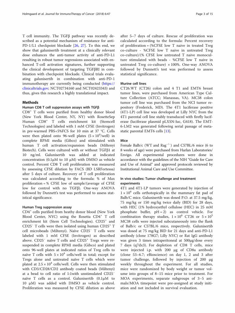
ResultsGalunisertib blocks TGFβ1 mediated suppression of naïveT cell proliferation and blocks Treg mediated suppressionof naïve T cellsTGFβ signaling plays an important role in suppressingan immune reaction and inducing tolerance. In particu-lar, TGFβ signaling inhibits innate and adaptive immunefunctions and induces suppressive immune cells. To testif galunisertib could rescue TGFβ suppressed immunecell subsets, naïve T cell suppression assays were estab-lished, and suppression mediated by TGFβ1 or by Tregulatory cells (Tregs) was tested in in vitro culture sys-tems. For these experiments, naïve human CD8+ T cellswere stimulated with anti-CD3/anti-CD28 beads in thepresence or absence of TGFβ1. As shown in Fig. 1a,while TGFβ1 potently suppressed the proliferation ofCD8+ T cells, addition of galunisertib resulted in a dose-dependent rescue of proliferation in the TGFβ1 treatedcultures, with enhanced proliferation observed at thehigher doses of galunisertib. To evaluate the ability ofgalunisertib to modulate Treg suppressive activity,CD4+CD25+ Treg cells were co-cultured with naïve T cells(CD4+CD25−) in the presence of anti-CD3/anti-28/anti-CD2 stimulation. While CD4+CD25+ Tregs potentlysuppressed naïve T cell proliferation, addition ofgalunisertib fully reversed the suppression of proliferation,demonstrating a role for galunisertib in reversing Tregmediated immune suppression (Fig. 1b).
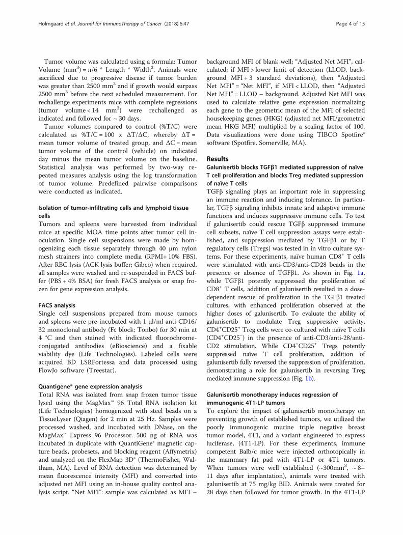
Galunisertib monotherapy induces regression ofimmunogenic 4T1-LP tumorsTo explore the impact of galunisertib monotherapy onpreventing growth of established tumors, we utilized thepoorly immunogenic murine triple negative breasttumor model, 4T1, and a variant engineered to expressluciferase, (4T1-LP). For these experiments, immunecompetent Balb/c mice were injected orthotopically inthe mammary fat pad with 4T1-LP or 4T1 tumors.When tumors were well established (~300mm3, ~ 8–11 days after implantation), animals were treated withgalunisertib at 75 mg/kg BID. Animals were treated for28 days then followed for tumor growth. In the 4T1-LP
Holmgaard et al. Journal for ImmunoTherapy of Cancer (2018) 6:47 Page 4 of 15
model, the majority of mice (10/12) responded to galuni-sertib therapy, including 4/12 complete responses (Fig.2a); in contrast, none of the poorly-immunogenic 4T1bearing mice responded to galunisertib therapy (Fig. 2b),suggesting that the presence of a foreign antigen (i.e. LP), potentially enhanced the ability of galunisertib to in-duce the rejection of the 4T1-LP derivative. In a previ-ous study, a survival benefit advantage with galunisertibwas observed in the poorly-immunogenic 4T1 tumormodel (Yingling et al., [25]). This may reflect the earlystart of treatment in that study (day 4 after tumor im-plantation compared to 8–11 days in the study pre-sented here) or it may be a result of an anti-metastaticactivity rather than an effect only on primary tumorgrowth.A few mice in the vehicle control group in the 4T1-LP
model, but not in the parental 4T1 model, showed aninitial tumor response before eventually progressing (Fig.2a), suggesting that spontaneous responses to immuno-genic tumor cell lines can occur in some mice. This mayreflect the different T cell repertoire between individualmice as we did not use TCR transgenic mice, or suggest-ing that these mice developed an immune response to adominant CTL epitope of LP, which may lead to reducedtumor invasiveness and spontaneous regression.[28]. Although, a few mice of the control group
showed initial spontaneous activity, all untreated tumors
eventually progressed without treatment. The spontan-eous activity we observe in a few of the 4T1-LP controlmice (Fig. 2a) is likely reflective of an immune responseto the implanted tumors, and this immune responsemay in fact be the mechanism by which galunisertib isso much more active as a monotherapy in 4T1-LP com-pared to 4T1 parental. We speculate that the immuno-genic nature of 4T1-LP is likely what impactsgalunisertib monotherapy activity in this model, while inless immunogenic tumor models, combination withanti-PD-L1 is needed (described below).To further evaluate and interrogate the impact of
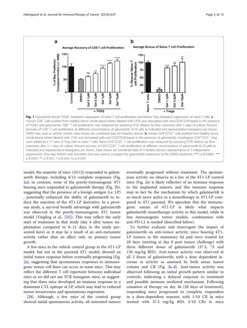
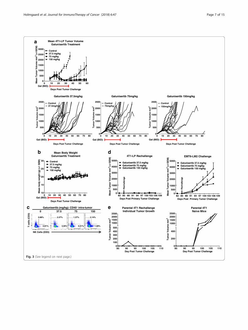
galunisertib on anti-tumor activity, mice bearing 4T1-LP tumors in the mammary fat pad were treated for28 days (starting at day 8 post tumor challenge) withthree different doses of galunisertib (37.5, 75 and150 mg/kg BID). Anti-tumor activity was observed atall 3 doses of galunisertib, with a dose dependent in-crease in activity as assessed by both mean tumorvolume and CR (Fig. 3a-d). Anti-tumor activity wasobserved following an initial growth pattern similar tocontrols, indicating a delayed response to treatmentand possible immune mediated mechanism. Followingcessation of therapy on day 36 (28 days of treatment),responding mice progressed to complete respondersin a dose-dependent manner, with 1/10 CR in micetreated with 37.5 mg/kg BID, 3/10 CRs in mice
Nai
ve
T c
ells
DM
SO
Gal
1.1
µM
Gal
3.3
µM
Gal
10µ
M
0
25
50
75
100
125
% R
escu
e N
aive
Tce
ll P
rolif
erat
ion
Co
ntr
ol
DM
SO
Gal
0.1
µM
Gal
0.3
µM
Gal
1µM
Gal
3µM
Gal
10µ
M
0
50
100
150%
of M
ax P
rolif
erio
nC
ompa
red
Co
ntro
l (+/
- S
EM
)
+ Tregs
a b
+ TGF
ns
Fig. 1 Galunisertib blocks TGFβ1 mediated suppression of naïve T cell proliferation and blocks Treg mediated suppression of naïve T cells. aHuman CD8+ cells purified from healthy donor whole blood where labeled with CFSE and stimulated with anti-CD3/CD28 beads in the presenceof TGFβ1 and galunisertib. CD8+ T cell proliferation was measured by assessing CFSE dilation by flow cytometry after 5 days of culture. Percentrecovery of CD8+ T cell proliferation at different concentrations of galunisertib (0-10 μM) as indicated and representative histograms are shown.DMSO was used as vehicle control. Data shown are combined data of 4 healthy donors. b Human CD4+CD25+ cells purified from healthy donorwhole blood where labeled with CFSE and stimulated with anti-CD3/CD28 beads in the presence of galunisertib. Autologous CD4+CD25− Tregwere added at a 1:1 ratio of Treg cells to naïve T cells. Naïve CD4+CD25− T cell proliferation was measured by assessing CFSE dilation by flowcytometry after 5–7 days of culture. Percent recovery of CD4+CD25− T cell proliferation at different concentrations of galunisertib (0-10 μM) asindicated and representative histograms are shown. Data shown are combined data of 4 healthy donors; representative of 3 independentexperiments. One-way ANOVA with Dunnett’s test was used to compare the galunisertib treatments to the DMSO treatment. ****: p≤ 0.0001; ***:p≤ 0.001; **: p≤ 0.01; *: p≤ 0.05; ns: p ≥ 0.05
Holmgaard et al. Journal for ImmunoTherapy of Cancer (2018) 6:47 Page 5 of 15
treated with 75 mg/kg BID, and 5/10 CRs observed inmice treated with 150 mg/kg BID (Fig. 3a). CR miceremained tumor free for an additional 49 days in theabsence of further treatment. These data indicate thatgalunisertib induces a potent, dose-dependent durableanti-tumor response. Metastases to lungs were notobserved in this tumor model.Treatments with galunisertib were well-tolerated
with no body weight loss observed with any of thedoses tested (Fig. 3b).
Importantly, PK/PD profiling studies of galunisertibsuggest that administration of 75 mg/kg BID in preclin-ical models or 150 mg/kg BID in patients can achievesignificant target modulation in vivo over a 24-h period[1, 29]. Thus, we show anti-tumor activity with galuni-sertib at clinically relevant doses.To begin to understand how galunisertib treatment
modulated immune cells within the tumor, tumors frommice galunisertib treated or vehicle control mice wereharvested 8 days after therapy initiation and the changesin T cell infiltration were analyzed by flow cytometry.Relative to control animals, a modest increase in bothCD3 T cells, mainly CD8 T cells, and NK cells wasobserved in tumors of mice treated with the clinicallyrelevant dose of 75 mg/kg galunisertib (Fig. 3c), indicat-ing a role of galunisertib on T-cell expansion or T-celltrafficking to the tumor site. These differences did notreach statistical significance. No significant changes wereobserved in the myeloid compartment in tumors of galu-nisertib treated mice compared to control treated micein this model. However, only the number of myeloidcells was analyzed and not the function; thus, whethergalunisertib induces reprogramming toward an antitu-mor phenotype was not explored. This may also reflectthe time point of tumor collection. To this end, a priorstudy with anti-mouse TGFβRII showed modulation ofMDSCs by blocking the TGFβ signaling pathway [13].
Galunisertib monotherapy induces immunologic memoryand demonstrates antigen spreading4T1-LP tumor bearing mice that completely respondedto galunisertib therapy remained tumor free for up to85 days (49 days after treatment completed) (Fig. 3a),indicating a durable response. To test the ability of galu-nisertib to induce immunologic memory to 4T1-LP tu-mors, mice which had completely regressed 4T1-LPwere re-challenged orthotopically with 4T1-LP on theopposite flank of the original tumor injection site andadditionally received a primary challenge of a differenttriple negative breast cancer tumor, EMT6-LM2, on theflank of the original tumor injection site. In all micetested, complete responders rejected the re-challengewith the 4T1-LP tumors (Fig. 3d, left panel), but did notreject EMT6-LM2 tumors (Fig. 3d, middle panel), dem-onstrating immunologic memory to the 4T1-LP tumorcells, but not the heterologous tumor. To evaluate thepotential for epitope spreading as a result of galunisertibanti-tumor activity, mice which had completelyregressed 4T1-LP after being treated with 75 mg/kg ofgalunisertib were re-challenged in a separate experimentwith the poorly immunogenic parental 4T1 tumors,which lack the immunogenic LP transgene and is not re-sponsive to de-novo galunisertib monotherapy (Fig. 2b);in all mice tested, 4T1-LP complete repressors also
0 20 40 60 800
100200300400500
1000
1500
2000
2500
4T1 Parental ModelIndividual Tumor Growth Curves
Day Post Tumor Challenge
Tum
or V
olu
me
mm
3
ControlGalunisertib
0 20 40 60 800
100200300400500
1000
1500
2000
2500
4T1-LP ModelIndividual Tumor Growth Curves
Day Post Tumor Challenge
Tum
or V
olu
me
mm
3
ControlGalunisertib
a
b
Gal (BID):
Gal (BID):
Fig. 2 Galunisertib monotherapy induces regression ofimmunogenic 4T1-LP variant breast tumors. Mean and individualtumor growth curves for Balb/c mice injected orthotopically in themammary fat pad with 4T1-LP (a) or parental 4T1 (b) tumor cellsand treated with galunisertib (75 mg/kg BID) when tumors reached~300mm3 (6–8 days after implantation). The number of mice/grouprejecting tumors (complete responders, CRs) was: control (0/10 mice)and galunisertib (4/12) for 4T1-LP, and control (0/10 mice) andgalunisertib (0/12) for 4T1 parental as indicated. Data shown arerepresentative of two independent experiments with10–12 mice/group
Holmgaard et al. Journal for ImmunoTherapy of Cancer (2018) 6:47 Page 6 of 15
0 10 20 30 40 50 600
500
1000
1500
2000
2500
3000
Mean 4T1-LP Tumor VolumeGalunisertib Treatment
Days Post Tumor Challenge
Mea
n Tu
mor
Vol
um
e m
m3
(+/-S
EM
)
Control37.5 mg/kg75 mg/kg150 mg/kg
a
0 10 20 30 40 50 60 70 800
500
1000
1500
2000
2500
Galunisertib 150mg/kg
Days Post Tumor Challenge
Tum
or
Vo
lum
e m
m3
Control150mg/kg
0 10 20 30 40 50 60 70 800
500
1000
1500
2000
2500
Galunisertib 75mg/kg
Days Post Tumor Challenge
Tum
or
Vo
lum
e m
m3
Control75mg/kg
0 10 20 30 40 50 60 70 800
500
1000
1500
2000
2500
Galunisertib 37.5mg/kg
Days Post Tumor Challenge
Tum
or
Vo
lum
e m
m3
Control37.5mg/kg
Gal (BID):
Gal (BID): Gal (BID): Gal (BID):
b
c Galunisertib (mg/kg): CD45+ intra-tumor
3.37% 1.27% 3.14%
NK Cells (DX5)
T c
ells
(C
D3)
0 10 20 30 40 50 60 70 8010
15
20
25
30
Mean Body WeightGalunisertib Treatment
Days Post Tumor Challenge
Mea
n b
ody
wei
ght (
g) (
+/-
SE
M)
Control37.5 mg/kg75 mg/kg150 mg/kg
0 37.5 75 150
0.88%
3.05% 0.51% 1.69%0.81%
Gal (BID):
d
82 85 88 91 94 97 100 103 106 1090
500
1000
2000
3000
4000
5000
4T1-LP Rechallenge
Days Post Primary Tumor Challenge
Mea
n T
umo
r V
olu
me
mm
3(+
/-S
EM
)
Galunisertib 37.5 mg/kgGalunisertib 75 mg/kgGalunisertib 150 mg/kg
Rec
hal
leng
e
82 85 88 91 94 97 100 103 106 1090
500
1000
2000
3000
4000
5000
EMT6-LM2 Challenge
Days Post Primary Tumor Challenge
Mea
n T
umo
r V
olu
me
mm
3(+
/-S
EM
)
Galunisertib 37.5 mg/kgGalunisertib 75 mg/kgGalunisertib 150 mg/kg
Rec
hal
leng
e
e
85 90 95 100 105 1100
100
200
300
400
500
1000150020002500
Day Post Tumor Challenge
Tum
or V
olum
e m
m3
Parental 4T1 RechallengeIndividual Tumor Growth
85 90 95 100 105 1100
200
400
1000150020002500
Day Post Tumor Challenge
Tum
or V
olum
e m
m3
Parental 4T1Naive Mice
Fig. 3 (See legend on next page.)
Holmgaard et al. Journal for ImmunoTherapy of Cancer (2018) 6:47 Page 7 of 15
rejected the parental 4T1 challenge (Fig. 3e, right panel),demonstrating the potential for galunisertib anti-tumoractivity to mediate antigen spreading.
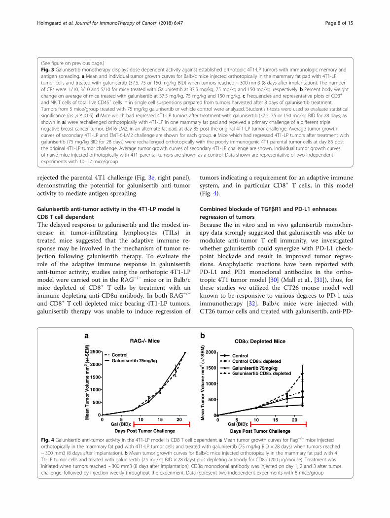
Galunisertib anti-tumor activity in the 4T1-LP model isCD8 T cell dependentThe delayed response to galunisertib and the modest in-crease in tumor-infiltrating lymphocytes (TILs) intreated mice suggested that the adaptive immune re-sponse may be involved in the mechanism of tumor re-jection following galunisertib therapy. To evaluate therole of the adaptive immune response in galunisertibanti-tumor activity, studies using the orthotopic 4T1-LPmodel were carried out in the RAG−/− mice or in Balb/cmice depleted of CD8+ T cells by treatment with animmune depleting anti-CD8α antibody. In both RAG−/−
and CD8+ T cell depleted mice bearing 4T1-LP tumors,galunisertib therapy was unable to induce regression of
tumors indicating a requirement for an adaptive immunesystem, and in particular CD8+ T cells, in this model(Fig. 4).
Combined blockade of TGFβR1 and PD-L1 enhnacesregression of tumorsBecause the in vitro and in vivo galunisertib monother-apy data strongly suggested that galunisertib was able tomodulate anti-tumor T cell immunity, we investigatedwhether galunisertib could synergize with PD-L1 check-point blockade and result in improved tumor regres-sions. Anaphylactic reactions have been reported withPD-L1 and PD1 monoclonal antibodies in the ortho-tropic 4T1 tumor model [30] (Mall et al., [31]), thus, forthese studies we utilized the CT26 mouse model wellknown to be responsive to various degrees to PD-1 axisimmunotherapy [32]. Balb/c mice were injected withCT26 tumor cells and treated with galunisertib, anti-PD-
(See figure on previous page.)Fig. 3 Galunisertib monotherapy displays dose dependent activity against established orthotopic 4T1-LP tumors with immunologic memory andantigen spreading. a Mean and individual tumor growth curves for Balb/c mice injected orthotopically in the mammary fat pad with 4T1-LPtumor cells and treated with galunisertib (37.5, 75 or 150 mg/kg BID) when tumors reached ~ 300 mm3 (8 days after implantation). The numberof CRs were: 1/10, 3/10 and 5/10 for mice treated with Galunisertib at 37.5 mg/kg, 75 mg/kg and 150 mg/kg, respectively. b Percent body weightchange on average of mice treated with galunisertib at 37.5 mg/kg, 75 mg/kg and 150 mg/kg. c Frequencies and representative plots of CD3+
and NK T cells of total live CD45+ cells in in single cell suspensions prepared from tumors harvested after 8 days of galunisertib treatment.Tumors from 5 mice/group treated with 75 mg/kg galunisertib or vehicle control were analyzed. Student’s t-tests were used to evaluate statisticalsignificance (ns: p≥ 0.05). d Mice which had regressed 4T1-LP tumors after treatment with galunisertib (37.5, 75 or 150 mg/kg BID for 28 days; asshown in a) were rechallenged orthotopically with 4T1-LP in one mammary fat pad and received a primary challenge of a different triplenegative breast cancer tumor, EMT6-LM2, in an alternate fat pad, at day 85 post the original 4T1-LP tumor challenge. Average tumor growthcurves of secondary 4T1-LP and EMT-6-LM2 challenge are shown for each group. e Mice which had regressed 4T1-LP tumors after treatment withgalunisertib (75 mg/kg BID for 28 days) were rechallenged orthotopically with the poorly immunogenic 4T1 parental tumor cells at day 85 postthe original 4T1-LP tumor challenge. Average tumor growth curves of secondary 4T1-LP challenge are shown. Individual tumor growth curvesof naïve mice injected orthotopically with 4T1 parental tumors are shown as a control. Data shown are representative of two independentexperiments with 10–12 mice/group
0 5 10 15 200
500
1000
1500
2000
CD8α Depleted Mice
Days Post Tumor Challenge
Mea
n T
umor
Vol
ume
mm
3(+
/-S
EM
)
Control
Galunisertib 75mg/kgControl CD8α depleted
Galunisertib CD8α depleted
Gal (BID):
a b
0 5 10 15 200
500
1000
1500
2000
2500
RAG-/- Mice
Days Post Tumor Challenge
Mea
n T
umor
Vol
ume
mm
3(+
/-S
EM
)
Galunisertib 75mg/kgControl
Gal (BID):
Fig. 4 Galunisertib anti-tumor activity in the 4T1-LP model is CD8 T cell dependent. a Mean tumor growth curves for Rag−/− mice injectedorthotopically in the mammary fat pad with 4T1-LP tumor cells and treated with galunisertib (75 mg/kg BID × 28 days) when tumors reached~ 300 mm3 (8 days after implantation). b Mean tumor growth curves for Balb/c mice injected orthotopically in the mammary fat pad with 4T1-LP tumor cells and treated with galunisertib (75 mg/kg BID × 28 days) plus depleting antibody for CD8α (200 μg/mouse). Treatment wasinitiated when tumors reached ~ 300 mm3 (8 days after implantation). CD8α monoclonal antibody was injected on day 1, 2 and 3 after tumorchallenge, followed by injection weekly throughout the experiment. Data represent two independent experiments with 8 mice/group
Holmgaard et al. Journal for ImmunoTherapy of Cancer (2018) 6:47 Page 8 of 15
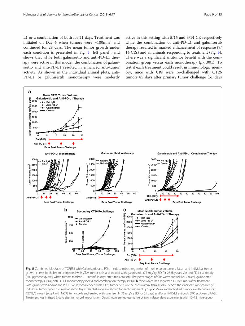
L1 or a combination of both for 21 days. Treatment wasinitiated on Day 6 when tumors were ~100mm3 andcontinued for 28 days. The mean tumor growth undereach condition is presented in Fig. 5 (left panel), andshows that while both galunsertib and anti-PD-L1 ther-apy were active in this model, the combination of galuni-sertib and anti-PD-L1 resulted in enhanced anti-tumoractivity. As shown in the individual animal plots, anti-PD-L1 or galunisertib monotherapy were modestly
active in this setting with 5/15 and 3/14 CR respectivelywhile the combination of anti-PD-L1 and galunisertibtherapy resulted in marked enhancement of response (9/14 CRs) and all animals responding to treatment (Fig. 5).There was a significant antitumor benefit with the com-bination group versus each monotherapy (p < .001). Totest if each treatment could result in immunologic mem-ory, mice with CRs were re-challenged with CT26tumors 85 days after primary tumor challenge (51 days
0 10 20 30 40 50 600
500
1000
1500
2000
2500
3000
Days Post Tumor Challenge
Tum
or
Vo
lum
e m
m3
Galunisertib Monotherapy
GalunisertibRat IgG
0 10 20 30 40 50 60 70 80 90 1000
500
1000
1500
2000
2500
3000
Galunisertib and Anti-PD-L1 Combination Therapy
Days Post Tumor Challenge
Tu
mo
r V
olu
me
mm
3
Rat IgGCombo
*
a
0 5 10 15 20 250
500
1000
1500
2000
2500
3000
Mean CT26 Tumor VolumeGalunisertib and Anti-PD-L1 Therapy
Days Post Tumor Challenge
Mea
n T
umor
Vol
ume
mm
3(+
/-S
EM
)
Rat IgG
GalunisertibAnti-PD-L1
Combo
Gal (BID):
Anti-PD-L1:
0 10 20 30 40 50 600
500
1000
1500
2000
2500
3000
Days Post Tumor Challenge
Tum
or
Vo
lum
e m
m3
Anti-PD-L1 Monotherapy
PD-L1Rat IgG
Gal (BID):Gal (BID):
Anti-PD-L1:Anti-PD-L1:
80 85 90 95 100 1050
250
500
750
1000
Secondary CT26 Rechallenge
Days Post Primary Tumor Challenge
Tum
or V
olu
me
mm
3
GalunisertibAnti-PD-L1Combo
Rec
hal
leng
e
*
b
0 5 10 15 200
500
1000
1500
2000
2500
3000
Mean MC38 Tumor VolumeGalunisertib and Anti-PD-L1 Therapy
Day Post Tumor Challenge
Mea
n T
umor
Vol
ume
mm
3(+
/-S
EM
)
Rat IgG
GalunisertibAnti-PD-L1
Combo
c
Gal (BID):
Anti-PD-L1:
Fig. 5 Combined blockade of TGFβR1 with Galunisertib and PD-L1 induce robust regression of murine colon tumors. Mean and individual tumorgrowth curves for Balb/c mice injected with CT26 tumor cells and treated with galunisertib (75 mg/kg BID for 28 days) and/or anti-PD-L1 antibody(500 μg/dose, q7dx3) when tumors reached ~100mm3 (6 days after implantation). The percentages of CRs were: control (0/15 mice), galunisertibmonotherapy (3/14), anti-PD-L1 monotherapy (5/15) and combination therapy (9/14). b Mice which had regressed CT26 tumors after treatmentwith galunisertib and/or anti-PD-L1 were rechallenged with CT26 tumor cells on the contralateral flank at day 85 post the original tumor challenge.Individual tumor growth curves of secondary CT26 challenge are shown for each treatment group. c Mean and individual tumor growth curves forC57BL/6 mice injected with MC38 tumor cells and treated with galunisertib (75 mg/kg BID for 21 days) and/or anti-PD-L1 antibody (500 μg/dose, q7dx3).Treatment was initiated 3 days after tumor cell implantation. Data shown are representative of two independent experiments with 10–12 mice/group
Holmgaard et al. Journal for ImmunoTherapy of Cancer (2018) 6:47 Page 9 of 15
after initial treatment cessation). All complete re-sponders in monotherapy and combination treatmentgroups rejected the re-challenge with the CT26 tumors(Fig. 5b, left panel); however, one animal from the com-bination cohort, defined as a long-term partial responder(indicated by * in Fig. 5a right panel) was unable to rejectsecondary tumor challenge. The ability of galunisertib toenhance the activity of anti-PD-L1 immunotherapy wasconfirmed in the MC38 tumor model, which is historicallyless responsive to checkpoint immunotherapy and consid-ered to be more myeloid biology driven [32]. In thismodel, where treatment began on day 3 after tumor chal-lenge, similar monotherapy and combination therapy ac-tivity was observed albeit with more moderate activityoverall (Fig. 5c).
Combination of galunisertib and anti-PD-L1 checkpointblockade induces an intra-tumor immune related geneexpression profile that is accelerated and enhancedcompared to anti-PD-L1 monotherapyTo further elucidate the mechanism of action of thecombination activity of galunisertib and anti-PD-L1,gene expression studies were carried out on tumorsfrom CT26 tumor bearing mice treated with control,anti-PD-L1, galunisertib or a combination of anti-PD-L1plus galunisertib. For these studies, tumors from treated
mice were collected day 10, 16, and 22 after tumor chal-lenge (i.e. 4, 10, and 16 days after initiation of therapy)and subjected to high-content molecular profiling usinga custom designed QuantigeneTM gene panel to detect Tcell activation and intra-tumoral inflammation (Table 1).Galunisertib monotherapy (75 mg/kg) did not appre-ciably alter the set of immune genes analyzed relative tocontrol tumors at any time point evaluated (Fig. 6, toppanel). Anti-PD-L1 monotherapy resulted in an en-hanced T cell infiltration and activation profile, exempli-fied by the increase in multiple immune activation andinflammation transcripts such as Ccl5, Itgax, Icam1,Foxp3, Lag3 by day 22 (Fig. 6a). On the other hand, thecombination treatment demonstrated an early signatureof enhanced T cell activation and inflammation exempli-fied by the upregulation of transcripts for Ifnγ, Lag3,Ccl3, Ccl4, Ccl5, and Tnfrsf18) beginning on Day 16(after only 10 days of therapy, where only a minorchange in activation was detected with anti-PD-L1monotherapy), and continuing at day 22 with an en-hanced gene expression related to T cell infiltration(Ptprc, Cd8b1, Cd3e, and Cd4) and T cell activation andinflammation (Il2, Il4, Il17a, Lag3, Ifnγ, Ifnα, Ifnβ1,Foxp3, Cd274, and Pdcdlg2) (Fig. 6a). The gene profilefor the combination cohort was similar but more robustcompared to PD-L1 monotherapy detected at the later
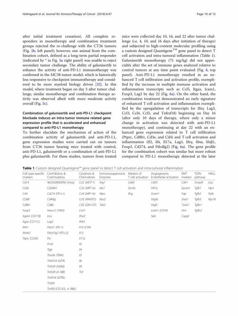
Table 1 Custom designed Quantigene™ gene panel to detect T cell activation and intra-tumoral inflammation
Cell type-specificmarkers
Coinhibitory &Costimulatory
Cytokines &Chemokines
ImmunosuppressiveEnzymes
Markers ofT cell activation
Angiogenesis;Endothelial activation
EMTmarkers
TGFbpathway
HKGs
Cd14 4632428N05Rik (Vista) Ccl2 (MCP-1) Arg1 Cd69 Cdh5 Cdh1 Smad4 Gus
Cd3e Cd200r1 Ccl3 (MIP-1a) Ido1 Gzmb Hif1a Epcam Tgfb1 Hprt
Cd4 Cd274 (PD-L1) Ccl4 (MIP-1b) Mpo Ifng Vcam1 Fap Tgfb2 Ppib
CD68 Cd40lg Ccl5 (RANTES) Nos2 Vegfa Snai1 Tgfb3 Rps18
Cd8b1 Cd86 Csf2 (GM-CSF) Tdo2 Vegfc Twist1 Tgfbr1
Foxp3 Havcr2 (TIM3) Cxcl1 Icam1 (CD54) Vim Tgfbr2
Itgam (CD11B) Icos Ifna2 Sele Cspg4
Itgax (CD11C) Lag3 Ifnb1
Klrk1 Pdcd1 (PD-1) Il10 (CSIF)
Ms4a1 Pdcd1lg2 (PD-L2) Il13
Ptprc (CD45) Pvr Il17a
Pvrl2 Il2
Tigit Il4
Timd4 (TIM4) Il5
Tnfrsf18 (GITR) Il6
Tnfrsf4 (OX40) Il9
Tnfrsf9 (4-1BB) Tnf
Tnfsf18 (GITRL)
Tnfsf4
Tnfsf9 (CD137L, 4-1BBL)
Holmgaard et al. Journal for ImmunoTherapy of Cancer (2018) 6:47 Page 10 of 15
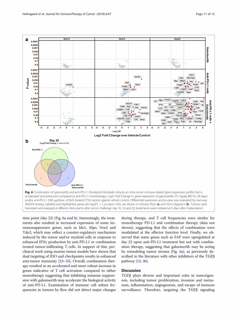
time point (day 22) (Fig. 6a and b). Interestingly, the treat-ments also resulted in increased expression of some im-munosuppressive genes, such as Ido1, Mpo, Nos2 andTdo2, which may reflect a counter-regulatory mechanisminduced by the tumor and/or myeloid cells in response toenhanced IFNγ production by anti-PD-L1 or combinationtreated tumor-infiltrating T cells. In support of this, pre-clinical work using murine tumor models have shown thatdual targeting of IDO and checkpoints results in enhancedanti-tumor immunity [33–35]. Overall, combination ther-apy resulted in an accelerated and more robust increase ingenes indicative of T cell activation compared to eithermonotherapy suggesting that inhibiting immune suppres-sion with galunisertib may accelerate the biological activityof anti-PD-L1. Examination of immune cell subset fre-quencies in tumors by flow did not detect major changes
during therapy, and T cell frequencies were similar formonotherapy PD-L1 and combination therapy (data notshown), suggesting that the effects of combination weremodulated at the effector function level. Finally, we ob-served that some genes such as FAP were upregulated atday 22 upon anti-PD-L1 treatment but not with combin-ation therapy, suggesting that galunisertib may be actingby remodeling tumor stroma (Fig. 6a), as previously de-scribed in the literature with other inhibitors of the TGFβpathway [13, 36].
DiscussionTGFβ plays diverse and important roles in tumorigen-esis, including tumor proliferation, invasion and metas-tasis, inflammation, angiogenesis, and escape of immunesurveillance. Therefore, targeting the TGFβ signaling
Fig. 6 Combination of galunisertib and anti-PD-L1 checkpoint blockade induces an intra-tumor immune related gene expression profile that isaccelerated and enhanced compared to anti-PD-L1 monotherapy. Log2 Fold Change in gene expression of galunisertib (75 mg/kg BID for 28 days)and/or anti-PD-L1 (500 μg/dose, q7dx3) treated CT26 tumors against vehicle control. Differential expression and p-value was evaluated by two-wayANOVA testing. Labelled and highlighted genes are log2FC > 1, p-value< 0.05, are shown in Volcano Plots (a) and Venn Diagrams (b). Tumors wereharvested and analyzed at different time points after tumor challenge: day 10, 16 and 22 (treatments were initiated at 6 days after implantation)
Holmgaard et al. Journal for ImmunoTherapy of Cancer (2018) 6:47 Page 11 of 15
pathway has been an attractive objective for cancer ther-apy, and several drugs have been identified and areunder clinical development [37–39]. Galunisertib is asmall molecule inhibitor of the kinase domain ofTGFβRI. Binding of TGFβ ligands to TGFβRII is the firststep to initiate activation of the TGFβ signaling pathway.Once bound to TGFβRII, this ligand/receptor complexbinds to TGFβRI to form a heterotrimeric complex. For-mation of this complex results in phosphorylation of theserine/threonine kinase domains of the receptors,followed by activation of the canonical SMAD2/3 signal-ing pathways as well as non-canonical (i.e. MAPK) path-ways [39]. These pathways modulate transcription ofnumerous target genes, resulting in a variety of effects.By blocking the kinase domain of TGFβRI, galunisertibmay effectively inhibit signaling via the TGFβ pathway.Preclinical and clinical research on galunisertib,
including the treatment of over 800 patients, has dem-onstrated that SMIs of TGFβ can safely be developed forclinical testing, provided there is an adequate under-standing of the pharmacokinetic/pharmacodynamics(PK/PD) relationship, as most of the toxicities in animalmodels that were of concern prior to the start of clinicaldevelopment of galunisertib have not been observed inhumans [1]. Furthermore, preclinical and clinical effortssuggest that the biology of the TGFβ inhibition is largelydependent on the microenvironment, perhaps more thanoriginally anticipated. For example, TGFβ1 is a potentinducer of angiogenesis [40], by directly inducing VEGFexpression [41], or recruiting other cells, such as mono-cytes, which in turn secrete proangiogenic molecules[42]. TGFβ can also manipulate the tumor microenvir-onment to favor the evasion of cancer cells from im-mune surveillance via tampering with the antitumorfunctions of T cells, NK cells, B cells, and other cells[43–45]. This activity of TGFβ may be mediated throughits direct effect on these cells, as well as via its ability toinduce Foxp3+ Tregs [46]. Both cancer-intrinsic andimmune-mediated effect of TGFβ in breast cancers havebeen described [47–49]. Thus, a focus on direct tumorcell cytotoxicity may be misleading and provide incon-clusive observations that will not be helpful to advanceclinical development of future TGFβ inhibitors. Earlystudies using immune-compromised animals may there-fore also have limited the screening for TGFβ inhibi-tors. It now appears that an active immune responseis essential to assess the effect of TGFβ signaling in-hibition in animal models; thus, immune-competentanimal models may be more predictive to evaluateTGFβ inhibitors. Consequently, more novel preclinicaltesting assays are required than those traditionallyused in oncology research.Here we describe the impact of galunisertib to modu-
late the immune system and its ability to enhance anti-
tumor activity in immune-competent murine tumormodels. We show that galunisertib monotherapy inducesdose-dependent regression of well-established immuno-genic 4T1-LP breast tumors. The responses were dur-able with immunological memory as demonstrated byrechallenge experiments with 4T1-LP tumors as well asa second triple negative breast tumor cell line. Of note,mice that rejected the immunogenic 4T1-LP tumorswere also able to reject 4T1 parental cells upon rechal-lenge, suggesting the development of a secondaryimmune response via antigen spreading as a conse-quence of effective tumor targeting. The anti-tumoractivity of galunisertib in the 4T1-LP tumor model wasCD8+ T cell dependent and associated with a modestincrease in T-cell infiltration in tumors. The increasewas modest and did not reach statistical significancethough, which might reflect the time point chosen fortumor collection. It is well established in the immuno-oncology field that the spatial distribution and location ofimmune cells is highly important. In fact, the recent publi-cations combining TGFβ inhibition and PD-L1 blockadeshow that the main mechanism of action of TGFβ inhib-ition is to increase T-cell infiltration into tumor [26, 27].In addition, using an anti-TGFβRII blocking antibody wehave previously shown that blocking TGFβ signaling inthe EMT6 tumor model induces immune infiltration [13].We did not investigate metastasis to lungs in either
the 4T1 or 4T1-LP tumor models used here. However,we have previously shown that the anti-mouse TGFβRIIantibody significantly inhibits the growth of established4T1-parental primary tumors and diminishes the spon-taneous pulmonary metastasis [13]. In addition, it wasshown that galunisertib in combination with anti-CTLA4 therapy suppresses both primary melanomatumor growth as well as metastases in a physiologicalrelevant trangenic melanoma model (Hanks et al. [50]).Furthermore, we described that galunisertib inhibitsTGFβ mediated migration of U87MG glioblastoma cellsin vitro in a dose-dependent manner [51]. Notably, inthis model system, galunisertib reduced baseline migra-tion of U87MG cells in the absence of exogenousTGFβ1, presumably by inhibiting autocrine signalingthrough TGFβRI. Together this suggests that galuniser-tib has the capacity to suppress the development of me-tastasis and that TGFβ pathway blockade of the parental4T1 model is sufficient to inhibit metastasis to lung.Importantly, we have previously shown that the TGFβ
pathway is abrogated upon treatment with galunisertibboth in vitro and in vivo [51]. We demonstrated thatgalunisertib inhibited TGFβ-induced pSMAD in varioustumor cell lines, including 4 T1-LP, in vitro in a dose-dependent manner [51]. Furthermore, we reported agalunisertib time and dose-dependent inhibition ofendogenous TGFβ-dependent signal transduction in vivo
Holmgaard et al. Journal for ImmunoTherapy of Cancer (2018) 6:47 Page 12 of 15
in EMT6-LM2 murine syngenic tumor models [51].These data suggest that the effects of galunisertib areon-target. Potential off-target effects of galunisertib arefurther diminished, as treatment with an anti-mouseTGFβRII antibody similarly inhibits the growth of estab-lished mouse 4T1 and EMT6 primary [13].Immunotherapeutic strategies such as immune check-
point blockade have shown significant promise for treat-ment of cancers resistant to conventional modalities,leading to Food and Drug Administration (FDA)approval in advanced melanoma, renal cell carcinomaand non-small cell lung cancer (NSCLC) [52]. Despiteclinical results, even with combined checkpoint blockade[53], therapeutic success has so far been limited to asubset of patients, calling for identification of markerspredicting response, identification of resistance mecha-nisms and development of combinatorial therapeuticapproaches. To this end, TGFβ pathway inhibition repre-sents an attractive strategy with its multitude of effectson cancer progression and on the immune system to en-hance the development of anti-tumor T-cell immunity.Indeed, a recent study by Powles et al., reports that lackof response to atzeolizumab (anti-PDL1) in bladder can-cer patients was associated with an immune-excludedphenotype that corresponded with active TGFβ in peritu-moral stroma and a signature of TGFβ signaling [26]. Usingmouse models that recapitulate the immune-excludedphenotype they further show that co-administration ofblocking antibodies to TGFβ and PDL1 reduced TGFβ sig-naling, facilitated T-cell penetration of tumors, and pro-voked vigorous anti-tumor immunity leading to tumorregressions. In a second recent study published by Batlleand colleagues, combinatorial activity of galunisertib withanti-PDL1 in murine colon cancer models was recently de-scribed [27]. Combination therapy induced pronounced im-mune responses which eradicated most metastases,prolonged recurrence-free survival, and was associated withdisruption of a T-cell exclusion phenotype. These resultssuggest that clinical co-administration of TGFβ and PDL1blocking agents may provide a subset of patients morefavorable outcomes; however, preclinical validation was per-formed with either a research-grade reagent [26] or a sig-nificantly excessive amount of galunisertib (800 mg/kg BIDcompared to the clinically relevant dose of 75 mg/kgdescribed in [1, 29]. In agreement, we demonstrate thatcombination of galunisertib with PD-L1 checkpoint block-ade results in a robust regression of CT26 tumors whencompared to single agents. The observed antitumor benefitwas associated with enhanced expression of genes indica-tive of immune activation and this gene expression profilewas accelerated compared to anti-PD-L1 monotherapy.Galunisertib alone resulted in no alteration of any of thetested genes. Considering the critical role of TGFβ in can-cer immunity, we speculate that this may be a result of the
gene panel tested, the dose chosen or the day of collection.Similar combination therapy activity was observed inthe PD-L1 insensitive tumor model, MC38, albeit withmore moderate activity overall, suggesting at leastadditive activity with potential for synergy when tar-geting the TGFβ and PD-1 pathways. The anti-tumoractivity of galunisertib was tested in a broad range ofmurine tumor models with similar results, furthersuggesting that TGFβ inhibition is immune mediatedand thus not restricted to specific tumor indications.Finally, we show that galunisertib reverses both TGFβ
and Treg mediated suppression of T cell proliferation inhuman cell cultures in vitro, which further highlight theimportant role of galunisertib to overcome immune sup-pression and promote anti-tumor immunity.Taken together, the results presented here demonstrate
the impact of blocking TGFβ signaling and provide astrong incentive to clinically explore the potential ofgalunisertib treatment to enhance the development ofanti-tumor T cell immunity, which may be enhanced bycombinations with immune checkpoint inhibitors. Ourresults expand on other reports demonstrating that sys-temic treatment with monoclonal antibodies targetingthe TGFβ ligands or the TGFβRII inhibit metastaticinvasion of breast cancer cells in murine tumor models[2, 13], and previous work reporting that blocking TGFβsignaling with SMIs suppresses metastasis in murinepancreatic cancer models [54], and enhances radiationresponse and prolongs survival in xenograft models ofglioblastoma [55].Galunisertib continues to advance in clinical trials hav-
ing completed Phase I [56] and is currently under inves-tigation in several Phase I and Phase II trials. Thus far,galunisertib has been very well-tolerated as a first-in-class, oral cancer therapy, and remains a promising com-pound in clinical development (http://clinicaltrials.gov/ct2/results?term=LY2157299). Our data presented heresupport continued clinical development of galunisertibto target tumors dependent on TGFβ-driven biology forgrowth, metastasis, and immune evasion. Whether TGFβinhibition applies to all tumors is not clear at this time.For clinical development, patient selection tools, definingwho will most likely benefit from TGFβ inhibition, remaina challenging question. Among others, the activity ofTGFβ inhibition appears to be dependent on immunefunction; thus, it will be important to investigate new bio-markers that are related to immune responses which mayhelp with patient selection in future studies.
ConclusionIn many advanced cancers, TGFβ ligands are overex-pressed and the outcome of signaling is diverted towarddisease progression. A concerted effort has thereforebeen to develop drugs that block TGFβ signaling for
Holmgaard et al. Journal for ImmunoTherapy of Cancer (2018) 6:47 Page 13 of 15
therapeutic benefit. Galunisertib is a pharmacologicalsmall molecule inhibitor of the TGFβ pathway that actsby inhibiting signaling through TGFβ receptor I. As amonotherapy, galunisertib has shown some antitumoractivity in a variety of tumors, including durable andlong-term responses in patients with glioma. Here, wedemonstrate the ability of galunisertib to modulate anti-tumor T cell immunity, alone and in combination withPD-L1 checkpoint blockade, in preclinical models. Ourdata provide a strong rationale to clinically explore thepotential of galunisertib to enhance anti-tumor immuneresponse, particularly, in combinations with PD-L1/PD-1checkpoint inhibitors. Galunisertib is currently underclinical development in combination with checkpointinhibitors (including nivolumab and durvalumab) in pa-tients with NSCLC, HCC, or pancreatic cancer.
Authors' contributionsRBH, DS, YL, SC, MM, XX, II, JD, DS, GH, RN, and KED designed and/orperformed experiments. KB provided information on ongoing clinical trials.JM and PI performed statistical analysis. GP and MDK provided experimentalguidance and gave conceptual advice. RBH and KED wrote the manuscript.All authors read and approved the final manuscript.
Ethics approvalAll animal studies were performed in accordance with federal and local laws,policies, regulations, and standards in effect at the time of their conduct.Laboratories conducting these studies were accredited by the Association forAssessment and Accreditation of Laboratory Animal Care, International andall study protocols were approved by each laboratory’s Institutional AnimalCare and Use Committee.
Competing interestsThe authors disclose no potential conflicts of interest. All authors are currentor former employees of Eli Lilly and Company.
Publisher’s NoteSpringer Nature remains neutral with regard to jurisdictional claims inpublished maps and institutional affiliations.
Author details1Lilly Research Laboratories, Eli Lilly and Company, Indianapolis, IN 46285,USA. 2Eli Lilly and Company, 450 East 29th Street, New York, USA. 3JanssenPharmaceutical Companies of Johnson and Johnson, Spring House, PA, USA.
Received: 13 February 2018 Accepted: 11 May 2018
References1. Herbertz S, et al. Clinical development of galunisertib (LY2157299
monohydrate), a small molecule inhibitor of transforming growth factor-beta signaling pathway. Drug Des Devel Ther. 2015;9:4479–99.
2. Akhurst RJ. Targeting TGF-beta signaling for therapeutic gain. Cold SpringHarb Perspect Biol. 2017;9(10)
3. Bhola NE, et al. TGF-beta inhibition enhances chemotherapy action againsttriple-negative breast cancer. J Clin Invest. 2013;123(3):1348–58.
4. Massague J. TGFbeta in Cancer. Cell. 2008;134(2):215–30.5. Ishigame H, et al. Truncated form of TGF-betaRII, but not its absence,
induces memory CD8+ T cell expansion and lymphoproliferative disorder inmice. J Immunol. 2013;190(12):6340–50.
6. Levy L, Hill CS. Alterations in components of the TGF-beta superfamilysignaling pathways in human cancer. Cytokine Growth Factor Rev. 2006;17(1–2):41–58.
7. Teicher BA. Transforming growth factor-beta and the immune response tomalignant disease. Clin Cancer Res. 2007;13(21):6247–51.
8. Ganapathy V, et al. Targeting the transforming growth factor-beta pathwayinhibits human basal-like breast cancer metastasis. Mol Cancer. 2010;9:122.
9. Wick W, Naumann U, Weller M. Transforming growth factor-beta: amolecular target for the future therapy of glioblastoma. Curr Pharm Des.2006;12(3):341–9.
10. Ivanovic V, et al. Elevated plasma TGF-beta1 levels correlate withdecreased survival of metastatic breast cancer patients. Clin Chim Acta.2006;371(1–2):191–3.
11. Gorelik L, Flavell RA. Transforming growth factor-beta in T-cell biology. NatRev Immunol. 2002;2(1):46–53.
12. Li MO, Sanjabi S, Flavell RA. Transforming growth factor-beta controlsdevelopment, homeostasis, and tolerance of T cells by regulatory T cell-dependent and -independent mechanisms. Immunity. 2006;25(3):455–71.
13. Zhong Z, et al. Anti-transforming growth factor beta receptor II antibodyhas therapeutic efficacy against primary tumor growth and metastasisthrough multieffects on cancer, stroma, and immune cells. Clin Cancer Res.2010;16(4):1191–205.
14. Novitskiy SV, et al. Deletion of TGF-beta signaling in myeloid cells enhancestheir anti-tumorigenic properties. J Leukoc Biol. 2012;92(3):641–51.
15. Kulkarni AB, et al. Transforming growth factor beta 1 null mutation in micecauses excessive inflammatory response and early death. Proc Natl Acad SciU S A. 1993;90(2):770–4.
16. Shull MM, et al. Targeted disruption of the mouse transforming growthfactor-beta 1 gene results in multifocal inflammatory disease. Nature. 1992;359(6397):693–9.
17. Bommireddy R, et al. TGF-beta 1 regulates lymphocyte homeostasis bypreventing activation and subsequent apoptosis of peripheral lymphocytes.J Immunol. 2003;170(9):4612–22.
18. Marie JC, Liggitt D, Rudensky AY. Cellular mechanisms of fatal early-onsetautoimmunity in mice with the T cell-specific targeting of transforminggrowth factor-beta receptor. Immunity. 2006;25(3):441–54.
19. Sakaguchi S, Powrie F. Emerging challenges in regulatory T cell functionand biology. Science. 2007;317(5838):627–9.
20. Chen W, et al. Conversion of peripheral CD4+CD25- naive T cells to CD4+CD25+ regulatory T cells by TGF-beta induction of transcription factorFoxp3. J Exp Med. 2003;198(12):1875–86.
21. Marie JC, et al. TGF-beta1 maintains suppressor function and Foxp3expression in CD4+CD25+ regulatory T cells. J Exp Med. 2005;201(7):1061–7.
22. Fahlen L, et al. T cells that cannot respond to TGF-beta escape control byCD4(+)CD25(+) regulatory T cells. J Exp Med. 2005;201(5):737–46.
23. Yang L, et al. Expansion of myeloid immune suppressor Gr+CD11b+ cells intumor-bearing host directly promotes tumor angiogenesis. Cancer Cell.2004;6(4):409–21.
24. Yang L, et al. Abrogation of TGF beta signaling in mammary carcinomasrecruits Gr-1+CD11b+ myeloid cells that promote metastasis. Cancer Cell.2008;13(1):23–35.
25. Yingling JM, et al. Preclinical assessment of galunisertib (LY2157299monohydrate), a first-in-class transforming growth factor-β1 receptorinhibitor. Accepted 2017.
26. Mariathasan S, et al. TGFbeta attenuates tumour response to PD-L1 blockadeby contributing to exclusion of T cells. Nature. 2018;554(7693):544–8.
27. Tauriello DVF, et al. TGFbeta drives immune evasion in geneticallyreconstituted colon cancer metastasis. Nature. 2018;554(7693):538–43.
28. Baklaushev VP, et al. Luciferase expression allows bioluminescence imagingbut imposes limitations on the Orthotopic mouse (4T1) model of breastCancer. Sci Rep. 2017;7(1):7715.
29. Gueorguieva I, et al. Defining a therapeutic window for the novel TGF-betainhibitor LY2157299 monohydrate based on a pharmacokinetic/pharmacodynamic model. Br J Clin Pharmacol. 2014;77(5):796–807.
30. Mall C, et al. Repeated PD-1/PD-L1 monoclonal antibody administrationinduces fatal xenogeneic hypersensitivity reactions in a murine model ofbreast cancer. Oncoimmunology. 2016;5(2):e1075114.
31. Mall, et al. Monoclonal antibody therapies targeting immune checkpointsinduce fatal anaphylactic reactions in a murine model of breast cancer.J ImmunoTherapy Cancer. 2014;2(Suppl 3):P111.
32. Mosely SI, et al. Rational selection of syngeneic preclinical tumor models forimmunotherapeutic drug discovery. Cancer Immunol Res. 2017;5(1):29–41.
33. Spranger S, et al. Mechanism of tumor rejection with doublets of CTLA-4,PD-1/PD-L1, or IDO blockade involves restored IL-2 production andproliferation of CD8(+) T cells directly within the tumor microenvironment.J Immunother Cancer. 2014;2:3.
Holmgaard et al. Journal for ImmunoTherapy of Cancer (2018) 6:47 Page 14 of 15
34. Holmgaard RB, et al. Indoleamine 2,3-dioxygenase is a critical resistancemechanism in antitumor T cell immunotherapy targeting CTLA-4. J ExpMed. 2013;210(7):1389–402.
35. Spranger S, et al. Up-regulation of PD-L1, IDO, and T(regs) in the melanomatumor microenvironment is driven by CD8(+) T cells. Sci Transl Med. 2013;5(200):200ra116.
36. Ostapoff KT, et al. Neutralizing murine TGFbetaR2 promotes a differentiatedtumor cell phenotype and inhibits pancreatic cancer metastasis. Cancer Res.2014;74(18):4996–5007.
37. Yingling JM, Blanchard KL, Sawyer JS. Development of TGF-beta signallinginhibitors for cancer therapy. Nat Rev Drug Discov. 2004;3(12):1011–22.
38. Nagaraj NS, Datta PK. Targeting the transforming growth factor-betasignaling pathway in human cancer. Expert Opin Investig Drugs.2010;19(1):77–91.
39. Akhurst RJ, Hata A. Targeting the TGFbeta signalling pathway in disease.Nat Rev Drug Discov. 2012;11(10):790–811.
40. Roberts AB, et al. Transforming growth factor type beta: rapid induction offibrosis and angiogenesis in vivo and stimulation of collagen formation invitro. Proc Natl Acad Sci U S A. 1986;83(12):4167–71.
41. Pertovaara L, et al. Vascular endothelial growth factor is induced inresponse to transforming growth factor-beta in fibroblastic and epithelialcells. J Biol Chem. 1994;269(9):6271–4.
42. Sunderkotter C, et al. Macrophage-derived angiogenesis factors. PharmacolTher. 1991;51(2):195–216.
43. Wrzesinski SH, Wan YY, Flavell RA. Transforming growth factor-beta and theimmune response: implications for anticancer therapy. Clin Cancer Res.2007;13(18 Pt 1):5262–70.
44. Kopp HG, Placke T, Salih HR. Platelet-derived transforming growth factor-beta down-regulates NKG2D thereby inhibiting natural killer cell antitumorreactivity. Cancer Res. 2009;69(19):7775–83.
45. Kehrl JH, et al. Production of transforming growth factor beta by human Tlymphocytes and its potential role in the regulation of T cell growth. J ExpMed. 1986;163(5):1037–50.
46. Li MO, Flavell RA. TGF-beta: a master of all T cell trades. Cell. 2008;134(3):392–404.
47. Hanks BA, et al. Type III TGF-beta receptor downregulation generatesan immunotolerant tumor microenvironment. J Clin Invest. 2013;123(9):3925–40.
48. Bierie B, Moses HL. Tumour microenvironment: TGFbeta: the molecularJekyll and Hyde of cancer. Nat Rev Cancer. 2006;6(7):506–20.
49. Padua D, et al. TGFbeta primes breast tumors for lung metastasis seedingthrough angiopoietin-like 4. Cell. 2008;133(1):66–77.
50. Hanks BA, et al. Combinatorial TGF-β signaling blockade and anti-CTLA-4antibody immunotherapy in a murine BRAFV600E-PTEN-/- transgenic modelof melanoma. J Clin Oncol. 2014;32(15_suppl):3011.
51. Yingling JM, et al. Preclinical assessment of galunisertib (LY2157299monohydrate), a first-in-class transforming growth factor-beta receptor typeI inhibitor. Oncotarget. 2018;9(6):6659–77.
52. Callahan MK, Postow MA, Wolchok JD. Targeting T cell co-receptors forCancer therapy. Immunity. 2016;44(5):1069–78.
53. Larkin J, et al. Combined Nivolumab and Ipilimumab or monotherapy inuntreated melanoma. N Engl J Med. 2015;373(1):23–34.
54. Melisi D, et al. LY2109761, a novel transforming growth factor beta receptortype I and type II dual inhibitor, as a therapeutic approach to suppressingpancreatic cancer metastasis. Mol Cancer Ther. 2008;7(4):829–40.
55. Zhang M, et al. Blockade of TGF-beta signaling by the TGFbetaR-I kinaseinhibitor LY2109761 enhances radiation response and prolongs survival inglioblastoma. Cancer Res. 2011;71(23):7155–67.
56. Rodon J, et al. First-in-human dose study of the novel transforming growthfactor-beta receptor I kinase inhibitor LY2157299 monohydrate in patientswith advanced cancer and glioma. Clin Cancer Res. 2015;21(3):553–60.
Holmgaard et al. Journal for ImmunoTherapy of Cancer (2018) 6:47 Page 15 of 15


![r l SSN -2230 46 Journal of Global Trends in … M. Nagmoti[61] Bark Anti-Diabetic Activity Anti-Inflammatory activity Anti-Microbial Activity αGlucosidase & αAmylase inhibitory](https://static.fdocument.org/doc/165x107/5affe29e7f8b9a256b8f2763/r-l-ssn-2230-46-journal-of-global-trends-in-m-nagmoti61-bark-anti-diabetic.jpg)