REGULACIÓN DE LAS METALOTIONEÍNAS · IL-6 deficiency on brain MTs expression. However, since IL-6...
15
Trabajo 5 Interleukin-6 and tumor necrosis factor-α type 1 receptor deficient mice reveal a role of IL-6 and TNF-α on brain metallothionein-I and –III regulation Molecular Brain Research 57: 221-243, 1998
Transcript of REGULACIÓN DE LAS METALOTIONEÍNAS · IL-6 deficiency on brain MTs expression. However, since IL-6...
Trabajo 5
Interleukin-6 and tumor necrosis factor-α type 1 receptordeficient mice reveal a role of IL-6 and TNF-α on brain
metallothionein-I and –III regulation
Molecular Brain Research 57: 221-243, 1998
Ž .Molecular Brain Research 57 1998 221–234
Research report
Interleukin-6 and tumor necrosis factor-a type 1 receptor deficient micereveal a role of IL-6 and TNF-a on brain metallothionein-I and -III
regulation
Javier Carrasco a, Joaquin Hernandez a, Horst Bluethmann b, Juan Hidalgo a,)
a Departamento de Biologia Celular y Fisiologıa, Unidad de Fisiologıa Animal, Facultad de Ciencias, UniÕersidad Autonoma de Barcelona, Bellaterra´ ´ ´08193, Barcelona, Spain
b Department of Biology, Pharmaceutical Research Gene Technology, F. Hoffmann-La Roche, Basel, Switzerland
Accepted 24 March 1998
Abstract
Ž . ŽMetallothioneins MTs are a family of low molecular weight proteins which in rodents is comprised of several isoforms MT-I to.MT-IV . MT-I and MT-II are widely expressed isoforms, whereas MT-III is mainly expressed in the central nervous system and is the
only isoform that inhibits survival and neurite formation of rat cortical neurons in vitro. However, the physiological roles and regulationof these proteins in the brain are poorly characterized. In this report we have studied the putative role of IL-6 and TNF-a on theregulation of brain MT-I and MT-III, by using mice carrying a null mutation in the IL-6 or the TNF-a type 1 receptor genes or both. In
Ž .situ hybridization analysis revealed that brain MT-I induction by bacterial lipopolysaccharide LPS was significantly lower in IL-6- andTNFR1-deficient mice, and to a greater extent in the double mutant mice, in most brain areas studied. These results suggest that the MT-Iisoform could be considered an acute-phase protein in the brain, which is consistent with previous studies in transgenic miceoverexpressing IL-6 in astrocytes. In contrast to LPS, brain MT-I induction by restraint stress was not affected significantly by IL-6 orTNFR1 deficiencies, suggesting that these cytokines are not important during the stress response in the brain. In basal conditions, it wasalso observed that the double mutant mice had diminished MT-I mRNA levels in several brain areas. In contrast to MT-I, MT-III mRNAlevels were minimally affected by either LPS or stress. Yet, significant decreasing effects of IL-6 and TNFR1 deficiencies were observed
Ž . Ž .in the Purkinje neuronal layer of the cerebellum after LPS and ependymal cells after LPS and stress . In contrast, significant increasingeffects, especially of TNFR1 deficiency, were observed in CA1 hippocampal area, retrosplenial and parietal cortex, and in thalamic nucleiŽ .after LPS . These results demonstrate that IL-6 and TNF-a are involved in brain MTs regulation during LPS-elicited inflammatoryresponse but not during the stress response. q 1998 Elsevier Science B.V. All rights reserved.
Keywords: IL-6; TNF; Metallothionein-I, -II and -III; Stress; Endotoxin; Turpentine
1. Introduction
Ž .Metallothioneins MTs are a family of small, cysteine-w xrich, metal-binding proteins 28 . There are several iso-
forms in mammals; MT-I and MT-II are expressed virtu-Žally in all tissues, whereas MT-III or growth inhibitory
.factor and MT-IV are two tissue specific isoforms whichw xare mainly localized in the brain 37,49 and stratified
) Corresponding author. Fax: q34-3-581-23-90; E-mail:[email protected]
w xsquamous epithelia 40 , respectively. The biochemical andmolecular properties of the MT-I and MT-II isoforms arewell documented, but their physiological role and regula-tion in the brain in physiological conditions are poorlycharacterized; the scarcity of information in this regard for
w x w xMT-III is even more dramatic 25 . Uchida et al. 49discovered MT-III unexpectedly as a protein markedly
Ž .decreased in human brains with Alzheimer disease AD .In vitro, the protein had the capability to inhibit survivaland neurite formation of cortical neurons. Yuguchi et al.w x55 later suggested that such an inhibiting role of MT-IIIcould be relevant after neuronal injury in vivo. Furtherstudies, however, have challenged not only the involve-
0169-328Xr98r$19.00 q 1998 Elsevier Science B.V. All rights reserved.Ž .PII S0169-328X 98 00087-4
( )J. Carrasco et al.rMolecular Brain Research 57 1998 221–234222
w xment of MT-III in AD 16 but also the view of this proteinw xas an inhibitory factor for neurons in vivo 15,47 . Several
studies have in addition demonstrated that MT-III is farless responsive to metals and other agents than the MT-I
w xand MT-II isoforms 11,37,57 . Thus, more studies areneeded to clarify the physiological regulation of brain MTisoforms.
It is known that the exogenous administration of aŽ .number of cytokines including interleukin 6 IL-6 and
Ž .tumor necrosis factor-a TNF-a increases MT-Iq II ex-w xpression in different tissues 5,10,12,34,44 and cell lines
w x4,17,29,45,51 . Furthermore, transgenic mice overexpress-ing IL-6 in astrocytes also showed a marked upregulation
w xof these MT isoforms 22 . However, whether or not IL-6and TNF-a would be involved in brain MT regulation inphysiological conditions is unknown.
IL-6 is a major mediator of the immune and inflamma-tory responses in the organism. Originally, IL-6 was named‘hepatocyte-stimulating factor’ because of its induction of
w xthe acute-phase response in the liver 1,20 . IL-6 is alsoinvolved in the B-cell differentiation, the activation of
w xT-cells and the promotion of hematopoiesis 50 . Further-more, IL-6 has specific effects in the nervous system,stimulating the activity of the hypothalamus–adrenocorti-cal axis, inducing fever or causing neuronal differentiationw x35,39,42 . It has also been demonstrated that transgenicmice expressing IL-6 under the control of the glial fibril-lary acidic protein gene promoter develop a chronic pro-gressive neurodegenerative disease characterized by neu-rodegeneration, astrocytosis, microgliosis, angiogenesis,
w xand induction of acute-phase protein synthesis 9 . It istherefore clear that IL-6 has major functions in both theliver and the brain. The cytokine tumor necrosis factor a
Ž . w xTNF-a is also a functionally pleiotropic hormone 7,8that shares a number of physiological functions with IL-6,including the liver acute-phase response. IL-6 and TNF-aare released not only after inflammatory stimuli but also
w xafter stressful stimuli 3,32,52,54,58 . Therefore, taken to-gether the previous studies suggest that both IL-6 andTNF-a could have a role on brain MT regulation. In thisreport we have undertaken studies employing knock-out
w xmice carrying a null mutation in the IL-6 gene 30 androrw xthe TNF-a type 1 receptor gene 41 which demonstrate
that both cytokines are major physiological regulators ofthe expression of brain MT isoforms during the LPS-elicited inflammatory response but not during the stressresponse.
2. Materials and methods
2.1. Production of IL-6 and TNFR1 deficient mice
Generation and development of the IL-6 deficient miceŽ yry.IL-6 and TNF-a type 1 receptor was as described
w xpreviously 30,41 . We used as controls the wild-typeŽ .strains, C57BLr6 Jackson, Germany and the F mice1
ŽC57BLr6=129rSv provided by Biological Research.Lab., Basel, Switzerland .
2.2. Maintenance of the animals
The animals were maintained under standard laboratoryŽconditions light cycle from 07:30 to 19:30, temperature
.228C, food and water provided ad libitum for at least oneweek before starting the experiments.
2.3. Experiment 1. Effect of restraint stress on brain MT-Iand MT-III expression of IL-6 yry mice
In this first experiment we studied the widely expressedMT-I isoform and also the brain-specific isoform, MT-III,in several brain areas by using in situ hybridization experi-ments. To this end, some animals were immobilized for4–5 h by wrapping them in a metallic net and then killedalong with unstressed mice and brain MT-I and MT-IIImRNAs were assayed as described below. We usedC57BLr6 mice as IL-6qrq controls.
2.4. Experiment 2. Effect of restraint stress and lipopoly-saccharide on brain MT-I and MT-III expression in IL-6 yry andror TNFR1yry mice
The previous experiment revealed only minor effects ofIL-6 deficiency on brain MTs expression. However, sinceIL-6 has overlapping functions with other cytokines but
w xespecially TNF-a 8 , and both are known to induceŽ .MT-Iq II synthesis see above , it was possible that brain
MTs response to stress was not significantly affected byIL-6 deficiency because of a compensatory effect of TNF-a . Therefore, we carried out this experiment where brainMT response to stress was evaluated in IL-6yry,TNFR1yry and IL-6yry=TNFR1yry mice. The animalswere again stressed for 4–5 h and then killed along basalanimals. In this experiment, we also evaluated brain MTs
Ž .response to lipopolysaccharide 0.1 mgrkg, s.c. in thethree mutant mice strains, which were killed 4–5 h afterthe injection. As controls, we used C57BLr6 andC57BLr6=129rSv mice.
2.5. Experiment 3. Effect of turpentine
Turpentine is a toxic agent that elicits an inflammatoryresponse where the cellular mediators released differ fromthose released after lipopolysaccharide, and, more specifi-
w xcally, IL-6 is not released by turpentine 46 . Therefore, wecarried out an experiment where IL-6 deficient mice were
Ž .injected with turpentine 50 ml, s.c. and killed 4–5 h lateralong uninjected mice. Controls were C57BLr6 mice.
( )J. Carrasco et al.rMolecular Brain Research 57 1998 221–234 223
2.6. In situ hybridization
Immediately after the animals were killed, their brainswere removed, frozen in liquid nitrogen and stored at
Ž .y808C. Serial coronal sections 20 mm in thickness wereobtained from the frozen brains with a cryostat and mountedon slides coated with poly-L-lysine, which were then main-tained at y808C until the day of analysis. We haveanalyzed the MT-I and MT-III isoforms. Since MT-I and
w xMT-II are coordinately regulated 53 , we assume that theresults described for MT-I are representative of the MT-Iq II isoforms.
In experiment 1, sections were prepared from severalŽ .representative brain areas. For MT-I mRNA studies: i
sections containing the hypothalamic arcuate nucleus wereused for determining the MT-I expression in the hippocam-
Ž .pal layers, habenula, cortex and hypothalamus, and ii incerebellar sections MT-I expression was determined in thePurkinje cell layer, raphe nuclei and white matter. ForMT-III mRNA studies the following sections were pre-
Ž .pared: i sections containing the arcuate nucleus, whereMT-III expression was determined in pyramidal neurons ofCA1 and CA2 and in lacunosum moleculare of hippocam-
Ž .pus, and in retrosplenial cortex; ii in sections obtained atthe end of the pineal recess of the third ventricle, MT-IIIexpression was studied in CA1, CA3, dentate gyrus andlacunosum moleculare of hippocampus, and in retrosple-
Ž .nial cortex; iii in sections starting at the beginning of thesupraquiasmatic nuclei, MT-III mRNA was determined in
Ž .globus pallidus and piriform and parietal cortex; and ivwe finally determined MT-III expression in sections whichincluded the cerebellar cortex and white matter, and theraphe nuclei. In Experiments 2 and 3, we studied MT-I andMT-III mRNA levels in sections containing the hypothala-mic arcuate nucleus and in cerebellar sections.
For MT-I mRNA studies, we used the mouse cDNAŽgenerously provided by Dr. R.D. Palmiter University of
.Washington, Seattle, WA . For MT-III mRNA studies, andin order to avoid cross-hybridization with MT-I and MT-IImRNAs, we have used a specific DNA fragment of 153 bpthat contains the coding region for the terminal 15 aminoacids and the 3X untranslated region until the poly G stretch
Žof MT-III mRNA generously provided by Dr. G.K. An-.drews, Dept. Biochemistry, Kansas City, KS, USA . Both
w35 xthe MT-I and MT-III cDNAs were labelled with SŽa-UTP using a SP6rT7 transcription kit Boehringer
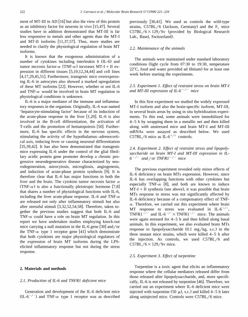
Ž . Ž .Fig. 1. Representative in situ hybridization analysis of the effect of immobilization stress STRESS and lipopolysaccharide LPS on MT-I and MT-IIIexpression in coronal sections of the cerebrum. Significant effects on MT-I expression was observed in stressed and endotoxin-injected mice. IL-6 androrTNF-a functional deficiencies affected significantly MT-I response to LPS, and to a much lower extent, to stress. In contrast, MT-III expression was onlymodestly affected. Several animals per group were studied and the mRNA levels quantitated in specific brain areas, which are shown in Figs. 3 and 4.
( )J. Carrasco et al.rMolecular Brain Research 57 1998 221–234224
.Mannheim, Mannheim, Germany . In brief, the DNA waslinearized by digestion with restriction endonucleasesŽ .HindIII to antisense probes and XhoI to sense probes .
Ž .Six ml of the linearized DNA 0.5 mgrml was incubatedwith 2 ml of transcription buffer, 2 ml of 100 mMdithiotreitol, 0.8 ml of RNase inhibitor, 1 ml of 10 mMATP, CTP and GTP, 2.4 ml of 0.1 mM cold UTP, 2 ml ofw35 x Ž . Ž .S a-UTP 20 mCirml Amersham , 0.8 ml of RNase
Žfree H O and 1 ml of RNA polymerase T7 RNA poly-2
merase for antisense probes, SP6 RNA polymerase for.sense probes , at 378C for 30 min. After the transcription
process, the DNA was digested by adding 1 ml of DNaseand incubating for 30 min at 378C. After DNAse treat-ment, the RNA was extracted with phenol and
Ž .phenol:chloroform:isoamyl alcohol 25:24:1 . The upperŽ .phase 200 ml was recovered and incubated overnight
Ž .with 1 ml of tRNA 10 mgrml , 10 ml of 3M sodiumacetate and 500 ml of 100% ethanol to precipitate theRNA. After centrifugation the precipitated probe was dis-solved in 50 ml of RNAse free H O. In situ hybridization2
was performed using procedures described by Yuguchi etw xal. 55 with some modifications: the sections were incu-
bated with 0.1 N HCl instead of proteinase K, and we usedRNAse at 10 mgrml instead of 1 mgrml to digest the freeprobe. The concentration of probe used was approximately1=106 dpmr90 mlrslide. Autoradiography was per-
Ž .formed exposing the film hyperfilm-MP, Amersham tothe slides for several days. All sections to be comparedwere prepared simultaneously and exposed to the sameautoradiographic film. MT-I or MT-III mRNA levels weresemiquantitatively determined in four sections per brainarea and animal, by measuring the optical densities and the
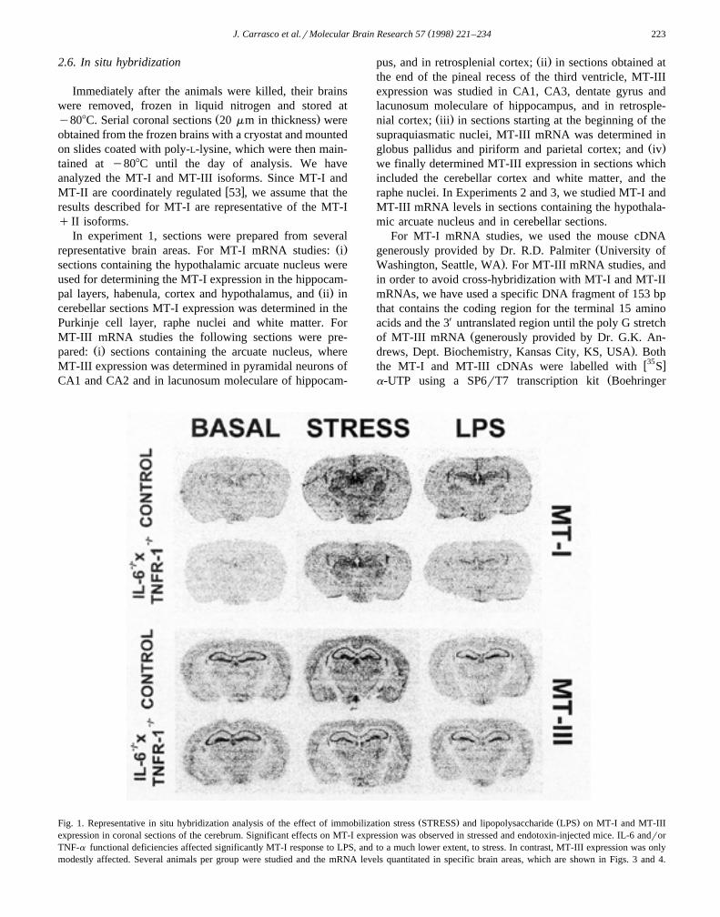
Ž . Ž .Fig. 2. Representative in situ hybridization analysis of the effect of immobilization stress STRESS and lipopolysaccharide LPS on MT-I and MT-IIIexpression in sagittal sections of the cerebellum and brain stem. Significant effects on MT-I expression was observed in stressed and endotoxin-injectedmice. IL-6 andror TNF-a functional deficiencies affected significantly MT-I response to LPS, but not to stress. MT-III expression was also modestlyaffected. Several animals per group were studied and the mRNA levels quantitated in specific brain areas, which are shown in Figs. 5 and 6.
( )J. Carrasco et al.rMolecular Brain Research 57 1998 221–234 225
number of pixels in defined areas with a Leica Q 500 MCsystem. The MT-I and MT-III mRNA values shown are
Žexpressed in arbitrary units number of pixels=optic den-.sity .
2.7. Statistical assays
Results were analyzed with Two-way andror One-wayANOVA followed by SNK multiple comparisons of themeans.
3. Results
3.1. Experiment 1
Figs. 1 and 2 show representative in situ hybridizationŽresults in sections from the cerebrum coronal sections
.containing the hypothalamic arcuate nucleus and the cere-bellum, respectively. It is clear that the sense probesproduced a very weak signal compared with the antisenseMT-I and MT-III probes. Also, the pattern of hybridization
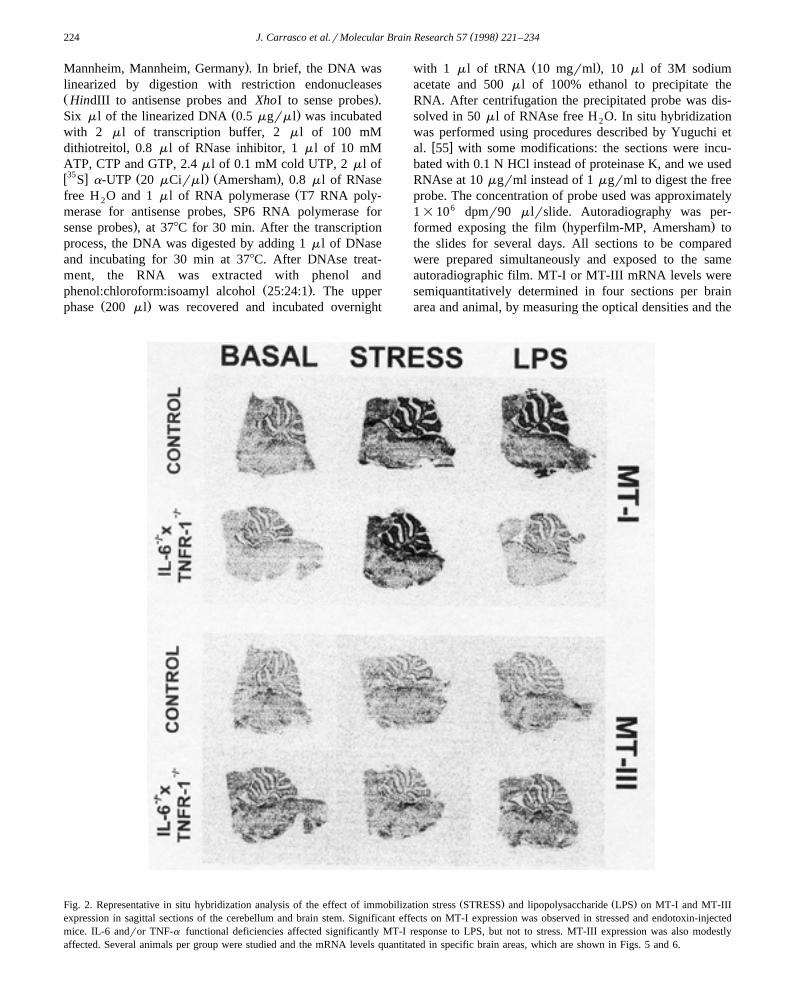
Table 1In situ hybridization of MT-I mRNA in brain of control and IL-6yry mice in basal and stress situations
Areas Film MT-IyryC57BLr6 IL-6
Basal Stress Basal Stress' 'Ž .CA1 Pyramidal neurons 1 11.7"1.2 61.3"11.5) 43.6"14.7 120.4"15.4)
Ž .CA2 Pyramidal neurons 1 120.8"23.4 187.8"38.0 145.6"37.9 197.4"41.6Ž .CA1 Lacunosum moleculare 1 83.0"27.0 379.8"61.6) 125.0"27.3 352.0"39.1)
Dentate gyrus 1 96.6"22.7 553.8"70.9) 145.8"16.6 510.6"39.3)
' 'Habenula 1 213.2"16.9 688.4"80.3) 172.0"49.6 570.6"17.8)
Choroid plexus 1 421"48.4 893.4"50.1) 455.2"13.7 839.8"41.1)
Retrosplenial cortex 1 45"19.5 274.2"70.1) 94.4"26.6 310.8"52.6)
Parietal cortex 1 83.6"13.3 282.8"20.7) 143.4"36.1 257.4"36.8)
Hypothalamus 1 138.0"50.6 337.0"91.6) 122.2"7.6 335.8"62.6)
Cerebellar cortex 2 254.7"41.4 1531.6"101.0) 158.3"48.6 1579.4"338.0)
Raphe nucleus 2 24.7"4.0 44.4"9.5) 29.9"6.0 68.5"19.1)
yry Ž . Ž . ŽC57BLr6 and IL-6 mice were killed in unstressed basal or stressed stress, 4 h of immobilization conditions. Coronal sections 4 per area and. Ž .animal were prepared containing the hypothalamic arcuate nucleus, processed simultaneously and exposed to the same autoradiographic film film 1 .
Ž .Other sections were prepared for the cerebellum which were processed separately and exposed to a different film film 2 . MT-I signal wassemiquantitatively determined by measuring the optical densities and the number of pixels in defined areas of the stated brain regions. Results are
Ž . 'mean"S.E. ns3–5 ; they were analyzed with Two-way MANOVA with strain and stress as main factors. ) p-0.05 vs. basal mice, p-0.05 vs.C57BLr6 strain.
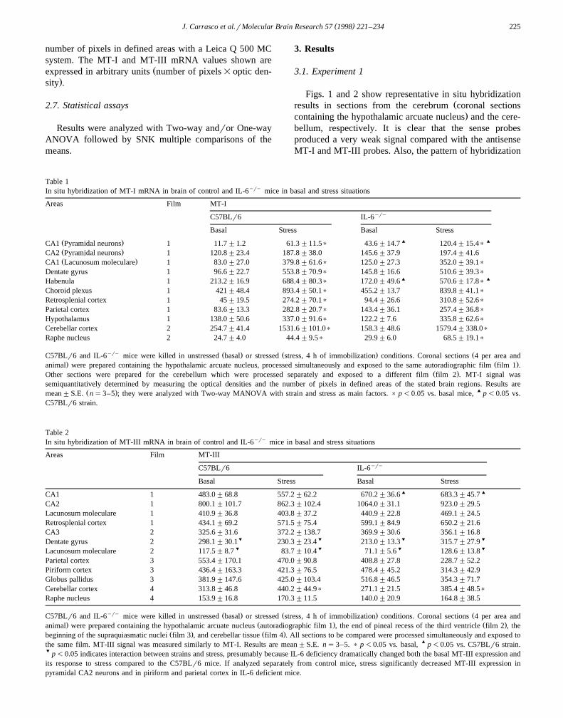
Table 2In situ hybridization of MT-III mRNA in brain of control and IL-6yry mice in basal and stress situations
Areas Film MT-IIIyryC57BLr6 IL-6
Basal Stress Basal Stress' 'CA1 1 483.0"68.8 557.2"62.2 670.2"36.6 683.3"45.7
CA2 1 800.1"101.7 862.3"102.4 1064.0"31.1 923.0"29.5Lacunosum moleculare 1 410.9"36.8 403.8"37.2 440.9"22.8 469.1"24.5Retrosplenial cortex 1 434.1"69.2 571.5"75.4 599.1"84.9 650.2"21.6CA3 2 325.6"31.6 372.2"138.7 369.9"30.6 356.1"16.8
% % % %Dentate gyrus 2 298.1"30.1 230.3"23.4 213.0"13.3 315.7"27.9% % % %Lacunosum moleculare 2 117.5"8.7 83.7"10.4 71.1"5.6 128.6"13.8
Parietal cortex 3 553.4"170.1 470.0"90.8 408.8"27.8 228.7"52.2Piriform cortex 3 436.4"163.3 421.3"76.5 478.4"45.2 314.3"42.9Globus pallidus 3 381.9"147.6 425.0"103.4 516.8"46.5 354.3"71.7Cerebellar cortex 4 313.8"46.8 440.2"44.9) 271.1"21.5 385.4"48.5)
Raphe nucleus 4 153.9"16.8 170.3"11.5 140.0"20.9 164.8"38.5
yry Ž . Ž . ŽC57BLr6 and IL-6 mice were killed in unstressed basal or stressed stress, 4 h of immobilization conditions. Coronal sections 4 per area and. Ž . Ž .animal were prepared containing the hypothalamic arcuate nucleus autoradiographic film 1 , the end of pineal recess of the third ventricle film 2 , the
Ž . Ž .beginning of the supraquiasmatic nuclei film 3 , and cerebellar tissue film 4 . All sections to be compared were processed simultaneously and exposed tothe same film. MT-III signal was measured similarly to MT-I. Results are mean"S.E. ns3–5. ) p-0.05 vs. basal, ' p-0.05 vs. C57BLr6 strain.% p-0.05 indicates interaction between strains and stress, presumably because IL-6 deficiency dramatically changed both the basal MT-III expression andits response to stress compared to the C57BLr6 mice. If analyzed separately from control mice, stress significantly decreased MT-III expression inpyramidal CA2 neurons and in piriform and parietal cortex in IL-6 deficient mice.
( )J. Carrasco et al.rMolecular Brain Research 57 1998 221–234226
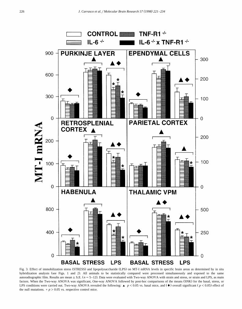
Ž . Ž .Fig. 3. Effect of immobilization stress STRESS and lipopolysaccharide LPS on MT-I mRNA levels in specific brain areas as determined by in situŽ .hybridization analysis see Figs. 1 and 2 . All animals to be statistically compared were processed simultaneously and exposed to the same
Ž .autoradiographic film. Results are mean"S.E. ns5–12 . Data were evaluated with Two-way ANOVA with strain and stress, or strain and LPS, as mainŽ .factors. When the Two-way ANOVA was significant, One-way ANOVA followed by post-hoc comparisons of the means SNK for the basal, stress, or
Ž . Ž .LPS conditions were carried out. Two-way ANOVA revealed the following: ' p-0.05 vs. basal mice, and l overall significant p-0.05 effect ofthe null mutations. ) p)0.05 vs. respective control mice.
( )J. Carrasco et al.rMolecular Brain Research 57 1998 221–234 227
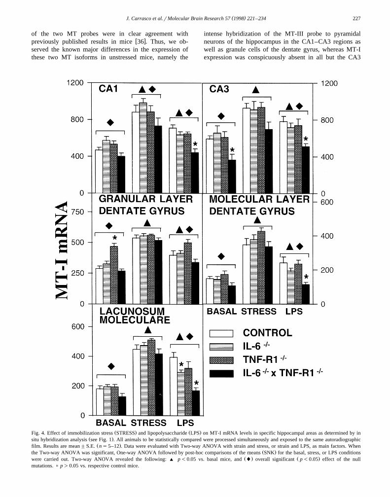
of the two MT probes were in clear agreement withw xpreviously published results in mice 36 . Thus, we ob-
served the known major differences in the expression ofthese two MT isoforms in unstressed mice, namely the
intense hybridization of the MT-III probe to pyramidalneurons of the hippocampus in the CA1–CA3 regions aswell as granule cells of the dentate gyrus, whereas MT-Iexpression was conspicuously absent in all but the CA3
Ž . Ž .Fig. 4. Effect of immobilization stress STRESS and lipopolysaccharide LPS on MT-I mRNA levels in specific hippocampal areas as determined by inŽ .situ hybridization analysis see Fig. 1 . All animals to be statistically compared were processed simultaneously and exposed to the same autoradiographic
Ž .film. Results are mean"S.E. ns5–12 . Data were evaluated with Two-way ANOVA with strain and stress, or strain and LPS, as main factors. WhenŽ .the Two-way ANOVA was significant, One-way ANOVA followed by post-hoc comparisons of the means SNK for the basal, stress, or LPS conditions
Ž . Ž .were carried out. Two-way ANOVA revealed the following: ' p-0.05 vs. basal mice, and l overall significant p-0.05 effect of the nullmutations. ) p)0.05 vs. respective control mice.
( )J. Carrasco et al.rMolecular Brain Research 57 1998 221–234228
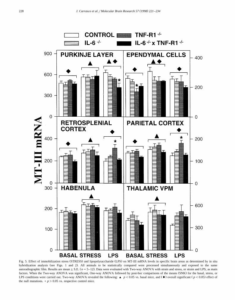
Ž . Ž .Fig. 5. Effect of immobilization stress STRESS and lipopolysaccharide LPS on MT-III mRNA levels in specific brain areas as determined by in situŽ .hybridization analysis see Figs. 1 and 2 . All animals to be statistically compared were processed simultaneously and exposed to the same
Ž .autoradiographic film. Results are mean"S.E. ns5–12 . Data were evaluated with Two-way ANOVA with strain and stress, or strain and LPS, as mainŽ .factors. When the Two-way ANOVA was significant, One-way ANOVA followed by post-hoc comparisons of the means SNK for the basal, stress, or
Ž . Ž .LPS conditions were carried out. Two-way ANOVA revealed the following: ' p-0.05 vs. basal mice, and l overall significant p-0.05 effect ofthe null mutations. ) p)0.05 vs. respective control mice.
( )J. Carrasco et al.rMolecular Brain Research 57 1998 221–234 229
neurons but was prominent in the lacunosum moleculare, astructure abundantly populated of astrocytes. In the re-maining brain areas, the MT-I and MT-III antisense probesproduced comparable signals in unstressed mice, with clear
Žhybridization throughout the neocortex with significantly
.lower levels in layer IV than in the remaining layers ,thalamus, hypothalamus, brainstem and cerebellar cortexŽ .especially the Purkinje cell layer . A prominent MT-IIIsignal was also observed in the choroid plexus and ependy-mal cells, and of MT-I in the later.
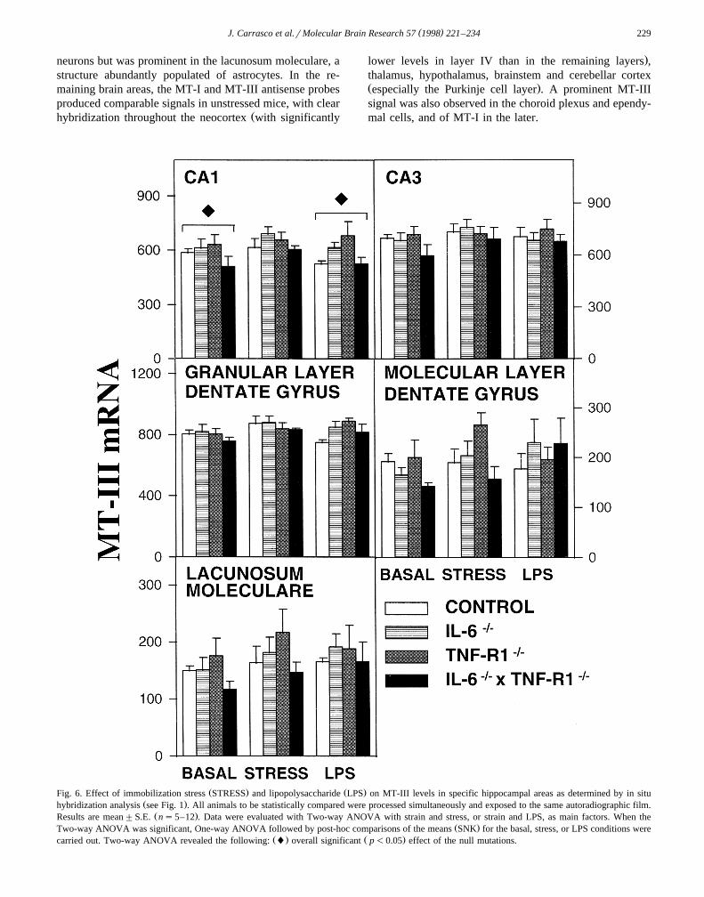
Ž . Ž .Fig. 6. Effect of immobilization stress STRESS and lipopolysaccharide LPS on MT-III levels in specific hippocampal areas as determined by in situŽ .hybridization analysis see Fig. 1 . All animals to be statistically compared were processed simultaneously and exposed to the same autoradiographic film.
Ž .Results are mean"S.E. ns5–12 . Data were evaluated with Two-way ANOVA with strain and stress, or strain and LPS, as main factors. When theŽ .Two-way ANOVA was significant, One-way ANOVA followed by post-hoc comparisons of the means SNK for the basal, stress, or LPS conditions were
Ž . Ž .carried out. Two-way ANOVA revealed the following: l overall significant p-0.05 effect of the null mutations.
( )J. Carrasco et al.rMolecular Brain Research 57 1998 221–234230
For studying the effect of IL-6 deficiency in both basaland stress situations, MT-I and MT-III mRNA levels of3–5 mice per group were quantitated in defined workingfields in representative brain areas stated in Section 2. Theresults demonstrate that the effects of stress and IL-6
Ždeficiency differ for both brain MT isoforms Tables 1 and.2 . Thus, there was a clear general increasing effect of
stress on MT-I mRNA levels in most brain areas studiedŽ .with the notable exception of the medial thalamic nuclei .The effect of IL-6 deficiency was not dramatic, sincestatistical significant effects were only observed in the
Ž .CA1 pyramidal neuron layer area and the habenula,increasing and decreasing MT-I mRNA levels, respec-tively.
Ž .The results for MT-III mRNA Table 2 demonstratedthat this isoform behaves differently from MT-I. Since thehippocampus has been suggested as one of the brain areas
w xwhere MT-III might be physiologically relevant 36 , weevaluated the effect of stress and IL-6 deficiency onpyramidal neurons of the CA1–CA3 areas, granule cells of
Ž .the dentate gyrus DG and lacunosum moleculare ofrostral and caudal sections of this brain area. Only minoreffects of both stress and IL-6 deficiency were noticed inall brain areas studied, in contrast to MT-I mRNA, withtendencies to increase or decrease MT-III mRNA depend-ing on the brain area and physiological condition. Finally,MT-III expression was significantly increased in the cere-bellum in both control and IL-6 deficient mice.
3.2. Experiment 2
Figs. 3 and 4 show brain MT-I mRNA levels of thethree mutant mice studied in comparison with controls.Since the C57BLr6 mice did not differ from the F1C57BLr6=129rSv mice, they were pooled. In line withthe previous experiment, stress increased MT-I mRNAlevels in all areas studied. Also as expected, LPS signifi-
cantly increased MT-I expression throughout the brain. Ingeneral, the brain MT-I response to stress was mostlyunaltered by the IL-6 and TNFR1 deficiencies, although ageneral tendency was observed in the double mutants tohave lower MT-I mRNA levels than the other mice. Thisprobably mostly reflects simply what is observed in basaldouble mutant mice, which also tend to have lower levels,while the fold induction in all mice is comparable. Interest-ingly, IL-6 deficiency increased MT-I mRNA levels in thehippocampal CA1 area in basal and stress conditions, andtended to decrease them in the habenula, in agreement withthe previous experiment. In contrast, the response of brainMT-I mRNA to LPS was significantly impaired in IL-6yry
and TNFR1yry mice in most brain areas. The response toLPS in the double mutant was almost completely bluntedin all brain areas studied.
Figs. 5 and 6 show brain MT-III mRNA levels. Incontrast to MT-I mRNA, stress and endotoxin had onlyminor effects on MT-III expression, although significantincreases were observed in stressed mice in brain areassuch as the cerebellar Purkinje layer, the cerebrum cortex,habenula, thalamic VPM, and ependymal cells. As forMT-I, the double mutant mice appeared to have lowerMT-III mRNA in basal and stress conditions in most brainareas. LPS was without effect in any of the brain areasanalyzed other than the Purkinje cell layer, where thecytokine deficiencies appeared to mediate the moderateMT-III mRNA increases. Interestingly, the TNFR1yry
mice showed higher MT-III mRNA levels in the retrosple-nial and parietal cortex in LPS-injected animals.
3.3. Experiment 3
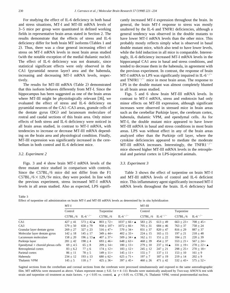
Table 3 shows the effect of turpentine on brain MT-Iand MT-III mRNA levels of control and IL-6 deficientmice. This inflammatory agent significantly increased MT-ImRNA levels throughout the brain. IL-6 deficiency had
Table 3Effect of turpentine oil administration on brain MT-I and MT-III mRNA levels as determined by in situ hybridization
MT-I MT-III
Control Turpentine Control Turpentineyry yry yry yryC57BLr6 IL-6 C57BLr6 IL-6 C57BLr6 IL-6 C57BLr6 IL-6
CA1 427"41 573"42' 803"72) 1037"66)' 583"25 613"49 663"23) 708"45)
CA3 579"50 658"79 958"107) 1072"66) 705"31 684"46 763"37 758"24Granular layer dentate gyrus 269"27 327"23 516"47) 570"34) 831"37 820"47 816"29 887"37Molecular layer dentate gyrus 142"18 145"17 349"44) 402"33) 224"15 165"15 197"21 218"48Lacunosum moleculare 158"20 196"13' 407"37) 509"34)' 162"11 151"22 184"21 220"39Purkinje layer 281"42 198"4 693"46) 648"63) 468"39 454"37 553"21) 567"24)
Ependymalqchoroid plexus cells 69"4.5 65"8 209"14) 180"13) 279"10 237"16' 331"19) 278"22)'
Retrosplenial cortex 83"6.2 77"6 174"12) 183"12) 245"12 247"21 298"25) 270"18)
Parietal cortex 86"11 78"5 149"11) 145"11) 155"7 137"11 153"10 161"9Habenula 234"12 193"13 680"62) 625"71) 197"7 187"19 219"14 202"9Thalamic VPM 145"5 118"7 425"36) 397"45) 460"26 471"42 532"43) 575"52)
Sagittal sections from the cerebellum and coronal sections from the cerebrum were processed simultaneously and exposed to the same autoradiographicŽ .film. MT mRNAs were measured as above. Values represent mean"S.E. ns4–11 . Results were statistically analyzed by Two-way ANOVA test with
strain and turpentine oil treatment as main factors. ) p-0.05 vs. control, ' p-0.05 vs. C57BLr6. Thalamic VPM, ventral posteromedial nucleus.
( )J. Carrasco et al.rMolecular Brain Research 57 1998 221–234 231
only modest effects, but, again interestingly, MT-I mRNAlevels of IL-6yry mice were increased in the hippocampalCA1 area and tended to decreased in the habenula, inagreement with the previous experiments. MT-III mRNAlevels were also increased in many of the brain areasstudied, but to a lower extent than MT-I isoform. IL-6deficiency did not affect the MT-III expression.
4. Discussion
The presence of MTs in the brain has long been recog-w xnized 25 . However, the research on brain MT has gained
great importance since the discovery of the brain specificŽ .isoform, MT-III or growth inhibitory factor , and its
w xputative relationship to Alzheimer disease 49 . However,w xthe latter has been challenged 16 , and, furthermore, MT-
III has now been shown to be expressed in tissues otherw xthan the brain 33 . In addition, the isoforms MT-Iq II
may also be equally important for normal brain physiologysince significant upregulations have been observed in hu-
w xman pathologies such as Alzheimer and Pick diseases 13w xand amyotrophic lateral sclerosis 48 , and in animals
w xsubjected to stress, LPS or brain damage 22,24,38 . Pre-sumably, this upregulation could provide protection against
w xfree radical induced brain damage 43 , or could assist inzinc andror copper metabolism in the brain. Recent stud-ies with transgenic and knock-out mice for MT-III alsoprovide evidence of a protective role of this isoform
w xagainst kainic acid brain damage 14 . A number of previ-ous studies demonstrated that MT-III responds promptly to
w xbrain damage in several animal models 2,55,56 . Thus, thethree brain isoforms may be relevant for brain physiology,and it is therefore of the utmost importance to characterizetheir regulation. In this study, brain MT regulation wasexamined in mice carrying a null mutation in the IL-6gene, TNF-a type 1 receptor gene, or both, a uniqueapproach for determining the physiological role of thesecytokines.
In previous studies it has been established that IL-6 andTNF-a are significant inducers of the MT-Iq II isoforms
w xin tissues such as the liver 12,44 , but little is known oftheir effect in the brain. We have shown that the i.c.v.injection of IL-6 in rats increases MT-Iq II levels in some
w xbrain areas 21 , and transgenic mice with targeted IL-6expression to astrocytes also show increased MT-Iq IIlevels in those brain areas with significant transgenic IL-6
w xexpression 22 , suggesting that IL-6 could be important inbrain MT regulation. However, IL-6yry mice did notdiffer dramatically from the control mice in basal condi-tions, and the only consistent effects noticed were in theCA1 pyramidal neurons and the habenula, where IL-6deficiency increased and decreased MT-I mRNA levels,respectively. This general lack of effect of IL-6 deficiencyis in principle not surprising since the MT-Iq II isoforms
are multiregulated proteins, but nevertheless significanteffects, and more important, opposite trends, were ob-served in specific areas. TNFR1yry mice did not differdramatically either from the control mice, although in thegranular layer of the dentate gyrus a significant increase ofMT-I mRNA levels was observed. Interestingly, the dou-ble mutant mice showed a much more significant alterationof their brain MT-I mRNA levels, since a clear trend tohave decreased levels was observed in most brain areas.IL-6 and TNF-a are pleiotropic cytokines which share
w xsome functions 8 , and thus it seems feasible that in basalconditions the two of them participate in the control ofMT-I expression and could compensate to some extent thefunctional loss of each other, so that a significant effect isobserved when both cytokines are missing simultaneously.Regarding MT-III mRNA, no dramatic effects of the func-tional IL-6 and TNF deficiencies were noticed, although inagreement with MT-I mRNA a clear trend to show de-creased MT-III signal in most brain areas was observed inthe double mutant.
We next examined the putative mediating role of IL-6and TNF-a on brain MT response to stress and LPS, twomajor physiological inducers of the MT-Iq II isoforms. In
w xagreement with previous reports 6,18,19 , stress signifi-cantly upregulated MT-I expression, indicating that this
ŽMT isoform and likely MT-II, which is coordinately.regulated with MT-I is important for the adaptation of the
w xorganism to stress 26 . This response was basically unal-tered in the IL-6yry andror TNFR1yry mice, indicatingthat a mediating role of IL-6 or TNF-a during stress isunlikely. Since both acute and chronic increases of IL-6 do
Ž .increase brain MT-Iq II levels see above , it might beconcluded that IL-6 is not increased during stress in thebrain, or that necessary factors are not co-released duringstress in the brain. It is important to mention that incontrast to the brain, we find a significant effect of IL-6
Ždeficiency on liver MT response to stress manuscript in.preparation , which is consistent with the increased circu-
lating IL-6 levels during stress and the role of the cytokineŽ .on liver MT regulation see above . Whether this high-
lights a tissue-specific role of IL-6 regarding brain MTregulation during stress or reflects the inability of thecytokines to cross the blood–brain barrier remains to beestablished.
A different pattern emerged when brain MT-I wasw xinduced with LPS. As expected 27 , the MT-I signal was
significantly increased throughout the brain. Although themechanism is unclear, it is well known that peripheral LPSadministration increases cytokine production including IL-6
w xin the brain 31 . The present results clearly demonstratethat brain MT-I response to LPS is mediated by IL-6 andTNF-a , since the IL-6yry and TNFR-1yry mice showedsignificant diminished responses which were almost com-pletely blunted in the double mutants. This is the first timethat a physiological role of these cytokines on brain MT-Iq II regulation is demonstrated.
( )J. Carrasco et al.rMolecular Brain Research 57 1998 221–234232
We also evaluated the effect of another inflammatorymodel, turpentine oil administration. A very significantMT-I upregulation was observed throughout the brain,suggesting again a relevant role of this MT isoform. IL-6deficiency did not affect this response in any of the brainareas analyzed, which is in contrast with the inflammatoryresponse elicited by LPS. A likely explanation of thisapparent discrepancy is that turpentine and LPS couldproduce a different set of pro-inflammatory cytokines. For
w xinstance, a previous study 46 has demonstrated that LPSbut not turpentine caused an upregulation of IL-1, IL-6 andTNF-a in circulating monocytes, peritoneal macrophagesand the liver. Despite these results, we do find a reductionof liver MT induction by turpentine in IL-6yry miceŽ .manuscript in preparation , suggesting again a tissue-specific role of IL-6. It is even feasible that brain MT-Iinduction by turpentine is at least in part a stress response,but in any case it is clear that IL-6 is not relevant in theinflammatory response elicited in the brain regarding MT-Iregulation.
Regarding MT-III, the basically brain-specific MT iso-form, the in situ hybridization results demonstrate that itremains mostly unaffected in comparison with MT-I, in
w xagreement with previous studies 11,37,57 . Nevertheless,stress slightly but consistently increased MT-III mRNAlevels in most brain areas analyzed. We have studiedpreviously the effect of stress by Northern-blot and dot-blot
w xin normal mice and rats 6,23 . Taken together, the resultssuggest that stress increases brain MT-III mRNA levelsalthough to a lower extent than MT-I isoform, and that theapparent discrepancies between experiments may be ex-plained by stress-induced changes in g-actin mRNA levels.IL-6 and TNF-a functional deficiencies appeared to bluntthe increase only in the ependymalqchoroid plexus cells.The double mutant tended to have less MT-III mRNAlevels than the other mice, but this appears to simplyreflect what is observed in basal mice. In contrast to stress,
Ž .LPS at the dosage given did not affect significantlyMT-III mRNA levels in most of the brain areas studied,with the exception of the Purkinje cell layer of the cerebel-lum where a slight increasing effect was noticed. The IL-6and TNF-a functional deficiencies diminished the effect ofLPS in this area. Interestingly, the TNFR1yry LPS-in-jected mice showed higher MT-III mRNA levels in theretrosplenial and parietal cortex, and the same tendencywas observed in the hippocampal CA1 area, the habenulaand the thalamic VPM, suggesting an inhibitory role ofTNF-a in these brain areas. Finally, turpentine signifi-cantly increased MT-III mRNA in several brain areas, andIL-6 deficiency decreased the MT-III signal in the ependy-malqchoroid plexus cells, which also supports the as-sumption above stated that brain MT response in this caseis at least partially a stress response.
In summary, the present results demonstrate that in vivoIL-6 and TNF-a are major regulators of the metal-bindingproteins, metallothioneins, in the brain. Their role is impor-
tant in one scenario, the inflammatory response elicited byLPS, but not in others, namely the stress response and theinflammatory response elicited by turpentine.
Acknowledgements
This work was supported by the DGICYT PB94-0667,CICYT SAF96-0189, and the CIRIT 1995SGR-00499. Thehelp of the Laboratori d’Analisi Bioquımica of the Depar-` ´tament de Bioquımica i Biologia Molecular is appreciated.´J. Carrasco is supported by a fellowship of the CIRIT,FIr96-2613. The help of J. Canto and G. Gordillo from´the Servei d’Estabulari is gratefully appreciated.
References
w x1 T. Andus, J. Bauer, W. Gerok, Effects of cytokines on the liver,Ž .Hepatology 13 1991 364–375.
w x2 T. Anezaki, H. Ishiguro, I. Hozumi, T. Inuzuka, M. Hiraiwa, H.Kobayashi, T. Yuguchi, A. Wanaka, Y. Uda, T. Miyatake, K.Yamada, M. Tohyama, S. Tsuji, Expression of growth inhibitory
Ž .factor GIF in normal and injured rat brains, Neurochem. Int. 27Ž .1995 89–94.
w x3 P.C. Arck, S. Merali, J. Manuel, G. Chaouat, D.A. Clark, Stress-tri-ggered abortion: inhibition of protective suppression and promotion
Ž .of tumor necrosis factor-a TNF-a release as a mechanism trigger-Ž .ing resorptions in mice, Am. J. Reprod. Immunol. 33 1995 74–80.
w x4 J. Bauer, U. Ganter, J. Abel, S. Strauss, U. Jonas, R. Weiss, P.Gebicke-Haerter, B. Volk, M. Berger, Effects of interleukin-1 andinterleukin-6 on metallothionein and amyloid precursor protein ex-pression in human neuroblastoma cells. Evidence that interleukin-6possibly acts via a receptor different from the 80-kDa interleukin-6
Ž .receptor, J. Neuroimmunol. 45 1993 163–173.w x5 J.U. Bell, M.J. Lawman, J.M. Lopez, L.E. DesJardin, L.A. Apple-
white, Effects of type I interferon-inducing agents on hepatic metal-Ž .lothionein, Exs 1987 581–586.
w x6 E. Belloso, J. Hernandez, M. Giralt, P. Kille, J. Hidalgo, Effect ofstress on mouse and rat brain metallothionein I and III mRNA
Ž .levels, Neuroendocrinology 64 1996 430–439.w x7 B. Beutler, Tumour Necrosis Factors: The molecules and their
emerging role in medicine, Raven Press, New York, 1992.w x8 H. Bluethmann, J. Rothe, N. Schultze, M. Tkachuk, P. Koebel,
Establishment of the role of IL-6 and TNF receptor using geneŽ .knockout mice, J. Leukocyte Biol. 56 1994 565–570.
w x9 I.L. Campbell, C.R. Abraham, E. Masliah, P. Kemper, J.D. Inglis,M.B.A. Oldstone, L. Mucke, Neurologic disease in transgenic miceby cerebral overexpression of interleukin-6, Proc. Natl. Acad. Sci.
Ž .U.S.A. 90 1993 10061–10065.w x10 R.J. Cousins, A.S. Leinart, Tissue-specific regulation of zinc
metabolism and metallothionein genes by interleukin-1, FASEB J. 2Ž .1988 2884–2890.
w x11 T. Dalton, T.L. Pazdernik, J. Wagner, F. Samson, G.K. Andrews,Temporal–spatial patterns of expression of metallothionein-I and -IIIand other stress related genes in rat brain after kainic acid-induced
Ž .seizures, Neurochem. Int. 27 1995 59–71.w x12 S.K. De, M.T. McMaster, G.K. Andrews, Endotoxin induction of
Ž .murine metallothionein gene expression, J. Biol. Chem. 265 199015267–15274.
w x13 J.R. Duguid, C.W. Bohmont, N.G. Liu, W.W. Tourtellotte, Changesin brain gene expression shared by scrapie and Alzheimer disease,
Ž .Proc. Natl. Acad. Sci. U.S.A. 86 1989 7260–7264.w x14 J.C. Erickson, G. Hollopeter, S.A. Thomas, G.J. Froelick, R.D.
( )J. Carrasco et al.rMolecular Brain Research 57 1998 221–234 233
Palmiter, Disruption of the metallothionein-III gene in mice: analysisof brain zinc, behavior, and neuron vulnerability to metals, aging,
Ž .and seizures, J. Neurosci. 17 1997 1271–1281.w x15 J.C. Erickson, B.A. Masters, E.J. Kelly, R.L. Brinster, R.D. Palmiter,
Expression of human metallothionein-III in transgenic mice, Neu-Ž .rochem. Int. 27 1995 35–41.
w x16 J.C. Erickson, A.K. Sewell, L.T. Jensen, D.R. Winge, R.D. Palmiter,Enhanced neurotrophic activity in Alzheimer’s disease cortex is not
Ž .associated with down-regulation of metallothionein-III GIF , BrainŽ .Res. 649 1994 297–304.
w x17 R.L. Friedman, G.R. Stark, a-Interferon-induced transcription ofHla and metallothionein genes containing homologous upstream
Ž .sequences, Nature 314 1985 637–639.w x18 T. Gasull, M. Giralt, A. Garcia, J. Hidalgo, Regulation of metalloth-
ionein-IqII levels in specific brain areas and liver in the rat: role ofŽ .catecholamines, Glia 12 1994 135–143.
w x19 T. Gasull, M. Giralt, J. Hernandez, P. Martinez, I. Bremner, J.Hidalgo, Regulation of metallothionein concentrations in rat brain:effect of glucocorticoids, zinc, copper, and endotoxin, Am. J. Phys-
Ž .iol. 266 1994 E760–E767.w x20 J. Gauldie, C. Richards, D. Harnish, P. Landsorp, H. Baumann,
Interferon-b 2rB-cell stimulatory factor type 2 shares identity withmonocyte-derived hepatocyte-stimulating factor and regulates themajor acute phase protein response in liver cells, Proc. Natl. Acad.
Ž .Sci. U.S.A. 84 1987 7251–7255.w x21 J. Hernandez, J. Hidalgo, Endotoxin and intracerebroventricular´
injection of IL-1 and IL-6 induce rat brain metallothionein-I and -III,Ž .Neurochem. Int. 32 1998 369–373.
w x22 J. Hernandez, A. Molinero, I.L. Campbell, J. Hidalgo, Transgenic´expression of interleukin 6 in the central nervous system regulatesbrain metallothionein-I and -III expression in mice, Mol. Brain Res.
Ž .48 1997 125–131.w x23 J. Hidalgo, E. Belloso, J. Hernandez, T. Gasull, A. Molinero, Role´
of glucocorticoids on rat brain metallothionein-I and -III response toŽ .stress, Stress 1 1997 231–240.
w x24 J. Hidalgo, M. Borras, J.S. Garvey, A. Armario, Liver, brain, andŽ .heart metallothionein induction by stress, J. Neurochem. 55 1990
651–654.w x25 J. Hidalgo, B. Castellano, I.L. Campbell, Regulation of brain metal-
Ž .lothioneins, Curr. Top. Neurochem. 1 1997 1–26.w x26 J. Hidalgo, T. Gasull, M. Giralt, A. Armario, Brain metallothionein
Ž .in stress, Biol. Signals 3 1994 198–210.w x27 Y. Itano, S. Noji, E. Koyama, S. Taniguchi, N. Taga, T. Takahashi,
K. Ono, F. Kosaka, Bacterial endotoxin-induced expression of met-allothionein genes in rat brain, as revealed by in situ hybridization,
Ž .Neurosci. Lett. 124 1991 13–16.w x28 J.H. Kagi, A. Schaffer, Biochemistry of metallothionein, Biochem-
Ž .istry 27 1988 8509–8515.w x29 M. Karin, R.J. Imbra, A. Heguy, G. Wong, Interleukin-1 regulates
Ž .human metallothionein gene expression, Mol. Cell Biol. 5 19852866–2869.
w x30 M. Kopf, H. Baumann, G. Freer, M. Freudenberg, M. Lamers, T.Kishimoto, R. Zinkernagel, H. Bluethmann, G. Kohler, Impaired¨immune and acute-phase responses in interleukin-6-deficient mice,
Ž .Nature 368 1994 339–342.w x31 S. Laye, P. Parnet, E. Goujon, R. Dantzer, Peripheral administration
of lipopolysaccharide induces the expression of cytokine transcriptsŽ .in the brain and pituitary, Mol. Brain Res. 27 1994 157–162.
w x32 L.G. Lemay, A.J. Vander, M. Kluger, The effects of psychologicalstress on plasma interleukin-6 activity in rats, Physiol. Behav. 47Ž .1990 957–961.
w x33 L. Liang, K. Fu, D.K. Lee, R.J. Sobieski, T. Dalton, G.K. Andrews,Activation of the complete mouse metallothionein gene locus in the
Ž .maternal deciduum, Mol. Reprod. Dev. 43 1996 25–37.w x34 J. Liu, Y.P. Liu, L.E. Sendelbach, C.D. Klassen, Endotoxin induc-
tion of hepatic metallothionein is mediated through cytokines, Toxi-Ž .col. Appl. Pharmacol. 109 1991 235–240.
w x35 T. Mandrup-Poulsen, J. Nerup, J.I. Reimers, F. Pociot, H.U. Ander-sen, A. Karlsen, U. Bjerre, R. Bergholdt, Cytokines and the en-docrine system: I. The immunoendocrine network, Eur. J. En-
Ž .docrinol. 133 1995 660–671.w x36 B.A. Masters, C.J. Quaife, J.C. Erickson, E.J. Kelly, G.J. Froelick,
B.P. Zambrowicz, R.L. Brinster, R.D. Palmiter, Metallothionein IIIis expressed in neurons that sequester zinc in synaptic vesicles, J.
Ž .Neurosci. 14 1994 5844–5857.w x37 R.D. Palmiter, S.D. Findley, T.E. Whitmore, D.M. Durnam, MT-III,
a brain-specific member of the metallothionein gene family, Proc.Ž .Natl. Acad. Sci. U.S.A. 89 1992 6333–6337.
w x38 M. Penkowa, T. Moos, Disruption of the blood–brain interface inneonatal rat neocortex induces a transient expression of metalloth-
Ž .ionein in reactive astrocytes, Glia 13 1995 217–227.w x39 F. Pousset, Cytokines as mediators in the central nervous system,
Ž .Biomed. Pharmacother. 48 1994 425–431.w x40 C.J. Quaife, S.D. Findley, J.C. Erickson, G.J. Froelick, E.J. Kelly,
B.P. Zambrowicz, R.D. Palmiter, Induction of a new metallothioneinŽ .isoform MT-IV occurs during differentiation of stratified squa-
Ž .mous epithelia, Biochemistry 33 1994 7250–7259.w x41 J. Rothe, W. Lesslauer, H. Lotscher, Y. Lang, P. Koebel, F. Kont-
gen, A. Althage, R. Zinkernagel, M. Steinmetz, H. Bluethmann,Mice lacking the tumor necrosis factor receptor 1 are resistant toTNF-mediated toxicity but highly susceptible to infection by Listeria
Ž .monocytogenes, Nature 364 1993 798–802.w x42 N.J. Rothwell, S.J. Hopkins, Cytokines and the nervous system II:
Ž .actions and mechanisms of action, Trends Neurol. Sci. 18 1995130–136.
w x43 M. Sato, I. Bremner, Oxygen free radicals and metallothionein, FreeŽ .Radic. Biol. Med. 14 1993 325–337.
w x44 M. Sato, M. Sasaki, H. Hojo, Tissue specific induction of metalloth-ionein synthesis by tumor necrosis factor-a , Res. Commun. Chem.
Ž .Pathol. Pharmacol. 75 1992 159–172.w x45 J.J. Schroeder, R.J. Cousins, Interleukin-6 regulates metallothionein
gene expression and zinc metabolism in hepatocyte monolayer cul-Ž .tures, Proc. Natl. Acad. Sci. U.S.A. 87 1990 3137–3141.
w x46 M. Scotte, M. Hiron, S. Masson, S. Lyoumi, F. Banine, P. Teniere,J.P. Lebreton, M. Daveau, Differential expression of cytokine genesin monocytes, peritoneal macrophages and liver following endo-
Ž .toxin- or turpentine-induced inflammation in rat, Cytokine 8 1996115–120.
w x47 A.K. Sewell, L.T. Jensen, J.C. Erickson, R.D. Palmiter, D.R. Winge,Bioactivity of metallothionein-3 correlates with its novel beta do-main sequence rather than metal binding properties, Biochemistry 34Ž .1995 4740–4747.
w x48 P.A. Sillevis Smitt, T.P. Mulder, H.W. Verspaget, H.G. Blaauwgeers,D. Troost, J.M. de Jong, Metallothionein in amyotrophic lateral
Ž .sclerosis, Biol. Signals 3 1994 193–197.w x49 Y. Uchida, K. Takio, K. Titani, Y. Ihara, M. Tomonaga, The growth
inhibitory factor that is deficient in the Alzheimer’s disease brain isŽ .a 68 amino acid metallothionein-like protein, Neuron 7 1991
337–347.w x50 J. van Snick, Interleukin-6: an overview, Annu. Rev. Immunol. 8
Ž .1990 253–278.w x51 P. Vanguri, Interferon-gamma-inducible genes in primary glial cells
of the central nervous system: comparisons of astrocytes with mi-croglia and Lewis with brown Norway rats, J. Neuroimmunol. 56Ž .1995 35–43.
w x52 C. Weinstock, D. Konig, R. Harnischmacher, J. Keul, A. Berg, H.Northoff, Effect of exhaustive exercise stress on the cytokine re-
Ž .sponse, Med. Sci. Sports Exerc. 29 1997 345–354.w x53 M.K. Yagle, R.D. Palmiter, Coordinate regulation of mouse metal-
lothionein I and II genes by heavy metals and glucocorticoids, Mol.Ž .Cell Biol. 5 1985 291–294.
w x54 K. Yamasu, Y. Shimada, M. Sakaizumi, G. Soma, D. Mizuno,Activation of the systemic production of tumor necrosis factor after
Ž .exposure to acute stress, Eur. Cytokine Netw. 3 1992 391–398.

( )J. Carrasco et al.rMolecular Brain Research 57 1998 221–234234
w x55 T. Yuguchi, E. Kohmura, K. Yamada, T. Sakaki, T. Yamashita, H.Otsuki, K. Kataoka, S. Tsuji, T. Hayakawa, Expression of growthinhibitory factor mRNA following cortical injury, J. Neurotrauma 12Ž .1995 299–306.
w x56 T. Yuguchi, E. Kohmura, K. Yamada, T. Sakaki, T. Yamashita, H.Otsuki, A. Wanaka, M. Tohyama, S. Tsuji, T. Hayakawa, Changesin growth inhibitory factor mRNA expression compared with thosein c-jun mRNA expression following facial nerve transection, Mol.
Ž .Brain Res. 28 1995 181–185.
w x57 H. Zheng, N.E. Berman, C.D. Klaassen, Chemical modulation ofmetallothionein I and III mRNA in mouse brain, Neurochem. Int. 27Ž .1995 43–58.
w x58 D. Zhou, A.W. Kusnecov, M.R. Shurin, M. Depaoli, B.S. Rabin,Exposure to physical and psychological stressors elevates plasmaIL-6: relationship to the activation of hypothalamic–pituitary–
Ž .adrenal axis, Endocrinology 133 1993 2523–2530.