ORIGINAL ARTICLE Neurotensin regulated miR-133 is involved...
10
ORIGINAL ARTICLE Neurotensin—regulated miR-133α is involved in proinflammatory signalling in human colonic epithelial cells and in experimental colitis Ivy Ka Man Law, 1 Kyriaki Bakirtzi, 1 Christos Polytarchou, 2 Angelos Oikonomopoulos, 1 Daniel Hommes, 1 Dimitrios Iliopoulos, 2 Charalabos Pothoulakis 1 ▸ Additional material is published online only. To view please visit the journal online (http://dx.doi.org/10.1136/ gutjnl-2014-307329). 1 Inflammatory Bowel Disease Center, Division of Digestive Diseases, David Geffen School of Medicine, UCLA, Los Angeles, California, USA 2 Center for Systems Biomedicine, Division of Digestive Diseases, David Geffen School of Medicine, UCLA, Los Angeles, California, USA Correspondence to Professor Charalabos Pothoulakis, Inflammatory Bowel Disease Center, Division of Digestive Diseases, David Geffen School of Medicine, University of California at Los Angeles, 675 Charles E Young Dr, South MRL building 1240, Los Angeles, CA 90095, USA; [email protected] Received 26 March 2014 Revised 2 July 2014 Accepted 22 July 2014 Published Online First 11 August 2014 To cite: Law IKM, Bakirtzi K, Polytarchou C, et al. Gut 2015;64: 1095–1104. ABSTRACT Objective Neurotensin (NT) mediates colonic inflammation through its receptor neurotensin receptor 1 (NTR1). NT stimulates miR-133α expression in colonic epithelial cells. We investigated the role of miR-133α in NT-associated colonic inflammation in vitro and in vivo. Design miR-133α and aftiphilin (AFTPH) levels were measured by quantitative PCR. Antisense (as)-miR-133α was administrated intracolonicaly prior to induction of 2, 4, 6-trinitrobenzene sulfonic acid (TNBS)-induced colitis and dextran sodium sulfate (DSS)-induced colitis. The effect of AFTPH was examined by gene silencing in vitro. Results NT increased miR-133α levels in NCM-460 overexpressing NTR1 (NCM460-NTR1) and HCT-116 cells. NT-induced p38, ERK1/2, c-Jun, and NF- κB activation, as well as IL-6, IL-8 and IL-1β messenger RNA (mRNA) expression in NCM-460-NTR1 cells were reduced in miR- 133α-silenced cells, while overexpression of miR-133α reversed these effects. MiR-133α levels were increased in TNBS (2 day) and DSS (5 day) colitis, while NTR1 deficient DSS-exposed mice had reduced miR-133α levels, compared to wild-type colitic mice. Intracolonic as-miR-133α attenuated several parameters of colitis as well expression of proinflammatory mediators in the colonic mucosa. In silico search coupled with qPCR identi fied AFTPH as a downstream target of miR-133α, while NT decreased AFTPH expression in NCM-460-NTR1 colonocytes. Gene silencing of AFTPH enhanced NT-induced proinflammatory responses and AFTPH levels were downregulated in experimental colitis. Levels of miR-133α were signi ficantly upregulated, while AFTPH levels were downregulated in colonic biopsies of patients with ulcerative colitis compared to controls. Conclusions NT-associated colitis and inflammatory signalling are regulated by miR-133α-AFTPH interactions. Targeting of miR-133α or AFTPH may represent a novel therapeutic approach in inflammatory bowel disease. INTRODUCTION Neurotensin (NT) is a neuropeptide expressed in the central nervous system and the intestine, 1 2 while its high-affinity receptor, NT receptor 1 (NTR1) 3 is expressed in neurones, 4 colonic epithe- lial, 25–7 immune cells, 89 as well as different colon cancer cell lines. 10 In the colon, NT/NTR1 signal- ling promotes inflammation in acute colitis animal models. 2 5 Colonic NT and NTR1 expression increases during enterotoxin-mediated intestinal inflammation, 2 dextran sodium sulfate (DSS) colitis, 6 11 12 and in the colon of patients with ulcerative colitis (UC). 6 Moreover, NTR1 defi- ciency in mice is associated with reduced weight loss and mortality in experimental colitis. 5 13 MicroRNAs (miRs) are short (19–25 nucleo- tides), single-stranded RNA molecules, acting as Open Access Scan to access more free content Significance of this study What is already known on this subject? ▸ Neurotensin (NT) mediates colonic inflammation through its high-affinity receptor (neurotensin receptor 1 (NTR1)). ▸ NT and NTR1 expressions are upregulated in inflamed colon tissues in experimental colitis and ulcerative colitis (UC). ▸ NT modulates differential expression of 38 microRNAs in colonocytes, among them miR-133α. ▸ MicroRNA-133α modulates signalling pathways related to proliferation, differentiation and hypertrophy in cancer cell lines and myocytes. What are the new findings? ▸ miR-133α modulates NT-induced MAP kinase, and NF-κB activation, as well as transcription of proinflammatory cytokines in vitro. ▸ Reducing the levels of miR-133α in human colonocytes and mouse colon attenuates proinflammatory signalling and development of experimental colitis in vitro and in vivo. ▸ Aftiphilin (AFTPH) is a novel downstream target of NT-induced miR-133α expression in human colonic epithelial cells involved in proinflammatory signalling pathways. ▸ Colon tissues from patients with UC and mice with colitis have higher miR-133α but lower AFTPH levels when compared with controls. How might it impact on clinical practice in the foreseeable future? ▸ Inhibiting miR-133α and restoring AFTPH expression in inflammatory bowel disease (IBD) colon may represent promising approaches for the treatment of IBD. ▸ The reverse association of the levels of miR-133α and AFTPH together with increased expression of NTR1 in UC may represent novel biomarkers for UC. In fl ammatory bowel disease Law IKM, et al. Gut 2015;64:1095–1104. doi:10.1136/gutjnl-2014-307329 1095 on 2 February 2019 by guest. Protected by copyright. http://gut.bmj.com/ Gut: first published as 10.1136/gutjnl-2014-307329 on 11 August 2014. Downloaded from
Transcript of ORIGINAL ARTICLE Neurotensin regulated miR-133 is involved...
ORIGINAL ARTICLE
Neurotensin—regulated miR-133α is involvedin proinflammatory signalling in human colonicepithelial cells and in experimental colitisIvy Ka Man Law,1 Kyriaki Bakirtzi,1 Christos Polytarchou,2 Angelos Oikonomopoulos,1
Daniel Hommes,1 Dimitrios Iliopoulos,2 Charalabos Pothoulakis1
▸ Additional material ispublished online only. To viewplease visit the journal online(http://dx.doi.org/10.1136/gutjnl-2014-307329).1Inflammatory Bowel DiseaseCenter, Division of DigestiveDiseases, David Geffen Schoolof Medicine, UCLA, LosAngeles, California, USA2Center for SystemsBiomedicine, Division ofDigestive Diseases, DavidGeffen School of Medicine,UCLA, Los Angeles, California,USA
Correspondence toProfessor CharalabosPothoulakis, InflammatoryBowel Disease Center, Divisionof Digestive Diseases, DavidGeffen School of Medicine,University of California at LosAngeles, 675 Charles E YoungDr, South MRL building 1240,Los Angeles, CA 90095, USA;[email protected]
Received 26 March 2014Revised 2 July 2014Accepted 22 July 2014Published Online First11 August 2014
To cite: Law IKM,Bakirtzi K, Polytarchou C,et al. Gut 2015;64:1095–1104.
ABSTRACTObjective Neurotensin (NT) mediates colonicinflammation through its receptor neurotensin receptor 1(NTR1). NT stimulates miR-133α expression in colonicepithelial cells. We investigated the role of miR-133α inNT-associated colonic inflammation in vitro and in vivo.Design miR-133α and aftiphilin (AFTPH) levels weremeasured by quantitative PCR. Antisense (as)-miR-133αwas administrated intracolonicaly prior to induction of2, 4, 6-trinitrobenzene sulfonic acid (TNBS)-inducedcolitis and dextran sodium sulfate (DSS)-induced colitis.The effect of AFTPH was examined by gene silencing in vitro.Results NT increased miR-133α levels in NCM-460overexpressing NTR1 (NCM460-NTR1) and HCT-116 cells.NT-induced p38, ERK1/2, c-Jun, and NF-κB activation, aswell as IL-6, IL-8 and IL-1β messenger RNA (mRNA)expression in NCM-460-NTR1 cells were reduced in miR-133α-silenced cells, while overexpression of miR-133αreversed these effects. MiR-133α levels were increased inTNBS (2 day) and DSS (5 day) colitis, while NTR1 deficientDSS-exposed mice had reduced miR-133α levels, comparedto wild-type colitic mice. Intracolonic as-miR-133α attenuatedseveral parameters of colitis as well expression ofproinflammatory mediators in the colonic mucosa. In silicosearch coupled with qPCR identified AFTPH as adownstream target of miR-133α, while NT decreased AFTPHexpression in NCM-460-NTR1 colonocytes. Gene silencing ofAFTPH enhanced NT-induced proinflammatory responses andAFTPH levels were downregulated in experimental colitis.Levels of miR-133α were significantly upregulated, whileAFTPH levels were downregulated in colonic biopsies ofpatients with ulcerative colitis compared to controls.Conclusions NT-associated colitis and inflammatorysignalling are regulated by miR-133α-AFTPH interactions.Targeting of miR-133α or AFTPH may represent a noveltherapeutic approach in inflammatory bowel disease.
INTRODUCTIONNeurotensin (NT) is a neuropeptide expressed inthe central nervous system and the intestine,1 2
while its high-affinity receptor, NT receptor 1(NTR1)3 is expressed in neurones,4 colonic epithe-lial,2 5–7 immune cells,8 9 as well as different coloncancer cell lines.10 In the colon, NT/NTR1 signal-ling promotes inflammation in acute colitis animalmodels.2 5 Colonic NT and NTR1 expressionincreases during enterotoxin-mediated intestinalinflammation,2 dextran sodium sulfate (DSS)colitis,6 11 12 and in the colon of patients with
ulcerative colitis (UC).6 Moreover, NTR1 defi-ciency in mice is associated with reduced weightloss and mortality in experimental colitis.5 13
MicroRNAs (miRs) are short (19–25 nucleo-tides), single-stranded RNA molecules, acting as
Open AccessScan to access more
free content
Significance of this study
What is already known on this subject?▸ Neurotensin (NT) mediates colonic
inflammation through its high-affinity receptor(neurotensin receptor 1 (NTR1)).
▸ NT and NTR1 expressions are upregulated ininflamed colon tissues in experimental colitisand ulcerative colitis (UC).
▸ NT modulates differential expression of 38microRNAs in colonocytes, among themmiR-133α.
▸ MicroRNA-133α modulates signalling pathwaysrelated to proliferation, differentiation andhypertrophy in cancer cell lines and myocytes.
What are the new findings?▸ miR-133α modulates NT-induced MAP kinase,
and NF-κB activation, as well as transcriptionof proinflammatory cytokines in vitro.
▸ Reducing the levels of miR-133α in humancolonocytes and mouse colon attenuatesproinflammatory signalling and development ofexperimental colitis in vitro and in vivo.
▸ Aftiphilin (AFTPH) is a novel downstream targetof NT-induced miR-133α expression in humancolonic epithelial cells involved inproinflammatory signalling pathways.
▸ Colon tissues from patients with UC and micewith colitis have higher miR-133α but lowerAFTPH levels when compared with controls.
How might it impact on clinical practice inthe foreseeable future?▸ Inhibiting miR-133α and restoring AFTPH
expression in inflammatory bowel disease (IBD)colon may represent promising approaches forthe treatment of IBD.
▸ The reverse association of the levels ofmiR-133α and AFTPH together with increasedexpression of NTR1 in UC may represent novelbiomarkers for UC.
Inflammatory bowel disease
Law IKM, et al. Gut 2015;64:1095–1104. doi:10.1136/gutjnl-2014-307329 1095
on 2 February 2019 by guest. P
rotected by copyright.http://gut.bm
j.com/
Gut: first published as 10.1136/gutjnl-2014-307329 on 11 A
ugust 2014. Dow
nloaded from
negative transcriptional regulators. They bind to the 30 untrans-lated regions (UTR) of transcripts14 and lead to messenger RNA(mRNA) degradation, or inhibition of translation into protein.15
Recently, several studies identified the differential miR expres-sion in colonic mucosa in patients with UC,16–19 Crohn’sDisease (CD)18 20 and experimental colitis.17 However, the roleof individual miRs in inflammatory bowel disease (IBD) patho-physiology is still under investigation.
We have recently shown that NT/NTR1 coupling in humancolonic epithelial NCM460 overexpressing NTR1(NCM460-NTR1) cells induces differential expression ofmiRs,10 including miR-133α. Dysregulation of miR-133α andits target genes has been correlated with colorectal cancer devel-opment21–28 through regulating ERK29 30 and Akt21 31 activa-tion, pathways also targeted by NT/NTR1 signalling in humancolonic epithelial cells.7 10 32 Based on these considerations, weexamined the role of NT-miR-133α interactions in colonicinflammation in vitro and in two experimental colitis modelsinduced by trinitrobenzene sulfonic acid (TNBS) and DSS invivo. We also performed studies to identify the target ofNT-NTR1-modulated miR-133α that mediates these effects.Our results point to aftiphilin (AFTPH), as a novel downstreamtarget for miR-133α, and present evidence for the association ofmiR-133α and its downstream target, AFTPH, in the patho-physiology of colitis.
MATERIALS AND METHODSCell culture and reagentsNCM460-NTR1 cells were generated from human colonic epi-thelial NCM460 cells (INCELL, San Antonio, Texas, USA)transduced with lentivirus particles as previously described10
and maintained in M3:D culture media (INCELL) supplemen-ted with 10% fetal bovine serum (FBS, Life Technologies,Grand Island, New York, USA). Human embryonic kidney fibro-blasts, HEK293, and colonic cancer HCT-116 cells were main-tained in Eagle’s Minimum Essential Medium (MEM, LifeTechnologies) and McCoy5a (ATCC, Manassas, Virginia, USA),respectively, and supplemented with 10% FBS. Lipofectamine2000, lipofectamine RNAimax and OptiMEM were from LifeTechnologies. NT was from Bachem Americas (Torrance,California, USA). TNBS and SR48962 were from Sigma Aldrich(St Louis, Missouri, USA), and DSS was from MP Biomedicals(Santa Ana, California, USA). Goat anti-AFTPH (sc-167055),mouse antivillin and rabbit anti-β tubulin were from Santa CruzBiotechnology (Santa Cruz, California, USA).
Transfection experimentsSmall interfering RNA (siRNA) was purchased from Santa CruzBiotechnology (Santa Cruz). NCM460-NTR1cells were trans-fected with siRNA against AFTPH (si-AFTPH) usingLipofectamine RNAiMAX. All miRs were purchased from LifeTechnologies. For miR-133α silencing or overexpression, cellswere transfected with antisense-miR-133α (as-miR-133α) ormiR-133α precursor (miR-133α), respectively, usingLipofectamine RNAiMAX. Cells transfected with siRNA-A(si-control), antisense-control miR (as-miR-control) ormiR-negative control precursor (miR-control) served as controls.
NT/NTR1-mediated phosphoprotein activationand cytokine productionNCM460-NTR1 cells were transfected with as-miR-133α,miR-133α and si-AFTPH and their respective controls. Cellswere incubated in serum-free media overnight and stimulatedwith NT (100 nM). Cell lysates were collected for
phosphoprotein detection, and media were collected 6 h afterstimulation for cytokine neasurements. Custom designedBio-Plex Pro Cell Signalling assay panel (Bio-Rad, Hercules,Californai, USA) and Bio-Plex Pro Human Cytokine 27-plexAssay (Bio-Rad) were used for phosphoprotein activation andcytokine production (except IL-8), respectively, according tomanufacturer’s instructions.
Animal models and establishment of experimentalcolitis modelsNeurotensin receptor 1 knockout mouse modelNtsr1tmDgen (hereafter called NTR1KO) were purchased fromJackson Laboratories and bred in our facility. Animals wereobtained as fifth generation backcross of 129 onto C57BL6/J.We performed one additional backcross before intercrossinganimals as heterozygous NTR1KO crosses to generate littermatecontrols.
TNBS-induced colitisC57BL6/J wild-type (WT) mice were purchased from JacksonLaboratories. TNBS was dissolved in 30% ethanol (5 mg/kg)was intracolonicaly administered (50 μL) to mice as previouslydescribed.5 33 Briefly, TNBS was slowly infused via a 1 mLsyringe (Becton Dickinson, Laguna Hills, California, USA) fittedwith a polyethylene cannula (Intramedic PE-20 tubing; BectonDickinson). Colon tissues were collected 48 h after TNBSadministration.
DSS-induced colitisNTR1KO and its WT counterparts, or C57BL6/J WT animals,were treated with 5% DSS in drinking water as previouslydescribed,13 34 and colon tissues were collected 5 days after thestart of the treatment.
In vivo NT infusion. NT was dissolved in PBS supplementedwith 1% bovine serum albumin (BSA)35 and was administeredintracolonicaly (300 μg/kg) to C57BL6/J WT mice twice per dayfor 4 days. Control mice were infused with 1% BSA in PBS.Colon tissues were collected 5 days after NTadministration.
In vivo knockdown of miR-133αEndogenous expression of miR-133α in colon tissues ofC57BL6/J WT mice was silenced by intracolonic administrationof miRCURY LNA Inhibitor probe, in vivo againstmmu-miR-133a (20 μg/mouse, Exiqon). Briefly, the appropriateamount of oligonucleotides against mmu-miR-133a and itsrespective control were resuspended in 100 μL Opti-MEM with2 μL lipofectamine 2000 and administered intracolonicaly 24 hand 72 h prior to TNBS or DSS treatment.
MicroRNA in situ hybridisationColon tissues (3 cm from distal end) were obtained from WTmice 2 day after TNBS treatment. The tissues were immediatelyfixed with 4% paraformaldehyde, paraffin-embedded and sec-tioned (5 μm) for histological study. 50-DIG and 30-DIG labelleddetection probes specific for mouse miR-133α (Cat. No.39270-15) and miRCURY LNA microRNA ISH optimisationKit (FFPE) were purchased from Exiqon, and the experimentswere performed according to the manufacturer’s instructions.
RNA expression studies in patient samplesTotal RNAs from colon tissues obtained from patients with UC(n=12), CD (n=8) and normal subjects (n=9) were purchasedfrom Origene (Rockville, Maryland, USA). Conversion of cDNA
Inflammatory bowel disease
1096 Law IKM, et al. Gut 2015;64:1095–1104. doi:10.1136/gutjnl-2014-307329
on 2 February 2019 by guest. P
rotected by copyright.http://gut.bm
j.com/
Gut: first published as 10.1136/gutjnl-2014-307329 on 11 A
ugust 2014. Dow
nloaded from
of RNA samples was performed as described above, and levelsof miR-133α and AFTPH were determined by qPCR analysis.
ImmunohistochemistryFrozen tissue sections (5 μm) obtained from chronic activepatients with UC (n=3) and their normal control (n=3) werepurchased from Origene. The tissues were fixed with 4% paraf-ormaldehyde and blocked with 0.3% Triton X-100 in PBS(PBS-Triton) supplemented with 5% normal bovine serum.Appropriate antibodies were incubated with the tissue sectionsovernight at 4°C, washed with PBS-Triton and incubated withappropriate secondary antibodies (Santa Cruz Biotechnology).The tissues were imaged using Axio Imager.Z1 microscope(Carl Zeiss).
Please refer to online supplementary methods for site-directedmutagenesis, luciferase assay, NF-κB p65 translocation, measure-ment of interleukin-8 (IL-8) production, immunoblot analysis,mRNA and miR expression analysis and microRNA profiling inblood samples.
Statistical analysisAll in vitro results derived from at least three sets of experi-ments, expressed as means±SD and analysed with Student ttests. Results from animal experiments and studies in humantissues were analysed with Student t tests and expressed asmeans±SEM. In all statistical comparisons, p<0.05 was used toindicate significant differences.
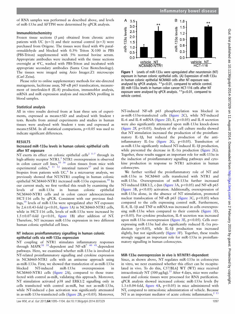
RESULTSIncreased miR-133α levels in human colonic epithelial cellsafter NT exposureNT exerts its effect on colonic epithelial cells2 5–7 through itshigh-affinity receptor NTR1.3 NTR1 overexpression is observedin colon cancer cell lines,10 36 colon tissues from mice withexperimental colitis,6 11 12 intestinal tumors37 and in tissuebiopsies from patients with UC.6 In a microarray analysis, wepreviously showed that NT/NTR1 coupling in human colonicepithelial NCM460-NTR1 increased miR-133α expression.10 Inour current study, we first verified this result by examining thelevels of miR-133α in human colonic epithelialNCM460-NTR1 cells and in colon cancer adenocarcinomaHCT-116 cells by qPCR. Consistent with our previous find-ings,10 levels of miR-133α were upregulated after NT exposureby 2.6±0.43-fold (p<0.01, figure 1A) in NCM460-NTR1 cells,while in HCT-116 cells, levels of miR-133α were increased by1.5±0.07-fold (p<0.01, figure 1B) after addition of NT.Therefore, NT increases miR-133α expression in two differenthuman colonic epithelial cell lines.
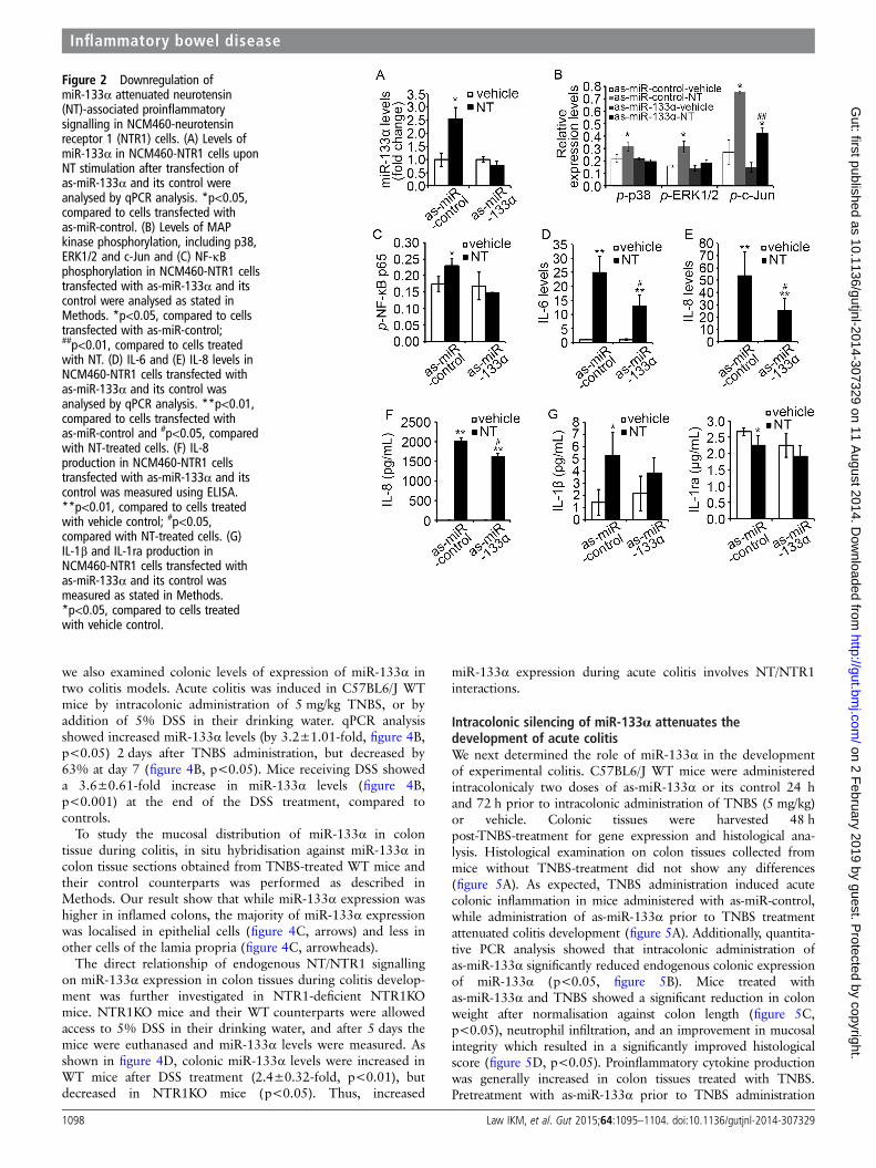
NT induces proinflammatory signalling in human colonicepithelial cells via miR-133α expressionNT coupling of NTR1 stimulates inflammatory responsesthrough MAPK38 39-dependent and NF-κB7 40 41-dependentpathways. Here, we examined whether miR-133α is involved inNT-related proinflammatory signalling and cytokine expressionin NCM460-NTR1 cells with an antisense approach usingas-miR-133α. First, we showed that transfection of as-miR-133αblocked NT-induced miR-133α overexpression inNCM460-NTR1 cells (figure 2A), compared to those trans-fected with control as-miR, validating this approach. Moreover,NT stimulation activated p38 and ERK1/2 signalling only incells transfected with control as-miR, but not as-miR-133α,while NT-induced c-Jun activation was significantly attenuatedin as-miR-133α-transfected cells (figure 2B, p<0.05). Moreover,
NT-induced NF-κB p65 phosphorylation was blocked inas-miR-133α-transfected cells (figure 2C), while NT-inducedIL-6 and IL-8 mRNA (figure 2D, E, p<0.05) and IL-8 secretionwere also significantly attenuated upon miR-133α knock-down(figure 2F, p<0.05). Analysis of the cell culture media showedthat NT stimulation increased the production of the proinflam-matory IL-1β, but reduced the production of the anti-inflammatory IL-1ra (figure 2G, p<0.05). Transfection ofas-miR-133α significantly reduced NT-induced IL-1β production,while prevented the decrease in IL-1ra production (figure 2G).Together, these results suggest an important role for miR-133α inthe induction of proinflammatory signalling pathways and cyto-kine production in response to NTR1 activation in humancolonocytes.
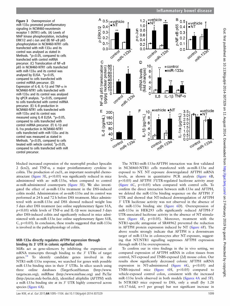
We further verified the proinflammatory role of NT andmiR-133α in NCM460 cells transfected with NTR1 andmiR-133α. Overexpression of miR-133α further increasedNT-induced ERK1/2, c-Jun (figure 3A, p<0.05) and NF-κB p65(figure 3B, p<0.05) activation. Additionally, overexpression ofmiR-133α alone, in the absence of NT stimulation, promotednuclear translocation of NF-κB p65 (figure 3C, p<0.05) whencompared to the cells expressing control miR. Furthermore,IL-8, IL-1β and TNF-α mRNAwas increased in cells overexpres-sing miR-133α when compared to their controls (figure 3D,p<0.05). For cytokine production, IL-8 secretion was increasedupon miR-133α overexpression (figure 3E, p<0.05). Cells over-expressing miR-133α had also significantly reduced IL-1ra pro-duction (p<0.05), while IL-1β production was increasedslightly, but not significantly (figure 3F). Together, these resultsstrongly suggest an important role for miR-133α in proinflam-matory signalling in human colonocytes.
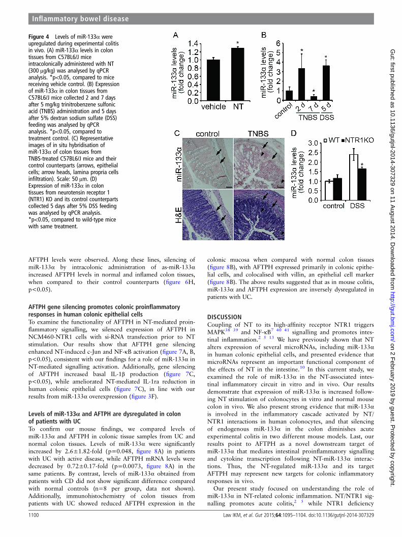
MiR-133α overexpression in vivo is NT/NTR1-dependentSince, as shown above, NT regulates miR-133α in colonocytesin vitro, we next examined whether this effect can be recapitu-lated in vivo. To do this, C57/BL6J WT (WT) mice receivedintracolonicaly NT (300 μg/kg).35 After 4 days, mice were eutha-nased and colonic tissues were processed for RNA purification.qPCR analysis showed increased colonic miR-133α levels (by1.3±0.04-fold, figure 4A, p<0.05) in mice administered withNT, compared to intracolonic administration of vehicle. BecauseNT is an important mediator of acute colonic inflammation,2 13
Figure 1 Levels of miR-133α were upregulated after neurotensin (NT)exposure in human colonic epithelial cells. (A) Expression of miR-133αin human colonic epithelial NCM460 cells after NT exposure wasanalysed by qPCR analysis. **p<0.01, compared to vehicle control.(B) miR-133α levels in human colon cancer HCT-116 cells after NTexposure were analysed by qPCR analysis. **p<0.01, compared tovehicle control.
Inflammatory bowel disease
Law IKM, et al. Gut 2015;64:1095–1104. doi:10.1136/gutjnl-2014-307329 1097
on 2 February 2019 by guest. P
rotected by copyright.http://gut.bm
j.com/
Gut: first published as 10.1136/gutjnl-2014-307329 on 11 A
ugust 2014. Dow
nloaded from
we also examined colonic levels of expression of miR-133α intwo colitis models. Acute colitis was induced in C57BL6/J WTmice by intracolonic administration of 5 mg/kg TNBS, or byaddition of 5% DSS in their drinking water. qPCR analysisshowed increased miR-133α levels (by 3.2±1.01-fold, figure 4B,p<0.05) 2 days after TNBS administration, but decreased by63% at day 7 (figure 4B, p<0.05). Mice receiving DSS showeda 3.6±0.61-fold increase in miR-133α levels (figure 4B,p<0.001) at the end of the DSS treatment, compared tocontrols.
To study the mucosal distribution of miR-133α in colontissue during colitis, in situ hybridisation against miR-133α incolon tissue sections obtained from TNBS-treated WT mice andtheir control counterparts was performed as described inMethods. Our result show that while miR-133α expression washigher in inflamed colons, the majority of miR-133α expressionwas localised in epithelial cells (figure 4C, arrows) and less inother cells of the lamia propria (figure 4C, arrowheads).
The direct relationship of endogenous NT/NTR1 signallingon miR-133α expression in colon tissues during colitis develop-ment was further investigated in NTR1-deficient NTR1KOmice. NTR1KO mice and their WT counterparts were allowedaccess to 5% DSS in their drinking water, and after 5 days themice were euthanased and miR-133α levels were measured. Asshown in figure 4D, colonic miR-133α levels were increased inWT mice after DSS treatment (2.4±0.32-fold, p<0.01), butdecreased in NTR1KO mice (p<0.05). Thus, increased
miR-133α expression during acute colitis involves NT/NTR1interactions.
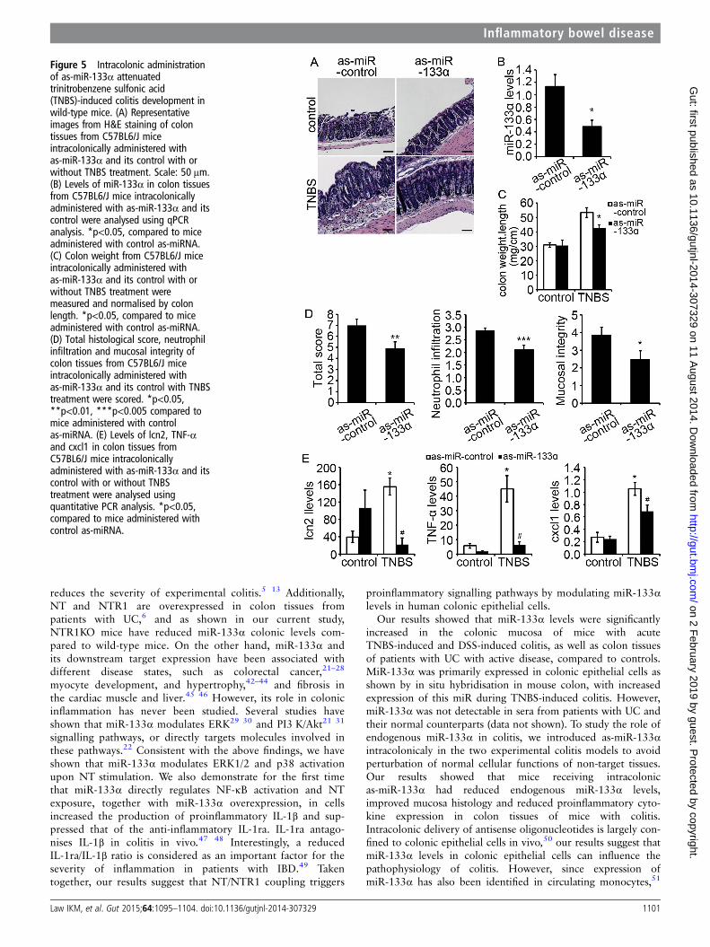
Intracolonic silencing of miR-133α attenuates thedevelopment of acute colitisWe next determined the role of miR-133α in the developmentof experimental colitis. C57BL6/J WT mice were administeredintracolonicaly two doses of as-miR-133α or its control 24 hand 72 h prior to intracolonic administration of TNBS (5 mg/kg)or vehicle. Colonic tissues were harvested 48 hpost-TNBS-treatment for gene expression and histological ana-lysis. Histological examination on colon tissues collected frommice without TNBS-treatment did not show any differences(figure 5A). As expected, TNBS administration induced acutecolonic inflammation in mice administered with as-miR-control,while administration of as-miR-133α prior to TNBS treatmentattenuated colitis development (figure 5A). Additionally, quantita-tive PCR analysis showed that intracolonic administration ofas-miR-133α significantly reduced endogenous colonic expressionof miR-133α (p<0.05, figure 5B). Mice treated withas-miR-133α and TNBS showed a significant reduction in colonweight after normalisation against colon length (figure 5C,p<0.05), neutrophil infiltration, and an improvement in mucosalintegrity which resulted in a significantly improved histologicalscore (figure 5D, p<0.05). Proinflammatory cytokine productionwas generally increased in colon tissues treated with TNBS.Pretreatment with as-miR-133α prior to TNBS administration
Figure 2 Downregulation ofmiR-133α attenuated neurotensin(NT)-associated proinflammatorysignalling in NCM460-neurotensinreceptor 1 (NTR1) cells. (A) Levels ofmiR-133α in NCM460-NTR1 cells uponNT stimulation after transfection ofas-miR-133α and its control wereanalysed by qPCR analysis. *p<0.05,compared to cells transfected withas-miR-control. (B) Levels of MAPkinase phosphorylation, including p38,ERK1/2 and c-Jun and (C) NF-κBphosphorylation in NCM460-NTR1 cellstransfected with as-miR-133α and itscontrol were analysed as stated inMethods. *p<0.05, compared to cellstransfected with as-miR-control;##p<0.01, compared to cells treatedwith NT. (D) IL-6 and (E) IL-8 levels inNCM460-NTR1 cells transfected withas-miR-133α and its control wasanalysed by qPCR analysis. **p<0.01,compared to cells transfected withas-miR-control and #p<0.05, comparedwith NT-treated cells. (F) IL-8production in NCM460-NTR1 cellstransfected with as-miR-133α and itscontrol was measured using ELISA.**p<0.01, compared to cells treatedwith vehicle control; #p<0.05,compared with NT-treated cells. (G)IL-1β and IL-1ra production inNCM460-NTR1 cells transfected withas-miR-133α and its control wasmeasured as stated in Methods.*p<0.05, compared to cells treatedwith vehicle control.
Inflammatory bowel disease
1098 Law IKM, et al. Gut 2015;64:1095–1104. doi:10.1136/gutjnl-2014-307329
on 2 February 2019 by guest. P
rotected by copyright.http://gut.bm
j.com/
Gut: first published as 10.1136/gutjnl-2014-307329 on 11 A
ugust 2014. Dow
nloaded from
blocked increased expression of the neutrophil product lipocalin2 (lcn2), and TNF-α, a major proinflammatory cytokine incolitis. The production of cxcl1, an important neutrophil chemo-attractant (figure 5E, p<0.05) was significantly reduced in miceadministered with as- miR-133α, when compared to controlas-miR-administered counterparts (figure 5E). We also investi-gated the effect of as-miR-133α treatment in the DSS-inducedcolitis model. Administration of as-miR-133α and its control wasperformed at 24 h and 72 h before DSS treatment. Mice adminis-tered with as-miR-133α and DSS showed reduced weight loss5 days after DSS treatment (see online supplementary figure S1A,p<0.05) while levels of TNF-α and IL-1β were increased 5 daysafter DSS-induced colitis and significantly reduced in mice admi-nistered with as-miR-133α (see online supplementary figure S1B,C, p<0.05). In conclusion, these results suggested that miR-133αis involved in the pathophysiology of colitis.
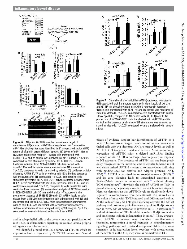
MiR-133α directly regulates AFTPH expression throughbinding its 30 UTR in colonic epithelial cellsMiRs act as gene-silencers by inhibiting the expression ofmRNA transcripts through binding to the 30 UTRs of targetgenes.14 To identify candidate genes involved in theNTR1-miR-133α response, we searched for genes with possiblemiR-133α binding sites in their 30 UTRs. In silico search usingthree online databases (TargetScanHuman (http://www.targetscan.org); miRBase (http://www.mirbase.org) and PicTar(http://pictar.mdc-berlin.de)), identified aftiphilin (AFTPH) witha miR-133α binding site at its 30 UTR highly conserved acrossspecies (figure 6A).
The NTR1-miR-133α-AFTPH interaction was first validatedin NCM460-NTR1 cells transfected with as-miR-133α andexposed to NT. NT exposure downregulated AFTPH mRNAlevels, as shown in quantitative PCR analysis (figure 6B,p<0.05) and AFTPH 30UTR-regulated luciferase activity assay(figure 6C, p<0.05) when compared with control cells. Toconfirm the direct interaction between miR-133α and AFTPH,we deleted the miR-133α binding sequence on the AFTPH 30
UTR and showed that NT-induced downregulation of AFTPH30 UTR luciferase activity was not observed in the absence ofthe miR-133α binding site (figure 6D). Overexpression ofmiR-133α in HEK293 cells significantly reduced AFTPH-30
UTR-associated luciferase activity in the absence of NT stimula-tion (figure 6E, p<0.05). Moreover, treatment with theNTR1-specific antagonist of SR48962 prevented the reductionin AFTPH protein expression induced by NT (figure 6F). Theabove results strongly indicate that AFTPH is a downstreamtarget of miR-133α in colonocytes after NT exposure, suggest-ing that NT/NTR1 signalling suppresses AFTPH expressionthrough miR-133α overexpression.
To confirm our in vitro findings in the in vivo setting, wecompared expression of AFTPH mRNA in colon tissues fromcontrol, NT-exposed and TNBS-exposed (2d) mouse colon. Ourresults show significantly decreased colonic AFTPH mRNAexpression in NT-administered (figure 6G, p<0.05) andTNBS-injected mice (figure 6H, p<0.05) compared tovehicle-exposed control colon, consistent with the increasedmiR-133α levels observed in both models (figures 4A and 5F).In NTR1KO mice exposed to DSS, only a small (by 1.28±0.17-fold, n=5 per group) but not significant increase in
Figure 3 Overexpression ofmiR-133α promoted proinflammatorysignaling in NCM460-neurotensinreceptor 1 (NTR1) cells. (A) Levels ofMAP kinase phosphorylation, includingERK1/2 and c-Jun and (B) NF-κB p65phosphorylation in NCM460-NTR1 cellstransfected with miR-133α and itscontrol was analysed as stated inMethods. *p<0.05, compared to cellstransfected with control miRNAprecursor. (C) Translocation of NF-κBp65 in NCM460-NTR1 cells transfectedwith miR-133α and its control wasanalysed by ELISA. *p<0.05,compared to cells transfected withcontrol miRNA precursor. (D)Expression of IL-8, IL-1β and TNF-α inNCM460-NTR1 cells transfected withmiR-133α and its control was analysedby qPCR analysis. *p<0.05, comparedto cells transfected with control miRNAprecursor. (E) IL-8 production inNCM460-NTR1 cells transfected withmiR-133α and its control wasmeasured using IL-8 ELISA. *p<0.05,compared to cells transfected withcontrol miRNA precursor. (F) IL-1β andIL-1ra production in NCM460-NTR1cells transfected with miR-133α and itscontrol was measured as stated inMethods. *p<0.05, compared to cellstreated with vehicle control; #p<0.05,compared to cells transfected with miRcontrol precursor.
Inflammatory bowel disease
Law IKM, et al. Gut 2015;64:1095–1104. doi:10.1136/gutjnl-2014-307329 1099
on 2 February 2019 by guest. P
rotected by copyright.http://gut.bm
j.com/
Gut: first published as 10.1136/gutjnl-2014-307329 on 11 A
ugust 2014. Dow
nloaded from
AFTPH levels were observed. Along these lines, silencing ofmiR-133α by intracolonic administration of as-miR-133αincreased AFTPH levels in normal and inflamed colon tissues,when compared to their control counterparts (figure 6H,p<0.05).
AFTPH gene silencing promotes colonic proinflammatoryresponses in human colonic epithelial cellsTo examine the functionality of AFTPH in NT-mediated proin-flammatory signalling, we silenced expression of AFTPH inNCM460-NTR1 cells with si-RNA transfection prior to NTstimulation. Our results show that AFTPH gene silencingenhanced NT-induced c-Jun and NF-κB activation (figure 7A, B,p<0.05), consistent with our findings for a role of miR-133α inNT-mediated signalling activation. Additionally, gene silencingof AFTPH increased basal IL-1β production (figure 7C,p<0.05), while ameliorated NT-mediated IL-1ra reduction inhuman colonic epithelial cells (figure 7C), in line with ourresults from miR-133α overexpression (figure 3F).
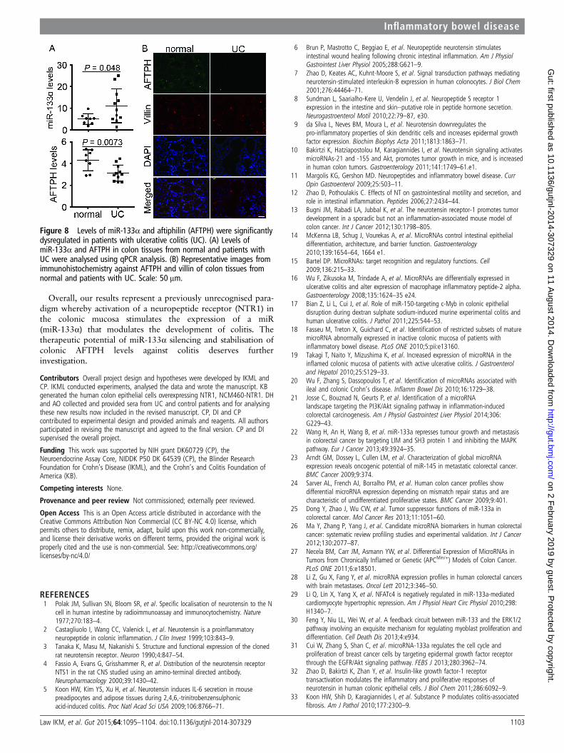
Levels of miR-133α and AFTPH are dysregulated in colonof patients with UCTo confirm our mouse findings, we compared levels ofmiR-133α and AFTPH in colonic tissue samples from UC andnormal colon tissues. Levels of miR-133α were significantlyincreased by 2.6±1.82-fold (p=0.048, figure 8A) in patientswith UC with active disease, while AFTPH mRNA levels weredecreased by 0.72±0.17-fold (p=0.0073, figure 8A) in thesame patients. By contrast, levels of miR-133α obtained frompatients with CD did not show significant difference comparedwith normal controls (n=8 per group, data not shown).Additionally, immunohistochemistry of colon tissues frompatients with UC showed reduced AFTPH expression in the
colonic mucosa when compared with normal colon tissues(figure 8B), with AFTPH expressed primarily in colonic epithe-lial cells, and colocalised with villin, an epithelial cell marker(figure 8B). The above results suggested that as in mouse colitis,miR-133α and AFTPH expression are inversely dysregulated inpatients with UC.
DISCUSSIONCoupling of NT to its high-affinity receptor NTR1 triggersMAPK38 39 and NF-κB7 40 41 signalling and promotes intes-tinal inflammation.2 5 13 We have previously shown that NTalters expression of several microRNAs, including miR-133αin human colonic epithelial cells, and presented evidence thatmicroRNAs represent an important functional component ofthe effects of NT in the intestine.10 In this current study, weexamined the role of miR-133α in the NT-associated intes-tinal inflammatory circuit in vitro and in vivo. Our resultsdemonstrate that expression of miR-133α is increased follow-ing NT stimulation of colonocytes in vitro and normal mousecolon in vivo. We also present strong evidence that miR-133αis involved in the inflammatory cascade activated by NT/NTR1 interactions in human colonocytes, and that silencingof endogenous miR-133α in the colon diminishes acuteexperimental colitis in two different mouse models. Last, ourresults point to AFTPH as a novel downstream target ofmiR-133α that mediates intestinal proinflammatory signallingand cytokine transcription following NT-miR-133α interac-tions. Thus, the NT-regulated miR-133α and its targetAFTPH may represent new targets for colonic inflammatoryresponses in vivo.
Our present study focused on understanding the role ofmiR-133α in NT-related colonic inflammation. NT/NTR1 sig-nalling promotes acute colitis,2 5 while NTR1 deficiency
Figure 4 Levels of miR-133α wereupregulated during experimental colitisin vivo. (A) miR-133α levels in colontissues from C57BL6/J miceintracolonically administered with NT(300 μg/kg) was analysed by qPCRanalysis. *p<0.05, compared to micereceiving vehicle control. (B) Expressionof miR-133α in colon tissues fromC57BL6/J mice collected 2 and 7 daysafter 5 mg/kg trinitrobenzene sulfonicacid (TNBS) administration and 5 daysafter 5% dextran sodium sulfate (DSS)feeding was analysed by qPCRanalysis. *p<0.05, compared totreatment control. (C) Representativeimages of in situ hybridisation ofmiR-133α of colon tissues fromTNBS-treated C57BL6/J mice and theircontrol counterparts (arrows, epithelialcells; arrow heads, lamina propria cellsinfiltration). Scale: 50 μm. (D)Expression of miR-133α in colontissues from neurotensin receptor 1(NTR1) KO and its control counterpartscollected 5 days after 5% DSS feedingwas analysed by qPCR analysis.*p<0.05, compared to wild-type micewith same treatment.
Inflammatory bowel disease
1100 Law IKM, et al. Gut 2015;64:1095–1104. doi:10.1136/gutjnl-2014-307329
on 2 February 2019 by guest. P
rotected by copyright.http://gut.bm
j.com/
Gut: first published as 10.1136/gutjnl-2014-307329 on 11 A
ugust 2014. Dow
nloaded from
reduces the severity of experimental colitis.5 13 Additionally,NT and NTR1 are overexpressed in colon tissues frompatients with UC,6 and as shown in our current study,NTR1KO mice have reduced miR-133α colonic levels com-pared to wild-type mice. On the other hand, miR-133α andits downstream target expression have been associated withdifferent disease states, such as colorectal cancer,21–28
myocyte development, and hypertrophy,42–44 and fibrosis inthe cardiac muscle and liver.45 46 However, its role in colonicinflammation has never been studied. Several studies haveshown that miR-133α modulates ERK29 30 and PI3 K/Akt21 31
signalling pathways, or directly targets molecules involved inthese pathways.22 Consistent with the above findings, we haveshown that miR-133α modulates ERK1/2 and p38 activationupon NT stimulation. We also demonstrate for the first timethat miR-133α directly regulates NF-κB activation and NTexposure, together with miR-133α overexpression, in cellsincreased the production of proinflammatory IL-1β and sup-pressed that of the anti-inflammatory IL-1ra. IL-1ra antago-nises IL-1β in colitis in vivo.47 48 Interestingly, a reducedIL-1ra/IL-1β ratio is considered as an important factor for theseverity of inflammation in patients with IBD.49 Takentogether, our results suggest that NT/NTR1 coupling triggers
proinflammatory signalling pathways by modulating miR-133αlevels in human colonic epithelial cells.
Our results showed that miR-133α levels were significantlyincreased in the colonic mucosa of mice with acuteTNBS-induced and DSS-induced colitis, as well as colon tissuesof patients with UC with active disease, compared to controls.MiR-133α was primarily expressed in colonic epithelial cells asshown by in situ hybridisation in mouse colon, with increasedexpression of this miR during TNBS-induced colitis. However,miR-133α was not detectable in sera from patients with UC andtheir normal counterparts (data not shown). To study the role ofendogenous miR-133α in colitis, we introduced as-miR-133αintracolonicaly in the two experimental colitis models to avoidperturbation of normal cellular functions of non-target tissues.Our results showed that mice receiving intracolonicas-miR-133α had reduced endogenous miR-133α levels,improved mucosa histology and reduced proinflammatory cyto-kine expression in colon tissues of mice with colitis.Intracolonic delivery of antisense oligonucleotides is largely con-fined to colonic epithelial cells in vivo,50 our results suggest thatmiR-133α levels in colonic epithelial cells can influence thepathophysiology of colitis. However, since expression ofmiR-133α has also been identified in circulating monocytes,51
Figure 5 Intracolonic administrationof as-miR-133α attenuatedtrinitrobenzene sulfonic acid(TNBS)-induced colitis development inwild-type mice. (A) Representativeimages from H&E staining of colontissues from C57BL6/J miceintracolonically administered withas-miR-133α and its control with orwithout TNBS treatment. Scale: 50 μm.(B) Levels of miR-133α in colon tissuesfrom C57BL6/J mice intracolonicallyadministered with as-miR-133α and itscontrol were analysed using qPCRanalysis. *p<0.05, compared to miceadministered with control as-miRNA.(C) Colon weight from C57BL6/J miceintracolonically administered withas-miR-133α and its control with orwithout TNBS treatment weremeasured and normalised by colonlength. *p<0.05, compared to miceadministered with control as-miRNA.(D) Total histological score, neutrophilinfiltration and mucosal integrity ofcolon tissues from C57BL6/J miceintracolonically administered withas-miR-133α and its control with TNBStreatment were scored. *p<0.05,**p<0.01, ***p<0.005 compared tomice administered with controlas-miRNA. (E) Levels of lcn2, TNF-αand cxcl1 in colon tissues fromC57BL6/J mice intracolonicallyadministered with as-miR-133α and itscontrol with or without TNBStreatment were analysed usingquantitative PCR analysis. *p<0.05,compared to mice administered withcontrol as-miRNA.
Inflammatory bowel disease
Law IKM, et al. Gut 2015;64:1095–1104. doi:10.1136/gutjnl-2014-307329 1101
on 2 February 2019 by guest. P
rotected by copyright.http://gut.bm
j.com/
Gut: first published as 10.1136/gutjnl-2014-307329 on 11 A
ugust 2014. Dow
nloaded from
and in subepithelial cells of the colonic mucosa, participation ofmiR-133α in inflammatory signalling in colonic lamina propriacell types cannot be excluded.
We identified a novel miR-133α target, AFTPH, in which itsexpression level is regulated by NT/NTR1 interactions. Several
pieces of evidence support our identification of AFTPH as amiR-133α downstream target. Incubation of human colonic epi-thelial cells with NT decreases AFTPH mRNA levels, as well asAFTPH 30UTR-regulated luciferase activity. Most importantly,expression of AFTPH with a deleted miR-133α bindingsequence on its 30 UTR is no longer downregulated in responseto NT exposure. The presence of AFTPH has not been previ-ously recognised in the intestine, and its cellular function is notwell characterised. AFTPH is essential to intracellular traffickingwith binding sites for clathrin and adaptor proteins (AP-1,AP-2).52 AFTPH is localised in trans-golgi network (TGN),53
and its gene silencing leads to unregulated exocytosis ofWeibel-Palade bodies in endothelial cells54 without alteringTGN morphology.55 However, the role of AFTPH or TGN inproinflammatory signalling cascades has not been investigated.Here, we demonstrate that AFTPH levels are significantly down-regulated in colon tissues from patients with UC, as well as inthe colon of mice with TNBS-induced and DSS-induced colitis.At the cellular level, AFTPH gene silencing activates the NF-κBpathway and promotes proinflammatory cytokine IL-1β produc-tion in vitro. NF-κB activation is involved in IL-1β expression56
and administration of NF-κB inhibitor reduce IL-1β productionand ameliorates colonic inflammation in mice.57 Thus, dysregu-lated AFTPH expression may modulate proinflammatoryresponses, representing a novel regulator in proinflammatorysignalling pathways and colitis development. Moreover, mea-surements of its expression levels, together with measurementsof the levels of miR-133α, may serve as biomarkers in UC.
Figure 6 Aftiphilin (AFTPH) was the downstream target ofneurotensin (NT)-induced miR-133α upregulation. (A) ConservativemiR-133α binding sites were identified in 30 untranslated region (UTR)region of aftiphilin across different species. (B) Levels of miR-133α inNCM460-neurotensin receptor 1 (NTR1) cells transfected withas-miR-133α and its control was analysed by qPCR analysis. *p<0.05,compared to cells stimulated by vehicle. (C) AFTPH 30UTR-drivenluciferase activities from NCM460-NTR1 cells transfected withas-miR-133α and its control were measured after NT stimulation.*p<0.05, compared to cells stimulated by vehicle. (D) Luciferase activitydriven by AFTPH 30UTR with or without miR-133α binding sequencewas measured after NT stimulation. *p<0.05, compared to cellsstimulated by vehicle. (E) AFTPH 30UTR-driven luciferase activities fromHEK293 cells transfected with miR-133α precursor (miR-133α) and itscontrol were measured. *p<0.05, compared to cells transfected withcontrol miRNA precursor. (F) Immunoblot analysis of AFTPH expressionin NCM460-NTR1 cells 30 min and 6 h after NT exposure in thepresence or absence of SR48962 (10 nM). (G) AFTPH levels in colontissues from C57BL6/J mice intracolonically administered with NT andits control and (H) from C57BL6/J mice intracolonically administeredwith as-miR-133α and its control with or without trinitrobenzenesulfonic acid treatment were analysed using qPCR analysis. *p<0.05,compared to mice administered with control as-miRNA.
Figure 7 Gene silencing of aftiphilin (AFTPH)-promoted neurotensin(NT)-associated proinflammatory response in vitro. Levels of (A) c-Junand (B) NF-κB phosphorylation in NCM460-neurotensin receptor 1(NTR1) cells transfected with si-AFTPH and its control was measured asstated in Methods. *p<0.05, compared to cells transfected with controlsiRNA; #p<0.05, compared to NT-treated cells. (C) IL-1β and IL-1raproduction of NCM460-NTR1 cells transfected with si-AFTPH and itscontrol in the presence or absence of NT stimulation was analysed asstated in Methods. *p<0.05, compared to cells transfected with controlsiRNA.
Inflammatory bowel disease
1102 Law IKM, et al. Gut 2015;64:1095–1104. doi:10.1136/gutjnl-2014-307329
on 2 February 2019 by guest. P
rotected by copyright.http://gut.bm
j.com/
Gut: first published as 10.1136/gutjnl-2014-307329 on 11 A
ugust 2014. Dow
nloaded from
Overall, our results represent a previously unrecognised para-digm whereby activation of a neuropeptide receptor (NTR1) inthe colonic mucosa stimulates the expression of a miR(miR-133α) that modulates the development of colitis. Thetherapeutic potential of miR-133α silencing and stabilisation ofcolonic AFTPH levels against colitis deserves furtherinvestigation.
Contributors Overall project design and hypotheses were developed by IKML andCP. IKML conducted experiments, analysed the data and wrote the manuscript. KBgenerated the human colon epithelial cells overexpressing NTR1, NCM460-NTR1. DHand AO collected and provided sera from UC and control patients and for analysingthese new results now included in the revised manuscript. CP, DI and CPcontributed to experimental design and provided animals and reagents. All authorsparticipated in revising the manuscript and agreed to the final version. CP and DIsupervised the overall project.
Funding This work was supported by NIH grant DK60729 (CP), theNeuroendocrine Assay Core, NIDDK P50 DK 64539 (CP), the Blinder ResearchFoundation for Crohn’s Disease (IKML), and the Crohn’s and Colitis Foundation ofAmerica (KB).
Competing interests None.
Provenance and peer review Not commissioned; externally peer reviewed.
Open Access This is an Open Access article distributed in accordance with theCreative Commons Attribution Non Commercial (CC BY-NC 4.0) license, whichpermits others to distribute, remix, adapt, build upon this work non-commercially,and license their derivative works on different terms, provided the original work isproperly cited and the use is non-commercial. See: http://creativecommons.org/licenses/by-nc/4.0/
REFERENCES1 Polak JM, Sullivan SN, Bloom SR, et al. Specific localisation of neurotensin to the N
cell in human intestine by radioimmunoassay and immunocytochemistry. Nature1977;270:183–4.
2 Castagliuolo I, Wang CC, Valenick L, et al. Neurotensin is a proinflammatoryneuropeptide in colonic inflammation. J Clin Invest 1999;103:843–9.
3 Tanaka K, Masu M, Nakanishi S. Structure and functional expression of the clonedrat neurotensin receptor. Neuron 1990;4:847–54.
4 Fassio A, Evans G, Grisshammer R, et al. Distribution of the neurotensin receptorNTS1 in the rat CNS studied using an amino-terminal directed antibody.Neuropharmacology 2000;39:1430–42.
5 Koon HW, Kim YS, Xu H, et al. Neurotensin induces IL-6 secretion in mousepreadipocytes and adipose tissues during 2,4,6,-trinitrobenzensulphonicacid-induced colitis. Proc Natl Acad Sci USA 2009;106:8766–71.
6 Brun P, Mastrotto C, Beggiao E, et al. Neuropeptide neurotensin stimulatesintestinal wound healing following chronic intestinal inflammation. Am J PhysiolGastrointest Liver Physiol 2005;288:G621–9.
7 Zhao D, Keates AC, Kuhnt-Moore S, et al. Signal transduction pathways mediatingneurotensin-stimulated interleukin-8 expression in human colonocytes. J Biol Chem2001;276:44464–71.
8 Sundman L, Saarialho-Kere U, Vendelin J, et al. Neuropeptide S receptor 1expression in the intestine and skin--putative role in peptide hormone secretion.Neurogastroenterol Motil 2010;22:79–87, e30.
9 da Silva L, Neves BM, Moura L, et al. Neurotensin downregulates thepro-inflammatory properties of skin dendritic cells and increases epidermal growthfactor expression. Biochim Biophys Acta 2011;1813:1863–71.
10 Bakirtzi K, Hatziapostolou M, Karagiannides I, et al. Neurotensin signaling activatesmicroRNAs-21 and -155 and Akt, promotes tumor growth in mice, and is increasedin human colon tumors. Gastroenterology 2011;141:1749–61.e1.
11 Margolis KG, Gershon MD. Neuropeptides and inflammatory bowel disease. CurrOpin Gastroenterol 2009;25:503–11.
12 Zhao D, Pothoulakis C. Effects of NT on gastrointestinal motility and secretion, androle in intestinal inflammation. Peptides 2006;27:2434–44.
13 Bugni JM, Rabadi LA, Jubbal K, et al. The neurotensin receptor-1 promotes tumordevelopment in a sporadic but not an inflammation-associated mouse model ofcolon cancer. Int J Cancer 2012;130:1798–805.
14 McKenna LB, Schug J, Vourekas A, et al. MicroRNAs control intestinal epithelialdifferentiation, architecture, and barrier function. Gastroenterology2010;139:1654–64, 1664 e1.
15 Bartel DP. MicroRNAs: target recognition and regulatory functions. Cell2009;136:215–33.
16 Wu F, Zikusoka M, Trindade A, et al. MicroRNAs are differentially expressed inulcerative colitis and alter expression of macrophage inflammatory peptide-2 alpha.Gastroenterology 2008;135:1624–35 e24.
17 Bian Z, Li L, Cui J, et al. Role of miR-150-targeting c-Myb in colonic epithelialdisruption during dextran sulphate sodium-induced murine experimental colitis andhuman ulcerative colitis. J Pathol 2011;225:544–53.
18 Fasseu M, Treton X, Guichard C, et al. Identification of restricted subsets of maturemicroRNA abnormally expressed in inactive colonic mucosa of patients withinflammatory bowel disease. PLoS ONE 2010;5:pii:e13160.
19 Takagi T, Naito Y, Mizushima K, et al. Increased expression of microRNA in theinflamed colonic mucosa of patients with active ulcerative colitis. J Gastroenteroland Hepatol 2010;25:S129–33.
20 Wu F, Zhang S, Dassopoulos T, et al. Identification of microRNAs associated withileal and colonic Crohn’s disease. Inflamm Bowel Dis 2010;16:1729–38.
21 Josse C, Bouznad N, Geurts P, et al. Identification of a microRNAlandscape targeting the PI3K/Akt signaling pathway in inflammation-inducedcolorectal carcinogenesis. Am J Physiol Gastrointest Liver Physiol 2014;306:G229–43.
22 Wang H, An H, Wang B, et al. miR-133a represses tumour growth and metastasisin colorectal cancer by targeting LIM and SH3 protein 1 and inhibiting the MAPKpathway. Eur J Cancer 2013;49:3924–35.
23 Arndt GM, Dossey L, Cullen LM, et al. Characterization of global microRNAexpression reveals oncogenic potential of miR-145 in metastatic colorectal cancer.BMC Cancer 2009;9:374.
24 Sarver AL, French AJ, Borralho PM, et al. Human colon cancer profiles showdifferential microRNA expression depending on mismatch repair status and arecharacteristic of undifferentiated proliferative states. BMC Cancer 2009;9:401.
25 Dong Y, Zhao J, Wu CW, et al. Tumor suppressor functions of miR-133a incolorectal cancer. Mol Cancer Res 2013;11:1051–60.
26 Ma Y, Zhang P, Yang J, et al. Candidate microRNA biomarkers in human colorectalcancer: systematic review profiling studies and experimental validation. Int J Cancer2012;130:2077–87.
27 Necela BM, Carr JM, Asmann YW, et al. Differential Expression of MicroRNAs inTumors from Chronically Inflamed or Genetic (APCMin/+) Models of Colon Cancer.PLoS ONE 2011;6:e18501.
28 Li Z, Gu X, Fang Y, et al. microRNA expression profiles in human colorectal cancerswith brain metastases. Oncol Lett 2012;3:346–50.
29 Li Q, Lin X, Yang X, et al. NFATc4 is negatively regulated in miR-133a-mediatedcardiomyocyte hypertrophic repression. Am J Physiol Heart Circ Physiol 2010;298:H1340–7.
30 Feng Y, Niu LL, Wei W, et al. A feedback circuit between miR-133 and the ERK1/2pathway involving an exquisite mechanism for regulating myoblast proliferation anddifferentiation. Cell Death Dis 2013;4:e934.
31 Cui W, Zhang S, Shan C, et al. microRNA-133a regulates the cell cycle andproliferation of breast cancer cells by targeting epidermal growth factor receptorthrough the EGFR/Akt signaling pathway. FEBS J 2013;280:3962–74.
32 Zhao D, Bakirtzi K, Zhan Y, et al. Insulin-like growth factor-1 receptortransactivation modulates the inflammatory and proliferative responses ofneurotensin in human colonic epithelial cells. J Biol Chem 2011;286:6092–9.
33 Koon HW, Shih D, Karagiannides I, et al. Substance P modulates colitis-associatedfibrosis. Am J Pathol 2010;177:2300–9.
Figure 8 Levels of miR-133α and aftiphilin (AFTPH) were significantlydysregulated in patients with ulcerative colitis (UC). (A) Levels ofmiR-133α and AFTPH in colon tissues from normal and patients withUC were analysed using qPCR analysis. (B) Representative images fromimmunohistochemistry against AFTPH and villin of colon tissues fromnormal and patients with UC. Scale: 50 μm.
Inflammatory bowel disease
Law IKM, et al. Gut 2015;64:1095–1104. doi:10.1136/gutjnl-2014-307329 1103
on 2 February 2019 by guest. P
rotected by copyright.http://gut.bm
j.com/
Gut: first published as 10.1136/gutjnl-2014-307329 on 11 A
ugust 2014. Dow
nloaded from
34 Koon HW, Zhao D, Xu H, et al. Substance P-mediated expression of the pro-angiogenicfactor CCN1 modulates the course of colitis. Am J Pathol 2008;173:400–10.
35 Evers BM, Izukura M, Townsend CM Jr, et al. Neurotensin prevents intestinalmucosal hypoplasia in rats fed an elemental diet. Dig Dis Sci 1992;37:426–31.
36 Maoret JJ, Pospai D, Rouyerfessard C, et al. Neurotensin receptor and its mRNA areexpressed in many human colon cancer cell lines but not in normal colonicepithelium: binding studies and RT-PCR experiments. Biochem Biophys ResCommun 1994;203:465–71.
37 Gui X, Guzman G, Dobner PR, et al. Increased neurotensin receptor-1 expressionduring progression of colonic adenocarcinoma. Peptides 2008;29:1609–15.
38 Zhao D, Zhan Y, Koon HW, et al. Metalloproteinase-dependent transforming growthfactor-alpha release mediates neurotensin-stimulated MAP kinase activation inhuman colonic epithelial cells. J Biol Chem 2004;279:43547–54.
39 Zhao D, Zhan Y, Zeng H, et al. Neurotensin stimulates expression of early growthresponse gene-1 and EGF receptor through MAP kinase activation in human colonicepithelial cells. Int J Cancer 2007;120:1652–6.
40 Zhao D, Kuhnt-Moore S, Zeng H, et al. Neurotensin stimulates IL-8 expression inhuman colonic epithelial cells through Rho GTPase-mediated NF-kappa B pathways.Am J Physiol Cell Physiol 2003;284:C1397–404.
41 Zhao D, Zhan Y, Zeng H, et al. Neurotensin stimulates interleukin-8 expressionthrough modulation of I kappa B alpha phosphorylation and p65 transcriptionalactivity: involvement of protein kinase C alpha. Mol Pharmacol 2005;67:2025–31.
42 Rao PK, Kumar RM, Farkhondeh M, et al. Myogenic factors that regulate expressionof muscle-specific microRNAs. Proc Natl Acad Sci USA 2006;103:8721–6.
43 Chen X, Wang K, Chen J, et al. In vitro evidence suggests that miR-133a-mediatedregulation of uncoupling protein 2 (UCP2) is an indispensable step in myogenicdifferentiation. J Biol Chem 2009;284:5362–9.
44 Hua Y, Zhang Y, Ren J. IGF-1 deficiency resists cardiac hypertrophy and myocardialcontractile dysfunction: role of microRNA-1 and microRNA-133a. J Cell Mol Med2012;16:83–95.
45 Roderburg C, Luedde M, Vargas Cardenas D, et al. miR-133a mediatesTGF-β-dependent derepression of collagen synthesis in hepatic stellate cells duringliver fibrosis. J Hepatol 2013;58:736–42.
46 Castoldi G, di Gioia CRT, Bombardi C, et al. MiR-133a regulates collagen 1A1:potential role of miR-133a in myocardial fibrosis in angiotensin II-dependenthypertension. J Cell Physiol 2012;227:850–6.
47 Cominelli F, Nast CC, Clark BD, et al. Interleukin 1 (IL-1) gene expression, synthesis,and effect of specific IL-1 receptor blockade in rabbit immune complex colitis. J ClinInvest 1990;86:972–80.
48 Ferretti M, Casini-Raggi V, Pizarro TT, et al. Neutralization of endogenous IL-1receptor antagonist exacerbates and prolongs inflammation in rabbit immune colitis.J Clin Invest 1994;94:449–53.
49 Casini-Raggi V, Kam L, Chong YJ, et al. Mucosal imbalance of IL-1 and IL-1receptor antagonist in inflammatory bowel disease. A novel mechanism of chronicintestinal inflammation. J Immunol 1995;154:2434–40.
50 Huang Z, Shi T, Zhou Q, et al. miR-141 Regulates colonic leukocytic trafficking bytargeting CXCL12β during murine colitis and human Crohn’s disease. Gut2014;63:1247–57.
51 Wang Y, Li L, Moore BT, et al. MiR-133a in human circulating monocytes: apotential biomarker associated with postmenopausal osteoporosis. PLoS ONE2012;7:e34641.
52 Mattera R, Ritter B, Sidhu SS, et al. Definition of the consensus motif recognized bygamma-adaptin ear domains. J Biol Chem 2004;279:8018–28.
53 Burman JL, Wasiak S, Ritter B, et al. Aftiphilin is a component of the clathrinmachinery in neurons. FEBS Lett 2005;579:2177–84.
54 Lui-Roberts WW, Ferraro F, Nightingale TD, et al. Aftiphilin and gamma-synergin arerequired for secretagogue sensitivity of Weibel-Palade bodies in endothelial cells.Mol Biol Cell 2008;19:5072–81.
55 Hirst J, Borner GH, Harbour M, et al. The aftiphilin/p200/gamma-synergin complex.Mol Biol Cell 2005;16:2554–65.
56 Carvalho FA, Aitken JD, Gewirtz AT, et al. TLR5 activation induces secretoryinterleukin-1 receptor antagonist (sIL-1Ra) and reduces inflammasome-associatedtissue damage. Mucosal Immunol 2011;4:102–11.
57 Funakoshi T, Yamashita K, Ichikawa N, et al. A novel NF-κB inhibitor,dehydroxymethylepoxyquinomicin, ameliorates inflammatory colonic injury in mice.J Crohns Colitis 2012;6:215–25.
Inflammatory bowel disease
1104 Law IKM, et al. Gut 2015;64:1095–1104. doi:10.1136/gutjnl-2014-307329
on 2 February 2019 by guest. P
rotected by copyright.http://gut.bm
j.com/
Gut: first published as 10.1136/gutjnl-2014-307329 on 11 A
ugust 2014. Dow
nloaded from