Ingested human insulin inhibits the mosquito NF-κB-dependent immune response 1 to
9
Ingested Human Insulin Inhibits the Mosquito NF-B-Dependent Immune Response to Plasmodium falciparum Nazzy Pakpour, a Vanessa Corby-Harris, b Gabriel P. Green, a Hannah M. Smithers, a Kong W. Cheung, a Michael A. Riehle, c and Shirley Luckhart a Department of Medical Microbiology and Immunology, School of Medicine, University of California, Davis, California, USA a ; Carl Hayden Bee Research Center, Agricultural Research Service, U.S. Department of Agriculture, Tucson, Arizona, USA b ; and Department of Entomology, University of Arizona, Tucson, Arizona, USA c We showed previously that ingested human insulin activates the insulin/IGF-1 signaling pathway in Anopheles stephensi and increases the susceptibility of these mosquitoes to Plasmodium falciparum. In other organisms, insulin can alter immune re- sponsiveness through regulation of NF-B transcription factors, critical elements for innate immunity that are also central to mosquito immunity. We show here that insulin signaling decreased expression of NF-B-regulated immune genes in mosquito cells stimulated with either bacterial or malarial soluble products. Further, human insulin suppressed mosquito immunity through sustained phosphatidylinositol 3-kinase activation, since inhibition of this pathway led to decreased parasite develop- ment in the mosquito. Together, these data demonstrate that activation of the insulin/IGF-1 signaling pathway by ingested hu- man insulin can alter NF-B-dependent immunity, and ultimately the susceptibility, of mosquitoes to P. falciparum. A nnually, there are over 250 million new malaria cases, the majority due to infection with the parasite Plasmodium fal- ciparum (58). When a female mosquito ingests an infectious blood meal, insulin from vertebrate blood collects in the mosquito midgut, a critical site for Plasmodium development (reviewed in reference 57) and a tissue that is exquisitely responsive to ingested human insulin (9, 31). The amount of insulin ingested by a feed- ing mosquito is directly influenced by malaria infection. That is, patients with severe malaria can develop blood insulin levels that exceed that of a normal healthy adult by as much as 10- to 35-fold (13, 56). Ingested human insulin enhances malaria parasite devel- opment in exposed mosquitoes relative to mosquitoes that do not receive insulin in the infectious blood meal (50). Collectively, these observations suggest that control strategies for malaria should recognize that the burden of transmission is influenced, in part, by changes in insulin physiology in the human host that cross talk with analogous physiology in the mosquito host. The highly conserved insulin/IGF-1 signaling (IIS) pathway consists of two main signaling branches, a mitogen-activated pro- tein kinase (MAPK)-dependent branch and phosphatidylinositol 3-kinase (PI3K)/Akt-dependent branch, both of which are in- volved in the regulation of innate immune responses (26, 38). Ingested insulin can activate both the PI3K and MAPK branches of the IIS pathway in mosquitoes (9, 31, 37, 38, 50). In addition, at least two IIS proteins—Akt/PKB and ERK— have proven critical for the control of malaria infection in the mosquito host (9, 51). Our previous work suggested that human insulin ingested in a blood meal can signal mosquito midgut epithelial cells directly (31), altering the susceptibility of mosquitoes to malaria infection (50). Specifically, when mosquitoes were provided human insulin in an infectious blood meal, we observed a significant increase in P. falciparum development relative to mosquitoes that did not receive insulin in the blood meal (50). While these data suggest that human insulin-induced signaling in the mosquito midgut alters the immune responsiveness of mosquitoes to malaria para- sites, the effects of insulin signaling on mosquito immunity have not been well characterized, and no direct connections between the IIS pathway and mosquito immunity have been established. Plasmodium parasites undergo a series of complicated devel- opmental transformations upon ingestion by the Anopheles mos- quito and, during this process, mosquito immunity can result in significant reductions in parasite numbers (reviewed in reference 48). The regulation of mosquito immunity during Plasmodium infection occurs in part through the activation of the highly con- served NF-B transcription factors Rel1 and Rel2 that control mosquito responses to bacterial, fungal, and parasitic pathogens (7, 20, 39–41). NF-B binding motifs have been found in the upstream re- gions of numerous insect immune genes, including antimicrobial effectors such as defensin 1 (Def), cecropin 1 (Cec), and gambicin (Gam), as well as nitric oxide synthase (18, 25, 39, 45, 54, 55). Increased NF-B-dependent transcription can reduce both bacte- rial load and Plasmodium development in anopheline mosquitoes (2, 18, 20, 23, 41). In mammals, a number of well-characterized cell signaling pathways, including IIS, can network with NF-B activation to fine tune the response to infection. For example, physiological levels of human insulin can suppress both lipopoly- saccharide (LPS)- and tumor necrosis factor alpha (TNF-)-in- duced NF-B activation in human cell lines (11, 28). In addition, IIS activation in human cell lines can attenuate the degradation of the negative regulators of NF-B, resulting in increased NF-B sequestration in the cytoplasm (10). Therefore, we hypothesized that human insulin ingested in an infectious blood meal alters mosquito immunity to malaria parasite infection through the reg- ulation of NF-B activity. We show here that human insulin can inhibit the expression of Received 6 January 2012 Returned for modification 3 February 2012 Accepted 22 March 2012 Published ahead of print 2 April 2012 Editor: J. H. Adams Address correspondence to Nazzy Pakpour, [email protected]. Copyright © 2012, American Society for Microbiology. All Rights Reserved. doi:10.1128/IAI.00024-12 June 2012 Volume 80 Number 6 Infection and Immunity p. 2141–2149 iai.asm.org 2141 Downloaded from https://journals.asm.org/journal/iai on 16 February 2022 by 175.196.156.152.
Transcript of Ingested human insulin inhibits the mosquito NF-κB-dependent immune response 1 to
Ingested Human Insulin Inhibits the Mosquito NF-�B-DependentImmune Response to Plasmodium falciparum
Nazzy Pakpour,a Vanessa Corby-Harris,b Gabriel P. Green,a Hannah M. Smithers,a Kong W. Cheung,a Michael A. Riehle,c
and Shirley Luckharta
Department of Medical Microbiology and Immunology, School of Medicine, University of California, Davis, California, USAa; Carl Hayden Bee Research Center, AgriculturalResearch Service, U.S. Department of Agriculture, Tucson, Arizona, USAb; and Department of Entomology, University of Arizona, Tucson, Arizona, USAc
We showed previously that ingested human insulin activates the insulin/IGF-1 signaling pathway in Anopheles stephensi andincreases the susceptibility of these mosquitoes to Plasmodium falciparum. In other organisms, insulin can alter immune re-sponsiveness through regulation of NF-�B transcription factors, critical elements for innate immunity that are also central tomosquito immunity. We show here that insulin signaling decreased expression of NF-�B-regulated immune genes in mosquitocells stimulated with either bacterial or malarial soluble products. Further, human insulin suppressed mosquito immunitythrough sustained phosphatidylinositol 3-kinase activation, since inhibition of this pathway led to decreased parasite develop-ment in the mosquito. Together, these data demonstrate that activation of the insulin/IGF-1 signaling pathway by ingested hu-man insulin can alter NF-�B-dependent immunity, and ultimately the susceptibility, of mosquitoes to P. falciparum.
Annually, there are over 250 million new malaria cases, themajority due to infection with the parasite Plasmodium fal-
ciparum (58). When a female mosquito ingests an infectious bloodmeal, insulin from vertebrate blood collects in the mosquitomidgut, a critical site for Plasmodium development (reviewed inreference 57) and a tissue that is exquisitely responsive to ingestedhuman insulin (9, 31). The amount of insulin ingested by a feed-ing mosquito is directly influenced by malaria infection. That is,patients with severe malaria can develop blood insulin levels thatexceed that of a normal healthy adult by as much as 10- to 35-fold(13, 56). Ingested human insulin enhances malaria parasite devel-opment in exposed mosquitoes relative to mosquitoes that do notreceive insulin in the infectious blood meal (50). Collectively,these observations suggest that control strategies for malariashould recognize that the burden of transmission is influenced, inpart, by changes in insulin physiology in the human host that crosstalk with analogous physiology in the mosquito host.
The highly conserved insulin/IGF-1 signaling (IIS) pathwayconsists of two main signaling branches, a mitogen-activated pro-tein kinase (MAPK)-dependent branch and phosphatidylinositol3-kinase (PI3K)/Akt-dependent branch, both of which are in-volved in the regulation of innate immune responses (26, 38).Ingested insulin can activate both the PI3K and MAPK branchesof the IIS pathway in mosquitoes (9, 31, 37, 38, 50). In addition, atleast two IIS proteins—Akt/PKB and ERK— have proven criticalfor the control of malaria infection in the mosquito host (9, 51).
Our previous work suggested that human insulin ingested in ablood meal can signal mosquito midgut epithelial cells directly(31), altering the susceptibility of mosquitoes to malaria infection(50). Specifically, when mosquitoes were provided human insulinin an infectious blood meal, we observed a significant increase inP. falciparum development relative to mosquitoes that did notreceive insulin in the blood meal (50). While these data suggestthat human insulin-induced signaling in the mosquito midgutalters the immune responsiveness of mosquitoes to malaria para-sites, the effects of insulin signaling on mosquito immunity havenot been well characterized, and no direct connections betweenthe IIS pathway and mosquito immunity have been established.
Plasmodium parasites undergo a series of complicated devel-opmental transformations upon ingestion by the Anopheles mos-quito and, during this process, mosquito immunity can result insignificant reductions in parasite numbers (reviewed in reference48). The regulation of mosquito immunity during Plasmodiuminfection occurs in part through the activation of the highly con-served NF-�B transcription factors Rel1 and Rel2 that controlmosquito responses to bacterial, fungal, and parasitic pathogens(7, 20, 39–41).
NF-�B binding motifs have been found in the upstream re-gions of numerous insect immune genes, including antimicrobialeffectors such as defensin 1 (Def), cecropin 1 (Cec), and gambicin(Gam), as well as nitric oxide synthase (18, 25, 39, 45, 54, 55).Increased NF-�B-dependent transcription can reduce both bacte-rial load and Plasmodium development in anopheline mosquitoes(2, 18, 20, 23, 41). In mammals, a number of well-characterizedcell signaling pathways, including IIS, can network with NF-�Bactivation to fine tune the response to infection. For example,physiological levels of human insulin can suppress both lipopoly-saccharide (LPS)- and tumor necrosis factor alpha (TNF-�)-in-duced NF-�B activation in human cell lines (11, 28). In addition,IIS activation in human cell lines can attenuate the degradation ofthe negative regulators of NF-�B, resulting in increased NF-�Bsequestration in the cytoplasm (10). Therefore, we hypothesizedthat human insulin ingested in an infectious blood meal altersmosquito immunity to malaria parasite infection through the reg-ulation of NF-�B activity.
We show here that human insulin can inhibit the expression of
Received 6 January 2012 Returned for modification 3 February 2012Accepted 22 March 2012
Published ahead of print 2 April 2012
Editor: J. H. Adams
Address correspondence to Nazzy Pakpour, [email protected].
Copyright © 2012, American Society for Microbiology. All Rights Reserved.
doi:10.1128/IAI.00024-12
June 2012 Volume 80 Number 6 Infection and Immunity p. 2141–2149 iai.asm.org 2141
Dow
nloa
ded
from
http
s://j
ourn
als.
asm
.org
/jour
nal/i
ai o
n 16
Feb
ruar
y 20
22 b
y 17
5.19
6.15
6.15
2.
the NF-�B-regulated immune gene expression both in immortal-ized mosquito cells in vitro and in the Anopheles stephensi midgutepithelium in vivo in response to bacterial and malarial solubleproducts. The presence of human insulin resulted in the sustainedactivation of the PI3K, but not the MAPK, branch of the mosquitoIIS pathway. In addition, inhibition of PI3K activity both in im-mortalized mosquito cells and in the A. stephensi midgut resultedin the reversal of the immunosuppressive effects of human insu-lin. Taken together, our data indicate that activation of the mos-quito IIS by human insulin inhibits the mosquito immune re-sponse to malaria parasites at least in part through the regulationof NF-�B activity.
MATERIALS AND METHODSCell culture, transfection, and luciferase reporter assays. A. stephensiembryonic (ASE) cells (a gift from H.-M. Müller, EMBL) were maintainedas previously described (51). Cec and Gam promoters were cloned into thepromoterless luciferase reporter pGL3 (Promega, Madison, WI). Trans-fections were performed using Effectene transfection reagent (Qiagen) aspreviously described (43). At 24 h posttransfection, cells were challengedwith 100 �g of LPS (Escherichia coli serotype O26:B6; Sigma-Aldrich, St.Louis, MO)/ml or with 150 � 106 parasite equivalents of P. falciparumsoluble products (PfsPs) with or without 0.017 to 1.7 �M human insulin(Sigma-Aldrich). For assays of kinase inhibition, cells were treated withthe inhibitors LY294002 (50 �M; Sigma-Aldrich), wortmannin (1 �M;EMD Chemicals, Gibbstown, NJ), PD98059 (10 �M; Sigma-Aldrich), orU0126 (10 �M; Promega) for 1 h prior to immune challenge. Luciferaseactivity was measured 24 h after immune challenge with the Dual-Glosystem (Promega).
Immunoblotting. Protein extracts were prepared, separated, andtransferred to membranes as previously described (37). Membranes wereblocked in 5% nonfat dry milk (wt/vol) in Tris-buffered saline (pH 7.0)containing 0.1% Triton-100 (TBS-T) for 1 h at room temperature. Mem-branes were incubated at 4°C overnight with the following: 1:10,000mouse monoclonal anti-diphosphorylated ERK1/2 (p-ERK; Sigma-Al-drich), 1:1,000 anti-phospho-FOXO1/FOXO3a (p-FOXO; Cell Signaling,Danvers, MA), or 1:10,000 anti-GAPDH (Sigma-Aldrich) antibody in 5%nonfat dry milk in TBS-T. Membranes were washed three times, for 5 mineach time, and incubated with a 1:2,000 (p-FOXO) or 1:20,000 (GAPDH)dilution of horseradish peroxidase (HRP)-conjugated goat anti-rabbitIgG (Biosource International, Camarillo, CA) or with 1:20,000 (p-ERK)HRP-conjugated rabbit anti-mouse IgG (Pierce, Rockford, IL) at 4°Covernight. To reveal antibody-bound proteins, membranes were incu-bated with SuperSignal West Dura chemiluminescent reagent for 5 minand visualized using the Kodak Image Station 4000MM Pro and Car-estream molecular imaging software (Carestream Health, New Haven,CT). The levels of phospho-proteins in each treatment were first normal-ized to total protein levels as determined by GAPDH and then to theappropriate control group.
Mosquito rearing and experimental treatments. A. stephensi Liston(Indian wild-type strain) were reared and maintained at 27°C and 75%humidity. All mosquito rearing and feeding protocols were approved andin accordance with regulatory guidelines and standards set by the Institu-tional Animal Care and Use Committee of the University of California,Davis. For experimental treatments, laboratory reared 3- to 5-day-oldfemale mosquitoes were kept on water for 48 h and then allowed to feedfor 30 min on reconstituted human blood meals provided through a He-motek insect feeding system (Discovery Workshops, Accrington, UnitedKingdom). For signaling studies, mosquitoes were fed washed red bloodcells (RBCs) alone or with 170 pM human insulin, 20 �M LY294002, orinsulin plus LY294002. For Western blot analyses, midguts were dissectedfrom 60 mosquitoes in each treatment group at various time points afterblood feeding and processed as previously described (51).
For life span studies, �1-day-old A. stephensi females (n � 50) were
placed into four treatment groups and fed a single blood meal containing(i) 170 pM human insulin, (ii) heat-killed bacteria (HK-bacteria), (iii)HK-bacteria plus insulin, or (iv) an equivalent volume of phosphate-buffered saline (PBS) as a control. HK-bacterial stocks were prepared bycombining equivalent volumes of log-phase liquid cultures of Staphylo-coccus aureus and Pseudomonas aeruginosa (Carolina Biological Supply,Burlington, NC) grown overnight and autoclaving the liquid culture for�20 min. The HK-bacteria were pelleted, and the supernatant was re-moved. HK-bacteria were washed twice with sterile PBS before beingadded to a blood meal. After provision of this single blood meal, mosqui-toes were provided 10% sucrose solution ad libitum thereafter. Experi-ments were repeated four times separately, and mortality was recordeddaily.
PfsP preparation. To produce P. falciparum soluble products (PfsPs)for in vitro experiments, infected RBCs (iRBCs) from day 15 parasitecultures were lysed in 15 ml of 1� RBC lysis buffer (eBioscience, SanDiego, CA) for 15 min and then brought to a final volume of 100 ml inPBS. Paramagnetic, hemozoin-containing parasites were enriched using aMACS LS column (Miltenyi Biotec, Auburn, CA) in a magnetic holder asdescribed previously (33). Column elutant, consisting primarily of para-sites, was then frozen at �80°C for 10 min and thawed at 37°C 10 timesand applied to new LS columns to remove the hemozoin. Pooled PfsPspreparations were determined to be endotoxin-free (E-Toxate kit; Sigma-Aldrich) and contained �1 �M hemin/heme/hemozoin (hemin assay kit;BioVision, Mountain View, CA). The number of parasite equivalents wasdetermined with the following formula: (iRBCS/RBCs) � (total numberof RBCs) minus 30% estimated loss during purification process (32, 33).
P. falciparum culture and mosquito infection. For mosquito infec-tion, cultures of P. falciparum strain NF54 MCB (a gift from Sanaria, Inc.,Rockville, MD) were grown in 10% heat-inactivated human serum and5% washed human RBCs (Interstate Blood Bank) in RPMI 1640 withHEPES (Invitrogen) and hypoxanthine (Sigma-Aldrich). Mosquitoeswere allowed to feed on day 15 mature gametocyte cultures diluted withhuman RBCs and heat-inactivated human serum with or without 170 pMhuman insulin and with or without 20 �M LY294002. All treatments wereadded to the diluted P. falciparum culture immediately prior to bloodfeeding. Protocols involving the culture and handling of P. falciparum formosquito feeding were approved and in accordance with regulatoryguidelines and standards set by the Biological Safety Administrative Advi-sory Committee of the University of California, Davis. After 10 days, midg-uts from 50 mosquitoes with fully developed eggs (to confirm completeengorgement) from each group were dissected in PBS and stained with0.1% mercurochrome for direct counting of P. falciparum oocysts. Meansof oocysts per midgut in each treatment group were calculated from alldissected mosquitoes, including zeros for mosquitoes that contained nooocysts.
P. falciparum growth assays. Aliquots of P. falciparum NF54 culturewere synchronized 48 h prior to the assay as previously described (35) andthen plated in 96-well flat-bottom plates in complete RPMI 1640 withHEPES, hypoxanthine, and 10% heat-inactivated human serum. Parasiteswere treated with LY294002 inhibitor at 2, 20, or 200 �M or with anequivalent volume of dimethyl sulfoxide (DMSO) diluent for 48 and 96 hin a candle jar in a 37°C incubator. The assays were terminated by re-placing the culture medium with RPMI 1640 with 10% formalin inPBS. Erythrocytes were stained with 10 �g of propidium iodide (Sig-ma-Aldrich)/ml in PBS for 1 h at room temperature. Infected RBCswere counted with FACSCalibur flow cytometer (BD Biosciences, SanJose, CA). The levels of parasite growth in response to treatment werenormalized to DMSO controls, which were set to 100%.
RESULTSHuman insulin inhibits LPS-induced antimicrobial promoteractivity in immortalized mosquito cells. To determine whetherhuman insulin could regulate the mosquito immune response invitro, we utilized a luciferase-reporter assay to quantify LPS-in-
Pakpour et al.
2142 iai.asm.org Infection and Immunity
Dow
nloa
ded
from
http
s://j
ourn
als.
asm
.org
/jour
nal/i
ai o
n 16
Feb
ruar
y 20
22 b
y 17
5.19
6.15
6.15
2.
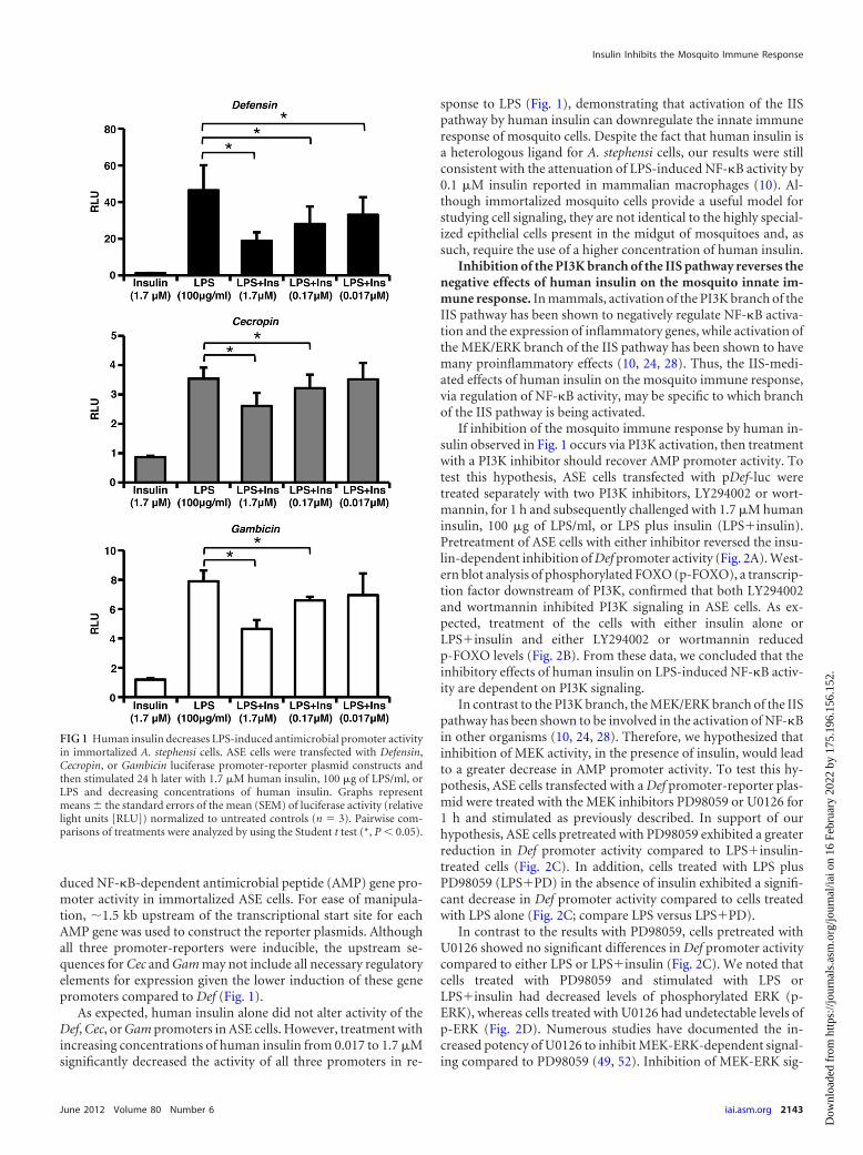
duced NF-�B-dependent antimicrobial peptide (AMP) gene pro-moter activity in immortalized ASE cells. For ease of manipula-tion, �1.5 kb upstream of the transcriptional start site for eachAMP gene was used to construct the reporter plasmids. Althoughall three promoter-reporters were inducible, the upstream se-quences for Cec and Gam may not include all necessary regulatoryelements for expression given the lower induction of these genepromoters compared to Def (Fig. 1).
As expected, human insulin alone did not alter activity of theDef, Cec, or Gam promoters in ASE cells. However, treatment withincreasing concentrations of human insulin from 0.017 to 1.7 �Msignificantly decreased the activity of all three promoters in re-
sponse to LPS (Fig. 1), demonstrating that activation of the IISpathway by human insulin can downregulate the innate immuneresponse of mosquito cells. Despite the fact that human insulin isa heterologous ligand for A. stephensi cells, our results were stillconsistent with the attenuation of LPS-induced NF-�B activity by0.1 �M insulin reported in mammalian macrophages (10). Al-though immortalized mosquito cells provide a useful model forstudying cell signaling, they are not identical to the highly special-ized epithelial cells present in the midgut of mosquitoes and, assuch, require the use of a higher concentration of human insulin.
Inhibition of the PI3K branch of the IIS pathway reverses thenegative effects of human insulin on the mosquito innate im-mune response. In mammals, activation of the PI3K branch of theIIS pathway has been shown to negatively regulate NF-�B activa-tion and the expression of inflammatory genes, while activation ofthe MEK/ERK branch of the IIS pathway has been shown to havemany proinflammatory effects (10, 24, 28). Thus, the IIS-medi-ated effects of human insulin on the mosquito immune response,via regulation of NF-�B activity, may be specific to which branchof the IIS pathway is being activated.
If inhibition of the mosquito immune response by human in-sulin observed in Fig. 1 occurs via PI3K activation, then treatmentwith a PI3K inhibitor should recover AMP promoter activity. Totest this hypothesis, ASE cells transfected with pDef-luc weretreated separately with two PI3K inhibitors, LY294002 or wort-mannin, for 1 h and subsequently challenged with 1.7 �M humaninsulin, 100 �g of LPS/ml, or LPS plus insulin (LPSinsulin).Pretreatment of ASE cells with either inhibitor reversed the insu-lin-dependent inhibition of Def promoter activity (Fig. 2A). West-ern blot analysis of phosphorylated FOXO (p-FOXO), a transcrip-tion factor downstream of PI3K, confirmed that both LY294002and wortmannin inhibited PI3K signaling in ASE cells. As ex-pected, treatment of the cells with either insulin alone orLPSinsulin and either LY294002 or wortmannin reducedp-FOXO levels (Fig. 2B). From these data, we concluded that theinhibitory effects of human insulin on LPS-induced NF-�B activ-ity are dependent on PI3K signaling.
In contrast to the PI3K branch, the MEK/ERK branch of the IISpathway has been shown to be involved in the activation of NF-�Bin other organisms (10, 24, 28). Therefore, we hypothesized thatinhibition of MEK activity, in the presence of insulin, would leadto a greater decrease in AMP promoter activity. To test this hy-pothesis, ASE cells transfected with a Def promoter-reporter plas-mid were treated with the MEK inhibitors PD98059 or U0126 for1 h and stimulated as previously described. In support of ourhypothesis, ASE cells pretreated with PD98059 exhibited a greaterreduction in Def promoter activity compared to LPSinsulin-treated cells (Fig. 2C). In addition, cells treated with LPS plusPD98059 (LPSPD) in the absence of insulin exhibited a signifi-cant decrease in Def promoter activity compared to cells treatedwith LPS alone (Fig. 2C; compare LPS versus LPSPD).
In contrast to the results with PD98059, cells pretreated withU0126 showed no significant differences in Def promoter activitycompared to either LPS or LPSinsulin (Fig. 2C). We noted thatcells treated with PD98059 and stimulated with LPS orLPSinsulin had decreased levels of phosphorylated ERK (p-ERK), whereas cells treated with U0126 had undetectable levels ofp-ERK (Fig. 2D). Numerous studies have documented the in-creased potency of U0126 to inhibit MEK-ERK-dependent signal-ing compared to PD98059 (49, 52). Inhibition of MEK-ERK sig-
FIG 1 Human insulin decreases LPS-induced antimicrobial promoter activityin immortalized A. stephensi cells. ASE cells were transfected with Defensin,Cecropin, or Gambicin luciferase promoter-reporter plasmid constructs andthen stimulated 24 h later with 1.7 �M human insulin, 100 �g of LPS/ml, orLPS and decreasing concentrations of human insulin. Graphs representmeans the standard errors of the mean (SEM) of luciferase activity (relativelight units [RLU]) normalized to untreated controls (n � 3). Pairwise com-parisons of treatments were analyzed by using the Student t test (*, P � 0.05).
Insulin Inhibits the Mosquito Immune Response
June 2012 Volume 80 Number 6 iai.asm.org 2143
Dow
nloa
ded
from
http
s://j
ourn
als.
asm
.org
/jour
nal/i
ai o
n 16
Feb
ruar
y 20
22 b
y 17
5.19
6.15
6.15
2.
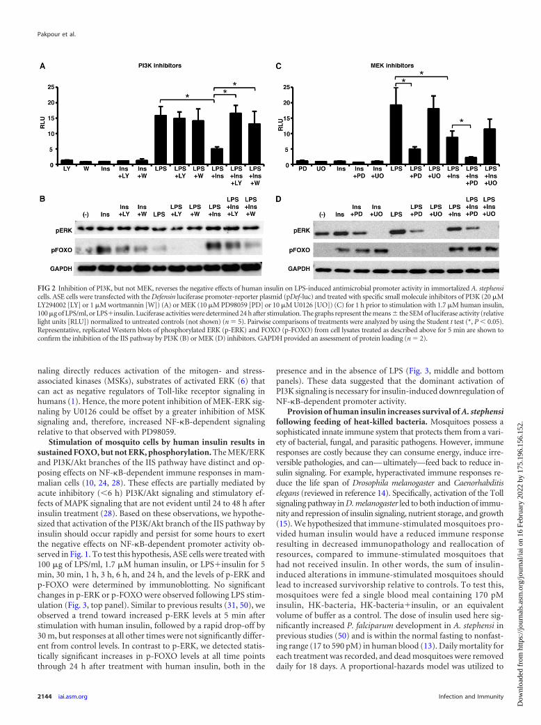
naling directly reduces activation of the mitogen- and stress-associated kinases (MSKs), substrates of activated ERK (6) thatcan act as negative regulators of Toll-like receptor signaling inhumans (1). Hence, the more potent inhibition of MEK-ERK sig-naling by U0126 could be offset by a greater inhibition of MSKsignaling and, therefore, increased NF-�B-dependent signalingrelative to that observed with PD98059.
Stimulation of mosquito cells by human insulin results insustained FOXO, but not ERK, phosphorylation. The MEK/ERKand PI3K/Akt branches of the IIS pathway have distinct and op-posing effects on NF-�B-dependent immune responses in mam-malian cells (10, 24, 28). These effects are partially mediated byacute inhibitory (�6 h) PI3K/Akt signaling and stimulatory ef-fects of MAPK signaling that are not evident until 24 to 48 h afterinsulin treatment (28). Based on these observations, we hypothe-sized that activation of the PI3K/Akt branch of the IIS pathway byinsulin should occur rapidly and persist for some hours to exertthe negative effects on NF-�B-dependent promoter activity ob-served in Fig. 1. To test this hypothesis, ASE cells were treated with100 �g of LPS/ml, 1.7 �M human insulin, or LPSinsulin for 5min, 30 min, 1 h, 3 h, 6 h, and 24 h, and the levels of p-ERK andp-FOXO were determined by immunoblotting. No significantchanges in p-ERK or p-FOXO were observed following LPS stim-ulation (Fig. 3, top panel). Similar to previous results (31, 50), weobserved a trend toward increased p-ERK levels at 5 min afterstimulation with human insulin, followed by a rapid drop-off by30 m, but responses at all other times were not significantly differ-ent from control levels. In contrast to p-ERK, we detected statis-tically significant increases in p-FOXO levels at all time pointsthrough 24 h after treatment with human insulin, both in the
presence and in the absence of LPS (Fig. 3, middle and bottompanels). These data suggested that the dominant activation ofPI3K signaling is necessary for insulin-induced downregulation ofNF-�B-dependent promoter activity.
Provision of human insulin increases survival of A. stephensifollowing feeding of heat-killed bacteria. Mosquitoes possess asophisticated innate immune system that protects them from a vari-ety of bacterial, fungal, and parasitic pathogens. However, immuneresponses are costly because they can consume energy, induce irre-versible pathologies, and can—ultimately—feed back to reduce in-sulin signaling. For example, hyperactivated immune responses re-duce the life span of Drosophila melanogaster and Caenorhabditiselegans (reviewed in reference 14). Specifically, activation of the Tollsignaling pathway in D. melanogaster led to both induction of immu-nity and repression of insulin signaling, nutrient storage, and growth(15). We hypothesized that immune-stimulated mosquitoes pro-vided human insulin would have a reduced immune responseresulting in decreased immunopathology and reallocation ofresources, compared to immune-stimulated mosquitoes thathad not received insulin. In other words, the sum of insulin-induced alterations in immune-stimulated mosquitoes shouldlead to increased survivorship relative to controls. To test this,mosquitoes were fed a single blood meal containing 170 pMinsulin, HK-bacteria, HK-bacteriainsulin, or an equivalentvolume of buffer as a control. The dose of insulin used here sig-nificantly increased P. falciparum development in A. stephensi inprevious studies (50) and is within the normal fasting to nonfast-ing range (17 to 590 pM) in human blood (13). Daily mortality foreach treatment was recorded, and dead mosquitoes were removeddaily for 18 days. A proportional-hazards model was utilized to
FIG 2 Inhibition of PI3K, but not MEK, reverses the negative effects of human insulin on LPS-induced antimicrobial promoter activity in immortalized A. stephensicells. ASE cells were transfected with the Defensin luciferase promoter-reporter plasmid (pDef-luc) and treated with specific small molecule inhibitors of PI3K (20 �MLY294002 [LY] or 1 �M wortmannin [W]) (A) or MEK (10 �M PD98059 [PD] or 10 �M U0126 [UO]) (C) for 1 h prior to stimulation with 1.7 �M human insulin,100 �g of LPS/ml, or LPSinsulin. Luciferase activities were determined 24 h after stimulation. The graphs represent the means the SEM of luciferase activity (relativelight units [RLU]) normalized to untreated controls (not shown) (n � 5). Pairwise comparisons of treatments were analyzed by using the Student t test (*, P � 0.05).Representative, replicated Western blots of phosphorylated ERK (p-ERK) and FOXO (p-FOXO) from cell lysates treated as described above for 5 min are shown toconfirm the inhibition of the IIS pathway by PI3K (B) or MEK (D) inhibitors. GAPDH provided an assessment of protein loading (n � 2).
Pakpour et al.
2144 iai.asm.org Infection and Immunity
Dow
nloa
ded
from
http
s://j
ourn
als.
asm
.org
/jour
nal/i
ai o
n 16
Feb
ruar
y 20
22 b
y 17
5.19
6.15
6.15
2.
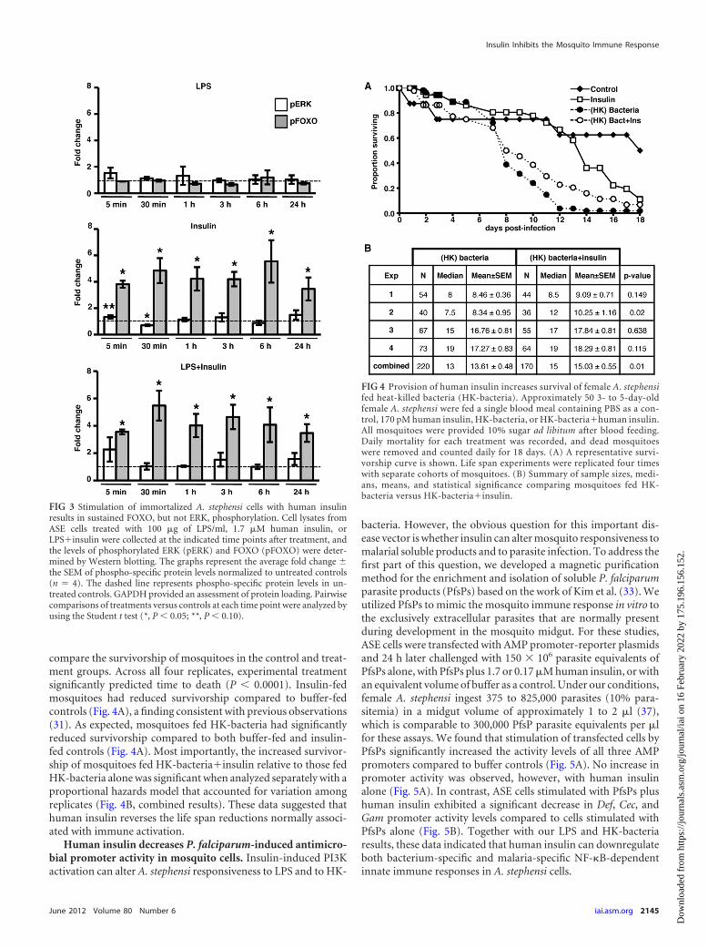
compare the survivorship of mosquitoes in the control and treat-ment groups. Across all four replicates, experimental treatmentsignificantly predicted time to death (P � 0.0001). Insulin-fedmosquitoes had reduced survivorship compared to buffer-fedcontrols (Fig. 4A), a finding consistent with previous observations(31). As expected, mosquitoes fed HK-bacteria had significantlyreduced survivorship compared to both buffer-fed and insulin-fed controls (Fig. 4A). Most importantly, the increased survivor-ship of mosquitoes fed HK-bacteriainsulin relative to those fedHK-bacteria alone was significant when analyzed separately with aproportional hazards model that accounted for variation amongreplicates (Fig. 4B, combined results). These data suggested thathuman insulin reverses the life span reductions normally associ-ated with immune activation.
Human insulin decreases P. falciparum-induced antimicro-bial promoter activity in mosquito cells. Insulin-induced PI3Kactivation can alter A. stephensi responsiveness to LPS and to HK-
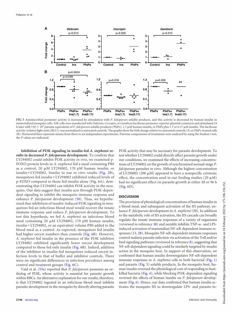
bacteria. However, the obvious question for this important dis-ease vector is whether insulin can alter mosquito responsiveness tomalarial soluble products and to parasite infection. To address thefirst part of this question, we developed a magnetic purificationmethod for the enrichment and isolation of soluble P. falciparumparasite products (PfsPs) based on the work of Kim et al. (33). Weutilized PfsPs to mimic the mosquito immune response in vitro tothe exclusively extracellular parasites that are normally presentduring development in the mosquito midgut. For these studies,ASE cells were transfected with AMP promoter-reporter plasmidsand 24 h later challenged with 150 � 106 parasite equivalents ofPfsPs alone, with PfsPs plus 1.7 or 0.17 �M human insulin, or withan equivalent volume of buffer as a control. Under our conditions,female A. stephensi ingest 375 to 825,000 parasites (10% para-sitemia) in a midgut volume of approximately 1 to 2 �l (37),which is comparable to 300,000 PfsP parasite equivalents per �lfor these assays. We found that stimulation of transfected cells byPfsPs significantly increased the activity levels of all three AMPpromoters compared to buffer controls (Fig. 5A). No increase inpromoter activity was observed, however, with human insulinalone (Fig. 5A). In contrast, ASE cells stimulated with PfsPs plushuman insulin exhibited a significant decrease in Def, Cec, andGam promoter activity levels compared to cells stimulated withPfsPs alone (Fig. 5B). Together with our LPS and HK-bacteriaresults, these data indicated that human insulin can downregulateboth bacterium-specific and malaria-specific NF-�B-dependentinnate immune responses in A. stephensi cells.
FIG 3 Stimulation of immortalized A. stephensi cells with human insulinresults in sustained FOXO, but not ERK, phosphorylation. Cell lysates fromASE cells treated with 100 �g of LPS/ml, 1.7 �M human insulin, orLPSinsulin were collected at the indicated time points after treatment, andthe levels of phosphorylated ERK (pERK) and FOXO (pFOXO) were deter-mined by Western blotting. The graphs represent the average fold change the SEM of phospho-specific protein levels normalized to untreated controls(n � 4). The dashed line represents phospho-specific protein levels in un-treated controls. GAPDH provided an assessment of protein loading. Pairwisecomparisons of treatments versus controls at each time point were analyzed byusing the Student t test (*, P � 0.05; **, P � 0.10).
FIG 4 Provision of human insulin increases survival of female A. stephensifed heat-killed bacteria (HK-bacteria). Approximately 50 3- to 5-day-oldfemale A. stephensi were fed a single blood meal containing PBS as a con-trol, 170 pM human insulin, HK-bacteria, or HK-bacteriahuman insulin.All mosquitoes were provided 10% sugar ad libitum after blood feeding.Daily mortality for each treatment was recorded, and dead mosquitoeswere removed and counted daily for 18 days. (A) A representative survi-vorship curve is shown. Life span experiments were replicated four timeswith separate cohorts of mosquitoes. (B) Summary of sample sizes, medi-ans, means, and statistical significance comparing mosquitoes fed HK-bacteria versus HK-bacteriainsulin.
Insulin Inhibits the Mosquito Immune Response
June 2012 Volume 80 Number 6 iai.asm.org 2145
Dow
nloa
ded
from
http
s://j
ourn
als.
asm
.org
/jour
nal/i
ai o
n 16
Feb
ruar
y 20
22 b
y 17
5.19
6.15
6.15
2.
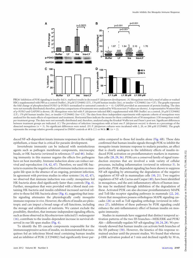
Inhibition of PI3K signaling in insulin-fed A. stephensi re-sults in decreased P. falciparum development. To confirm thatLY294002 could inhibit PI3K activity in vivo, we examined p-FOXO protein levels in A. stephensi fed a meal containing PBSas a control, 20 �M LY294002, 170 pM human insulin, orinsulinLY294002. Similar to our in vitro results (Fig. 2B),mosquitoes fed insulinLY294002 exhibited reduced levels ofp-FOXO compared to those fed insulin alone (Fig. 6A), dem-onstrating that LY294002 can inhibit PI3K activity in the mos-quito. Our data suggest that insulin acts through PI3K-depen-dent signaling to inhibit the mosquito immune response andenhance P. falciparum development (50). Thus, we hypothe-sized that inhibition of insulin-induced PI3K signaling in mos-quitoes fed an infectious blood meal would recover the innateimmune response and reduce P. falciparum development. Totest this hypothesis, we fed A. stephensi an infectious bloodmeal containing 20 �M LY294002, 170 pM human insulin,insulinLY294002, or an equivalent volume PBS added to theblood meal as a control. As expected, mosquitoes fed insulinhad higher oocyst numbers than controls (Fig. 6B). However,A. stephensi fed insulin in the presence of the PI3K inhibitorLY294002 exhibited significantly lower oocyst developmentcompared to those fed only insulin (Fig. 6B). Indeed, additionof the inhibitor to insulin-fed mosquitoes reduced oocyst in-fection levels to that of buffer and inhibitor controls. Therewere no significant differences in infection prevalence amongcontrol and treatment groups (Fig. 6C).
Vaid et al. (53a) reported that P. falciparum possesses an or-tholog of PI3K, whose activity is essential for parasite growthwithin RBCs. An alternative explanation for our results, therefore,is that LY294002 ingested in an infectious blood meal inhibitsparasite development in the mosquito by directly altering parasite
PI3K activity that may be necessary for parasite development. Totest whether LY294002 could directly affect parasite growth underour conditions, we examined the effects of increasing concentra-tions of LY294002 on the growth of synchronized asexual-stage P.falciparum parasites in vitro. Although the highest concentrationof LY294002 (200 �M) appeared to have a nonspecific cytotoxiceffect, the concentration used in our feeding studies (20 �M)had no significant effect on parasite growth at either 48 or 96 h(Fig. 6D).
DISCUSSION
The provision of physiological concentrations of human insulin ina blood meal, and subsequent activation of the IIS pathway, en-hance P. falciparum development in A. stephensi (50). In additionto the metabolic role of IIS activation, the IIS cascade can broadlyregulate the innate immune responses of a variety of organisms(reviewed in reference 38) and notably inhibits TNF-�- and LPS-induced activation of mammalian NF-�B-dependent immune re-sponses (11, 28). Mosquito NF-�B-dependent immune responsescontrol malaria parasite infection via activation of the Toll and/orImd signaling pathways (reviewed in reference 8), suggesting thatNF-�B-dependent signaling could be similarly targeted by insulinaction in the mosquito host. In support of this observation, weconfirmed that human insulin downregulates NF-�B-dependentimmune responses in A. stephensi cells to both bacterial (Fig. 1)and parasite (Fig. 5) soluble products. In the mosquito host, hu-man insulin reversed the physiological cost of responding to heat-killed bacteria (Fig. 4), while blocking PI3K-dependent signalingreversed the effects of human insulin on P. falciparum develop-ment (Fig. 6). Hence, our data confirmed that human insulin ac-tivates the mosquito IIS to downregulate LPS- and parasite-in-
FIG 5 Antimicrobial promoter activity is increased by stimulation with P. falciparum soluble products, and this activity is decreased by human insulin inimmortalized mosquito cells. ASE cells were transfected with Defensin, Cecropin, or Gambicin luciferase promoter-reporter plasmid constructs and stimulated 24h later with 150 � 106 parasite equivalents of P. falciparum soluble products (PfsPs), 1.7 �M human insulin, or PfsPs plus 1.7 or 0.17 �M insulin. The luciferaseactivity (relative light units [RLU]) was normalized to untreated controls. The graphs show the fold change relative to untreated controls (A) or PfsPs-treated cells(B). Horizontal lines represent means from three to six independent experiments. Pairwise comparisons of treatments were analyzed by using the Student t test;the P values are indicated.
Pakpour et al.
2146 iai.asm.org Infection and Immunity
Dow
nloa
ded
from
http
s://j
ourn
als.
asm
.org
/jour
nal/i
ai o
n 16
Feb
ruar
y 20
22 b
y 17
5.19
6.15
6.15
2.
duced NF-�B-dependent innate immune responses in the midgutepithelium, a tissue that is critical for parasite development.
Invertebrate immunity can be induced with noninfectiousagents such as pathogen membrane components, microscopicbeads, or HK-bacteria (reviewed in references 17 and 46). Induc-ing immunity in this manner negates the effects live pathogenshave on host mortality. Immune induction alone can reduce sur-vival and reproduction (14, 42, 47). Therefore, we used HK-bac-teria to examine the negative effects of immune induction on mos-quito life span in the absence of an ongoing, persistent infection.In agreement with previous studies in other systems (14, 42, 47),we observed that immune induction was costly: mosquitoes fedHK-bacteria alone died significantly faster than controls (Fig. 4).Further, mosquitoes that were provided with a blood meal con-taining HK-bacteria and insulin exhibited increased survival rel-ative to those fed HK-bacteria alone (Fig. 4), suggesting that insu-lin signaling attenuates the physiologically costly mosquitoimmune response in vivo. However, the effects of insulin are pleio-tropic and can impact a broad range of cell functions, includingthe storage and utilization of nutrients. We cannot exclude thepossibility, therefore, that immune-independent signaling effects,such as those observed in Mycobacterium-infected D. melanogaster(16), contribute to the insulin-dependent increase in survival ob-served in our life span studies (Fig. 4).
To identify the IIS cascade components responsible for theimmunosuppressive action of insulin, we demonstrated that mos-quitoes fed an infectious blood meal containing human insulinand an inhibitor of PI3K (LY294002) had significantly fewer par-
asites compared to those fed insulin alone (Fig. 6B). These dataconfirmed that human insulin signals through PI3K to inhibit themosquito innate immune response to malaria parasites, an effectthat is clearly analogous to the inhibitory effects of insulin-in-duced PI3K activation on proinflammatory markers in mamma-lian cells (28, 29, 36). PI3Ks are a conserved family of signal trans-duction enzymes that are involved a wide variety of cellularprocesses, including inflammation (reviewed in reference 5). Inparticular, PI3K-dependent signaling has been shown to suppressNF-�B signaling by attenuating the degradation of the negativeregulators of NF-�B in mammalian cells (10, 21). Two negativeregulators of NF-�B, Cactus and Caspar (40), have been identifiedin mosquitoes, and the anti-inflammatory effects of human insu-lin may be mediated through inhibition of the degradation ofthese. Activated PI3K can also decrease proinflammatory MAPKand Toll-like receptor signaling pathways in mammals (22, 24).Given that mosquitoes possess orthologous MAPK signaling cas-cades (26) as well as Toll signaling orthologs (reviewed in refer-ence 27), inhibition of these pathways by PI3K signaling couldenhance the anti-inflammatory effect of human insulin on mos-quito immunity.
Studies in mammals have suggested that distinct temporal ac-tivation patterns of the two IIS branches—MEK/ERK and PI3K/Akt— differentially regulate NF-�B signaling (10, 24, 28). We pre-viously showed that human insulin can activate both branches ofthe IIS pathway (50). However, the kinetics of this response re-mained unclear until the present studies. We found that whereasp-ERK activation peaked at 5 min and declined rapidly by 30 m,
FIG 6 Inhibition of PI3K signaling in insulin-fed A. stephensi results in decreased P. falciparum development. (A) Mosquitoes were fed a meal of saline or washedRBCs supplemented with PBS as a control (buffer), 20 �M LY294002 (LY), 170 pM human insulin (Ins), or insulinLY294002 (insLY). The graphs representthe fold change of phosphorylated FOXO (p-FOXO) normalized to untreated controls (n � 4). GAPDH provided an assessment of protein loading. The datawere not normally distributed; therefore, pairwise comparisons of treatments were analyzed by Wilcoxon test (P values are shown). A representative Western blotof p-FOXO and GAPDH is shown. (B) Mosquitoes were fed with P. falciparum-infected RBCs supplemented with PBS (buffer) as a control, 20 �M LY294002(LY), 170 pM human insulin (Ins), or insulinLY294002 (InsLY). The data from three independent experiments with separate cohorts of mosquitoes wereanalyzed for the main effects of experiment and treatment. Horizontal lines indicate the means for three combined sets of 50 mosquitoes (150 mosquitoes total)per treatment group. The data were not normally distributed and, therefore, analyzed using the Kruskal-Wallis test and Dunn’s post test. Significant differencesbetween treatment groups are indicated. (C) The prevalence of infection (mosquitoes with at least one P. falciparum oocyst) is shown as a percentage of thedissected mosquitoes (n � 3). No significant differences were noted. (D) P. falciparum cultures were incubated with 2, 20, or 200 �M LY294002. The graphrepresents the average relative growth compared to DMSO controls at 48 h (�) or 96 h (�) (n � 2).
Insulin Inhibits the Mosquito Immune Response
June 2012 Volume 80 Number 6 iai.asm.org 2147
Dow
nloa
ded
from
http
s://j
ourn
als.
asm
.org
/jour
nal/i
ai o
n 16
Feb
ruar
y 20
22 b
y 17
5.19
6.15
6.15
2.
p-FOXO levels were maintained to 24 h after insulin treatment(Fig. 3). These results suggested that PI3K/Akt signaling, and theassociated downstream inhibitory effects on the mosquito im-mune response, are sustained after insulin stimulation. In the con-text of P. falciparum development, this insulin-induced signalingpersistence would extend from zygote and ookinete developmentto the early invasion of the midgut epithelium, thereby protectingdeveloping parasites during a broad period of critical morphogen-esis.
In previous work, we showed that P. falciparum infection of A.stephensi can be completely inhibited by the overexpression of acti-vated Akt in the midgut epithelium (9), an outcome that initiallyappears to be at odds with the effects of insulin-induced activation ofPI3K/Akt presented here. However, Akt can interact with a variety ofproteins that both regulate its activity and allow for stimulus-specificdifferences in its substrate utilization (reviewed in reference 19).Therefore, we would not expect the overexpression of constitutivelyactive Akt in transgenic mosquitoes to replicate the ligand-specificeffects of insulin stimulation in nontransgenic mosquitoes. Certainly,insulin-induced activation of MEK/ERK (Fig. 3) and p38 MAPK ac-tivation (26) would not contribute to the phenotype of Akt-overex-pressing transgenic A. stephensi since these signaling molecules arenot downstream of Akt. Finally, our observations regarding the dif-ferences between overexpression of a signaling regulator versus sig-naling by a ligand are not unique. The overexpression of antioxidantgenes, purported to extend life span, in fact reduce the life span of D.melanogaster in the absence of oxidative stress (3). Hence, functionalbiology is often only revealed when ligand and signaling are con-nected.
By 2030, one in five adults will have type 2 diabetes in Africa(53). Our data, combined with these predictions, suggest that hy-perinsulinemia in diabetics could reach levels that would be pre-dicted to inhibit the mosquito immune response to P. falciparum,thereby increasing the ability of mosquitoes to transmit malaria.These interactions could increase the spread of malaria infectionin populations where this comorbidity is increasing in prevalenceand where malaria transmission is already a significant problem.Our observations suggest that a critical component of future con-trol strategies for both of these diseases will be based on under-standing the effects of hyperinsulinemia and clinical interventionson the relative risk of parasite infection and transmission byAnopheles mosquitoes.
ACKNOWLEDGMENTS
We thank Edwin E. Lewis for statistical analyses and Andrew Ross fortechnical assistance in completing these studies. We also thank Joseph L.DeRisi and Charles C. Kim for the kind gift of the magnetic purificationstand.
Funding for these studies was provided by NIH NIAID R01 AI080799and R01 AI073745.
REFERENCES1. Ananieva O, et al. 2008. The kinases MSK1 and MSK2 act as negative
regulators of Toll-like receptor signaling. Nat. Immunol. 9:1028 –1036.2. Barillas-Mury C, et al. 1996. Immune factor Gambif1, a new rel family
member from the human malaria vector, Anopheles gambiae. EMBO J.15:4691– 4701.
3. Bayne AC, Mockett RJ, Orr WC, Sohal RS. 2005. Enhanced catabolismof mitochondrial superoxide/hydrogen peroxide and aging in transgenicDrosophila. Biochem. J. 391:277–284.
4. Reference deleted.5. Cantley LC. 2002. The phosphoinositide 3-kinase pathway. Science 296:
1655–1657.
6. Cargnello M, Roux PP. 2011. Activation and function of the MAPKs andtheir substrates, the MAPK-activated protein kinases. Microbiol. Mol.Biol. Rev. 75:50 – 83.
7. Christophides GK, et al. 2002. Immunity-related genes and gene familiesin Anopheles gambiae. Science 298:159 –165.
8. Cirimotich CM, Dong Y, Garver LS, Sim S, Dimopoulos G. 2010.Mosquito immune defenses against Plasmodium infection. Dev. Comp.Immunol. 34:387–395.
9. Corby-Harris V, et al. 2010. Activation of Akt signaling reduces theprevalence and intensity of malaria parasite infection and lifespan inAnopheles stephensi mosquitoes. PLoS Pathog. 6:e1001003.
10. Cuschieri J, Bulger E, Grinsell R, Garcia I, Maier RV. 2008. Insulinregulates macrophage activation through activin A. Shock 29:285–290.
11. Dandona P, et al. 2001. Insulin inhibits intranuclear nuclear factor �Band stimulates I�B in mononuclear cells in obese subjects: evidence for ananti-inflammatory effect? J. Clin. Endocrinol. Metab. 86:3257–3265.
12. Reference deleted.13. Darby SM, Miller ML, Allen RO, LeBeau M. 2001. A mass spectrometric
method for quantitation of intact insulin in blood samples. J. Anal. Toxi-col. 25:8 –14.
14. DeVeale B, Brummel T, Seroude L. 2004. Immunity and aging: theenemy within? Aging Cell 3:195–208.
15. DiAngelo JR, Bland ML, Bambina S, Cherry S, Birnbaum MJ. 2009. Theimmune response attenuates growth and nutrient storage in Drosophila byreducing insulin signaling. Proc. Natl. Acad. Sci. U. S. A. 106:20853–20858.
16. Dionne MS, Pham LN, Shirasu-Hiza M, Schneider DS. 2006. Akt andFOXO dysregulation contribute to infection-induced wasting in Drosoph-ila. Curr. Biol. 16:1977–1985.
17. Dionne MS, Schneider DS. 2008. Models of infectious diseases in the fruitfly Drosophila melanogaster. Dis. Model Mech. 1:43– 49.
18. Dong Y, et al. 2006. Anopheles gambiae immune responses to human androdent Plasmodium parasite species. PLoS Pathog. 2:e52.
19. Franke TF. 2008. PI3K/Akt: getting it right matters. Oncogene 27:6473–6488.
20. Frolet C, Thoma M, Blandin S, Hoffmann JA, Levashina EA. 2006.Boosting NF-�B-dependent basal immunity of Anopheles gambiae abortsdevelopment of Plasmodium berghei. Immunity 25:677– 685.
21. Fukao T, Koyasu S. 2003. PI3K and negative regulation of TLR signaling.Trends Immunol. 24:358 –363.
22. Fukao T, et al. 2002. PI3K-mediated negative feedback regulation ofIL-12 production in DCs. Nat. Immunol. 3:875– 881.
23. Garver LS, Dong Y, Dimopoulos G. 2009. Caspar controls resistance toPlasmodium falciparum in diverse anopheline species. PLoS Pathog.5:e1000335.
24. Guha M, Mackman N. 2002. The phosphatidylinositol 3-kinase-Aktpathway limits lipopolysaccharide activation of signaling pathways andexpression of inflammatory mediators in human monocytic cells. J. Biol.Chem. 277:32124 –32132.
25. Hillyer JF, Estevez-Lao TY. 2010. Nitric oxide is an essential componentof the hemocyte-mediated mosquito immune response against bacteria.Dev. Comp. Immunol. 34:141–149.
26. Horton AA, et al. 2011. The mitogen-activated protein kinome fromAnopheles gambiae: identification, phylogeny and functional characteriza-tion of the ERK, JNK and p38 MAP kinases. BMC Genomics 12:574.
27. Imler JL, Zheng L. 2004. Biology of Toll receptors: lessons from insectsand mammals. J. Leukoc. Biol. 75:18 –26.
28. Iwasaki Y, et al. 2009. Insulin exhibits short-term anti-inflammatory butlong-term proinflammatory effects in vitro. Mol. Cell Endocrinol. 298:25–32.
29. Jeschke MG, Klein D, Herndon DN. 2004. Insulin treatment improvesthe systemic inflammatory reaction to severe trauma. Ann. Surg. 239:553–560.
30. Reference deleted.31. Kang MA, Mott TM, Tapley EC, Lewis EE, Luckhart S. 2008. Insulin
regulates aging and oxidative stress in Anopheles stephensi. J. Exp. Biol.211:741–748.
32. Karl S, Davis TM, St Pierre TG. 2010. Parameterization of high magneticfield gradient fractionation columns for applications with Plasmodiumfalciparum infected human erythrocytes. Malaria J. 9:116.
33. Kim CC, Wilson EB, DeRisi JL. 2010. Improved methods for magneticpurification of malaria parasites and haemozoin. Malaria J. 9:17.
34. Reference deleted.
Pakpour et al.
2148 iai.asm.org Infection and Immunity
Dow
nloa
ded
from
http
s://j
ourn
als.
asm
.org
/jour
nal/i
ai o
n 16
Feb
ruar
y 20
22 b
y 17
5.19
6.15
6.15
2.
35. Lambros C, Vanderberg JP. 1979. Synchronization of Plasmodium fal-ciparum erythrocytic stages in culture. J. Parasitol. 65:418 – 420.
36. Leffler M, et al. 2007. Insulin attenuates apoptosis and exerts anti-inflammatory effects in endotoxemic human macrophages. J. Surg. Res.143:398 – 406.
37. Lim J, Gowda DC, Krishnegowda G, Luckhart S. 2005. Induction ofnitric oxide synthase in Anopheles stephensi by Plasmodium falciparum:mechanism of signaling and the role of parasite glycosylphosphatidyli-nositols. Infect. Immun. 73:2778 –2789.
38. Luckhart S, Riehle MA. 2007. The insulin signaling cascade from nema-todes to mammals: insights into innate immunity of Anopheles mosqui-toes to malaria parasite infection. Dev. Comp. Immunol. 31:647– 656.
39. Luna C, et al. 2006. Expression of immune responsive genes in cell linesfrom two different anopheline species. Insect Mol. Biol. 15:721–729.
40. Meister S, et al. 2005. Immune signaling pathways regulating bacterialand malaria parasite infection of the mosquito Anopheles gambiae. Proc.Natl. Acad. Sci. U. S. A. 102:11420 –11425.
41. Meredith JM, et al. 2006. A novel association between clustered NF-�Band C/EBP binding sites is required for immune regulation of mosquitoDefensin genes. Insect Mol. Biol. 15:393– 401.
42. Moret Y, Schmid-Hempel P. 2000. Survival for immunity: the price ofimmune system activation for bumblebee workers. Science 290:1166 –1168.
43. Pakpour N, Cheung KW, Souvannaseng L, Concordet JP, Luckhart S.2010. Transfection and mutagenesis of target genes in mosquito cells bylocked nucleic acid-modified oligonucleotides. J. Vis. Exp. pii:e2355.
44. Reference deleted.45. Richman AM, Dimopoulos G, Seeley D, Kafatos FC. 1997. Plasmodium
activates the innate immune response of Anopheles gambiae mosquitoes.EMBO J. 16:6114 – 6119.
46. Schmid-Hempel P. 2005. Evolutionary ecology of insect immune de-fenses. Annu. Rev. Entomol. 50:529 –551.
47. Schmid-Hempel P. 2005. Natural insect host-parasite systems show im-mune priming and specificity: puzzles to be solved. Bioessays 27:1026 –1034.
48. Sinden RE. 1999. Plasmodium differentiation in the mosquito. Parasito-logia 41:139 –148.
49. Son MH, Kang KW, Lee CH, Kim SG. 2001. Potentiation of cadmium-induced cytotoxicity by sulfur amino acid deprivation through activationof extracellular signal-regulated kinase1/2 (ERK1/2) in conjunction withp38 kinase or c-jun N-terminal kinase (JNK): complete inhibition of thepotentiated toxicity by U0126 an ERK1/2 and p38 kinase inhibitor.Biochem. Pharmacol. 62:1379 –1390.
50. Surachetpong W, Pakpour N, Cheung KW, Luckhart S. 2011. Reactiveoxygen species-dependent cell signaling regulates the mosquito immuneresponse to Plasmodium falciparum. Antioxid. Redox Signal. 14:943–955.
51. Surachetpong W, Singh N, Cheung KW, Luckhart S. 2009. MAPK ERKsignaling regulates the TGF-�1-dependent mosquito response to Plasmo-dium falciparum. PLoS Pathog. 5:e1000366.
52. Traore K, Sharma RB, Burek CL, Trush MA. 2007. Role of ROS andMAPK in TPA-induced ICAM-1 expression in the myeloid ML-1 cell line.J. Cell Biochem. 100:1010 –1021.
53. Unwin N, Gan D, Whiting D. 2010. The IDF Diabetes Atlas: providingevidence, raising awareness, and promoting action. Diabetes Res. Clin.Pract. 87:2–3.
53a.Vaid A, Ranjan R, Smythe WA, Hoppe HC, Sharma P. 2010. PfPI3K, aphosphatidylinositol-3 kinase from Plasmodium falciparum, is exportedto the host erythrocyte and is involved in hemoglobin trafficking. Blood115:2500 –2507.
54. Vizioli J, et al. 2000. Cloning and analysis of a cecropin gene from themalaria vector mosquito, Anopheles gambiae. Insect Mol. Biol. 9:75– 84.
55. Vizioli J, et al. 2001. Gambicin: a novel immune responsive antimicrobialpeptide from the malaria vector Anopheles gambiae. Proc. Natl. Acad. Sci.U. S. A. 98:12630 –12635.
56. White NJ, et al. 1983. Severe hypoglycemia and hyperinsulinemia infalciparum malaria. N. Engl. J. Med. 309:61– 66.
57. Whitten MM, Shiao SH, Levashina EA. 2006. Mosquito midguts andmalaria: cell biology, compartmentalization and immunology. ParasiteImmunol. 28:121–130.
58. World Health Organization. 2008. World malaria report 2008. WorldHealth Organization, Geneva, Switzerland.
Insulin Inhibits the Mosquito Immune Response
June 2012 Volume 80 Number 6 iai.asm.org 2149
Dow
nloa
ded
from
http
s://j
ourn
als.
asm
.org
/jour
nal/i
ai o
n 16
Feb
ruar
y 20
22 b
y 17
5.19
6.15
6.15
2.