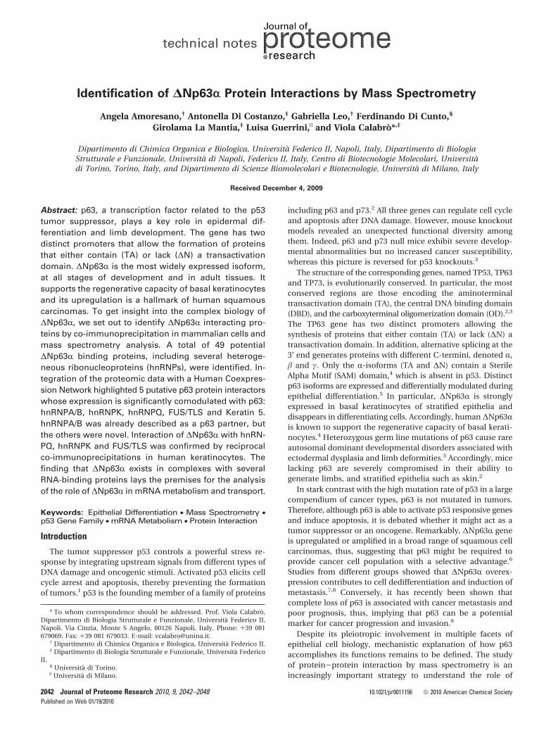
Identification of ΔNp63α Protein Interactions by Mass Spectrometry
7
Identification of ∆Np63r Protein Interactions by Mass Spectrometry Angela Amoresano, † Antonella Di Costanzo, ‡ Gabriella Leo, † Ferdinando Di Cunto, § Girolama La Mantia, ‡ Luisa Guerrini, | and Viola Calabro `* ,‡ Dipartimento di Chimica Organica e Biologica, Universita ` Federico II, Napoli, Italy, Dipartimento di Biologia Strutturale e Funzionale, Universita ` di Napoli, Federico II, Italy, Centro di Biotecnologie Molecolari, Universita ` di Torino, Torino, Italy, and Dipartimento di Scienze Biomolecolari e Biotecnologie, Universita ` di Milano, Italy Received December 4, 2009 Abstract: p63, a transcription factor related to the p53 tumor suppressor, plays a key role in epidermal dif- ferentiation and limb development. The gene has two distinct promoters that allow the formation of proteins that either contain (TA) or lack (∆N) a transactivation domain. ∆Np63R is the most widely expressed isoform, at all stages of development and in adult tissues. It supports the regenerative capacity of basal keratinocytes and its upregulation is a hallmark of human squamous carcinomas. To get insight into the complex biology of ∆Np63R, we set out to identify ∆Np63R interacting pro- teins by co-immunoprecipitation in mammalian cells and mass spectrometry analysis. A total of 49 potential ∆Np63R binding proteins, including several heteroge- neous ribonucleoproteins (hnRNPs), were identified. In- tegration of the proteomic data with a Human Coexpres- sion Network highlighted 5 putative p63 protein interactors whose expression is significantly comodulated with p63: hnRNPA/B, hnRNPK, hnRNPQ, FUS/TLS and Keratin 5. hnRNPA/B was already described as a p63 partner, but the others were novel. Interaction of ∆Np63R with hnRN- PQ, hnRNPK and FUS/TLS was confirmed by reciprocal co-immunoprecipitations in human keratinocytes. The finding that ∆Np63R exists in complexes with several RNA-binding proteins lays the premises for the analysis of the role of ∆Np63R in mRNA metabolism and transport. Keywords: Epithelial Differentiation • Mass Spectrometry • p53 Gene Family • mRNA Metabolism • Protein Interaction Introduction The tumor suppressor p53 controls a powerful stress re- sponse by integrating upstream signals from different types of DNA damage and oncogenic stimuli. Activated p53 elicits cell cycle arrest and apoptosis, thereby preventing the formation of tumors. 1 p53 is the founding member of a family of proteins including p63 and p73. 2 All three genes can regulate cell cycle and apoptosis after DNA damage. However, mouse knockout models revealed an unexpected functional diversity among them. Indeed, p63 and p73 null mice exhibit severe develop- mental abnormalities but no increased cancer susceptibility, whereas this picture is reversed for p53 knockouts. 3 The structure of the corresponding genes, named TP53, TP63 and TP73, is evolutionarily conserved. In particular, the most conserved regions are those encoding the aminoterminal transactivation domain (TA), the central DNA binding domain (DBD), and the carboxyterminal oligomerization domain (OD). 2,3 The TP63 gene has two distinct promoters allowing the synthesis of proteins that either contain (TA) or lack (∆N) a transactivation domain. In addition, alternative splicing at the 3′ end generates proteins with different C-termini, denoted R, and γ. Only the R-isoforms (TA and ∆N) contain a Sterile Alpha Motif (SAM) domain, 4 which is absent in p53. Distinct p63 isoforms are expressed and differentially modulated during epithelial differentiation. 5 In particular, ∆Np63R is strongly expressed in basal keratinocytes of stratified epithelia and disappears in differentiating cells. Accordingly, human ∆Np63R is known to support the regenerative capacity of basal kerati- nocytes. 4 Heterozygous germ line mutations of p63 cause rare autosomal dominant developmental disorders associated with ectodermal dysplasia and limb deformities. 3 Accordingly, mice lacking p63 are severely compromised in their ability to generate limbs, and stratified epithelia such as skin. 2 In stark contrast with the high mutation rate of p53 in a large compendium of cancer types, p63 is not mutated in tumors. Therefore, although p63 is able to activate p53 responsive genes and induce apoptosis, it is debated whether it might act as a tumor suppressor or an oncogene. Remarkably, ∆Np63R gene is upregulated or amplified in a broad range of squamous cell carcinomas, thus, suggesting that p63 might be required to provide cancer cell population with a selective advantage. 6 Studies from different groups showed that ∆Np63R overex- pression contributes to cell dedifferentiation and induction of metastasis. 7,8 Conversely, it has recently been shown that complete loss of p63 is associated with cancer metastasis and poor prognosis, thus, implying that p63 can be a potential marker for cancer progression and invasion. 9 Despite its pleiotropic involvement in multiple facets of epithelial cell biology, mechanistic explanation of how p63 accomplishes its functions remains to be defined. The study of protein-protein interaction by mass spectrometry is an increasingly important strategy to understand the role of * To whom correspondence should be addressed. Prof. Viola Calabro `, Dipartimento di Biologia Strutturale e Funzionale, Universita ` Federico II, Napoli. Via Cinzia, Monte S Angelo, 80126 Napoli, Italy. Phone: +39 081 679069. Fax: +39 081 679033. E-mail: [email protected]. † Dipartimento di Chimica Organica e Biologica, Universita ` Federico II. ‡ Dipartimento di Biologia Strutturale e Funzionale, Universita ` Federico II. § Universita ` di Torino. | Universita ` di Milano. 2042 Journal of Proteome Research 2010, 9, 2042–2048 10.1021/pr9011156 2010 American Chemical Society Published on Web 01/19/2010
Transcript of Identification of ΔNp63α Protein Interactions by Mass Spectrometry
Identification of ∆Np63r Protein Interactions by Mass Spectrometry
Angela Amoresano,† Antonella Di Costanzo,‡ Gabriella Leo,† Ferdinando Di Cunto,§
Girolama La Mantia,‡ Luisa Guerrini,| and Viola Calabro*,‡
Dipartimento di Chimica Organica e Biologica, Universita Federico II, Napoli, Italy, Dipartimento di BiologiaStrutturale e Funzionale, Universita di Napoli, Federico II, Italy, Centro di Biotecnologie Molecolari, Universitadi Torino, Torino, Italy, and Dipartimento di Scienze Biomolecolari e Biotecnologie, Universita di Milano, Italy
Received December 4, 2009
Abstract: p63, a transcription factor related to the p53tumor suppressor, plays a key role in epidermal dif-ferentiation and limb development. The gene has twodistinct promoters that allow the formation of proteinsthat either contain (TA) or lack (∆N) a transactivationdomain. ∆Np63R is the most widely expressed isoform,at all stages of development and in adult tissues. Itsupports the regenerative capacity of basal keratinocytesand its upregulation is a hallmark of human squamouscarcinomas. To get insight into the complex biology of∆Np63R, we set out to identify ∆Np63R interacting pro-teins by co-immunoprecipitation in mammalian cells andmass spectrometry analysis. A total of 49 potential∆Np63R binding proteins, including several heteroge-neous ribonucleoproteins (hnRNPs), were identified. In-tegration of the proteomic data with a Human Coexpres-sion Network highlighted 5 putative p63 protein interactorswhose expression is significantly comodulated with p63:hnRNPA/B, hnRNPK, hnRNPQ, FUS/TLS and Keratin 5.hnRNPA/B was already described as a p63 partner, butthe others were novel. Interaction of ∆Np63R with hnRN-PQ, hnRNPK and FUS/TLS was confirmed by reciprocalco-immunoprecipitations in human keratinocytes. Thefinding that ∆Np63R exists in complexes with severalRNA-binding proteins lays the premises for the analysisof the role of ∆Np63R in mRNA metabolism and transport.
Keywords: Epithelial Differentiation • Mass Spectrometry •p53 Gene Family • mRNA Metabolism • Protein Interaction
Introduction
The tumor suppressor p53 controls a powerful stress re-sponse by integrating upstream signals from different types ofDNA damage and oncogenic stimuli. Activated p53 elicits cellcycle arrest and apoptosis, thereby preventing the formationof tumors.1 p53 is the founding member of a family of proteins
including p63 and p73.2 All three genes can regulate cell cycleand apoptosis after DNA damage. However, mouse knockoutmodels revealed an unexpected functional diversity amongthem. Indeed, p63 and p73 null mice exhibit severe develop-mental abnormalities but no increased cancer susceptibility,whereas this picture is reversed for p53 knockouts.3
The structure of the corresponding genes, named TP53, TP63and TP73, is evolutionarily conserved. In particular, the mostconserved regions are those encoding the aminoterminaltransactivation domain (TA), the central DNA binding domain(DBD), and the carboxyterminal oligomerization domain (OD).2,3
The TP63 gene has two distinct promoters allowing thesynthesis of proteins that either contain (TA) or lack (∆N) atransactivation domain. In addition, alternative splicing at the3′ end generates proteins with different C-termini, denoted R,� and γ. Only the R-isoforms (TA and ∆N) contain a SterileAlpha Motif (SAM) domain,4 which is absent in p53. Distinctp63 isoforms are expressed and differentially modulated duringepithelial differentiation.5 In particular, ∆Np63R is stronglyexpressed in basal keratinocytes of stratified epithelia anddisappears in differentiating cells. Accordingly, human ∆Np63Ris known to support the regenerative capacity of basal kerati-nocytes.4 Heterozygous germ line mutations of p63 cause rareautosomal dominant developmental disorders associated withectodermal dysplasia and limb deformities.3 Accordingly, micelacking p63 are severely compromised in their ability togenerate limbs, and stratified epithelia such as skin.2
In stark contrast with the high mutation rate of p53 in a largecompendium of cancer types, p63 is not mutated in tumors.Therefore, although p63 is able to activate p53 responsive genesand induce apoptosis, it is debated whether it might act as atumor suppressor or an oncogene. Remarkably, ∆Np63R geneis upregulated or amplified in a broad range of squamous cellcarcinomas, thus, suggesting that p63 might be required toprovide cancer cell population with a selective advantage.6
Studies from different groups showed that ∆Np63R overex-pression contributes to cell dedifferentiation and induction ofmetastasis.7,8 Conversely, it has recently been shown thatcomplete loss of p63 is associated with cancer metastasis andpoor prognosis, thus, implying that p63 can be a potentialmarker for cancer progression and invasion.9
Despite its pleiotropic involvement in multiple facets ofepithelial cell biology, mechanistic explanation of how p63accomplishes its functions remains to be defined. The studyof protein-protein interaction by mass spectrometry is anincreasingly important strategy to understand the role of
* To whom correspondence should be addressed. Prof. Viola Calabro,Dipartimento di Biologia Strutturale e Funzionale, Universita Federico II,Napoli. Via Cinzia, Monte S Angelo, 80126 Napoli, Italy. Phone: +39 081679069. Fax: +39 081 679033. E-mail: [email protected].
† Dipartimento di Chimica Organica e Biologica, Universita Federico II.‡ Dipartimento di Biologia Strutturale e Funzionale, Universita Federico
II.§ Universita di Torino.| Universita di Milano.
2042 Journal of Proteome Research 2010, 9, 2042–2048 10.1021/pr9011156 2010 American Chemical SocietyPublished on Web 01/19/2010
multifunctional proteins. To shed light on the role of ∆Np63Rin epithelial biology, we set out to identify ∆Np63R bindingproteins by co-immunoprecipitation in H1299 cells followedby mass spectrometry analysis. We identified 49 putative∆Np63R molecular partners, including several hnRNPs. Thefinding that ∆Np63R associates with multiple RNA-bindingproteins suggests that it may play a relevant role in RNAmetabolism processes.
Materials and Methods
Cell Culture and Transfections. H1299 cells derived fromhuman lung carcinoma were provided by ATCC (CRL-5803).Cells were grown at 37 °C and 5% (v/v) CO2 in Dulbecco’smodified Eagle medium supplemented with 10% fetal bovineserum (Euroclone). HaCaT cells were maintained in RPMImedium and 10% fetal calf serum. H1299 cells were seeded ata density of about 70% confluence and transfected with 0.2 µgof plasmid encoding myc∆Np63R using LipofectAMINE 2000(Life Technologies, Inc.).
Immunoaffinity Purification of p63-Associated Proteins.p63 complexes were affinity purified from 10 mg of totalextracts prepared from Myc∆Np63R transfected H1299 cells.At 24 h after transfection, cells were washed with PBS and lysedin 50 mM Tris/HCl, pH 7.5, 150 mM NaCl, 0.5% NP40, 5 mMEDTA, 10% glycerol, and protease inhibitors (Sigma). Totalprotein in cell extracts was quantified using the BioRad proteinassay and incubated with mouse anti-IgG agarose conjugatedbeads (Sigma) overnight (o.n.) at 4 °C. Beads were pretreatedwith 5% nonfat milk. After clarification, extracts were centri-fuged at 3000 rpm for 5 min at 4 °C. Cell extracts were thenincubated o.n. with immobilized anti-Myc 9E10 antibody (SantaCruz). The beads were washed extensively with lysis buffer (150mM NaCl, 10% glycerol, and 50 mM Tris/HCl, pH 7.5). Controlsamples were prepared in parallel using untransfected cellextracts. Protein samples were eluted with Myc competitorpeptide (200 µg/mL in BC100), TCA precipitated, resuspendedin 2× loading buffer (Sigma) and loaded on a 12.5% SDSpolyacrylamide gel. The gel was run for 1 h in Tris-Glycinebuffer at 25 mA and stained with colloidal Coomassie blue(Pierce).
Western Blotting Analysis. Each step of the experimentalprocedure was paralleled by Western blotting step that servedas control. Samples aliquots were resuspended in 2× loadingbuffer (Sigma) and loaded in a SDS-10% polyacrylamide gel.The gel was run in 10× Tris-Glycine buffer at 200 V, andtransferred to a PVDF membrane (Hybond-P, AmershamBiosciences). The membrane was blocked with 4% nonfat milkin TBS (25 mM Tris, pH 7.4, and 125 mM NaCl) for 1 h at roomtemperature, washed 3 times with TBS and then incubated withmouse anti-Myc horseradish peroxidase-conjugated secondaryantibodies (Alexis Biochemicals) Protein were revealed byenhanced chemiluminescence (RPN2132; Amersham, Buck-inghamshire, U.K.).
In Situ Digestion and MALDI Analysis. Trypsin, dithiothrei-tol (DTT), iodoacetamide and R-cyano-4-hydroxycinnamic acidwere purchased from Sigma. NH4HCO3 was from Fluka. Trif-luoroacetic acid (TFA)-HPLC grade was from Carlo Erba. Allother reagents and solvents were of the highest purity availablefrom Baker.
Slices containing protein bands were excised from the geland destained by repetitive washes with 0.1 M NH4HCO3, pH7.5, and acetonitrile. Samples were reduced by incubation with50 µL of 10 mM DTT in 0.1 M NH4HCO3 buffer, pH 7.5, and
carboxyamidomethylated with 50 µL of 55 mM iodoacetamidein the same buffer. Enzymatic digestion was carried out withtrypsin (12.5 ng/µL) in 10 mM ammonium bicarbonate buffer,pH 7.8. Gel pieces were incubated at 4 °C for 2 h. Trypsinsolution was then removed and a new aliquot of the samesolution was added; samples were incubated for 18 h at 37 °C.A minimum reaction volume was used as to obtain thecomplete rehydratation of the gel. Peptides were then extractedby washing the gel particles with 10 mM ammonium bicarbon-ate and 1% formic acid in 50% acetonitrile at room tempera-ture. The resulting peptide mixtures were desalted using ZipTippipettes from Millipore, following the recommended purifica-tion procedure.
MALDI-TOF mass spectra were recorded using an AppliedBiosystem Voyager STR instrument equipped with a nitrogenlaser (337 nm). One microliter of the analyte mixture was mixed(1/1, v/v) with a 10 mg/mL solution of R-cyano-hydroxycin-namic acid in acetonitrile/50 mM citrate buffer (2/3, v/v) andwas applied to the metallic sample plate and dried down atroom temperature. Acceleration and reflector voltage were setup as follows: target voltage at 20 kV, grid at 66% of targetvoltage, delayed extraction at 150 ns to obtain the best signalto-noise ratios and the best possible isotopic resolution. Masscalibration was performed using external peptide standardspurchased from Applied Biosystems. Raw data were analyzedusing the computer software provided by the manufacturer asmonoisotopic masses.
NanoHPLC-chip MS/MS Analysis. LC/MS/MS analyses wereperformed on a LC/MSD Trap XCT Ultra (Agilent Technologies,Palo Alto, CA) equipped with a 1100 HPLC system and a chipcube (Agilent Technologies). After loading, the peptide mixturewas first concentrated and washed in 40 nL enrichment column(Agilent Technologies chip), with 0.2% formic acid in 2%acetonitrile as the eluent. The sample was then fractionatedon a C18 reverse-phase capillary column (Agilent Technologieschip) at flow rate of 300 nL/min, with a linear gradient of eluentB (0.2% formic acid in 95% acetonitrile) in A (0.2% formic acidin 2% acetonitrile) from 7 to 60% in 50 min. Peptide analysiswas performed using data-dependent acquisition of one MSscan (mass range from 300 to 1800 m/z) followed by MS/MSscans of the three most abundant ions in each MS scan.Dynamic exclusion was used to acquire a more complete surveyof the peptides by automatic recognition and temporaryexclusion (2 min) of ions from which definitive mass spectraldata was previously acquired. Nitrogen at a flow rate of 3 L/minand heated to 325 °C was used as the dry gas for spraydesolvation. MS/MS spectra were measured automaticallywhen the MS signal surpassed the threshold of 50 000 counts.Double charged ions were preferably isolated and fragmentedover single charged ions. The acquired MS/MS spectra weretransformed in Mascot generic file format and used for peptidesidentification with a licensed version of MASCOT, in a localdatabase.
Protein Identification. Spectral data were analyzed usingAnalyst software (version 1.4.1) and MSMS centroid peak listswere generated using the MASCOT.dll script (version 1.6b9).MSMS centroid peaks were threshold at 0.1% of the base peak.MSMS spectra having less than 10 peaks were rejected. MSMSspectra were searched against NCBInr database (2009/07/10version, 211 309 entries) and then converted in Swiss-proteincode, using the licensed version of Mascot 2.1 (Matrix Science),after converting the acquired MSMS spectra in mascot genericfile format. The Mascot search parameters were taxonomy
Identification of p63 Interacting Partners technical notes
Journal of Proteome Research • Vol. 9, No. 4, 2010 2043

human; “trypsin” as enzyme allowing up to 3 missed cleavages,carbamidomethyl on as fixed modification, oxidation of M,pyroGlu N-term Q, as variable modifications, 600 ppm MSMStolerance and 0.6 Da peptide tolerance and top 20 proteinentries. Spectra with a MASCOT score <25 having low qualitywere rejected. The score used to evaluate quality of matchesfor MSMS data was higher than 30.
Comparison with IntAct Data. Data from mass spectrometryanalysis were compared with those provided by IntAct database(htpp://www.ebi.ac.uk/intact/search/do/search?searchString){0})linked in the corresponding Swiss-Prot entries (htpp://www.expasy.org/sprot/). Each protein candidate was submitted to the IntActdatabank obtaining a list of all proteins involved in interactionwith the query protein.
HierarchView (htpp://www.ebi.ac.uk/intact/hierarchView/display.do) was used to construct a graphical representationof the interaction network centered on each protein of interestthat had been used as bait in pull-down experiments. IntActdatabase was also searched for selecting proteins belonging tothe functionally different complexes recruited by our bait toobtain graph view of the interactions between these complexes.
Coexpression Analysis. The list of human genes whoseexpression is comodulated with p63 was derived essentially asdescribed.10 Briefly, we collected 353 human normal tissueAffymetrix microarray experiments, corresponding to 65 dif-ferent tissues.11
We then calculated the Pearson correlation coefficients ofevery probe set (r1) in the expression matrix with all other probesets (rx), and ranked the probe sets according to the values ofthis index. A directed edge was established from r1 to rx if rx
fell within the top 1% rows in terms of correlation with r1.These directed networks where then converted into undi-
rected networks by mapping the probe sets to the correspond-ing Entrez Gene identifiers. The list of genes whose expressionis strictly comodulated with p63 is defined as the ensemble ofthe first-level links of p63 in this network.
Co-Immunoprecipitation. HaCaT cells from subconfluent100 mm plates were collected for preparation of whole-cellextracts as previously described.12 Lysates containing 1 mg ofproteins were precleared with 30 µL of protein A-agarose (50%slurry; Roche) and then incubated overnight at 4 °C with 3 µgof anti-p63 monoclonal antibody (4A4; Santa Cruz), anti-hnRNPK (H-300: sc-25373 Santa Cruz), anti-hnRNPQ (I8E4; sc-56703 Santa Cruz), or anti-FUS (A300-302A Bethyl Laborato-ries) antibodies. Anti-GFP antibodies (Roche) were used ascontrol of specificity. Anti-hnRNPA/B (H-200: sc-15385 SantaCruz) antibodies were used as control of positive interaction.The immunocomplexes were collected by incubating withprotein A agarose at 4 °C for 4 h. Beads were washed vigorouslywith 50 mM Tris-HCl, pH 7.5, 150 mN NaCl, 5 mM EDTA, 0.5%NP40, and 10% glycerol, resuspended in 4× SDS-PAGE loadingbuffer and loaded directly in an 8% SDS-polyacrylamide gel.
Results
Identification of Proteins Specifically Interacting withthe Bait. The proteins specifically interacting with p63 weredefined by functional proteomics using transfected Myc-tagged∆Np63R protein as bait. Two biological replicates were per-formed using different cellular extracts. Briefly, total proteinextract (10 mg) from ∆Np63R transfected H1299 cells wasprecleared with underivatized agarose beads to minimizenonspecific binding. The unbound material was immunopre-cipitated with polyclonal anti-Myc IgG conjugated agarose
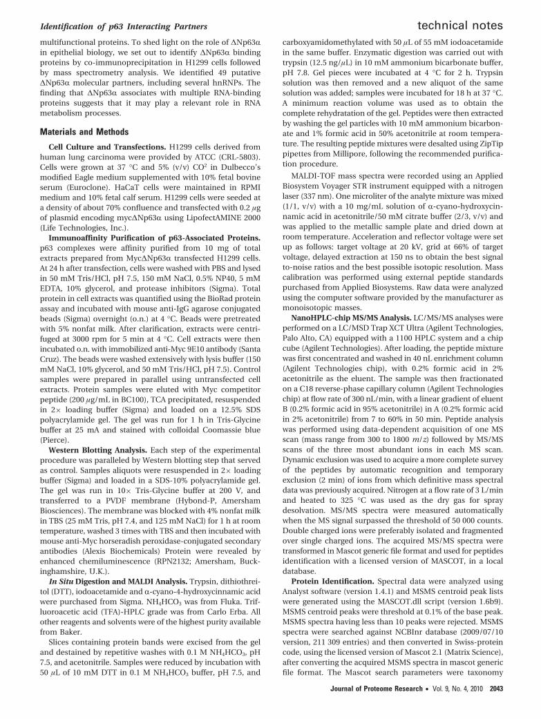
beads. The proteins bound to the affinity column were exten-sively washed and eluted with two different concentration ofMyc peptide solution (Figure 1A, sample I and sample II).Control experiments were carried out, in parallel, using extractsfrom untransfected H1299 cells. Following each step of theprocedure, Western blotting was performed on a sample aliquotusing anti-Myc antibodies as a control (Figure 1A). The proteinmixture from the first myc elution was subjected to SDS-PAGEand visualized with Coomassie Blue staining (Figure 1B).
Figure 1. Western blot analysis of ∆Np63R during the affinitypurification procedure. (A)Western blotting of extracts from Myc-∆Np63R transfected H1299 cells (sample). Untransfected H1299cell extracts were used as control. Aliquots (40 µg) correspondingto each step of the experimental procedure were subjected toWestern blotting and revealed with anti-Myc and peroxidase-conjugated secondary antibodies. Whole cell extract (lane 1).Unbound control (lane 6) and sample (lane 7) after precleaningwith underivatised agarose beads. Elution with myc-peptidecompetitor, control (lanes 2 and 4), sample (lanes 3 and 5). (B)SDS-PAGE fractionation of ∆Np63R protein complexes. ∆Np63Rprotein complexes were isolated by immunoprecipitation experi-ment. Following SDS-PAGE fractionation, Coomassie Blue stainedprotein bands occurring in the sample (lane 5) and in the control(lane 4) were submitted to mass spectral analyses. Correspond-ing gel slices from the sample and control lanes (slices 1-25)were submitted to the identification procedure. Common proteinsidentified in both the sample and the control gel slices wereeliminated. Molecular weight markers are also shown (lane 1).
technical notes Amoresano et al.
2044 Journal of Proteome Research • Vol. 9, No. 4, 2010
Stained gel displayed a number of discrete bands both in thecontrol and sample lanes (lanes 4 and 5, respectively). Becauseof the complexity of the gel patterns and the low resolution of1D electrophoresis, several proteins can occur in the same gelband. Therefore, protein bands specifically present in thesample lane and absent in the control lane cannot be identifiedby simply comparing the two gel profiles. Thus, the entire lanesfrom the sample and the control were cut in 25 slices each andthe slices were destained and trypsin digested, in situ. Theresulting peptide mixtures were extracted from the gel, testedby MALDI-MS, and analyzed by nanoLC/MS/MS analysisgenerating sequence information on individual peptides.
This information, together with the peptide mass values, wasthen used to search protein databases, leading to the identi-fication of the protein components. All the gel bands from thesample and control lanes were then compared on the basis ofthe proteins effectively identified in each gel slice. Commonproteins identified both in the control and the sample sliceswere discarded and only those proteins solely identified in thesample slices and absent in the control were considered. Asfurther selection criteria, only the proteins, identified byMASCOT search with at least 3 peptides, and found exclusivelyin both biological replicates were selected, providing a full listof putative ∆Np63R interactors.13 As an example, slice 11 fromthe sample lanes, in Figure 1B, revealed the presence of 9proteins identified with at least 3 peptides. However, six ofthese putative interactors were also found in the correspondingslices from the control lanes and were then discarded. Theremaining three proteins fulfilled the selection criteria and werethen considered as putative p63 interactors.
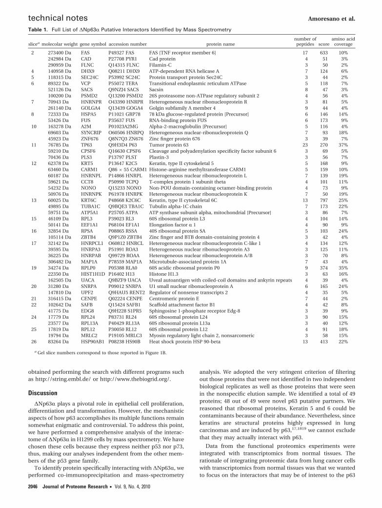
This strategy let us to obtain more confident identificationof truly ∆Np63R binding candidates minimizing the numberof false positives.14 It should be noted that some proteinsshowed a higher electrophoretic mobility, with respect to theircorresponding molecular weight. This finding could be due tosome proteolytic processing. On the contrary, other proteincandidates exhibited a molecular mass lower than expected onthe basis of the electrophoretic mobility, likely due to theoccurrence of post-translational modifications. A total of 49putative p63-interacting proteins were identified and listed inTable 1 together with their corresponding genes and thenumber of peptides used for their identification.
Database Analysis of Protein-Protein Interactions. Toassess whether the proteins identified by the proteomic screen-ing were novel p63 interacting partners, we extensively searchedthe literature and protein interaction databases for knowninteractors. Interestingly, hnRNPA/B (ABBP1) was alreadydescribed as a p63 direct interactor.15
To define the functional relationships between the putativep63 interactors, we classified them according to the GeneOntology annotation and analyzed if some of the GO keywordswere significantly enriched. Six distinct functional clusters wererepresented: transcription, mRNA processing, mitosis andprotein folding factors, ribosomal and structural proteins(Figure 2). Statistical analysis revealed that proteins involvedin RNA-binding and mRNA processing were significantlyenriched in our list of candidates (GO keywords: RNA binding,1.8 × 10-13; mRNA processing, 1.9 × 10⁻⁵).
Integration of the Proteomic Screening with Coexpres-sion Analysis. Integration of proteomic and transcriptomic datacan be of great help to identify functionally relevant protein-protein interactions.16
To increase the likelihood of selecting the most functionallysignificant interactors, among the p63-binders, we searchedmicroarray data for those genes having an expression profilevery similar to p63. To address this question, we extracted alist of genes highly comodulated with p63 (Supporting Infor-mation) from a recently described human coexpression net-work obtained from a large meta analysis of normal tissuesmicroarray data.10 Although it is conceivable that the proteinexpression profiles in cancer cells could be different fromnormal cells, we reasoned that, in tumors, p63 should still retainfunctionally relevant interactions with proteins that are nor-mally coexpressed. Moreover, integration of proteomic datafrom lung cancer cells with transcriptomics from normal tissuesshould lead to the identification of interactors that may be ofinterest to the p63 functions, far beyond the particular cell lineused for the proteomic screening.
Interestingly, five proteins included in our list of p63 putativepartners, Keratin 5, hnRNPA/B, hnRNPK, hnRNPQ (Syncrip)and FUS/TLS, are also significantly comodulated with p63, inhuman tissues. Keratin 5 is a structural protein; since it is highlyexpressed in squamous cell lung carcinomas,,17,18 19 it mightlikely represent a false positive.
FUS, hnRNPQ, hnRNPK, and hnRNPA/B (ABBP1) are all RNAbinding proteins involved in pre-mRNA processing, mRNAexport and turnover mechanisms.20 Since hnRNPA/B wasalready validated as a p63 interactor,15 we decided to furthervalidate the interaction of p63 with hnRNPK, hnRNPQ andFUS/TLS.
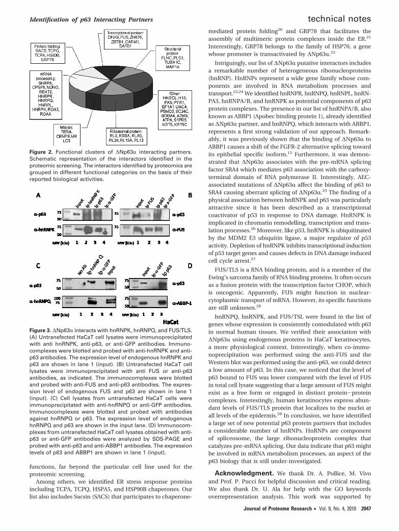
Confirmation of ∆Np63r Association with FUS/TLS,hnRNPQ, and hnRNPK. To validate the interaction of ∆Np63Rwith FUS/TLS, hnRNPK, or hnRNPQ, we performed co-immu-noprecipitation assays in HaCaT human keratinocytes, a morephysiological environment. First, we verified that hnRNPK,hnRNPQ, and FUS proteins are endogenously expressed inHaCaT cells by Western blot (data not shown). Then, weimmunoprecipitated endogenous ∆Np63R, from HaCaT cells,with the 4A4 antibody. Specific immunodetection showed thatFUS, hnRNPK, and hnRNPQ were present in p63 complexes(Figure 3A,B and data not shown). Conversely, p63 was specif-ically pulled down by antibodies against hnRNPQ, hnRNPK,or FUS (Figure 3C and data not shown). Interestingly, theamount of associated p63 was much higher for hnRNPK thanfor FUS or hnRNPQ, indicating that the fraction of FUS orhnRNPQ associated with p63 was a minor pool of the total FUSand hnRNPQ proteins. Neither FUS nor hnRNP proteins wereimmunoprecipitated using anti-GFP antibodies (Figure 3).Immunoprecipitation with antibodies against ABBP1 was per-formed as control of positive interaction (Figure 3D). Theseresults, together with the GO keyword overrepresentationanalysis, indicate that p63 might play a role in association withcomponents of ribonuclear protein complexes.
IntAct Data. Finally, we compared our proteomic results withthose provided by IntAct database (see Materials and Methods).Each protein candidate was submitted to the IntAct databankobtaining a list of all proteins involved in interaction with thequery protein. HierarchView (htpp://www.ebi.ac.uk/intact/hierarchView/display.do) was used to construct a graphicalrepresentation of the interaction network centered on eachprotein of interest. IntAct database was also searched forselecting proteins belonging to functionally different complexesrecruited by our bait. Total interaction graph view is reported(Supporting Information), where the putative interactors iden-tified by our MS approach are evidenced. Similar results were
Identification of p63 Interacting Partners technical notes
Journal of Proteome Research • Vol. 9, No. 4, 2010 2045
obtained performing the search with different programs suchas http://string.embl.de/ or http://www.thebiogrid.org/.
Discussion
∆Np63R plays a pivotal role in epithelial cell proliferation,differentiation and transformation. However, the mechanisticaspects of how p63 accomplishes its multiple functions remainsomewhat enigmatic and controversial. To address this point,we have performed a comprehensive analysis of the interac-tome of ∆Np63R in H1299 cells by mass spectrometry. We havechosen these cells because they express neither p53 nor p73,thus, making our analyses independent from the other mem-bers of the p53 gene family.
To identify protein specifically interacting with ∆Np63R, weperformed co-immunoprecipitation and mass-spectrometry
analysis. We adopted the very stringent criterion of filteringout those proteins that were not identified in two independentbiological replicates as well as those proteins that were seenin the nonspecific elution sample. We identified a total of 49proteins; 48 out of 49 were novel p63 putative partners. Wereasoned that ribosomal proteins, Keratin 5 and 6 could becontaminants because of their abundance. Nevertheless, sincekeratins are structural proteins highly expressed in lungcarcinomas and are induced by p63,17,1819 we cannot excludethat they may actually interact with p63.
Data from the functional proteomics experiments wereintegrated with transcriptomics from normal tissues. Therationale of integrating proteomic data from lung cancer cellswith transcriptomics from normal tissues was that we wantedto focus on the interactors that may be of interest to the p63
Table 1. Full List of ∆Np63R Putative Interactors Identified by Mass Spectrometry
slicea molecolar weight gene symbol accession number protein namenumber of
peptides scoreamino acid
coverage
2 273400 Da FAS P49327 FAS FAS [TNF receptor member 6] 17 633 10%242984 Da CAD P27708 PYR1 Cad protein 4 51 3%290959 Da FLNC Q14315 FLNC Filamin-C 3 50 2%
4 140958 Da DHX9 Q08211 DHX9 ATP-dependent RNA helicase A 7 124 6%5 118315 Da SEC24C P53992 SC24C Protein transport protein Sec24C 3 44 2%6 89322 Da VCP P55072 TERA Transitional endoplasmic reticulum ATPase 5 118 7%
521126 Da SACS Q9NZJ4 SACS Sacsin 8 47 3%100200 Da PSMD2 Q13200 PSMD2 26S proteasome non-ATPase regulatory subunit 2 4 56 4%
7 70943 Da HNRNPR O43390 HNRPR Heterogeneous nuclear ribonucleoprotein R 3 81 5%261140 Da GOLGA4 Q13439 GOGA4 Golgin subfamily A member 4 9 44 4%
8 72333 Da HSPA5 P11021 GRP78 78 kDa glucose-regulated protein [Precursor] 6 146 14%53426 Da FUS P35637 FUS RNA-binding protein FUS 6 173 9%
10 163278 Da A2M P01023A2MG Alpha-2-macroglobulin [Precursor] 5 116 4%69603 Da SYNCRIP O60506 HNRPQ Heterogeneous nuclear-ribonucleoprotein Q 7 93 18%45923 Da ZNF676 Q8N7Q3 ZN676 Zinc finger protein 676 3 39 7%
11 76785 Da TP63 Q9H3D4 P63 Tumor protein 63 23 270 37%59210 Da CPSF6 Q16630 CPSF6 Cleavage and polyadenylation specificity factor subunit 6 3 69 5%70436 Da PLS3 P13797 PLST Plastin-3 3 56 7%
12 62378 Da KRT5 P13647 K2C5 Keratin, type II cytoskeletal 5 5 168 9%63460 Da CARM1 Q86 × 55 CARM1 Histone-arginine methyltransferase CARM1 5 159 10%60187 Da HNRNPL P14866 HNRPL Heterogeneous nuclear ribonucleoprotein L 7 139 19%59621 Da CCT8 P50990 TCPQ T-complex protein 1 subunit theta 4 101 11%54232 Da NONO Q15233 NONO Non-POU domain-containing octamer-binding protein 4 73 9%50976 Da HNRNPK P61978 HNRPK Heterogeneous nuclear ribonucleoprotein K 7 50 19%
13 60025 Da KRT6C P48668 K2C6C Keratin, type II cytoskeletal 6C 13 797 25%49895 Da TUBA1C Q9BQE3 TBA1C Tubulin alpha-1C chain 7 173 22%59751 Da ATP5A1 P25705 ATPA ATP synthase subunit alpha, mitochondrial [Precursor] 3 86 7%
15 46109 Da RPL3 P39023 RL3 60S ribosomal protein L3 4 104 14%50141 Da EEF1A1 P68104 EF1A1 Elongation factor R 1 4 90 9%
16 32854 Da RPSA P08865 RSSA 40S ribosomal protein SA 5 103 24%105114 Da ZBTB4 Q9P1Z0 ZBTB4 Zinc finger and BTB domain-containing protein 4 3 42 4%
17 32142 Da HNRPCL1 O60812 HNRCL Heterogeneous nuclear ribonucleoprotein C-like 1 4 134 12%39595 Da HNRPA3 P51991 ROA3 Heterogeneous nuclear ribonucleoprotein A3 3 125 11%36225 Da HNRPAB Q99729 ROAA Heterogeneous nuclear ribonucleoprotein A/B 3 70 8%306482 Da MAP1A P78559 MAP1A Microtubule-associated protein 1A 7 43 4%
19 34274 Da RPLP0 P05388 RLA0 60S acidic ribosomal protein P0 9 374 35%22350 Da HIST1H1D P16402 H13 Histone H1.3 3 63 16%162505 Da UACA Q9BZF9 UACA Uveal autoantigen with coiled-coil domains and ankyrin repeats 4 39 4%
20 31280 Da SNRPA P09012 SNRPA U1 small nuclear ribonucleoprotein A 6 165 24%147810 Da UPF2 Q9HAU5 RENT2 Regulator of nonsense transcripts 2 4 35 5%
21 316415 Da CENPE Q02224 CENPE Centromeric protein E 7 44 2%22 102642 Da SAFB Q15424 SAFB1 Scaffold attachment factor B1 4 42 8%
41775 Da EDG8 Q9H228 S1PR5 Sphingosine 1-phosphate receptor Edg-8 3 39 9%24 17779 Da RPL24 P83731 RL24 60S ribosomal protein L24 3 90 15%
23577 Da RPL13A P40429 RL13A 60S ribosomal protein L13a 3 40 12%25 17819 Da RPL12 P30050 RL12 60S ribosomal protein L12 4 91 18%
19794 Da MRLC2 P19105 MRLC3 Myosin regulatory light chain 2, nonsarcomeric 3 58 15%26 83264 Da HSP90AB1 P08238 HS90B Heat shock protein HSP 90-beta 13 413 22%
a Gel slice numbers correspond to those reported in Figure 1B.
technical notes Amoresano et al.
2046 Journal of Proteome Research • Vol. 9, No. 4, 2010
functions, far beyond the particular cell line used for theproteomic screening.
Among others, we identified ER stress response proteinsincluding TCPA, TCPQ, HSPA5, and HSP90B chaperones. Ourlist also includes Sacsin (SACS) that participates to chaperone-
mediated protein folding20 and GRP78 that facilitates theassembly of multimeric protein complexes inside the ER.21
Interestingly, GRP78 belongs to the family of HSP70, a genewhose promoter is transactivated by ∆Np63R.22
Intriguingly, our list of ∆Np63R putative interactors includesa remarkable number of heterogeneous ribonucleoproteins(hnRNP). HnRNPs represent a wide gene family whose com-ponents are involved in RNA metabolism processes andtransport.23,24 We identified hnRNPR, hnRNPQ, hnRNPL, hnRN-PA3, hnRNPA/B, and hnRNPK as potential components of p63protein complexes. The presence in our list of hnRNPA/B, alsoknown as ABBP1 (Apobec binding protein 1), already identifiedas ∆Np63R partner, and hnRNPQ, which interacts with ABBP1,represents a first strong validation of our approach. Remark-ably, it was previously shown that the binding of ∆Np63R toABBP1 causes a shift of the FGFR-2 alternative splicing towardits epithelial specific isoform.15 Furthermore, it was demon-strated that ∆Np63R associates with the pre-mRNA splicingfactor SRA4 which mediates p63 association with the carboxy-terminal domain of RNA polymerase II. Interestingly, AEC-associated mutations of ∆Np63R affect the binding of p63 toSRA4 causing aberrant splicing of ∆Np63R.25 The finding of aphysical association between hnRNPK and p63 was particularlyattractive since it has been described as a transcriptionalcoactivator of p53 in response to DNA damage. HnRNPK isimplicated in chromatin remodelling, transcription and trans-lation processes.26 Moreover, like p53, hnRNPK is ubiquitinatedby the MDM2 E3 ubiquitin ligase, a major regulator of p53activity. Depletion of hnRNPK inhibits transcriptional inductionof p53 target genes and causes defects in DNA damage inducedcell cycle arrest.27
FUS/TLS is a RNA binding protein, and is a member of theEwing’s sarcoma family of RNA binding proteins. It often occursas a fusion protein with the transcription factor CHOP, whichis oncogenic. Apparently, FUS might function in nuclear-cytoplasmic transport of mRNA. However, its specific functionsare still unknown.28
hnRNPQ, hnRNPK, and FUS/TSL were found in the list ofgenes whose expression is consistently comodulated with p63in normal human tissues. We verified their association with∆Np63R using endogenous proteins in HaCaT keratinocytes,a more physiological context. Interestingly, when co-immu-noprecipitation was performed using the anti-FUS and theWestern blot was performed using the anti-p63, we could detecta low amount of p63. In this case, we noticed that the level ofp63 bound to FUS was lower compared with the level of FUSin total cell lysate suggesting that a large amount of FUS mightexist as a free form or engaged in distinct protein-proteincomplexes. Interestingly, human keratinocytes express abun-dant levels of FUS/TLS protein that localizes to the nuclei atall levels of the epidermis.28 In conclusion, we have identifieda large set of new potential p63 protein partners that includesa considerable number of hnRNPs. HnRNPs are componentof spliceosome, the large ribonucleoprotein complex thatcatalyzes pre-mRNA splicing. Our data indicate that p63 mightbe involved in mRNA metabolism processes, an aspect of thep63 biology that is still under-investigated.
Acknowledgment. We thank Dr. A. Pollice, M. Vivoand Prof. P. Pucci for helpful discussion and critical reading.We also thank Dr. U. Ala for help with the GO keywordsoverrepresentation analysis. This work was supported by
Figure 2. Functional clusters of ∆Np63R interacting partners.Schematic representation of the interactors identified in theproteomic screening. The interactors identified by proteomics aregrouped in different functional categories on the basis of theirreported biological activities.
Figure 3. ∆Np63R interacts with hnRNPK, hnRNPQ, and FUS/TLS.(A) Untransfected HaCaT cell lysates were immunoprecipitatedwith anti hnRNPK, anti-p63, or anti-GFP antibodies. Immuno-complexes were blotted and probed with anti-hnRNPK and anti-p63 antibodies. The expression level of endogenous hnRNPK andp63 are shown in lane 1 (input). (B) Untransfected HaCaT celllysates were immunoprecipitated with anti FUS or anti-p63antibodies, as indicated. The immunocomplexes were blottedand probed with anti-FUS and anti-p63 antibodies. The expres-sion level of endogenous FUS and p63 are shown in lane 1(input). (C) Cell lysates from untransfected HaCaT cells wereimmunoprecipitated with anti-hnRNPQ or anti-GFP antibodies.Immunocomplexes were blotted and probed with antibodiesagainst hnRNPQ or p63. The expression level of endogenoushnRNPQ and p63 are shown in the input lane. (D) Immunocom-plexes from untransfected HaCaT cell lysates obtained with anti-p63 or anti-GFP antibodies were analyzed by SDS-PAGE andprobed with anti-p63 and anti-ABBP1 antibodies. The expressionlevels of p63 and ABBP1 are shown in lane 1 (input).
Identification of p63 Interacting Partners technical notes
Journal of Proteome Research • Vol. 9, No. 4, 2010 2047

AIRC and MIUR to G. La Mantia, MIUR to A. Amoresanoand Telethon GGP05056 to L. Guerrini.
Supporting Information Available: SupplementaryFigure, interactome of ∆Np63R protein partners showing theentire network of interacting proteins involving ∆Np63R, asavailable in the web by IntAct database. Supplementary Table1, list of genes highly comodulated with p63 extracted from ahuman coexpression network obtained from a large metaanalysis of normal tissues microarray data. This material isavailable free of charge via the Internet at http://pubs.acs.org.
References(1) Pietsch, E. C.; Sykes, S. M.; McMahon, S. B.; Murphy, M. E. The
p53 family and programmed cell death. Oncogene 2008, 27, 6507–6521.
(2) Levrero, M.; De Laurenzi, V.; Costanzo, A.; Gong, J.; Wang, J. Y.;Melino, G. The p53/p63/p73 family of transcription factors:overlapping and distinct functions. J. Cell Sci. 2000, 113, 1661–1670.
(3) Yang, A.; Schweitzer, R.; Sun, D.; Kaghad, M.; Walker, N.; Bronson,R. T.; Tabin, C.; Sharpe, A.; Caput, D.; Crum, C.; McKeon, F. p63is essential for regenerative proliferation in limb, craniofacial andepithelial development. Nature 1999, 398, 714–718.
(4) Chi, S. W.; Ayed, A.; Arrowsmith, C. H. Solution structure of aconserved C-terminal domain of p73 with structural homology tothe SAM domain. EMBO J. 1999, 18, 4438–4445.
(5) Di Costanzo, A.; Festa, L.; Duverger, O.; Vivo, M.; Guerrini, L.; LaMantia, G.; Morasso, M. I.; Calabro, V. Homeodomain protein Dlx3induces phosphorylation-dependent p63 degradation. Cell Cycle2009, 8, 1185–1195.
(6) Parsa, R.; Yang, A.; McKeon, F. J. Association of p63 with prolifera-tive potential in normal and neoplastic human keratinocytes.Invest. Dermatol. 1999, 113, 1099–105.
(7) Urist, M. J.; Di Como, C. J.; Lu, M. L.; Charytonowicz, E.; Verbel,D.; Crum, C. P.; Ince, T. A.; McKeon, F. D.; Cordon-Cardo, C. Lossof p63 expression is associated with tumor progression in bladdercancer. Am. J. Pathol. 2002, 161, 1199–206.
(8) Koga, F.; Kawakami, S.; Fujii, Y.; Saito, K.; Ohtsuka, Y.; Iwai, A.;Ando, N.; Takizawa, T.; Kageyama, Y.; Kihara, K. Impaired p63expression associates with poor prognosis and uroplakin IIIexpression in invasive urothelial carcinoma of the bladder. Clin.Cancer Res. 2003, 15, 5501–7.
(9) Adorno, A.; Cordenonsi, M.; Montagner, M.; Dupont, S.; Wong,C.; Hann, B.; Solari, A.; Bobisse, S.; Rondina, M. B.; Guzzardo, V.;Parenti, A. R.; Rosato, A.; Bicciato, S.; Balmain, A.; Piccolo, S. Amutant p53/Smad complex opposes p63 to empowered TGB�induced metastasis. Cell 2009, 137, 87–98.
(10) Ala, U.; Piro, R. M.; Grassi, E.; Damasco, C.; Silengo, L.; Oti, M.;Provero, P.; Di Cunto, F. Prediction of human diseases genes byhuman-mouse conserved coexpression analysis. PLoS Comput.Biol. 2008, 28, 37–43.
(11) Roth, R. B.; Hevezi, P.; Lee, J.; Willhite, D.; Lechner, S. M.; Foster,A. C.; Zlotnik, A. Gene expression analyses reveal molecularrelationship among 20 regions of the human CNS. Neurogenetics2006, 7, 67–80.
(12) Rossi, M.; De Simone, M.; Pollice, A.; Santoro, R.; La Mantia, G.;Guerrini, L.; Calabro, V. Itch/AIP4 associates with and promotesp63 protein degradation. Cell Cycle 2006, 5, 1816–1822.
(13) Kocher, T.; Superti-Furga, G. Mass spectrometry-based functionalproteomics: from molecular machines to protein networks. Nat.Methods 2007, 4, 807–815.
(14) Monti, M.; Orru, S.; Pagnozzi, D.; Pucci, P. Interaction proteomics.Biosci. Rep. 2005, 25, 45–56.
(15) Fomenkov, A.; Huang, Y. P.; Topaloglu, O.; Brechman, A.; Osada,M.; Fomenkova, T.; Yuriditsky, E.; Trink, B.; Sidransky, D.; Rato-vitski, E. p63R mutations lead to aberrant splicing of keratinocytegrowth factor receptor in the Hay-Wells syndrome. J. Biol. Chem.2003, 278, 23906–23914.
(16) Lemeer, S.; Pinkse, M. W.; Mohammed, S.; van Breukelen, B.; denHertog, J.; Slijper, M.; Heck, A. J. On line automated in vivozebrafish phosphoproteomics. J. Proteome Res. 2008, 7, 1555–1564.
(17) Camilo, R.; Capelozzi, V. L.; Siqueira, S. A.; Del Carlo Bernardi, F.Expression of p63, keratin 5/6, keratin 7, and surfactant-A in non-small cell lung carcinomas. Hum. Pathol. 2006, 37, 542–546.
(18) Boldrup, L.; Coates, P. J.; Gu, X.; Nylander, K. DeltaNp63 isoformsregulate CD44 and keratins 4, 5/6, 14 and 19 in squamous cellcarcinoma of head and neck. J. Pathol. 2007, 213, 384–391.
(19) Romano, R. A.; Birkaya, B.; Sinha, S. A functional enhancer ofkeratin 14 is a direct transcriptional target of ∆Np63. J. Invest.Dermatol. 2008, 127, 1175–1186.
(20) Takiyama, Y. Sacsinopathies: sacsin-related ataxia. Cerebellum2007, 28, 1–7.
(21) Fu, Y.; Wey, S.; Wang, M.; Ye, R.; Liao, C. P.; Roy-Burman, P.; Lee,A. S. Pten null prostate tumorigenesis and AKT activation areblocked by targeted knockout of ER chaperone GRP78/BiP inprostate epithelium. Proc. Natl. Acad. Sci. U.S.A. 2008, 105, 19444–519449.
(22) Wu, G.; Nomoto, S.; Hoque, M. O.; Dracheva, T.; Osada, M.; Lee,C. C.; Dong, S. M.; Guo, Z.; Benoit, N.; Cohen, Y.; Califano, J.;Moon, C.S,; Ratoviski, E.; Jen, J.; Sidranski, D.; Trink, B. ∆Np63Rand TAp63R regulate transcription of genes with distinct biologicalfunctions in cancer development. Cancer Res. 2003, 63, 2351–2357.
(23) Kim, J. H.; Hahm, B.; Kim, Y. K.; Choi, M.; Jang, S. K. Protein-protein interaction among hnRNPs shuttling between nucleus andcytoplasm. J. Mol. Biol. 2000, 298, 395–405.
(24) Martinez-Contreras, R.; Cloutier, P.; Shkreta, L.; Fisette, J. F.; Revil,T.; Chabot, B. hnRNP protein and splicing control. Adv. Exp. Med.Biol. 2007, 623, 123–147.
(25) Huang, Y. P.; Kim, Y.; Li, Z.; Fomenkov, T.; Fomenkov, A.;Ratovitski, E. A. AEC-associated p63 mutations lead to alternativesplicing/protein stabilization of p63 and modulation of Notchsignaling. Cell Cycle 2005, 4, 1440–1447.
(26) Bomsztyk, K.; Denisenko, O.; Ostrowski, J. hnRNP K: one proteinmultiple processes. BioEssays 2004, 26, 629–638.
(27) Moumen, A.; Masterson, P.; O’Connor, M. J.; Jackson, S. P. hnRNPK: An HDM2 target and transcriptional coactivator of p53 inresponse to DNA damage. Cell 2005, 123, 1065–1078.
(28) Champliaud, M. F.; Champliaud, D.; Albalat, R.; Burgeson, R.;Magro, C.; Baden, D. Localization and characterization of the RNAbinding protein TLS in skin and stratified mucosa. J. Invest.Dermatol. 1998, 110, 277–281.
PR9011156
technical notes Amoresano et al.
2048 Journal of Proteome Research • Vol. 9, No. 4, 2010