Traditional Culture and Identification Methods
14
81 6 Traditional Culture and Identification Methods Maria E. Delost, PhD, MT(ASCP) Chapter Outline Introduction Colonial Morphology Preliminary Biochemical Tests Multitest Systems Detection of Metabolic Activity Automated Identification Systems Matrix-Assisted Laser Desorption Ionization Time of Flight Mass Spectrometry (MALDI-TOF MS) Blood Culture Systems Key Terms α hemolytic β hemolytic Colonial characteristics Colorimetry Fluorometry MALDI-TOF MS Nephelometry Nonhemolytic Phenotypic characteristics Learning Objectives Upon successful study and review of this chapter, the learner should be able to: 1. Describe the common bacterial streaking techniques. 2. Explain the importance of colonial morphology in clinical microbiology. 3. Describe the major phenotypic characteristics used to evaluate colonial morphology. 4. Identify and describe the types of hemolysis observed on sheep blood agar. 5. Discuss how the following tests can be used in the preliminary identification of bacteria: catalase, cytochrome oxidase, coagulase, PYR hydrolysis, and carbohydrate utilization. 6. Explain the three methods to detect bacterial metabolism. 7. Discuss the use of manual multitest systems in the microbiology laboratory. 8. State the principle of the following detection methods and give an application of each: colorim- etry, nephelometry, and fluorometry. 9. Describe the principle of MALDI-TOF MS and its applications in clinical microbiology. 10. State the principle of operation and capabilities of automated microbiology systems, including identi- fication and antimicrobial testing. 11. Discuss manual and automated blood culture systems. © Cavan Images RF/Getty Images Copyright © 2022 by Jones & Bartlett Learning, LLC, an Ascend Learning Company
Transcript of Traditional Culture and Identification Methods
81
6
Traditional Culture and Identification MethodsMaria E. Delost, PhD, MT(ASCP)
Chapter OutlineIntroductionColonial MorphologyPreliminary Biochemical TestsMultitest SystemsDetection of Metabolic Activity
Automated Identification SystemsMatrix-Assisted Laser Desorption Ionization Time of
Flight Mass Spectrometry (MALDI-TOF MS)Blood Culture Systems
Key Termsα hemolyticβ hemolyticColonial characteristics
ColorimetryFluorometry
MALDI-TOF MSNephelometry
NonhemolyticPhenotypic characteristics
Learning ObjectivesUpon successful study and review of this chapter, the learner should be able to:
1. Describe the common bacterial streaking techniques. 2. Explain the importance of colonial morphology in
clinical microbiology. 3. Describe the major phenotypic characteristics used
to evaluate colonial morphology. 4. Identify and describe the types of hemolysis
observed on sheep blood agar. 5. Discuss how the following tests can be used in
the preliminary identification of bacteria: catalase, cytochrome oxidase, coagulase, PYR hydrolysis, and carbohydrate utilization.
6. Explain the three methods to detect bacterial metabolism.
7. Discuss the use of manual multitest systems in the microbiology laboratory.
8. State the principle of the following detection methods and give an application of each: colorim-etry, nephelometry, and fluorometry.
9. Describe the principle of MALDI-TOF MS and its applications in clinical microbiology.
10. State the principle of operation and capabilities of automated microbiology systems, including identi-fication and antimicrobial testing.
11. Discuss manual and automated blood culture systems.
© Cavan Im
ages RF/Getty Im
ages
Copyright © 2022 by Jones & Bartlett Learning, LLC, an Ascend Learning Company
82 Part I Introduction to Clinical Microbiology
Copyright © 2022 by Jones & Bartlett Learning, LLC, an Ascend Learning Company
IntroductionTraditional methods of identification using phenotypic characteristics to identify microorganisms include microscopic properties, such as the Gram stain reaction and morphology, and macroscopic properties, such as colonial morphology on artificial media. Chapter 6 will discuss the importance of colonial morphology or iso-lates recovered from clinical specimens in preliminary identification and how these phenotypic characteristics are used to identify microorganisms. This includes colo-nial morphology, preliminary tests, and confirmatory methods. Manual identification methods and instru-mentation will be discussed. An outline of identification methods is found in Box 6-1.
Colonial MorphologyClinical specimens are cultivated on artificial media to determine if bacteria are present in the specimen, and to identify any pathogens. Media selected for primary inoculation depend on the site of the specimen and which microorganisms are most likely associated with infection in that site. Media should support the growth of those bacteria most often known to cause infection in that site. Colonial morphology or colony charac-teristics of bacteria are important observations in the preliminary identification of bacteria. These criteria are used to differentiate closely related species and genera. Colonial morphology can provide a preliminary diagno-sis and guides the identification scheme. By examining
primary plates, the microbiologist evaluates the growth to determine if the colonies represent pathogens, normal microbiota, or contaminants. The importance of colo-nial morphology is summarized in Box 6-2.
Because clinical specimens often contain more than one organism, all primary plates are streaked for isolation. Isolated colonies provide pure growth of the organism that is needed for all subsequent laboratory testing. Methods for obtaining isolated colonies are illustrated in Figure 6-1A-C and are summarized below. After streaking plates for isola-tion, the inoculated media are incubated in ambient air at 35ºC to 37oC; certain primary plates are incu-bated in increased carbon dioxide (CO2). If anaerobic cultures are requested, anaerobic media are set up and incubated in 85% nitrogen gas, 10% hydrogen gas, and 5% CO2 gas using a glove box or anaerobic jar. For some specimens, when fungi or mycobacteria are suspected, a second set of plates may be inoculated and incubated at room temperature (20ºC to 25oC).
Method A can be used on either liquid specimens or swabs.
1. Transfer a drop of the liquid specimen using a sterile pipette to a corner of the agar plate. Swabs are plated directly by rolling over an area in the corner of the plate.
BOX 6-1
CHARACTERISTICS OF MICROORGANISMSPhenotypic Characteristics
Microscopic morphology and staining characteristics: Gram stain reaction
Colonial morphology: appearance on artificial plating media
Environmental requirements for growth: oxygen, capnophilic
Nutritional needs: enriched or supplemented mediaAntimicrobial susceptibility: resistance or sensitive to
specific antibiotic(s)
Genetic Characteristics
Molecular techniques to identify bacterial genomeIdentification of specific gene that identifies the
organismIdentification of specific gene products
BOX 6-2
THE VALUE OF COLONIAL MORPHOLOGY1. Provides a presumptive identification to guide
the diagnostic identification procedures used in the work-up of pathogenic organisms. Prevents unnecessary, costly, time-consuming work-up of normal microbiota and contaminants.
2. Communicating preliminary results to the healthcare provider can provide information on a possible potential pathogen, which can assist in the patient’s diagnosis and treatment. In some cases, antimicrobial therapy can be initiated on preliminary findings. Reporting the isolation of normal microbiota or contaminants and not a potential pathogen prevents unnecessary patient treatment.
3. The early identification of an organism associated with healthcare-associated infections or community-acquired infections so that infection control and public health officials can be notified, and appropriate measures taken.
4. Antimicrobial susceptibility testing can be performed based on preliminary findings, including identification of antimicrobial resistance to guide patient therapy.
Chapter 6 Traditional Culture and Identification Methods 83
Copyright © 2022 by Jones & Bartlett Learning, LLC, an Ascend Learning Company
Growth rating Quadrant
2 3 4
1+ Less than 10
2+ More than 10 Less than 5
3+ More than 10 More than 5 Less than 5
4+ More than 10 More than 5 More than 5
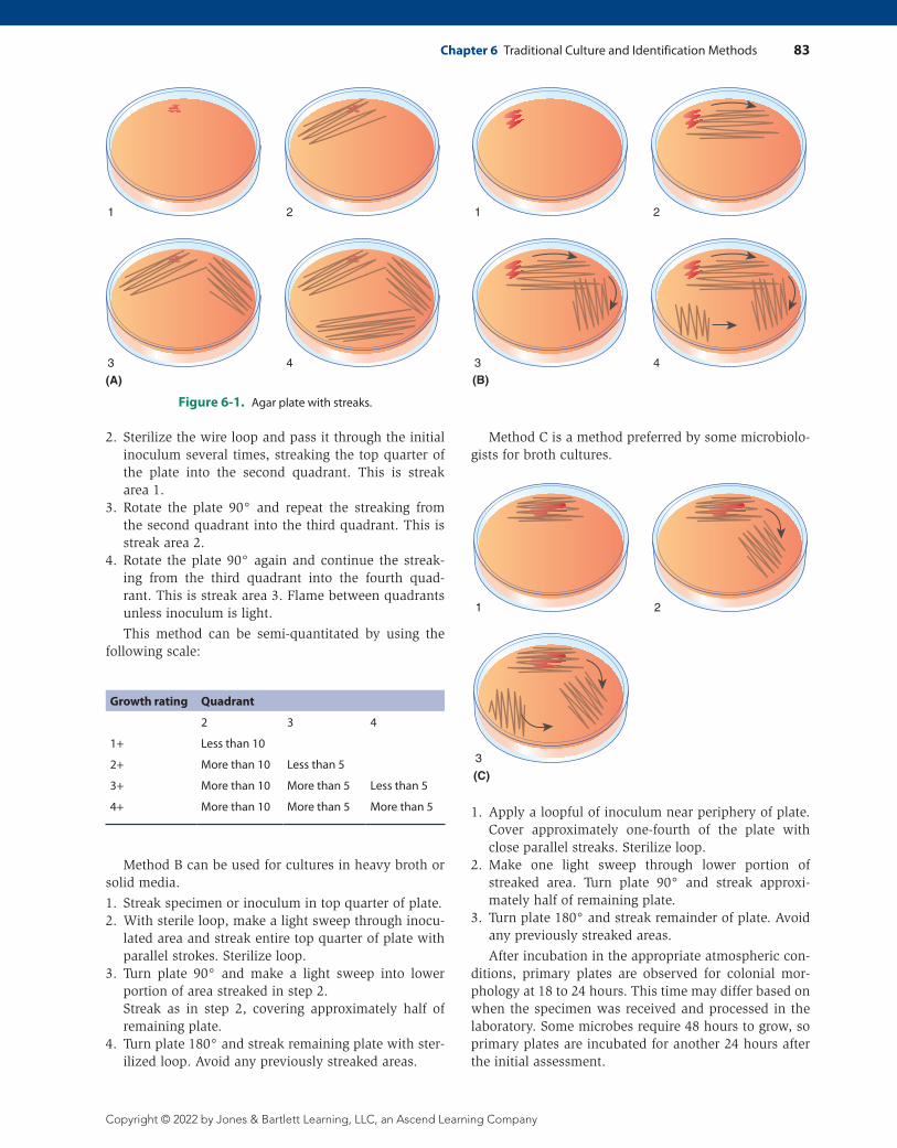
Figure 6-1. Agar plate with streaks.
1 2
43
(A)
1 2
3 4
(B)
1 2
3
(C)
2. Sterilize the wire loop and pass it through the initial inoculum several times, streaking the top quarter of the plate into the second quadrant. This is streak area 1.
3. Rotate the plate 90° and repeat the streaking from the second quadrant into the third quadrant. This is streak area 2.
4. Rotate the plate 90° again and continue the streak-ing from the third quadrant into the fourth quad-rant. This is streak area 3. Flame between quadrants unless inoculum is light.
This method can be semi-quantitated by using the following scale:
Method C is a method preferred by some microbiolo-gists for broth cultures.
1. Apply a loopful of inoculum near periphery of plate. Cover approximately one-fourth of the plate with close parallel streaks. Sterilize loop.
2. Make one light sweep through lower portion of streaked area. Turn plate 90° and streak approxi-mately half of remaining plate.
3. Turn plate 180° and streak remainder of plate. Avoid any previously streaked areas.
After incubation in the appropriate atmospheric con-ditions, primary plates are observed for colonial mor-phology at 18 to 24 hours. This time may differ based on when the specimen was received and processed in the laboratory. Some microbes require 48 hours to grow, so primary plates are incubated for another 24 hours after the initial assessment.
Method B can be used for cultures in heavy broth or solid media.
1. Streak specimen or inoculum in top quarter of plate.2. With sterile loop, make a light sweep through inocu-
lated area and streak entire top quarter of plate with parallel strokes. Sterilize loop.
3. Turn plate 90° and make a light sweep into lower portion of area streaked in step 2.
Streak as in step 2, covering approximately half of remaining plate.
4. Turn plate 180° and streak remaining plate with ster-ilized loop. Avoid any previously streaked areas.
84 Part I Introduction to Clinical Microbiology
Copyright © 2022 by Jones & Bartlett Learning, LLC, an Ascend Learning Company
This process is known as plate reading and includes the following assessments:
• The set of primary plates (enriched, differential, selective) are evaluated together for a particular site. Compare the growth characteristics on each plate, including the amount of growth and description.
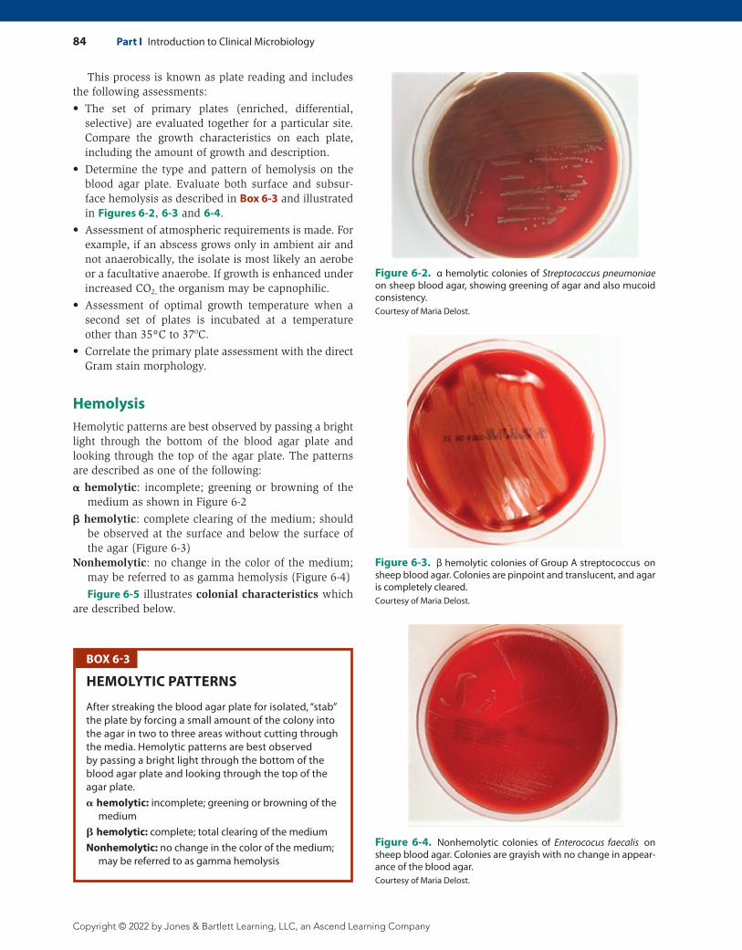
• Determine the type and pattern of hemolysis on the blood agar plate. Evaluate both surface and subsur-face hemolysis as described in Box 6-3 and illustrated in Figures 6-2, 6-3 and 6-4.
• Assessment of atmospheric requirements is made. For example, if an abscess grows only in ambient air and not anaerobically, the isolate is most likely an aerobe or a facultative anaerobe. If growth is enhanced under increased CO2, the organism may be capnophilic.
• Assessment of optimal growth temperature when a second set of plates is incubated at a temperature other than 35ºC to 37oC.
• Correlate the primary plate assessment with the direct Gram stain morphology.
HemolysisHemolytic patterns are best observed by passing a bright light through the bottom of the blood agar plate and looking through the top of the agar plate. The patterns are described as one of the following:
hemolytic: incomplete; greening or browning of the medium as shown in Figure 6-2
hemolytic: complete clearing of the medium; should be observed at the surface and below the surface of the agar (Figure 6-3)
Nonhemolytic: no change in the color of the medium; may be referred to as gamma hemolysis (Figure 6-4)
Figure 6-5 illustrates colonial characteristics which are described below.
Figure 6-2. α hemolytic colonies of Streptococcus pneumoniae on sheep blood agar, showing greening of agar and also mucoid consistency.Courtesy of Maria Delost.
Figure 6-3. β hemolytic colonies of Group A streptococcus on sheep blood agar. Colonies are pinpoint and translucent, and agar is completely cleared.Courtesy of Maria Delost.
Figure 6-4. Nonhemolytic colonies of Enterococus faecalis on sheep blood agar. Colonies are grayish with no change in appear-ance of the blood agar.Courtesy of Maria Delost.
BOX 6-3
HEMOLYTIC PATTERNS
After streaking the blood agar plate for isolated, “stab” the plate by forcing a small amount of the colony into the agar in two to three areas without cutting through the media. Hemolytic patterns are best observed by passing a bright light through the bottom of the blood agar plate and looking through the top of the agar plate. hemolytic: incomplete; greening or browning of the
medium hemolytic: complete; total clearing of the medium Nonhemolytic: no change in the color of the medium;
may be referred to as gamma hemolysis
Chapter 6 Traditional Culture and Identification Methods 85
Copyright © 2022 by Jones & Bartlett Learning, LLC, an Ascend Learning Company
Other Colonial CharacteristicsSize is described as pinpoint, small, medium, or large, or in millimeters, but is usually not measured.
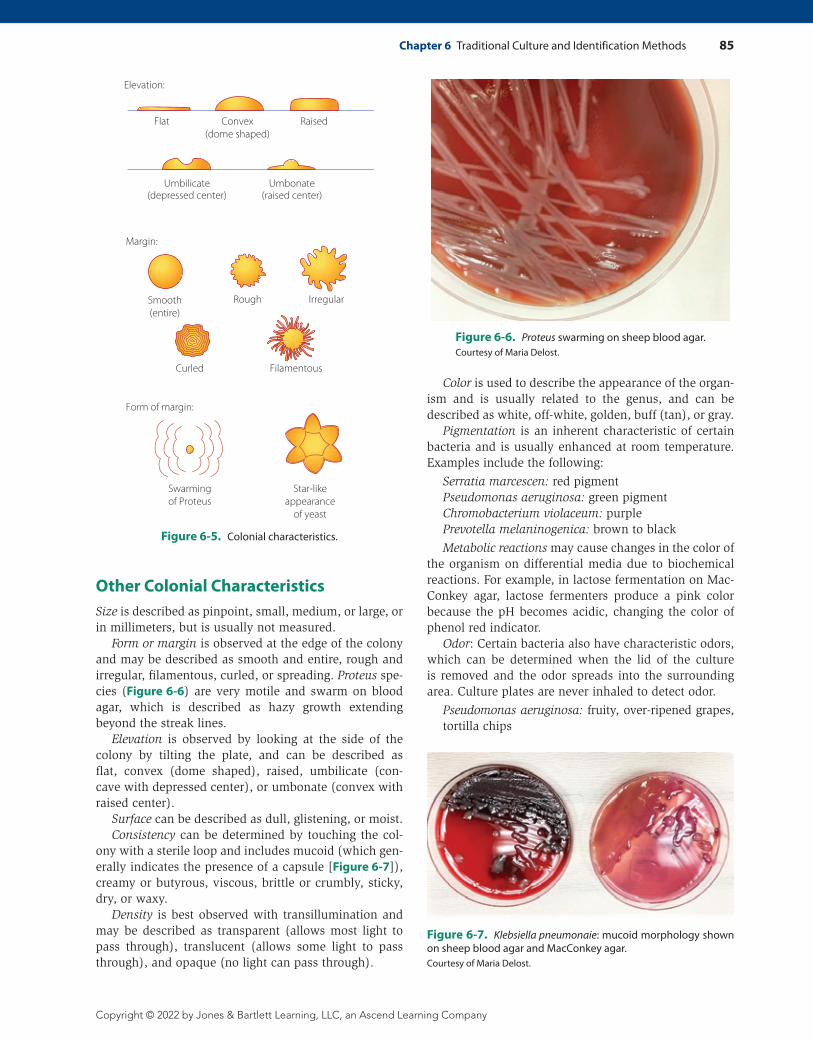
Form or margin is observed at the edge of the colony and may be described as smooth and entire, rough and irregular, filamentous, curled, or spreading. Proteus spe-cies (Figure 6-6) are very motile and swarm on blood agar, which is described as hazy growth extending beyond the streak lines.
Elevation is observed by looking at the side of the colony by tilting the plate, and can be described as flat, convex (dome shaped), raised, umbilicate (con-cave with depressed center), or umbonate (convex with raised center).
Surface can be described as dull, glistening, or moist.Consistency can be determined by touching the col-
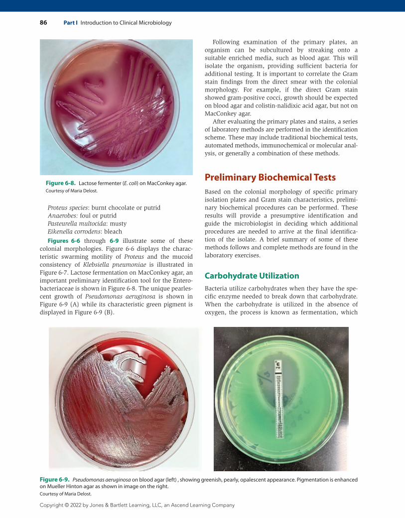
ony with a sterile loop and includes mucoid (which gen-erally indicates the presence of a capsule [Figure 6-7]), creamy or butyrous, viscous, brittle or crumbly, sticky, dry, or waxy.
Density is best observed with transillumination and may be described as transparent (allows most light to pass through), translucent (allows some light to pass through), and opaque (no light can pass through).
Color is used to describe the appearance of the organ-ism and is usually related to the genus, and can be described as white, off-white, golden, buff (tan), or gray.
Pigmentation is an inherent characteristic of certain bacteria and is usually enhanced at room temperature. Examples include the following:
Serratia marcescen: red pigmentPseudomonas aeruginosa: green pigmentChromobacterium violaceum: purplePrevotella melaninogenica: brown to black
Metabolic reactions may cause changes in the color of the organism on differential media due to biochemical reactions. For example, in lactose fermentation on Mac-Conkey agar, lactose fermenters produce a pink color because the pH becomes acidic, changing the color of phenol red indicator.
Odor: Certain bacteria also have characteristic odors, which can be determined when the lid of the culture is removed and the odor spreads into the surrounding area. Culture plates are never inhaled to detect odor.
Pseudomonas aeruginosa: fruity, over-ripened grapes, tortilla chips
Curled
Swarmingof Proteus
Star-likeappearance
of yeast
Filamentous
Elevation:
Flat Convex(dome shaped)
Raised
Umbilicate(depressed center)
Umbonate(raised center)
Margin:
Form of margin:
Rough IrregularSmooth(entire)
Figure 6-5. Colonial characteristics.
Figure 6-6. Proteus swarming on sheep blood agar.Courtesy of Maria Delost.
Figure 6-7. Klebsiella pneumonaie: mucoid morphology shown on sheep blood agar and MacConkey agar.Courtesy of Maria Delost.
86 Part I Introduction to Clinical Microbiology
Copyright © 2022 by Jones & Bartlett Learning, LLC, an Ascend Learning Company
Following examination of the primary plates, an organism can be subcultured by streaking onto a suitable enriched media, such as blood agar. This will isolate the organism, providing sufficient bacteria for additional testing. It is important to correlate the Gram stain findings from the direct smear with the colonial morphology. For example, if the direct Gram stain showed gram- positive cocci, growth should be expected on blood agar and colistin-nalidixic acid agar, but not on MacConkey agar.
After evaluating the primary plates and stains, a series of laboratory methods are performed in the identification scheme. These may include traditional biochemical tests, automated methods, immunochemical or molecular anal-ysis, or generally a combination of these methods.
Preliminary Biochemical TestsBased on the colonial morphology of specific primary isolation plates and Gram stain characteristics, prelimi-nary biochemical procedures can be performed. These results will provide a presumptive identification and guide the microbiologist in deciding which additional procedures are needed to arrive at the final identifica-tion of the isolate. A brief summary of some of these methods follows and complete methods are found in the laboratory exercises.
Carbohydrate UtilizationBacteria utilize carbohydrates when they have the spe-cific enzyme needed to break down that carbohydrate. When the carbohydrate is utilized in the absence of oxygen, the process is known as fermentation, which
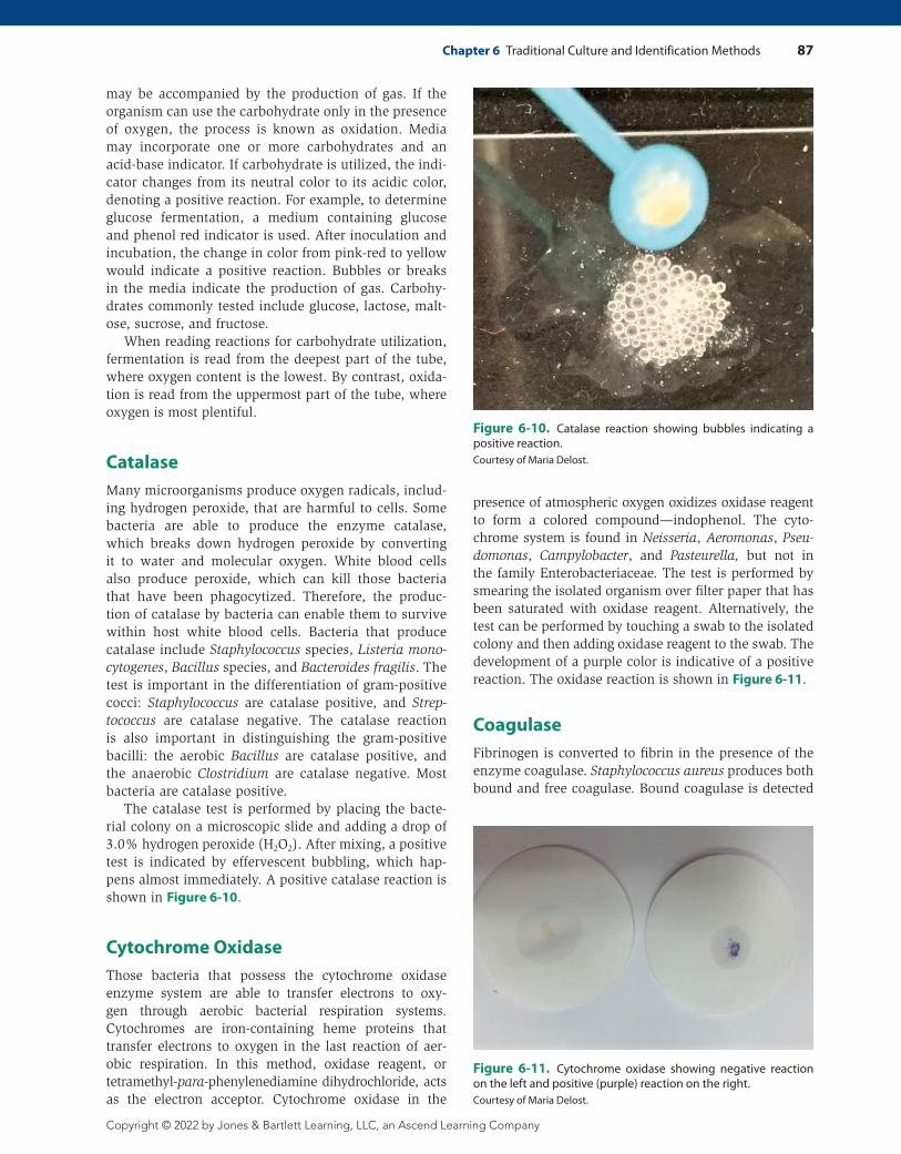
Figure 6-8. Lactose fermenter (E. coli) on MacConkey agar.Courtesy of Maria Delost.
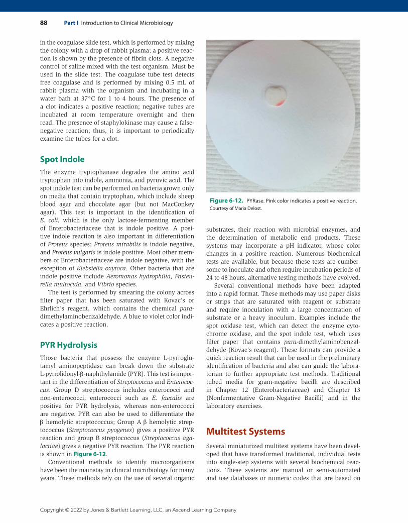
Figure 6-9. Pseudomonas aeruginosa on blood agar (left) , showing greenish, pearly, opalescent appearance. Pigmentation is enhanced on Mueller Hinton agar as shown in image on the right. Courtesy of Maria Delost.
Proteus species: burnt chocolate or putridAnaerobes: foul or putridPasteurella multocida: mustyEikenella corrodens: bleach
Figures 6-6 through 6-9 illustrate some of these colonial morphologies. Figure 6-6 displays the charac-teristic swarming motility of Proteus and the mucoid consistency of Klebsiella pneumoniae is illustrated in Figure 6-7. Lactose fermentation on MacConkey agar, an important preliminary identification tool for the Entero-bacteriaceae is shown in Figure 6-8. The unique pearles-cent growth of Pseudomonas aeruginosa is shown in Figure 6-9 (A) while its characteristic green pigment is displayed in Figure 6-9 (B).
Chapter 6 Traditional Culture and Identification Methods 87
Copyright © 2022 by Jones & Bartlett Learning, LLC, an Ascend Learning Company
may be accompanied by the production of gas. If the organism can use the carbohydrate only in the presence of oxygen, the process is known as oxidation. Media may incorporate one or more carbohydrates and an acid-base indicator. If carbohydrate is utilized, the indi-cator changes from its neutral color to its acidic color, denoting a positive reaction. For example, to determine glucose fermentation, a medium containing glucose and phenol red indicator is used. After inoculation and incubation, the change in color from pink-red to yellow would indicate a positive reaction. Bubbles or breaks in the media indicate the production of gas. Carbohy-drates commonly tested include glucose, lactose, malt-ose, sucrose, and fructose.
When reading reactions for carbohydrate utilization, fermentation is read from the deepest part of the tube, where oxygen content is the lowest. By contrast, oxida-tion is read from the uppermost part of the tube, where oxygen is most plentiful.
CatalaseMany microorganisms produce oxygen radicals, includ-ing hydrogen peroxide, that are harmful to cells. Some bacteria are able to produce the enzyme catalase, which breaks down hydrogen peroxide by converting it to water and molecular oxygen. White blood cells also produce peroxide, which can kill those bacteria that have been phagocytized. Therefore, the produc-tion of catalase by bacteria can enable them to survive within host white blood cells. Bacteria that produce catalase include Staphylococcus species, Listeria mono-cytogenes, Bacillus species, and Bacteroides fragilis. The test is important in the differentiation of gram-positive cocci: Staphylococcus are catalase positive, and Strep-tococcus are catalase negative. The catalase reaction is also important in distinguishing the gram-positive bacilli: the aerobic Bacillus are catalase positive, and the anaerobic Clostridium are catalase negative. Most bacteria are catalase positive.
The catalase test is performed by placing the bacte-rial colony on a microscopic slide and adding a drop of 3.0% hydrogen peroxide (H2O2). After mixing, a positive test is indicated by effervescent bubbling, which hap-pens almost immediately. A positive catalase reaction is shown in Figure 6-10.
Cytochrome OxidaseThose bacteria that possess the cytochrome oxidase enzyme system are able to transfer electrons to oxy-gen through aerobic bacterial respiration systems. Cytochromes are iron-containing heme proteins that transfer electrons to oxygen in the last reaction of aer-obic respiration. In this method, oxidase reagent, or tetramethyl-para-phenylenediamine dihydrochloride, acts as the electron acceptor. Cytochrome oxidase in the
presence of atmospheric oxygen oxidizes oxidase reagent to form a colored compound—indophenol. The cyto-chrome system is found in Neisseria, Aeromonas, Pseu-domonas, Campylobacter, and Pasteurella, but not in the family Enterobacteriaceae. The test is performed by smearing the isolated organism over filter paper that has been saturated with oxidase reagent. Alternatively, the test can be performed by touching a swab to the isolated colony and then adding oxidase reagent to the swab. The development of a purple color is indicative of a positive reaction. The oxidase reaction is shown in Figure 6-11.
CoagulaseFibrinogen is converted to fibrin in the presence of the enzyme coagulase. Staphylococcus aureus produces both bound and free coagulase. Bound coagulase is detected
Figure 6-10. Catalase reaction showing bubbles indicating a positive reaction.Courtesy of Maria Delost.
Figure 6-11. Cytochrome oxidase showing negative reaction on the left and positive (purple) reaction on the right.Courtesy of Maria Delost.
88 Part I Introduction to Clinical Microbiology
Copyright © 2022 by Jones & Bartlett Learning, LLC, an Ascend Learning Company
in the coagulase slide test, which is performed by mixing the colony with a drop of rabbit plasma; a positive reac-tion is shown by the presence of fibrin clots. A negative control of saline mixed with the test organism. Must be used in the slide test. The coagulase tube test detects free coagulase and is performed by mixing 0.5 mL of rabbit plasma with the organism and incubating in a water bath at 37°C for 1 to 4 hours. The presence of a clot indicates a positive reaction; negative tubes are incubated at room temperature overnight and then read. The presence of staphylokinase may cause a false-
negative reaction; thus, it is important to periodically examine the tubes for a clot.
Spot IndoleThe enzyme tryptophanase degrades the amino acid tryptophan into indole, ammonia, and pyruvic acid. The spot indole test can be performed on bacteria grown only on media that contain tryptophan, which include sheep blood agar and chocolate agar (but not MacConkey agar). This test is important in the identification of E. coli, which is the only lactose-fermenting member of Enterobacteriaceae that is indole positive. A posi-tive indole reaction is also important in differentiation of Proteus species; Proteus mirabilis is indole negative, and Proteus vulgaris is indole positive. Most other mem-bers of Enterobacteriaceae are indole negative, with the exception of Klebsiella oxytoca. Other bacteria that are indole positive include Aeromonas hydrophilia, Pasteu-rella multocida, and Vibrio species.
The test is performed by smearing the colony across filter paper that has been saturated with Kovac’s or Ehrlich’s reagent, which contains the chemical para-dimethylaminobenzaldehyde. A blue to violet color indi-cates a positive reaction.
PYR HydrolysisThose bacteria that possess the enzyme L-pyrroglu-tamyl aminopeptidase can break down the substrate L-pyrrolidonyl-β-naphthylamide (PYR). This test is impor-tant in the differentiation of Streptococcus and Enterococ-cus. Group D streptococcus includes enterococci and non-enterococci; enterococci such as E. faecalis are positive for PYR hydrolysis, whereas non-enterococci are negative. PYR can also be used to differentiate the β hemolytic streptococcus; Group A β hemolytic strep-tococcus (Streptococcus pyogenes) gives a positive PYR reaction and group B streptococcus (Streptococcus aga-lactiae) gives a negative PYR reaction. The PYR reaction is shown in Figure 6-12.
Conventional methods to identify microorganisms have been the mainstay in clinical microbiology for many years. These methods rely on the use of several organic
substrates, their reaction with microbial enzymes, and the determination of metabolic end products. These systems may incorporate a pH indicator, whose color changes in a positive reaction. Numerous biochemical tests are available, but because these tests are cumber-some to inoculate and often require incubation periods of 24 to 48 hours, alternative testing methods have evolved.
Several conventional methods have been adapted into a rapid format. These methods may use paper disks or strips that are saturated with reagent or substrate and require inoculation with a large concentration of substrate or a heavy inoculum. Examples include the spot oxidase test, which can detect the enzyme cyto-chrome oxidase, and the spot indole test, which uses filter paper that contains para-dimethylaminobenzal-dehyde (Kovac’s reagent). These formats can provide a quick reaction result that can be used in the preliminary identification of bacteria and also can guide the labora-torian to further appropriate test methods. Traditional tubed media for gram-negative bacilli are described in Chapter 12 (Enterobacteriaceae) and Chapter 13 (Nonfermentative Gram-Negative Bacilli) and in the laboratory exercises.
Multitest SystemsSeveral miniaturized multitest systems have been devel-oped that have transformed traditional, individual tests into single-step systems with several biochemical reac-tions. These systems are manual or semi-automated and use databases or numeric codes that are based on
Figure 6-12. PYRase. Pink color indicates a positive reaction.Courtesy of Maria Delost.
Chapter 6 Traditional Culture and Identification Methods 89
Copyright © 2022 by Jones & Bartlett Learning, LLC, an Ascend Learning Company
the biochemical profile of the organism. Binary codes based on positive or negative reactions are derived and converted into a code profile that “matches” the pheno-type of specific organisms. The databases are available by website or in a code book.
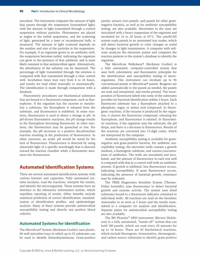
One example is the Analytical Profile Index (API®) of bioMérieux, which utilizes microtubes with dehydrated substrates that are reconstituted by adding a bacterial suspension. After incubation for 18–24 hours at 35oC, the reactions are read, and a profile number is deter-mined that can be matched with that in a profile index from a database. Most of the microorganisms can be identified to the species level using bioMérieux’s API® identification products. A large database is accessible through the Internet-based APIwebTM service. Manual bioMérieux’s API® products for gram-negative bacteria include the API® 20E for 18- to 24-hour identification of Enterobacteriaceae and other nonfastidious gram- negative bacilli and the API® Rapid 20E for 4-hour iden-tification of Enterobacteriaceae. There are also API® products to identify non-fermentative gram-negative bacilli, Neisseria and Haemophilus, and gram-positive bacteria including staphylococci, micrococci, strepto-cocci, and enterococci. An API is shown in Figure 6-13.
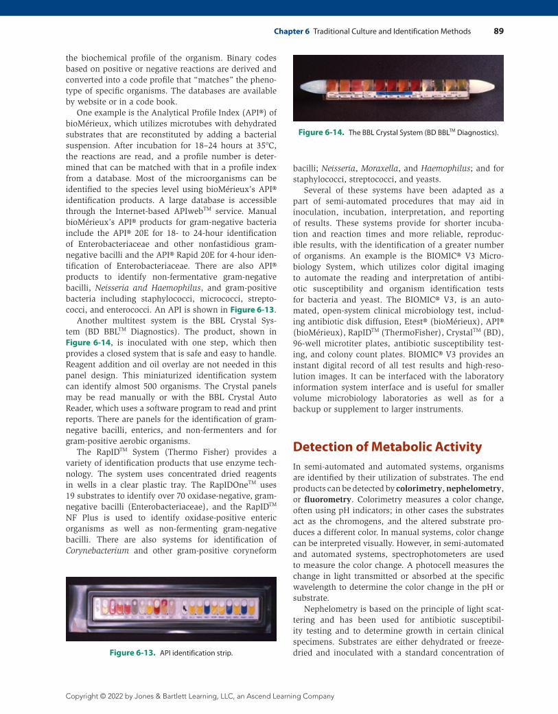
Another multitest system is the BBL Crystal Sys-tem (BD BBLTM Diagnostics). The product, shown in Figure 6-14, is inoculated with one step, which then provides a closed system that is safe and easy to handle. Reagent addition and oil overlay are not needed in this panel design. This miniaturized identification system can identify almost 500 organisms. The Crystal panels may be read manually or with the BBL Crystal Auto Reader, which uses a software program to read and print reports. There are panels for the identification of gram-negative bacilli, enterics, and non-fermenters and for gram-positive aerobic organisms.
The RapIDTM System (Thermo Fisher) provides a variety of identification products that use enzyme tech-nology. The system uses concentrated dried reagents in wells in a clear plastic tray. The RapIDOneTM uses 19 substrates to identify over 70 oxidase-negative, gram-negative bacilli (Enterobacteriaceae), and the RapIDTM NF Plus is used to identify oxidase-positive enteric organisms as well as non-fermenting gram- negative bacilli. There are also systems for identification of Corynebacterium and other gram-positive coryneform
bacilli; Neisseria, Moraxella, and Haemophilus; and for staphylococci, streptococci, and yeasts.
Several of these systems have been adapted as a part of semi-automated procedures that may aid in inoculation, incubation, interpretation, and reporting of results. These systems provide for shorter incuba-tion and reaction times and more reliable, reproduc-ible results, with the identification of a greater number of organisms. An example is the BIOMIC® V3 Micro-biology System, which utilizes color digital imaging to automate the reading and interpretation of antibi-otic susceptibility and organism identification tests for bacteria and yeast. The BIOMIC® V3, is an auto-mated, open-system clinical microbiology test, includ-ing antibiotic disk diffusion, Etest® (bioMérieux), API® ( bioMérieux), RapIDTM (ThermoFisher), CrystalTM (BD), 96-well microtiter plates, antibiotic susceptibility test-ing, and colony count plates. BIOMIC® V3 provides an instant digital record of all test results and high-reso-lution images. It can be interfaced with the laboratory information system interface and is useful for smaller volume microbiology laboratories as well as for a backup or supplement to larger instruments.
Detection of Metabolic ActivityIn semi-automated and automated systems, organisms are identified by their utilization of substrates. The end products can be detected by colorimetry, nephelometry, or fluorometry. Colorimetry measures a color change, often using pH indicators; in other cases the substrates act as the chromogens, and the altered substrate pro-duces a different color. In manual systems, color change can be interpreted visually. However, in semi-automated and automated systems, spectrophotometers are used to measure the color change. A photocell measures the change in light transmitted or absorbed at the specific wavelength to determine the color change in the pH or substrate.
Nephelometry is based on the principle of light scat-tering and has been used for antibiotic susceptibil-ity testing and to determine growth in certain clinical specimens. Substrates are either dehydrated or freeze-dried and inoculated with a standard concentration of Figure 6-13. API identification strip.
Figure 6-14. The BBL Crystal System (BD BBLTM Diagnostics).
90 Part I Introduction to Clinical Microbiology
Copyright © 2022 by Jones & Bartlett Learning, LLC, an Ascend Learning Company
inoculum. The instrument compares the amount of light that passes through the suspension (transmitted light) with the amount of light transmitted through a control suspension without particles. Photometers are placed at angles to the turbid suspension, and the scattering of light, generated by a laser or incandescent bulb, is measured. The amount of light scattered depends on the number and size of the particles in the suspension. For example, if an organism grows in an antibiotic well, the suspension becomes turbid, indicating the organism can grow in the presence of that antibiotic and is most likely resistant to that antimicrobial agent. Alternatively, the absorbance of the solution can be measured. The percentage of light transmitted through the solution is compared with that transmitted through a clear control well. Incubation times may vary from 4 to 24 hours, and results are read either manually or automatically. The identification is made through comparison with a database.
Fluorometric procedures use biochemical substrates that are bound to a fluorescent component, such as fluo-rophores. If the organism has the enzyme to metabo-lize a substrate, the fluorophore is released from the substrate, and fluorescence is exhibited. In some reac-tions, fluorescence is used to detect a change in pH. In pH-driven fluorometric reactions, the pH change results in the fluorophore becoming fluorescent. In other reac-tions, the fluorescence may be quenched or lost. For example, the pH increases in a positive decarboxylase reaction resulting in the production of fluorescence. In other reactions, an acidic pH would be noted by the lack of fluorescence. Fluorescence is detected by using ultraviolet light of a specific wavelength that is directed toward the reaction chamber while a fluorometer mea-sures the fluorescence.
Automated Identification SystemsThere are several automated identification systems with various features and capacities. Fully automated sys-tems incubate, read the reactions, interpret the results, and identify the microorganism. These systems have an interface to the laboratory information system, which expedites reporting of results. Other benefits include statistical prediction of correct identification, standard-ization of identification profiles, and epidemiologic analysis. Many of these systems provide antimicrobial susceptibility testing and directly test positive blood cultures.
Automated Systems for IdentificationThe MicroScan® System (Beckman-Coulter) uses plastic, 96-well microtiter trays in which up to 32 substrates can be used to identify Enterobacteriaceae. Gram-positive
panels, urinary tract panels, and panels for other gram-negative bacteria, as well as for antibiotic susceptibility testing, are also available. Microtubes in the trays are inoculated with a heavy suspension of the organism and incubated for 16 to 20 hours at 35°C. The autoSCAN system reads panels in an automated tray reader, which will detect bacterial growth or color changes as noted by changes in light transmission. A computer with soft-ware analyzes the electronic pulses and compares the reaction patterns to the system’s database to identify the organism.
The MicroScan WalkAway® (Beckman Coulter) is a fully automated, computer-controlled system that uses both colorimetric and fluorometric principles for the identification and susceptibility testing of micro-organisms. This instrument can incubate up to 96 conventional panels or MicroScan® panels. Reagents are added automatically to the panels as needed, the panels are read and interpreted, and results printed. The incor-poration of fluorescent labels into some of the substrates provides for bacterial identification within 2 hours. Each fluorescent substrate has a fluorophore attached to a phosphate, sugar, or amino acid compound. In fluoro-genic reactions, if the enzyme is produced by the organ-ism, it cleaves the fluorescent compound, releasing the fluorophore, and fluorescence is emitted. In fluoromet-ric reactions, if the organism uses the substrate, the pH drops, and there is a decrease in fluorescence. Results of the reactions are converted into 15-digit codes, which are interpreted by the computer.
Antibiotic susceptibility testing is available for gram-negative and gram-positive bacteria. For antibiotic sus-ceptibility testing, the microtiter wells contain a growth medium, a fluorogenic substrate, and various concentra-tions of antibiotics. The wells are inoculated and incu-bated, and the amount of fluorescence in each test well is compared with that in a control well with no antibiotic present. If growth is inhibited, less fluorescence occurs, indicating susceptibility. If more fluorescence occurs, indicating the presence of bacterial growth, resistance may be indicated.
The TREK Diagnostics Sensititre System (Thermo-Fisher Scientific) uses fluorescence to detect bacterial growth and enzyme activity. The system uses dried substrates bound to a fluorescent indicator contained in individual wells. All reactions are read on the Sensititre Autoreader in as soon as 5 hours and the results trans-mitted to a computer for analysis and identification. Separate plates for antimicrobial susceptibility testing are also available.
The BD Phoenix™ M50 instrument (Becton Dickin-son) is a fully automated, “hands off” system that can hold 100 panels, which are read every 20 minutes for up to 16 hours. There are 45 biochemical reactions, which include fluorogenic, fermentative, chromogenic, and carbon source substrates to identify gram- positive
Chapter 6 Traditional Culture and Identification Methods 91
Copyright © 2022 by Jones & Bartlett Learning, LLC, an Ascend Learning Company
and gram-negative bacilli. Results are available in 2–12 hours for bacteria and 4–15 hours for yeasts. The system uses both fluorometric and colorimetric endpoint detection, depending on the substrate. There are panels for identification only and susceptibility only, as well as combination panels for both identifi-cation and susceptibility. The system identifies most clinically significant aerobic and facultative anaerobic gram-negative and gram-positive bacteria, as well as yeast and yeast-like organisms. A colorimetric redox indicator and turbidity are used for antimicrobial sus-ceptibility testing.
The VITEK® System (bioMérieux) identifies most clinically significant bacteria and yeast and also per-forms qualitative and quantitative antibiotic susceptibil-ity tests. The VITEK® 2 is an automated microbiology system utilizing growth-based technology that uses reagent cards, which test substrate wells. Substrates measure various metabolic activities such as acidifica-tion, alkalinization, enzyme hydrolysis, and growth in the presence of inhibitory substances. VITEK 2 systems use chromogenic substrates and carbohydrate utiliza-tion measured with Advanced Colorimetry™ identifica-tion technology.
The VITEK® has several modules, including a reader/incubator module, which has a capacity of 15, 30, 60, or 120 cards depending on the VITEK® model. The inoculum is prepared from a growing isolate, and the cards are inoculated automatically. The card is placed in the reader/incubator module, where it is scanned by a series of emitted diodes. The light source directs light through the wells to the reader. The data are sent to a computer, which records and interprets the read-ings. Changes in absorbance, resulting from changes in turbidity as the organism grows, or from color changes that occur as the pH changes are measured. The com-puter module stores, analyzes, and interprets test data and also controls reader/incubator functions. The data terminal and printer module allow the visual examina-tion of results and provide an automatic printed copy of results.
The VITEK® also can be used for antibiotic suscepti-bility testing for gram-positive and gram-negative organ-isms. A gram-positive or gram-negative susceptibility card is inoculated, and the reader measures the amount of light passing through each well hourly for 15 hours. As the organism grows, the amount of light transmitted decreases. Increased turbidity and resulting decreased transmission of light usually indicate the organism is resistant to the antibiotic. A growth curve is developed based on the decline of the percentage of transmis-sion. The minimal inhibitory concentration (MICs), the lowest antibiotic concentration that shows no visible growth, can be determined based on the growth curve and slope of the curve.
Matrix-Assisted Laser Desorption Ionization Time of Flight Mass Spectrometry (MALDI-TOF MS)Matrix-assisted laser desorption ionization time of flight mass spectrometry (MALDI-TOF MS) relies on the principle of mass spectrometry and uses chemical compounds to protect large biologic compounds, such as proteins, to withstand the high intensity of lasers. This methodology has made a tremendous impact on clinical microbiology by identifying organism in a rapid and cost-effective manner and by replacing or supple-menting other identification methods. MALDI-TOF MS requires isolated colonies.
In MALDI-TOF MS, a single colony is selected from an agar plate and transferred onto a MALDI metal target plate and then overlaid with 1–2 mL of matrix. The matrix is a low-mass organic compound, usually a cyano-4-hydroxycinnamic acid, a crystalizing agent, which is solubilized in a mixture of trifluoroacetic acid and acetonitrile, which are organic solvents. The specimen can be directly applied onto a target plate or prepared as a protein extract before application. The proteins and other cellular components are extracted, evaporate from the solvents, and crystallize.
The target plate is placed into the ionization cham-ber, and a precise laser ionizes the sample with pulses. A cloud of proteins is released, which are accelerated by an electric charge through a ring electrode. The transfer of light energy into heat allows the sample to be released (desorbed) as single charged ions. The desorbed and ionized molecules pass through a vacuum tube and con-tact a detector. The time, in nanoseconds, needed for the ions to travel through the vacuum tube is known as their “time of flight.” Smaller-sized molecules travel faster than larger molecules. When the ions hit the detector, they generate a spectrum based on their mass-to-charge ratio and signal intensity. Small and abundant proteins, such as ribosomal proteins, are efficiently detected. The spectrum is unique to each species. Each spectrum is digitalized and compared to spectra in a database for identification.
MALDI-TOF provides quick results, but there are some shortcomings. The identification is limited to the size of the proprietary database. This is cur-rently expanding to include more microbes. The origi-nal sample requires a pure culture, so the specimen must be streaked, incubated, and an isolated colony selected. MALDI-TOF MS cannot be used directly on clinical specimens or samples containing contaminants or multiple organisms. However, with good technical expertise, the method does provide accurate and repro-ducible results. Its accuracy is comparable to or may even exceed that of those systems that use biochemical
92 Part I Introduction to Clinical Microbiology
Copyright © 2022 by Jones & Bartlett Learning, LLC, an Ascend Learning Company
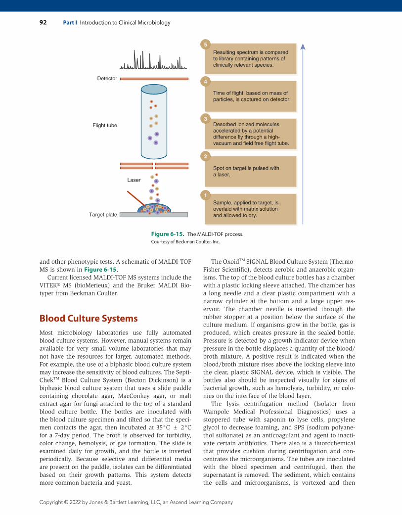
and other phenotypic tests. A schematic of MALDI-TOF MS is shown in Figure 6-15.
Current licensed MALDI-TOF MS systems include the VITEK® MS (bioMerieux) and the Bruker MALDI Bio-typer from Beckman Coulter.
Blood Culture SystemsMost microbiology laboratories use fully automated blood culture systems. However, manual systems remain available for very small volume laboratories that may not have the resources for larger, automated methods. For example, the use of a biphasic blood culture system may increase the sensitivity of blood cultures. The Septi-ChekTM Blood Culture System (Becton Dickinson) is a biphasic blood culture system that uses a slide paddle containing chocolate agar, MacConkey agar, or malt extract agar for fungi attached to the top of a standard blood culture bottle. The bottles are inoculated with the blood culture specimen and tilted so that the speci-men contacts the agar, then incubated at 35°C ± 2°C for a 7-day period. The broth is observed for turbidity, color change, hemolysis, or gas formation. The slide is examined daily for growth, and the bottle is inverted periodically. Because selective and differential media are present on the paddle, isolates can be differentiated based on their growth patterns. This system detects more common bacteria and yeast.
The OxoidTM SIGNAL Blood Culture System (Thermo-Fisher Scientific), detects aerobic and anaerobic organ-isms. The top of the blood culture bottles has a chamber with a plastic locking sleeve attached. The chamber has a long needle and a clear plastic compartment with a narrow cylinder at the bottom and a large upper res-ervoir. The chamber needle is inserted through the rubber stopper at a position below the surface of the culture medium. If organisms grow in the bottle, gas is produced, which creates pressure in the sealed bottle. Pressure is detected by a growth indicator device when pressure in the bottle displaces a quantity of the blood/broth mixture. A positive result is indicated when the blood/broth mixture rises above the locking sleeve into the clear, plastic SIGNAL device, which is visible. The bottles also should be inspected visually for signs of bacterial growth, such as hemolysis, turbidity, or colo-nies on the interface of the blood layer.
The lysis centrifugation method (Isolator from Wampole Medical Professional Diagnostics) uses a stoppered tube with saponin to lyse cells, propylene glycol to decrease foaming, and SPS (sodium polyane-thol sulfonate) as an anticoagulant and agent to inacti-vate certain antibiotics. There also is a fluorochemical that provides cushion during centrifugation and con-centrates the microorganisms. The tubes are inoculated with the blood specimen and centrifuged, then the supernatant is removed. The sediment, which contains the cells and microorganisms, is vortexed and then
Sample, applied to target, isoverlaid with matrix solutionand allowed to dry.
Spot on target is pulsed witha laser.
Desorbed ionized moleculesaccelerated by a potentialdifference fly through a high-vacuum and field free flight tube.
Time of flight, based on mass ofparticles, is captured on detector.
Resulting spectrum is comparedto library containing patterns of clinically relevant species.
Detector
Flight tube
Laser
Target plate
1
2
3
4
5
Figure 6-15. The MALDI-TOF process.Courtesy of Beckman Coulter, Inc.
Chapter 6 Traditional Culture and Identification Methods 93
Copyright © 2022 by Jones & Bartlett Learning, LLC, an Ascend Learning Company
used to inoculate media. This system is believed to pro-vide better recovery of fragile bacteria and some fungi, but is more susceptible to contamination and aerosol production.
Automated Blood Culture SystemsAutomation in the detection of bacteria in the blood has significantly improved the detection of bacteremia. These systems are fully automated and offer continuous monitoring. The BACTECTM system (Becton Dickinson) was the first automated blood culture system. The early models, such as the BACTEC 460, used a radio-metric principle and relied on the release of 14CO2 from 14C radioactively labeled substrates. These models were replaced by newer versions, such as the BACTEC 660 and BACTEC NR 860, that detect the release of CO2 directly with an infrared spectrophotometer. The newer models alleviate the disadvantages associated with radioactiv-ity, including safety hazards and strict disposal guide-lines. The BACTEC 860 is fully automated and includes incubators, shakers, and detectors.
The newest versions of the BACTEC (9240, 9120, and 9050) are fully automated, with continuous monitor-ing, and use fluorescence to measure released CO2 or consumed oxygen. There is a gas-permeable CO2 sensor on the bottom of each vial. When released from bacte-rial metabolism, the CO2 dissolves in water and forms hydrogen ions (H+), which causes the pH to decrease, initiating a fluorescent signal. Using photodetectors, the instrument measures the amount of fluorescence every 10 minutes, which corresponds to the amount of CO2 being produced by the microorganism. A computer pro-gram interprets the data using several algorithms to flag a bottle as positive. A message is displayed on the com-puter screen, alerting the microbiologist when a vial is positive.
The BacT/ALERT® 3D (bioMérieux) uses colorimet-ric technology and measures microbial-produced CO2 photometrically. The system is fully automated and noninvasive and utilizes a colorimetric CO2 sensor, which is placed at the bottom of each blood culture bottle. A CO2-permeable membrane separates the sen-sor from microbial growth. CO2 generated from micro-bial metabolism diffuses across the membrane into the sensor and dissolves in a pH-sensitive water solution. This results in the production of hydrogen ions, which lowers the pH, changing the color of the sensor from dark green to yellow. Light reflected from the sensor is detected and measured photometrically. Changes in reflectance are determined, and a voltage signal pro-portional to the intensity of the reflected light is gen-erated. The voltage signal is analyzed by a computer, which determines whether growth is present based on changes in absorbance, rate of change, and the color threshold produced.
The latest version is the BacT/ALERT® VIRTUO® (bioMérieux) offers advanced robotics and hand-free bottle loading and unloading.
The VersaTREK® System (Trek Diagnostic Systems) monitors pressure changes in the bottle headspace cre-ated from bacterial metabolism. It detects the production or consumption of CO2, H2, and O2 by organisms growing in the culture medium. The gases are detected by changes in head space pressure. Advantages of the VersaTREK® System include earlier detection because of the measure-ment of all three gases and the system is not limited by organisms that produce low CO2 concentrations.
This chapter described traditional cultivation and identification of microorganisms from primary plating; culture characteristics; preliminary reactions; manual and semi-automated identification; and principles of automation in identification antimicrobial testing, and blood cultures. Chapter 7 will describe immunochemi-cal methods and the use of molecular diagnostics.
94 Part I Introduction to Clinical Microbiology
Copyright © 2022 by Jones & Bartlett Learning, LLC, an Ascend Learning Company
Review Questions
Multiple Choice 1. An organism that produced a greening of the
blood agar plate would be described as: a. α hemolytic.b. β hemolytic.c. nonhemolytic.d. ∆ hemolytic.
2. Which of the following is not considered to be a phenotypic characteristic?a. Gram stain morphologyb. Nucleic acid identificationc. Colonial size and margind. Change in color of pH indicator
3. The automated principle that utilizes a spectrophotometer to detect changes in color is:a. colorimetry.b. nephelometry.c. turbimetry.d. fluorometry.
MatchingPhenotypic characteristics and preliminary testing can provide important clues about a microorganism. For the following, match each description with the most likely microorganism.
4. Indole positive, gram-negative bacilli that produced pink colonies of MacConkey agar.
5. Gram-positive cocci that arranged in clusters and were catalase positive.
6. Gram-negative bacillus with a fruity odor that produced a green pigment and was oxidase negative.
7. Gram-negative bacillus that was oxidase negative and produced swarming colonies on sheep blood agar.
8. Gram-positive diplococci that produced mucoid, hemolytic colonies on sheep blood agar.a. Streptococcus pneumoniaeb. Pseudomonas aeruginosac. Escherichia colid. Proteus speciese. Staphylococcus species
Short Answer 9. List some of the phenotypic characteristics that
are observed in primary culture.
10. State the principle of MALDI-TOF MS.
ReferencesBeckman Coulter. (2020). Microbiology panels and systems.
Retrieved from https://www.beckmancoulter.com/en/products /microbiology
BioMérieux USA. (2020). VITEK® 2: Healthcare. Retrieved from https://www.biomerieux-usa.com/clinical/vitek-2-healthcare
Bourbeau, P. P., & Ledeboer, N. A. (2013). Automation in clinical microbiology. Journal of Clinical Microbiology, 51, 1658–1665. DOI: 10.1128/JCM.00301-13
Carroll, K. C., Glanz, B. D., Borek, A. P., Burger, C., Bhally, H. S., Henciak, S., & Flayhart, D. (2006). Evaluation of the BD Phoenix automated system for identification and susceptibility testing of Enterobacteriaceae. Journal of Clinical Microbiology, 44, 3506–3509.
Dallas, S. D., Avery, A. L., Pekarek, P. M., Mills, T. J., Neal, W. J., Smallbrook, A. G., & Hejna, J. (2005). Comparison of BD Phoenix to Biomerieux Vitek for the identification and susceptibility testing of common bacterial isolates. White paper presented at the 105th General Meeting of the American Society for Microbiology. Retrieved from http://www.bd.com/ds /technicalCenter/whitepapers/lr905.pdf
Florio, W., Tavanti, A., Barnini, S., Ghelardi, E., & Lupetti, A. (2018). Recent advances and ongoing challenges in the diag-nosis of microbial infections by MALDI-TOF Mass Spectrom-etry. Frontiers in microbiology, 9, 1097. https://doi.org/10.3389 /fmicb.2018.01097
Pincus, D. H. (2006). Microbial identification using the bioMéreiux VITEK 2 system. In M. J. Miller (Ed.), Encyclopedia of rapid microbiological methods, vol. II. PDA/DHI. Retrieved from https://store.pda.org/TableOfContents/ERMM_V2_Ch01.pdf
Sellenriek, P., Holmes, J., Ferrett, R., Drury, R., & Storch, G. A. (2005). Comparison of MicroScan Walk-Away®, PhoenixTM, and VITEK-TWO® microbiology systems used in the identification and susceptibility testing of bacteria. White paper presented at the 105th General Meeting of the American Society for Microbiology. Retrieved from http://www.bd.com/ds/technical Center/whitepapers/lr900.pdf
Stager, C. E., & Davis, J. R. (1992). Automated systems for iden-tification of microorganisms. Clinical Microbiology Review, 5, 302–327.