CHARACTERIZATION AND IDENTIFICATION OF A ...
274
CHARACTERIZATION AND IDENTIFICATION OF A GALACTURONOSYLTRANSFERASE INVOLVED IN PECTIN BIOSYNTHESIS by JASON DWIGHT STERLING (Under the Direction of Debra Mohnen) ABSTRACT Pectins are a class of acidic, plant cell wall polysaccharides that contain (1,4)-linked α-D- galactopyranosyluronic acid (D-GalpA) as part of their backbone structure. Pectin is involved in the regulation of plant growth and development, the elucidation of plant defense responses, and the maintenance of plant cell adhesion. While a significant amount of published information is available on the structure and function of pectin in the plant cell wall, relatively little is known about pectin biosynthesis. This study centers around the characterization and identification of an α-1,4-galacturonosyltransferase (GalAT) involved in the biosynthesis of the backbone structures of pectin. The subcellular localization of a GalAT was determined by sucrose density gradient centrifugation of pea epicotyl membranes. GalAT activity was found to co-fractionate with Golgi-resident latent UDPase activity. Treatment of Golgi vesicles with Proteinase K in the absence of detergent showed that intact Golgi membranes from pea protected GalAT activity from proteolytic degradation. These results show that pectin biosynthesis occurs in the Golgi apparatus and that the pea GalAT has its catalytic site facing the Golgi lumen. A radioactive assay was developed to assay GalAT activity. The assay uses cetylpyridinium chloride-treated filters to separate unincorporated UDP-D-[ 14 C]GalpA from radioactive products formed during the GalAT reaction. The versatility of this assay was demonstrated by its ability to rapidly and
Transcript of CHARACTERIZATION AND IDENTIFICATION OF A ...
CHARACTERIZATION AND IDENTIFICATION OF A
GALACTURONOSYLTRANSFERASE INVOLVED IN PECTIN BIOSYNTHESIS
by
JASON DWIGHT STERLING
(Under the Direction of Debra Mohnen)
ABSTRACT
Pectins are a class of acidic, plant cell wall polysaccharides that contain (1,4)-linked α-D-
galactopyranosyluronic acid (D-GalpA) as part of their backbone structure. Pectin is involved in
the regulation of plant growth and development, the elucidation of plant defense responses, and
the maintenance of plant cell adhesion. While a significant amount of published information is
available on the structure and function of pectin in the plant cell wall, relatively little is known
about pectin biosynthesis. This study centers around the characterization and identification of an
α-1,4-galacturonosyltransferase (GalAT) involved in the biosynthesis of the backbone structures
of pectin. The subcellular localization of a GalAT was determined by sucrose density gradient
centrifugation of pea epicotyl membranes. GalAT activity was found to co-fractionate with
Golgi-resident latent UDPase activity. Treatment of Golgi vesicles with Proteinase K in the
absence of detergent showed that intact Golgi membranes from pea protected GalAT activity
from proteolytic degradation. These results show that pectin biosynthesis occurs in the Golgi
apparatus and that the pea GalAT has its catalytic site facing the Golgi lumen. A radioactive
assay was developed to assay GalAT activity. The assay uses cetylpyridinium chloride-treated
filters to separate unincorporated UDP-D-[14C]GalpA from radioactive products formed during
the GalAT reaction. The versatility of this assay was demonstrated by its ability to rapidly and
accurately measure GalAT activity in multiple chromatography fractions during the partial
purification of a GalAT from tobacco (Nicotiana tabacum L. cv. Samsun) and Arabidopsis
thaliana (cv. Columbia). Using a new detergent solubilization technique and a combination of
SP-Sepharose, Reactive Yellow 3, and UDP-agarose chromatography, GalAT activity was
purified 17-fold from solubilized Arabidopsis membranes. The partially purified fraction was
digested with sequencing-grade trypsin and the released peptides were analyzed by liquid
chromatography-tandem mass spectrometry. Peptide analysis revealed the presence of two
proteins (JS33 and JS36) with sequence similarity to other glycosyltransferases. Truncated
versions of these genes lacking their N-terminal transmembrane domain were cloned into a
vector containing an N-terminal signal sequence designed for secretion of the recombinant
proteins. Both gene constructs were transiently and stably expressed in human embryonic
kidney (HEK) 293 cells. Preliminary transient transfection experiments indicated that GalAT
activity was detectable in media from HEK293 cells transiently expressing the JS36 gene
construct providing evidence that JS36 encoded a putative GalAT. Inactive, recombinant
proteins were detected in cell lysates from stable HEK293 lines expressing either JS33 or JS36.
Database analysis indicates that JS33 and JS36 are part of a 25 member gene family in
Arabidopsis. Mutation of two members of this putative GalAT gene family gives phenotypes
that are similar to previously characterized pectin mutants. Further research needs to be
conducted to determine what role(s) these genes play in pectin biosynthesis.
INDEX WORDS: Galacturonosyltransferase, pectin, glycosyltransferase, Golgi apparatus,
Arabidopsis thaliana, Pisum sativum, galacturonic acid, assay,
cetylpyridinium chloride, plant cell wall, heterologous expression,

multiple antigenic peptide, sucrose gradient, biosynthesis, CAZy family 8,
gene family.
CHARACTERIZATION AND IDENTIFICATION OF A
GALACTURONOSYLTRANSFERASE INVOLVED IN PECTIN BIOSYNTHESIS
by
JASON DWIGHT STERLING
B.S., McGill University, Canada, 1996
A Dissertation Submitted to the Graduate Faculty of The University of Georgia in Partial
Fulfillment of the Requirements for the Degree
DOCTOR OF PHILOSOPHY
ATHENS, GEORGIA
2004
© 2004
Jason Dwight Sterling
All Rights Reserved
CHARACTERIZATION AND IDENTIFICATION OF A
GALACTURONOSYLTRANSFERASE INVOLVED IN PECTIN BIOSYNTHESIS
by
JASON DWIGHT STERLING
Major Professor: Debra Mohnen
Committee: Alan Darvill Russell Malmberg Kelley Moremen Michael Pierce
Electronic Version Approved:
Maureen Grasso Dean of the Graduate School The University of Georgia December 2004

iv
DEDICATION
I dedicate this dissertation to my family (Suzanne, Tyrone, and T-man), the most
important thing in my life. I would not have been able to do it without you.
v
ACKNOWLEDGEMENTS
I would like to thank Dr. Debra Mohnen for her guidance and support throughout my
graduate school career and for the being the best mentor a graduate student could ever have. I
would also like to thank Dr. Kelley Moremen, Dr. Michael Pierce, Dr. Alan Darvill, Dr. Russell
Malmberg, and Dr. Carl Bergmann for being great committee members, for providing guidance
when I had questions about my research and for never letting me settle for “I don’t know”.
I am extremely grateful to all the members of the Mohnen lab, past and present, that
made lab life so much fun over the years. I also thank all of the undergraduate students that have
ever worked with me on my graduate research.
I would like to thank Dr. Malcolm O’Neill for generously providing the figures on pectin
structure used in Chapter 1. I would also like to thank Dr. Debra Mohnen for providing some of
the tables used in the Biosynthesis section of Chapter 1.
The research presented in the thesis would not have been possible without materials
generously provided by members of the CCRC, including Stefan Eberhard (for providing the
initial Arabidopsis tissue cultures), Dr. Carl Bergmann (Chapter 2), Dr. Maor Bar-Peled (Chapter
4), Dr. Kelley Moremen (Chapter 4), and Dr. Michael Pierce (Chapter 4).
The research presented in Chapter 2 was conducted with the assistance of Lorena
Norambuena from the University of Chile, Santiago, Chile. We also thank Dr. Malcolm O’Neill
for the critical reading of Chapter 2.
The research presented in Chapter 4 would not have been possible without the guidance
of April Harper, Leslie Stanton, Intaek Lee, Dr. Steve Mast, Dr. Alison Vandersall-Nairn, and
Dr. Michael Hahn.
vi
TABLE OF CONTENTS
Page
ACKNOWLEDGEMENTS.............................................................................................................v
INTRODUCTION ...........................................................................................................................1
CHAPTER
1 LITERATURE REVIEW: PECTIN STRUCTURE, FUNCTION, AND
BIOSYNTHESIS.......................................................................................................4
Introduction ...............................................................................................................5
Structure ....................................................................................................................6
Pectin in the Primary Wall ......................................................................................21
Pectin Function........................................................................................................36
Biosynthesis.............................................................................................................42
Conclusions and Relevance...................................................................................109
2 THE CATALYTIC SITE OF THE PECTIN BIOSYNTHETIC ENZYME α-(1,4)-
GALACTURONOSYLTRANSFERASE (GALAT) IS LOCATED IN THE
LUMEN OF THE GOLGI ....................................................................................112
Abstract .................................................................................................................113
Introduction ...........................................................................................................114
Materials and Methods ..........................................................................................115
Results ...................................................................................................................122
Discussion .............................................................................................................139
vii
3 DEVELOPMENT OF A FILTER ASSAY FOR MEASURING
HOMOGALACTURONAN:α-(1,4)-GALACTURONOSYLTRANSFERASE
ACTIVITY ............................................................................................................145
Abstract .................................................................................................................146
Introduction ...........................................................................................................147
Materials and Methods ..........................................................................................149
Results ...................................................................................................................153
Discussion .............................................................................................................159
4 IDENTIFICATION AND HETEROLOGOUS EXPRESSION OF TWO PROPOSED
GALACTURONOSYLTRANSFERASES FROM ARABIDOPSIS THALIANA .161
Abstract .................................................................................................................162
Introduction ...........................................................................................................164
Materials and Methods ..........................................................................................165
Results ...................................................................................................................177
Discussion .............................................................................................................220
CONCLUSION............................................................................................................................229
REFERENCES ............................................................................................................................231
INTRODUCTION
All plant cells are surrounded by a complex extracellular matrix known as the plant cell
wall. The plant cell wall defines overall cell shape and provides each cell with mechanical
strength and a protective barrier against environmental hazards. The cell wall is the first line of
defense against pathogen attack and is a repository for a variety of biologically active molecules
involved in plant cell signaling. The plant cell wall also has nutritive value and is the main
source of dietary fiber found in plant-based foods.
Structural analysis of plant cell walls indicates that they can be subdivided into two types:
primary and secondary cell walls. The secondary cell wall surrounds cells that differentiate to
perform specialized structural support functions within the plant and generally contain lignin. In
contrast, the primary cell wall surrounds cells in elongating, meristematic and succulent regions
of the plant, and is the first type of wall laid down by all plant cells.
The primary cell wall is mainly composed of polysaccharides (~90%) and these
polysaccharides have been divided into three main types: cellulose, hemicelluloses, and pectin.
An in-depth review on what is currently known about pectin structure, function and biosynthesis
is presented in Chapter 1. In brief, pectins are a diverse group of acidic polysaccharides found
almost exclusively in plant primary walls. They comprise between 10-30% of the primary walls
of plants, and are involved in the regulation of plant cell growth and development, the elicitation
of plant defense responses, and the maintenance of cell-cell adhesion. Furthermore, pectins are
used extensively in the food and cosmetic industries as a gelling and stabilizing agent, and as a
dietary supplement to aid in the prevention of high cholesterol and cancer. While much is
known about pectin structure and function within the plant, relatively little is known about how
2
plant cells make pectin, including the site(s) of synthesis and the plant genes that encode pectin
biosynthetic enzymes.
Research presented in this dissertation is aimed at increasing our understanding of how
pectin is synthesized. The research centers around a pectin glycosyltransferase known as α-1,4-
galacturonosyltransferase (GalAT). GalATs are involved in the synthesis of approximately 70%
of the backbone structures of pectin. They catalyze the addition of D-GalpA from UDP-D-GalpA
onto the non-reducing ends of oligomeric or polymeric pectin acceptors. The first study was
aimed at determining the site of pectin biosynthesis within the plant cell. The subcellular
location of a homogalacturonan GalAT from pea epicotyl membranes was determined by sucrose
density gradient centrifugation (Chapter 2).
The results from this study indicate that the pea GalAT is a Golgi-localized enzyme that
has its catalytic site facing the Golgi lumen. These results extend previous in vivo labeling and
immunocytochemical studies (using antibodies against pectin epitopes) by demonstrating that a
pectin biosynthetic glycosyltransferase functions in the Golgi lumen. The study also implicates
the Golgi as the initial site of pectin biosynthesis and allows us to design a model for pectin
biosynthesis in vivo.
The second study was aimed at identifying putative GalAT genes from thale cress
(Arabidopsis thaliana). GalAT activity was partially purified from solubilized Arabidopsis
membranes with the aid of a newly developed GalAT activity assay presented in Chapter 3. This
assay uses filters coated with cetylpyridinium chloride to separate unincorporated UDP-D-
[14C]GalpA from radioactive products generated during the GalAT reaction. The assay also
greatly simplifies the process of identifying GalAT activity in multiple chromatography
fractions.
3
Detergent-solubilized GalAT activity was purified 17-fold using a combination of SP-
Sepharose, Reactive Yellow 3, and UDP-agarose chromatography (Chapter 4). Trypsin
digestion of the most purified GalAT-containing fraction yielded peptides that were analyzed by
liquid chromatography-tandem mass spectrometry. Two proteins (JS33 and JS36) were
identified that had conserved domains found in other glycosyltransferases. Truncated versions of
these genes lacking their N-terminal transmembrane domain were cloned into a vector containing
an N-terminal signal sequence (for secretion of the recombinant proteins) and heterologously
expressed in human embryonic kidney (HEK) 293 cells. JS36 was shown to possess GalAT
activity in media from HEK293 cells in a preliminary transient transfection experiment,
providing further evidence for the identification of a putative GalAT gene from Arabidopsis.
In addition, BLAST analysis of the Arabidopsis genome reveal that JS33 and JS36 are
part of a 25 member gene family in Arabidopsis. Mutants in two members of the gene family
(qua1 and parvus) display phenotypes consistent with those observed in other pectin structure
mutant plants. Future experiments aimed at determining the function of JS33 and JS36, and their
role in pectin biosynthesis, are also discussed.

CHAPTER 1
LITERATURE REVIEW: PECTIN STRUCTURE, FUNCTION, AND BIOSYNTHESIS
5
INTRODUCTION
All plant cells are surrounded by an extracellular matrix known as the plant cell wall
(Bacic et al., 1988; Brett and Waldron, 1990). Cell walls are divided into two types based on
their structure and the developmental state of the cell. The primary cell wall is the first type of
wall that is laid down by all plant cells. It surrounds meristematic, undifferentiated cells and
cells of succulent tissues of the plant (McNeil et al., 1984; Albersheim et al., 1996). Plant cells
that undergo further differentiation may also produce a secondary wall (Bacic et al., 1988).
Secondary cell wall formation is most commonly associated with lignin deposition during xylem
differentiation (Fukuda, 1996; Mellerowicz et al., 2001), but certain cell types, for example
cotton fibers, may also lay down a specialized unlignified secondary cell wall (Delmer, 1999).
The primary cell wall is composed almost entirely of polysaccharides (90%; McNeil et
al., 1984). These polysaccharides have been classified into three major categories: cellulose,
hemicelluloses, and pectins (Bacic et al., 1988). Pectins are a class of plant cell wall
polysaccharides that contain (1,4)-linked α-D-galactopyranosyluronic acid (D-GalpA) as part of
their backbone structure (O'Neill et al., 1990). They are a major component (~30%) of the
primary walls of dicots, non-graminaceous monocots and gymnosperms (type I primary walls)
and are present in lesser amounts (~10%) in the primary walls of plants of the grass or Poaceae
family (type II primary walls; Carpita and Gibeaut, 1993; Mohnen, 1999). Pectic
polysaccharides have also been detected in lycophyte, pteridophyte, and (to a lesser extent)
bryophyte cell walls (Matsunaga et al., 2004), suggesting that they have persisted throughout
plant evolution (O'Neill et al., 2004). Studies conducted on pectin structure, function and
biosynthesis have focused on pectins found in primary cell walls due to their natural abundance
6
and ease of extraction (McNeil et al., 1984; O'Neill et al., 1990; McCann and Roberts, 1991;
Carpita and Gibeaut, 1993; Albersheim et al., 1996).
Pectins are the most structurally complex of all the plant cell wall polysaccharides
(Mohnen, 2002; O'Neill et al., 2004). The fine structure of pectins has proven to be important to
plant homeostasis as slight alterations in pectin structure have been shown to cause severe
phenotypes in wildtype and mutant plants (O'Neill et al., 2001; Iwai et al., 2002; Jones et al.,
2003). It has been hypothesized that pectin biosynthesis will require the coordinated action of
over 100 proteins (Mohnen, 2002). Now, after over 70 years of pectin research, genes thought to
be involved in pectin biosynthesis are finally being discovered.
There have been several new developments in pectin research since the last major
reviews on pectin were published (Ridley et al., 2001; Mohnen, 2002). This review expands on
what is currently known about pectin structure, function, and biosynthesis including progress
made on the localization of pectin epitopes within the wall, the characterization of pectin
mutants, and the identification of several genes involved in pectin biosynthesis.
STRUCTURE
The structure of pectic polysaccharides has been exhaustively studied (reviewed in
O’Neill et al., 1990; Ridley et al., 2001; O’Neill et al., 2004). Pectic polysaccharides can be
released from primary cell walls by sequential aqueous buffer extractions (Zablackis et al., 1995;
Schols et al., 1995a; Nakamura et al., 2001), acid hydrolysis (Nothnagel et al., 1983; Cardoso et
al., 2002), or by treating cell walls with pectinolytic enzymes (McNeil et al., 1980; McNeil et al.,
1984; York et al., 1986; Stevenson et al., 1988; O'Neill et al., 1990; Ishii and Matsunaga, 1996).
Analysis of pectic polysaccharides released from plant primary cell walls has demonstrated that
pectin is composed of three main types of polysaccharides: homogalacturonan (HGA),
7
rhamnogalacturonan I (RG-I) and substituted galacturonans (SGA) such as rhamnogalacturonan
II (RG-II; York et al., 1986; O’Neill et al., 1990). While the elucidation of the composition and
linkages of the glycosyl residues that make up pectin has been difficult, techniques involving 2D
NMR (Vidal et al., 2000; Rodriguez-Carvajal et al., 2003), mass spectrometry (Ishii et al., 1999;
Vidal et al., 2000), molecular modeling (Rodriguez-Carvajal et al., 2003), and the use of well
characterized endo- and exoglycanases (McNeil et al., 1980; Lerouge et al., 1993; An et al.,
1994a; Vidal et al., 2000; Ishii et al., 2001a; Nakamura et al., 2002a) have greatly enhanced our
understanding of the fine structures of pectin in the wall.
Homogalacturonan
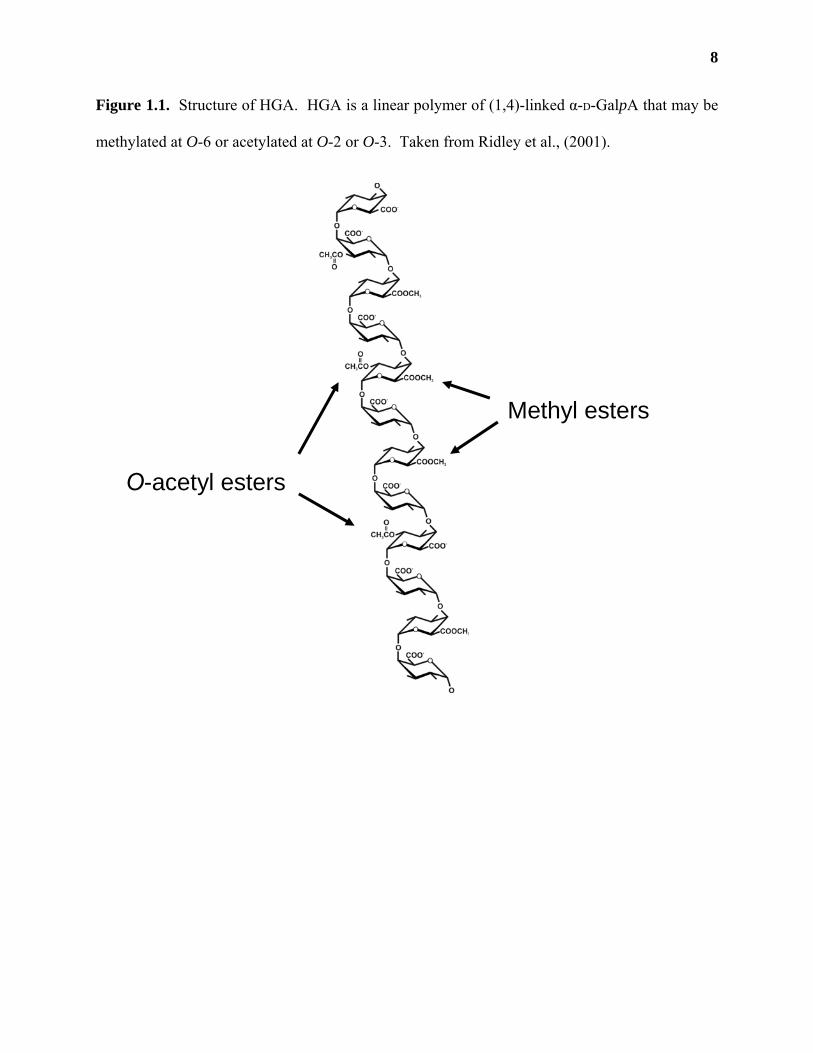
Homogalacturonan (HGA) is a linear polymer of (1,4)-linked α-D-GalpA (Figure 1.1).
Cell wall analysis of sycamore (Acer pseudoplatanus) suspension-cultured cells (O'Neill et al.,
1990; Mohnen et al., 1996) and leaves of Arabidopsis thaliana (Zablackis et al., 1995) shows
that HGA comprises roughly 24% of the primary wall. The exact size of HGA in the wall is not
known, although polymers consisting of 30-200 GalpA residues have been reported (Nothnagel
et al., 1983; Carpita and Gibeaut, 1993; Willats et al., 2001). HGA may be methylated at O-6
(Mort et al., 1993) and may contain acetyl esters at O-2 or O-3 (Ishii, 1997; Pauly and Scheller,
2000; Willats et al., 2001a). The degree and pattern of methylation and/or acetylation of HGA
chains has not been completely elucidated, possibly due to the heterogeneity of HGA polymers
or to changes in methylation/acetylation during the extraction of HGA from the wall (O'Neill et
al., 1990; Pauly and Scheller, 2000; Willats et al., 2001a). Studies on HGA fragments released
from cell wall residues by Driselase (a commercial mixture of fungal endo- and
exopolysaccharidases; (Brown and Fry, 1993; Ishii, 1997; Needs et al., 1998) and on the
8
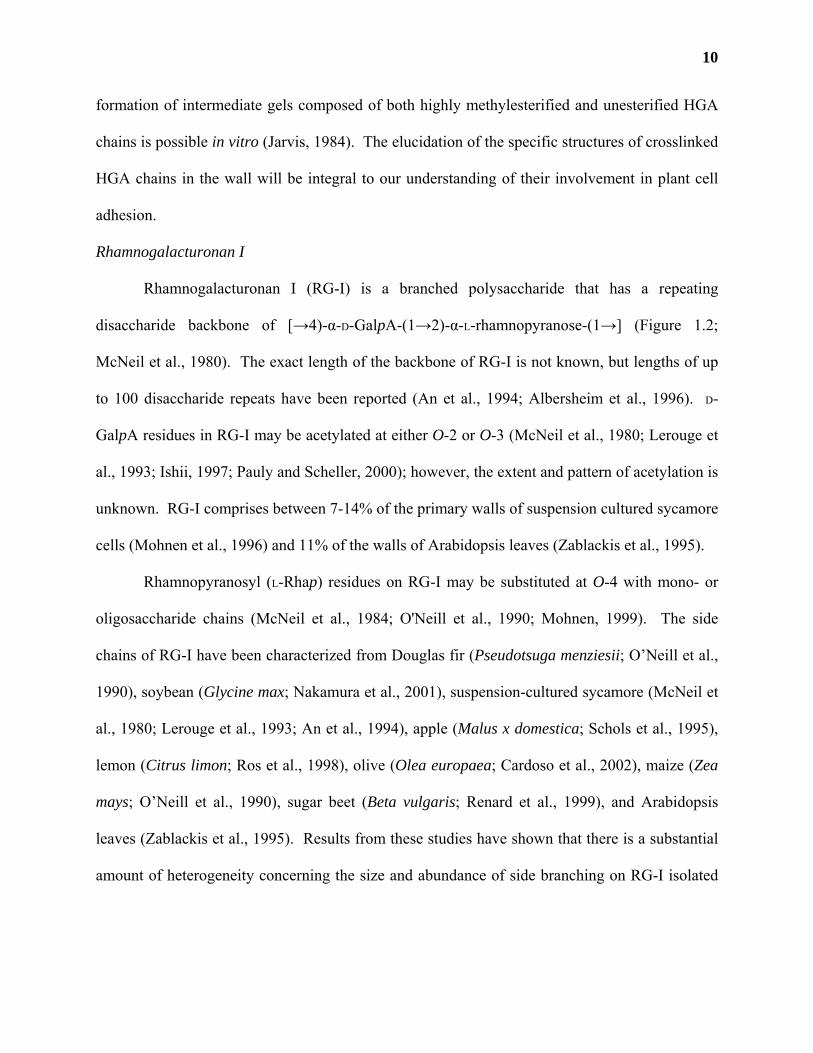
Figure 1.1. Structure of HGA. HGA is a linear polymer of (1,4)-linked α-D-GalpA that may be
methylated at O-6 or acetylated at O-2 or O-3. Taken from Ridley et al., (2001).
Methyl esters
O-acetyl esters
9
changes in apparent molecular weight of pectins treated with pectinesterases (Lee et al., 2003),
suggest that HGA may contain other as yet unidentified esters. Techniques are currently being
developed which may enable the determination of methylation and acetylation patterns of HGA
(Willats and Knox, 1999a; Clausen et al., 2003; Goubet et al., 2003).
HGA chains can form ionic crosslinks in the presence of Ca2+
Unesterified HGA chains can form ionic crosslinks in the presence of Ca2+ (Kohn, 1975;
Jarvis, 1984). The properties of Ca2+ crosslinked HGA has been extensively studied in vitro, as
Ca2+-HGA gels are used extensively in the food industry as thickening and stabilizing agents
(Thakur et al., 1997). Ca2+ crosslinked HGA is also thought to play an important role in plant
cell adhesion, as treatment of plant tissues with Ca2+ chelating agents solubilizes some of the
pectic polysaccharides from the wall and releases individual plant cells (McCann and Roberts,
1991; McCartney and Knox, 2002; Jarvis et al., 2003).
The binding of two Ca2+ crosslinked HGA molecules is thought to require HGA chains
that have a degree of polymerization of at least 7 GalpA residues (Thakur et al., 1997), with
optimal binding occurring between HGA chains that are at least 14 residues in length (Jarvis,
1984). The two chains are thought to form an “egg-box” structure, such as those described for
the Ca2+-induced gelation of alginates (Kohn, 1975). Molecular modeling studies suggest that
Ca2+ crosslinked HGA chains have a 2-fold helical conformation and are likely oriented in an
anti-parallel orientation (Braccini et al., 1999; Braccini and Perez, 2001). The specific
orientations of Ca2+ crosslinked HGA chains within the cell wall are unknown. Studies in this
area are further complicated by the fact that highly methylesterified HGA chains can form gels in
vitro in the presence of high solute concentrations at low pH (Jarvis, 1984; Tibbits et al., 1998).
While these conditions may not be suitable for gel formation within the primary wall, the
10
formation of intermediate gels composed of both highly methylesterified and unesterified HGA
chains is possible in vitro (Jarvis, 1984). The elucidation of the specific structures of crosslinked
HGA chains in the wall will be integral to our understanding of their involvement in plant cell
adhesion.
Rhamnogalacturonan I
Rhamnogalacturonan I (RG-I) is a branched polysaccharide that has a repeating
disaccharide backbone of [→4)-α-D-GalpA-(1→2)-α-L-rhamnopyranose-(1→] (Figure 1.2;
McNeil et al., 1980). The exact length of the backbone of RG-I is not known, but lengths of up
to 100 disaccharide repeats have been reported (An et al., 1994; Albersheim et al., 1996). D-
GalpA residues in RG-I may be acetylated at either O-2 or O-3 (McNeil et al., 1980; Lerouge et
al., 1993; Ishii, 1997; Pauly and Scheller, 2000); however, the extent and pattern of acetylation is
unknown. RG-I comprises between 7-14% of the primary walls of suspension cultured sycamore
cells (Mohnen et al., 1996) and 11% of the walls of Arabidopsis leaves (Zablackis et al., 1995).
Rhamnopyranosyl (L-Rhap) residues on RG-I may be substituted at O-4 with mono- or
oligosaccharide chains (McNeil et al., 1984; O'Neill et al., 1990; Mohnen, 1999). The side
chains of RG-I have been characterized from Douglas fir (Pseudotsuga menziesii; O’Neill et al.,
1990), soybean (Glycine max; Nakamura et al., 2001), suspension-cultured sycamore (McNeil et
al., 1980; Lerouge et al., 1993; An et al., 1994), apple (Malus x domestica; Schols et al., 1995),
lemon (Citrus limon; Ros et al., 1998), olive (Olea europaea; Cardoso et al., 2002), maize (Zea
mays; O’Neill et al., 1990), sugar beet (Beta vulgaris; Renard et al., 1999), and Arabidopsis
leaves (Zablackis et al., 1995). Results from these studies have shown that there is a substantial
amount of heterogeneity concerning the size and abundance of side branching on RG-I isolated
11
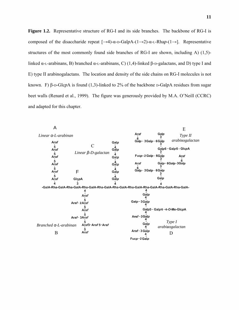
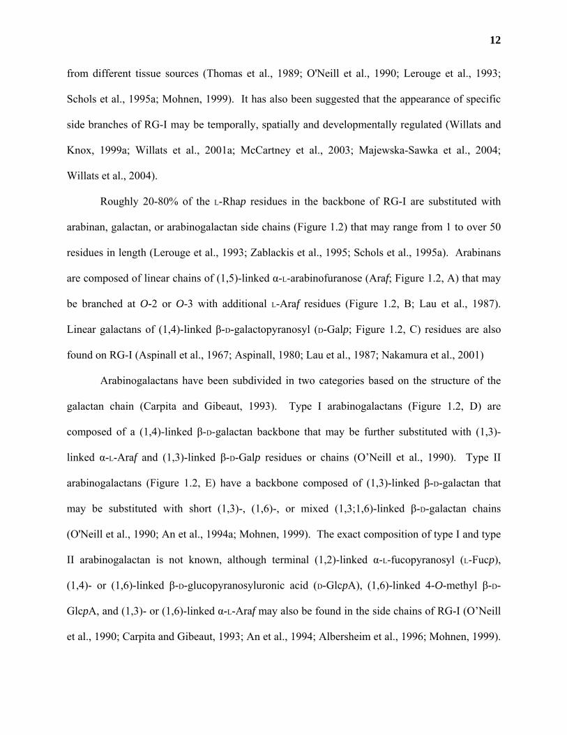
Figure 1.2. Representative structure of RG-I and its side branches. The backbone of RG-I is
composed of the disaccharide repeat [→4)-α-D-GalpA-(1→2)-α-L-Rhap-(1→]. Representative
structures of the most commonly found side branches of RG-I are shown, including A) (1,5)-
linked α-L-arabinans, B) branched α-L-arabinans, C) (1,4)-linked β-D-galactans, and D) type I and
E) type II arabinogalactans. The location and density of the side chains on RG-I molecules is not
known. F) β-D-GlcpA is found (1,3)-linked to 2% of the backbone D-GalpA residues from sugar
beet walls (Renard et al., 1999). The figure was generously provided by M.A. O’Neill (CCRC)
and adapted for this chapter.
2
A
B
C
D
E
F
Branched α-L-arabinan
Linear α-L-arabinan
2
46
2
2
Linear β-D-galactan
Type IIarabinogalactan
Type Iarabinogalactan
p
p p p
pp p
p p p
p p
p
p
p p p
f
f
f
12
from different tissue sources (Thomas et al., 1989; O'Neill et al., 1990; Lerouge et al., 1993;
Schols et al., 1995a; Mohnen, 1999). It has also been suggested that the appearance of specific
side branches of RG-I may be temporally, spatially and developmentally regulated (Willats and
Knox, 1999a; Willats et al., 2001a; McCartney et al., 2003; Majewska-Sawka et al., 2004;
Willats et al., 2004).
Roughly 20-80% of the L-Rhap residues in the backbone of RG-I are substituted with
arabinan, galactan, or arabinogalactan side chains (Figure 1.2) that may range from 1 to over 50
residues in length (Lerouge et al., 1993; Zablackis et al., 1995; Schols et al., 1995a). Arabinans
are composed of linear chains of (1,5)-linked α-L-arabinofuranose (Araf; Figure 1.2, A) that may
be branched at O-2 or O-3 with additional L-Araf residues (Figure 1.2, B; Lau et al., 1987).
Linear galactans of (1,4)-linked β-D-galactopyranosyl (D-Galp; Figure 1.2, C) residues are also
found on RG-I (Aspinall et al., 1967; Aspinall, 1980; Lau et al., 1987; Nakamura et al., 2001)
Arabinogalactans have been subdivided in two categories based on the structure of the
galactan chain (Carpita and Gibeaut, 1993). Type I arabinogalactans (Figure 1.2, D) are
composed of a (1,4)-linked β-D-galactan backbone that may be further substituted with (1,3)-
linked α-L-Araf and (1,3)-linked β-D-Galp residues or chains (O’Neill et al., 1990). Type II
arabinogalactans (Figure 1.2, E) have a backbone composed of (1,3)-linked β-D-galactan that
may be substituted with short (1,3)-, (1,6)-, or mixed (1,3;1,6)-linked β-D-galactan chains
(O'Neill et al., 1990; An et al., 1994a; Mohnen, 1999). The exact composition of type I and type
II arabinogalactan is not known, although terminal (1,2)-linked α-L-fucopyranosyl (L-Fucp),
(1,4)- or (1,6)-linked β-D-glucopyranosyluronic acid (D-GlcpA), (1,6)-linked 4-O-methyl β-D-
GlcpA, and (1,3)- or (1,6)-linked α-L-Araf may also be found in the side chains of RG-I (O’Neill
et al., 1990; Carpita and Gibeaut, 1993; An et al., 1994; Albersheim et al., 1996; Mohnen, 1999).
13
Type II arabinogalactans are also associated with arabinogalactan proteins (AGPs); however, it is
commonly accepted that these polysaccharides are indeed present as side chains of RG-I
(McNeil et al., 1984; Bacic et al., 1988; Mohnen, 1999).
Other substitutions on RG-I side chains are known to occur. For example, L-Araf and D-
Galp residues may also contain ferulic or coumaric acid (Ishii, 1997a), and in the walls of sugar
beet 2% of the D-GalpA backbone residues are substituted with D-GlcpA (Figure 1.2, F; Renard
et al., 1999). The exact roles these different side chains play in RG-I function has not yet been
elucidated, although certain tissues have been found to be enriched in RG-I molecules that have
specific side chains (Schols et al., 1995a; Ros et al., 1998; Cardoso et al., 2002; Nakamura et al.,
2002a).
Substituted galacturonans
Some polymers of HGA are substituted with mono- or oligosaccharide side branches.
These branched HGA polysaccharides are collectively known as substituted galacturonans
(SGAs). To date, there are only three types of SGA polysaccharides known to exist in primary
cell walls: xylogalacturonan (XGA), apiogalacturonan (AGA) and rhamnogalacturonan II (RG-
II).
Xylogalacturonan
XGA has been isolated from pine (Pinus mugo) pollen (Bouveng, 1965), carrot (Daucus
carota) callus (Kikuchi et al., 1996), soybean cotyledons (Nakamura et al., 2002a), cocoa
(Theobroma cacao) beans (Redgwell and Hansen, 2000), pea (Pisum sativum) hulls (Le Goff et
al., 2001), and the fruits of watermelon (Citrullus lanatus; Yu and Mort, 1996), lemon (Ros et
al., 1998), and apple (Schols et al., 1995a). XGA has a backbone of (1,4)-linked α-D-GalpA that
is substituted at O-3 with β-D-xylopyranosyl residues (D-Xylp; Figure 1.3, A; O’Neill et al.,
14
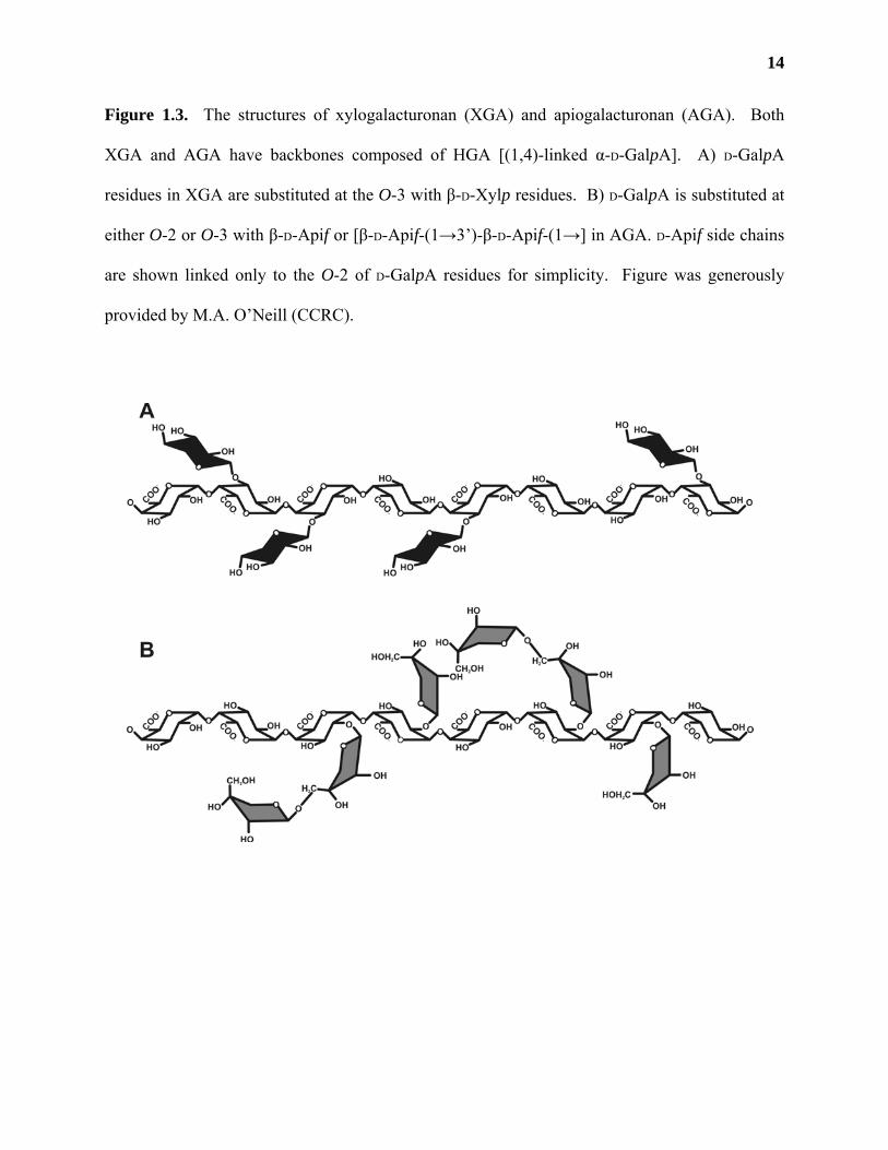
Figure 1.3. The structures of xylogalacturonan (XGA) and apiogalacturonan (AGA). Both
XGA and AGA have backbones composed of HGA [(1,4)-linked α-D-GalpA]. A) D-GalpA
residues in XGA are substituted at the O-3 with β-D-Xylp residues. B) D-GalpA is substituted at
either O-2 or O-3 with β-D-Apif or [β-D-Apif-(1→3’)-β-D-Apif-(1→] in AGA. D-Apif side chains
are shown linked only to the O-2 of D-GalpA residues for simplicity. Figure was generously
provided by M.A. O’Neill (CCRC).
15
1990; Schols et al., 1995; Kikuchi et al., 1996; Yu and Mort, 1996). The degree of D-Xylp
branching on XGA is not known.
Nakamura et al. (2002) found that polygalacturonase treatment of hot-water soluble,
soybean cotyledon polysaccharides that had previously had their arabinan and galactan chains
removed by treatment with an α-(1,3)-L-arabinosidase and a β-(1,4)-D-galactosidase, respectively
(Nakamura et al., 2001), yielded two oligosaccharides rich in D-Xylp and D-GalpA. Linkage
analysis revealed the presence of terminal, 4-, and 3,4-linked GalpA and terminal, 4-, and 2,4-
linked D-Xylp. Although treatment of these oligosaccharides with an β-(1,4)-D-xylosidase
released some D-Xylp residues from the oligosaccharides, the authors were unable to prove that
4- or 2,4-linked D-Xylp were indeed attached to D-GalpA (Nakamura et al., 2002a).
Apiogalacturonan
Apiogalacturonan (AGA; Figure 1.3, B) has been isolated from the cell walls of only a
limited number of plants species, for example duckweed (Lemna minor L; Golovchenko et al.,
2002). It has a linear backbone of HGA that can be substituted at the O-2 or O-3 with β-D-
apiofuranose (D-Apif) or [β-D-Apif-(1→3’)-β-D-Apif-(1→] (Cheng and Kindel, 1997; Ridley et
al., 2001; Golovchenko et al., 2002). Whether AGA is a component of the cell walls of different
plant species, or has special functions in aquatic monocots like duckweed, has yet to be
addressed.
Rhamnogalacturonan II
The structure of RG-II is by far the most complicated of any of the primary cell wall
polysaccharides (Figure 1.4). It is composed of 13 different monosaccharides in over 20
different linkages (O'Neill et al., 1997). Many of the sugars that make up RG-II are unusual,
such as 2-keto-3-deoxy-D-manno-octulopyranosyluronic acid (D-Kdop) and 3-deoxy-D-lyxo-2-
16
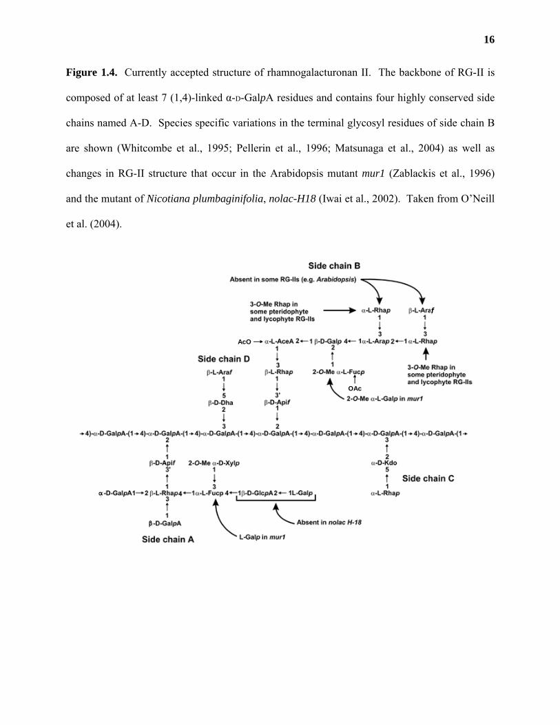
Figure 1.4. Currently accepted structure of rhamnogalacturonan II. The backbone of RG-II is
composed of at least 7 (1,4)-linked α-D-GalpA residues and contains four highly conserved side
chains named A-D. Species specific variations in the terminal glycosyl residues of side chain B
are shown (Whitcombe et al., 1995; Pellerin et al., 1996; Matsunaga et al., 2004) as well as
changes in RG-II structure that occur in the Arabidopsis mutant mur1 (Zablackis et al., 1996)
and the mutant of Nicotiana plumbaginifolia, nolac-H18 (Iwai et al., 2002). Taken from O’Neill
et al. (2004).
f
OAc
AcO
α
β
17
heptulopyranosylaric acid (D-Dhap). Some sugars, such as L-aceric acid (3-C-carboxy-5-deoxy-
L-xylofuranose; L-AcefA) are specific for RG-II and are not found anywhere else in nature.
There is some evidence that the HGA backbone of RG-II contain methylesters (Pellerin et al.,
1996) and that acetylesters may be present on the 2-O-methyl L-Fucp and L-AcefA residues of
side chain B (Whitcombe et al., 1995; Glushka et al., 2003); however, the exact pattern of
methyl- and/or acetylation has not yet been determined and may be dependent on the original
source of RG-II (Whitcombe et al., 1995; Glushka et al., 2003). RG-II accounts for 1-4% of the
walls of dicots, non-graminaceous monocots and gymnosperms (O'Neill et al., 1990; Mohnen et
al., 1996), 0.2-2% of the walls of lycophytes and pteridophytes (Matsunaga et al., 2004; O'Neill
et al., 2004), less that 0.1% of the walls of grasses (Thomas et al., 1989; O'Neill et al., 1990), and
can barely be detected in the walls of bryophytes (Matsunaga et al., 2004).
RG-II has a backbone of at least 7 (1,4)-linked α-D-GalpA residues (O'Neill et al., 1996)
and is surprisingly resistant to microbial and fungal glycosidases and glycanases, such as those
found in commercial preparations (i.e. Pectinol AC and Driselase; Stevenson et al., 1988; Ishii
and Matsunaga, 1996). RG-II can be released from primary walls by endopolygalacturonase
(EPGase) treatment (York et al., 1986) or by buffer extraction followed by EPGase treatment
(Thomas et al., 1989; Ishii et al., 2001a). Structural characterization of the side chains of RG-II
was done mainly by partial acid hydrolysis of purified RG-II, followed by analysis of the
released oligosaccharides using a combination of mass spectrometry (MS) and nuclear magnetic
resonance (NMR) techniques (Spellman et al., 1983a; Thomas et al., 1989; Puvanesarajah et al.,
1991; Whitcombe et al., 1995; Pellerin et al., 1996).
It is currently believed that RG-II consists of 4 distinct side branches named A-D.
Evidence for the attachment sites of these side branches to the backbone of RG-II has been
18
obtained by periodate oxidation (Puvanesarajah et al., 1991) and by selectively cleaving NaBH4-
reduced RG-II with a cell free extract from Penicillium daleae that is known to contain RG-II-
degrading glycanases (Vidal et al., 2000). Recent studies using 2D NMR have confirmed some
of these results (Rodriguez-Carvajal et al., 2003) and have allowed for the further refinement of
RG-II structure (Reuhs et al., 2004). These studies have led to a consensus structure of RG-II
(Figure 1.4; O’Neill et al., 2004). However, the inability to unequivocally assign all of the peaks
from 2D NMR experiments that correspond to backbone D-GalpA residues and to residues that
are directly attached to D-GalpA (Vidal et al., 2000; Ridley et al., 2001; Rodriguez-Carvajal et
al., 2003) may require that this currently accepted RG-II structure be further refined in the future.
For example, it is currently believed that L-Araf may be linked to backbone D-GalpA residues on
RG-II (Thomas et al., 1989; Pellerin et al., 1996; Matsunaga et al., 2004) and may even
constitute a new side chain (Rodriguez-Carvajal et al., 2003).
RG-II has been isolated from a number of different tissue sources, including sycamore
(Darvill et al., 1978; Whitcombe et al., 1995), sugar beet (Ishii and Matsunaga, 1996), potato
(Solanum tuberosum; Ishii, 1997), pea (O'Neill et al., 1996), rice (Oryza sativa; Thomas et al.,
1989), radish (Raphanus sativus; Kobayashi et al., 1996), Arabidopsis (Zablackis et al., 1995),
and red wine (Vidal et al., 2000). Glycosyl composition and linkage analysis have shown that
the structure of RG-II is highly conserved amongst plant species. The discovery of conserved
RG-II structures in the cell walls of lesser plants (e.g. lycophytes and pteridophytes; Matsunaga
et al., 2004) has led to the proposal that RG-II may have been required for the evolution of land
plants (Matsunaga et al., 2004; O'Neill et al., 2004).
Structural differences in glycosyl composition of RG-II isolated from different plant
species are found only on the residues attached to the O-2 and/or O-3 of the L-Arap residue of

19
side chain B (Figure 1.4; Thomas et al., 1989; Whitcombe et al., 1995; Pellerin et al., 1996;
Vidal et al., 2000; Matsunaga et al., 2004). The L-Arap is not substituted in species such as sugar
beet (Ishii and Matsunaga, 1996) and contains (1,2)-linked α-L-Rhap in Arabidopsis (Glushka et
al., 2003) and rice (Thomas et al., 1989). RG-II from red wine has α-L-Rhap and [β-L-Araf-(1→
2)-α-L-Rhap-(1→] linked to the O-3 and O-2 of the L-Arap residue, respectively (Pellerin et al.,
1996). Only the disaccharide is found attached to the O-2 of the L-Arap residue in RG-II from
sycamore (Whitcombe et al., 1995), while RG-II isolated from some lycophytes and
pteridophytes may contain 3-O-methyl β-D-Rhap linked to the O-3 and/or O-2 of the L-Arap
residue (Matsunaga et al., 2004). The functions of the different substitutions on the L-Arap
residue of side chain B are unknown, as they do not seem to have any effect on the function of
RG-II in vivo (O'Neill et al., 2004).
RG-II molecules can dimerize in the presence of boron
RG-II isolated from the primary walls of a number of different tissue sources is
predominantly (~95%) in the form of a dimer crosslinked by a 1:2 borate-diol ester (Figure 1.5;
Ishii and Matsunaga, 1996; Kobayashi et al., 1996; O'Neill et al., 1996; Pellerin et al., 1996).
The presence of dimeric RG-II (dRG-II) complexed by borate di-esters has been confirmed in all
plant species in which RG-II has been isolated, including the cell walls of lycophytes and
pteridophytes (Matsunaga et al., 2004; O’Neill et al., 2004). The approximate molecular weight
of monomeric RG-II (mRG-II) and dRG-II are 5 and 10 kDa, respectively (O'Neill et al., 1996;
Pellerin et al., 1996; Matsunaga et al., 2004). The relative proportion of dRG-II:mRG-II isolated
from walls depends on the plant source (O'Neill et al., 1996; Matsunaga et al., 2004), but the
characteristics of RG-II dimerization are the same across plant species. This suggests that the
20
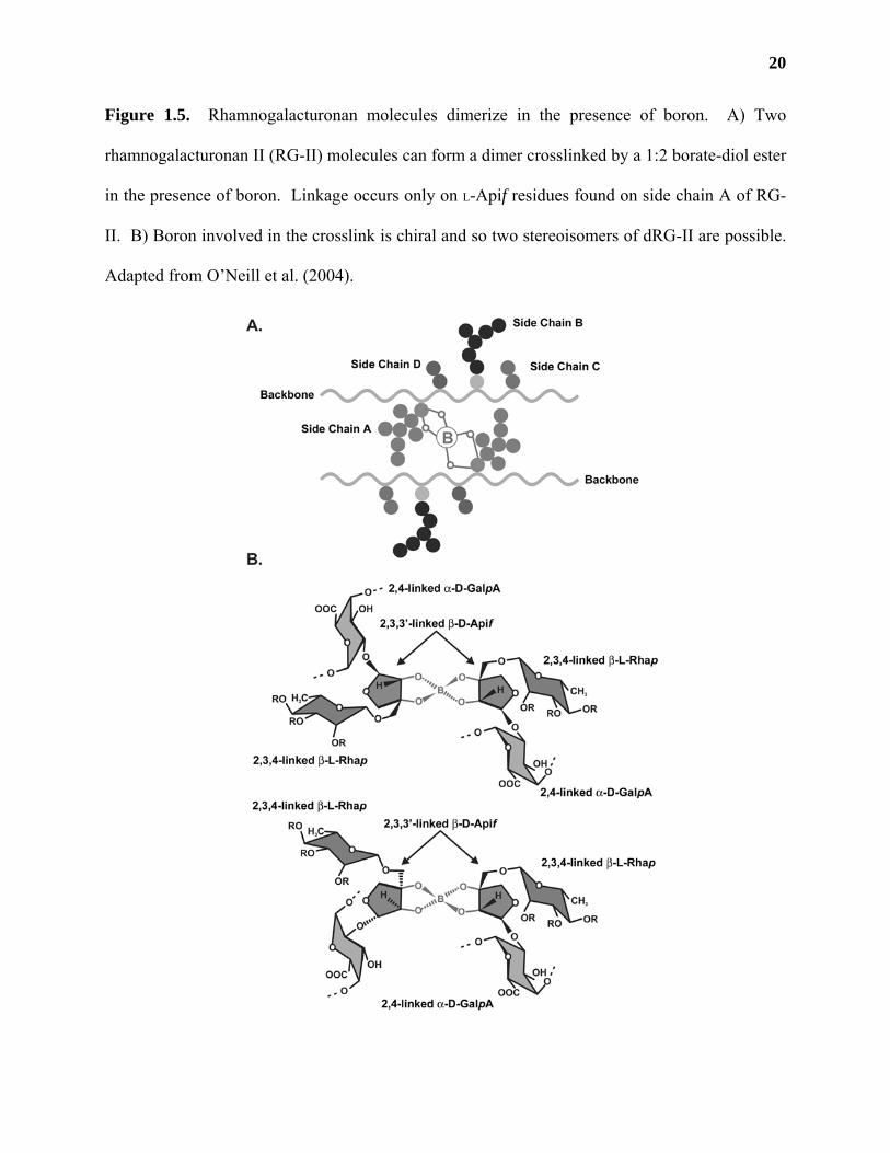
Figure 1.5. Rhamnogalacturonan molecules dimerize in the presence of boron. A) Two
rhamnogalacturonan II (RG-II) molecules can form a dimer crosslinked by a 1:2 borate-diol ester
in the presence of boron. Linkage occurs only on L-Apif residues found on side chain A of RG-
II. B) Boron involved in the crosslink is chiral and so two stereoisomers of dRG-II are possible.
Adapted from O’Neill et al. (2004).
21
chemical properties of RG-II are the same irrespective of the tissue source and the differences in
side chain B structure (O'Neill et al., 1996; Ishii et al., 1999; Matsunaga et al., 2004).
The formation of dRG-II from mRG-II in the presence of boron occurs spontaneously in
vitro at an optimum pH of 3-4 without the addition of any exogenous proteins (O'Neill et al.,
1996; Ishii et al., 1999). The addition of di- and trivalent cations with an ionic radii >1.0, such
as Pb2+, Sr2+, Ba2+, La3+, and Ce3+, greatly increases the rate of dimer formation (O'Neill et al.,
1996; Ishii et al., 1999). Ca2+ also increases this rate, but to a much lesser extent than that of the
larger cations (Ridley et al., 2001). Conversely, incubation of dRG-II at pH 1-3 in the absence of
boron causes its conversion into mRG-II (Ishii and Matsunaga, 1996; Kobayashi et al., 1996;
O'Neill et al., 1996). Glycosyl composition and linkage analysis of in vitro synthesized mRG-II
or dRG-II are identical, except for the presence of 2,3,3’-linked L-Apif found in dRG-II
molecules (O'Neill et al., 1996), providing evidence that the site of borate di-ester cross-linking
of RG-II is on L-Apif residues (Ishii et al., 1999). Selective labeling of derivatives from dRG-II
demonstrates that only the L-Apif residues on side chain A (Figure 1.5, A) participate in RG-II
dimerization (Ishii et al., 1999). Two diastereoisomers of dRG-II are possible due to the fact that
the boron molecule involved in the crosslink is chiral (Figure 1.5, B; O’Neill et al., 2004).
Further research needs to be conducted to determine which of these stereoisomers is commonly
found in nature.
PECTIN IN THE PRIMARY WALL
HGA, RG-I, and RG-II may be covalently linked in the primary cell wall
There is compelling evidence that HGA, RG-I and RG-II may be covalently linked in the
primary walls of plant cells (Thomas et al., 1989). HGA, RG-I and RG-II can be isolated as a
high molecular complex (>100 kDa) by extracting primary walls with aqueous buffers (O'Neill et
22
al., 1990; Ishii et al., 2001a; Reuhs et al., 2004). Saponification of this complex followed by
enzymic digestion with purified EPGases causes the release of HGA oligosaccharides
(oligogalacturonides or OGAs) and RG-II from the remaining high molecular weight material,
which consists mainly of RG-I (McNeil et al., 1980; York et al., 1986; O'Neill et al., 1990;
Lerouge et al., 1993; O'Neill et al., 1997). Ishii et al. (2001a) found that treatment of the original
high molecular weight complex for one hour with 1 N HCl (a treatment that is known to strip
boron from dRG-II in vitro) changed its elution profile following size exclusion chromatography.
Incubation of the acid-treated material for 16 h in a buffer containing boric acid and lead acetate
at pH 7.3 (conditions known to cause the formation of dRG-II from mRG-II in vitro), caused the
acid-treated pectin to elute at the same retention time as the original high molecular weight
complex. These results suggest that RG-II dimerization can change the molecular weight
distribution of the high molecular weight pectin complex and provides further evidence that
pectic polysaccharides may be covalently linked in the wall.
Evidence that RG-II may be covalently linked to HGA stems from the observation that
the lengths of the HGA backbones on EPGase-released RG-II molecules are heterogeneous
(Whitcombe et al., 1995; Pellerin et al., 1996; Vidal et al., 2000). Partial acid hydrolysis of RG-
II from sycamore (Whitcombe et al., 1995) and from red wine (Pellerin et al., 1996) releases RG-
II side chains and OGAs of degrees of polymerization of 7-15 (Ishii et al., 1999) with the
average length being between 7 and 9 D-GalpA residues for sycamore and red wine RG-II,
respectively (O'Neill et al., 1996). This variation in the length of the RG-II backbones is most
likely the result of the incomplete hydrolysis of RG-II from the high molecular weight complex
by EPGase (Whitcombe et al., 1995; Ishii et al., 1999) or the incomplete digestion of cell wall
polysaccharides during the fermentation of red wine (Pellerin et al., 1996). Furthermore,
23
treatment of sycamore RG-II with a purified exopolygalacturonase (exoPGase) releases D-GalpA
residues from RG-II (Whitcombe et al., 1995), suggesting that RG-II molecules have (1,4)-linked
α-D-GalpA residues attached to their backbones that are accessible to exoPGase.
The high molecular weight material that remains following treatment of buffer-soluble
pectic polysaccharides with EPGase is composed mainly of RG-I (McNeil et al., 1980; York et
al., 1986). Lerouge et al. (1993) conducted glycosyl composition analysis of this fraction from
sycamore suspension-cultured cells and found that the molar ratios of D-GalpA and L-Rhap
varied in several RG-I preparations. Treatment of these RG-I preparations with exoPGase
released D-GalpA as the only monosaccharide and resulted in RG-I fractions that had equimolar
ratios of D-GalpA:L-Rhap. These results suggested that (1,4)-linked α-D-GalpA residues were
covalently attached to RG-I molecules.
Further evidence of a covalent attachment between HGA and RG-I was demonstrated
using a hot-water soluble polysaccharide fraction from defatted soybean cotyledons (Nakamura
et al., 2002a). Treatment of this fraction with a mixture of purified pectinolytic enzymes [(1,3)-
α-L-arabinosidase, (1,5)-α-L-endoarabinanase, (1,4)-β-D-galactosidase and (1,4)-β-D-
endogalactanase, EPGase, and a rhamnogalacturonan α-D-galactopyranosyluronohydolase]
released oligosaccharides that, upon separation by anion exchange chromatography, had varying
ratios of L-Rhap:D-GalpA (ranging from 2.7 to 10.4). Fast atom bombardment (FAB) MS
analysis of these oligosaccharides suggested the presence of L-Rhap residues within short
stretches of (1,4)-linked α-D-GalpA chains; however, the relative placement of these L-Rhap
residues could not be determined due to the low abundance of fragmentation ions during analysis.
Structural analysis of fragments released by enzymic digestion of RG-I from the high
molecular weight complex from soybean walls revealed the presence of segments of XGA
24
(Nakamura et al., 2002, 2002a). XGA fragments associated with RG-I molecules have also been
detected in extracts from apple (Schols et al., 1995a), lemon (Ros et al., 1998), pea (Le Goff et
al., 2001) and sugar beet (Ishii and Matsunaga, 2001). It is unknown at this time whether or not
the association between RG-I and XGA is due to a covalent interaction, or is simply caused by
the natural propensity of pectic polysaccharides to aggregate in solution (Mort et al., 1991).
Evidence for the presence of other covalent linkages between pectic polysaccharides
HGA, RG-I and RG-II may also be held in the wall by other types of covalent linkages.
Approximately 50% of the pectic polysaccharides found in primary walls are solubilized by
treatment with aqueous buffers or purified EPGase (O'Neill et al., 1990; Zablackis et al., 1995;
Ishii and Matsunaga, 2001; Reuhs et al., 2004). The remaining pectic material can be solubilized
from walls by treating them with mild alkali, a procedure known to break ester linkages and to
solubilize neutral hemicelluloses, such as xyloglucan (York et al., 1986; Zablackis et al., 1995).
Cold alkali treatment of a commercially available fractionated pectin powder (FPP) from
citrus peels caused the high molecular weight component of this fraction to migrate as uniformly
sized OGAs on polyacrylamide gels (Jackson et al., 2004). OGA formation was not due to β-
elimination as an increase in A232 was not observed following base treatment. These OGAs were
not produced by treating FPP with a purified EPGase alone or in combination with a pectin
methylesterase; however, cold alkali followed by EPGase treatment of FPP caused its complete
degradation. These results suggested that the OGAs present in FPP were resistant to EPGase
treatment and that this resistance was not due to O-6 methylation. Driselase treatment of cell
wall material from spinach (Spinacia oleracea), carrot, rose (Rosa sp. 'Paul's Scarlet'), and tall
fescue (Festuca arundinacea) suspension cultured cells (Brown and Fry, 1993) and from carrot
root (Needs et al., 1998) also released OGAs that possessed unidentified esters that were resistant
25
to pectinolytic degradation. The structures of these esters, and of those found in FPP, have yet to
be described.
Pectin methylesterases (PEs) are enzymes that remove methyl groups from pectin
molecules (Micheli, 2001). It has been suggested that another function of pectinesterases may be
to covalently link pectic polysaccharides in the wall (Lee et al., 2003). Treatment of citrus pectin
with a purified pectinesterase (PE) in vitro caused it to migrate as a higher molecular weight
complex on size exclusion columns (Lee et al., 2003), suggesting that PEs may be able to
catalyze reactions that increase the molecular weight of pectin molecules in the wall.
Ferulic and coumaric esters have been found attached to L-Araf and D-Galp residues from
Driselase-treated walls of spinach suspension-cultured cells and sugar beet pulp (Fry, 1982; Ishii,
1994). These esters could allow pectic molecules to crosslink to other cell wall components by
oxidative coupling (Ishii, 1997a); however, evidence for such crosslinked components has yet to
be demonstrated.
Mild alkali treatment of suspension-cultured rose cells released a polysaccharide complex
containing both pectin and xyloglucan molecules (Thompson and Fry, 2000). These molecules
co-eluted on anion exchange columns, even though xyloglucan itself contains no acidic residues.
Furthermore, it has been demonstrated that a membrane-enriched fraction from pea synthesized a
galactan that could be solubilized by treating the radioactive products with pectin- or xyloglucan-
degrading enzymes (Abdel-Massih et al., 2003). Both of these studies suggest that pectin and
xyloglucan are associated in the wall, although data describing the nature of this attachment has
yet to be presented.
26
Simplified model of pectin in the wall
Several models describing the association of pectic polysaccharides within the primary
wall have been presented (Keegstra et al., 1973; McCann and Roberts, 1991; Carpita and
Gibeaut, 1993). The first models depicted pectic polysaccharides crosslinked to each other and
to other cell wall components through covalent linkages (Keegstra et al., 1973). The basis for
this model was derived from the fact that 1) pectins isolated from the wall by chemical or
enzymic methods co-chromatographed on ion exchange and size exclusion columns with
xyloglucan polymers (Keegstra et al., 1973; Thompson and Fry, 2000), 2) that EPGase treatment
of wall material allowed the release of higher amounts of cell wall proteins following protease
digestion than untreated walls (Keegstra et al., 1973), and 3) that associations between RG-I and
AGPs are possible as both molecules contain type II arabinogalactans (McNeil et al., 1984; Bacic
et al., 1988; Carpita et al., 2001). The validity of this model was questioned when glycosyl
composition and linkage analysis of cell wall material failed to demonstrate the presence of
covalent crosslinks between pectin and other wall components (McCann and Roberts, 1991;
Carpita et al., 2001). Furthermore, electron microscopy of CDTA or Na2CO3 extracted walls
revealed an independent, crosslinked network of cellulose and hemicellulose polysaccharides
(McCann and Roberts, 1991), suggesting that pectins in the wall formed an independent, gel-like
matrix that could be extracted from the other cell wall components.
Recent structural analysis of the wall has revealed the presence of covalent crosslinks
between specific classes of pectic polysaccharides (Spellman et al., 1983; Whitcombe et al.,
1995; Pellerin et al., 1996; Ishii and Matsunaga, 2001; Nakamura et al., 2002, 2002a). Based on
these observations, newer models for the association of pectic polysaccharides within the wall
have been presented (Carpita and Gibeaut, 1993; Willats et al., 2001; Nakamura et al., 2002a;
27
Vincken et al., 2003). Most models of pectin in the wall depict it as stretches of RG-I covalently
attached to polymers of HGA in an alternating pattern (Carpita and Gibeaut, 1993; Nakamura et
al., 2002). However, one model suggests that pectic polysaccharides are composed of large RG-I
polymers that have side branches of HGA, RG-II, XGA, arabinans, galactans and
arabinogalactans (Vincken et al., 2003). These authors go on to suggest that associations
between RG-I polymers occur in areas of overlapping regions of HGA and RG-II side branches,
and that these overlapping regions are the primary sites of Ca2+ and boron crosslinking of pectic
polysaccharides, respectively.
Glycosyl composition and linkage analysis of RG-I polymers from sycamore (McNeil et
al., 1980; York et al., 1986; O'Neill et al., 1990; Lerouge et al., 1993; An et al., 1994; An et al.,
1994a), soybean (Nakamura et al., 2001; Nakamura et al., 2002a), cocoa (Redgwell and Hansen,
2000), rice (Thomas et al., 1989), lemon (Ros et al., 1998), Douglas fir (O'Neill et al., 1990),
sugar beet (Renard et al., 1999; Ishii and Matsunaga, 2001), apple (Schols et al., 1995a), pea (Le
Goff et al., 2001), and Arabidopsis (Zablackis et al., 1995) failed to provide any evidence for the
existence of D-GalpA residues attached as side chains to the backbone of RG-I. Therefore, a new
model of the association of pectic polysaccharides in the wall based on current structural data is
presented (Figure 1.6).
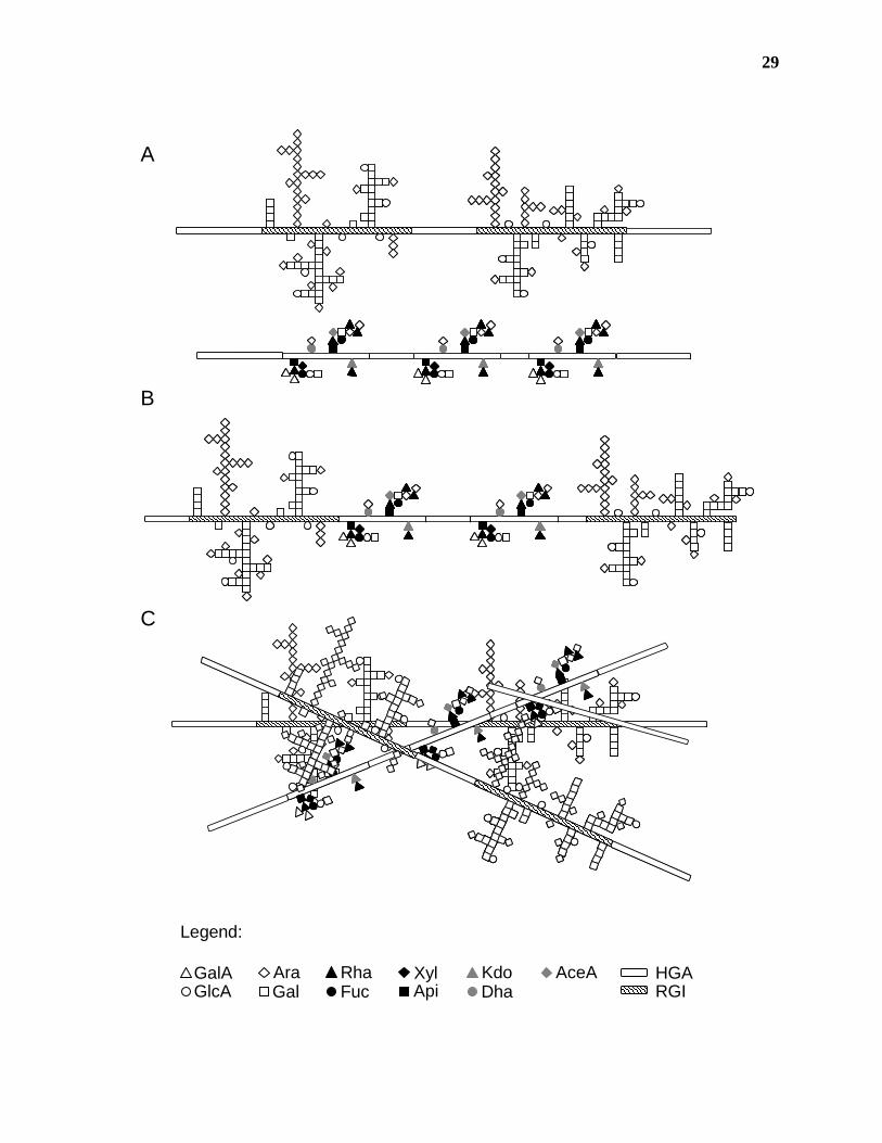
The covalent attachment of HGA to either RG-I or RG-II polymers has been
demonstrated (Figure 1.6, A; Lerouge et al., 1993; Ishii and Matsunaga, 2001; Nakamura et al.,
2002a; Reuhs et al., 2004). The high molecular weight of pectins extracted from the wall
suggests that HGA stretches link several RG-I and RG-II molecules either as independent
polymers (Figure 1.6, A) or to each other (Figure 1.6, B). The possibility also exists that
28
Figure 1.6. Model of the association of pectic polysaccharides in the primary cell wall. A)
Recent structural analyses of primary cell wall polysaccharides provide evidence for the covalent
attachment of HGA to independent polymers of RG-I and RG-II. Pectin isolated from the cell
wall is composed of a high molecular weight complex of all three pectic polysaccharides. B) It
is not known whether this complex is composed of HGA, RG-I and RG-II polymers covalently
attached to each other in a random or ordered fashion, or C) if the RG-I:HGA and RG-II:HGA
molecules are separate pectic species that associate in the wall due to intermolecular attractions.
29
GlcAAraGal
GalA Rha XylApiFuc
KdoDha
AceA
Legend:
HGARGI
A
B
C
30
independent RG-I:HGA and RG-II:HGA polymers form the high molecular weight complex due
to ionic or other intermolecular interactions, such as Ca2+ crosslinking or hydrogen bonding
(Figure 1.6, C; Mort et al., 1991; Tibbits et al., 1998; Braccini et al., 1999; Braccini and Perez,
2001). Further research into the structure of pectic polysaccharides in muro is required before
accurate models describing pectin function and biosynthesis can be designed.
Location of pectin epitopes in the primary wall
One of the reasons that it has been difficult to design accurate models of pectin structure
in the wall is that pectic polysaccharides are heterogeneous and specific pectin populations can
be isolated from the walls of different tissues (Thomas et al., 1989; O'Neill et al., 1990; Oxenboll
Sorensen et al., 2000; Nakamura et al., 2001). Furthermore, it has been shown that the presence
of pectic polysaccharides in the wall may be developmentally (McCartney et al., 2003), spatially
(Willats et al., 2001a), and temporally (McCartney and Knox, 2002) regulated. Differences in
pectic polysaccharide expression may also occur within the walls of individual plant cells (Orfila
and Knox, 2000).
Immunocytochemistry using antibodies generated against purified pectin fragments
(Knox et al., 1990; Willats et al., 1999b; Willats et al., 2004) or isolated by screening phage
display libraries (Williams et al., 1996; Willats et al., 1999) has increased our understanding of
the distribution of pectic polysaccharides in the primary wall. Antibodies such as LM5 (Jones et
al., 1997) and LM6 (Willats et al., 1998) have been well characterized and bind to specific side
chains of RG-I (Table 1.1). JIM5, JIM7, LM7, and PAM1 all bind to HGA chains with differing
degrees of O-6 methylesterification (Knox et al., 1990; Willats et al., 2000), while LM8 and
CCRC-R1 are thought to bind to XGA and RG-II, respectively. The exact pectic epitope(s)
recognized by these latter antibodies is still largely unknown. Synthesis and/or
31
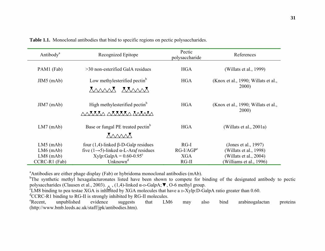
Table 1.1. Monoclonal antibodies that bind to specific regions on pectic polysaccharides.
Antibodya Recognized Epitope Pectic polysaccharide References
PAM1 (Fab) >30 non-esterified GalA residues HGA (Willats et al., 1999)
JIM5 (mAb) Low methylesterified pectinb
HGA (Knox et al., 1990; Willats et al., 2000)
JIM7 (mAb) High methylesterified pectinb
HGA (Knox et al., 1990; Willats et al., 2000)
LM7 (mAb) Base or fungal PE treated pectinb
HGA (Willats et al., 2001a)
LM5 (mAb) four (1,4)-linked β-D-Galp residues RG-I (Jones et al., 1997) LM6 (mAb) five (1→5)-linked α-L-Araf residues RG-I/AGPe (Willats et al., 1998) LM8 (mAb) Xylp:GalpA = 0.60-0.95c XGA (Willats et al., 2004)
CCRC-R1 (Fab) Unknownd RG-II (Williams et al., 1996) aAntibodies are either phage display (Fab) or hybridoma monoclonal antibodies (mAb). bThe synthetic methyl hexagalacturonates listed have been shown to compete for binding of the designated antibody to pectic polysaccharides (Clausen et al., 2003). , (1,4)-linked α-D-GalpA;▼, O-6 methyl group. cLM8 binding to pea testae XGA is inhibited by XGA molecules that have a D-Xylp:D-GalpA ratio greater than 0.60. dCCRC-R1 binding to RG-II is strongly inhibited by RG-II molecules. eRecent, unpublished evidence suggests that LM6 may also bind arabinogalactan proteins (http://www.bmb.leeds.ac.uk/staff/jpk/antibodies.htm).
32
purification of well-defined pectic structures coupled with immunodot binding and competitive
enzyme-linked immunoabsorbant assays (ELISAs) will aid in the determination of the specific
epitope(s) that these antibodies recognize (Willats and Knox, 1999a; Clausen et al., 2003). The
antibodies listed in Table 1.1 have been extensively used to analyze the cell- and tissue-
dependent distribution of specific pectin epitopes during different developmental stages of the
plant life cycle and have given new insight into the potential roles that pectin may play in the
wall and in the plant (McCann and Roberts, 1991; Willats et al., 2001).
The degree and pattern of methylesterified HGA seems to be spatially and
developmentally regulated (Willats et al., 2001). Analysis of the cell walls of tissues from plants
such as pea (Willats et al., 2001a), sugar beet (Majewska-Sawka et al., 2002; Majewska-Sawka
et al., 2004), carrot (Willats et al., 1999b), clover (Trifolium pratense L.; Lynch and Staehelin,
1992), Arabidopsis (Dolan et al., 1997; Willats et al., 1999), potato (Bush et al., 2001), and
tomato (Lycopersicon esculentum; Orfila and Knox, 2000) have shown that HGA chains with
different patterns of methyl esterification are differentially distributed across the primary cell
wall. Unesterified HGA chains (recognized by PAM1) or those with relatively low degrees of
methylation (DM; recognized by JIM5) are localized to the middle lamella, the cell corners, and
at the interface between the cell wall and the plasma membrane. This distribution is even more
restricted in walls stained with LM7, since the epitope it recognizes is found only at the
expanded middle lamella of cell corners and/or the outer edges of the wall that line the
intercellular space (Willats et al., 2001a). LM7 is thought to recognize HGA with a non block-
wise pattern of methylesterification based on its ability to bind only to pectins that have been
generated by base de-esterification or by treatment with fungal pectin methylesterases (Willats et
al., 2001a). However, recent studies using synthetic methyl hexagalacturonates with varying
33
patterns of methylesterification suggest that the LM7 epitope may recognize HGA chains with a
more sparsely distributed pattern of methylesterification than that recognized by JIM5 (Clausen
et al., 2003).
Conversely, more highly methylated HGA chains recognized by JIM7 are distributed
evenly throughout the cell walls of all tissues that have been studied (Carpita and Gibeaut, 1993;
Willats et al., 2001). JIM7 also uniformly labels the walls of pea testae at different days post
anthesis (McCartney and Knox, 2002), and the walls of sugar beet during different stages of
anther development (Majewska-Sawka et al., 2004). JIM5 and PAM1 epitopes are restricted to
specific cell layers in pea testae (McCartney and Knox, 2002) and JIM5 labeling in anthers from
sugar beet shows that the epitope appears only in meiocyte walls after cells have entered meiotic
prophase (McCartney and Knox, 2000). These results indicate that different regions of the cell
wall may have discrete domains of HGA polymers that differ in their degree and pattern of
methylesterification.
Great diversity in pectic epitope distribution is also visualized in cells stained with
antibodies against side chains of RG-I (e.g. LM5 and LM6). LM5 and LM6 recognize (1,4)-
linked β-D-galactans and [1,5]-linked α-L-arabinans, respectively; however, recent, unpublished
results suggest that LM6 may also bind to arabinogalactan proteins (AGPs;
http://www.bmb.leeds.ac.uk/staff/jpk/antibodies.htm). In pea cotyledons, LM5 epitopes appears
late in seed development, occurring at 25-34 days after anthesis (McCartney et al., 2000). This
epitope is also found adjacent to the plasma membrane of parenchyma cells, and is absent from
the outer epidermal cell wall. LM6 epitopes are seen throughout the wall in all cell layers, and
its appearance is not as highly regulated as that of LM5. Distal root cap, vascular cylinder and
vascular cortex cells from carrot exhibit strong LM5 labeling, while cells of the meristem and of
34
the cortical cells emerging from it stain with LM6 (Willats et al., 1999b). The appearance of the
galactan epitope also correlates with root cell elongation as strong LM5 staining is seen in cells
of the stele, and the endodermal and cortical cell layers in transition zone of the root (McCartney
et al., 2003). The LM5 epitope is absent in the endodermal and cortical cell layers in more
mature regions of the root, suggesting that the appearance of the LM5 epitope in these cell layers
is a transient event (McCartney et al., 2003). The appearance of the LM5 epitope also seems to
occur transiently in pea testae, as LM5 labeling of the macrosclereid layer attenuates with
increasing days after anthesis. In contrast, strong LM6 labeling appears only in the crushed
parenchyma layer late (30 days after anthesis) in testae development (McCartney and Knox,
2002). LM5 and LM6 epitopes are present in different cell types and at different stages of sugar
beet anther development. (Majewska-Sawka et al., 2004). Weak LM5 labeling is detected in
walls of the epidermis and endothecium in premeiotic anthers while strong labeling of meiocyte
walls appears during meiotic prophase and persists as microspores mature. LM6 labels anther
walls during all stages of anther development whereas meiocytes lose LM6 labeling as they
mature into microspores. The walls of pit fields from mature green tomato pericarp tissue also
show differential staining of LM5 and LM6 (Orfila and Knox, 2000). The LM6 epitope is
present throughout the cell wall while the LM5 epitope is absent from walls of pit fields. Results
from these studies indicate that subsets of RG-I molecules, and possibly AGPs, possessing
distinct side chains exist in specific tissues, at different times, and at different developmental
stages within the wall.
LM8 is an antibody that recognizes XGA from pea testae (Le Goff et al., 2001; Willats et
al., 2004). The LM8 epitope is detected in only two areas of the pea plant: the crushed
parenchyma layer of pea testae that is recognized by LM6, and cells of the root cap. LM8
35
staining of the crushed parenchyma layer is developmentally regulated. Its appearance in this
cell layer mimics that of the LM6 epitope, with greater staining appearing late in pea testae
development (~25 days after anthesis). A gradient of LM8 staining occurs in the root cap, with
greater staining occurring in cells that are in the process of separating from the root. The
localization of the LM8 epitope to these specific tissues seems to be common to angiosperms, as
LM8 labeling was detected in the same tissues from lupin (Lupinus arboreus), carrot, maize and
Arabidopsis (Willats et al., 2004).
The epitope that is recognized by the RG-II-specific antibody CCRC-R1, has not been
defined (Williams et al., 1996). Competitive ELISA and immunolabeling studies show that
binding to RG-II is only inhibited by excess RG-II, and not by individual RG-II
monosaccharides or OGAs. Labeling of sycamore suspension-cultured cells with CCRC-R1
occurs in all areas of the cell wall except the middle lamella. Further characterization of the
epitope recognized by CCRC-R1 will be required before definitive conclusions can be made
concerning the localization of RG-II in the wall.
The differential staining of pectic epitopes spatially within cell walls and tissues, and
during different developmental processes, suggests that specific subsets of pectin molecules may
play specific roles in the wall. One of the problems that is associated with the interpretation of
immunocytochemistry studies such as the ones listed above is that it is difficult to determine
whether the appearance of these epitopes is due to de novo biosynthesis of the pectic epitopes or
to the unmasking of existing epitopes within the wall (Mohnen, 1999).
Furthermore, the appearance of specific epitopes does not automatically prove a
particular function within the wall. For example, transgenic potato plants expressing a cell wall-
targeted fungal (1,4)-β-D-endogalactanase (Oxenboll Sorensen et al., 2000) or a Golgi-targeted
36
(1,5)-α-L-endoarabinanase (Skjot et al., 2002) were decreased by roughly 70% in tuber cell walls
in galactan and arabinan content, respectively. The transgenic plants grew normally and had no
apparent phenotypes, suggesting that the loss of these polysaccharides in the tuber had no effect
on plant viability.
The effects of transgenic expression of glycanases in these studies may be dependent on
the location of enzyme expression and on the type of enzyme that is expressed. For example,
cell wall-targeted expression of the fungal (1,5)-α-L-endoarabinanase produced transgenic plants
with severe phenotypes, such as the production of plants lacking side shoots, flowers, stolons and
tubers (Skjot et al., 2002). Furthermore, potato plants expressing a fungal rhamnogalacturonan
lyase had a variety of adverse phenotypes including the production of smaller tubers, changes in
pectic polysaccharide glycosyl composition and extractability, and altered localization of LM5
and LM6 epitopes in the wall (Oomen et al., 2002). Results such as these suggest that more
research into the effects of changes in pectin structure on plant growth and development will be
required before accurate models describing pectin function can be designed.
PECTIN FUNCTION
The elucidation of the functions of pectin in the wall has been challenging due to the
paucity of published studies where specific well-characterized changes in pectin structure caused
changes in pectin function. Based on correlations between the location of pectic epitopes,
changes in the expression of pectinolytic enzymes during different developmental stages, and the
mechanical properties of pectins in vitro, pectic polysaccharides have been implicated in fruit
ripening (Rose et al., 1998; Orfila et al., 2001), flower and leaf abscission (Roberts et al., 2002),
pollen differentiation (Rhee and Somerville, 1998), root cap cell differentiation (Stephenson and
Hawes, 1994; Willats et al., 2004), and the control of cell wall porosity (McCann and Roberts,
37
1991). Pectin has also been implicated in the elicitation of plant defense responses based on the
ability of purified oligogalacturonides (OGAs) from plant cell walls or purified pectins (Spiro et
al., 1993) to cause the accumulation of phytoalexins (Nothnagel et al., 1983), and reactive
oxygen species (Ridley et al., 2001). Additionally, these OGAs are involved in the regulation of
plant growth and development as it has been shown that discretely sized OGAs can change the
morphogenetic fate of tobacco (Nicotiana tabacum L. cv. Samsun) thin cell-layer explants
(Eberhard et al., 1989). Recently, a pectic fraction purified from lily (Lilium longiflorum Thumb
cv. Nelly White) styles has been shown to be required for pollen tube binding in an in vitro
adhesion assay, suggesting that pectin is also involved in pollen tube adhesion (Mollet et al.,
2000).
Several excellent reviews on the possible involvement of pectic polysaccharides in these
processes have been written (Darvill et al., 1992; Carpita and Gibeaut, 1993; Mohnen and Hahn,
1993; Hadfield and Bennett, 1998; Brummell and Harpster, 2001; Ridley et al., 2001; Willats et
al., 2001; Roberts et al., 2002). The functions of pectins described in this section represent cases
where changes in pectin function occurred due to well-defined changes in cell wall pectin
structure.
Boron-induced RG-II dimerization is important for plant growth
Boron is an essential micronutrient in plants and is absolutely required for plant growth
(Blevins and Lukaszewski, 1998). The basis for this requirement went largely unknown for
almost a century, until the recent discovery that RG-II forms dimers in the presence of boron
(Ishii and Matsunaga, 1996; Kobayashi et al., 1996; O'Neill et al., 1996). This discovery has led
to the proposal that one of the functions of boron in plants is to cross-link RG-II polymers
(O'Neill et al., 2004). Evidence for the importance of boron-induced RG-II dimer (dRG-II)
38
formation in plants is seen when plants are grown in the absence of boron (Fleischer et al., 1999;
Ishii et al., 2001a). Pumpkin (Cucurbia moschata Duchesne, cv. Tokyou-Kabocha) seedlings
grown in the absence of boron for one week had approximately a 10-fold reduction in the amount
of boron in the second, third and fourth leaves compared to normally grown seedlings (Ishii et
al., 2001a). This reduction resulted in a corresponding increase in the amount of RG-II monomer
(mRG-II) that could be extracted from the cell walls. Boron deficient cell walls were
significantly swollen compared to wild type walls, a phenotype that could be reversed by treating
boron-deficient plants for 5 h with boric acid. Similarly, suspension-cultured Chenopodium
album cells grown in the absence of boron have significantly greater wall pore size (Fleischer et
al., 1999). The difference in wall pore size between boron-deficient and wild type cells could be
corrected within 50 minutes by the addition of boric acid to the culture medium. Glycosyl
composition and linkage analysis showed no difference in the structure of RG-II isolated from
wild type and boron-deficient tissues (Fleischer et al., 1999; Ishii et al., 2001a) and incubation of
mRG-II from deficient or wild type pumpkin seedlings in the presence of lead acetate and boric
acid showed that RG-II molecules were equally capable of forming dRG-II (Ishii et al., 2001a).
Further evidence for the requirement of dRG-II formation for plant growth is seen in
mur1 plants of Arabidopsis (O'Neill et al., 2001). MUR1 encodes a GDP-D-mannose-4,6-
dehydratase that is required for the synthesis of GDP-L-Fucp (Bonin et al., 1997). Mutant plants
exhibiting the mur1 mutation have 5% of the wild type levels of fucose in their cell walls (Reiter
et al., 1993). These plants are dwarfed, have brittle leaves, and have significantly reduced wall
mechanical strength (Reiter et al., 1997; Ryden et al., 2003).
Cell wall analysis of mur1 plants demonstrated that L-Fucp was replaced by L-Galp on the
side chains of RG-II (Figure 1.4), causing a concurrent reduction (50%) in the level of 2-O-
39
methyl D-Xylp in side chain A (O'Neill et al., 2001; Reuhs et al., 2004). Further analysis
demonstrated that dRG-II accounted for only 50% of the total RG-II in mur1 plants, while the
dimer accounted for 95% of the total RG-II in wild-type plants, suggesting that the exchange of
L-Fucp for L-Galp and/or the reduction in 2-O-methyl D-Xylp inhibited the ability of RG-II to
dimerize in the presence of boron. This hypothesis was confirmed by the fact that mur1 dRG-II
forms less rapidly and is less stable than wild-type dRG-II in vitro (O'Neill et al., 2001).
The mur1 phenotype can be corrected by spraying plants with L-Fucp (Reiter et al., 1993)
or with boric acid (O'Neill et al., 2001). Spraying mur1 plants with L-Fucp restores the wild-type
glycosyl composition of RG-II, wild-type levels of fucose in the wall, and wild-type levels of
dRG-II that can be extracted from the walls of mur1 plants (O'Neill et al., 2001). Mur1 plants
sprayed with boric acid are not altered in the glycosyl composition of RG-II; however, these
plants grow normally, have wild-type levels of dRG-II that can be isolated from their walls, and
have walls with comparable mechanical strength as those of wild type walls (O'Neill et al., 2001;
Ryden et al., 2003). These results suggest that a plant’s ability to form dRG-II in the presence of
boron has a direct effect on plant growth and on the mechanical properties of the cell wall.
Pectins are involved in cell adhesion
The Ca2+ cross-linked matrix formed by HGA chains at the middle lamella is thought to
be one of the main forces involved in plant cell adhesion (McCann and Roberts, 1991; Willats et
al., 2001a). This is based in part by the fact that pectins and individual plant cells can be
released from plant material by treatment by with Ca2+ chelating agents such as trans-(1,2)-
diaminocyclohexane-N, N, N’, N’-tetraacetic acid (CDTA; Thomas et al., 1989; McCann and
Roberts, 1991; McCartney and Knox, 2002). However, a substantial amount of pectin remains in
the primary wall, even after treatment with strong base (O'Neill et al., 1990; Zablackis et al.,
40
1995). Furthermore, treatment of pea testae with CDTA results in the separation of some, but
not all, cells found within this tissue (McCartney and Knox, 2002), suggesting that there may be
other linkages involved in maintaining pectin structures within the wall (Ridley et al., 2001;
Jarvis et al., 2003).
A series of mutants with cell adhesion and shoot development defects were generated by
T-DNA transformation of in vitro cultures of haploid leaf discs of Nicotiana plumbaginifolia
(Iwai et al., 2001). One of the mutants, nolac-H18, was defective in a proposed
glycosyltransferase gene, NpGUT1, with high amino acid identity to mammalian
glucuronosyltransferases (Iwai et al., 2002). nolac-H18 mutants had several differences in cell
wall glycosyl composition compared to wild-type walls. RG-II isolated from mutant walls
lacked the disaccharide [α-L-Galp-(1→2)-β-D-GlcpA-(1→ ] from side chain A (Figure 1.4),
suggesting that the mutated gene encoded a β-(1,4)-glucuronosyltransferase. dRG-II accounted
for only 50% of the total RG-II that could be isolated from nolac-H18 walls, compared to 95% in
wild-type cells, and nolac-H18 mRG-II had a severe reduction in its ability to form RG-II dimers
in the presence of boron. The changes in wall composition of nolac-H18 mutants, as well as the
cell adhesion and RG-II dimerization defects, could be complemented by transforming mutant
cells with wild-type NpGUT1 DNA. These results suggest that the removal of the β-D-GlcpA-
containing disaccharide from side chain A of RG-II severely reduces the ability of RG-II to
dimerize in the presence of boron, and causes defects in cell adhesion.
The arabinan side chains of RG-I are essential for proper stomatal function
Stomata are pores on the surface of plant leaves that allow for the exchange of gases
required for photosynthesis. The control of stomatal opening is a tightly regulated process due to
the excessive loss of water vapor, or transpiration, which can occur when stomata are open.
41
Elongated, kidney-shaped cells that lie adjacent to the stomatal pore, called guard cells, regulate
the opening and/or closing of stomatal pores (Salisbury and Ross, 1992). Stomatal guard cells
regulate the size of the stomatal pore by expanding or contracting in response to changes in
internal hydrostatic (or turgor) pressure (Franks et al., 2001), and hence, the cell walls of guard
cells must be extremely elastic.
An investigation into the cell wall polysaccharides that may be involved in regulating
guard cell function was conducted using epidermal strips from Commelina communis (Jones et
al., 2003). Epidermal strips were treated with a number of purified endoglycanases. Normal
guard cell function was then tested using fusicoccin and either abscisic acid (ABA) or external
osmoticant (i.e. mannitol) which induced stomatal opening and stomatal closure, respectively.
Treatment of guard cells with a purified (1,5)-α-L-endoarabinanase disrupted their ability
to open in the presence of fusicoccin. This effect could be blocked by pre-incubating epidermal
strips with the arabinan/AGP-binding antibody LM6. A similar effect on stomatal opening was
seen (to a lesser extent) when guard cells were treated with a feruloyl esterase. In contrast,
treatment of guard cells with both the endopolygalacturonase (EPGase) and the pectin
methylesterase (PME) caused greater stomatal opening compared to untreated cells, while
treatment of the guard cells with either the EPGase or the PME alone had very little effect on
stomatal opening.
Guard cells incubated with (1,5)-α-L-endoarabinanase or with feruloyl esterase also failed
to close in the presence of ABA or mannitol. No effect on stomatal closure was observed in cells
treated with both EPGase and PME. However, (1,5)-α-L-endoarabinanase or feruloyl esterase-
treated guard cells closed normally after they were treated with both EPGase and PME. These
results suggest that the disruption of arabinan chains from RG-I disrupt normal guard cell
42
function and that this effect may be due to the increased association of HGA chains following
arabinan digestion.
The exact roles of pectic polysaccharides in the wall are still poorly understood. The
study of pectin structure mutants or the directed alteration of pectin structures during
physiological events will greatly aid in our understanding of pectin function. These studies,
coupled with the discovery of pectin biosynthetic genes, should allow us to acquire a more
complete picture of the role of pectin in the wall and its physiological function in the plant.
BIOSYNTHESIS
Pectic polysaccharides are synthesized in the Golgi apparatus
Pectin was hypothesized to be synthesized in the Golgi as early as the 1960’s (Northcote
and Pickett-Heaps, 1966). Autoradiographical analysis of plant tissues incubated with
radiolabeled glucose demonstrated the appearance of radiolabeled D-Galp, L-Araf, and D-GalpA
associated with the Golgi fraction from pea (Ray, 1967; Harris and Northcote, 1971), oat (Arena
sativa) (Ray, 1967) and wheat (Triticum aestivum) (Northcote and Pickett-Heaps, 1966).
Immunocytochemistry studies using antibodies such as JIM5 (Knox et al., 1990) and JIM7
(Zhang and Staehelin, 1992) confirmed these results and suggested that HGA was synthesized in
the cis and medial Golgi stacks, with methyl-esterification and pectin branching occurring in the
medial to trans Golgi (Zhang and Staehelin, 1992; Vicre et al., 1998). Pectins are believed to be
packaged into vesicles and secreted into the cell wall in a highly esterified form (Zhang and
Staehelin, 1992; Carpita and Gibeaut, 1993; Dolan et al., 1997), although this observation may
be dependent on the cell and/or tissues that are examined (Lynch and Staehelin, 1992; Dolan et
al., 1997).
43
Recent enzymatic evidence demonstrating the subcellular localization of pectin
biosynthetic enzymes (Sterling et al., 2001; Geshi et al., 2004) supports the Golgi apparatus as
the proposed site for pectin biosynthesis (Mohnen, 2002). The first enzymatic evidence for the
subcellular localization and topology of a pectin biosynthetic glycosyltransferase was conducted
using pea epicotyl membranes (Chapter 3). An α-(1,4)-D-galacturonosyltransferase (GalAT)
involved in HGA biosynthesis was shown to co-localize with Golgi membrane markers in both
linear and discontinuous sucrose gradients. Proteinase K protection experiments confirmed that
the catalytic site of the enzyme faced the lumen of the Golgi apparatus. Similar results were
obtained for a β-(1,4)-D-galactosyltransferase (GalT) from potato involved in RG-I β-D-galactan
biosynthesis (Geshi et al., 2004), and a HGA methyltransferase from tobacco (Goubet and
Mohnen, 1999a). Subcellular fractionations of French (Phaseolus vulgaris; (Bolwell and
Northcote, 1983) and mung (Vigna radiata var. radiata) (Nunan and Scheller, 2003) bean
membranes using discontinuous sucrose density gradient centrifugation indicated the presence of
Golgi-resident arabinosyltransferase activity in these systems. The presence of Golgi-localized
galactosyltransferase activity thought to be involved in the biosynthesis of RG-I side chains has
also been identified in pea (Baydoun et al., 2001) and radish (Kato et al., 2003) seedlings and in
flax (Linum usitatissimum) suspension cultured cells (Goubet and Morvan 1993). These results
demonstrate that pectin biosynthetic transferases are Golgi-localized and that the primary site for
pectin biosynthesis is in the Golgi apparatus.
Nucleotide sugars appear to be the activated sugar donors for pectin biosynthetic
glycosyltransferase activity (Hassid et al., 1959; Hassid, 1967; Brett and Waldron, 1990;
Mohnen, 2002). The biosynthesis of nucleotide sugars is thought to occur mainly in the cytosol
via the action of nucleotide-sugar interconverting enzymes (Seifert et al., 2004a), although recent
44
studies have suggested that nucleotide biosynthesis may also occur in the Golgi (Hayashi et al.,
1988; Harper and Bar-Peled, 2002; Burget et al., 2003). Nucleotide sugars made in the cytosol
gain access into the lumen of the Golgi via the action of Golgi-localized nucleotide sugar
transporters (Munoz et al., 1996; Baldwin et al., 2001; Norambuena et al., 2002; Seifert et al.,
2002).
The biosynthesis of pectin is likely to involve the coordinated action of close to 100
proteins, including nucleotide-sugar interconverting enzymes (NIEs), nucleotide sugar
transporters (NSTs), and a variety of methyl-, acetyl- and glycosyltransferases (Figure 1.7). Due
to the intricate relationship between pectin structure and function, increased efforts have been
made to identify pectin biosynthetic genes. This section will detail what is currently known
about pectin biosynthesis, with an emphasis on the identification of genes that are known, or
hypothesized, to be involved in this process.
Nucleotide-sugar substrates are required for pectin biosynthesis
Early experiments testing the substrate specificities of pectin glycosyltransferases have
shown that nucleotide-sugars are the activated sugar substrates for these enzymes (Hassid et al.,
1959; Hassid, 1967). At least 12 different nucleotide-sugar substrates are required to produce all
of the known pectic structures (O'Neill et al., 2004). The activated sugar forms of most of these
nucleotide-sugars are known; however, those of D-Dhap and L-AcefA have yet to be discovered
(Mohnen, 2002).
The synthesis of nucleotide-sugars in plants occurs via the interconversion of existing
nucleotide-sugars by NIEs (Figure 1.8; Reiter and Vanzin, 2001; Seifert, 2004) or via the
activation of monosaccharides by sugar kinases and pyrophosphorylases (the salvage pathway;
Feingold and Avigad, 1980). NIEs are thought to be responsible for the production of
45
Figure 1.7. Proposed model of pectin biosynthesis. A representative picture of a plant cell is
drawn, showing several Golgi stacks and a close-up of an individual Golgi cisterna. A) Pectin
biosynthesis occurs in the Golgi apparatus. B) Nucleotide-sugars are synthesized in the cytosol
by nucleotide-sugar interconverting enzymes (NIE). Membrane-associated activity has also been
detected for several NIEs (Feingold and Barber, 1990) and several genes encoding NIEs have
putative transmembrane domains (Harper and Bar-Peled, 2002; Burget et al., 2003). The
subcellular localization and topology of these membrane-bound NIEs is largely unknown (the
catalytic site can be cytosolically or lumenally oriented); however, MUR4 has been shown to
associate with the Golgi (Burget et al., 2003) and a UDP-D-GlcA decarboxylase has been shown
to co-localize with Golgi membranes and to have its catalytic site facing the Golgi lumen
(Hayashi et al., 1988). C) Nucleotide-sugars made in the cytosol are transported into the Golgi
apparatus by nucleotide-sugar transporters (NSTs). These proteins catalyze the specific import
of nucleotide-sugars in an equimolar exchange for NMP (Hirschberg et al., 1998). D) Golgi
lumenal pectin glycosyl- (GlyT), acetyl-, and methyltransferases (M/AT), with a predicted type
II membrane topology (Gleeson, 1998), catalyze the formation of pectic polysaccharides
(Sterling et al., 2001; Geshi et al., 2004). E) UDP released from nucleotide-sugars by the action
of glycosyltransferases is hydrolyzed by a Golgi lumenal nucleoside diphosphatase (NDPase)
into the corresponding nucleoside monophosphate (Orellana et al., 1997; Neckelmann and
Orellana, 1998). F) Synthesized pectic polysaccharides are packaged into Golgi-derived
vesicles, sent to the plasma membrane and ultimately fuse with it, releasing their contents into
the cell wall.
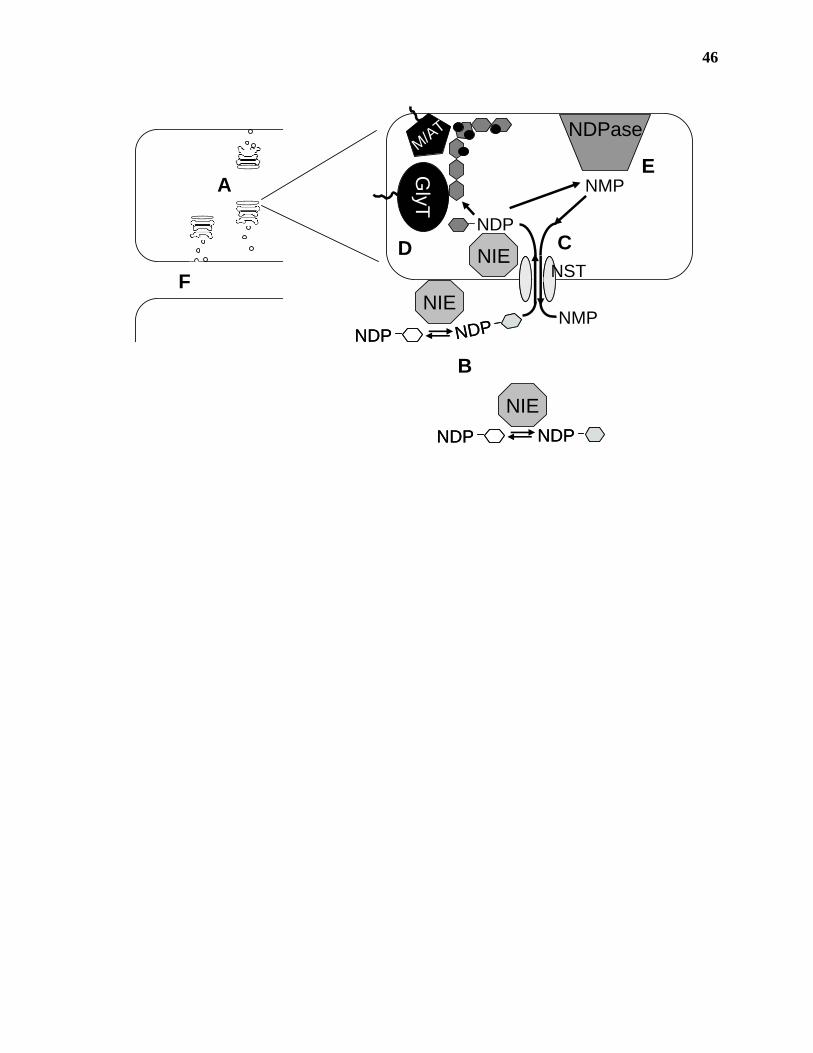
46
GlyT
NIE
NIE
NIE
NDPase
NDPNDP NDPNDP
NDPNDP NDPNDP NMP
NDP
NMP
M/AT
A
B
CD
E
NSTF
47
Figure 1.8. Simplified nucleotide-sugar interconversion pathways in plants. Most of the
nucleotide-sugars involved in pectin biosynthesis can be made through the interconversion of
UDP-D-Glcp or GDP-D-Manp. The genes involved in the interconversion of nucleotide sugars are
shown adjacent to the reactions they catalyze. The function of the RHM gene has not been
demonstrated, although it is thought to be involved in the biosynthesis of UDP-L-Rhap. A)
Nucleotide-sugars that arise from the conversion of UDP-D-Glcp. B) Nucleotide-sugars that
arise from the conversion of GDP-D-Manp. UGE, UDP-D-Glcp 4-epimerase; RHM, NDP-L-
Rhap biosynthetic gene; NRS/ER, UDP-4-keto-6-deoxy-D-Glc 3’,5’-epimerase-4-reductase
(NDP-L-Rhap synthase/epimerase-reductase); UGD, UDP-D-Glcp dehydrogenase; GAE, UDP-D-
GlcpA 4-epimerase; UXS, UDP-D-GlcpA decarboxylase (UDP-D-Xylp synthase); AXS, UDP-D-
Apif/UDP-D-Xylp synthase; MUR4, UDP-D-Xylp 4-epimerase; GMD, GDP-D-mannose 4,6-
dehydratase; GER, GDP-4-keto-6-deoxy-D-Man 3’,5’-epimerase-4-reductase; GME, GDP-D-
Manp 3’,5’-epimerase.
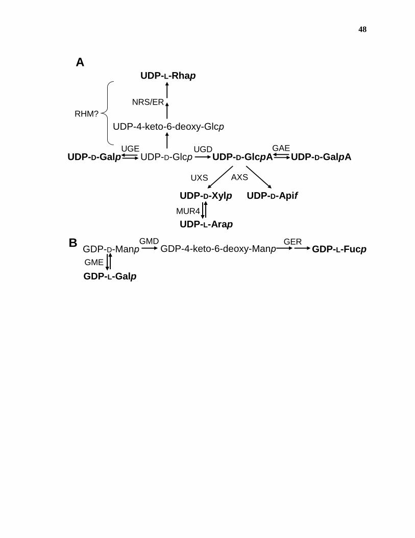
48
B
A
GDP-D-Manp
GDP-L-Galp
GDP-L-FucpGDP-4-keto-6-deoxy-ManpGMD GER
GME
UDP-D-Glcp
UDP-L-Rhap
UDP-D-GlcpA
UDP-L-Arap
UDP-D-Xylp
UDP-D-Galp UDP-D-GalpA
UDP-D-Apif
UDP-4-keto-6-deoxy-Glcp
UGE
RHM?
UXS
GAE
MUR4
AXS
NRS/ER
UGD
49
the majority of the nucleotide-sugars required for pectin biosynthesis (Mohnen, 1999; Reiter and
Vanzin, 2001; Seifert, 2004). Nucleotide-sugar interconversion starts with either UDP-D-Glcp or
GDP-D-Manp produced by the activation of either α-D-Manp-1-phosphate or α-D-Glcp-1-
phosphate into the corresponding NDP-sugar (Figure 1.8). Through the actions of several
enzymes, UDP-D- Galp, UDP-L-Rhap, UDP-D-GlcpA, UDP-D-GalpA, UDP-D-Xylp, UDP-L-
Arap, UDP-D-Apif, GDP-L-Fucp, and GDP-L-Galp can be synthesized (Hassid et al., 1959;
Feingold and Avigad, 1980; Feingold and Barber, 1990).
Plant extracts have been shown to salvage free D-Galp, D-GlcpA, D-GalpA, D-Xylp, and L-
Araf and L-Fucp released as a result of metabolic processes into the corresponding sugar-1-
phosphate by C-1 kinases (Hassid et al., 1959; Feingold and Avigad, 1980).
Pyrophosphorylases then convert the sugar-1-phosphates into NDP-sugars in the presence of the
appropriate NTP. It is commonly believed that the salvage pathway is used mainly in the
recycling of monosaccharides from the cell wall (Feingold and Avigad, 1980; Mohnen, 2002).
However, recent studies on the conversion of myo-inositol into UDP-D-GlcpA (Seitz et al., 2000)
suggest that the salvage pathway may play a role in the tissue-specific biosynthesis of some
nucleotide-sugars (Figure 1.9, B). In light of these results, certain aspects of the salvage pathway
will be considered in this section. For a more detailed summary of the salvage pathways in
plants, the reader is directed to several excellent reviews (Hassid et al., 1959; Hassid, 1967;
Feingold and Avigad, 1980; Feingold and Barber, 1990).
The synthesis of CMP-D-Kdop (Figure 1.9, A) is unique in that it does not involve the
interconversion of existing nucleotide-sugars and instead starts with the condensation of D-Arap-
5-phosphate and phosphenolpyruvate (PEP) to form D-Kdop-8-phosphate (Doong et al., 1991).
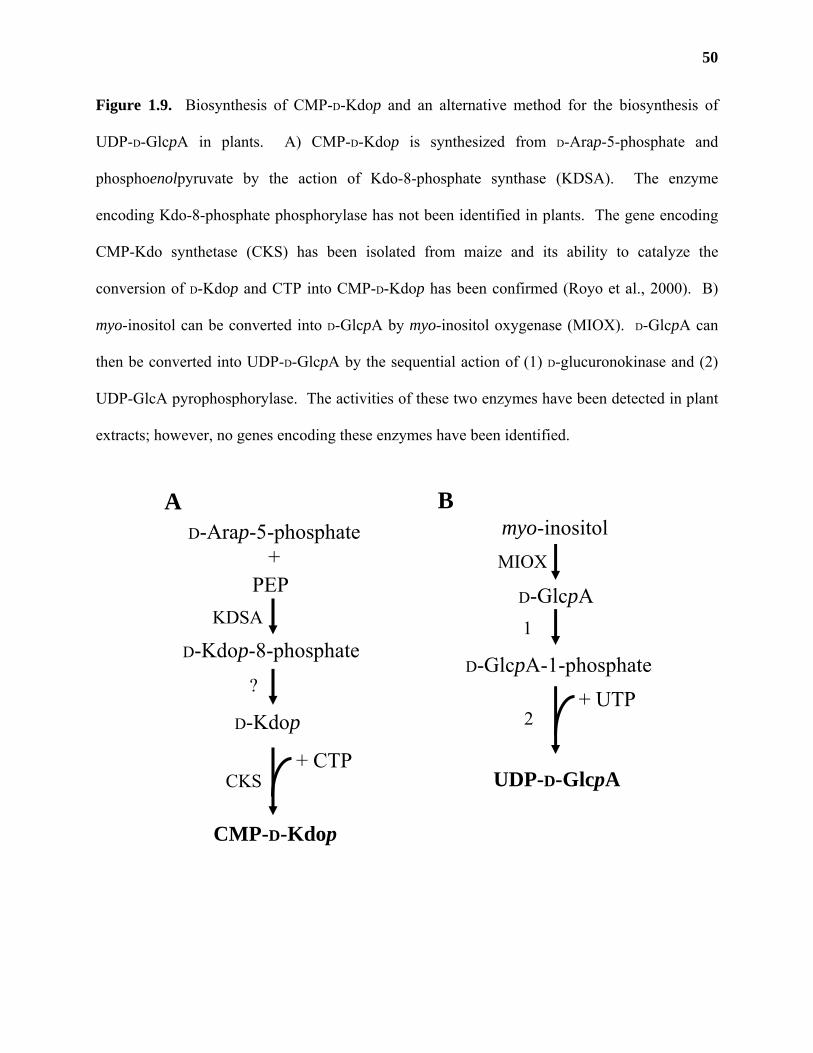
50
Figure 1.9. Biosynthesis of CMP-D-Kdop and an alternative method for the biosynthesis of
UDP-D-GlcpA in plants. A) CMP-D-Kdop is synthesized from D-Arap-5-phosphate and
phosphoenolpyruvate by the action of Kdo-8-phosphate synthase (KDSA). The enzyme
encoding Kdo-8-phosphate phosphorylase has not been identified in plants. The gene encoding
CMP-Kdo synthetase (CKS) has been isolated from maize and its ability to catalyze the
conversion of D-Kdop and CTP into CMP-D-Kdop has been confirmed (Royo et al., 2000). B)
myo-inositol can be converted into D-GlcpA by myo-inositol oxygenase (MIOX). D-GlcpA can
then be converted into UDP-D-GlcpA by the sequential action of (1) D-glucuronokinase and (2)
UDP-GlcA pyrophosphorylase. The activities of these two enzymes have been detected in plant
extracts; however, no genes encoding these enzymes have been identified.
D-Arap-5-phosphate
PEP+
D-Kdop-8-phosphate
D-Kdop
CMP-D-Kdop
+ CTP
KDSA
?
CKS
A
UDP-D-GlcpA
myo-inositol
D-GlcpA
D-GlcpA-1-phosphate+ UTP
MIOX
1
2
B
51
D-Kdop-8-phosphate is then dephosphorylated (Wu and Woodard, 2003) and converted into
CMP-D-Kdop in the presence of CTP (Royo et al., 2000).
The recent identification and characterization of a number of NIEs has greatly enhanced
our understanding of the synthesis of these substrates. Proof of their activity has been
demonstrated in most cases by heterologous expression. Our current understanding of the
biosynthesis of each activated sugar substrate will be considered in this section.
UDP-D-Galp: UDP-Glc 4-epimerase (EC 5.1.3.2) catalyzes the reversible conversion of
UDP-D-Glcp to UDP-D-Galp. The reaction involves the transfer of a hydride ion from the C-4 of
the sugar moiety to an enzyme-bound NAD+, creating a 4-keto intermediate. The non-
stereospecific return of the hydride ion to the 4-keto intermediate causes the formation of either
UDP-D-Glcp or UDP-D-Galp.
Soluble and membrane-bound UDP-Glc 4-epimerase activity could be detected in several
plant species (Feingold and Avigad, 1980) and was purified 1000-fold from wheat germ (Fan
and Feingold, 1969). The first gene encoding an active UDP-Glc 4-epimerase (UGE1) was
isolated from an Arabidopsis cDNA library by searching for genes that had sequence similarity
to cloned UDP-Glc 4-epimerases from rat and bacteria (Table 1.2; Dormann and Benning, 1996).
The cDNA encoded a 39 kDa protein that could partially complement a Saccharomyces
cerevisiae mutant (gal10) deficient in its ability to grow on galactose as a sole carbon source.
The recombinantly expressed protein purified from Escherichia coli was active in the absence of
exogenous NAD+, had a Km for UDP-D-Glcp of 0.11 mM, and a Vmax of 3.96 mmol min-1 mg-1
E-NAD+ + UDP-D-Glcp E-NAD+ + UDP-D-GalpE-NADH + UDP-4-keto-Glcp
E-NAD+ + UDP-D-Glcp(1)
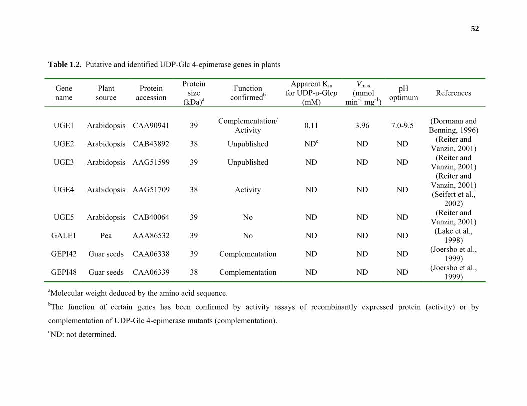
52
Table 1.2. Putative and identified UDP-Glc 4-epimerase genes in plants
Gene name
Plant source
Protein accession
Protein size
(kDa)a
Function confirmedb
Apparent Km for UDP-D-Glcp
(mM)
Vmax (mmol
min-1 mg-1)
pH optimum References
UGE1 Arabidopsis CAA90941 39 Complementation/Activity 0.11 3.96 7.0-9.5 (Dormann and
Benning, 1996)
UGE2 Arabidopsis CAB43892 38 Unpublished NDc ND ND (Reiter and Vanzin, 2001)
UGE3 Arabidopsis AAG51599 39 Unpublished ND ND ND (Reiter and Vanzin, 2001)
UGE4 Arabidopsis AAG51709 38 Activity ND ND ND
(Reiter and Vanzin, 2001) (Seifert et al.,
2002)
UGE5 Arabidopsis CAB40064 39 No ND ND ND (Reiter and Vanzin, 2001)
GALE1 Pea AAA86532 39 No ND ND ND (Lake et al., 1998)
GEPI42 Guar seeds CAA06338 39 Complementation ND ND ND (Joersbo et al., 1999)
GEPI48 Guar seeds CAA06339 38 Complementation ND ND ND (Joersbo et al., 1999)
aMolecular weight deduced by the amino acid sequence. bThe function of certain genes has been confirmed by activity assays of recombinantly expressed protein (activity) or by
complementation of UDP-Glc 4-epimerase mutants (complementation). cND: not determined.
53
(Dormann and Benning, 1996). Antisense suppression of UGE1 expression in Arabidopsis
caused a 90% reduction in UDP-Glc 4-epimerase activity in stems, yet these plants had no
discernable phenotype when compared to wildtype plants (Dormann and Benning, 1998).
Transgenic plants overexpressing UGE1 had 3-fold higher levels of UDP-Glc 4-epimerase
activity in leaves and grew normally in the presence of toxic levels of galactose (Dormann and
Benning, 1998). Putative UDP-Glc 4-epimerases have also been isolated from pea (Lake et al.,
1998) and from developing guar (Cyamopsis tetragonoloba) seeds (Joersbo et al., 1999). The
function of the gene from pea has not been confirmed; however, the two cDNA’s from guar
seeds were identified by functional complementation of a UDP-Glc 4-epimerase mutant of E.
coli, suggesting that they encode functional UDP-Glc 4-epimerases (Table 1.2).
Analysis of the Arabidopsis genome revealed the presence of 4 open reading frames
(UGE2-5) with 65-89% amino acid sequence identity to UGE1 (Reiter and Vanzin, 2001). UGE
isoforms have been shown by β-glucuronidase (GUS) fusions to be differentially expressed in
Arabidopsis roots (Seifert et al., 2004a). The gene encoding UGE4 is allelic to the Arabidopsis
mutants root-epidermal-bulger 1 (Reiter and Vanzin, 2001; Seifert et al., 2002) and root hair
development 1 (Schiefelbein and Somerville, 1990) that are characterized by bulging root
epidermal cells. The bulging of these cells is most likely due to altered levels of galactose in
epidermal cell wall polymers; evidenced by the differential visualization of wildtype and root
hair development 1 (rdh1) plants using antibodies that recognize galactose-containing epitopes
(Seifert et al., 2002). The rdh1 phenotype can be corrected by growing plants on galactose
(Seifert et al., 2002) or in the presence of ethylene (Seifert et al., 2004a). Ethylene treatment
reduces the expression of UGE1 and UGE3 in roots and causes wildtype staining of specific
galactose-containing epitopes found on arabinogalactans and xyloglucans. The staining of other
54
galactose-containing epitopes, such as those recognized by LM5, is even more reduced in
ethylene-treated rdh1 plants, suggesting that the limited UDP-D-Galp pool is preferentially used
for the synthesis of arabinogalactans and xyloglucan, instead of the synthesis of the (1,4)-linked
β-D-galactan chains found on RG-I. These results led the authors to speculate that specific UGE
isoforms are involved in the channeling of the UDP-D-Galp pool into specific polysaccharide
synthase complexes that synthesize cell wall polymers (Seifert et al., 2002; Seifert et al., 2004a).
Evidence supporting the existence of glycosyltransferase-glycosyltransferase and
glycosyltransferase-NST enzyme complexes involved in glycoconjugate production has been
obtained using mammalian cell lines (Opat et al., 2000; Bieberich et al., 2002; Sprong et al.,
2003); however, substrate channeling may not be the sole explanation for the effect of ethylene
treatment on the Arabidopsis rdh1 mutant.
Differences in cell surface glycosyl residue composition have also been well documented
in NST mutants of yeast and mammalian cell lines (Deutscher and Hirschberg, 1986; Hirschberg
et al., 1998; Kawakita et al., 1998). Mutation of the NST in these cell lines resulted in the
reduction of the nucleotide-sugars accessible to the Golgi-localized glycosyltransferases. This
has led some researchers to speculate that the differences in glycosyl residue content and
composition at the cell surface of NST mutant lines were due to differences in the affinities of
the Golgi-localized glycosyltransferases (Km) for their nucleotide-sugar substrates (Kawakita et
al., 1998). Glycosyltransferases with the lowest Km for a particular nucleotide-sugar substrate
would sequester the available nucleotide-sugar pool for the production of specific carbohydrate
epitopes (Kawakita et al., 1998) and cause these epitopes to be detected at the cell surface.
Such a scenario could also be occurring in rdh1 mutants of Arabidopsis. Mutations in
rdh1 reduce the amount of UDP-D-Galp synthesized in the root, causing cell wall
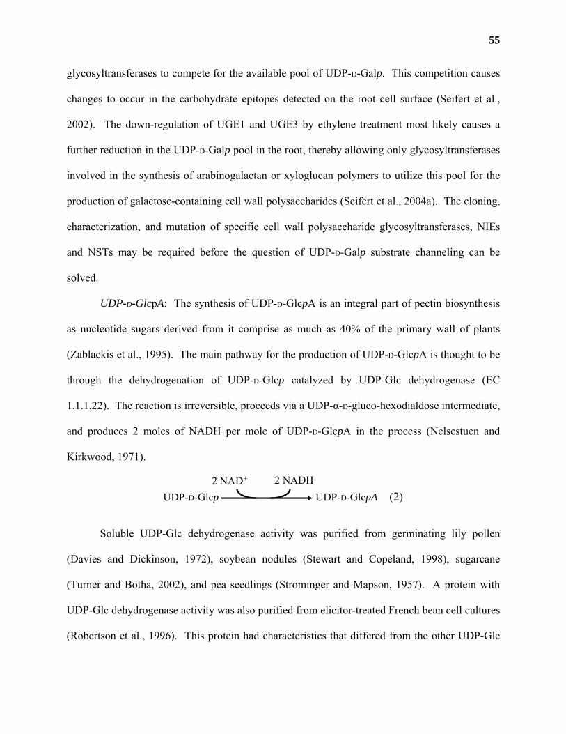
55
glycosyltransferases to compete for the available pool of UDP-D-Galp. This competition causes
changes to occur in the carbohydrate epitopes detected on the root cell surface (Seifert et al.,
2002). The down-regulation of UGE1 and UGE3 by ethylene treatment most likely causes a
further reduction in the UDP-D-Galp pool in the root, thereby allowing only glycosyltransferases
involved in the synthesis of arabinogalactan or xyloglucan polymers to utilize this pool for the
production of galactose-containing cell wall polysaccharides (Seifert et al., 2004a). The cloning,
characterization, and mutation of specific cell wall polysaccharide glycosyltransferases, NIEs
and NSTs may be required before the question of UDP-D-Galp substrate channeling can be
solved.
UDP-D-GlcpA: The synthesis of UDP-D-GlcpA is an integral part of pectin biosynthesis
as nucleotide sugars derived from it comprise as much as 40% of the primary wall of plants
(Zablackis et al., 1995). The main pathway for the production of UDP-D-GlcpA is thought to be
through the dehydrogenation of UDP-D-Glcp catalyzed by UDP-Glc dehydrogenase (EC
1.1.1.22). The reaction is irreversible, proceeds via a UDP-α-D-gluco-hexodialdose intermediate,
and produces 2 moles of NADH per mole of UDP-D-GlcpA in the process (Nelsestuen and
Kirkwood, 1971).
Soluble UDP-Glc dehydrogenase activity was purified from germinating lily pollen
(Davies and Dickinson, 1972), soybean nodules (Stewart and Copeland, 1998), sugarcane
(Turner and Botha, 2002), and pea seedlings (Strominger and Mapson, 1957). A protein with
UDP-Glc dehydrogenase activity was also purified from elicitor-treated French bean cell cultures
(Robertson et al., 1996). This protein had characteristics that differed from the other UDP-Glc
UDP-D-Glcp UDP-D-GlcpA2 NAD+ 2 NADH
(2)
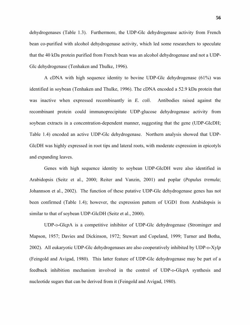
56
dehydrogenases (Table 1.3). Furthermore, the UDP-Glc dehydrogenase activity from French
bean co-purified with alcohol dehydrogenase activity, which led some researchers to speculate
that the 40 kDa protein purified from French bean was an alcohol dehydrogenase and not a UDP-
Glc dehydrogenase (Tenhaken and Thulke, 1996).
A cDNA with high sequence identity to bovine UDP-Glc dehydrogenase (61%) was
identified in soybean (Tenhaken and Thulke, 1996). The cDNA encoded a 52.9 kDa protein that
was inactive when expressed recombinantly in E. coli. Antibodies raised against the
recombinant protein could immunoprecipitate UDP-glucose dehydrogenase activity from
soybean extracts in a concentration-dependent manner, suggesting that the gene (UDP-GlcDH;
Table 1.4) encoded an active UDP-Glc dehydrogenase. Northern analysis showed that UDP-
GlcDH was highly expressed in root tips and lateral roots, with moderate expression in epicotyls
and expanding leaves.
Genes with high sequence identity to soybean UDP-GlcDH were also identified in
Arabidopsis (Seitz et al., 2000; Reiter and Vanzin, 2001) and poplar (Populus tremula;
Johannson et al., 2002). The function of these putative UDP-Glc dehydrogenase genes has not
been confirmed (Table 1.4); however, the expression pattern of UGD1 from Arabidopsis is
similar to that of soybean UDP-GlcDH (Seitz et al., 2000).
UDP-D-GlcpA is a competitive inhibitor of UDP-Glc dehydrogenase (Strominger and
Mapson, 1957; Davies and Dickinson, 1972; Stewart and Copeland, 1999; Turner and Botha,
2002). All eukaryotic UDP-Glc dehydrogenases are also cooperatively inhibited by UDP-D-Xylp
(Feingold and Avigad, 1980). This latter feature of UDP-Glc dehydrogenase may be part of a
feedback inhibition mechanism involved in the control of UDP-D-GlcpA synthesis and
nucleotide sugars that can be derived from it (Feingold and Avigad, 1980).
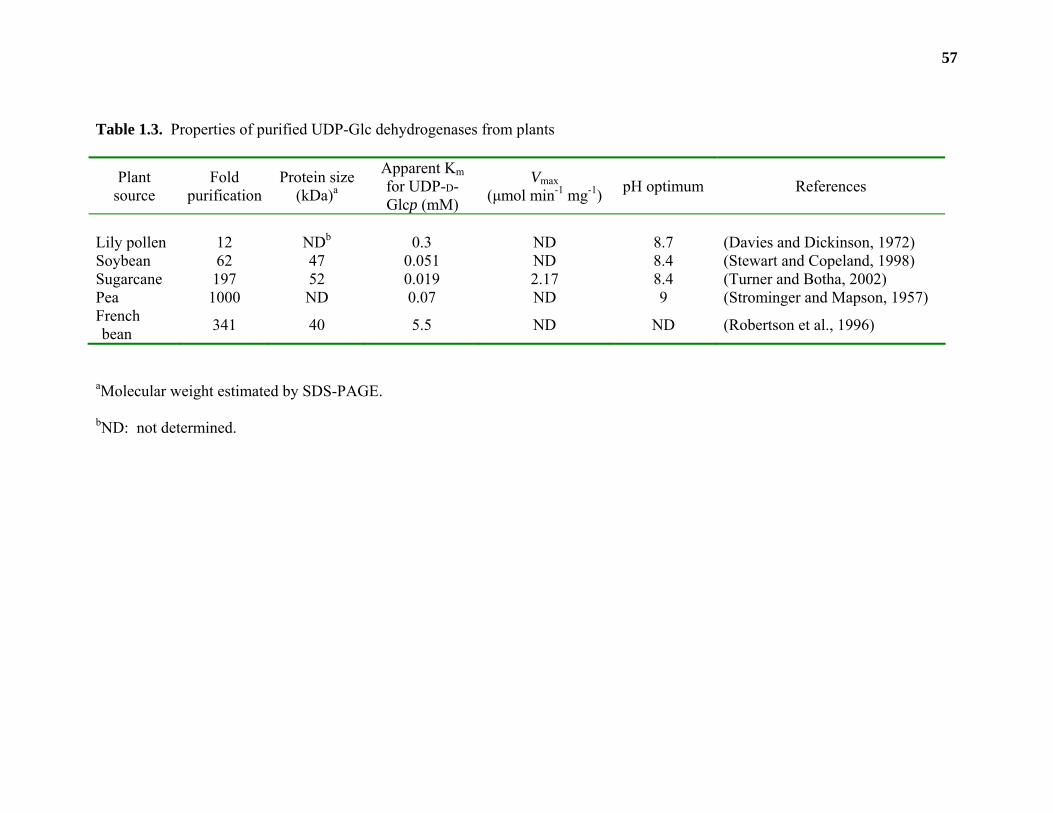
57
Table 1.3. Properties of purified UDP-Glc dehydrogenases from plants
Plant source
Fold purification
Protein size (kDa)a
Apparent Km for UDP-D-Glcp (mM)
Vmax (µmol min-1 mg-1) pH optimum References
Lily pollen 12 NDb 0.3 ND 8.7 (Davies and Dickinson, 1972) Soybean 62 47 0.051 ND 8.4 (Stewart and Copeland, 1998) Sugarcane 197 52 0.019 2.17 8.4 (Turner and Botha, 2002) Pea 1000 ND 0.07 ND 9 (Strominger and Mapson, 1957) French bean 341 40 5.5 ND ND (Robertson et al., 1996)
aMolecular weight estimated by SDS-PAGE.
bND: not determined.
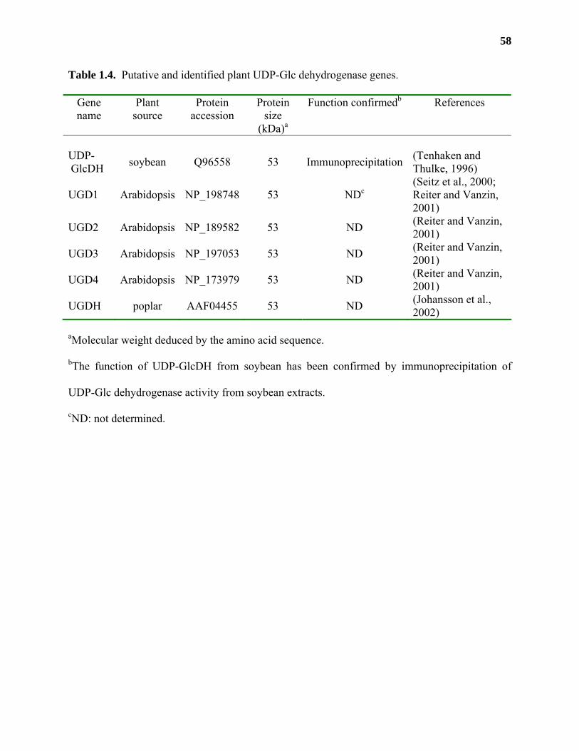
58
Table 1.4. Putative and identified plant UDP-Glc dehydrogenase genes.
Gene name
Plant source
Protein accession
Protein size
(kDa)a
Function confirmedb References
UDP-GlcDH soybean Q96558 53 Immunoprecipitation (Tenhaken and
Thulke, 1996)
UGD1 Arabidopsis NP_198748 53 NDc (Seitz et al., 2000; Reiter and Vanzin, 2001)
UGD2 Arabidopsis NP_189582 53 ND (Reiter and Vanzin, 2001)
UGD3 Arabidopsis NP_197053 53 ND (Reiter and Vanzin, 2001)
UGD4 Arabidopsis NP_173979 53 ND (Reiter and Vanzin, 2001)
UGDH poplar AAF04455 53 ND (Johansson et al., 2002)
aMolecular weight deduced by the amino acid sequence.
bThe function of UDP-GlcDH from soybean has been confirmed by immunoprecipitation of
UDP-Glc dehydrogenase activity from soybean extracts.
cND: not determined.
59
UDP-D-GlcpA can also be synthesized through the conversion of myo-inositol into D-
GlcpA (Figure 1.9, B). Whole plants or extracts from parsley (Petroselinum crispum) leaves and
strawberry (Fragaria ananassa) fruits can convert radioactive myo-inositol into radioactive
pectin (Loewus and Kelly, 1963). Roughly 40% of the radioactivity from parsley leaves and
strawberry fruits is found in D-GalpA, D-Xylp, L-Araf, and D-GlcpA, and this conversion is
performed without rearrangement or cleavage of the carbon chain of myo-inositol (Loewus and
Kelly, 1963).
Myo-inositol is produced in plants via the conversion of D-Glc-6-P or phytic acid by myo-
inositol-1-P synthetase or phytase, respectively (Loewus and Loewus, 1983). Both enzymes
produce myo-inositol-1-P which is then converted into myo-inositol by inositol monophosphatase
(IMP; EC 3.1.3.25). Three genes encoding IMPs have been cloned from tomato and shown to be
active by recombinant expression in E. coli (Gillaspy et al., 1995).
Myo-inositol oxygenase (MIOX; EC 1.13.99.1) converts myo-inositol into D-GlcpA
(Figure 1.9, B). A gene encoding a myo-inositol oxygenase (MIOX4) was recently cloned from
Arabidopsis (Lorence et al., 2004). Recombinant MIOX4 expressed in E. coli could convert
myo-inositol into D-GlcpA with a specific activity of 2.2 µmol min-1 mg-1. MIOX4 is highly
expressed in leaf and flower tissue and is part of a 4 member gene family in Arabidopsis (Table
1.5).
D-GlcpA applied to plant extracts or produced by MIOX can be converted into UDP-D-
GlcpA by sequential action of D-glucuronokinase (EC 2.7.1.43) and UDP-GlcA
pyrophosphorylase (EC 2.7.7.44). The activity of both enzymes have been detected in mung
bean seedlings (Feingold et al., 1958; Neufeld et al., 1961). UDP-GlcA pyrophosphorylase
activity has been purified 80-fold from barley (Hordeum vulgare) seedlings (Roberts, 1971).
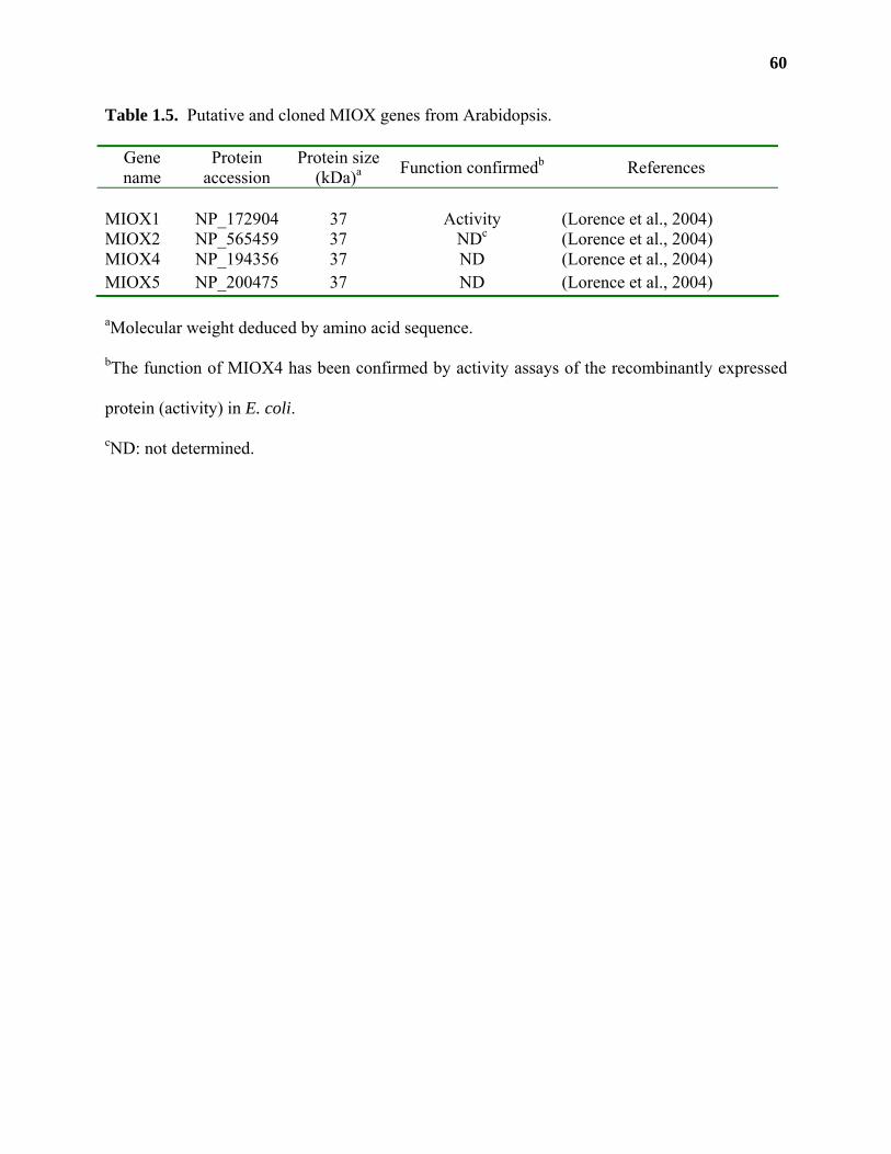
60
Table 1.5. Putative and cloned MIOX genes from Arabidopsis.
Gene name
Protein accession
Protein size (kDa)a Function confirmedb References
MIOX1 NP_172904 37 Activity (Lorence et al., 2004) MIOX2 NP_565459 37 NDc (Lorence et al., 2004) MIOX4 NP_194356 37 ND (Lorence et al., 2004) MIOX5 NP_200475 37 ND (Lorence et al., 2004) aMolecular weight deduced by amino acid sequence.
bThe function of MIOX4 has been confirmed by activity assays of the recombinantly expressed
protein (activity) in E. coli.
cND: not determined.
61
The protein had an estimated molecular weight of 35 kDa by size exclusion chromatography, had
a Km for UDP-D-GlcpA of 0.5 mM, and pH optimum of 8-9. No genes encoding either D-
glucuronokinase or UDP-GlcA pyrophosphorylase have been cloned from any plant source.
It has been hypothesized that the conversion of myo-inositol into UDP-D-GlcpA may be a
major pathway involved in the synthesis of this nucleotide-sugar (Roberts, 1971; Loewus and
Loewus, 1983). Firstly, UDP-Glc dehydrogenase activity can not be detected in some plant
species (Feingold and Avigad, 1980). Secondly, it has been shown that the incubation of
Arabidopsis seedlings with [3H]myo-inositol causes the incorporation of radioactivity into areas
of the plant where UGD1 expression is absent, such as the hypocotyl and the cotyledons (Seitz et
al., 2000). The incubation of Arabidopsis seedlings with myo-inositol does not change the level
of UGD1 expression, suggesting that the synthesis of UDP-D-GlcpA may occur through different
biochemical pathways in different parts of the plant or during specific developmental stages.
Alternatively, the differential locations of these two biochemical pathways may reflect their
involvement in different plant processes. For example, UGD1 and UDP-GlcDH are highly
expressed in the root, which is the major site of D-GlcpA uptake by the plant (Hassid, 1967;
Feingold and Avigad, 1980). Myo-inositol, on the other hand, is a precursor for the biosynthesis
of L-ascorbic acid in leaves, flowers and other above-ground organs (Smirnoff et al., 2001; Agius
et al., 2002; Lorence et al., 2004). Plants fed [3H]myo-inositol may be converting the surplus
myo-inositol into UDP-D-GlcpA (and ultimately pectin) as a means of controlling the production
of L-ascorbic acid in above-ground tissues. Although it is commonly believed that the
dehydrogenation of UDP-D-Glcp is the main pathway involved in the production of UDP-D-
GlcpA, further research into the role of the myo-inositol pathway needs to be conducted in order
to determine its role in pectin biosynthesis.
62
UDP-D-GalpA: UDP-D-GalpA is the source of the most abundant glycosyl residue found
in pectic polysaccharides (Mohnen, 2002). It is synthesized by the reversible 4-epimeration of
UDP-D-GlcpA catalyzed by UDP-GlcA 4-epimerase (EC 5.1.3.6; Neufeld et al., 1958). The
catalytic mechanism of UDP-GlcA 4-epimerase is similar to that of UDP-Glc 4 epimerase and
proceeds via the oxidation of the C-4 of UDP-D-GlcpA by an enzyme bound NAD(P)+ resulting
in a 4-keto intermediate (Feingold and Avigad, 1980). Subsequent non-stereospecific reduction
of the 4-keto group causes the production of equimolar concentrations of UDP-D-GlcpA and
UDP-D-GalpA.
UDP-GlcA 4-epimerase activity was first identified in particulate fractions from mung
bean, and later detected in membrane fractions from asparagus (Asparagus officinalis), radish
and spinach (Neufeld et al., 1958; Feingold et al., 1960). Enzyme activity was later found to also
be associated with soluble fractions from mung bean (Feingold et al., 1960). Membrane-bound
activities were also found in radish roots (Liljebjelke et al., 1995) and Arabidopsis leaves
(Molhoj et al., 2004). Soluble UDP-D-GlcpA 4-epimerase was partially purified from the blue-
green algae Anabaena flos-aquae (Gaunt et al., 1974). The partially purified enzyme had a pH
optimum of 8.5, a Km for UDP-D-GlcpA of 37 µM, and an approximate molecular weight of 54
kDa. The enzyme was also allosterically inhibited by UDP-D-Xylp (Gaunt et al., 1974).
A gene encoding an active UDP-GlcA 4-epimerase (GAE1) was recently cloned from
Arabidopsis and expressed recombinantly in Pichia pastoris (Molhoj et al., 2004). GAE1 was
identified by screening the Arabidopsis genome for sequences that were similar to a UDP-GlcA
4-epimerase (Cap1J) from Streptococcus pneumoniae (Munoz et al., 1999). Recombinant GAE1
(3)E-NAD+ + UDP-D-GlcpA E-NAD+ + UDP-D-GalpAE-NADH + UDP-4-keto-GlcpA
63
reacted with UDP-D-GlcpA producing roughly equimolar concentrations of UDP-D-GalpA and
UDP-D-GlcpA. The enzyme had a pH optimum of 7.6, a Km for UDP-D-GlcpA of 0.19 mM and
was inactive on UDP-D-Glcp and UDP-D-Xylp. GAE1 activity was significantly inhibited by
UDP-D-Xylp, a property that was associated with the partially purified enzyme from A. flos-
aquae. GAE1 activity was not dependent on the presence of divalent cations, nor on the
exogenous addition of NAD(P)+, suggesting that the recombinant enzyme contained a tightly
bound nicotinamide derivative. GAE1:β-glucuronidase fusions suggested that GAE1 was
expressed in all major tissues of Arabidopsis (Molhoj et al., 2004).
GAE1 is part of a 6 member gene family in Arabidopsis (Table 1.6; Reiter and Vanzin,
2001). All GAE genes are predicted type II membrane proteins and have an N-terminal
extension that encodes a putative transmembrane domain (Reiter and Vanzin, 2001; Molhoj et
al., 2004). While the absolute topology of any GAE gene has not been determined, recombinant
GAE1 activity associates only with the membrane fraction from P. pastoris. Soluble UDP-GlcA
4-epimerase activity was not detected in Arabidopsis leaves, contrary to results obtained from
mung bean seedlings. At this point, it is not known whether the soluble UDP-GlcA 4-epimerase
activity represents a different subset of proteins in these tissues, or whether the soluble enzymes
are produced by proteolytic events that occur during tissue preparation. The isolation of UDP-
GlcA 4-epimerase genes from different plant species will allow us to determine whether soluble
isoforms of these enzymes exist in other plants.
UDP-D-Xylp: UDP-D-Xylp is produced by the irreversible decarboxylation of UDP-D-
GlcpA that is catalyzed by UDP-GlcA decarboxylase (EC 4.1.1.35). The reaction mechanism
for plant UDP-GlcA decarboxylases was investigated (Schutzbach and Feingold, 1970) using the
purified enzyme from wheat germ (Ankel and Feingold, 1965). The reaction commences with
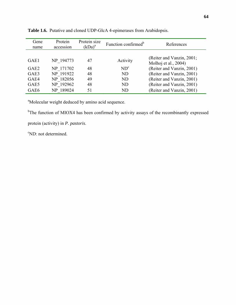
64
Table 1.6. Putative and cloned UDP-GlcA 4-epimerases from Arabidopsis.
Gene name
Protein accession
Protein size (kDa)a Function confirmedb References
GAE1 NP_194773 47 Activity (Reiter and Vanzin, 2001; Molhoj et al., 2004)
GAE2 NP_171702 48 NDc (Reiter and Vanzin, 2001) GAE3 NP_191922 48 ND (Reiter and Vanzin, 2001) GAE4 NP_182056 49 ND (Reiter and Vanzin, 2001) GAE5 NP_192962 48 ND (Reiter and Vanzin, 2001) GAE6 NP_189024 51 ND (Reiter and Vanzin, 2001) aMolecular weight deduced by amino acid sequence.
bThe function of MIOX4 has been confirmed by activity assays of the recombinantly expressed
protein (activity) in P. pastoris.
cND: not determined.
65
the oxidation of C-4 by an enzyme-bound NAD+ producing a 4-keto intermediate. The
irreversible decarboxylation of C-5 on the intermediate occurs with the release of CO2. The
stereospecific reduction of the C-5 and the 4-keto group by a proton from water and by an
enzyme-bound NADH, respectively, produces UDP-D-Xylp as the final product.
All plant UDP-GlcA decarboxylases are allosterically inhibited by UDP-D-Xylp and do
not require the addition of exogenous NAD+ for activity (John et al., 1977; Hayashi et al., 1988;
Harper and Bar-Peled, 2002). UDP-GlcA decarboxylase activity from wheat germ is also
allosterically activated by low concentrations of UDP-D-GlcpA (John et al., 1977). The
inhibition of NIEs by UDP-D-Xylp has also been demonstrated for UDP-Glc dehydrogenase, and
suggests that the synthesis of UDP-D-Xylp may be one of the main regulatory factors controlling
the production of many of the nucleotide-sugars involved in pectin biosynthesis (John et al.,
1977; Harper and Bar-Peled, 2002).
UDP-GlcA decarboxylase exists in many plant species as both soluble and membrane-
bound isoforms (Table 1.7). Activity was first detected in particulate fractions from mung bean,
and later detected in membrane fractions from radish, asparagus and spinach (Neufeld et al.,
1958; Feingold et al., 1960). Soluble UDP-GlcA decarboxylase was first purified from wheat
germ by Ankel and Feingold (1965). The enzyme was later found to exist in wheat germ as two
different 210 kDa isoforms that differed in their binding to DEAE-cellulose, kinetic properties,
and response to allosteric effectors (John et al., 1977).
(4)E-NAD+ + UDP-D-GlcpA E-NAD+ + UDP-D-XylpE-NADH + UDP-4-keto-GlcpACO2
H+
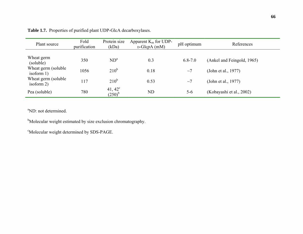
66
Table 1.7. Properties of purified plant UDP-GlcA decarboxylases.
Plant source Fold purification
Protein size (kDa)
Apparent Km for UDP-D-GlcpA (mM) pH optimum References
Wheat germ (soluble) 350 NDa 0.3 6.8-7.0 (Ankel and Feingold, 1965)
Wheat germ (soluble isoform 1) 1056 210b 0.18 ~7 (John et al., 1977)
Wheat germ (soluble isoform 2) 117 210b 0.53 ~7 (John et al., 1977)
Pea (soluble) 780 41, 42c (250)b ND 5-6 (Kobayashi et al., 2002)
aND: not determined.
bMolecular weight estimated by size exclusion chromatography.
cMolecular weight determined by SDS-PAGE.
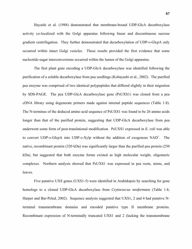
67
Hayashi et al. (1988) demonstrated that membrane-bound UDP-GlcA decarboxylase
activity co-localized with the Golgi apparatus following linear and discontinuous sucrose
gradient centrifugation. They further demonstrated that decarboxylation of UDP-D-GlcpA only
occurred within intact Golgi vesicles. These results provided the first evidence that some
nucleotide-sugar interconversions occurred within the lumen of the Golgi apparatus.
The first plant gene encoding a UDP-GlcA decarboxylase was identified following the
purification of a soluble decarboxylase from pea seedlings (Kobayashi et al., 2002). The purified
pea enzyme was comprised of two identical polypeptides that differed slightly in their migration
by SDS-PAGE. The pea UDP-GlcA decarboxylase gene (PsUXS1) was cloned from a pea
cDNA library using degenerate primers made against internal peptide sequences (Table 1.8).
The N-terminus of the deduced amino acid sequence of PsUXS1 was found to be 26 amino acids
longer than that of the purified protein, suggesting that UDP-GlcA decarboxylase from pea
underwent some form of post-translational modification. PsUXS1 expressed in E. coli was able
to convert UDP-D-GlcpA into UDP-D-Xylp without the addition of exogenous NAD+. The
native, recombinant protein (320 kDa) was significantly larger than the purified pea protein (250
kDa), but suggested that both enzyme forms existed as high molecular weight, oligomeric
complexes. Northern analysis showed that PsUXS1 was expressed in pea roots, stems, and
leaves.
Five putative UXS genes (UXS1-5) were identified in Arabidopsis by searching for gene
homologs to a cloned UDP-GlcA decarboxylase from Crytococcus neoformans (Table 1.8;
Harper and Bar-Peled, 2002). Sequence analysis suggested that UXS1, 2 and 4 had putative N-
terminal transmembrane domains and encoded putative type II membrane proteins.
Recombinant expression of N-terminally truncated UXS1 and 2 (lacking the transmembrane
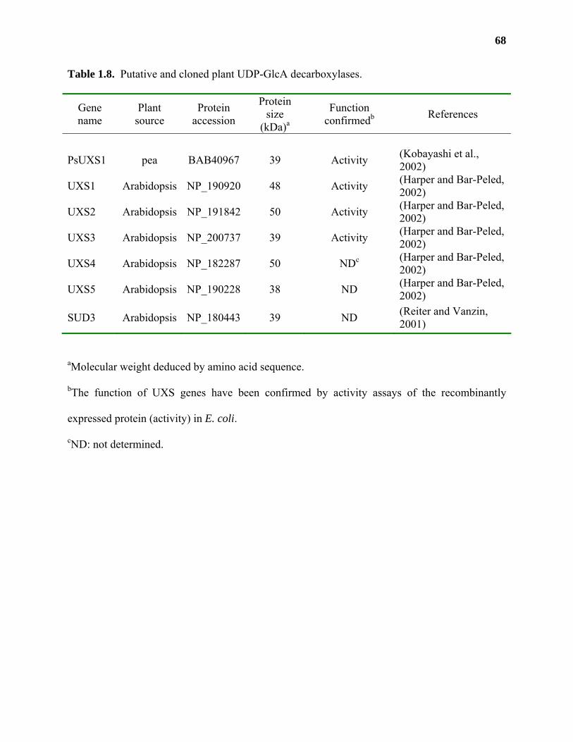
68
Table 1.8. Putative and cloned plant UDP-GlcA decarboxylases.
Gene name
Plant source
Protein accession
Protein size
(kDa)a
Function confirmedb References
PsUXS1 pea BAB40967 39 Activity (Kobayashi et al., 2002)
UXS1 Arabidopsis NP_190920 48 Activity (Harper and Bar-Peled, 2002)
UXS2 Arabidopsis NP_191842 50 Activity (Harper and Bar-Peled, 2002)
UXS3 Arabidopsis NP_200737 39 Activity (Harper and Bar-Peled, 2002)
UXS4 Arabidopsis NP_182287 50 NDc (Harper and Bar-Peled, 2002)
UXS5 Arabidopsis NP_190228 38 ND (Harper and Bar-Peled, 2002)
SUD3 Arabidopsis NP_180443 39 ND (Reiter and Vanzin, 2001)
aMolecular weight deduced by amino acid sequence.
bThe function of UXS genes have been confirmed by activity assays of the recombinantly
expressed protein (activity) in E. coli.
cND: not determined.

69
domain) and full length UXS3 demonstrated that all three genes encoded active UDP-GlcA
decarboxylases. Recombinant UXS3 was inactive on UDP-D-Glcp, UDP-D-Galp, and UDP-D-
GalpA, was inhibited by UDP-D-Xylp, and did not require the addition of exogenous NAD+ for
activity. Reverse transcriptase-PCR analysis demonstrated that UXS1, 2 and 3 were expressed in
stem, root, flower, and leaf tissues of Arabidopsis.
The UXS genes were also identified in Arabidopsis by searching for sequences that had
high similarity to a 4,6-dehydratase from E. coli (rmlB; Reiter and Vanzin, 2001). This search
identified one additional gene named SUD3 (soluble UDP-GlcA decarboxylase 3) that had high
sequence identity to the UXS genes (Seifert, 2004). The function of SUD3, UXS4 and UXS5
has not yet been elucidated.
UDP-L-Arap: UDP-L-Arap is synthesized by the reversible 4-epimerization of UDP-D-
Xylp catalyzed by UDP-Xyl 4-epimerase (EC 5.1.3.5) in a reaction mechanism that is thought to
be identical to that of UDP-Glc and UDP-GlcA 4-epimerases (Feingold and Avigad, 1980).
UDP-Xyl 4-epimerase activity was first detected in membrane fractions from mung bean,
radish, asparagus, and spinach (Neufeld et al., 1958; Feingold et al., 1960). The partially
purified enzyme from wheat germ and could catalyze the conversion of UDP-D-Xylp to UDP-L-
Arap without the addition of exogenous NAD+, had a Km for UDP-D-Xylp of 1.5 mM, and a pH
optimum of 8.0 (Fan and Feingold, 1970).
A mutant in Arabidopsis, mur4, was discovered that had a 50% reduction in L-Araf
content in leaves (Burget and Reiter, 1999). Wildtype MUR4 expressed in the mur4 mutant
background could complement the L-Araf deficiency and allowed for the recovery of up to 2.4-
fold higher levels of L-Araf in some transgenic lines (Burget et al., 2003). Expression of MUR4
E-NAD+ + UDP-D-Xylp E-NAD+ + UDP-L-ArapE-NADH + UDP-4-keto-Xylp(5)
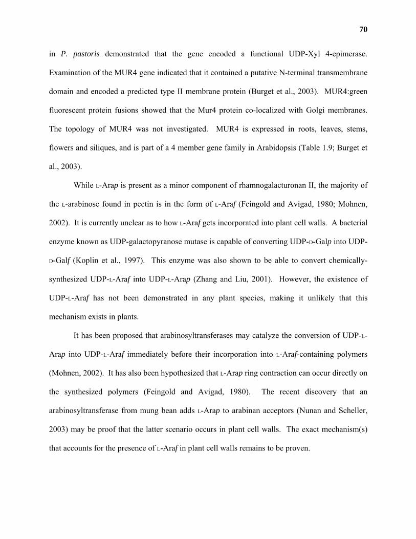
70
in P. pastoris demonstrated that the gene encoded a functional UDP-Xyl 4-epimerase.
Examination of the MUR4 gene indicated that it contained a putative N-terminal transmembrane
domain and encoded a predicted type II membrane protein (Burget et al., 2003). MUR4:green
fluorescent protein fusions showed that the Mur4 protein co-localized with Golgi membranes.
The topology of MUR4 was not investigated. MUR4 is expressed in roots, leaves, stems,
flowers and siliques, and is part of a 4 member gene family in Arabidopsis (Table 1.9; Burget et
al., 2003).
While L-Arap is present as a minor component of rhamnogalacturonan II, the majority of
the L-arabinose found in pectin is in the form of L-Araf (Feingold and Avigad, 1980; Mohnen,
2002). It is currently unclear as to how L-Araf gets incorporated into plant cell walls. A bacterial
enzyme known as UDP-galactopyranose mutase is capable of converting UDP-D-Galp into UDP-
D-Galf (Koplin et al., 1997). This enzyme was also shown to be able to convert chemically-
synthesized UDP-L-Araf into UDP-L-Arap (Zhang and Liu, 2001). However, the existence of
UDP-L-Araf has not been demonstrated in any plant species, making it unlikely that this
mechanism exists in plants.
It has been proposed that arabinosyltransferases may catalyze the conversion of UDP-L-
Arap into UDP-L-Araf immediately before their incorporation into L-Araf-containing polymers
(Mohnen, 2002). It has also been hypothesized that L-Arap ring contraction can occur directly on
the synthesized polymers (Feingold and Avigad, 1980). The recent discovery that an
arabinosyltransferase from mung bean adds L-Arap to arabinan acceptors (Nunan and Scheller,
2003) may be proof that the latter scenario occurs in plant cell walls. The exact mechanism(s)
that accounts for the presence of L-Araf in plant cell walls remains to be proven.
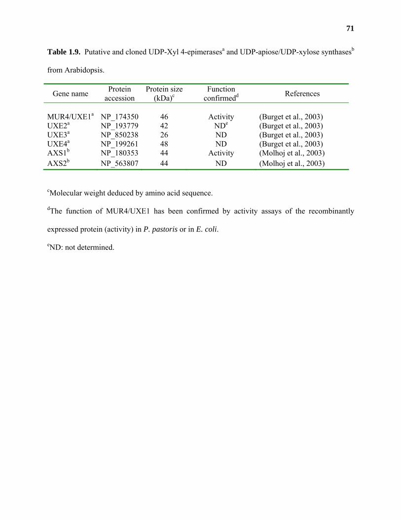
71
Table 1.9. Putative and cloned UDP-Xyl 4-epimerasesa and UDP-apiose/UDP-xylose synthasesb
from Arabidopsis.
Gene name Protein accession
Protein size (kDa)c
Function confirmedd References
MUR4/UXE1a NP_174350 46 Activity (Burget et al., 2003) UXE2a NP_193779 42 NDe (Burget et al., 2003) UXE3a NP_850238 26 ND (Burget et al., 2003) UXE4a NP_199261 48 ND (Burget et al., 2003) AXS1b NP_180353 44 Activity (Molhoj et al., 2003) AXS2b NP_563807 44 ND (Molhoj et al., 2003)
cMolecular weight deduced by amino acid sequence.
dThe function of MUR4/UXE1 has been confirmed by activity assays of the recombinantly
expressed protein (activity) in P. pastoris or in E. coli.
eND: not determined.
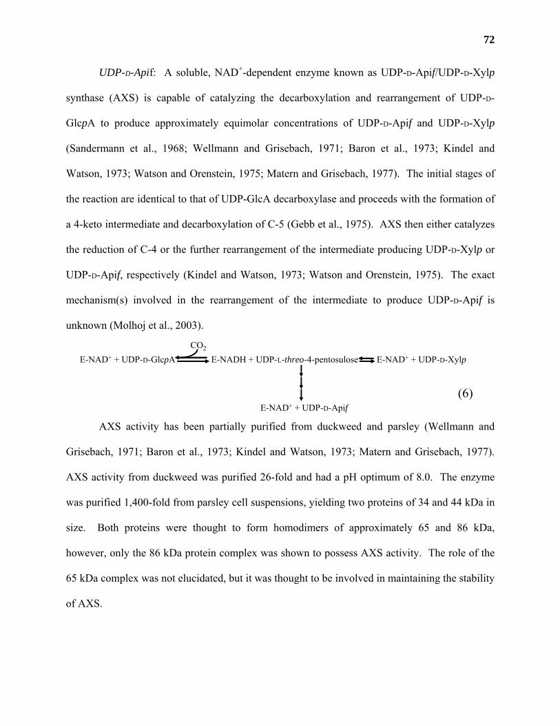
72
UDP-D-Apif: A soluble, NAD+-dependent enzyme known as UDP-D-Apif/UDP-D-Xylp
synthase (AXS) is capable of catalyzing the decarboxylation and rearrangement of UDP-D-
GlcpA to produce approximately equimolar concentrations of UDP-D-Apif and UDP-D-Xylp
(Sandermann et al., 1968; Wellmann and Grisebach, 1971; Baron et al., 1973; Kindel and
Watson, 1973; Watson and Orenstein, 1975; Matern and Grisebach, 1977). The initial stages of
the reaction are identical to that of UDP-GlcA decarboxylase and proceeds with the formation of
a 4-keto intermediate and decarboxylation of C-5 (Gebb et al., 1975). AXS then either catalyzes
the reduction of C-4 or the further rearrangement of the intermediate producing UDP-D-Xylp or
UDP-D-Apif, respectively (Kindel and Watson, 1973; Watson and Orenstein, 1975). The exact
mechanism(s) involved in the rearrangement of the intermediate to produce UDP-D-Apif is
unknown (Molhoj et al., 2003).
AXS activity has been partially purified from duckweed and parsley (Wellmann and
Grisebach, 1971; Baron et al., 1973; Kindel and Watson, 1973; Matern and Grisebach, 1977).
AXS activity from duckweed was purified 26-fold and had a pH optimum of 8.0. The enzyme
was purified 1,400-fold from parsley cell suspensions, yielding two proteins of 34 and 44 kDa in
size. Both proteins were thought to form homodimers of approximately 65 and 86 kDa,
however, only the 86 kDa protein complex was shown to possess AXS activity. The role of the
65 kDa complex was not elucidated, but it was thought to be involved in maintaining the stability
of AXS.
E-NAD+ + UDP-D-GlcpA E-NADH + UDP-L-threo-4-pentosuloseCO2
E-NAD+ + UDP-D-Xylp
E-NAD+ + UDP-D-Apif(6)
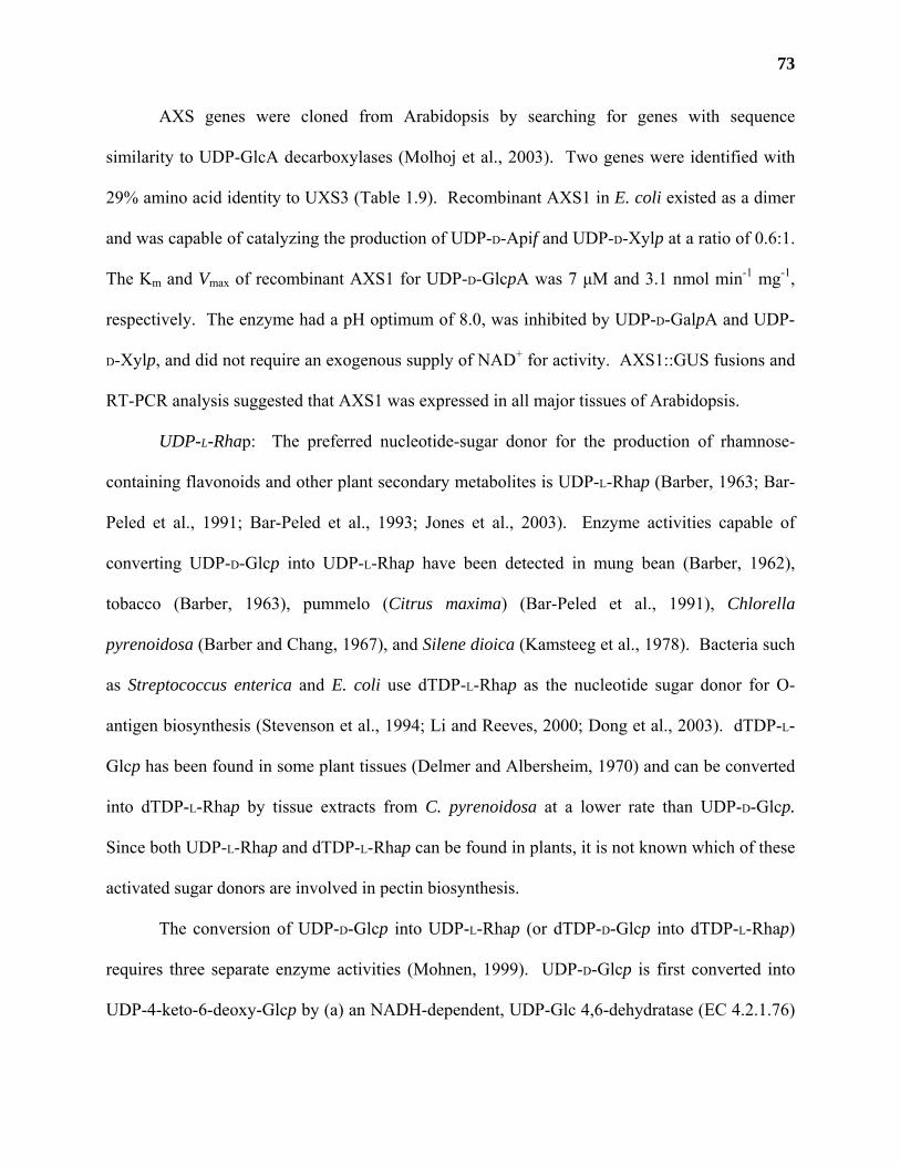
73
AXS genes were cloned from Arabidopsis by searching for genes with sequence
similarity to UDP-GlcA decarboxylases (Molhoj et al., 2003). Two genes were identified with
29% amino acid identity to UXS3 (Table 1.9). Recombinant AXS1 in E. coli existed as a dimer
and was capable of catalyzing the production of UDP-D-Apif and UDP-D-Xylp at a ratio of 0.6:1.
The Km and Vmax of recombinant AXS1 for UDP-D-GlcpA was 7 µM and 3.1 nmol min-1 mg-1,
respectively. The enzyme had a pH optimum of 8.0, was inhibited by UDP-D-GalpA and UDP-
D-Xylp, and did not require an exogenous supply of NAD+ for activity. AXS1::GUS fusions and
RT-PCR analysis suggested that AXS1 was expressed in all major tissues of Arabidopsis.
UDP-L-Rhap: The preferred nucleotide-sugar donor for the production of rhamnose-
containing flavonoids and other plant secondary metabolites is UDP-L-Rhap (Barber, 1963; Bar-
Peled et al., 1991; Bar-Peled et al., 1993; Jones et al., 2003). Enzyme activities capable of
converting UDP-D-Glcp into UDP-L-Rhap have been detected in mung bean (Barber, 1962),
tobacco (Barber, 1963), pummelo (Citrus maxima) (Bar-Peled et al., 1991), Chlorella
pyrenoidosa (Barber and Chang, 1967), and Silene dioica (Kamsteeg et al., 1978). Bacteria such
as Streptococcus enterica and E. coli use dTDP-L-Rhap as the nucleotide sugar donor for O-
antigen biosynthesis (Stevenson et al., 1994; Li and Reeves, 2000; Dong et al., 2003). dTDP-L-
Glcp has been found in some plant tissues (Delmer and Albersheim, 1970) and can be converted
into dTDP-L-Rhap by tissue extracts from C. pyrenoidosa at a lower rate than UDP-D-Glcp.
Since both UDP-L-Rhap and dTDP-L-Rhap can be found in plants, it is not known which of these
activated sugar donors are involved in pectin biosynthesis.
The conversion of UDP-D-Glcp into UDP-L-Rhap (or dTDP-D-Glcp into dTDP-L-Rhap)
requires three separate enzyme activities (Mohnen, 1999). UDP-D-Glcp is first converted into
UDP-4-keto-6-deoxy-Glcp by (a) an NADH-dependent, UDP-Glc 4,6-dehydratase (EC 4.2.1.76)
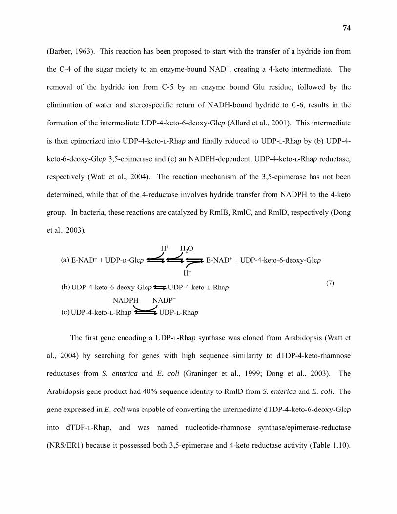
74
(Barber, 1963). This reaction has been proposed to start with the transfer of a hydride ion from
the C-4 of the sugar moiety to an enzyme-bound NAD+, creating a 4-keto intermediate. The
removal of the hydride ion from C-5 by an enzyme bound Glu residue, followed by the
elimination of water and stereospecific return of NADH-bound hydride to C-6, results in the
formation of the intermediate UDP-4-keto-6-deoxy-Glcp (Allard et al., 2001). This intermediate
is then epimerized into UDP-4-keto-L-Rhap and finally reduced to UDP-L-Rhap by (b) UDP-4-
keto-6-deoxy-Glcp 3,5-epimerase and (c) an NADPH-dependent, UDP-4-keto-L-Rhap reductase,
respectively (Watt et al., 2004). The reaction mechanism of the 3,5-epimerase has not been
determined, while that of the 4-reductase involves hydride transfer from NADPH to the 4-keto
group. In bacteria, these reactions are catalyzed by RmlB, RmlC, and RmlD, respectively (Dong
et al., 2003).
The first gene encoding a UDP-L-Rhap synthase was cloned from Arabidopsis (Watt et
al., 2004) by searching for genes with high sequence similarity to dTDP-4-keto-rhamnose
reductases from S. enterica and E. coli (Graninger et al., 1999; Dong et al., 2003). The
Arabidopsis gene product had 40% sequence identity to RmlD from S. enterica and E. coli. The
gene expressed in E. coli was capable of converting the intermediate dTDP-4-keto-6-deoxy-Glcp
into dTDP-L-Rhap, and was named nucleotide-rhamnose synthase/epimerase-reductase
(NRS/ER1) because it possessed both 3,5-epimerase and 4-keto reductase activity (Table 1.10).
E-NAD+ + UDP-D-Glcp E-NAD+ + UDP-4-keto-6-deoxy-GlcpH+ H2O
H+
UDP-4-keto-6-deoxy-Glcp UDP-4-keto-L-Rhap
UDP-4-keto-L-Rhap UDP-L-Rhap
(a)
(b)
(c)
(7)
NADPH NADP+
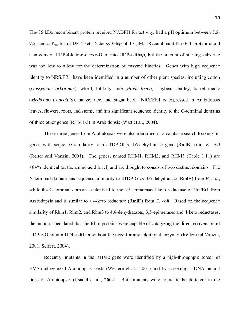
75
The 35 kDa recombinant protein required NADPH for activity, had a pH optimum between 5.5-
7.5, and a Km for dTDP-4-keto-6-deoxy-Glcp of 17 µM. Recombinant Nrs/Er1 protein could
also convert UDP-4-keto-6-deoxy-Glcp into UDP-L-Rhap, but the amount of starting substrate
was too low to allow for the determination of enzyme kinetics. Genes with high sequence
identity to NRS/ER1 have been identified in a number of other plant species, including cotton
(Gossypium arboreum), wheat, loblolly pine (Pinus taeda), soybean, barley, barrel medic
(Medicago truncatula), maize, rice, and sugar beet. NRS/ER1 is expressed in Arabidopsis
leaves, flowers, roots, and stems, and has significant sequence identity to the C-terminal domains
of three other genes (RHM1-3) in Arabidopsis (Watt et al., 2004).
These three genes from Arabidopsis were also identified in a database search looking for
genes with sequence similarity to a dTDP-Glcp 4,6-dehydratase gene (RmlB) from E. coli
(Reiter and Vanzin, 2001). The genes, named RHM1, RHM2, and RHM3 (Table 1.11) are
>84% identical (at the amino acid level) and are thought to consist of two distinct domains. The
N-terminal domain has sequence similarity to dTDP-Glcp 4,6-dehydratase (RmlB) from E. coli,
while the C-terminal domain is identical to the 3,5-epimerase/4-keto-reductase of Nrs/Er1 from
Arabidopsis and is similar to a 4-keto reductase (RmlD) from E. coli. Based on the sequence
similarity of Rhm1, Rhm2, and Rhm3 to 4,6-dehydratases, 3,5-epimerases and 4-keto reductases,
the authors speculated that the Rhm proteins were capable of catalyzing the direct conversion of
UDP-D-Glcp into UDP-L-Rhap without the need for any additional enzymes (Reiter and Vanzin,
2001; Seifert, 2004).
Recently, mutants in the RHM2 gene were identified by a high-throughput screen of
EMS-mutagenized Arabidopsis seeds (Western et al., 2001) and by screening T-DNA mutant
lines of Arabidopsis (Usadel et al., 2004). Both mutants were found to be deficient in the
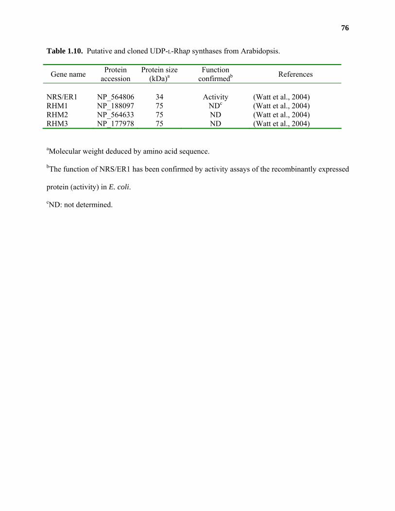
76
Table 1.10. Putative and cloned UDP-L-Rhap synthases from Arabidopsis.
Gene name Protein accession
Protein size (kDa)a
Function confirmedb References
NRS/ER1 NP_564806 34 Activity (Watt et al., 2004) RHM1 NP_188097 75 NDc (Watt et al., 2004) RHM2 NP_564633 75 ND (Watt et al., 2004) RHM3 NP_177978 75 ND (Watt et al., 2004)
aMolecular weight deduced by amino acid sequence.
bThe function of NRS/ER1 has been confirmed by activity assays of the recombinantly expressed
protein (activity) in E. coli.
cND: not determined.
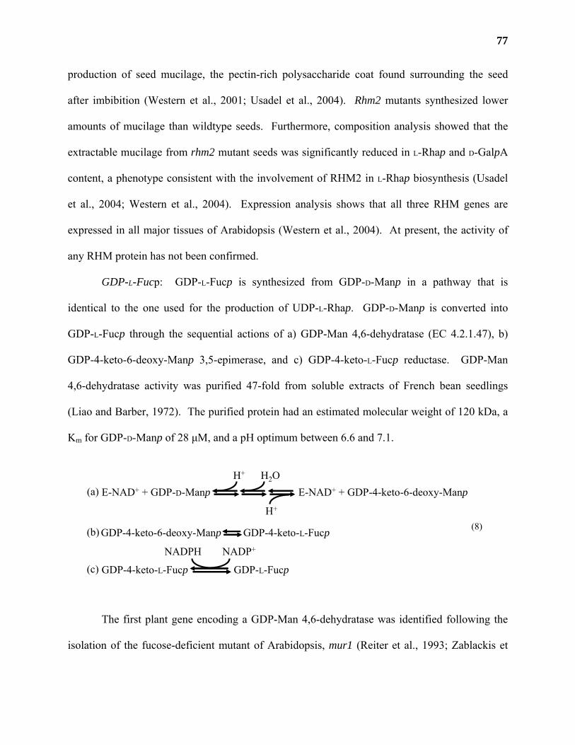
77
production of seed mucilage, the pectin-rich polysaccharide coat found surrounding the seed
after imbibition (Western et al., 2001; Usadel et al., 2004). Rhm2 mutants synthesized lower
amounts of mucilage than wildtype seeds. Furthermore, composition analysis showed that the
extractable mucilage from rhm2 mutant seeds was significantly reduced in L-Rhap and D-GalpA
content, a phenotype consistent with the involvement of RHM2 in L-Rhap biosynthesis (Usadel
et al., 2004; Western et al., 2004). Expression analysis shows that all three RHM genes are
expressed in all major tissues of Arabidopsis (Western et al., 2004). At present, the activity of
any RHM protein has not been confirmed.
GDP-L-Fucp: GDP-L-Fucp is synthesized from GDP-D-Manp in a pathway that is
identical to the one used for the production of UDP-L-Rhap. GDP-D-Manp is converted into
GDP-L-Fucp through the sequential actions of a) GDP-Man 4,6-dehydratase (EC 4.2.1.47), b)
GDP-4-keto-6-deoxy-Manp 3,5-epimerase, and c) GDP-4-keto-L-Fucp reductase. GDP-Man
4,6-dehydratase activity was purified 47-fold from soluble extracts of French bean seedlings
(Liao and Barber, 1972). The purified protein had an estimated molecular weight of 120 kDa, a
Km for GDP-D-Manp of 28 µM, and a pH optimum between 6.6 and 7.1.
The first plant gene encoding a GDP-Man 4,6-dehydratase was identified following the
isolation of the fucose-deficient mutant of Arabidopsis, mur1 (Reiter et al., 1993; Zablackis et
E-NAD+ + GDP-D-Manp E-NAD+ + GDP-4-keto-6-deoxy-ManpH+ H2O
H+
GDP-4-keto-6-deoxy-Manp GDP-4-keto-L-Fucp
GDP-4-keto-L-Fucp GDP-L-Fucp
(a)
(b)
(c)
(8)
NADPH NADP+
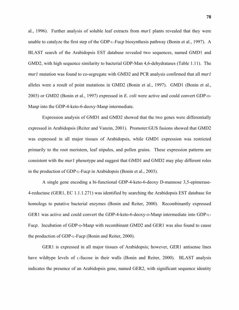
78
al., 1996). Further analysis of soluble leaf extracts from mur1 plants revealed that they were
unable to catalyze the first step of the GDP-L-Fucp biosynthesis pathway (Bonin et al., 1997). A
BLAST search of the Arabidopsis EST database revealed two sequences, named GMD1 and
GMD2, with high sequence similarity to bacterial GDP-Man 4,6-dehydratases (Table 1.11). The
mur1 mutation was found to co-segregate with GMD2 and PCR analysis confirmed that all mur1
alleles were a result of point mutations in GMD2 (Bonin et al., 1997). GMD1 (Bonin et al.,
2003) or GMD2 (Bonin et al., 1997) expressed in E. coli were active and could convert GDP-D-
Manp into the GDP-4-keto-6-deoxy-Manp intermediate.
Expression analysis of GMD1 and GMD2 showed that the two genes were differentially
expressed in Arabidopsis (Reiter and Vanzin, 2001). Promoter:GUS fusions showed that GMD2
was expressed in all major tissues of Arabidopsis, while GMD1 expression was restricted
primarily to the root meristem, leaf stipules, and pollen grains. These expression patterns are
consistent with the mur1 phenotype and suggest that GMD1 and GMD2 may play different roles
in the production of GDP-L-Fucp in Arabidopsis (Bonin et al., 2003).
A single gene encoding a bi-functional GDP-4-keto-6-deoxy D-mannose 3,5-epimerase-
4-reductase (GER1, EC 1.1.1.271) was identified by searching the Arabidopsis EST database for
homologs to putative bacterial enzymes (Bonin and Reiter, 2000). Recombinantly expressed
GER1 was active and could convert the GDP-4-keto-6-deoxy-D-Manp intermediate into GDP-L-
Fucp. Incubation of GDP-D-Manp with recombinant GMD2 and GER1 was also found to cause
the production of GDP-L-Fucp (Bonin and Reiter, 2000).
GER1 is expressed in all major tissues of Arabidopsis; however, GER1 antisense lines
have wildtype levels of L-fucose in their walls (Bonin and Reiter, 2000). BLAST analysis
indicates the presence of an Arabidopsis gene, named GER2, with significant sequence identity
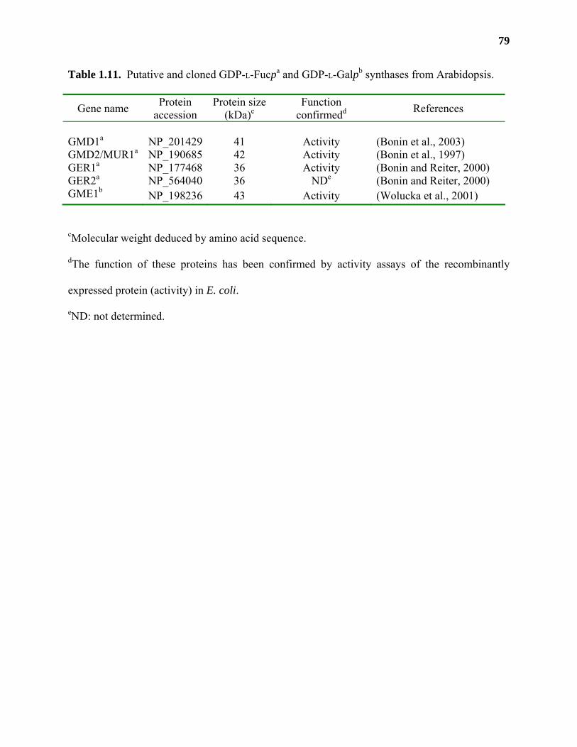
79
Table 1.11. Putative and cloned GDP-L-Fucpa and GDP-L-Galpb synthases from Arabidopsis.
Gene name Protein accession
Protein size (kDa)c
Function confirmedd References
GMD1a NP_201429 41 Activity (Bonin et al., 2003) GMD2/MUR1a NP_190685 42 Activity (Bonin et al., 1997) GER1a NP_177468 36 Activity (Bonin and Reiter, 2000) GER2a NP_564040 36 NDe (Bonin and Reiter, 2000) GME1b NP_198236 43 Activity (Wolucka et al., 2001)
cMolecular weight deduced by amino acid sequence.
dThe function of these proteins has been confirmed by activity assays of the recombinantly
expressed protein (activity) in E. coli.
eND: not determined.
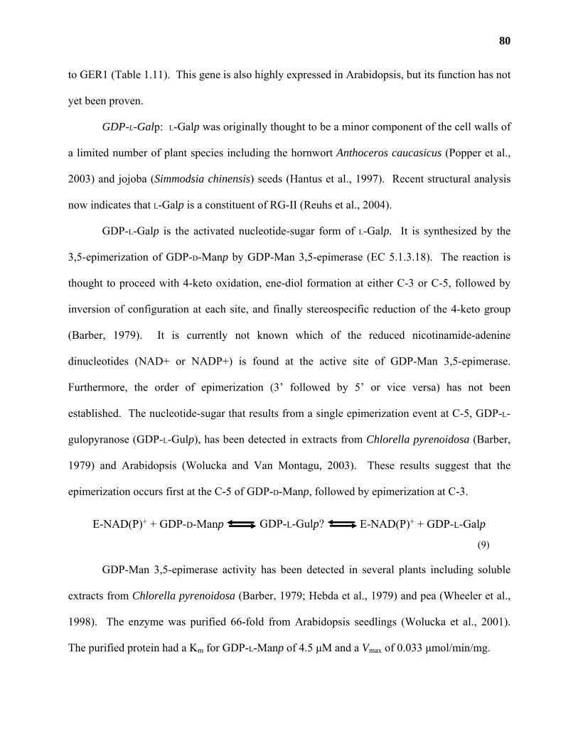
80
to GER1 (Table 1.11). This gene is also highly expressed in Arabidopsis, but its function has not
yet been proven.
GDP-L-Galp: L-Galp was originally thought to be a minor component of the cell walls of
a limited number of plant species including the hornwort Anthoceros caucasicus (Popper et al.,
2003) and jojoba (Simmodsia chinensis) seeds (Hantus et al., 1997). Recent structural analysis
now indicates that L-Galp is a constituent of RG-II (Reuhs et al., 2004).
GDP-L-Galp is the activated nucleotide-sugar form of L-Galp. It is synthesized by the
3,5-epimerization of GDP-D-Manp by GDP-Man 3,5-epimerase (EC 5.1.3.18). The reaction is
thought to proceed with 4-keto oxidation, ene-diol formation at either C-3 or C-5, followed by
inversion of configuration at each site, and finally stereospecific reduction of the 4-keto group
(Barber, 1979). It is currently not known which of the reduced nicotinamide-adenine
dinucleotides (NAD+ or NADP+) is found at the active site of GDP-Man 3,5-epimerase.
Furthermore, the order of epimerization (3’ followed by 5’ or vice versa) has not been
established. The nucleotide-sugar that results from a single epimerization event at C-5, GDP-L-
gulopyranose (GDP-L-Gulp), has been detected in extracts from Chlorella pyrenoidosa (Barber,
1979) and Arabidopsis (Wolucka and Van Montagu, 2003). These results suggest that the
epimerization occurs first at the C-5 of GDP-D-Manp, followed by epimerization at C-3.
GDP-Man 3,5-epimerase activity has been detected in several plants including soluble
extracts from Chlorella pyrenoidosa (Barber, 1979; Hebda et al., 1979) and pea (Wheeler et al.,
1998). The enzyme was purified 66-fold from Arabidopsis seedlings (Wolucka et al., 2001).
The purified protein had a Km for GDP-L-Manp of 4.5 µM and a Vmax of 0.033 µmol/min/mg.
E-NAD(P)+ + GDP-D-Manp E-NAD(P)+ + GDP-L-GalpGDP-L-Gulp?
(9)
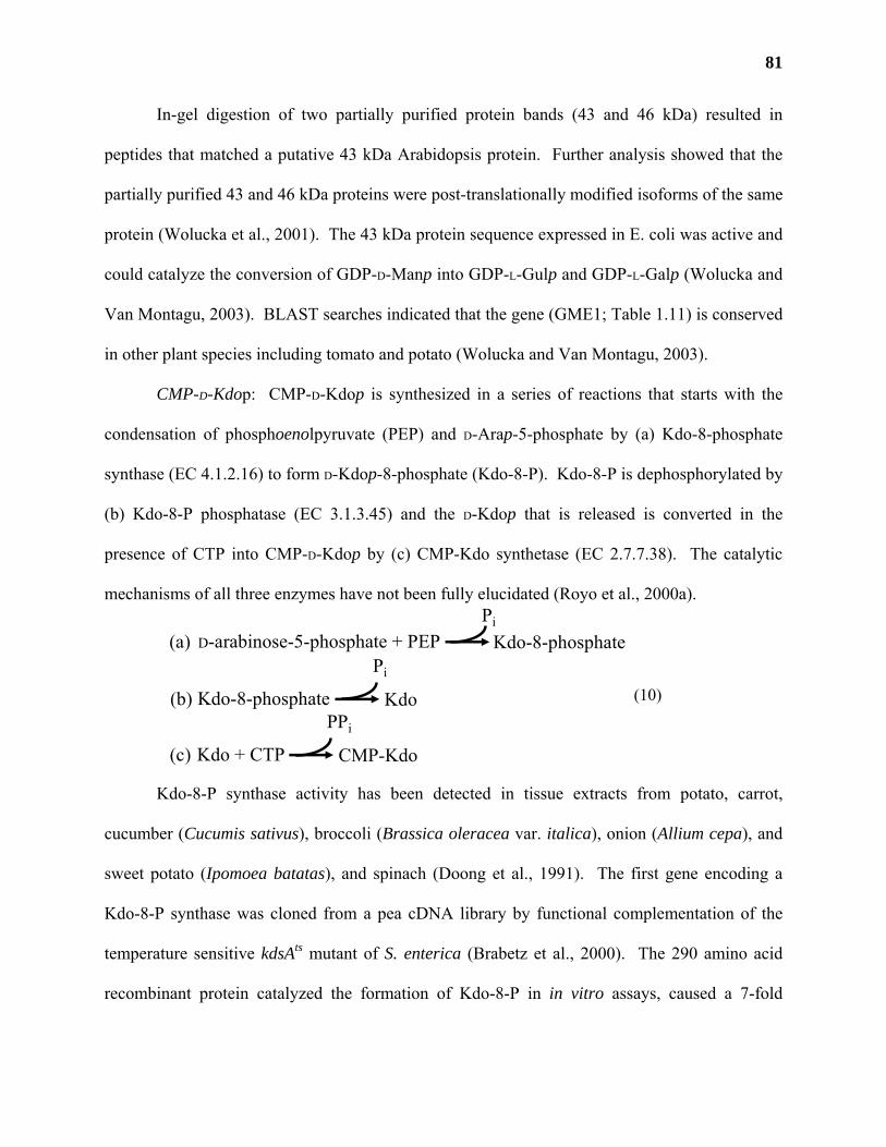
81
In-gel digestion of two partially purified protein bands (43 and 46 kDa) resulted in
peptides that matched a putative 43 kDa Arabidopsis protein. Further analysis showed that the
partially purified 43 and 46 kDa proteins were post-translationally modified isoforms of the same
protein (Wolucka et al., 2001). The 43 kDa protein sequence expressed in E. coli was active and
could catalyze the conversion of GDP-D-Manp into GDP-L-Gulp and GDP-L-Galp (Wolucka and
Van Montagu, 2003). BLAST searches indicated that the gene (GME1; Table 1.11) is conserved
in other plant species including tomato and potato (Wolucka and Van Montagu, 2003).
CMP-D-Kdop: CMP-D-Kdop is synthesized in a series of reactions that starts with the
condensation of phosphoenolpyruvate (PEP) and D-Arap-5-phosphate by (a) Kdo-8-phosphate
synthase (EC 4.1.2.16) to form D-Kdop-8-phosphate (Kdo-8-P). Kdo-8-P is dephosphorylated by
(b) Kdo-8-P phosphatase (EC 3.1.3.45) and the D-Kdop that is released is converted in the
presence of CTP into CMP-D-Kdop by (c) CMP-Kdo synthetase (EC 2.7.7.38). The catalytic
mechanisms of all three enzymes have not been fully elucidated (Royo et al., 2000a).
Kdo-8-P synthase activity has been detected in tissue extracts from potato, carrot,
cucumber (Cucumis sativus), broccoli (Brassica oleracea var. italica), onion (Allium cepa), and
sweet potato (Ipomoea batatas), and spinach (Doong et al., 1991). The first gene encoding a
Kdo-8-P synthase was cloned from a pea cDNA library by functional complementation of the
temperature sensitive kdsAts mutant of S. enterica (Brabetz et al., 2000). The 290 amino acid
recombinant protein catalyzed the formation of Kdo-8-P in in vitro assays, caused a 7-fold
D-arabinose-5-phosphate + PEPPi
Kdo-8-phosphate(a)
Kdo-8-phosphate
Pi
Kdo(b)
Kdo + CTP
PPi
CMP-Kdo(c)
(10)
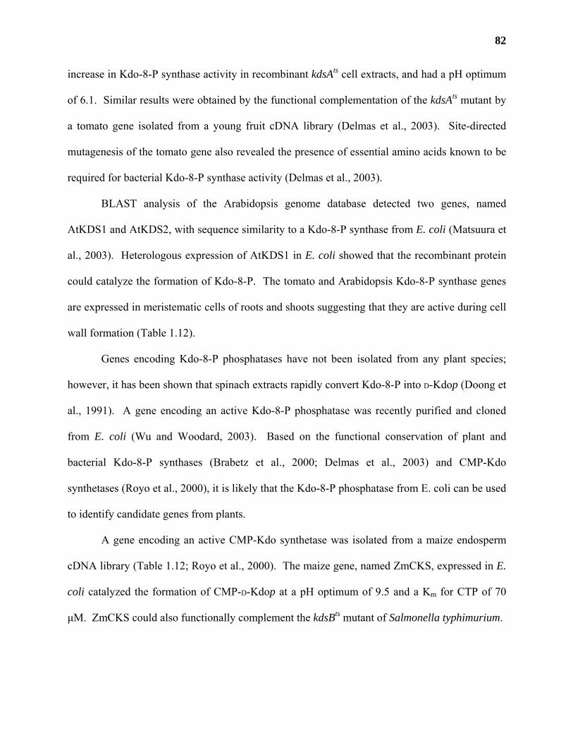
82
increase in Kdo-8-P synthase activity in recombinant kdsAts cell extracts, and had a pH optimum
of 6.1. Similar results were obtained by the functional complementation of the kdsAts mutant by
a tomato gene isolated from a young fruit cDNA library (Delmas et al., 2003). Site-directed
mutagenesis of the tomato gene also revealed the presence of essential amino acids known to be
required for bacterial Kdo-8-P synthase activity (Delmas et al., 2003).
BLAST analysis of the Arabidopsis genome database detected two genes, named
AtKDS1 and AtKDS2, with sequence similarity to a Kdo-8-P synthase from E. coli (Matsuura et
al., 2003). Heterologous expression of AtKDS1 in E. coli showed that the recombinant protein
could catalyze the formation of Kdo-8-P. The tomato and Arabidopsis Kdo-8-P synthase genes
are expressed in meristematic cells of roots and shoots suggesting that they are active during cell
wall formation (Table 1.12).
Genes encoding Kdo-8-P phosphatases have not been isolated from any plant species;
however, it has been shown that spinach extracts rapidly convert Kdo-8-P into D-Kdop (Doong et
al., 1991). A gene encoding an active Kdo-8-P phosphatase was recently purified and cloned
from E. coli (Wu and Woodard, 2003). Based on the functional conservation of plant and
bacterial Kdo-8-P synthases (Brabetz et al., 2000; Delmas et al., 2003) and CMP-Kdo
synthetases (Royo et al., 2000), it is likely that the Kdo-8-P phosphatase from E. coli can be used
to identify candidate genes from plants.
A gene encoding an active CMP-Kdo synthetase was isolated from a maize endosperm
cDNA library (Table 1.12; Royo et al., 2000). The maize gene, named ZmCKS, expressed in E.
coli catalyzed the formation of CMP-D-Kdop at a pH optimum of 9.5 and a Km for CTP of 70
µM. ZmCKS could also functionally complement the kdsBts mutant of Salmonella typhimurium.
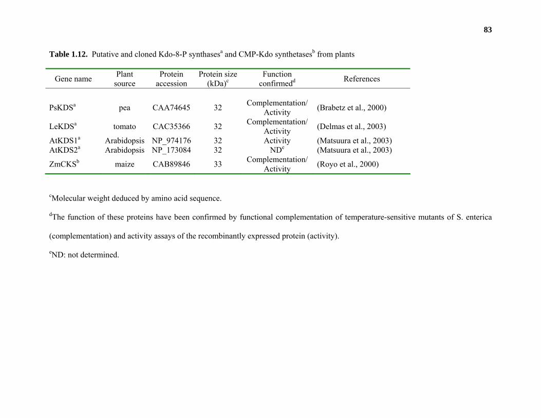
83
Table 1.12. Putative and cloned Kdo-8-P synthasesa and CMP-Kdo synthetasesb from plants
Gene name Plant source
Protein accession
Protein size (kDa)c
Function confirmedd References
PsKDSa pea CAA74645 32 Complementation/ Activity (Brabetz et al., 2000)
LeKDSa tomato CAC35366 32 Complementation/ Activity (Delmas et al., 2003)
AtKDS1a Arabidopsis NP_974176 32 Activity (Matsuura et al., 2003) AtKDS2a Arabidopsis NP_173084 32 NDe (Matsuura et al., 2003)
ZmCKSb maize CAB89846 33 Complementation/ Activity (Royo et al., 2000)
cMolecular weight deduced by amino acid sequence.
dThe function of these proteins have been confirmed by functional complementation of temperature-sensitive mutants of S. enterica
(complementation) and activity assays of the recombinantly expressed protein (activity).
eND: not determined.
84
ZmCKS is expressed in all major tissues of maize (Royo et al., 2000). Furthermore,
genes with high sequence similarity to ZmCKS have been detected in Arabidopsis, loblolly pine
tomato, rice, wheat, and barley (Royo et al., 2000; Royo et al., 2000a). All putative plant CMP-
Kdo synthetases differ from bacterial enzymes in that they are predicted to have an N-terminal
signal sequence that targets them to the endomembrane system (Royo et al., 2000a). The.
subcellular location of ZmCKS or any other putative CMP-Kdo synthetase has yet to be
determined.
XXP-D-Dhap and XXP-L-AcefA: The enzymes and/or pathways involved in synthesizing
D-Dhap, L-AcefA, or their activated nucleotide-sugar forms have not been determined. It has
been hypothesized that plant 2-keto-3-deoxy-D-arabino-heptulosonic acid-7-P (DAH-7-P)
synthases may be able to catalyze the condensation of D-threose-4-P and PEP to form D-Dhap-7-
P (Doong et al., 1992), a precursor of D-Dhap. At present, the absolute configuration of the
product from this reaction is not known (Doong et al., 1992). Further research on the synthesis
of D-Dhap and L-AcefA will be required before their biosynthetic pathways can be elucidated.
Nucleotide sugar transporters involved in pectin biosynthesis
The results from the previous section suggest that the nucleotide-sugar biosynthesis
occurs mainly in the cytosol. Nucleotide-sugar transporters (NSTs) are multiple-membrane
spanning, antiport proteins that transfer nucleotide-sugars into the lumen of the endoplasmic
reticulum (ER) or the Golgi in exchange for their corresponding nucleotide-monophosphates
(Hirschberg et al., 1998). Many NSTs are specific for only one type of nucleotide-sugar, while
others, such as the GDP-D-Manp transporter from Leishmania (Hong et al., 2000) and the UDP-
D-Galp transporter from humans and Drosophila (Segawa et al., 2002) are able to transport more
than one type of nucleotide-sugar. The study of NSTs has recently gained prominence with the
85
discovery that NST mutants are defective in protein glycosylation (Dean et al., 1997; Ma et al.,
1997), have altered cell surface decorations (Deutscher and Hirschberg, 1986; Kawakita et al.,
1998; Hoeflich et al., 2004), and can result in developmental defects (Berninsone et al., 2001).
Genes encoding NSTs have been identified in yeast (Dean et al., 1997), Leishmania
donovani (Ma et al., 1997), mammals (Deutscher and Hirschberg, 1986; Hirschberg et al., 1998;
Aoki et al., 2003), Drosophila melanogaster (Segawa et al., 2002), Caenorhabditis elegans
(Berninsone et al., 2001; Hoeflich et al., 2004), and Arabidopsis thaliana (Baldwin et al., 2001;
Norambuena et al., 2002; Bakker et al., 2004; Handford et al., 2004). The transport of GDP-L-
Fucp (Wulff et al., 2000), UDP-D-Glcp (Munoz et al., 1996), UDP-D-Galp (Norambuena et al.,
2002), UDP-D-GlcpA, and UDP-D-Xylp (Hayashi et al., 1988) has been shown to occur in pea
Golgi vesicles, and plant genes encoding NSTs have recently been identified and cloned. This
section will describe what is currently known about NST genes from plants that are involved in
pectin biosynthesis.
UDP-D-Glcp/UDP-D-Galp transporter: A NST capable of transporting both UDP-D-Glcp
and UDP-D-Galp was cloned from Arabidopsis by searching for genes with high sequence
similarity to UDP-D-Galp transporters from humans and yeast (Norambuena et al., 2002). The
gene (AtUTr1) could functionally complement a canine MDCK mutant cell line deficient in
UDP-D-Galp transport. Expression of AtUTr1 in S. cerevisiae yielded an increase in the
transport of UDP-D-Glcp and UDP-D-Galp in intact Golgi vesicles, but not the transport of any
other nucleotide-sugar that was tested. AtUTr1 is thought to be part of a 6 member gene family
in Arabidopsis (Table 1.13).
UDP-D-Galp transporter: Two genes from an Arabidopsis cDNA library were identified
that could functionally complement a Chinese hamster ovary cell line deficient in the transport of
86
UDP-D-Galp (Table 1.13). The genes (UDP-GalT1 and 2) expressed in S. cerevisiae, caused a
10-fold increase in UDP-D-Galp transport in intact Golgi vesicles (Bakker et al., 2004).
GONST transporters: A GDP-D-Manp transporter from Arabidopsis was recently cloned
and named GONST1 (Baldwin et al., 2001). While the transport of GDP-D-Manp is not
predicted to be required for pectin biosynthesis, GONST1 is part of a 5 member gene family for
which 3 of the members have no known function (Handford et al., 2004). BLAST analysis of
GONST5 shows that it has 91% sequence identity to UDP-GalT1, suggesting that it may also be
a UDP-D-Galp transporter. Further research on these and other putative NSTs should allow us to
determine which of these proteins are involved in pectin biosynthesis.
Pectin glycosyltransferases
Nucleotide-sugars transported into the lumen of the Golgi apparatus become substrates
for Golgi-localized glycosyltransferases. At least some of these glycosyltransferases are type II
membrane proteins that catalyze the transfer of the sugar moiety of the nucleotide-sugar onto the
non-reducing end of oligo- or polysaccharide acceptors (Keegstra and Raikhel, 2001; Perrin et
al., 2001). It has been estimated that at least 62 different glycosyltransferase activities would be
required to synthesize all of the pectic polysaccharides found in the primary wall based on the
current structure of pectin (Table 1.14; Mohnen, 2002). Of these predicted enzyme activities,
only three [α-(1,4)-galacturonosyltransferase, α-(1,2)-galacturonosyltransferase and α-(1,4)-
rhamnosyltransferase] are involved in the synthesis of the backbone structures of pectin (Table
1.14). These backbone glycosyltransferases may be processive enzymes that catalyze the
synthesis of polymeric products (Mohnen, 2002). The other glycosyltransferases are predicted to
catalyze the addition of glycosyl residues producing the side branches found in pectic
polysaccharides.
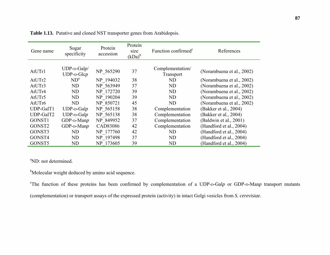
87
Table 1.13. Putative and cloned NST transporter genes from Arabidopsis.
Gene name Sugar specificity
Protein accession
Protein size
(kDa)b Function confirmedc References
AtUTr1 UDP-D-Galp/ UDP-D-Glcp NP_565290 37 Complementation/
Transport (Norambuena et al., 2002)
AtUTr2 NDa NP_194032 38 ND (Norambuena et al., 2002) AtUTr3 ND NP_563949 37 ND (Norambuena et al., 2002) AtUTr4 ND NP_172720 39 ND (Norambuena et al., 2002) AtUTr5 ND NP_190204 39 ND (Norambuena et al., 2002) AtUTr6 ND NP_850721 45 ND (Norambuena et al., 2002) UDP-GalT1 UDP-D-Galp NP_565158 38 Complementation (Bakker et al., 2004) UDP-GalT2 UDP-D-Galp NP_565138 38 Complementation (Bakker et al., 2004) GONST1 GDP-D-Manp NP_849952 37 Complementation (Baldwin et al., 2001) GONST2 GDP-D-Manp CAD83086 42 Complementation (Handford et al., 2004) GONST3 ND NP_177760 42 ND (Handford et al., 2004) GONST4 ND NP_197498 37 ND (Handford et al., 2004) GONST5 ND NP_173605 39 ND (Handford et al., 2004)
aND: not determined.
bMolecular weight deduced by amino acid sequence.
cThe function of these proteins has been confirmed by complementation of a UDP-D-Galp or GDP-D-Manp transport mutants
(complementation) or transport assays of the expressed protein (activity) in intact Golgi vesicles from S. cerevisiae.
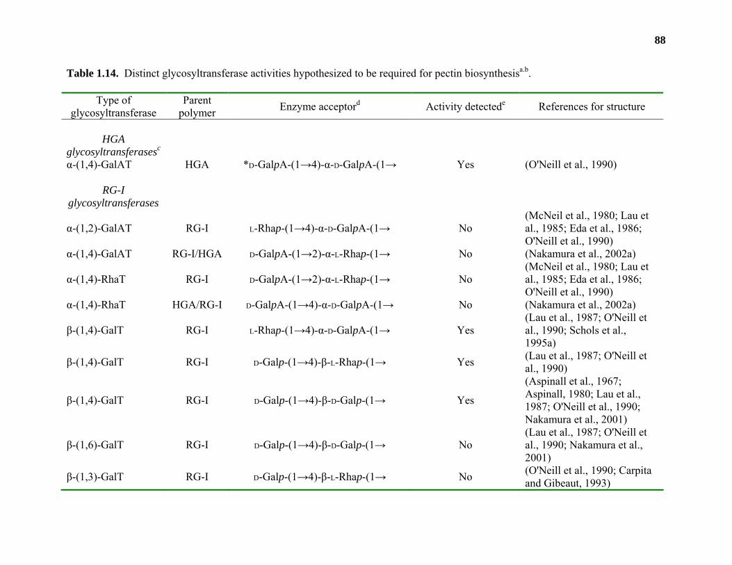
88
Table 1.14. Distinct glycosyltransferase activities hypothesized to be required for pectin biosynthesisa.b.
Type of glycosyltransferase
Parent polymer Enzyme acceptord Activity detectede References for structure
HGA
glycosyltransferasesc
α-(1,4)-GalAT HGA *D-GalpA-(1→4)-α-D-GalpA-(1→ Yes (O'Neill et al., 1990)
RG-I glycosyltransferases
α-(1,2)-GalAT RG-I L-Rhap-(1→4)-α-D-GalpA-(1→ No (McNeil et al., 1980; Lau et al., 1985; Eda et al., 1986; O'Neill et al., 1990)
α-(1,4)-GalAT RG-I/HGA D-GalpA-(1→2)-α-L-Rhap-(1→ No (Nakamura et al., 2002a)
α-(1,4)-RhaT RG-I D-GalpA-(1→2)-α-L-Rhap-(1→ No (McNeil et al., 1980; Lau et al., 1985; Eda et al., 1986; O'Neill et al., 1990)
α-(1,4)-RhaT HGA/RG-I D-GalpA-(1→4)-α-D-GalpA-(1→ No (Nakamura et al., 2002a)
β-(1,4)-GalT RG-I L-Rhap-(1→4)-α-D-GalpA-(1→ Yes (Lau et al., 1987; O'Neill et al., 1990; Schols et al., 1995a)
β-(1,4)-GalT RG-I D-Galp-(1→4)-β-L-Rhap-(1→ Yes (Lau et al., 1987; O'Neill et al., 1990)
β-(1,4)-GalT RG-I D-Galp-(1→4)-β-D-Galp-(1→ Yes
(Aspinall et al., 1967; Aspinall, 1980; Lau et al., 1987; O'Neill et al., 1990; Nakamura et al., 2001)
β-(1,6)-GalT RG-I D-Galp-(1→4)-β-D-Galp-(1→ No (Lau et al., 1987; O'Neill et al., 1990; Nakamura et al., 2001)
β-(1,3)-GalT RG-I D-Galp-(1→4)-β-L-Rhap-(1→ No (O'Neill et al., 1990; Carpita and Gibeaut, 1993)
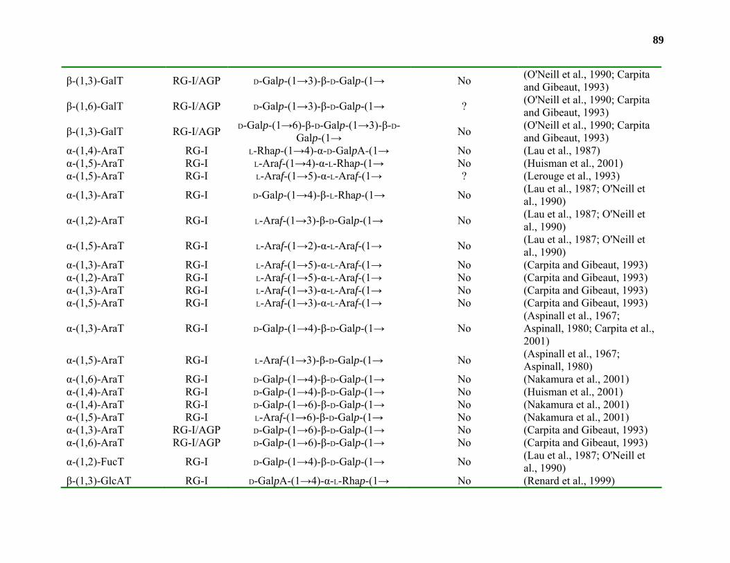
89
β-(1,3)-GalT RG-I/AGP D-Galp-(1→3)-β-D-Galp-(1→ No (O'Neill et al., 1990; Carpita and Gibeaut, 1993)
β-(1,6)-GalT RG-I/AGP D-Galp-(1→3)-β-D-Galp-(1→ ? (O'Neill et al., 1990; Carpita and Gibeaut, 1993)
β-(1,3)-GalT RG-I/AGP D-Galp-(1→6)-β-D-Galp-(1→3)-β-D-Galp-(1→ No (O'Neill et al., 1990; Carpita
and Gibeaut, 1993) α-(1,4)-AraT RG-I L-Rhap-(1→4)-α-D-GalpA-(1→ No (Lau et al., 1987) α-(1,5)-AraT RG-I L-Araf-(1→4)-α-L-Rhap-(1→ No (Huisman et al., 2001) α-(1,5)-AraT RG-I L-Araf-(1→5)-α-L-Araf-(1→ ? (Lerouge et al., 1993)
α-(1,3)-AraT RG-I D-Galp-(1→4)-β-L-Rhap-(1→ No (Lau et al., 1987; O'Neill et al., 1990)
α-(1,2)-AraT RG-I L-Araf-(1→3)-β-D-Galp-(1→ No (Lau et al., 1987; O'Neill et al., 1990)
α-(1,5)-AraT RG-I L-Araf-(1→2)-α-L-Araf-(1→ No (Lau et al., 1987; O'Neill et al., 1990)
α-(1,3)-AraT RG-I L-Araf-(1→5)-α-L-Araf-(1→ No (Carpita and Gibeaut, 1993) α-(1,2)-AraT RG-I L-Araf-(1→5)-α-L-Araf-(1→ No (Carpita and Gibeaut, 1993) α-(1,3)-AraT RG-I L-Araf-(1→3)-α-L-Araf-(1→ No (Carpita and Gibeaut, 1993) α-(1,5)-AraT RG-I L-Araf-(1→3)-α-L-Araf-(1→ No (Carpita and Gibeaut, 1993)
α-(1,3)-AraT RG-I D-Galp-(1→4)-β-D-Galp-(1→ No (Aspinall et al., 1967; Aspinall, 1980; Carpita et al., 2001)
α-(1,5)-AraT RG-I L-Araf-(1→3)-β-D-Galp-(1→ No (Aspinall et al., 1967; Aspinall, 1980)
α-(1,6)-AraT RG-I D-Galp-(1→4)-β-D-Galp-(1→ No (Nakamura et al., 2001) α-(1,4)-AraT RG-I D-Galp-(1→4)-β-D-Galp-(1→ No (Huisman et al., 2001) α-(1,4)-AraT RG-I D-Galp-(1→6)-β-D-Galp-(1→ No (Nakamura et al., 2001) α-(1,5)-AraT RG-I L-Araf-(1→6)-β-D-Galp-(1→ No (Nakamura et al., 2001) α-(1,3)-AraT RG-I/AGP D-Galp-(1→6)-β-D-Galp-(1→ No (Carpita and Gibeaut, 1993) α-(1,6)-AraT RG-I/AGP D-Galp-(1→6)-β-D-Galp-(1→ No (Carpita and Gibeaut, 1993)
α-(1,2)-FucT RG-I D-Galp-(1→4)-β-D-Galp-(1→ No (Lau et al., 1987; O'Neill et al., 1990)
β-(1,3)-GlcAT RG-I D-GalpA-(1→4)-α-L-Rhap-(1→ No (Renard et al., 1999)
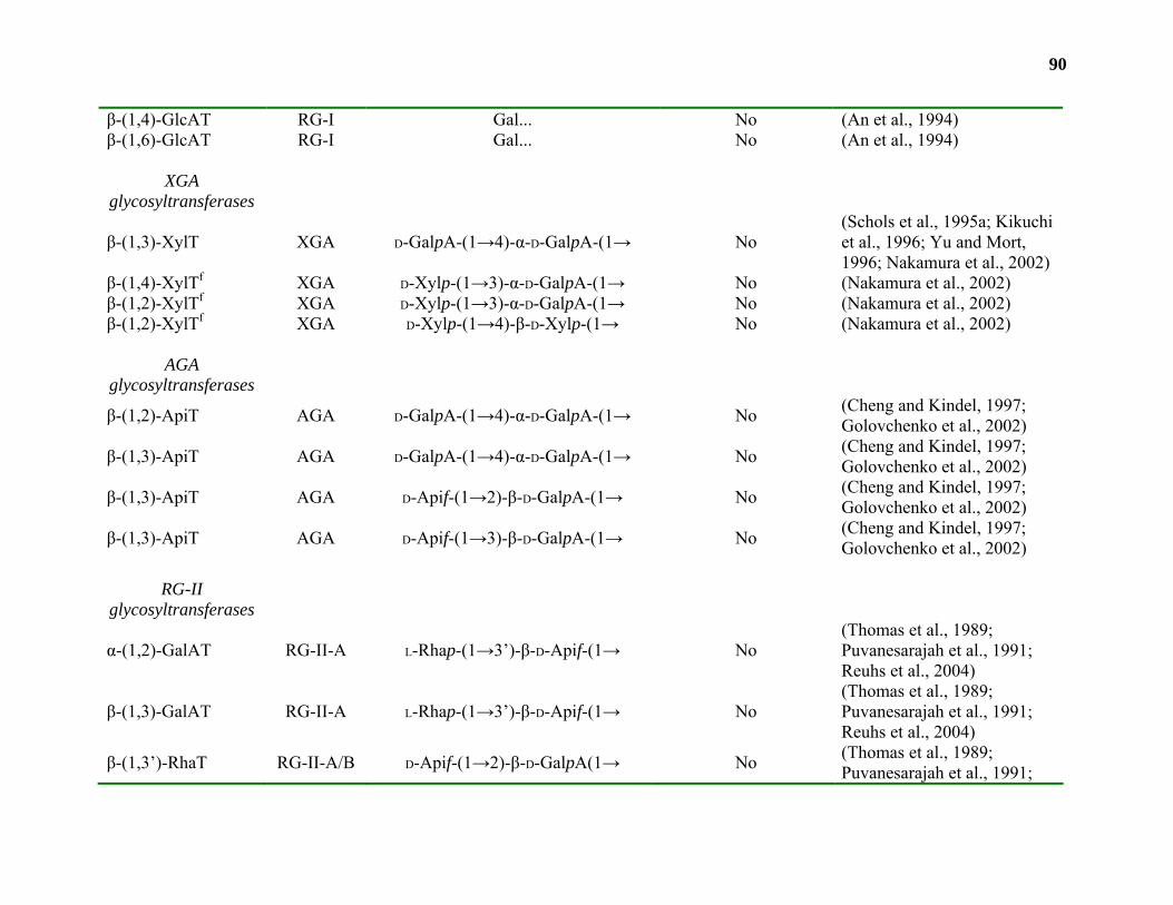
90
β-(1,4)-GlcAT RG-I Gal... No (An et al., 1994) β-(1,6)-GlcAT RG-I Gal... No (An et al., 1994)
XGA glycosyltransferases
β-(1,3)-XylT XGA D-GalpA-(1→4)-α-D-GalpA-(1→ No (Schols et al., 1995a; Kikuchi et al., 1996; Yu and Mort, 1996; Nakamura et al., 2002)
β-(1,4)-XylTf XGA D-Xylp-(1→3)-α-D-GalpA-(1→ No (Nakamura et al., 2002) β-(1,2)-XylTf XGA D-Xylp-(1→3)-α-D-GalpA-(1→ No (Nakamura et al., 2002) β-(1,2)-XylTf XGA D-Xylp-(1→4)-β-D-Xylp-(1→ No (Nakamura et al., 2002)
AGA
glycosyltransferases
β-(1,2)-ApiT AGA D-GalpA-(1→4)-α-D-GalpA-(1→ No (Cheng and Kindel, 1997; Golovchenko et al., 2002)
β-(1,3)-ApiT AGA D-GalpA-(1→4)-α-D-GalpA-(1→ No (Cheng and Kindel, 1997; Golovchenko et al., 2002)
β-(1,3)-ApiT AGA D-Apif-(1→2)-β-D-GalpA-(1→ No (Cheng and Kindel, 1997; Golovchenko et al., 2002)
β-(1,3)-ApiT AGA D-Apif-(1→3)-β-D-GalpA-(1→ No (Cheng and Kindel, 1997; Golovchenko et al., 2002)
RG-II
glycosyltransferases
α-(1,2)-GalAT RG-II-A L-Rhap-(1→3’)-β-D-Apif-(1→ No (Thomas et al., 1989; Puvanesarajah et al., 1991; Reuhs et al., 2004)
β-(1,3)-GalAT RG-II-A L-Rhap-(1→3’)-β-D-Apif-(1→ No (Thomas et al., 1989; Puvanesarajah et al., 1991; Reuhs et al., 2004)
β-(1,3’)-RhaT RG-II-A/B D-Apif-(1→2)-β-D-GalpA(1→ No (Thomas et al., 1989; Puvanesarajah et al., 1991;
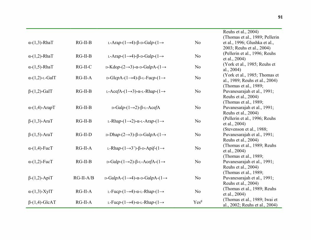
91
Reuhs et al., 2004)
α-(1,3)-RhaT RG-II-B L-Arap-(1→4)-β-D-Galp-(1→ No (Thomas et al., 1989; Pellerin et al., 1996; Glushka et al., 2003; Reuhs et al., 2004)
α-(1,2)-RhaT RG-II-B L-Arap-(1→4)-β-D-Galp-(1→ No (Pellerin et al., 1996; Reuhs et al., 2004)
α-(1,5)-RhaT RG-II-C D-Kdop-(2→3)-α-D-GalpA-(1→ No (York et al., 1985; Reuhs et al., 2004)
α-(1,2)-L-GalT RG-II-A D-GlcpA-(1→4)-β-L-Fucp-(1→ No (York et al., 1985; Thomas et al., 1989; Reuhs et al., 2004)
β-(1,2)-GalT RG-II-B L-AcefA-(1→3)-α-L-Rhap-(1→ No (Thomas et al., 1989; Puvanesarajah et al., 1991; Reuhs et al., 2004)
α-(1,4)-ArapT RG-II-B D-Galp-(1→2)-β-L-AcefA No (Thomas et al., 1989; Puvanesarajah et al., 1991; Reuhs et al., 2004)
β-(1,3)-AraT RG-II-B L-Rhap-(1→2)-α-L-Arap-(1→ No (Pellerin et al., 1996; Reuhs et al., 2004)
β-(1,5)-AraT RG-II-D D-Dhap-(2→3)-β-D-GalpA-(1→ No (Stevenson et al., 1988; Puvanesarajah et al., 1991; Reuhs et al., 2004)
α-(1,4)-FucT RG-II-A L-Rhap-(1→3’)-β-D-Apif-(1→ No (Thomas et al., 1989; Reuhs et al., 2004)
α-(1,2)-FucT RG-II-B D-Galp-(1→2)-β-L-AcefA-(1→ No (Thomas et al., 1989; Puvanesarajah et al., 1991; Reuhs et al., 2004)
β-(1,2)-ApiT RG-II-A/B D-GalpA-(1→4)-α-D-GalpA-(1→ No (Thomas et al., 1989; Puvanesarajah et al., 1991; Reuhs et al., 2004)
α-(1,3)-XylT RG-II-A L-Fucp-(1→4)-α-L-Rhap-(1→ No (Thomas et al., 1989; Reuhs et al., 2004)
β-(1,4)-GlcAT RG-II-A L-Fucp-(1→4)-α-L-Rhap-(1→ Yesg (Thomas et al., 1989; Iwai et al., 2002; Reuhs et al., 2004)
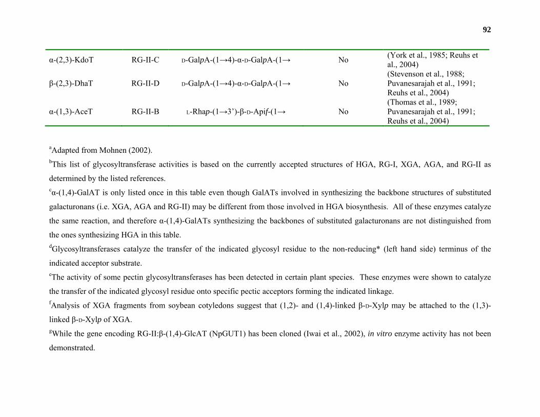
92
α-(2,3)-KdoT RG-II-C D-GalpA-(1→4)-α-D-GalpA-(1→ No (York et al., 1985; Reuhs et al., 2004)
β-(2,3)-DhaT RG-II-D D-GalpA-(1→4)-α-D-GalpA-(1→ No (Stevenson et al., 1988; Puvanesarajah et al., 1991; Reuhs et al., 2004)
α-(1,3)-AceT RG-II-B L-Rhap-(1→3’)-β-D-Apif-(1→ No (Thomas et al., 1989; Puvanesarajah et al., 1991; Reuhs et al., 2004)
aAdapted from Mohnen (2002). bThis list of glycosyltransferase activities is based on the currently accepted structures of HGA, RG-I, XGA, AGA, and RG-II as
determined by the listed references. cα-(1,4)-GalAT is only listed once in this table even though GalATs involved in synthesizing the backbone structures of substituted
galacturonans (i.e. XGA, AGA and RG-II) may be different from those involved in HGA biosynthesis. All of these enzymes catalyze
the same reaction, and therefore α-(1,4)-GalATs synthesizing the backbones of substituted galacturonans are not distinguished from
the ones synthesizing HGA in this table. dGlycosyltransferases catalyze the transfer of the indicated glycosyl residue to the non-reducing* (left hand side) terminus of the
indicated acceptor substrate. eThe activity of some pectin glycosyltransferases has been detected in certain plant species. These enzymes were shown to catalyze
the transfer of the indicated glycosyl residue onto specific pectic acceptors forming the indicated linkage. fAnalysis of XGA fragments from soybean cotyledons suggest that (1,2)- and (1,4)-linked β-D-Xylp may be attached to the (1,3)-
linked β-D-Xylp of XGA. gWhile the gene encoding RG-II:β-(1,4)-GlcAT (NpGUT1) has been cloned (Iwai et al., 2002), in vitro enzyme activity has not been
demonstrated.
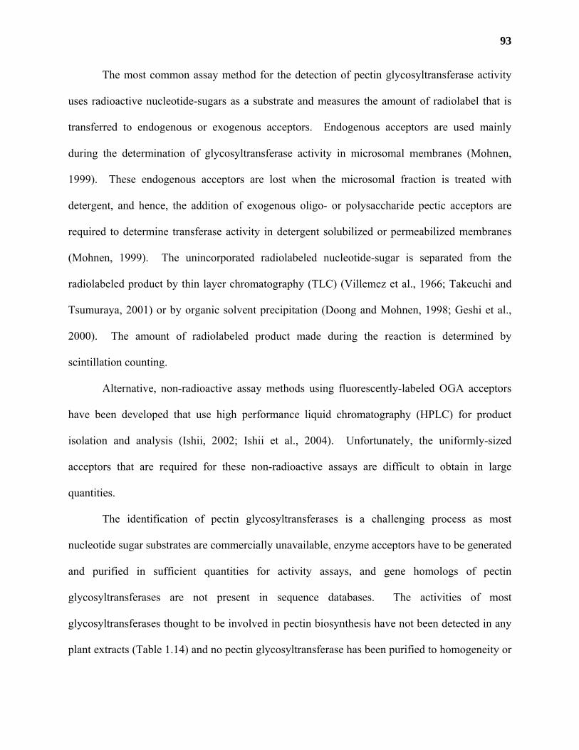
93
The most common assay method for the detection of pectin glycosyltransferase activity
uses radioactive nucleotide-sugars as a substrate and measures the amount of radiolabel that is
transferred to endogenous or exogenous acceptors. Endogenous acceptors are used mainly
during the determination of glycosyltransferase activity in microsomal membranes (Mohnen,
1999). These endogenous acceptors are lost when the microsomal fraction is treated with
detergent, and hence, the addition of exogenous oligo- or polysaccharide pectic acceptors are
required to determine transferase activity in detergent solubilized or permeabilized membranes
(Mohnen, 1999). The unincorporated radiolabeled nucleotide-sugar is separated from the
radiolabeled product by thin layer chromatography (TLC) (Villemez et al., 1966; Takeuchi and
Tsumuraya, 2001) or by organic solvent precipitation (Doong and Mohnen, 1998; Geshi et al.,
2000). The amount of radiolabeled product made during the reaction is determined by
scintillation counting.
Alternative, non-radioactive assay methods using fluorescently-labeled OGA acceptors
have been developed that use high performance liquid chromatography (HPLC) for product
isolation and analysis (Ishii, 2002; Ishii et al., 2004). Unfortunately, the uniformly-sized
acceptors that are required for these non-radioactive assays are difficult to obtain in large
quantities.
The identification of pectin glycosyltransferases is a challenging process as most
nucleotide sugar substrates are commercially unavailable, enzyme acceptors have to be generated
and purified in sufficient quantities for activity assays, and gene homologs of pectin
glycosyltransferases are not present in sequence databases. The activities of most
glycosyltransferases thought to be involved in pectin biosynthesis have not been detected in any
plant extracts (Table 1.14) and no pectin glycosyltransferase has been purified to homogeneity or

94
recombinantly expressed (Mohnen, 1999; Mohnen, 2002). Recent studies analyzing mutant
plants with altered pectin structures have allowed the identification of putative pectin
glycosyltransferase genes (Bouton et al., 2002; Iwai et al., 2002); however, the function of these
genes has not been demonstrated through heterologous expression.
Glycosyltransferase activities (EC 2.4.-) involved in pectin biosynthesis have been
discussed in great detail in previous reviews (Mohnen, 1999; Ridley et al., 2001; Mohnen, 2002).
The following section will summarize published results on the identification and characterization
of glycosyltransferases involved in pectin biosynthesis.
Galacturonosyltransferases (GalATs): GalATs catalyze the transfer of D-GalpA from
UDP-D-GalpA onto the non-reducing end of a pectin acceptor (Scheller et al., 1999). Plants are
expected to contain four distinct GalAT activities based on pectin structure (Table 1.14). Only
the activity of α-(1,4)-GalAT has been detected in plant extracts.
α-(1,4)-GalAT (EC 2.4.1.43) activity has been detected in membrane fractions from
mung bean (Villemez et al., 1965; Villemez et al., 1966), tomato (Lin et al., 1966), tobacco
(Doong et al., 1995; Doong and Mohnen, 1998; Scheller et al., 1999), Arabidopsis (Guillaumie et
al., 2003), sycamore (Bolwell et al., 1985), pumpkin (Ishii, 2002), azuki (Vigna angularis) bean
(Takeuchi and Tsumuraya, 2001), petunia (Petunia axillaris) (Akita et al., 2002), and pea
(Sterling et al., 2001). All α-(1,4)-GalATs have a strict requirement for Mn2+ and are strongly
inhibited by divalent cation chelators such as EDTA. While all α-(1,4)-GalATs have similar pH
optima, the published kinetic characteristics of these enzymes are variable, and depend on the
original plant tissue source and on the method used to test for enzyme activity (Table 1.15).
The type of acceptor used in the GalAT activity assay contributes to some of the
observed differences in the kinetic constants of membrane-bound and detergent-solubilized
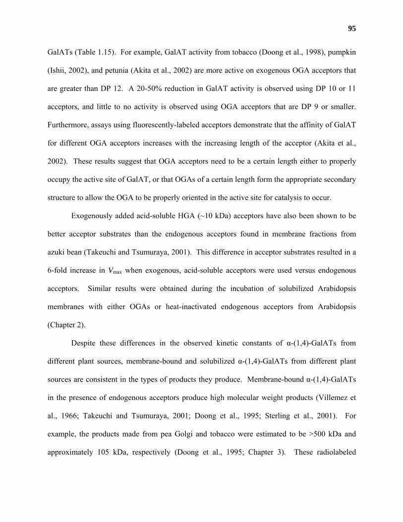
95
GalATs (Table 1.15). For example, GalAT activity from tobacco (Doong et al., 1998), pumpkin
(Ishii, 2002), and petunia (Akita et al., 2002) are more active on exogenous OGA acceptors that
are greater than DP 12. A 20-50% reduction in GalAT activity is observed using DP 10 or 11
acceptors, and little to no activity is observed using OGA acceptors that are DP 9 or smaller.
Furthermore, assays using fluorescently-labeled acceptors demonstrate that the affinity of GalAT
for different OGA acceptors increases with the increasing length of the acceptor (Akita et al.,
2002). These results suggest that OGA acceptors need to be a certain length either to properly
occupy the active site of GalAT, or that OGAs of a certain length form the appropriate secondary
structure to allow the OGA to be properly oriented in the active site for catalysis to occur.
Exogenously added acid-soluble HGA (~10 kDa) acceptors have also been shown to be
better acceptor substrates than the endogenous acceptors found in membrane fractions from
azuki bean (Takeuchi and Tsumuraya, 2001). This difference in acceptor substrates resulted in a
6-fold increase in Vmax when exogenous, acid-soluble acceptors were used versus endogenous
acceptors. Similar results were obtained during the incubation of solubilized Arabidopsis
membranes with either OGAs or heat-inactivated endogenous acceptors from Arabidopsis
(Chapter 2).
Despite these differences in the observed kinetic constants of α-(1,4)-GalATs from
different plant sources, membrane-bound and solubilized α-(1,4)-GalATs from different plant
sources are consistent in the types of products they produce. Membrane-bound α-(1,4)-GalATs
in the presence of endogenous acceptors produce high molecular weight products (Villemez et
al., 1966; Takeuchi and Tsumuraya, 2001; Doong et al., 1995; Sterling et al., 2001). For
example, the products made from pea Golgi and tobacco were estimated to be >500 kDa and
approximately 105 kDa, respectively (Doong et al., 1995; Chapter 3). These radiolabeled
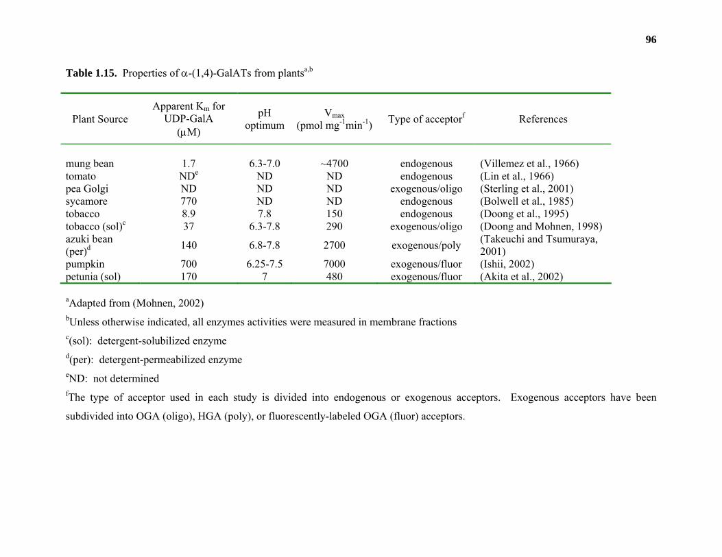
96
Table 1.15. Properties of α-(1,4)-GalATs from plantsa,b
Plant Source Apparent Km for
UDP-GalA
(µM)
pH optimum
Vmax (pmol mg-1min-1) Type of acceptorf References
mung bean 1.7 6.3-7.0 ~4700 endogenous (Villemez et al., 1966) tomato NDe ND ND endogenous (Lin et al., 1966) pea Golgi ND ND ND exogenous/oligo (Sterling et al., 2001) sycamore 770 ND ND endogenous (Bolwell et al., 1985) tobacco 8.9 7.8 150 endogenous (Doong et al., 1995) tobacco (sol)c 37 6.3-7.8 290 exogenous/oligo (Doong and Mohnen, 1998) azuki bean (per)d 140 6.8-7.8 2700 exogenous/poly (Takeuchi and Tsumuraya,
2001) pumpkin 700 6.25-7.5 7000 exogenous/fluor (Ishii, 2002) petunia (sol) 170 7 480 exogenous/fluor (Akita et al., 2002)
aAdapted from (Mohnen, 2002)
bUnless otherwise indicated, all enzymes activities were measured in membrane fractions c(sol): detergent-solubilized enzyme d(per): detergent-permeabilized enzyme eND: not determined fThe type of acceptor used in each study is divided into endogenous or exogenous acceptors. Exogenous acceptors have been
subdivided into OGA (oligo), HGA (poly), or fluorescently-labeled OGA (fluor) acceptors.
97
products could be almost completely degraded (89% and 91% for tobacco and pea Golgi,
respectively) into mono-, di- and triGalA following exhaustive treatment with a purified EPGase
(Doong and Mohnen, 1998; Sterling et al., 2001), indicating that the D-[14C]GalpA residues were
α-(1,4)-linked to the endogenous acceptors. Neither the size of the endogenous acceptors nor the
number of contiguous D-[14C]GalpA residues added onto the non-reducing termini have been
determined.
Solubilized or detergent-permeabilized α-(1,4)-GalAT from tobacco (Doong and
Mohnen, 1998; Scheller et al., 1999), petunia pollen tubes (Akita et al., 2002), and pea Golgi
(Chapter 3) in the presence of exogenous OGA acceptors (DP ≥ 10) add from 1 to 10 D-GalpA
residues onto the non-reducing end of the OGA acceptor in a non-processive manner. These
products can also be completely degraded into mono-, di- and triGalA following their treatment
with a purified EPGase (Doong and Mohnen, 1998; Sterling et al., 2001; Akita et al., 2002)
suggesting that the product was (1,4)-α-linked. Similar products were obtained from pumpkin
microsomal membranes incubated with fluorescently labeled OGAs (Ishii, 2002). These results
indicate that OGAs may not be the correct acceptor for processive α-(1,4)-GalAT activity or that
tissue homogenization may disrupt protein complexes required for processive
glycosyltransferase activity, or that the enzymes may not be processive in vivo. The purification
and eventual cloning of an α-(1,4)-GalAT should enable us to determine the catalytic mechanism
of the enzyme.
A T-DNA insertion mutant of Arabidopsis, named qua1, was recently discovered that had
phenotypes expected for a mutation in a α-(1,4)-GalAT gene, including dwarfism, reduced cell
adhesion, and a 25% reduction in D-GalpA content in leaves (Bouton et al., 2002). While the
enzymatic activity of the QUA1 gene has yet to be demonstrated, recent studies described in this
98
thesis have shown that QUA1 has high sequence similarity to two putative α-(1,4)-GalAT genes
from Arabidopsis (Chapter 4).
Arabinosyltransferases (AraTs): Arabinosyltransferase activity has been detected in
microsomal membranes from mung (Odzuck and Kauss, 1972; Nunan and Scheller, 2003) and
French bean (Bolwell and Northcote, 1983; Rodgers and Bolwell, 1992) using radiolabeled
UDP-L-Arap as a substrate. The products from these reactions have never been unequivocally
shown to be a part of any pectic structure (Mohnen, 1999; Mohnen, 2002). Questions remain as
to whether any of the detected activities are involved in pectin biosynthesis.
The first report of the addition of L-Ara to a well defined, pectic acceptor was conducted
using solubilized AraT activity from mung bean (Nunan and Scheller, 2003). Solubilized AraT
activity from mung bean catalyzed the transfer of a single L-Ara residue to a (1,5)-linked α-L-
arabinooctaose (Ara8) acceptor. The radiolabeled product synthesized by the enzyme could be
completely converted to arabinose by strong acid hydrolysis; however, the radiolabel was found
to be resistant to treatment with a purified (1,5)-α-L-endoarabinase and (1,2;1,3;1,5)-α-D-
arabinofuranosidase, respectively. Mild acid hydrolysis and methylation analysis of the
radiolabeled product indicated that the added L-Ara residue was in the pyranose ring form, but
linkage analysis of the product could not be determined due to the limited quantities of material
that were available. The presence of terminal L-Arap residues on pectic structures other than
RG-II has been observed in specific plant species, for example arabinans from pigeon pea
(Cajanus cajan) cotyledons (Swamy and Salimath, 1991), and arabinogalactans from Angelica
acutiloba (Kitagawa) roots (Kiyohara et al., 1987), and soybean cotyledons (Huisman et al.,
2001). These results suggest that L-Arap may be a common component of the arabinans and/or
99
arabinogalactans of RG-I, or that other plant enzymes are required to convert the incorporated L-
Arap residues into the L-furanose form during pectin arabinan biosynthesis.
(1,5)-linked α-L-arabinosyl oligomers smaller than DP 8 were less efficient acceptors for
solubilized AraT activity, as were large arabinans, galactans and RG-I fragments. AraT activity
was greatest in young (2 d old) tissue and decreased significantly with the age of the plant. The
AraT also had a requirement for Mn2+ in the reaction mixture with 3 mM MnCl2 giving the
greatest levels of incorporation. The AraT had a pH optimum of 6.5 and Km for UDP-L-Arap of
0.33 mM.
Recently, a mutant in Nicotiana plumbaginifolia, named nolac-H14, was discovered that
exhibited similar non-organogenic and cell adhesion defects as nolac-H18 (Iwai et al., 2001).
Glycosyl composition and linkage analysis of the pectic and hemicellulosic fractions indicate a
significant reduction in L-Araf in nolac-H14 mutant callus. These results suggest that the gene
encoded by nolac-H14 may be involved in the incorporation of L-Araf in Nicotiana
plumbaginifolia.
Galactosyltransferases (GalTs): An activity capable of transferring D-[14C]Galp from
UDP-D-[14C]Galp to an endogenous acceptor forming a water-soluble, high molecular weight
polymer was first detected in microsomal membranes from mung bean seedlings (McNab et al.,
1968). Strong acid hydrolysis indicated that the radiolabeled polymer was composed solely of D-
[14C]Galp (McNab et al., 1968). The majority of the D-[14C]Galp labeled product (>90%) made
by mung bean membranes was shown in a later study to be composed of (1,4)-linked β-D-
galactan by treatment of the product with a purified (1,4)-β-D-exogalactanase and a (1,4)-β-D-
endogalactanase, respectively (Brickell and Reid, 1996). The β-(1,4)-GalT from mung bean had
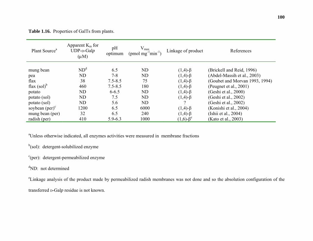
100
Table 1.16. Properties of GalTs from plants.
Plant Sourcea Apparent Km for
UDP-D-Galp
(µM)
pH optimum
Vmax (pmol mg-1min-1) Linkage of product References
mung bean NDd 6.5 ND (1,4)-β (Brickell and Reid, 1996) pea ND 7-8 ND (1,4)-β (Abdel-Massih et al., 2003) flax 38 7.5-8.5 75 (1,4)-β (Goubet and Morvan 1993, 1994) flax (sol)b 460 7.5-8.5 180 (1,4)-β (Peugnet et al., 2001) potato ND 6-6.5 ND (1,4)-β (Geshi et al., 2000) potato (sol) ND 7.5 ND (1,4)-β (Geshi et al., 2002) potato (sol) ND 5.6 ND ? (Geshi et al., 2002) soybean (per)c 1200 6.5 6000 (1,4)-β (Konishi et al., 2004) mung bean (per) 32 6.5 240 (1,4)-β (Ishii et al., 2004) radish (per) 410 5.9-6.3 1000 (1,6)-βe (Kato et al., 2003)
aUnless otherwise indicated, all enzymes activities were measured in membrane fractions
b(sol): detergent-solubilized enzyme
c(per): detergent-permeabilized enzyme
dND: not determined
eLinkage analysis of the product made by permeabilized radish membranes was not done and so the absolution configuration of the
transferred D-Galp residue is not known.
101
a pH optimum of 6.5 and was stimulated by Mg2+ (Table 1.16). A similar enzyme activity was
identified in pea epicotyl membranes (Abdel-Massih et al., 2003).
Two, pH-dependent, GalT activities were detected in microsomes from flax suspension-
cultured cells (Goubet and Morvan 1993). Both activities were capable of synthesizing a
polymeric product in the presence of endogenous acceptors composed entirely of D-Galp, but had
different pH optima. GalT activities with pH optima of 5-6.5 and 7.5-8.5 produced an alkali-
soluble product that was composed mainly of (1,3)- and (1,6)-linked β-D-galactan and a water-
soluble product that was composed mainly of (1,4)-linked β-D-galactan, respectively. The GalT
activities were solubilized from flax membranes and the activity with the higher pH optimum
(pH 8) was shown to catalyze the transfer of D-[14C]Galp onto purified RG-I acceptors (Table
1.16). The radiolabeled products from these reactions were highly resistant to
rhamnogalacturonan depolymerases, (i.e. rhamnogalacturonase A and B); however, a significant
portion (60%) of the D-[14C]Galp could be removed from the products by treatment with a
purified (1,4)-β-D-galactosidase suggesting that the product was (1,4)-β-linked (Peugnet et al.,
2001).
Microsomal membranes from potato suspension cultures incubated with UDP-D-
[14C]Galp in the presence of endogenous acceptors and Mn2+ at pH 6 were able to produce a
large molecular weight (>500 kDa) radiolabeled product that released D-[14C]Galp as the sole
glycosyl residue upon complete acid hydrolysis (Geshi et al., 2000). The majority of the
radioactivity from the product made by dark grown cells (73%) could be released by treatment
with a purified (1,4)-β-D-endogalactanase. Furthermore, treatment of the large molecular weight
product with base followed by rhamnogalacturonase A (RGase A), an endohydrolase that cleaves
the [→4)-α-D-GalpA-(1→2)-α-L-Rhap] linkage of RG-I backbone structures (Azadi et al., 1995),
102
released approximately 50% of the radioactivity as a series of smaller RG-I oligomers (~14
kDa). The combined treatment of the RG-I oligomers with a purified (1,4)-β-D-galactosidase and
a (1,4)-β-D-endogalactanase released all of the radioactivity from the RG-I oligomers as D-
[14C]Galp. These results suggest that membranes from potato suspension-cultured cells catalyze
the initiation and elongation of (1,4)-linked β-D-galactan chains found on RG-I (Table 1.14 and
1.16).
Two distinct β-(1,4)-GalT activities could be solubilized from potato suspension cultures
that differed in their pH optima and acceptor substrate specificities (Geshi et al., 2002). Both
enzyme activities catalyze the addition of D-[14C]Galp from UDP-D-[14C]Galp onto RG-I
acceptors that contained short (1,4)-β-D-galactan side chains consisting of a single D-Galp
residue. β-(1,4)-GalT activity at pH 5.6 and 7.5 preferred high molecular weight (~1.2 MDa)
RG-I acceptors and medium-sized (~21 kDa) acceptors, respectively. Shorter RG-I oligomers,
linear galactans, degalactosylated RG-I fragments, or RG-I molecules with high galactose
content did not function as viable acceptors for the solubilized activity. The radiolabeled product
made using the medium-sized acceptor at pH 7.5 was determined to be (1,4)-β-linked by
digestion with a purified (1,4)-β-D-endogalactanase (Table 1.16). Product made using the high
molecular weight acceptor at pH 5.6 was impervious to this treatment, suggesting that either the
β-(1,4)-GalT produced side chains that were resistant to (1,4)-β-D-endogalactanase cleavage
(meaning that they were shorter than 3 contiguous D-[14C]Galp residues) or that the enzyme
catalyzed the formation of a linkage other than (1,4)-linked β-D-Galp.
A β-(1,4)-GalT activity capable of transferring D-[14C]Galp to chemically modified, large
molecular weight (60-70 kDa) galactan acceptors from lupin seeds was identified in microsomal
membranes from soybean (Konishi et al., 2004). The radiolabeled product could be degraded by
103
treatment with a purified (1,4)-β-D-endogalactanase, indicating that it was (1,4)-β-linked. This β-
(1,4)-GalT from soybean membranes could also catalyze the addition of 1 to 5 D-Galp residues
onto fluorescently-labeled, (1,4)-linked β-D-galactosyl oligomers with a DP greater than 5. A
similar activity (capable of catalyzing the addition of D-Galp residues onto fluorescently-labeled
(1,4)-linked β-D-galactosyl oligomers) was detected in microsomal membranes from mung bean
(Ishii et al., 2004). Both the activity from soybean and mung bean had a pH optimum of 6.5 and
were stimulated by Mn2+ (Ishii et al., 2001a; Konishi et al., 2004).
Radish microsomal membranes incubated with a chemically-modified, (1,3)-linked β-D-
galactan acceptor from acacia gum in the presence of UDP-D-[14C]Galp produced a radiolabeled
product (Kato et al., 2003). Treatment of the radiolabeled product with a purified (1,3)-β-D-
exogalactanase released a product that migrated the same distance as (1,6)-linked β-D-
galactobiose upon TLC analysis (Kato et al., 2003). These results suggested that the enzyme
activity that was identified was a β-(1,6)-GalT capable of transferring a single (1,6)-linked β-D-
Galp residue onto (1,3)-linked β-D-galactans. Linkage analysis of the synthesized product was
not conducted and so the absolute configuration of the added β-D-Galp residue remains to be
confirmed.
Apiosyltransferases (ApiT): An activity capable of transferring D-[14C]Apif from UDP-D-
[14C]Apif onto an endogenous acceptor was detected in membrane preparations from duckweed
(Pan and Kindel, 1977). The radiolabeled product could be partially degraded by treatment with
a fungal pectinase and partial acid hydrolysis released 46% of the radioactivity from the product
as D-[14C]Apif and D-[14C]apiobiose. ApiT activity could be increased 2-fold with the addition of
UDP-D-GalpA to reaction mixtures, suggesting that the ApiT was partially dependent on HGA
biosynthesis. The absolute configuration of the radiolabeled product was not determined, nor
104
was the composition of the endogenous acceptor. The ApiT had a pH optimum of 5.7 and Km
for UDP-D-Apif of 4.9 µM.
Glucuronosyltransferases (GlcATs): A gene encoding a β-(1,4)-GlcAT involved in RG-
II biosynthesis was cloned by analyzing a T-DNA mutant of Nicotiana plumbaginifolia (Iwai et
al., 2002). The mutant, npgut1, exhibited a number of developmental and cell adhesion defects
that were caused by the absence of the [α-L-Galp-(1→2)-β-D-GlcpA-(1→] disaccharide from side
chain A of RG-II (Figure 1.4). Kinetic data on this enzyme is lacking as NpGUT1 has not been
heterologously expressed and β-(1,4)-GlcAT activity has not been detected in any plant tissue.
This is the first published report of the cloning of a pectin glycosyltransferase (Table 1.17).
Acetyl- and methyltransferases involved in pectin biosynthesis
The biosynthesis of pectic polysaccharides in the Golgi requires more than the catalytic
activities of pectin glycosyltransferases. Pectic structures made in Golgi are modified by a series
of acetyl- and methyltransferases that act late in pectin biosynthesis (Table 1.17; Zhang and
Staehelin, 1992). The methyl- and acetylation of pectic polymers may be an important process
regulating the secretion and assembly of pectins into the primary wall (Zhang and Staehelin,
1992; Carpita and Gibeaut, 1993; Dolan et al., 1997). Furthermore, pectin methyl- and
acetylation has also been associated with cell separation events (Willats et al., 2001a), with the
resistance of pectic polysaccharides to hydrolytic enzymes (Schols et al., 1990; Schols and
Voragen, 1994; Renard and Jarvis, 1999a) and may also be developmentally regulated (Kauss
and Hassid, 1967a; Vannier et al., 1992; Liners et al., 1994; Pauly and Scheller, 2000). No genes
encoding these enzymes have been cloned, but significant progress has been made in identifying
and characterizing pectin methyl- and acetyltransferases from a variety of plant sources.
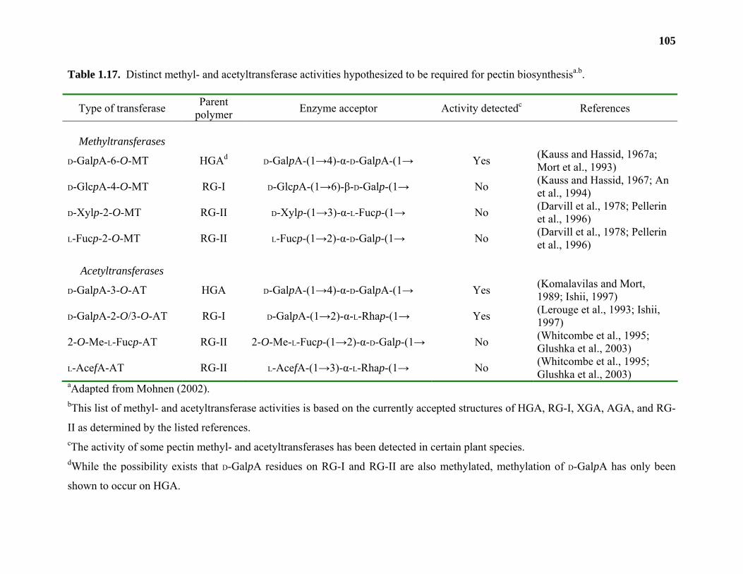
105
Table 1.17. Distinct methyl- and acetyltransferase activities hypothesized to be required for pectin biosynthesisa.b.
Type of transferase Parent polymer Enzyme acceptor Activity detectedc References
Methyltransferases
D-GalpA-6-O-MT HGAd D-GalpA-(1→4)-α-D-GalpA-(1→ Yes (Kauss and Hassid, 1967a;
Mort et al., 1993)
D-GlcpA-4-O-MT RG-I D-GlcpA-(1→6)-β-D-Galp-(1→ No (Kauss and Hassid, 1967; An et al., 1994)
D-Xylp-2-O-MT RG-II D-Xylp-(1→3)-α-L-Fucp-(1→ No (Darvill et al., 1978; Pellerin et al., 1996)
L-Fucp-2-O-MT RG-II L-Fucp-(1→2)-α-D-Galp-(1→ No (Darvill et al., 1978; Pellerin et al., 1996)
Acetyltransferases
D-GalpA-3-O-AT HGA D-GalpA-(1→4)-α-D-GalpA-(1→ Yes (Komalavilas and Mort, 1989; Ishii, 1997)
D-GalpA-2-O/3-O-AT RG-I D-GalpA-(1→2)-α-L-Rhap-(1→ Yes (Lerouge et al., 1993; Ishii, 1997)
2-O-Me-L-Fucp-AT RG-II 2-O-Me-L-Fucp-(1→2)-α-D-Galp-(1→ No (Whitcombe et al., 1995; Glushka et al., 2003)
L-AcefA-AT RG-II L-AcefA-(1→3)-α-L-Rhap-(1→ No (Whitcombe et al., 1995; Glushka et al., 2003)
aAdapted from Mohnen (2002). bThis list of methyl- and acetyltransferase activities is based on the currently accepted structures of HGA, RG-I, XGA, AGA, and RG-
II as determined by the listed references. cThe activity of some pectin methyl- and acetyltransferases has been detected in certain plant species. dWhile the possibility exists that D-GalpA residues on RG-I and RG-II are also methylated, methylation of D-GalpA has only been
shown to occur on HGA.
106
Pectin methyltransferases
HGA methyltransferase (HGA-MT) activity was first discovered in membranes from
mung bean seedlings (Kauss and Hassid, 1967a). Membranes incubated with S-adenosyl-L-
[14C]methionine catalyzed the incorporation of [14C]methyl groups onto endogenous acceptors to
form radiolabeled products. Approximately 95% of the radioactivity could be removed from the
product as [14C]methanol by treatment of the radiolabeled material with base or a purified pectin
methylesterase (PME), suggesting that the [14C]methyl groups were added onto HGA (Table
1.18). Addition of UDP-D-GalpA to the reaction mixture increased the rate of [14C]methyl
transfer (Kauss and Swanson, 1969). A similar activity was characterized in membranes from
suspension-cultured tobacco cells (Table 1.18) where approximately 59% of the radiolabeled
product made by tobacco membranes was shown to be methylated HGA (Goubet et al., 1998).
The results suggested that HGA-MT activity in membranes may be dependent upon HGA
biosynthesis.
In contrast, a membrane-bound HGA-MT activity from soybean hypocotyls was not
stimulated by the addition of UDP-D-GalpA (Ishikawa et al., 2000). The detergent-
permeabilized soybean enzyme catalyzed the transfer of [14C]methyl groups onto exogenous
pectin acceptors. Similar to HGA-MTs from mung bean and tobacco, treatment of the
radiolabeled products with a purified PME or EPGase released the incorporated radiolabel,
suggesting that the addition was onto HGA.
HGA-MT from tobacco was solubilized and shown to use endogenous acceptors, HGA or
pectins with a degree of esterification (DE) of 30 as an acceptor (Goubet and Mohnen, 1999).
The radiolabeled products from endogenous and exogenous acceptors were extracted with
boiling water and ammonium oxalate (conditions that solubilize pectins from the cell wall).
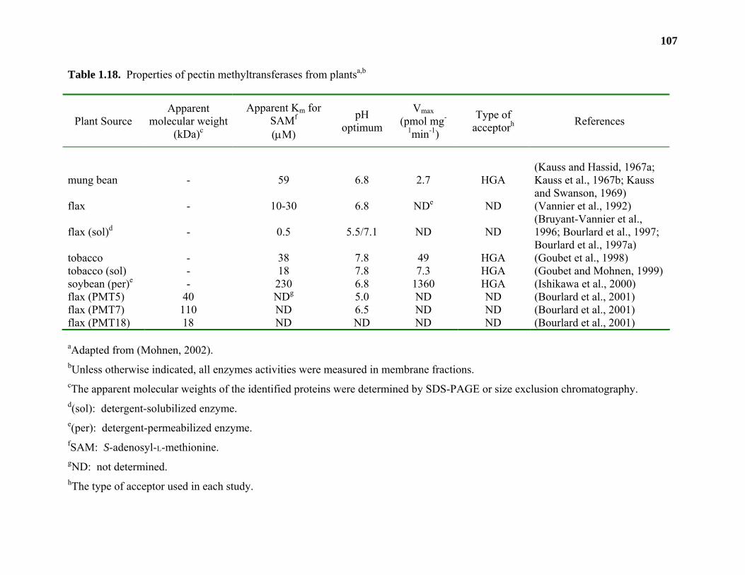
107
Table 1.18. Properties of pectin methyltransferases from plantsa,b
Plant Source Apparent
molecular weight (kDa)c
Apparent Km for SAMf
(µM)
pH optimum
Vmax (pmol mg-
1min-1)
Type of acceptorh References
mung bean - 59 6.8 2.7 HGA (Kauss and Hassid, 1967a; Kauss et al., 1967b; Kauss and Swanson, 1969)
flax - 10-30 6.8 NDe ND (Vannier et al., 1992)
flax (sol)d - 0.5 5.5/7.1 ND ND (Bruyant-Vannier et al., 1996; Bourlard et al., 1997; Bourlard et al., 1997a)
tobacco - 38 7.8 49 HGA (Goubet et al., 1998) tobacco (sol) - 18 7.8 7.3 HGA (Goubet and Mohnen, 1999) soybean (per)e - 230 6.8 1360 HGA (Ishikawa et al., 2000) flax (PMT5) 40 NDg 5.0 ND ND (Bourlard et al., 2001) flax (PMT7) 110 ND 6.5 ND ND (Bourlard et al., 2001) flax (PMT18) 18 ND ND ND ND (Bourlard et al., 2001)
aAdapted from (Mohnen, 2002).
bUnless otherwise indicated, all enzymes activities were measured in membrane fractions. cThe apparent molecular weights of the identified proteins were determined by SDS-PAGE or size exclusion chromatography. d(sol): detergent-solubilized enzyme. e(per): detergent-permeabilized enzyme. fSAM: S-adenosyl-L-methionine. gND: not determined. hThe type of acceptor used in each study.
108
Treatment of the extracted products with a purified PME and EPGase released approximately
64% and 62.5% of the incorporated [14C]methyl groups, respectively, suggesting that the bulk of
the [14C]methylated product was HGA.
Membrane-bound PMT activity was also detected and characterized from flax hypocotyls
and suspension-cultured cells (Bruyant-Vannier et al., 1996; Bourlard et al., 1997a). Flax PMT
transferred [14C]methyl groups from S-adenosyl-L-[14C]methionine onto an endogenous product.
The transferred radiolabel could be released as [14C]methanol by treatment with base. Although
the endogenous acceptor was not characterized, membrane-bound PMT activity from flax was
stimulated by the addition of exogenous pectins with both high (50%) and low (10%) degrees of
esterification (Bourlard et al., 1997a).
The flax PMT activity was solubilized using detergent and two PMT isoforms were
identified that differed in their pH optima and preference for pectins with low or high DE
(Bruyant-Vannier et al., 1996; Bourlard et al., 1997a). Purification of the two flax PMT isoforms
(PMT5 and PMT7) identified an 18 kDa protein (PMT18). It has been proposed that the PMT18
encodes the catalytic domain of both isoforms of flax PMT (Bourlard et al., 2001). At present,
the identity of the gene encoding the 18 kDa protein is unknown.
A solubilized PMT activity from suspension-cultured flax cells was also stimulated 1.6-
and 7-fold by the addition of exogenous RG-I or RG-II acceptors, respectively (Bourlard et al.,
1997). The location of [14C]methyl transfer onto the RG-I and RG-II acceptors was not
determined. Therefore, it is not known whether the transfer of [14C]methyl groups occurred onto
backbone D-GalpA residues or onto the methylated, side chain glycosyl residues found on RG-I
and RG-II (Table 1.18).
109
Pectin acetyltransferases
Membrane-bound pectin acetyltransferase (PAT) activity was recently characterized from
suspension-cultured potato cells (Pauly and Scheller, 2000). Potato PAT activity catalyzed the
transfer of [14C]acetyl groups from [14C]acetyl-CoA onto endogenous potato acceptors.
Approximately 23% of salt/ethanol precipitated, radiolabeled product could be solubilized
following its treatment with both EPGase and PME, suggesting that at least 23% of the
[14C]acetyl groups transferred were onto HGA. Approximately 33% of the [14C]acetyl groups
were transferred onto RG-I acceptors greater than 500 kDa in size as determined by their
susceptibility to an RG-I-specific acetylesterase. The RG-I acetyltransferase (RGAT) activity
was partially characterized and found to have a pH optimum of 7.0, a Km for [14C]acetyl-CoA of
35 µM and a Vmax of 54 pmol min-1 mg-1. The radiolabeled product generated by potato RGAT
could also be digested into smaller oligomers using rhamnogalacturonan lyase (RGase B)
providing further evidence supporting the existence of a RGAT activity in these membranes.
Acetyltransferases that transfer acetyl groups to the 2-O-Me-L-Fucp or L-AcefA residues on side
chain B of RG-II (Figure 1.4) have yet to be identified.
CONCLUSIONS AND RELEVANCE
It has been demonstrated that mutations in pectin biosynthetic genes create alterations in
pectin structure that can have severe deleterious effects on pectin function in the wall. While a
number of pectin biosynthetic genes, especially those involved in nucleotide-sugar biosynthesis,
have been discovered in plants, almost no genes encoding pectin methyl-, acetyl- and
glycosyltransferases have been identified to date. Furthermore, pectin transferase activities have
not been extensively characterized and information is lacking as to the location and mode of
action of pectin biosynthesis in plants.
110
At the commencement of this research, the subcellular localization of pectic
glycosyltransferases was not known. The elucidation of the subcellular location of a pectin
galacturonosyltransferase, α-(1,4)-GalAT, from pea epicotyl membranes confirmed more indirect
evidence suggesting that pectin biosynthesis occurred in the Golgi apparatus. Furthermore, it
presented the first evidence of the topology of a pectin glycosyltransferase and allowed for the
development of the currently accepted model for pectin biosynthesis in plants.
A new radioactive GalAT activity assay was developed that alleviates some of the time
and expense required to assay multiple samples for GalAT activity. Furthermore, this assay was
shown to be useful for the detection of GalAT activity using either endogenous or exogenous
acceptors and may be an effective tool for the detection of other pectin glycosyltransferase
activities.
Using this newly developed assay and a novel solubilization procedure, α-(1,4)-GalAT
activity was purified 17-fold from Arabidopsis membranes, the highest recorded purity of any α-
(1,4)-GalAT characterized to date. This allowed for the identification of two putative α-(1,4)-
GalAT genes (JS36 and JS33) from Arabidopsis. N-terminally truncated versions of these genes
were cloned into a vector designed for recombinant protein secretion, and were heterologously
expressed in human embryonic kidney (HEK) 293 cells. Media from cells transfected with the
JS36 gene construct was able to catalyze the transfer of D-[14C]GalpA from UDP-D-[14C]GalpA
onto exogenous OGA acceptors in one transient experiment. These results suggest that JS36
encodes an α-(1,4)-GalAT. However, subsequent transient and stable expression of JS33 or JS36
in HEK293 cells did not confirm these initial results.
Both JS33 and JS36 are part of a 25 member gene family in Arabidopsis. Mutants in two
members of this gene family, qua1 and parvus, give phenotypes that are expected for the

111
mutation of pectin glycosyltransferases. The characterization of mutant plants in putative
GALAT family members and their expression in heterologous systems may be required to
determine the role of the putative GALAT family in pectin biosynthesis.
CHAPTER 2
THE CATALYTIC SITE OF THE PECTIN BIOSYNTHETIC ENZYME α-(1,4)-
GALACTURONOSYLTRANSFERASE (GALAT) IS LOCATED IN THE LUMEN OF
THE GOLGI1
1 Jason Sterling, Heather F. Quigley, Ariel Orellana and Debra Mohnen 2001. Plant Physiol. 127: 360-371. Reprinted here with permission of publisher
113
ABSTRACT
α-(1,4)-galacturonosyltransferase (GalAT) is an enzyme required for the biosynthesis of
the plant cell wall pectic polysaccharide homogalacturonan (HGA). GalAT activity in
homogenates from pea (Pisum sativum L. var. Alaska) stem internodes co-localized in linear and
discontinuous sucrose gradients with latent UDPase activity, an enzyme marker specific for
Golgi membranes. GalAT activity was separated from antimycin A-insensitive
NADH:cytochrome c reductase and cytochrome c oxidase activities, enzyme markers for the
endoplasmic reticulum and the mitochondria, respectively. GalAT and latent UDPase activities
were separated from the majority (80%) of callose synthase activity, a marker for the plasma
membrane, suggesting that little or no GalAT is present in the plasma membrane. GalAT
activities in proteinase K-treated and untreated Golgi vesicles were similar, whereas no GalAT
activity was detected after treating Golgi vesicles with proteinase K in the presence of Triton X-
100. These results demonstrate that the catalytic site of GalAT resides within the lumen of the
Golgi. The products generated by Golgi-localized GalAT were converted by
endopolygalacturonase treatment to mono- and digalacturonic acid, thereby showing that GalAT
synthesizes (1,4)-linked α-D-galacturonan. Our data provide the first enzymatic evidence that a
glycosyltransferase involved in HGA synthesis is present in the Golgi apparatus. Together with
prior results of in vivo labeling and immunocytochemical studies, these results show that pectin
biosynthesis occurs in the Golgi. A model for the biosynthesis of the pectic polysaccharide HGA
is proposed.
114
INTRODUCTION
Pectins are a family of polysaccharides present in all plant primary walls (O'Neill et al.,
1990). Homogalacturonan (HGA) accounts for approximately 60% of the total pectin in plants
(O'Neill et al., 1990; Mohnen et al., 1996). HGA is a linear polymer composed of (1,4) linked α-
D-galactosyluronic acid residues (D-GalpA) that are often methylesterified at the C-6 carboxyl
group (Mort et al., 1993) and may also be O-acetylated at O-2 and O-3 (Ishii, 1997). The D-
GalpA residues may, in some plants, be substituted at O-3 with a β-linked xylosyl residue
(Schols et al., 1995; Yu and Mort, 1996).
An enzyme that catalyzes the transfer of D-GalpA from UDP-D-GalpA onto endogenous
HGA was first identified in particulate suspensions from Phaseolus aureus and was named
polygalacturonate 4-α-galacturonosyltransferase [EC 2.4.1.43] (Villemez et al., 1965; Villemez
et al., 1966). A similar enzyme was characterized in microsomal membranes from suspension-
cultured tobacco (Nicotiana tabacum L. cv. Samsun) cells (Doong et al., 1995) and solubilized
from tobacco membranes (Doong and Mohnen, 1998). The solubilized enzyme was shown to
catalyze the addition of an D-GalpA residue onto O-4 of the non-reducing end (Scheller et al.,
1999) of exogenous oligomeric HGA acceptors (Doong and Mohnen, 1998) and thus was named
homogalacturonan 4-α-galacturonosyltransferase (HGA-GalAT).
No direct evidence has yet been reported for the subcellular localization of the
glycosyltransferases responsible for pectin biosynthesis. However, the results of in vivo labeling
and immunocytochemical analyses have suggested that pectin is synthesized in the Golgi
apparatus (Northcote and Pickett-Heaps, 1966; Harris and Northcote, 1971; Staehelin and
Moore, 1995). Immunocytochemical studies using antibodies that recognize the pectic
polysaccharides homogalacturonan and rhamnogalacturonan I, suggest that these pectic
115
polysaccharides are synthesized within different compartments of the Golgi (Zhang and
Staehelin, 1992) and are transported from the Golgi in vesicles that migrate to, and fuse with, the
plasma membrane. The polysaccharides are then released into the extracellular space and
incorporated into the wall (Northcote and Pickett-Heaps, 1966).
We now provide evidence that an α-(1,4)-galacturonosyltransferase (GalAT) from pea
(Pisum sativum L. var. Alaska) epicotyls, with properties similar to the previously described
HGA-GalAT from tobacco, is located in the Golgi apparatus and that the catalytic site of this
enzyme is in the Golgi lumen. The third internodes of etiolated pea epicotyls were used for this
study since the different subcellular membranes from this tissue can be separated using Suc
density gradients, making such tissue useful for topological and localization studies of enzymes
involved in cell wall polysaccharide biosynthesis (Brummell et al., 1990; Orellana et al., 1997;
Wulff et al., 2000). Our data provide strong evidence that HGA is synthesized in the Golgi
apparatus and a model for the biosynthesis of this pectic polysaccharide is proposed.
MATERIALS AND METHODS
Chemicals
Uridine diphosphate-α-D-[14C]glucuronic acid (UDP-D-[14C]GlcpA, 293.6 mCi mmol-1)
and uridine diphosphate-α-D-[14C]glucose (UDP-D-[14C]Glcp, 286.2 mCi mmol-1) were
purchased from DuPont-New England Nuclear (Boston, MA). Uridine diphosphate-α-D-
[14C]galacturonic acid (UDP-D-[14C]GalpA) was prepared by the epimerization of UDP-D-
[14C]GlcpA as described previously (Liljebjelke et al., 1995). OGAs with a DP of 14 and a
mixture of OGAs of DP 7 to 23 were prepared according to Doong et al. (1995). Proteinase K
was purchased from Boehringer Mannheim (Indianapolis, IN). Homogeneous EPGase (748
units ml-1, 1 unit releases 1 µmole D-GalpA per min) from Fusarium moniliforme was a gift from
116
Carl Bergmann (Complex Carbohydrate Research Center, Athens, GA). All other reagents were
purchased from Sigma (St. Louis, MO).
Plant materials
Pea (Pisum sativum L. var. Alaska) seeds were purchased from the Green Seed Company
(Athens, GA). Seedlings were grown in moist vermiculite for 7 to 8 d at 25°C in the dark.
Linear sucrose gradient fractionation of pea homogenates
Third internodes (from the cotyledon, 1 cm in length, 5 g total weight) from 7 to 8 d old
etiolated peas (Pisum sativum L. var Alaska) were excised and homogenized in 50 mM HEPES
[4-(2-hydroxyethyl)-1-piperazineethanesulfonic acid]/KOH, pH 7.0, containing 0.4 M sucrose, 1
mM MgCl2, 10 mM KCl, 1 mM EDTA, and 1 mM dithiothreitol using a razor blade and a mortar
and pestle. The homogenate was filtered through two layers of Miracloth (Calbiochem, San
Diego, CA) and the filtrate centrifuged for 10 min at 1,000g. The 1,000g supernatant (5 mL)
was overlaid onto 24 mL of a 20% to 50% sucrose (Suc; w/w) gradient (Munoz et al., 1996) in
50 mM HEPES/KOH, pH 7.0 containing 0.1 mM MgCl2, 1 mM EDTA and 1 mM dithiothreitol
and centrifuged for 3.5 h at 100,000g. Fractions (0.5 mL) were collected and analyzed for
enzyme marker activity. All manipulations were done at 4°C. Suc concentrations were
determined using an Abbe refractometer (model 3L, Milton Roy Company, Rochester, NY) and
the densities were calculated using an ambient temperature of 20°C.
Isolation of Golgi membranes
Enriched Golgi membranes were isolated using discontinuous Suc gradients according to
a previously described method (Munoz et al., 1996). All manipulations were done on ice or at
4°C. The third internodes (1 cm, 40-70 g total mass) of 7 to 8 d old etiolated pea stem epicotyls
were excised and homogenized in 1 volume of buffer A (0.1 M KH2PO, pH 6.65 containing 5
117
mM MgCl2, 1 mM dithioerythritol, and 16% [w/w] Suc) using a razor blade and a mortar and
pestle. The homogenate was filtered through two layers of Miracloth and centrifuged for 2 min
at 1,000g. The supernatant was layered onto 38% (w/w) Suc in buffer A (8 ml) and centrifuged
for 90 min at 100,000g. The upper phase was removed without disturbing the interface layer and
replaced with 33% (w/w) Suc (15 ml) and 8% (w/w) Suc (5 ml) in buffer A. The gradient was
centrifuged for a further 100 min at 100,000g. The pellet was resuspended in 10 mM Tris, pH
7.5 containing 0.25 M Suc (ST buffer) and stored at −80°C. The membranes that equilibrated at
each of the interfaces were collected, diluted with an equal volume of water, and centrifuged for
50 min at 100,000g. These pellets were resuspended in ST buffer and stored at -80°C. In some
experiments (Table 2.2), membranes from the 33% and 38% Suc phases were also collected and
stored as described above. Otherwise, the 33% and 38% Suc phases with no prior membrane
collection were used for enzyme analysis (Table 2.1).
Enzyme assays
Cyt c oxidase activity was measured as described (Briskin et al., 1987) with some
modifications. Cyt c oxidase was assayed spectrophotometrically at 550 nm using a Shimadzu
scanning spectrophotometer (model UV-2101; Columbus, MD). A portion (35 µl) of each
gradient fraction was added to 30 mM K2HPO4, pH 7.4 (735 µl) containing 1 mM EDTA, 1 mM
NaHCO3, 0.00045% Triton X-100, and 50 µM Cyt c (reduced with sodium dithionite). The total
activity of Cyt c oxidase was calculated for each fraction using the initial linear rate of Cyt c
oxidation (decrease in A550) and the extinction coefficient of Cyt c (18.5 mM-1 cm-1).
Antimycin A-resistant NADH:Cyt c reductase was measured according to a previously
described method with some modifications (Vannier et al., 1992). A portion (0.1 ml) of each
gradient fraction was combined with 50 mM KH2PO4, pH 7.5 (0.9 ml) containing 50 µM Cyt c,
118
0.3 mM NADH, 5 mM KCN and 1 µM antimycin A. The linear increase in A550 was taken as
the rate of Cyt c reduction and was used to calculate the total activity in each fraction.
Latent UDPase activity was measured as described (Briskin et al., 1987) with some
modifications. A reaction mixture (15 µl) containing 6 mM UDP, 6 mM MgSO4, and 60 mM
Tris/Mes [2-(N-morpholino)-ethanesulfonic acid], pH 6.5, with or without 0.1% (v/v) Triton X-
100, was added to 15 µl of gradient fraction in a microtiter plate (Nunc, Suwanee, GA) and kept
for 15 min at 25°C. The reaction was stopped by the addition of Ames reagent (270 µl) and
processed as described (Ames, 1966). The A690 of the sample was measured using a Tikertek
Multiscan ELISA plate reader (model MCC/340 MKII; Flow Laboratories, McLean, VA) and
was converted to the amount of Pi released from UDP (UDPase activity) using a standard curve
of KH2PO4. Latent UDPase activity was considered to be present in those fractions in which an
increase in A690 was observed when fractions were incubated in the presence 0.1% (v/v) Triton
X-100. The latent UDPase activity reported in Tables 2.1 and 2.2 and in Figure 2.1 was
calculated by subtracting the amount of Pi released from UDP by enzyme in the absence of
Triton X-100 from the amount of Pi released from UDP by enzyme in the presence of Triton X-
100.
Callose synthase activity was measured according to a previously described method by
(Dhugga and Ray, 1994) with some modifications. Gradient fractions (10 µl of one-tenth diluted
sample) were incubated for 5 min in 10 mM MOPS [3-(N-morpholino)-propanesulfonic acid],
pH 7.0 containing 1 mM UDP-D-Glcp, 10 mM cellobiose, 2 mM CaCl2, 0.3 mM spermine,
0.02% (v/v) Triton X-100, and 0.32 µM UDP-D-[14C]Glcp. The reaction was stopped by the
addition of 70% (v/v) ethanol (2 ml) and the insoluble products were collected by filtration
through a GF/A glass-microfiber disc (Whatman, Maidstone, UK). The discs were washed with
119
70% ethanol (15 ml), dried, and the amount of radioactivity on the discs measured by
scintillation counting.
GalAT activity was measured as described by Doong et al. (1995) with some
modifications. Samples (10 µL) were incubated for 2 min in 50 mM HEPES, pH 7.8, containing
100 µg of OGAs (DP of 7-23), 1.1 µM UDP-D-[14C]GalpA (specific activity 293.6 mCi mmol-1),
0.25 mM MnCl2, 25 mM KCl and 0.1% (v/v) Triton X-100 in a total volume of 30 µl (unless
otherwise stated). The reaction was stopped by the addition of 700 µl chloroform:methanol (3:2,
v/v) and centrifuged to collect the precipitable products. The product was washed twice with
500 µl of 65% (v/v) ethanol and the amount of radioactivity quantified by scintillation counting.
All enzyme assays were conducted at room temperature with the exception of the GalAT
assay which was conducted at 30°C. Protein concentrations were determined by protein assay
(BioRad, Hercules, CA) using bovine serum albumin as a standard. Uronic acids were analyzed
colorimetrically using the meta-hydroxybiphenyl assay (Blumenkrantz and Asboe-Hansen,
1973).
Product characterization
The products generated by GalAT in pea Golgi vesicles were analyzed according to
Doong et al. (1995). The products were formed using either detergent-permeabilized Golgi in
the presence of OGAs of DP 14 as exogenous acceptor or using intact Golgi without exogenous
acceptor.
Four separate GalAT reactions containing 12 µl of Golgi vesicles (approximately 43 µg
of protein) and 5 µM UDP-D-[14C]GalpA in 50 mM HEPES, pH 7.8, 0.25 mM MnCl2, and 25
mM KCl were incubated with or without 0.1% (v/v) Triton X-100 and 15 µg of OGA 14 mer for
35 min at 30°C in a total volume of 60 µl. The products were recovered, washed as described
120
above, resuspended in 0.5 ml of 50 mM NaOAc, pH 6.0 containing 5 mM EDTA, and pooled
(total volume 2 ml). Individual pools were analyzed as follows by either HPAEC or SEC. For
example, a 500 µl aliquot was analyzed directly by HPAEC. A second aliquot of 500 µl was
treated for 8 h at 30°C with a homogeneous EPGase (2 units) and the products generated were
analyzed by HPAEC. A third aliquot of the pooled products (1 ml) was adjusted to pH 12 by the
addition of 1 N NaOH and kept at 4°C for 8 h. The solution was adjusted to pH 5.0 with 1 N
HCl and a portion (500 µl) was analyzed by HPAEC. The remaining base-treated product (500
µl) was treated with EPGase for 8 h and analyzed by HPAEC.
Alkaline phosphatase treatment of D-GalpA-1-P
Aqueous extracts from a GalAT reaction were prepared by incubating 12 µl of Golgi
vesicles (approximately 43 µg of protein) for 60 min at 30°C in 50 mM HEPES, pH 7.8,
containing 2.5 µM UDP-D-[14C]GalpA, and 15 µg of OGAs of DP 12, 0.25 mM MnCl2, 25 mM
KCl, 0.1% (v/v) Triton X-100 in a total volume of 60 µl. The reaction was terminated by the
addition of chloroform:methanol (5:1 [v/v], 300 µl) and 240 µl 50 mM NaOAc, pH 6.0
containing 5 mM EDTA was added. The mixture was centrifuged at 13,000g for 2 min. The
aqueous upper phase was mixed with 300 µl of chloroform:methanol (5:1 [v/v]), centrifuged and
removed. The organic phase from the first centrifugation was mixed with 50 mM NaOAc, pH
6.0 (300 µl) containing 5 mM EDTA, centrifuged, and the aqueous phase was combined with the
previously obtained aqueous phase. An aliquot (80 µl) of the combined aqueous phases was
adjusted to pH 8.5 with Tris (10 mM). Water or 10 units of alkaline phosphatase was added, and
the mixture kept for 60 min at 37°C. The reaction was stopped by heating at 85°C for 10 min.
The resulting mixture was filtered (0.2 µm) and chromatographed by HPAEC. Fractions (1 ml)
121
were collected and the amount of radioactivity in each fraction was quantified by liquid
scintillation counting.
Size-exclusion chromatography of the [14C]-labeled product generated using intact and
permeabilized Golgi
The products made by GalAT in intact Golgi without exogenous acceptor, and the
products formed by detergent-permeabilized (0.1% [v/v] Triton X-100) Golgi in the presence of
exogenous OGA 14 mer (15 µg) were separated using a Superose12 HR10/30 SEC column
(Pharmacia, Uppsala, Sweden). The column was eluted with 50 mM sodium acetate containing
5 mM EDTA at 0.35 ml min-1. Fractions (0.5 ml) were collected and the radioactivity in a
portion (400 µl) of each fraction was determined by scintillation counting. The SEC elution
profiles of dextran molecular mass standards, D-GalpA, triGalA, and OGAs of a DP of 7, 12, 15
and 16 were detected using a pulsed electrochemical detector (Dionex, Sunnyvale, CA) with
post-column addition of 400 mM NaOH at a flow rate of 0.2 ml min-1 (see below).
HPAEC of the [14C]-labeled product made using intact and permeabilized Golgi
D-[14C]GalpA-labeled product generated by GalAT in pea Golgi vesicles, and D-GalpA
and oligogalacturonide standards, were filtered (0.2 µm nylon microfilterfuge tubes, Rainin
Instrument Co., Inc., Woburn, MA) and chromatographed on a CarboPac PA100 column (4 X
250 mm) fitted with a CarboPac PA100 guard column (3 X 25 mm) using a Dionex DX 500 LC,
(Dionex, Sunnyvale, CA). Samples were eluted from the column at 1 ml min-1 with a gradient
formed from ultra-pure deionized water (eluant A) and 1 M sodium acetate containing 5 mM
EDTA (eluant B) as follows: 0% (v/v) B at 0-2 min, 40% (v/v) B at 17 min, 80% (v/v) B at 49
min, 100% (v/v) B at 50-52 min, 0% (v/v) B at 57 min (modified from Doong et al., 1995). The
non-radiolabeled standards were detected by pulsed electrochemical detection after post-column
122
addition of 400 mM NaOH at 0.5 ml min-1. The pulse sequence for the electrochemical detector
was 0.05 V at 0 s, 0.05 V at 0.2 s (begin integration), 0.05 V at 0.4 s (end integration), 0.75 V at
0.41 to 0.6 s, and −0.15 V at 0.61 to 1.0 s. Fractions (0.75 ml) were collected, an aliquot (500
µl) of each fraction was combined with 10 volumes of scintillation cocktail (Scintiverse, Fisher
Scientific, Norcross, GA), and the amount of radioactivity was measured using a scintillation
counter. Quenching by salt and/or base was found to be negligible.
Proteinase K digestion
Proteinase K treatment of Golgi vesicles was conducted according to Orellana et al.
(1997). In brief, Golgi vesicles (100 µg protein) were incubated for 30 min at 30°C in 10 mM
Tris, pH 7.5 containing 0.25 M Suc and 1 mM MgCl2 (100 µl total volume) in the presence and
absence of 0.1% (v/v) Triton X-100 and 4 µg Proteinase K. Proteolysis was stopped by the
addition of 1 mM phenylmethylsulfonylfluoride and 10 µl of each treatment was brought to a
final concentration of 0.1% (v/v) Triton X-100 and assayed for GalAT activity in the presence of
exogenous OGA acceptors.
RESULTS
Subcellular localization of GalAT activity in pea membranes
Subcellular membrane fractions obtained by linear sucrose (Suc) gradient centrifugation
of homogenates from etiolated pea third internodes were analyzed for GalAT activity and for
organelle-specific enzyme activity (Figure 2.1). GalAT activity was detected almost exclusively
in fractions 26 through 30 (1.17-1.14 g ml-1; Figure 2.1, A). The presence of GalAT activity in
such a limited number of fractions suggests that it is associated with an organelle of a distinct
density. Enzyme marker assays for the ER and for the mitochondria (antimycin A-insensitive
123
Figure 2.1. Location of GalAT activity and enzyme markers in pea membranes fractionated by
linear Suc density gradient centrifugation. A) GalAT activity ( ) and sucrose density ( ). The
data were obtained from a single analysis of each fraction. Similar results were obtained in three
separate gradients. B) Cytochrome (Cyt) c oxidase ( ) and Antimycin A resistant NADH Cyt c
reductase ( ), enzyme markers for mitochondria and endoplasmic reticulum (ER), respectively.
Activities are expressed as the percentage of the highest total activity (191.2 nmol Cyt c reduced
min-1 and 397.6 nmol Cyt c oxidized min-1 for Cyt c reductase and Cyt c oxidase, respectively)
obtained in the peak fraction. The data were obtained from a single analysis of each fraction.
Similar results were obtained in two independent gradients. C) Latent UDPase activity, a Golgi
marker, is detected as the increase in A690 obtained in fractions permeabilized with 0.1% (v/v)
Triton X-100 compared to the absorbance obtained in fractions in the absence of detergent. The
position of greatest latent activity is depicted with a double-headed arrow (Golgi). Data are the
average of duplicate samples from each fraction. Similar results were obtained in three separate
gradients.
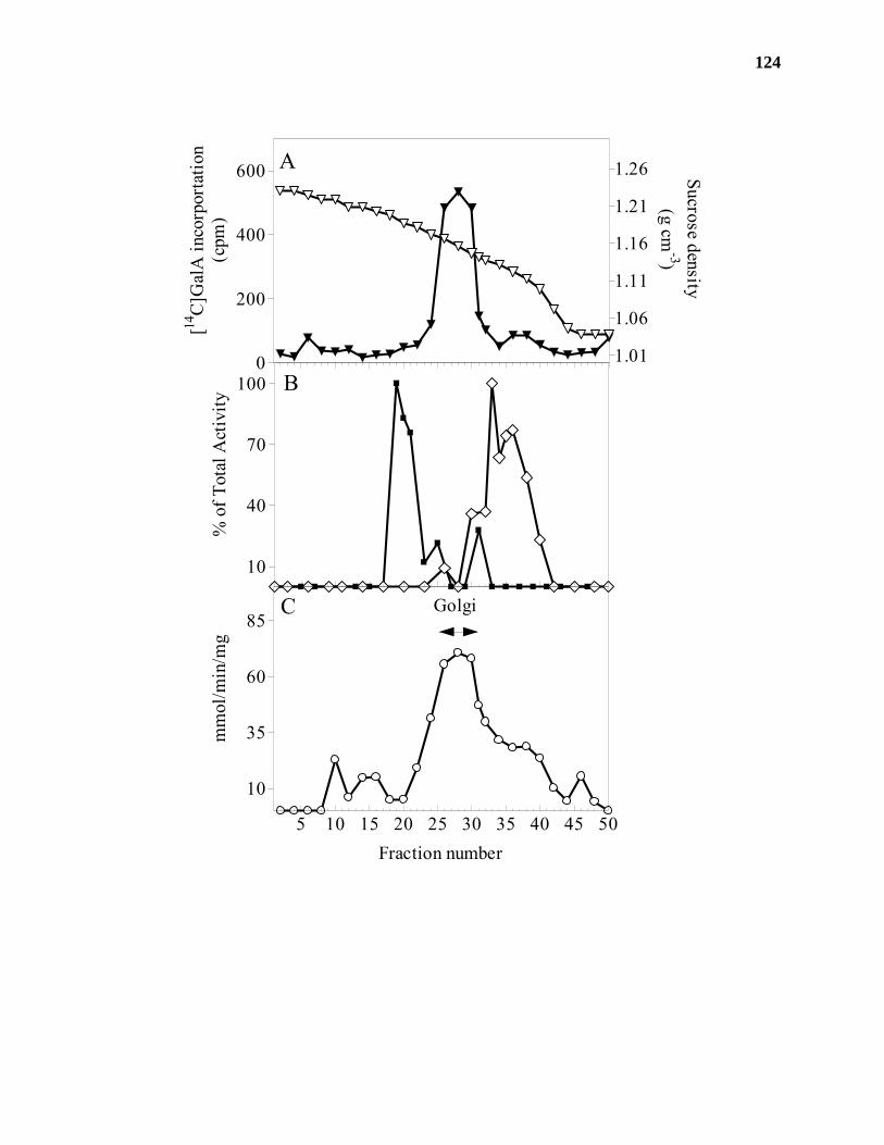
124
10
40
70
100
% o
f Tot
al A
ctiv
ity
5 10 15 20 25 30 35 40 45 50
10
35
60
85Golgi
Fraction number
mm
ol/m
in/m
g
A
B
C
0
200
400
600
1.01
1.06
1.11
1.16
1.21
1.26[14
C]G
alA
inco
rpor
tatio
n(c
pm)
Sucrose density(g cm
-3)
125
NADH:Cyt c reductase and Cyt c oxidase, respectively) showed that the ER (fractions 30-40,
1.14-1.10 g ml-1) and mitochondria (fractions 19-22, 1.20-1.18 g ml-1) were present in a different
subset of fractions (Figure 2.1, B) than those that contained the greatest amount of GalAT
activity.
Latent UDPase activity is a well-characterized Golgi-specific enzyme marker (Orellana et
al., 1997) that is detected following the permeabilization of membranes with detergent. The
greatest amount of latent UDPase activity was present in fractions 24 through 32 of a density of
1.17 to 1.14 g cm-3 (Figure 2.1, C). These fractions coincide with the fractions that contain the
greatest amount of GalAT activity, suggesting that GalAT is a Golgi-localized enzyme.
To confirm that GalAT is present in a Golgi-enriched fraction, pea homogenates were
separated using discontinuous Suc gradient centrifugation. Each fraction (Figure 2.2) was
analyzed for GalAT activity and for membrane marker activities (Table 2.1). Latent UDPase
activity was highest in the membranes that banded at the 8-33% (w/w) Suc interface suggesting
that these membranes were Golgi-enriched (Tanford and Reynolds, 1976). The Golgi-enriched
fraction also contained approximately 90% of the total GalAT activity (Table 2.1). Some (21%
of total) ER-specific enzyme marker activity was also detected in the Golgi-enriched fraction.
However, the ER-enriched fraction (33-38% [w/w] Suc interface) contained no GalAT activity
(Table 2.1). These results, when taken together with the results shown in Figure 2.1, strongly
suggest that the GalAT from pea membranes is a Golgi-localized enzyme.
The biosynthesis of cellulose and callose has been shown to occur at the plasma
membrane (Frederikson and Larsson, 1989; Delmer, 1999), and the possibility could not be
discounted that GalAT may also be localized to the plasma membrane. Thus, experiments were
conducted, using discontinuous Suc gradient centrifugation, to separate Golgi membranes from
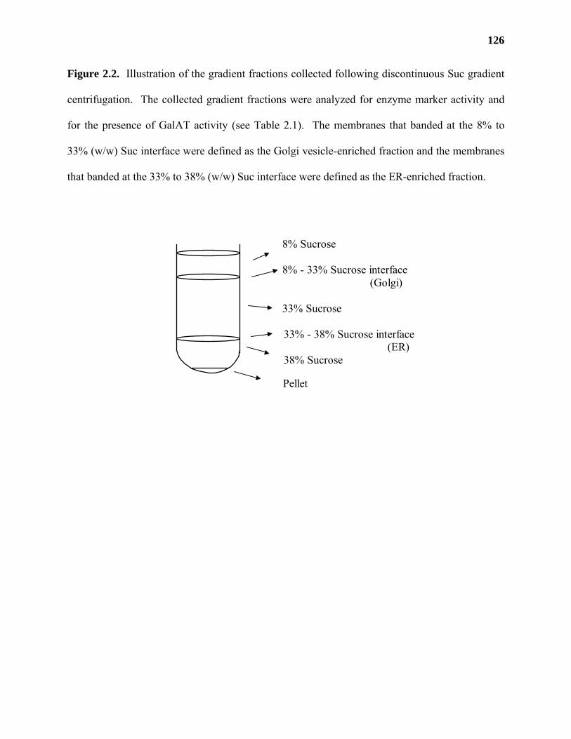
126
Figure 2.2. Illustration of the gradient fractions collected following discontinuous Suc gradient
centrifugation. The collected gradient fractions were analyzed for enzyme marker activity and
for the presence of GalAT activity (see Table 2.1). The membranes that banded at the 8% to
33% (w/w) Suc interface were defined as the Golgi vesicle-enriched fraction and the membranes
that banded at the 33% to 38% (w/w) Suc interface were defined as the ER-enriched fraction.
8% Sucrose 8% - 33% Sucrose interface (Golgi)
33% Sucrose 33% - 38% Sucrose interface (ER) 38% Sucrose
Pellet
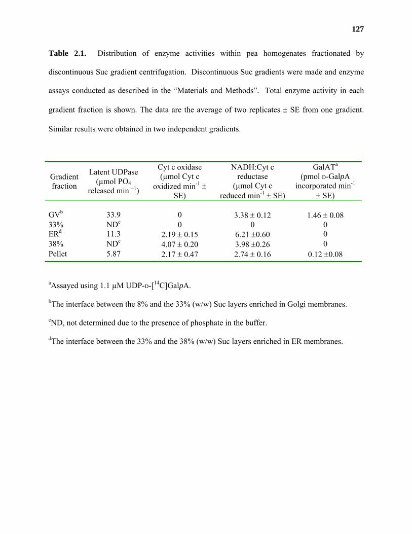
127
Table 2.1. Distribution of enzyme activities within pea homogenates fractionated by
discontinuous Suc gradient centrifugation. Discontinuous Suc gradients were made and enzyme
assays conducted as described in the “Materials and Methods”. Total enzyme activity in each
gradient fraction is shown. The data are the average of two replicates ± SE from one gradient.
Similar results were obtained in two independent gradients.
Gradient fraction
Latent UDPase (µmol PO4
released min –1)
Cyt c oxidase (µmol Cyt c
oxidized min-1 ± SE)
NADH:Cyt c reductase
(µmol Cyt c reduced min-1 ± SE)
GalATa (pmol D-GalpA
incorporated min-1 ± SE)
GVb 33.9 0 3.38 ± 0.12 1.46 ± 0.08 33% NDc 0 0 0 ERd 11.3 2.19 ± 0.15 6.21 ±0.60 0 38% NDc 4.07 ± 0.20 3.98 ±0.26 0 Pellet 5.87 2.17 ± 0.47 2.74 ± 0.16 0.12 ±0.08
aAssayed using 1.1 µM UDP-D-[14C]GalpA.
bThe interface between the 8% and the 33% (w/w) Suc layers enriched in Golgi membranes.
cND, not determined due to the presence of phosphate in the buffer.
dThe interface between the 33% and the 38% (w/w) Suc layers enriched in ER membranes.
128
plasma membranes. The gradient fractions were assayed for GalAT activity, for latent UDPase
and for callose synthase activity, a known marker for plasma membrane vesicles in plants (Morre
et al., 1987; Dhugga and Ray, 1994). Callose synthase activity was present in all of the
discontinuous sucrose gradient fractions assayed. These results indicate that the Golgi-enriched
membranes obtained using discontinuous Suc gradients also contained some plasma membranes.
Thus, the possibility cannot be ruled out that GalAT is localized to a subset of plasma membrane
vesicles that co-localize with Golgi membranes. Nevertheless, because approximately 80% of
the callose synthase activity is located in fractions that are distinct from those that contain the
majority of the GalAT and latent UDPase activities (Table 2.2), these results most strongly
support the conclusion that GalAT is Golgi localized and is not a component of the plasma
membrane.
Characterization of the products made by GalAT in enriched Golgi membranes from pea.
The subcellular fractionation of GalAT activity with the Golgi-enriched fraction from pea
suggested that a pectin biosynthetic enzyme was localized in the Golgi. To test whether HGA is
formed by the GalAT in pea Golgi, the [14C]-labeled products generated by incubating UDP-D-
[14C]GalpA with intact pea Golgi were analyzed by size exclusion chromatography (SEC) and by
high performance anion exchange chromatography (HPAEC). The products were analyzed both
before and after treatment with base (to remove methyl and other esters), with
endopolygalacturonase (EPGase; to fragment HGA), and with base plus EPGase. Fractions
were collected, analyzed by scintillation counting, and the elution times of the radioactive
products compared to the elution times of D-GalpA, oligogalacturonides (OGAs), and dextran
molecular mass standards.
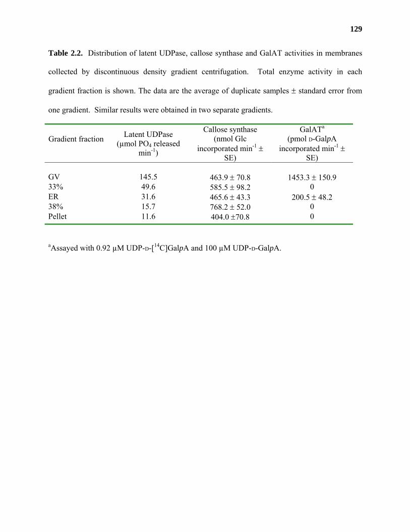
129
Table 2.2. Distribution of latent UDPase, callose synthase and GalAT activities in membranes
collected by discontinuous density gradient centrifugation. Total enzyme activity in each
gradient fraction is shown. The data are the average of duplicate samples ± standard error from
one gradient. Similar results were obtained in two separate gradients.
Gradient fraction
Latent UDPase (µmol PO4 released
min-1)
Callose synthase (nmol Glc
incorporated min-1 ± SE)
GalATa (pmol D-GalpA
incorporated min-1 ± SE)
GV 145.5 463.9 ± 70.8 1453.3 ± 150.9 33% 49.6 585.5 ± 98.2 0 ER 31.6 465.6 ± 43.3 200.5 ± 48.2 38% 15.7 768.2 ± 52.0 0 Pellet 11.6 404.0 ±70.8 0
aAssayed with 0.92 µM UDP-D-[14C]GalpA and 100 µM UDP-D-GalpA.
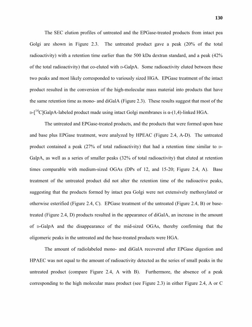
130
The SEC elution profiles of untreated and the EPGase-treated products from intact pea
Golgi are shown in Figure 2.3. The untreated product gave a peak (20% of the total
radioactivity) with a retention time earlier than the 500 kDa dextran standard, and a peak (42%
of the total radioactivity) that co-eluted with D-GalpA. Some radioactivity eluted between these
two peaks and most likely corresponded to variously sized HGA. EPGase treatment of the intact
product resulted in the conversion of the high-molecular mass material into products that have
the same retention time as mono- and diGalA (Figure 2.3). These results suggest that most of the
D-[14C]GalpA-labeled product made using intact Golgi membranes is α-(1,4)-linked HGA.
The untreated and EPGase-treated products, and the products that were formed upon base
and base plus EPGase treatment, were analyzed by HPEAC (Figure 2.4, A-D). The untreated
product contained a peak (27% of total radioactivity) that had a retention time similar to D-
GalpA, as well as a series of smaller peaks (32% of total radioactivity) that eluted at retention
times comparable with medium-sized OGAs (DPs of 12, and 15-20; Figure 2.4, A). Base
treatment of the untreated product did not alter the retention time of the radioactive peaks,
suggesting that the products formed by intact pea Golgi were not extensively methoxylated or
otherwise esterified (Figure 2.4, C). EPGase treatment of the untreated (Figure 2.4, B) or base-
treated (Figure 2.4, D) products resulted in the appearance of diGalA, an increase in the amount
of D-GalpA and the disappearance of the mid-sized OGAs, thereby confirming that the
oligomeric peaks in the untreated and the base-treated products were HGA.
The amount of radiolabeled mono- and diGalA recovered after EPGase digestion and
HPAEC was not equal to the amount of radioactivity detected as the series of small peaks in the
untreated product (compare Figure 2.4, A with B). Furthermore, the absence of a peak
corresponding to the high molecular mass product (see Figure 2.3) in either Figure 2.4, A or C
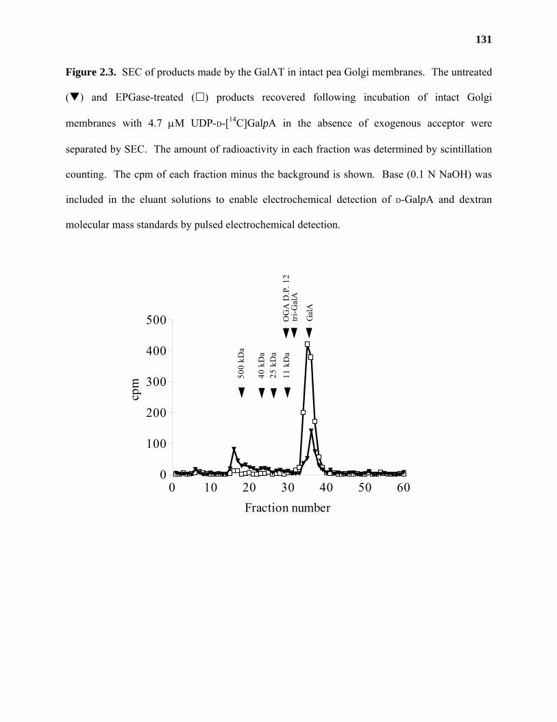
131
Figure 2.3. SEC of products made by the GalAT in intact pea Golgi membranes. The untreated
( ) and EPGase-treated ( ) products recovered following incubation of intact Golgi
membranes with 4.7 µM UDP-D-[14C]GalpA in the absence of exogenous acceptor were
separated by SEC. The amount of radioactivity in each fraction was determined by scintillation
counting. The cpm of each fraction minus the background is shown. Base (0.1 N NaOH) was
included in the eluant solutions to enable electrochemical detection of D-GalpA and dextran
molecular mass standards by pulsed electrochemical detection.
0 10 20 30 40 50 600
100
200
300
400
500
500
kDa
40 k
Da
25 k
Da
Gal
A
11 k
Da
OG
A D
.P. 1
2tri
-Gal
A
Fraction number
cpm
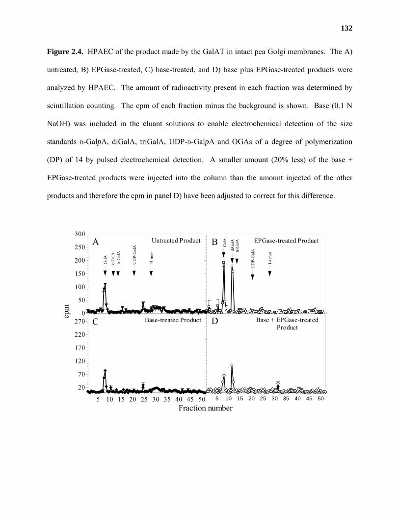
132
Figure 2.4. HPAEC of the product made by the GalAT in intact pea Golgi membranes. The A)
untreated, B) EPGase-treated, C) base-treated, and D) base plus EPGase-treated products were
analyzed by HPAEC. The amount of radioactivity present in each fraction was determined by
scintillation counting. The cpm of each fraction minus the background is shown. Base (0.1 N
NaOH) was included in the eluant solutions to enable electrochemical detection of the size
standards D-GalpA, diGalA, triGalA, UDP-D-GalpA and OGAs of a degree of polymerization
(DP) of 14 by pulsed electrochemical detection. A smaller amount (20% less) of the base +
EPGase-treated products were injected into the column than the amount injected of the other
products and therefore the cpm in panel D) have been adjusted to correct for this difference.
5 10 15 20 25 30 35 40 45 50
20
70
120
170
220
270 Base-treated Product
EPGase-treated Product
Gal
A
diG
alAtri
Gal
A
14 m
er
UD
P-G
alA
0
50
100
150
200
250
300Untreated Product
Gal
A
diG
alA
triG
alA
14 m
er
UD
P-G
alA
5 10 15 20 25 30 35 40 45 50
Base + EPGase-treatedProduct
Fraction number
A B
C Dcpm
133
suggests either that the high molecular mass material was unable to elute as a defined peak on
the anion exchange column or that the material was unable to pass through the 0.2 µm filters
used prior to HPAEC analysis (see “Materials and Methods”). Analysis of the 0.2 µm filters
revealed that approximately 47% and 26% (for untreated and base treated products, respectively)
of the total label incorporated into product remained on the filter following filtration.
In a preliminary study, we found that the amount of radiolabeled product formed by
membrane-bound GalAT from pea increased with the addition of exogenous OGA acceptors in
the presence of 0.1% (v/v) Triton X-100 (data not shown). Therefore, the product formed by
Golgi membranes in the presence of 0.1% (v/v) Triton X-100 and an OGA acceptor (DP 14) was
analyzed to confirm that pea GalAT produced HGA under these conditions.
Several differences were observed between the products made by intact and detergent-
permeabilized Golgi membranes. For example, approximately 47% of untreated product made
by intact Golgi bound to the HPLC prefilter, whereas only 12% of the untreated product from
Triton X-100-permeabilized Golgi bound to the filter. Comparable amounts of radioactive
product (83% for intact Golgi and 98% for permeabilized Golgi) passed through the filter after
base and EPGase treatment. These results suggested that the products formed using detergent
permeabilized Golgi and exogenous oligomeric HGA acceptors were smaller and/or had
different characteristics than the products made by intact Golgi vesicles.
The [14C]-labeled products formed using 0.1% Triton X-100-permeabilized pea Golgi in
the presence of an OGA acceptor (DP 14) were analyzed by SEC and HPAEC (Figures 2.5 and
2.6) before and after, base treatment, EPGase treatment, and base plus EPGase treatment. SEC
of the untreated product (Figure 2.5) gave two peaks that eluted with the same retention time as
an authentic OGA of DP 15 (15 mer, 32% of total) and D-GalpA (22% of total), respectively.
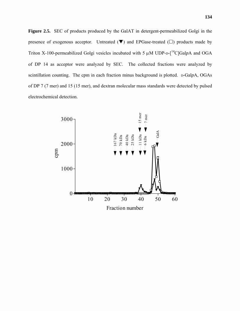
134
Figure 2.5. SEC of products produced by the GalAT in detergent-permeabilized Golgi in the
presence of exogenous acceptor. Untreated ( ) and EPGase-treated ( ) products made by
Triton X-100-permeabilized Golgi vesicles incubated with 5 µM UDP-D-[14C]GalpA and OGA
of DP 14 as acceptor were analyzed by SEC. The collected fractions were analyzed by
scintillation counting. The cpm in each fraction minus background is plotted. D-GalpA, OGAs
of DP 7 (7 mer) and 15 (15 mer), and dextran molecular mass standards were detected by pulsed
electrochemical detection.
10 20 30 40 50 600
1000
2000
3000
15 m
er
7 m
er
25 k
Da
6 kD
a11
kD
a
40 k
Da
70 k
Da
167
kDa
Gal
A
Fraction number
cpm
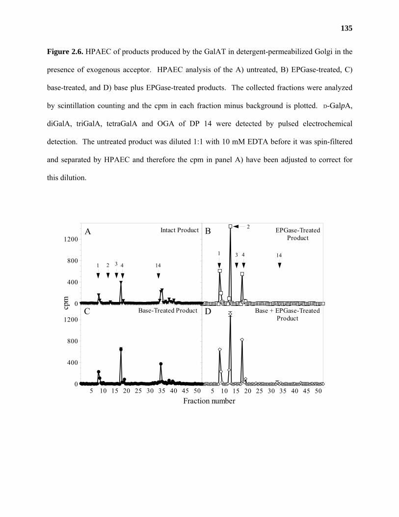
135
Figure 2.6. HPAEC of products produced by the GalAT in detergent-permeabilized Golgi in the
presence of exogenous acceptor. HPAEC analysis of the A) untreated, B) EPGase-treated, C)
base-treated, and D) base plus EPGase-treated products. The collected fractions were analyzed
by scintillation counting and the cpm in each fraction minus background is plotted. D-GalpA,
diGalA, triGalA, tetraGalA and OGA of DP 14 were detected by pulsed electrochemical
detection. The untreated product was diluted 1:1 with 10 mM EDTA before it was spin-filtered
and separated by HPAEC and therefore the cpm in panel A) have been adjusted to correct for
this dilution.
EPGase-TreatedProduct
1
2
3 4 14
5 10 15 20 25 30 35 40 45 50
Base + EPGase-TreatedProduct
0
400
800
1200Intact Product
1 2 3 4 14
5 10 15 20 25 30 35 40 45 500
400
800
1200Base-Treated Product
Fraction number
cpm
A B
C D
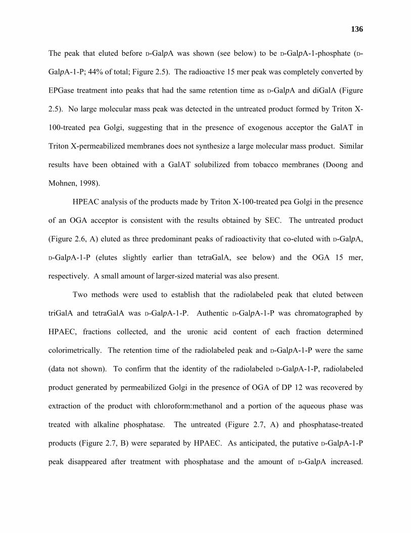
136
The peak that eluted before D-GalpA was shown (see below) to be D-GalpA-1-phosphate (D-
GalpA-1-P; 44% of total; Figure 2.5). The radioactive 15 mer peak was completely converted by
EPGase treatment into peaks that had the same retention time as D-GalpA and diGalA (Figure
2.5). No large molecular mass peak was detected in the untreated product formed by Triton X-
100-treated pea Golgi, suggesting that in the presence of exogenous acceptor the GalAT in
Triton X-permeabilized membranes does not synthesize a large molecular mass product. Similar
results have been obtained with a GalAT solubilized from tobacco membranes (Doong and
Mohnen, 1998).
HPEAC analysis of the products made by Triton X-100-treated pea Golgi in the presence
of an OGA acceptor is consistent with the results obtained by SEC. The untreated product
(Figure 2.6, A) eluted as three predominant peaks of radioactivity that co-eluted with D-GalpA,
D-GalpA-1-P (elutes slightly earlier than tetraGalA, see below) and the OGA 15 mer,
respectively. A small amount of larger-sized material was also present.
Two methods were used to establish that the radiolabeled peak that eluted between
triGalA and tetraGalA was D-GalpA-1-P. Authentic D-GalpA-1-P was chromatographed by
HPAEC, fractions collected, and the uronic acid content of each fraction determined
colorimetrically. The retention time of the radiolabeled peak and D-GalpA-1-P were the same
(data not shown). To confirm that the identity of the radiolabeled D-GalpA-1-P, radiolabeled
product generated by permeabilized Golgi in the presence of OGA of DP 12 was recovered by
extraction of the product with chloroform:methanol and a portion of the aqueous phase was
treated with alkaline phosphatase. The untreated (Figure 2.7, A) and phosphatase-treated
products (Figure 2.7, B) were separated by HPAEC. As anticipated, the putative D-GalpA-1-P
peak disappeared after treatment with phosphatase and the amount of D-GalpA increased.
137
Figure 2.7. Alkaline phosphatase treatment of an aqueous extract of the radiolabeled products
produced in pea Golgi. Permeabilized Golgi vesicles were incubated for 60 min at 30°C with an
exogenous OGA acceptor of DP 12 and 2.5 µM UDP-D-[14C]GalpA. The reaction was stopped
by the addition of chloroform:methanol (5:1 [v/v]) and the aqueous phase was extracted, washed,
and treated for 60 min at 37°C with A) water ( ), or with B) 10 units of alkaline phosphatase
( ). The reaction was terminated by incubation at 85°C for 10 min, filtered and analyzed by
HPAEC. Fractions were collected and analyzed by scintillation counting. The cpm in each
fraction minus background is shown.
0
1000
2000
3000
4000
Glc
GalA
Glc
-1-P
UD
P-G
alA
UD
P-G
lcA
13m
erGalA-1-P
0 10 20 30 40 50 600
1000
2000
3000
4000
Fraction number
A
B
cpm
138
Phosphatase treatment did not change the retention time of OGA (DP 13) or D-GalpA. These
results establish that the radioactive product that elutes by HPEAC between triGalA and
tetraGalA is phosphorylated D-GalpA.
Base treatment of the intact product made by detergent-permeabilized Golgi did not shift
the retention time of the peaks (Figure 2.6, C), suggesting that the products were not
methoxylated or otherwise esterified. Treatment of the untreated or base-treated products with
an EPGase resulted in the disappearance of the OGA 15 mer and larger peaks, the appearance of
diGalA, and an increase in the amount of D-GalpA (Figure 2.6, C and D). These results show
that most of the D-[14C]GalpA-labeled product made in Triton X-100-permeabilized Golgi
membranes in the presence of exogenous OGA 14 mer is OGA of DP 15.
The GalAT in permeabilized Golgi vesicles and the GalAT solubilized from tobacco
synthesize similar products. Therefore, it was possible that 0.1% (v/v) Triton X-100 was
solubilizing the enzyme from Golgi membranes rather than permeabilizing the membranes. To
test this, Golgi vesicles were treated with 0.1% (v/v) Triton X-100, centrifuged at 100,000g for 1
h, and the GalAT activity in the supernatant and in the pellet was determined (data not shown).
All of the activity was present in the pellet, suggesting that 0.1% (v/v) Triton X-100 alone was
not sufficient to solubilize GalAT activity from Golgi membranes and that the changes in
product formation upon Triton X-100 treatment were due either to the permeabilization of the
Golgi membranes or to the effect of the detergent on the GalAT itself. Nevertheless, the
characteristics of the products formed by GalAT from pea (Figures 2.3-2.6) are consistent with
the characteristics of the membrane-bound (Doong et al., 1995) and solubilized forms of GalAT
from tobacco (Doong and Mohnen, 1998).
139
Topography of GalAT in pea Golgi.
Pectin biosynthesis is believed to occur in the lumen of the Golgi endomembrane system
(Northcote and Pickett-Heaps, 1966; Zhang and Staehelin, 1992) and it is likely that the
glycosyltransferases, or at least their catalytic sites, face the lumen of the Golgi. To test this
hypothesis, isolated Golgi membranes were treated with proteinase K (a protease that cannot
pass through intact membranes) in the presence and in the absence of 0.1% (v/v) Triton X-100
and then assayed for GalAT activity. GalAT activity was similar in intact Golgi vesicles, Triton
X-100-treated vesicles, and Proteinase K-treated vesicles (Figure 2.8). However, GalAT activity
in Golgi membranes treated with both Triton X-100 and proteinase K was reduced to levels
comparable with that of boiled Golgi vesicles. These results demonstrate that the intact Golgi
membranes protect the catalytic site of GalAT from proteinase K digestion and strongly suggest
that the catalytic domain of GalAT is located within the lumen of the Golgi.
DISCUSSION
The results presented here provide the first direct enzymatic evidence that GalAT is
located in the Golgi apparatus. GalAT activity localizes exclusively to fractions that contain the
majority of the latent UDPase activity, a well characterized Golgi-specific enzyme marker. A
combination of detergent and proteinase K treatments show that pea GalAT is a membrane-
bound protein whose catalytic site faces the lumen of the Golgi apparatus. The catalytic site of
tobacco HGA methyltransferase has also been localized to the lumen of the Golgi apparatus
(Goubet and Mohnen, 1999a). Taken together, our results show that pectin biosynthetic enzymes
are present in the Golgi and confirm that pectin biosynthesis occurs in the Golgi endomembrane
system (Northcote and Pickett-Heaps, 1966; Harris and Northcote, 1971; Zhang and Staehelin,
1992).
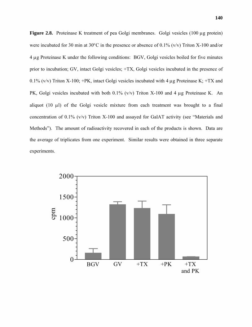
140
Figure 2.8. Proteinase K treatment of pea Golgi membranes. Golgi vesicles (100 µg protein)
were incubated for 30 min at 30°C in the presence or absence of 0.1% (v/v) Triton X-100 and/or
4 µg Proteinase K under the following conditions: BGV, Golgi vesicles boiled for five minutes
prior to incubation; GV, intact Golgi vesicles; +TX, Golgi vesicles incubated in the presence of
0.1% (v/v) Triton X-100; +PK, intact Golgi vesicles incubated with 4 µg Proteinase K; +TX and
PK, Golgi vesicles incubated with both 0.1% (v/v) Triton X-100 and 4 µg Proteinase K. An
aliquot (10 µl) of the Golgi vesicle mixture from each treatment was brought to a final
concentration of 0.1% (v/v) Triton X-100 and assayed for GalAT activity (see “Materials and
Methods”). The amount of radioactivity recovered in each of the products is shown. Data are
the average of triplicates from one experiment. Similar results were obtained in three separate
experiments.
0
500
1000
1500
2000
cpm
BGV GV +TX +PK +TXand PK
141
A model summarizing our current understanding (Doong et al., 1995; Liljebjelke et al.,
1995; Doong and Mohnen, 1998) of the biosynthesis of HGA is shown in Figure 2.9. UDP-D-
GalpA, the substrate for GalAT, is believed to be formed on the cytosolic side of the Golgi by
the epimerization of UDP-D-GlcpA catalyzed by a UDP-D-glucuronate-4-epimerase (EC 5.1.3.6)
(Feingold et al., 1960; Feingold and Avigad, 1980; Mohnen, 1999) that is associated with Golgi
membranes (K. Adams and D. Mohnen, unpublished results). UDP-D-GalpA is thought to be
transported into the lumen of the Golgi apparatus by a proposed membrane-bound UDP-D-
GalpA:UMP antiport transport protein. Similar NSTs involved in protein glycosylation have
been identified, purified and cloned from several mammalian and yeast sources (Hirschberg et
al., 1998). GalAT, a Golgi-resident protein with its catalytic site in the lumen of the Golgi, binds
UDP-D-GalpA and catalyzes the transfer of D-GalpA onto a growing HGA chain and the
production of UDP. The UDP is hydrolyzed in the lumen into inorganic phosphate and UMP by
a Golgi-resident, membrane-bound nucleoside diphosphatase (NDP/UDPase; Orellana et al.,
1997). UMP is exported out of the Golgi in exchange for UDP-D-GalpA by the proposed UDP-
D-GalpA:UMP antiport transporter.
The products synthesized by the GalAT from pea are comparable with the products
synthesized by the GalAT from tobacco (Doong et al., 1995; Doong and Mohnen, 1998). The
GalAT in detergent-permeabilized pea Golgi membranes produces a product that is
predominantly only one GalA residue longer than the exogenous acceptor. This result is similar
to that obtained with the solubilized GalAT from tobacco, where the product generated is also
only one residue longer than the exogenous acceptor (Doong and Mohnen, 1998). In contrast,
the GalAT in intact pea Golgi and in tobacco membranes both use endogenous acceptors to
generate high molecular mass products, with the product in pea (> 500 kDa) being larger than
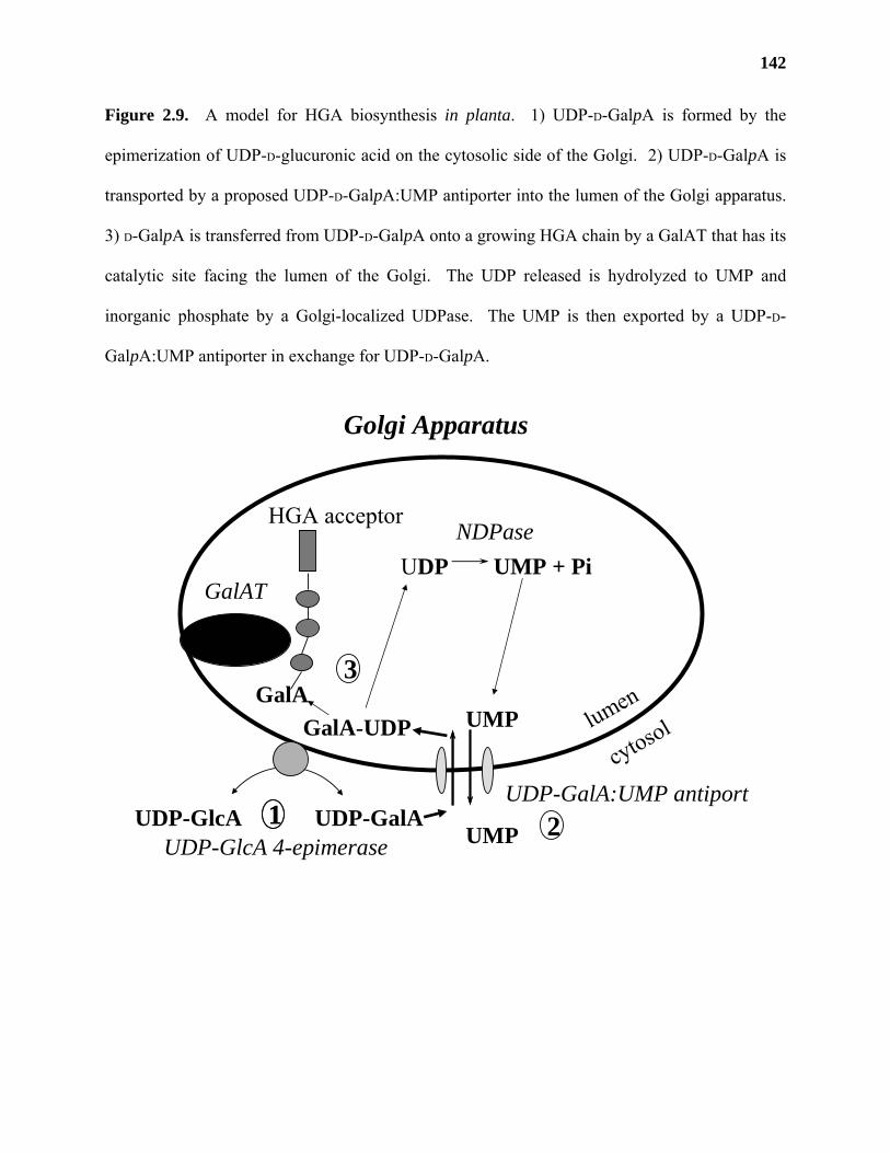
142
Figure 2.9. A model for HGA biosynthesis in planta. 1) UDP-D-GalpA is formed by the
epimerization of UDP-D-glucuronic acid on the cytosolic side of the Golgi. 2) UDP-D-GalpA is
transported by a proposed UDP-D-GalpA:UMP antiporter into the lumen of the Golgi apparatus.
3) D-GalpA is transferred from UDP-D-GalpA onto a growing HGA chain by a GalAT that has its
catalytic site facing the lumen of the Golgi. The UDP released is hydrolyzed to UMP and
inorganic phosphate by a Golgi-localized UDPase. The UMP is then exported by a UDP-D-
GalpA:UMP antiporter in exchange for UDP-D-GalpA.
UDP-GalA
UMPGalA
UDP UMP + PiNDPase
GalAT
UDP-GalA:UMP antiport
Golgi Apparatus
GalA-UDP
11
3
2UDP-GlcA 4-epimerase
HGA acceptor
cytosollumen
UDP-GlcAUMP
143
that produced in tobacco (approximately 105 kDa; Doong et al., 1995). The number of GalA
residues added onto the endogenous acceptor(s) in the pea and tobacco membranes in vitro has
not yet been determined.
There are a number of possible reasons why the pea detergent-permeabilized and the
tobacco solubilized GalATs add only a single D-GalpA to the end of the exogenous acceptors.
For example, treating pea Golgi vesicles with detergent and exogenous OGA acceptors may
cause the GalAT to preferentially use the exogenous OGAs as acceptors, whereas the GalAT in
intact Golgi uses an endogenous acceptor. The exogenous OGA (e.g. DP 14) acceptor may not
be the optimal substrate for the GalAT, whereas the endogenous product found in intact Golgi
membranes presumably contains the necessary structural information for the enzyme to
synthesize a polymeric product. Such acceptor-mediated control of enzyme processivity has
been demonstrated for a mammalian α-2,8-polysialic acid synthase (Kojima et al., 1996), where
the placement of an (1,6)-linked α-fucose residue in the acceptor was shown to be necessary for
polysialylation. It is also possible that treating Golgi membranes with Triton X-100 disrupts an
enzyme complex (protein-protein or other interaction) or removes an element that is required for
polymer synthase activity. Protein-protein complexes involved in glycan biosynthesis have been
demonstrated for glycogenin, a self-glucosylating protein that initiates glycogen biosynthesis
(Lin et al., 1999).
The presence of D-GalpA among the products formed by both intact and detergent-
permeabilized pea Golgi membranes, and the presence of D-GalpA-1-P in the products formed by
detergent-permeabilized pea Golgi membranes, are a major difference between the reaction
products formed by pea and tobacco membranes (Doong and Mohnen, 1998). The presence of D-
GalpA and D-GalpA1-P most likely results from the hydrolysis of UDP-D-[14C]GalpA by a
144
phosphodiesterase (Feingold et al., 1958) and a phosphatase (Neufeld et al., 1961). Such
hydrolases have been reported to be released during the homogenization and isolation of
membranes from some plant tissues (Villemez et al., 1966; Rodgers and Bolwell, 1992; Brickell
and Reid, 1996) and it is likely that such enzymes were released during the preparation of the pea
Golgi.
Several enzymes including latent UDPase (Orellana et al., 1997) and mammalian
glycosyltransferases (Paulson and Colley, 1989) such as the polysialyltransferases that function
in the polysialylation of neural cell adhesion molecules and other N-linked glycoproteins (Tsuji,
1996) have their catalytic sites located in the lumen of the Golgi. Most Golgi-localized
glycosyltransferases are type II integral membrane proteins with a single N-terminal membrane-
spanning region and a lumenal C-terminal catalytic domain (Paulson and Colley, 1989; Tsuji,
1996). On the other hand, plasma membrane-localized polymer synthases such as hyaluronan
synthase (Weigel et al., 1997) and the putative cellulose synthase (Delmer, 1999) are predicted to
have multiple transmembrane-spanning regions at the amino and carboxy termini and a large
central domain containing the catalytic site. The purification of the GalAT, the determination of
its amino acid sequence, and the identification of its gene will be required to elucidate the
structure of GalAT and will allow the comparison of GalAT structure with that of known
glycosyltransferases and polymer synthases.
CHAPTER 3
DEVELOPMENT OF A FILTER ASSAY FOR MEASURING
HOMOGALACTURONAN:α-(1,4)-GALACTURONOSYLTRANSFERASE ACTIVITY2
2 Jason D. Sterling, Jason A. Lemons, Ivy F. Forkner, and Debra Mohnen. To be submitted to Anal. Biochem.
146
ABSTRACT
α-(1,4)-galacturonosyltransferases (GalATs) are enzymes that catalyze the addition of
(1,4)-linked α-D-galacturonsyl residues onto the non-reducing end of homogalacturonan chains.
The nucleotide-sugar donor for the reaction is uridine diphospho-D-galacturonic acid (UDP-D-
GalpA). Many GalAT activity assays are based on the incorporation of D-[14C]GalpA from
UDP-D-[14C]GalpA onto exogenously added homogalacturonan acceptors. Reactions based on
this method can be time consuming as multiple centrifugations and labor intensive washes with
organic solvents are required to remove the unincorporated UDP-D-[14C]GalpA from the [14C]-
labeled products. An alternative GalAT filter assay was developed based on the ability of
homogalacturonan to bind to cetylpyridinium chloride (CPC). GalAT assay reaction products
made using radish (Raphanus sativus) microsomal membranes or solubilized proteins from
tobacco (Nicotiana tabacum L. cv. Samsun) and Arabidopsis thaliana (cv. Columbia) were
spotted onto Whatman 3MM paper impregnated with 2.5% (w/v) CPC. Unincorporated UDP-D-
[14C]GalpA could be selectively removed from the filters by washing with 150-250 mM NaCl.
The versatility of this assay is demonstrated by testing multiple chromatographic fractions
obtained during the partial purification of tobacco GalAT on the cation exchange resin SP
Sepharose, and by detecting the GalAT-catalyzed incorporation of D-[14C]GalpA onto
endogenous acceptors from Arabidopsis membranes.
147
INTRODUCTION
Pectins are classified as a group of acidic plant cell wall polysaccharides that contain
(1,4)-linked α-D-galacturonic acid (D-GalpA) as part of their backbone structure (O'Neill et al.,
1990; Ridley et al., 2001). Collectively, pectins comprise roughly 30% of the primary cell walls
of eudicots and non-graminaceous monocots and 10% of the walls of Poaceae (O'Neill et al.,
1990; Carpita and Gibeaut, 1993). It has been estimated that as many as 62 different
glycosyltransferases are required to synthesize all of the pectic polysaccharides found in plant
primary cell walls (Chapter 1), but to date, only a few glycosyltransferases believed to be
involved in pectin biosynthesis have been identified (Bouton et al., 2002; Iwai et al., 2002).
Much of the difficulty that researchers have encountered in being able to clone pectin
biosynthetic glycosyltransferases is due to the lack of gene homologs in the database. Once
putative pectin glycosyltransferase genes have been identified, the lack of commercially
available nucleotide-sugar substrates, oligo/polysaccharide acceptors, and/or reproducible
methods for assaying glycosyltransferase activities make it difficult to definitively prove the
functions of the cloned genes.
The glycosyltransferases responsible for the incorporation of D-GalpA into pectin are
known as galacturonosyltransferases (Ridley et al., 2001). Hassid and colleagues first detected
galacturonosyltransferase (GalAT) activity in the 1960’s in mung bean seedlings (Villemez et al.,
1965; Villemez et al., 1966) by incubating microsomes with radioactive uridine diphospho-
[14C]galacturonic acid (UDP-D-[14C]GalpA). The [14C]-labeled products generated from the
reaction were separated from the unincorporated UDP-D-[14C]GalpA by thin layer
chromatography (TLC; Villemez et al., 1966). Alternatively, the radiolabeled products could be
precipitated using trichloroacetic acid, washed extensively with water and precipitated with
148
organic solvents to remove the unincorporated nucleotide-sugar prior to analysis (Villemez et al.,
1965). The amount of D-[14C]GalpA incorporated into TLC-purified or precipitated products was
estimated by scintillation counting (Villemez et al., 1965; Villemez et al., 1966).
A number of researchers have used modifications of these initial methods to detect
GalAT activity in extracts from different plant species, including tobacco (Doong et al., 1995;
Doong and Mohnen, 1998; Scheller et al., 1999) and Arabidopsis (Guillaumie et al., 2003)
suspension cultured cells, sycamore cambial cells (Bolwell et al., 1985), pumpkin (Ishii, 2002),
and azuki bean (Takeuchi and Tsumuraya, 2001) seedlings, petunia pollen tubes (Akita et al.,
2002), and pea Golgi (Sterling et al., 2001). Methods that employ UDP-D-[14C]GalpA as a
substrate (Doong et al., 1995; Doong and Mohnen, 1998; Sterling et al., 2001; Takeuchi and
Tsumuraya, 2001) still require extensive washing of the precipitated products in order to remove
the unincorporated radiolabeled substrate. Other methods employ fluorescently-labeled
acceptors (Akita et al., 2002; Ishii, 2002), which eliminates the need for radioactively labeled
substrates, but adds the additional time and expense of producing uniformly labeled
oligogalacturonides acceptors. All of these methods are both quantitative and reliable; however,
they are too time consuming and labor intensive to be useful as a simple method to detect
enzyme activity in numerous plant extracts (i.e. mutant screens), in multiple media or cell extract
samples from recombinant tissue cultures, or to assay several column fractions during the
purification of native or recombinant GalAT.
In this paper, we report the development of a facile and rapid filter assay for measuring
GalAT activity in vitro. The assay is based on a method by Brandan and Hirschberg (1988) that
is used to detect N-heparan-sulfate sulfotransferase activity from rat liver. We show that the
filter assay gives results similar to those obtained using the well established GalAT precipitation
149
assay (Doong et al., 1995; Doong and Mohnen, 1998), that it can be used to detect GalAT
activity in microsomes and solubilized membrane proteins from different plant sources, and that
it is an effective method for the detection of GalAT activity in column fractions during enzyme
purification.
MATERIALS AND METHODS
Chemicals
Uridine diphospho-D-[14C]glucuronic acid (UDP-D-[14C]GlcpA; 272.54 mCi/mM) was
purchased from DuPont New England Nuclear (Boston, MA). Uridine diphospho-D-
[14C]galacturonic acid (UDP-D-[14C]GalpA) was prepared by the 4-epimerization of UDP-D-
[14C]GlcpA using an extract from radish roots as described (Liljebjelke et al., 1995) or was
synthesized using modifications of previously developed methods (Basu et al., 2000). Briefly,
50 µCi of uridine diphospho-D-[14C]galactose (UDP-D-[14C]Galp) was purified over a CarboPac
PA-1 column (Dionex; Sunnyvale, CA) using a 0.05-1 M linear gradient of ammonium formate,
pH 6.6, at a flow rate of 1 mL/min. Purified UDP-D-[14C]Galp was lyophilized and resuspended
in 0.5 mL of 25 mM NaH2PO4, pH 7.0, containing 250 units of galactose oxidase and 13,000
units of catalase. The reaction was incubated in the dark at room temperature for 24 h, at which
point another 250 units of galactose oxidase and 13,000 units of catalase were added. The
reaction was incubated for another 24 h in the dark and terminated by the addition of 600 µl of
5:1 (v/v), chloroform:methanol. The resultant mixture containing UDP-D-[14C]GalpA was
extracted and purified as described previously (Liljebjelke et al., 1995)..
Oligogalacturonides (OGAs) of degrees of polymerization (DP) from 7-23 were prepared
as described (Spiro et al., 1993). Chloroform, methanol, and glycerol were purchased from J.T.
150
Baker (Phillipsburg, NJ). Scintiverse was purchased from Fisher-Scientific (Springfield, NJ).
All other reagents were purchased from Sigma (St. Louis, MO).
Plant material
Radishes (Raphanus sativus) were obtained from a local grocer. The roots were cut in
fourths and stored at –80°C. Tobacco (Nicotiana tabacum L. cv. Samsun) cell suspension
cultures, originally isolated from pith callus tissue, were grown and harvested by filtration as
described (Doong and Mohnen, 1998). Suspension-cultured Arabidopsis thaliana (cv.
Columbia) cells derived from leaf calli were grown in the dark for 10 days in Gamborg’s B-5
basal medium containing 58.4 mM sucrose, 9 mM 2,4-dichlorophenoxyacetic acid, and 0.23 mM
kinetin. Cells were harvested by filtration, washed extensively with deionized water and stored
at –80°C until use.
Preparation of GalAT
Radish roots (50 g) were homogenized in a blender for 3 x 30s at high speed in 100 mL
extraction buffer (50 mM HEPES [4-(2-hydroxyethyl)-1-piperazineethanesulfonic acid], pH 7.3,
50% [v/v] gylcerol, 25 mM KCl, 0.25 mM MnCl2, and 0.1% [v/v] 2-mercaptoethanol) at 4°C.
The suspension was filtered through two layers of Miracloth (Calbiochem, La Jolla, CA) and the
filtrate was centrifuged at 4°C for 60 minutes at 40,000g. The supernatant was discarded and the
pellet was homogenized in 1 mL of storage buffer (50 mM HEPES, pH 6.8, 0.25 mM MnCl2, 25
mM KCl, 25% [v/v] glycerol) using a glass, hand-held homogenizer (Kontes Glass Co.,
Vineland, NJ). This suspension was used as a source of membrane-bound GalAT.
Solubilized tobacco GalAT was obtained as described (Doong and Mohnen, 1998) with
some modifications. Briefly, tobacco cells (20 g) were homogenized in 100 mL extraction buffer
for 6 x 30 seconds at 4°C using a polytron (Kinematica, Switzerland). The resulting suspension
151
was filtered through two layers of Miracloth and the filtrate was centrifuged for 60 minutes at
40,000g at 4°C. The supernatant was discarded and the pellet was homogenized in 2.5 mL of
solubilization buffer (100 mM NaOAc, 200 mM NaCl, 2 mM EDTA, and 0.04% [v/v] Triton X-
100) using a glass, hand-held homogenizer. The resulting suspension was centrifuged for 60
minutes at 100,000g at 4°C. The supernatant was used as a source of solubilized tobacco GalAT.
Arabidopsis cells (700 g) were homogenized in 1.2 L of extraction buffer at 4°C using a
Parr bomb. The homogenate was filtered through two layers of nylon mesh and the filtrate was
centrifuged at 28,000g for 5 h at 4°C. The supernatant was decanted and the top layer of the
microsomal pellet was resuspended in 9 mL of storage buffer. The resuspended membranes
could be stored for several months at –80°C before use. Arabidopsis membranes used as a
source of endogenous acceptors were heat-inactivated by incubating an aliquot at 95°C for 10
min.
Solubilized GalAT was isolated from resuspended Arabidopsis membranes by adding
Triton X-100, NaCl, NaOAc, and EDTA to a final concentration of 4% (v/v), 0.2 M, 0.1 M, and
2 mM, respectively. The mixture was centrifuged at 150,000g for 1.5 h at 4°C and the
supernatant containing proteins that were soluble in 4% (v/v) Triton X-100 was removed and
diluted with 4 volumes of 50 mM HEPES, pH 7.3, 0.25 mM MnCl2, 2 mM EDTA, 25% (v/v)
glycerol, and 0.25% (v/v) Triton X-100. The diluted fraction, subsequently called solubilized
enzyme, was filtered through a 0.2 µm nylon filter and either used immediately or stored at
−80°C until use.
Precipitation assay
The precipitation assay was performed as described (Doong et al., 1995) with some
modifications. An aliquot (10 µl) of membrane-bound or solubilized GalAT was incubated with
152
50 mM HEPES, pH 7.8, 0.2 M sucrose, 0.05% (w/v) bovine serum albumin, 100 µM UDP-D-
GalpA, 70 µg of OGAs, 1.1 µM UDP-D-[14C]GalpA, and with either 0.25 mM (membrane-bound
GalAT) or 1 mM (solubilized GalAT) MnCl2 in a total volume of 30 µl at 30°C for 30 min. The
reactions were terminated by the addition of 700 µL chloroform:methanol (3:2, v/v) and
centrifuged at 13,200g for five minutes. The pellet was resuspended in 65% (v/v) ethanol by
sonication for 15 min, centrifuged at 13,200g and the supernatant was removed. The 65% (v/v)
ethanol washing step was repeated and the final washed pellet was dissolved in 200 µL 10 mM
EDTA. The amount of D-[14C]GalpA incorporated into precipitated product was estimated by
scintillation counting.
SP Sepharose chromatography
An aliquot of solubilized tobacco GalAT (6 mL) was loaded onto a 5 mL SP sepharose
column (Amersham) equilibrated with binding buffer (50 mM Hepes, pH 7.3, 1% [v/v] TX-100,
25% [v/v] glycerol, 2 mM EDTA and 0.25 mM MnCl2). Unbound proteins (flow through) were
collected in 1 mL aliquots. The column was washed with 5 column volumes of binding buffer
and bound proteins were eluted from the column using a step gradient of NaCl in binding buffer
(5 mL; 100-600 mM, increasing in 100 mM increments). Column fractions (1 mL) were tested
for GalAT activity using the filter assay as described. All procedures were conducted at 4°C.
Original GalAT filter assay
The filter assay was developed based on the method of Brandan and Hirschberg (1988)
that was used to assay N-heparan sulfate sulfotransferase activity. Whatman (Maidstone,
England) 3MM paper was cut into 2x2 cm squares and soaked for 15 min in 2.5% (w/v)
cetylpyridinium chloride (CPC). The filters were dried at 65°C overnight prior to use. Reaction
mixtures prepared as described above were terminated by the addition of 5 µL 1 M NaOH and
153
the entire mixture from each individual reaction was spotted onto a CPC-coated filter. The filters
were allowed to air dry for 5 min and washed as described in the “Results”. Washed filters were
dried for 2 h and the amount of radioactivity bound to each filter was estimated by scintillation
counting.
Optimized GalAT filter assay
The filter assay was modified to improve reproducibility and to lower background levels
of unincorporated UDP-D-[14C]GalpA on the filters. Following the incubation of 3MM squares
in 2.5 % CPC, the filters were washed 3 times for 5 min with 1L of deionized H2O to remove
residual CPC prior to drying. Once the reaction mixtures had been spotted on the filters, the
filters were washed twice with 150 mM NaCl at a ratio of 1:100 (filter/ml of NaCl). Washed
filters were dried at room temperature for at least 1 h and the amount of radioactivity bound to
each filter was estimated by scintillation counting.
RESULTS
Comparison of the sensitivity of the GalAT filter assay to the GalAT precipitation assay
The development of an effective filter assay to detect GalAT activity required that the
unreacted UDP-D-[14C]GalpA substrate be separated from the radiolabeled products formed
during the GalAT reaction, and that the assay be as, or more, sensitive than previously
established methods. The anionic nature of OGAs acceptors and of the substrate, UDP-D-
[14C]GalpA, promotes their binding to the positively-charged, CPC-treated filter squares. UDP-
D-GalpA, however, has a lower total negative charge than the OGA acceptors and the products
formed from the GalAT reaction (Doong and Mohnen, 1998). It was thus predicted that washing
the filters with salt solutions would remove the unreacted substrate from the retained polyanionic
OGA products.
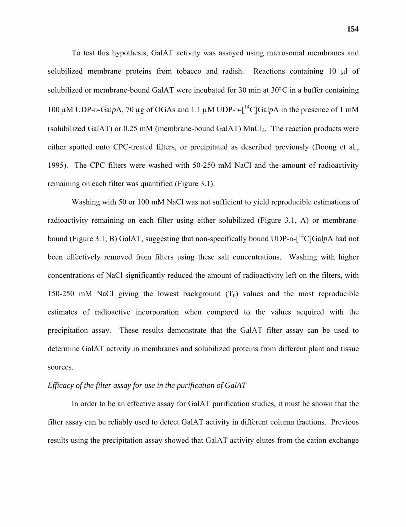
154
To test this hypothesis, GalAT activity was assayed using microsomal membranes and
solubilized membrane proteins from tobacco and radish. Reactions containing 10 µl of
solubilized or membrane-bound GalAT were incubated for 30 min at 30°C in a buffer containing
100 µM UDP-D-GalpA, 70 µg of OGAs and 1.1 µM UDP-D-[14C]GalpA in the presence of 1 mM
(solubilized GalAT) or 0.25 mM (membrane-bound GalAT) MnCl2. The reaction products were
either spotted onto CPC-treated filters, or precipitated as described previously (Doong et al.,
1995). The CPC filters were washed with 50-250 mM NaCl and the amount of radioactivity
remaining on each filter was quantified (Figure 3.1).
Washing with 50 or 100 mM NaCl was not sufficient to yield reproducible estimations of
radioactivity remaining on each filter using either solubilized (Figure 3.1, A) or membrane-
bound (Figure 3.1, B) GalAT, suggesting that non-specifically bound UDP-D-[14C]GalpA had not
been effectively removed from filters using these salt concentrations. Washing with higher
concentrations of NaCl significantly reduced the amount of radioactivity left on the filters, with
150-250 mM NaCl giving the lowest background (T0) values and the most reproducible
estimates of radioactive incorporation when compared to the values acquired with the
precipitation assay. These results demonstrate that the GalAT filter assay can be used to
determine GalAT activity in membranes and solubilized proteins from different plant and tissue
sources.
Efficacy of the filter assay for use in the purification of GalAT
In order to be an effective assay for GalAT purification studies, it must be shown that the
filter assay can be reliably used to detect GalAT activity in different column fractions. Previous
results using the precipitation assay showed that GalAT activity elutes from the cation exchange
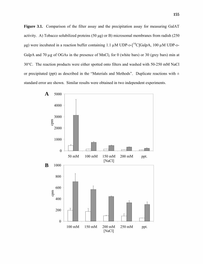
155
Figure 3.1. Comparison of the filter assay and the precipitation assay for measuring GalAT
activity. A) Tobacco solubilized proteins (50 µg) or B) microsomal membranes from radish (250
µg) were incubated in a reaction buffer containing 1.1 µM UDP-D-[14C]GalpA, 100 µM UDP-D-
GalpA and 70 µg of OGAs in the presence of MnCl2 for 0 (white bars) or 30 (grey bars) min at
30°C. The reaction products were either spotted onto filters and washed with 50-250 mM NaCl
or precipitated (ppt) as described in the “Materials and Methods”. Duplicate reactions with ±
standard error are shown. Similar results were obtained in two independent experiments.
B
A
0
200
400
600
800
1000
100 mM 150 mM 200 mM 250 mM ppt.[NaCl]
cpm
0
1000
2000
3000
4000
5000
50 mM 100 mM 150 mM 200 mM ppt.[NaCl]
cpm
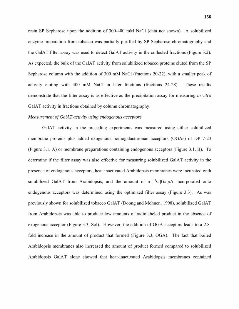
156
resin SP Sepharose upon the addition of 300-400 mM NaCl (data not shown). A solubilized
enzyme preparation from tobacco was partially purified by SP Sepharose chromatography and
the GalAT filter assay was used to detect GalAT activity in the collected fractions (Figure 3.2).
As expected, the bulk of the GalAT activity from solubilized tobacco proteins eluted from the SP
Sepharose column with the addition of 300 mM NaCl (fractions 20-22), with a smaller peak of
activity eluting with 400 mM NaCl in later fractions (fractions 24-28). These results
demonstrate that the filter assay is as effective as the precipitation assay for measuring in vitro
GalAT activity in fractions obtained by column chromatography.
Measurement of GalAT activity using endogenous acceptors
GalAT activity in the preceding experiments was measured using either solubilized
membrane proteins plus added exogenous homogalacturonan acceptors (OGAs) of DP 7-23
(Figure 3.1, A) or membrane preparations containing endogenous acceptors (Figure 3.1, B). To
determine if the filter assay was also effective for measuring solubilized GalAT activity in the
presence of endogenous acceptors, heat-inactivated Arabidopsis membranes were incubated with
solubilized GalAT from Arabidopsis, and the amount of D-[14C]GalpA incorporated onto
endogenous acceptors was determined using the optimized filter assay (Figure 3.3). As was
previously shown for solubilized tobacco GalAT (Doong and Mohnen, 1998), solubilized GalAT
from Arabidopsis was able to produce low amounts of radiolabeled product in the absence of
exogenous acceptor (Figure 3.3, Sol). However, the addition of OGA acceptors leads to a 2.8-
fold increase in the amount of product that formed (Figure 3.3, OGA). The fact that boiled
Arabidopsis membranes also increased the amount of product formed compared to solubilized
Arabidopsis GalAT alone showed that heat-inactivated Arabidopsis membranes contained
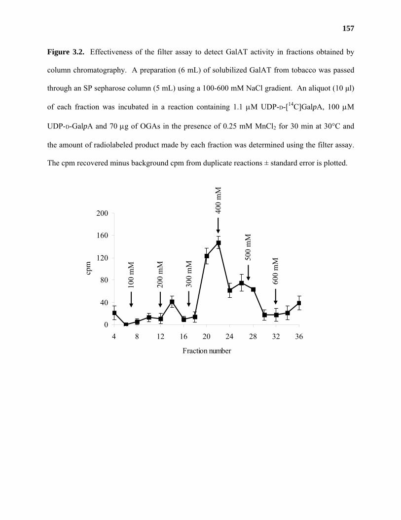
157
Figure 3.2. Effectiveness of the filter assay to detect GalAT activity in fractions obtained by
column chromatography. A preparation (6 mL) of solubilized GalAT from tobacco was passed
through an SP sepharose column (5 mL) using a 100-600 mM NaCl gradient. An aliquot (10 µl)
of each fraction was incubated in a reaction containing 1.1 µM UDP-D-[14C]GalpA, 100 µM
UDP-D-GalpA and 70 µg of OGAs in the presence of 0.25 mM MnCl2 for 30 min at 30°C and
the amount of radiolabeled product made by each fraction was determined using the filter assay.
The cpm recovered minus background cpm from duplicate reactions ± standard error is plotted.
0
40
80
120
160
200
4 8 12 16 20 24 28 32 36
Fraction number
cpm
100
mM
500
mM
200
mM
300
mM
400
mM
600
mM
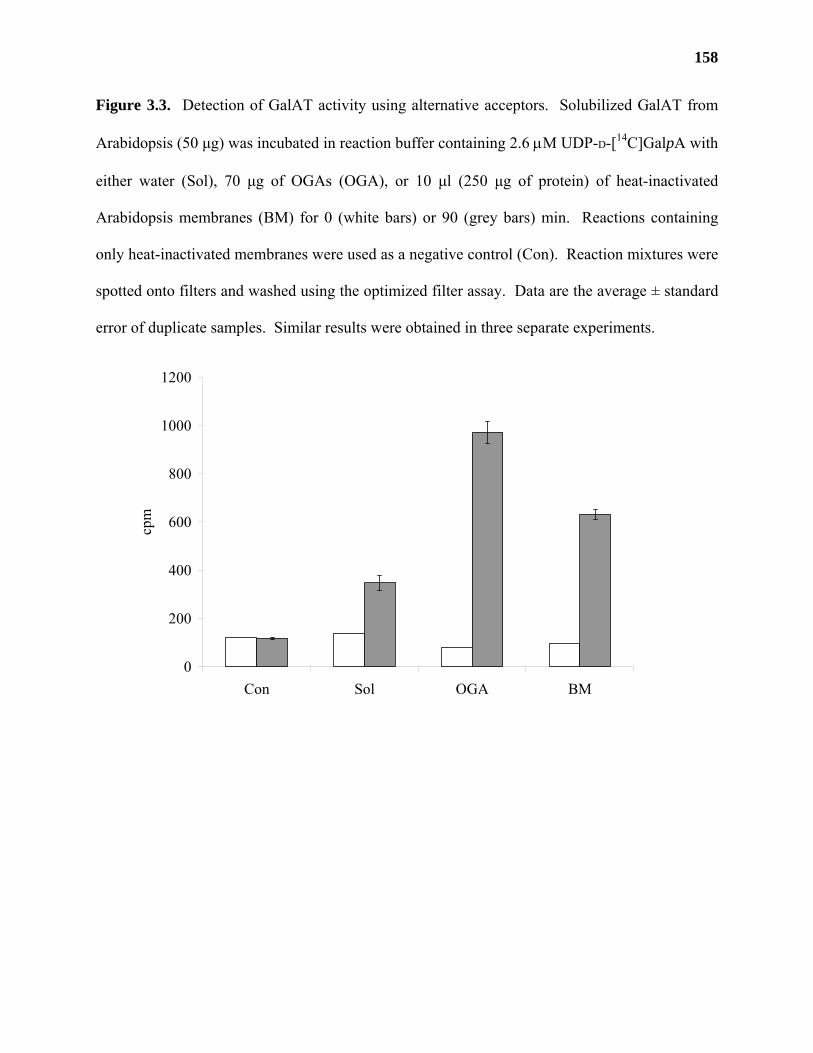
158
Figure 3.3. Detection of GalAT activity using alternative acceptors. Solubilized GalAT from
Arabidopsis (50 µg) was incubated in reaction buffer containing 2.6 µM UDP-D-[14C]GalpA with
either water (Sol), 70 µg of OGAs (OGA), or 10 µl (250 µg of protein) of heat-inactivated
Arabidopsis membranes (BM) for 0 (white bars) or 90 (grey bars) min. Reactions containing
only heat-inactivated membranes were used as a negative control (Con). Reaction mixtures were
spotted onto filters and washed using the optimized filter assay. Data are the average ± standard
error of duplicate samples. Similar results were obtained in three separate experiments.
0
200
400
600
800
1000
1200
Con Sol OGA BM
cpm
159
acceptors for GalAT. These results demonstrate that the filter assay can be used to detect the
incorporation of D-[14C]GalpA onto exogenously supplied endogenous acceptors.
DISCUSSION
The precipitation assay developed by Doong and co-workers (1995) is a commonly used
method for the detection of GalAT activity in plant tissue extracts. One of the main problems
with this assay is the amount of time required to separate the unincorporated UDP-D-[14C]GalpA
from the radiolabeled products made by GalAT. A rapid and reproducible filter assay was
developed that is based on the ability of GalAT reaction products to bind to positively-charged,
CPC-coated filters. CPC has previously been used to aid in the salt-facilitated precipitation of
radioactive GalAT products (Villemez et al., 1965) and our results show that this association is
strong enough to allow for the selective removal of unincorporated UDP-D-[14C]GalpA.
The original N-heparan-sulfate sulfotransferase assay used water and 25 mM sodium
sulfate to remove unincorporated radiolabeled [35S]-3’-phosphoadenylyl sulfate from filters
(Brandan and Hirschberg, 1988). In our hands, unincorporated UDP-D-[14C]GalpA was not
separated from the radiolabeled products by washing the filters with water (data not shown) or
dilute (50-100 mM) salt solutions. Instead, it was found that washing solutions containing 150-
250 mM NaCl were required to remove the unincorporated UDP-D-[14C]GalpA from the filters
and to obtain the reproducible binding of the radioactive products to filters in levels that were
comparable to the GalAT precipitation assay.
Our results show that the filter assay gives reliable and reproducible estimates of GalAT
activity. The GalAT filter assay has been shown to be effective for the determination of GalAT
activity in multiple plant species including radish membranes, and solubilized proteins from
tobacco and Arabidopsis. The rapidity with which many GalAT reactions can be assayed greatly
160
enhances our ability to determine GalAT activity in fractions obtained during column
chromatography and is an effective tool to aid in the purification of GalAT activity from plant
tissues.
The versatility of the GalAT filter assay is exemplified by our ability to test the activity
of GalAT using endogenous acceptors. This will allow the development of assays to test for the
activity of glycosyltransferases that specifically add neutral and/or acidic sugars onto
endogenous or well-defined acidic pectic structures. Products synthesized using such assays can
potentially be further analyzed by selective removal with high salt solutions or directly on the
filter by treatment of the radioactive products with purified exo- or endoglycanases. Future
experiments will be centered on establishing whether specific pectic polysaccharides, such as
RG-I oligosaccharides, can be bound to the filters and used to test for the activity of pectic
glycosyltransferases such as RG-I arabinosyl- (Nunan and Scheller, 2003) or
galactosyltransferases (Geshi et al., 2002).
CHAPTER 4
IDENTIFICATION AND HETEROLOGOUS EXPRESSION OF TWO PROPOSED
GALACTURONOSYLTRANSFERASES FROM ARABIDOPSIS THALIANA.3
3 Jason D. Sterling, Kumar Kolli, Ron Orlando, and Debra Mohnen. To be submitted to Plant Cell.
162
ABSTRACT
α-(1,4)-galacturonosyltransferases (GalATs) are enzymes that catalyze the incorporation
of α-D-galacturonic acid into pectic polysaccharides. In an attempt to identify putative GalAT
genes from Arabidopsis thaliana, we partially purified detergent-solubilized GalAT activity from
Arabidopsis membranes using a combination of SP-Sepharose, Reactive Yellow 3 (RY3) and
UDP-agarose chromatography. Analysis of the partially purified fractions by liquid
chromatography-tandem mass spectrometry (LC-MS/MS) led to the identification of two
proteins, named JS33 and JS36, which contained sequence motifs found in other
glycosyltransferases (Henrissat et al., 2001). JS33 and JS36 transcripts were detected in root,
stem, flower, and leaf tissues from Arabidopsis. N-terminally truncated and epitope-tagged
versions of JS33 and JS36 were cloned into an expression vector designed for recombinant
protein secretion and heterologously expressed in human embryonic kidney (HEK) 293 cells.
Transient expression of the JS36 gene construct resulted in low levels of GalAT activity in
HEK293 cell media in preliminary transient transfection experiments providing further evidence
that JS36 was a putative GalAT gene. HEK cell lines stably transformed with the JS33 and JS36
gene constructs were shown to produce mRNA transcripts and recombinant proteins of the sizes
expected for JS33 and JS36. However, recombinant proteins from the stably transformed lines
were only detected in cell lysates and did not possess any detectable GalAT activity. Selected
regions of JS33, JS36 and JS36L (a protein with high sequence identity to JS36) were used to
generate multiple antigenic peptides (MAPs) for the production of polyclonal antibodies.
Preliminary results showed that pools of antibodies generated against the three proteins were not
able to precipitate significant amounts of GalAT activity from solubilized Arabidopsis proteins.
A BLAST search of the Arabidopsis genome identified 23 additional coding regions with
163
significant sequence identity to JS36 and JS33. These results suggest that these genes comprise a
novel gene family in Arabidopsis. More experimentation needs to be conducted in order to
determine the roles that JS36 and JS33 play in pectin biosynthesis.
164
INTRODUCTION
Pectins are a family of plant cell wall polysaccharides that contain (1,4)-linked α-D-
galactopyranosyluronic acid (D-GalpA) as part of their backbone structure. Pectins are involved
in the regulation of a number of plant processes, including plant growth (O'Neill et al., 2001;
Willats et al., 2001), development (Willats et al., 1999b; McCartney et al., 2003) and the
initiation of plant defense responses (Mohnen and Hahn, 1993; Ridley et al., 2001). Disruption
of pectin structure by the mutation of pectin biosynthetic enzymes, the treatment of cells with
glycosyl hydrolases, or the expression of glycosyl hydrolases in transgenic plants cause a wide
variety of pleiotropic effects, including dwarfism (O'Neill et al., 2001; Bouton et al., 2002),
altered seed coats (Usadel et al., 2004; Western et al., 2004), reduced wall tensile properties
(Ryden et al., 2003), impaired guard cell function (Jones et al., 2003), and altered cell adhesion
and wall glycosyl residue composition (Bouton et al., 2002; Iwai et al., 2002). While the enzyme
activity encoded by some pectin biosynthetic genes has been well characterized (e.g. MUR1), the
activity of other genes, especially those encoding putative pectin glycosyltransferases (e.g.
QUA1), has not been determined.
α-(1,4)-Galacturonosyltransferases (GalATs) are glycosyltransferases that catalyze the
incorporation of D-GalpA into the backbone structures of pectin (Villemez et al., 1965; Doong et
al., 1995). D-GalpA is a major component of the backbone of all three types of pectin found in
plant primary walls, comprising roughly 70% of total cell wall pectin (Mohnen et al., 1996). It
has been estimated that between 5 and 7 distinct GalATs may be required to synthesize all of the
backbone structures and side branches found in primary cell wall pectins (Chapter 1; Mohnen
2002). However, genes encoding enzymatically active GalATs have yet to be identified.
165
In an attempt to identify putative GalAT genes, we partially purified GalAT activity from
Arabidopsis thaliana and identified two genes, named JS33 and JS36, which contained amino
acid motifs found in other glycosyltransferases. We heterologously expressed N-terminally
truncated forms of these proteins in HEK293 cells and showed that low levels of GalAT activity
could be detected in media from cells transfected with JS36 in preliminary experiments.
Analysis of the Arabidopsis genome indicate the presence of 23 additional genes with significant
sequence identity to JS33 and JS36, suggesting that these genes are part of a larger gene family
in Arabidopsis.
MATERIALS AND METHODS
Chemicals and kits
Uridine diphosphate-α-D-[14C]galactose (UDP-D-[14C]Galp, 11.0 GBq/mmol), PD-10
columns, and SP-, protein A- and protein G-Sepharose were purchased from Amersham
Biosciences (Piscataway, NJ). A mixture of OGAs with a degree of polymerization (DP) of 7 to
23 was prepared according to Doong et al. (1995). UDP-agarose was purchased from
Calbiochem (La Jolla, CA). Nickel-nitrilotriacetic acid agarose (Ni-NTA) resin, plasmid
maxiprep and gel extraction kits were purchased from Qiagen (Valencia, CA). EZNA plasmid
miniprep kits were purchased from Omega Bio-tek (Doraville, GA). All restriction enzymes
were purchased from New England Biolabs (Beverly, MA). Highly purified, monoclonal anti-
hemagluttinin (anti-HA) antibodies were purchased from Covance (Berkeley, CA). Anti-HA
conjugated to horseradish peroxidase was purchased from Roche (Indianapolis, CA). 3,3’,5,5’-
Tetramethylbenzidine (TMB) substrate kits were purchased from Vector Labs (Burlingame, CA).
Polyvinylidene difluoride (PVDF) membranes were purchased from Schleicher and Schuell
Biosciences (Keene, NH). Protein concentrations were determined using the Bradford assay
166
(Bio-Rad, Hercules, CA). Reactive Yellow 3 resin and all other chemicals were purchased from
Sigma.
Plant material
Cell-suspension cultures of Arabidopsis thaliana (cv. Columbia) derived from leaf calli
were grown in the dark for 7-10 days in Gamborg’s B-5 basal medium containing 58.4 mM
sucrose, 9 µM 2,4-dichlorophenoxyacetic acid, and 0.23 µM kinetin as described (Guillaumie et
al., 2003). Cells were collected by filtration, washed extensively with water, and stored at -80°C
until use.
Mammalian cell culture
Human embryonic kidney (HEK) 293 cells (Edge Biosystems; Gaithersburg, MD) were a
gift of Dr. Kelley Moremen (The University of Georgia, Athens). HEK cells were grown in 150
cm2 culture dishes (Corning, Corning, NY) in bicarbonate-buffered, Dulbeco’s modified Eagle’s
medium (Sigma), pH 7.4, with high glucose (4.5 g/L) supplemented with 10% (v/v) fetal bovine
serum (Sigma), 0.6 g/L L-glutamine, 100 µg/mL streptomycin sulfate, and 100 units/mL
penicillin. Cells were maintained at 37°C in a humidified incubator (Thermo Electron Corp.,
Newington, NH) containing 5% CO2 and passaged when the cells were 80% confluent.
Preparation of GalAT from Arabidopsis
Solubilized GalAT from Arabidopsis suspension cultured cells was prepared as described
in Guillaumie et al. (2003). Dark-grown Arabidopsis cells (700 g) were homogenized in 1.2 L of
homogenization buffer (50 mM 4-[2-hydroxyethyl]-1-piperazineethanesulfonic acid; HEPES, pH
7.3, 0.25 mM MnCl2, 25 mM KCl, 50% (v/v) glycerol and 0.1% 2-mercaptoethanol) at 4°C
using a Parr bomb. The homogenate was filtered through two layers of nylon mesh and the
filtrate was centrifuged at 28,000g for 5 h at 4°C. The supernatant was decanted and the top layer
167
of the microsomal pellet was resuspended in 9 mL of cold 50 mM HEPES, pH 6.8, 0.25 mM
MnCl2, 25 mM KCl, and 25% (v/v) glycerol. To the resuspended pellet, Triton X-100, NaCl,
NaOAc, and EDTA were added to final concentrations of 4% (v/v), 0.2 M, 0.1 M, and 2 mM,
respectively, and the mixture was centrifuged at 150,000g for 1.5 h at 4°C. The supernatant
containing proteins that were soluble in 4% (v/v) Triton X-100 was removed and diluted with 4
volumes of cold 50 mM HEPES, pH 7.3, 0.25 mM MnCl2, 2 mM EDTA, 25% (v/v) glycerol,
and 0.25% (v/v) Triton X-100. The diluted fraction, subsequently called solubilized GalAT, was
filtered through a 0.2 µm nylon filter (Fisher Scientific) and either used immediately or stored at
-80°C until use.
SP-Sepharose chromatography
Solubilized GalAT (125 mL) was loaded twice over a 50 mL column of SP-sepharose (5
x 2.5 cm) equilibrated with buffer A (50 mM HEPES, pH 7.3, 0.25 mM MnCl2, 1% Triton X-
100, 2 mM EDTA, and 25% [v/v] glycerol). The column was washed with 3 volumes of buffer
B (50 mM HEPES, pH 7.3, 0.25 mM MnCl2, 25% [v/v] glycerol, 2.5% [w/v] 3-[(3-
cholamidopropyl)dimethylammonio]propanesulfonic acid, CHAPS) and bound proteins were
eluted using a step gradient of NaCl in buffer B as follows: 50 mL of 100 mM, 50 mL of 200
mM, 35 mL of 300 mM, 70 mL of 400 mM. Proteins eluting with 300 and 400 mM NaCl were
pooled and desalted in buffer B over PD-10 columns according to the manufacturer’s
instructions. Briefly, 2.5 mL of sample was loaded per column. Proteins were eluted from the
column with the addition of 3.5 mL of buffer B. All operations were carried out at 4°C.
Reactive Yellow 3 chromatography
The pooled and desalted fraction from the SP-sepharose column was loaded onto a 15 mL
column (2 x 5 cm) of Reactive Yellow 3 (RY3) equilibrated with buffer B. Bound proteins were
168
eluted with the sequential addition of one column volume of buffer B containing 1) 10 µM UDP,
2) 10 µM UDP and 2 µM OGAs, 3) 10 µM UDP, 2 µM OGAs and 100 mM NaCl, and 4) 10 µM
UDP, 2 µM OGAs and 200 mM NaCl. The proteins that eluted with 100 and 200 mM NaCl
were pooled and desalted in buffer B using PD-10 columns.
UDP-agarose chromatography
The pooled RY3 fractions were loaded onto a 7 mL (2.1 x 2 cm) UDP-agarose column
that had been equilibrated with buffer B. The most purified fraction from UDP-agarose
chromatography shown in Table 4.2, Experiment 1 was obtained using the following elution
scheme: bound proteins were eluted with one column volume of buffer B containing 0.01
mg/mL OGAs and 1) 50 mM UDP, 2) 25 mM MnCl2, 3) 25 mM MnCl2 and 0.5 M NaCl, 4) 25
mM EDTA, and 5) 25 mM EDTA and 0.5 M NaCl. The fraction eluted with 25 mM MnCl2 and
0.5 M NaCl was determined to be the most pure following GalAT activity assays and the
determination of protein concentration in each of the eluted fractions.
The fraction analyzed by liquid chromatography, tandem mass spectrometry (LC-
MS/MS) was obtained by UDP-agarose chromatography in a second experiment (Table 4.2,
Experiment 2) using the following elution scheme: bound proteins from the UDP-agarose
column were first eluted with one column volume each of 0.01, 0.05, 0.1, 0.5, 1, 5, 10, 15 and 20
mM UDP in buffer B. The column was washed with 1 volume of 1 M NaCl in buffer B and all
of the eluted fractions from this step were collected, desalted, and tested for GalAT activity. The
fraction that eluted with 1 M NaCl was reapplied to the column and bound proteins were eluted
with one column volume of buffer B containing 0.01 mg/mL OGAs and 1) 50 mM UDP, 2) 25
mM MnCl2, 3) 25 mM MnCl2 and 0.5 M NaCl, 4) 25 mM EDTA, and 5) 25 mM EDTA and 0.5
M NaCl. The fraction that eluted with 25 mM MnCl2 and 0.5 M NaCl was further analyzed by
169
trypsin digestion and LC-MS/MS. OGAs were added to the elution buffers to inhibit the activity
of a contaminating exoPGase that co-purified with GalAT activity.
Trypsin digestion and analysis of Arabidopsis peptides
The most purified fraction from UDP-agarose chromatography was incubated with 940
units of sequencing grade, modified trypsin (Promega; Madison, WI) for 12 h at 37°C. Peptides
were dialyzed overnight in water using Spectro/Por CE 1,000 molecular weight cutoff dialysis
membranes (Spectrum Laboratories, Rancho Dominguez, CA), lyophilized and analyzed by LC
MS/MS.
Liquid chromatography- tandem mass spectrometry and identification of Arabidopsis proteins
Peptides were introduced into a Q-TOF2 (Waters Micromass, Milford, MA) tandem mass
spectrometer using a Waters CapLC delivery system. The mobile phases used for gradient
elution consisted of 0.1% formic acid in water (A) or acetonitrile (B). The gradient conditions
were 10% B to 70% B over 60 min at a flow rate of 1 µL/min. The Q-TOF2 was operated in a
data-dependant scan mode. The survey MS spectra were acquired from 450-1700 mass units.
The switch criteria for MS to MS/MS mode were ion count and charge state, with the Q-TOF set
to ignore singly charged ions and to acquire MS/MS data for up to 3 co-eluting peptides. The
collision energy was varied automatically depending on the peptide mass and charge state.
Acquired spectra were used to probe the Arabidopsis genome to identify candidate GalAT genes
using the Mascot search engine (www.matrixscience.com) using a peptide and MS/MS tolerance
of ±2 and ±1, respectively.
RT-PCR and cloning of JS33 and JS36
Total RNA from flowers, rosette leaves or stems obtained from 3- or 6-week-old
Arabidopsis ecotype Columbia plants as well as total RNA from roots of 4-week old Arabidopsis
170
plants grown in liquid media were gifts of Dr. Maor Bar-Peled (The University of Georgia,
Athens; Harper and Bar-Peled, 2002). First cDNA strand synthesis was conducted according to
the manufacturer’s instructions using 1 mM oligo(dT) primer, 200 units of Superscript II reverse
transcriptase (Invitrogen) and 8 µg total RNA. Products from these reactions were treated with 2
units of RNase H (Invitrogen) and used as a template for PCR using gene specific primers. PCR
reactions were conducted using high fidelity Platinum Taq polymerase (Invitrogen, Carlsbad,
CA) according to manufacturer’s instructions. Typical PCR reactions involved 1 cycle at 95°C
for 2 min followed by 30 cycles at 95°C for 30 s, 52°C for 30s, and 70°C for 2.5 min, and a final
extension at 70°C for 5 min. The PCR products were analyzed on 1% Tris-acetate EDTA (TAE)
agarose gels and DNA bands of the appropriate size were excised and purified using QIAquick
gel extraction kit (Qiagen; Valencia, CA). Control PCR reactions included actin primers
ACT119S and ACT284A (McKinney et al., 1995) as a positive control (a gift from Dr. Richard
Meagher, the University of Georgia, Athens).
A full-length cDNA encoding JS33 (accession no. AY091448) in vector pUNI51 was
obtained from the Arabidopsis Information Resource Center (ABRC). An N-terminally
truncated version of JS33 (JS33∆1-43) lacking the putative transmembrane domain was
generated using the sense primer FJS33Ntru (5’-ttattacccggggaattccacaatggctttcactctcctggattt-3’)
and the antisense primer RpUNI51 (5’-ggcggctatgatctcggcggccgctagaattg-3’). The primers
contained restriction sites for SmaI and NotI, respectively (underscored in the primers
sequences).
A version of JS36 (accession no. NM_115977) lacking its putative N-terminal
transmembrane domain (JS36∆1-41) was generated from total flower RNA using RT-PCR and
the gene specific sense primer FJS36Ntru (5’-ttattacccggggaattccgaggagtgtatatcgattcctcaaatg-3’)
171
and antisense primer RJS36 (5’-gcggccgcattttattcatgaaggttgcaacgacg-3’). The primers contained
restriction sites for SmaI and Not I, respectively (underscored in the primer sequences).
Transcript analysis of JS33 and JS36 in Arabidopsis tissues was conducted using the
previously described RT-PCR protocol and the primers FJS33Ntru and RJS33 (5’-
aagcattggccgtgtagctacttac-3’), and FJS36Ntru and RJS36 for JS33 and JS36, respectively.
Expression of JS33 and JS36 in HEK293 cells
The purified PCR products for truncated JS33 and JS36 were first cloned into pCR2.1-
TOPO (Invitrogen). Positive clones were selected based on their resistance to kanamycin and
SmaI/NotI fragments were subcloned into the mammalian expression vector pEAK10 (Edge
Biosystems) along with an N-terminal HindIII/SmaI fragment containing a Trypanosoma cruzi
mannosidase (TCMss) signal sequence (MRLLTALFAYFIVALILAFSVSAKARR), a poly-
histidine (HHHHHHHHHH) tag, and two copies of the hemagluttinin (HA) epitope
(YPYDVPDYA) that was a gift from Dr. Kelley Moremen (Vandersall-Nairn et al., 1998).
Correctly constructed pEAK10 vectors containing both the N-terminal fragment and the
appropriate JS gene (Figure 4.1, B) were confirmed by DNA sequencing.
Transient transfection and stable transformation of HEK293 cells (Jordan et al., 1996)
was conducted using 25-30 µg of DNA for transient experiments, 6 µg of DNA for the
production of clonal lines, and 25-30 µg of DNA for the creation of stable, mixed-population
lines. Transformed cells were selected on media containing puromycin (1 µg/mL) and positive
transformants were selected by PCR amplification of genomic DNA and/or RT-PCR of total
RNA using the gene specific primers FJS33Ntru and RJS33, and FJS36Ntru, RJS36 and. 36IP
(5’-tgattctgcagtccttggtaaatacag-3’) for JS33 and JS36, respectively. Cells transformed with
empty pEAK10 vector or expressing a recombinant, histidine-tagged, rat α-(2,6)-sialyltransferase
172
(Wlasichuk et al., 1993), a gift of Dr. Kelley Moremen (unpublished results), were used as
negative controls. Control RT-PCR reactions that were conducted to test transcript levels in
HEK293 cells included the use of primers (FP 5’-caatgaccccttcattgacc-3’, RP 5’-
gtcttctgggtggcagtgat-3') specific to positions 123 and 584, respectively, of human glyceraldehyde
3’-phosphate dehydrogenase (accession no. BC023632) that were gifts from Dr. Michael Pierce
(the University of Georgia, Athens, GA).
Ni2+-affinity column chromatography
Media from an HEK293 cell line expressing a recombinant α-(2,6)-sialyltransferase
(containing an N-terminal polyhistidine tag) was desalted over a PD-10 column equilibrated with
buffer C (50 mM KH2PO4, pH 7.5, 300 mM NaCl, 40 mM imidazole). The desalted media (30
mL) was poured over a 200 µL column of Ni-NTA agarose equilibrated with buffer C. The
column was washed with 10 volumes of buffer C. Proteins bound to the Ni-NTA resin were
denatured in 0.2 volumes of SDS-PAGE sample buffer (Laemmli, 1970) and separated by SDS-
PAGE.
Immunoprecipitation of recombinant JS33 and JS36 using anti-HA antibodies
Protein A-Sepharose, equilibrated in ice-cold phosphate-buffered-saline (PBS pH ; 137
mM NaCl, 2.7 mM KCl, 4.3 mM Na2HPO4, and 1.4 mM KH2PO4, pH 7.3), was incubated with
1 µg of purified monoclonal anti-HA antibodies (Covance, Princeton, NJ) at a ratio of 30:1 (v/v)
for 2 h unless otherwise specified. The resulting protein A-Sepharose:anti-HA conjugate was
washed 3 times with 10 volumes of cold PBS, brought to a 50% slurry with PBS (media) or cell
lysis buffer (cell lysates) and kept at 4°C until use.
Media (3.4 mL) from JS33 and JS36 expressing lines was harvested, brought to 10%
(v/v) glycerol, and incubated with 60 µl of protein A-Sepharose:anti-HA conjugate in the
173
presence of 310 µL of PBS and 80 mg bovine serum albumin (BSA) for 1 h. Proteins bound to
the protein A-Sepharose:anti-HA conjugate were washed 4 times with 10 volumes of 0.1% (w/v)
TX-100, 50 mM Tris/Cl, pH 7.4, and 300 mM NaCl followed by 2 washes with 10 volumes of
PBS and used immediately for GalAT activity assays (10 µL) or SDS-PAGE (10 µL).
Cell lysates from HEK lines were prepared by incubating one 150 cm2 dish of cells with
1 mL of cold cell lysis buffer (50 mM HEPES, pH 7.3, 1% [v/v] TX-100, 150 mM NaCl , 2 mM
EDTA, and 1 tablet of Complete Protease Inhibitor Cocktail, EDTA free; Roche, catalog no. 1-
873-580) for 30 min. Cell debris was pelleted by centrifugation and the clear supernatant was
incubated with 40 µL (Western blots) or 160 µL (GalAT activity assays) of protein A-
Sepharose:anti-HA conjugate for 1 h. Immunoprecipitates were either washed 5 times with 10
volumes of buffer A (activity assays) or 3 times with 10 volumes of buffer A followed by 2
washes of 10 volumes of PBS (Western blot). All manipulations were done at 4°C.
SDS-PAGE and immunoblotting
SDS-PAGE was carried out according to a previously described method (Laemmli,
1970). Samples (10-20 µL) were separated on 7.5 or 10% SDS-PAGE gels. Separated proteins
were either visualized by silver staining (Heukeshoven and Dernick, 1985), Coomassie staining
(Bio-Safe Coomassie G250 stain, Bio-Rad), or transferred to PVDF membranes for 2 h at 200
mA in 25 mM Tris, 192 mM glycine, and 20% methanol. Membranes were blocked for 1 h in
3% BSA in Tris-buffered-saline (TBS; 100 mM Tris/Cl, pH 7.5, 0.9% [w/v] NaCl) and incubated
with a 1:1000 dilution of anti-HA-peroxidase antibody in blocking buffer for 1 h. Membranes
were washed 4 times in TTBS (0.1% [v/v] Tween 20 in TBS) and developed using the 3,3’,5,5’-
tetramethylbenzidine (TMB) substrate kit (Vector Labs, Burlingame, CA) according to
manufacturer’s instructions.
174
Synthesis of uridine diphosho-D-[14C]galacturonic acid
Uridine diphosho-D-[14C]galacturonic acid (UDP-D-[14C]GalpA) was synthesized based
on previously developed methods (Liljebjelke et al., 1995; Basu et al., 2000) with some
modifications. Briefly, 50 µCi of uridine diphospho-D-[14C]galactose (UDP-D-[14C]Galp) was
purified over a CarboPac PA-1 column (Dionex; Sunnyvale, CA) using a 0.05-1 M linear
gradient of ammonium formate, pH 6.6, at a flow rate of 1 mL/min. Purified UDP-D-[14C]Galp
was lyophilized and resuspended in 0.5 mL of 25 mM NaH2PO4, pH 7.0, containing 250 units of
galactose oxidase and 13,000 units of catalase. The reaction was incubated in the dark at room
temperature for 24 h, at which point another 250 units of galactose oxidase and 13,000 units of
catalase were added. The reaction was incubated for another 24 h in the dark and terminated
with 600 µl of 5:1 (v/v), chloroform:methanol. The resultant mixture containing UDP-D-
[14C]GalpA was extracted and purified as described previously (Liljebjelke et al., 1995). The
typical yield of UDP-D-[14C]GalpA from the starting UDP-D-[14C]Galp was between 60 to 70%.
GalAT activity assay
The GalAT activity assay was a modification of Doong et al. (1995). Assay conditions
used to test solubilized or membrane-bound GalAT activity, or fractions from chromatography
columns were conducted in GalAT reaction buffer (50 mM HEPES, pH 7.8, 0.2 M sucrose,
0.05% (w/v) bovine serum albumin, and 25 mM KCl) containing 80 µg of OGAs (DP of 7-23),
1.1 µM UDP-D-[14C]GalpA (specific activity 298 mCi mmol-1), 0.25 mM MnCl2 and 140 µM
UDP-D-GalpA in a total volume of 30 µl for 15 min at 30°C (unless otherwise specified).
Reaction mixtures were terminated with 5 µL of 0.4 M NaOH.
Media and/or immunoprecipitated media (10 µL) from HEK cell lines were incubated in
GalAT reaction buffer containing 80 µg of OGAs (DP of 7-23), 2 µM UDP-D-[14C]GalpA
175
(specific activity 298 mCi mmol-1), 0.25 mM MnCl2 and 100 µM UDP-D-GalpA in a total
volume of 30 µl for 45 min (media) or 60 min (immunoprecipitated media). Reaction mixtures
were terminated with 5 µL of 0.4 M NaOH.
Cell lysates (20 µL) and/or immunoprecipitated cell lysates (40 µL) from HEK cell lines
were incubated in GalAT reaction buffer containing 50 µg of OGAs (DP of 7-23), 2 µM UDP-D-
[14C]GalpA, 0.25 mM MnCl2 and 125 µg of heat-inactivated Arabidopsis membranes (as an
alternative source of acceptors) in a total volume of 70 µl at 30°C for 30 min (cell lysates) or 90
min (immunoprecipitated cell lysates). Reaction mixtures were terminated with 10 µl of 0.4 M
NaOH.
Terminated reaction mixtures were spotted onto cetylpyridinium chloride-coated
Whatman (Maidstone, England) 3MM paper (4 cm2). The filters were allowed to dry for 5 min
and washed 3 times for 10 min each in 150 mM NaCl at a ratio of 1 filter/100 mL solution. The
amount of radioactivity bound to each filter was estimated by scintillation counting. Statistical
analysis of GalAT reaction data was performed using a two-sample, one-tailed Student’s t test
(Zar, 1999) with an a priori α of 0.05 (95% confidence).
Production of polyclonal antibodies
Three distinct regions of JS33, JS36 and JS36L (At5g47780), an Arabidopsis protein with
significant sequence identity to JS36, were selected and used for the production of soluble,
tetrameric, multiple antigenic peptides (MAPs). Peptides sequences (20-25 amino acids) were
selected by analysis using hydropathy plots (Kyte and Doolittle, 1982), analyzing beta-turn
character (Chou and Fasman, 1978), sequence alignments, and use of GenTHREADER
(http://bioinf.cs.ucl.ac.uk/psipred/). Stretches in the amino acid sequence of JS genes that had
low hydrophobicity, high beta-turn character (determined using ProtScale;
176
http://au.expasy.org/cgi-bin/protscale.pl), and were predicted to be loop regions in the protein by
sequence alignments (ClustalX, version 1.83) and by GenTHREADER, were chosen as
candidate peptides for MAP construction. MAPs were constructed at the Molecular Genetics
Instrument Facility (The University of Georgia). Rabbits (New Zealand White, two for each
antigen), were immunized (0.5 mL of 0.3 mg mL-1 antigen), maintained and bled by the
Polyclonal Antibody Facility (The University of Georgia).
Immunoprecipitation of Arabidopsis solubilized GalAT using polyclonal antibodies
Pre-immune serum or polyclonal antibodies (1 µL of pooled serum) from rabbits
inoculated with JS33, JS36 and/or JS36L MAP antigens were incubated with 40 µL of protein A-
Sepharose (equilibrated in ice-cold PBS) for 2 h at 4°C. Polyclonal antibody:protein A
conjugates were washed 6 times with buffer A, and 40 µL of conjugate was incubated with 0.5
mL of solubilized Arabidopsis GalAT overnight at 4°C. Immunoprecipitates were washed 5
times in buffer A and tested for GalAT activity as described above using 2 µM UDP-D-
[14C]GalpA and 80 µg of OGAs in a total volume of 70 µL for 30 min at 30°C.
Sequence alignment of JS family genes and phylogenetic analysis
Arabidopsis genes with high sequence identity to JS36 were aligned using ClustalX,
version 1.83 (Jeanmougin et al., 1998). Phylogenetic analysis of the Arabidopsis GalAT gene
family, along with EST (GenBank, http://www.ncbi.nlm.nih.gov/dbEST) or tentative consensus
(TC, http://www.tigr.org/tdb/tgi) sequences from soybean (Glycine max, Gm), barrel medic
(Medicago truncatula, Mt), rice (Oryza sativa, Os), tobacco (Nicotiana tobacum, Nt) and
chickpea (Cicer arietinum, Ca) was conducted using PAUP software, version 3.1 (Swofford,
1998).
177
RESULTS
Solubilization and partial purification of Arabidopsis GalAT
A reproducible method for the solubilization of roughly 20% of the GalAT activity from
tobacco membranes was previously established (Doong and Mohnen, 1998). The low yield of
total activity from the starting material was a major drawback in this solubilization method.
Therefore, an improved solubilization method was developed that allowed the recovery of up to
85% of the starting GalAT activity from tobacco membranes.
The original low-speed centrifugation pellet (4,000g, 15 min) generated during the
isolation of tobacco membranes (Doong and Mohnen, 1998) contained ~50% of the total activity
present in tobacco cell homogenates (data not shown). It was hypothesized that the elimination
of this step would greatly improve the yield of GalAT from these cells. Centrifugation of
tobacco homogenates at high speed (28,000g, 2 h) created a layered pellet. Resuspension and
solubilization of the top layer of this pellet with 4% (v/v) Triton X-100 resulted in the
solubilization of up to 85% of the total activity present in the top layer of the high speed pellet
(Table 4.1). Little or no activity was present in the other layers of the pellet. If the top layer was
mixed with the other layers prior to solubilization, the amount of solubilized GalAT activity that
could be recovered decreased dramatically (Table 4.1), suggesting that the other layers of the
pellet contained a potent inhibitor of GalAT activity and/or a protease capable of digesting
tobacco GalAT. This finding was supported by the fact that GalAT activity solubilized using the
new method was more stable than that obtained by previous solubilization methods, being stable
for up to one year at -80°C, with a 30% loss in activity occurring after one week at 4°C (data not
shown).
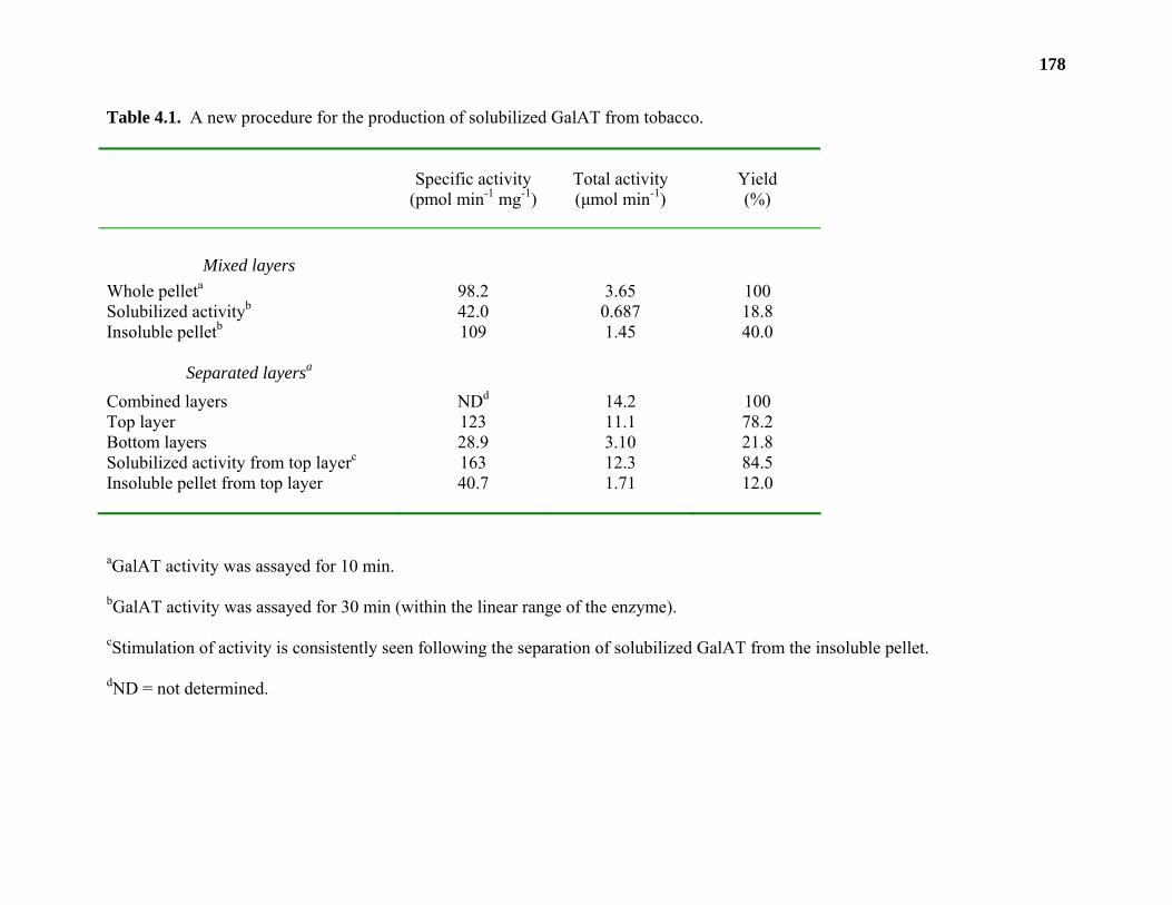
178
Table 4.1. A new procedure for the production of solubilized GalAT from tobacco.
Specific activity (pmol min-1 mg-1)
Total activity (µmol min-1)
Yield (%)
Mixed layers
Whole pelleta 98.2 3.65 100 Solubilized activityb 42.0 0.687 18.8 Insoluble pelletb 109 1.45 40.0
Separated layersa
Combined layers NDd 14.2 100 Top layer 123 11.1 78.2 Bottom layers 28.9 3.10 21.8 Solubilized activity from top layerc 163 12.3 84.5 Insoluble pellet from top layer 40.7 1.71 12.0
aGalAT activity was assayed for 10 min.
bGalAT activity was assayed for 30 min (within the linear range of the enzyme).
cStimulation of activity is consistently seen following the separation of solubilized GalAT from the insoluble pellet.
dND = not determined.
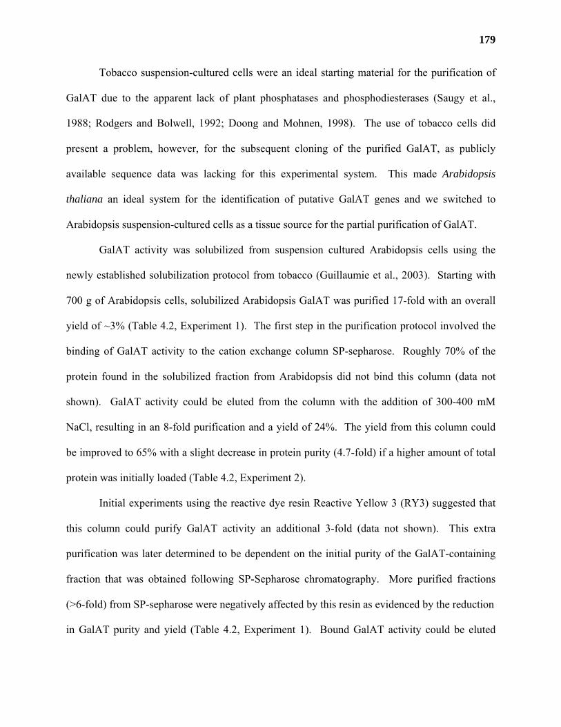
179
Tobacco suspension-cultured cells were an ideal starting material for the purification of
GalAT due to the apparent lack of plant phosphatases and phosphodiesterases (Saugy et al.,
1988; Rodgers and Bolwell, 1992; Doong and Mohnen, 1998). The use of tobacco cells did
present a problem, however, for the subsequent cloning of the purified GalAT, as publicly
available sequence data was lacking for this experimental system. This made Arabidopsis
thaliana an ideal system for the identification of putative GalAT genes and we switched to
Arabidopsis suspension-cultured cells as a tissue source for the partial purification of GalAT.
GalAT activity was solubilized from suspension cultured Arabidopsis cells using the
newly established solubilization protocol from tobacco (Guillaumie et al., 2003). Starting with
700 g of Arabidopsis cells, solubilized Arabidopsis GalAT was purified 17-fold with an overall
yield of ~3% (Table 4.2, Experiment 1). The first step in the purification protocol involved the
binding of GalAT activity to the cation exchange column SP-sepharose. Roughly 70% of the
protein found in the solubilized fraction from Arabidopsis did not bind this column (data not
shown). GalAT activity could be eluted from the column with the addition of 300-400 mM
NaCl, resulting in an 8-fold purification and a yield of 24%. The yield from this column could
be improved to 65% with a slight decrease in protein purity (4.7-fold) if a higher amount of total
protein was initially loaded (Table 4.2, Experiment 2).
Initial experiments using the reactive dye resin Reactive Yellow 3 (RY3) suggested that
this column could purify GalAT activity an additional 3-fold (data not shown). This extra
purification was later determined to be dependent on the initial purity of the GalAT-containing
fraction that was obtained following SP-Sepharose chromatography. More purified fractions
(>6-fold) from SP-sepharose were negatively affected by this resin as evidenced by the reduction
in GalAT purity and yield (Table 4.2, Experiment 1). Bound GalAT activity could be eluted
180
Table 4.2. Partial purification of Arabidopsis GalATa
Experiment 1:
Step Total protein (mg)
Specific activity (pmol
min-1 mg-1)
Purification (-fold)
Total activity (µmol min-1)
Yield (%)
1. Sol 794 304b 1 241 100 2. SP 23.6 2450b 8.06 57.7 24 3. RY3 13.7 1720c 5.66 23.6 9.8 4. UDP-agarose (2)
1.29 5120c 16.8 6.59 2.7
Experiment 2:
Sample Total protein (mg)
Specific activity (pmol
min-1 mg-1)
Purification (-fold)
Total activity (µmol min-1)
Yield (%)
1. Sol 2,200 26.0 1 57.2e 100 2. SP 314 121 4.7 37.5e 65.6 3. RY3 NDd ND ND 17.5f 30.6 4. UDP-agarose (1)
ND ND ND 7.09f 12.4
5. UDP-agarose (2)
ND ND ND 1.35f 2.4
aGalAT activity in Experiments 1 and 2 was measured using different amounts of cold UDP-D-
GalpA and are therefore not directly comparable.
bGalAT activity was measured for 15 min using 1 mM UDP-D-GalpA.
cGalAT activity was assayed for 30 min using 1 mM UDP-D-GalpA
dND = Not determined.
eGalAT activity was assayed for 15 min using 100 µM UDP-D-GalpA.
fGalAT activity was assayed for 1 h using 100 µM UDP-D-GalpA.
181
from the column with 100-200 mM NaCl in the presence of 10 µM UDP and 2 µM OGAs. UDP
and OGAs were included in the elution buffer to aid in the selective removal of GalAT activity
that had bound to the column due to an affinity-type interaction between the enzyme active site
and the column matrix.
The fraction from RY3 was applied to an UDP-agarose column and an elution scheme
was developed based on the purification of whey galactosyltransferase (Barker et al., 1972).
Whey galactosyltransferase was found to bind maximally to UDP-Sepharose in the presence of
25 mM MnCl2. This binding occurred even when the column was washed with high (0.5-1 M)
concentrations of NaCl (Barker et al., 1972).
Arabidopsis GalAT activity bound to UDP-agarose in the presence of low concentrations
of MnCl2 (0.25 mM). Following a washing step containing 50 mM UDP to remove any
contaminating proteins, the column was equilibrated with a buffer containing 25 mM MnCl2,
washed with NaCl, and finally eluted with a buffer containing 25 mM EDTA. Approximately
30% of the total activity was eluted with 25 mM MnCl2/0.5 M NaCl and GalAT activity present
in this fraction was purified an additional 3-fold compared to the most purified fraction from
Reactive Yellow 3 (Table 4.2, Experiment 1).
The relative purity of the fractions eluted from the UDP-agarose column was compared
to solubilized Arabidopsis proteins and to the eluted fractions from the SP-sepharose and RY3
columns (Figure 4.1). SDS-PAGE analysis showed that while the most purified fraction eluted
from the UDP-agarose column (Lane 6) was enriched in specific protein bands compared to
fractions from the earlier columns, it also contained a number of abundant protein bands that
were found in fractions that did not contain activity (compare Lane 6 to Lanes 4 and 7). Unique
bands suggestive of possible GalATs were not visible at this stage of purification suggesting that
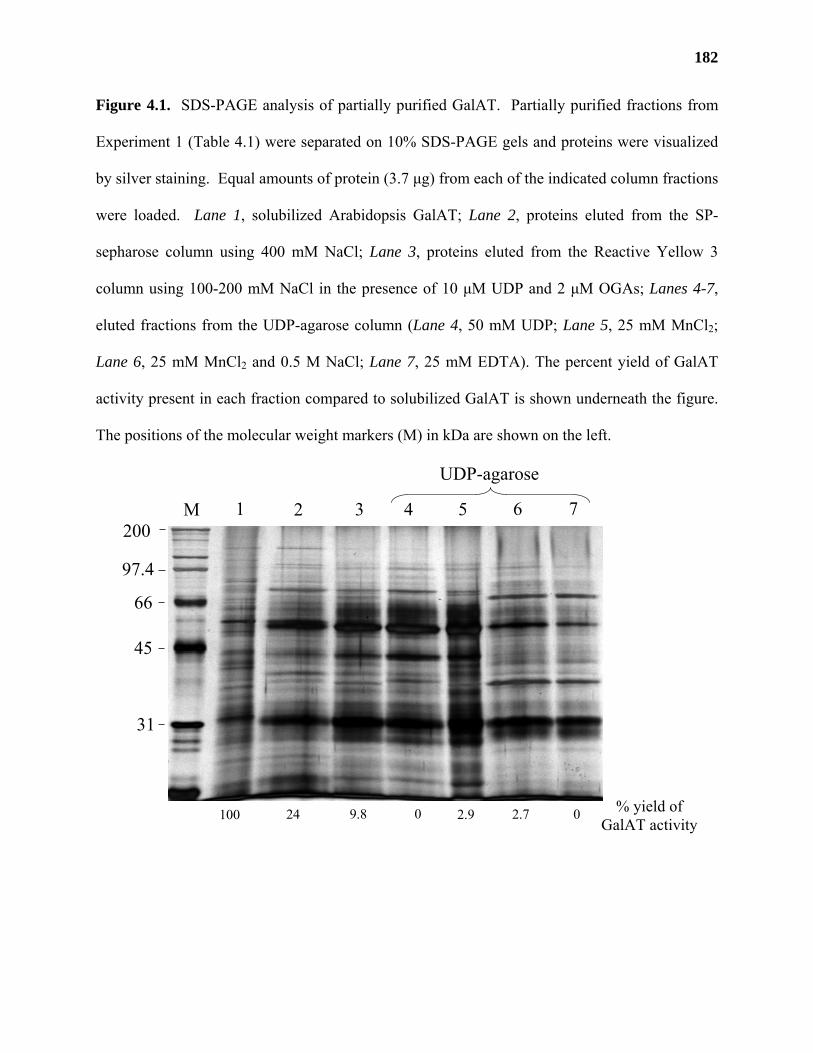
182
Figure 4.1. SDS-PAGE analysis of partially purified GalAT. Partially purified fractions from
Experiment 1 (Table 4.1) were separated on 10% SDS-PAGE gels and proteins were visualized
by silver staining. Equal amounts of protein (3.7 µg) from each of the indicated column fractions
were loaded. Lane 1, solubilized Arabidopsis GalAT; Lane 2, proteins eluted from the SP-
sepharose column using 400 mM NaCl; Lane 3, proteins eluted from the Reactive Yellow 3
column using 100-200 mM NaCl in the presence of 10 µM UDP and 2 µM OGAs; Lanes 4-7,
eluted fractions from the UDP-agarose column (Lane 4, 50 mM UDP; Lane 5, 25 mM MnCl2;
Lane 6, 25 mM MnCl2 and 0.5 M NaCl; Lane 7, 25 mM EDTA). The percent yield of GalAT
activity present in each fraction compared to solubilized GalAT is shown underneath the figure.
The positions of the molecular weight markers (M) in kDa are shown on the left.
% yield of GalAT activity
200
97.4
66
45
31
1 2 3 4 5 6M 7
100 24 9.8 0 2.9 2.7 0
UDP-agarose
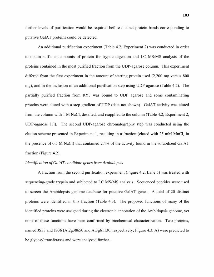
183
further levels of purification would be required before distinct protein bands corresponding to
putative GalAT proteins could be detected.
An additional purification experiment (Table 4.2, Experiment 2) was conducted in order
to obtain sufficient amounts of protein for tryptic digestion and LC MS/MS analysis of the
proteins contained in the most purified fraction from the UDP-agarose column. This experiment
differed from the first experiment in the amount of starting protein used (2,200 mg versus 800
mg), and in the inclusion of an additional purification step using UDP-agarose (Table 4.2). The
partially purified fraction from RY3 was bound to UDP agarose and some contaminating
proteins were eluted with a step gradient of UDP (data not shown). GalAT activity was eluted
from the column with 1 M NaCl, desalted, and reapplied to the column (Table 4.2, Experiment 2,
UDP-agarose [1]). The second UDP-agarose chromatography step was conducted using the
elution scheme presented in Experiment 1, resulting in a fraction (eluted with 25 mM MnCl2 in
the presence of 0.5 M NaCl) that contained 2.4% of the activity found in the solubilized GalAT
fraction (Figure 4.2).
Identification of GalAT candidate genes from Arabidopsis
A fraction from the second purification experiment (Figure 4.2, Lane 5) was treated with
sequencing-grade trypsin and subjected to LC MS/MS analysis. Sequenced peptides were used
to screen the Arabidopsis genome database for putative GalAT genes. A total of 20 distinct
proteins were identified in this fraction (Table 4.3). The proposed functions of many of the
identified proteins were assigned during the electronic annotation of the Arabidopsis genome, yet
none of these functions have been confirmed by biochemical characterization. Two proteins,
named JS33 and JS36 (At2g38650 and At3g61130, respectively; Figure 4.3, A) were predicted to
be glycosyltransferases and were analyzed further.
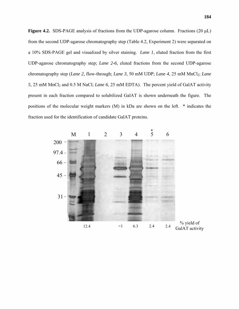
184
Figure 4.2. SDS-PAGE analysis of fractions from the UDP-agarose column. Fractions (20 µL)
from the second UDP-agarose chromatography step (Table 4.2, Experiment 2) were separated on
a 10% SDS-PAGE gel and visualized by silver staining. Lane 1, eluted fraction from the first
UDP-agarose chromatography step; Lane 2-6, eluted fractions from the second UDP-agarose
chromatography step (Lane 2, flow-through; Lane 3, 50 mM UDP; Lane 4, 25 mM MnCl2; Lane
5, 25 mM MnCl2 and 0.5 M NaCl; Lane 6, 25 mM EDTA). The percent yield of GalAT activity
present in each fraction compared to solubilized GalAT is shown underneath the figure. The
positions of the molecular weight markers (M) in kDa are shown on the left. * indicates the
fraction used for the identification of candidate GalAT proteins.
*
% yield of GalAT activity
200
97.4
66
45
31
1 2 3 4 5 6M
12.4 6.3 2.4 2.4<1
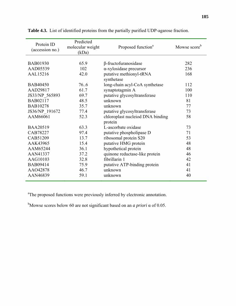
185
Table 4.3. List of identified proteins from the partially purified UDP-agarose fraction.
Protein ID (accession no.)
Predicted molecular weight
(kDa) Proposed functiona Mowse scoreb
BAB01930 65.9 β-fructofuranosidase 282 AAD05539 102 α-xylosidase precursor 236 AAL15216 42.0 putative methionyl-tRNA
synthetase 168
BAB40450 76..6 long-chain acyl-CoA synthetase 112 AAD29817 61.7 synaptotagmin A 100 JS33/NP_565893 69.7 putative glycosyltransferase 110 BAB02117 48.5 unknown 81 BAB10278 35.7 unknown 77 JS36/NP_191672 77.4 putative glycosyltransferase 73 AAM66061 52.3 chloroplast nucleiod DNA binding
protein 58
BAA20519 63.3 L-ascorbate oxidase 73 CAB78227 97.4 putative phospholipase D 71 CAB51209 13.7 ribosomal protein S20 53 AAK43965 15.4 putative HMG protein 48 AAM65244 36.1 hypothetical protein 48 AAN41337 37.2 quinone reductase-like protein 46 AAG10103 32.8 fibrillarin 1 42 BAB09414 75.9 putative ATP-binding protein 41 AAO42878 46.7 unknown 41 AAN46839 59.1 unknown 40
aThe proposed functions were previously inferred by electronic annotation.
bMowse scores below 60 are not significant based on an a priori α of 0.05.
186
Figure 4.3. Alignment of the amino acids sequences of JS33 and JS36. (A) The amino acid
sequences of JS33 and JS36 were aligned using ClustalX (version 1.83). The peptides obtained
by tryptic digestion of proteins found in the most purified fraction from UDP-agarose
chromatography are underlined. The site of N-terminal truncation is indicated by the arrows.
The conserved DxD motif found in both sequences is boxed. (B) Schematic representation of the
gene constructs used in the expression of the JS genes in HEK293 cells. The locations of the N-
terminal Trypanosoma cruzi mannosidase signal sequence (TCMss), the poly-histidine tag
(10xHis-tag) and 2 copies of the hemagluttinin epitope tag (HA-epitope tag) are indicated.
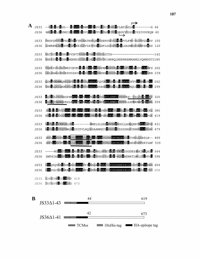
187
JS33 --MKGGGGGGG---GGGGGKRRWKVLVIGVLVLVILSMLVPLAFLLGLHN---------G 46
JS36 MALKRGLSGVNRIRGSGGGSRSVLVLLIFFCVFAPLCFFVGRGVYIDSSNDYSIVSVKQN 60
JS33 FHSPGFVTVQPASSFESFTRINATKHTQRDVSERVDEVLQKINPVLPKK-SDINVGSRDV 105
JS36 LDWRERLAMQSVRSLFSKEILDVIATSTADLGPLSLDSFKKNNLSASWRGTGVDPSFRHS 120
JS33 NATSGTDSKKRGLPVSPTVVANPSPANKTKSEASYTG-----------------------142
JS36 ENPATPDVKSNNLNEKRDSISKDSIHQKVETPTKIHRRQLREKRREMRANELVQHNDDTI 180
JS33 VQRKIVSGDETWRTCEVKYGSYCLWREENKEPMKDAKVKQMKDQLFVARAYYPSIAKMPS 202
JS36 LKLENAAIERSKSVDSAVLGKYSIWRRENENDNSDSNIRLMRDQVIMARVYS-GIAKLKN 239
JS33 QSKLTRDMKQNIQEFERILSESSQDADLPPQVDKKLQKMEAVIAKAKSFPVDCNNVDKKL 262
JS36 KNDLLQELQARLKDSQRVLGEATSDADLPRSAHEKLRAMGQVLAKAKMQLYDCKLVTGKL 299
JS33 RQILDLTEDEASFHMKQSVFLYQLAVQTMPKSLHCLSMRLTVEHFKS--DSLEDPISEKF 320
JS36 RAMLQTADEQVRSLKKQSTFLAQLAAKTIPNPIHCLSMRLTIDYYLLSPEKRKFPRSENL 359
JS33 SDPSLLHFVIISDNILASSVVINSTVVHARDSKNFVFHVLTDEQNYFAMKQWFIRNPCKQ 380
JS36 ENPNLYHYALFSDNVLAASVVVNSTIMNAKDPSKHVFHLVTDKLNFGAMNMWFLLNPPGK 419
JS33 STVQVLNIEKLELDDS---------DMKLSLSAEFRVSFPSGDLLASQQNRTHYLSLFSQ 431
JS36 ATIHVENVDEFKWLNSSYCPVLRQLESAAMREYYFKADHPTSGSSNLKYRNPKYLSMLNH 479
JS33 SHYLLPKLFDKLEKVVILDDDVVVQRDLSPLWDLDMEGKVNGAVKSCTVRLGQLRSLK-- 489
JS36 LRFYLPEVYPKLNKILFLDDDIIVQKDLTPLWEVNLNGKVNGAVETCGESFHRFDKYLNF 539
JS33 -----RGNFDTNACLWMSGLNVVDLARWRALGVSETYQKYYKEMSSGDESSEAIALQASL 544
JS36 SNPHIARNFNPNACGWAYGMNMFDLKEWKKRDITGIYHK-WQNMNENRTLWKLGTLPPGL 598
JS33 LTFQDQVYALDDKWALSGLGYDYYINAQAIKNAAILHYNGNMKPWLELGIPNYKNYWRRH 604
JS36 ITFYGLTHPLNKAWHVLGLGYNPSIDKKDIENAAVVHYNGNMKPWLELAMSKYRPYWTKY 658
JS33 LSREDRFLSDCNVNP 619
JS36 IKFDHPYLRRCNLHE 673
JS33 --MKGGGGGGG---GGGGGKRRWKVLVIGVLVLVILSMLVPLAFLLGLHN---------G 46
JS36 MALKRGLSGVNRIRGSGGGSRSVLVLLIFFCVFAPLCFFVGRGVYIDSSNDYSIVSVKQN 60
JS33 FHSPGFVTVQPASSFESFTRINATKHTQRDVSERVDEVLQKINPVLPKK-SDINVGSRDV 105
JS36 LDWRERLAMQSVRSLFSKEILDVIATSTADLGPLSLDSFKKNNLSASWRGTG
JS33 --MKGGGGGGG---GGGGGKRRWKVLVIGVLVLVILSMLVPLAFLLGLHN---------G 46
JS36 MALKRGLSGVNRIRGSGGGSRSVLVLLIFFCVFAPLCFFVGRGVYIDSSNDYSIVSVKQN 60
JS33 FHSPGFVTVQPASSFESFTRINATKHTQRDVSERVDEVLQKINPVLPKK-SDINVGSRDV 105
JS36 LDWRERLAMQSVRSLFSKEILDVIATSTADLGPLSLDSFKKNNLSASWRGTGVDPSFRHS 120
JS33 NATSGTDSKKRGLPVSPTVVANPSPANKTKSEASYTG-----------------------142
JS36 ENPATPDVKSNNLNEKRDSISKDSIHQKVETPTKIHRRQLREKRREMRANELVQHNDDTI 180
JS33 VQRKIVSGDETWRTCEVKYGSYCLWREENKEPMKDAKVKQMKDQLFVARAYYPSIAKMPS 202
JS36 LKLENAAIERSKSVDSAVLGKYSIWRRENENDNSDSNIRLMRDQVIMARVYS-GIAKLKN 239
VDPSFRHS 120
JS33 NATSGTDSKKRGLPVSPTVVANPSPANKTKSEASYTG-----------------------142
JS36 ENPATPDVKSNNLNEKRDSISKDSIHQKVETPTKIHRRQLREKRREMRANELVQHNDDTI 180
JS33 VQRKIVSGDETWRTCEVKYGSYCLWREENKEPMKDAKVKQMKDQLFVARAYYPSIAKMPS 202
JS36 LKLENAAIERSKSVDSAVLGKYSIWRRENENDNSDSNIRLMRDQVIMARVYS-GIAKLKN 239
JS33 QSKLTRDMKQNIQEFERILSESSQDADLPPQVDKKLQKMEAVIAKAKSFPVDCNNVDKKL 262
JS36 KNDLLQELQARLKDSQRVLGEATSDADLPRSAHEKLRAMGQVLAKAKMQLYDCKLVTGKL 299
JS33 RQILDLTEDEASFHMKQSVFLYQLAVQTMPKSLHCLSMRLTVEHFKS--DSLEDPISEKF 320
JS36 RAML
JS33 QSKLTRDMKQNIQEFERILSESSQDADLPPQVDKKLQKMEAVIAKAKSFPVDCNNVDKKL 262
JS36 KNDLLQELQARLKDSQRVLGEATSDADLPRSAHEKLRAMGQVLAKAKMQLYDCKLVTGKL 299
JS33 RQILDLTEDEASFHMKQSVFLYQLAVQTMPKSLHCLSMRLTVEHFKS--DSLEDPISEKF 320
JS36 RAMLQTADEQVRSLKKQSTFLAQLAAKTIPNPIHCLSMRLTIDYYLLSPEKRKFPRSENL 359
JS33 SDPSLLHFVIISDNILASSVVINSTVVHARDSKNFVFHVLTDEQNYFAMKQWFIRNPCKQ 380
JS36 ENPNLYHYALFSDNVLAASVVVNSTIMNAKDPSKHVFHLVTDKLNFGAMNMWFLLNPPGK 419
JS33 ST
QTADEQVRSLKKQSTFLAQLAAKTIPNPIHCLSMRLTIDYYLLSPEKRKFPRSENL 359
JS33 SDPSLLHFVIISDNILASSVVINSTVVHARDSKNFVFHVLTDEQNYFAMKQWFIRNPCKQ 380
JS36 ENPNLYHYALFSDNVLAASVVVNSTIMNAKDPSKHVFHLVTDKLNFGAMNMWFLLNPPGK 419
JS33 STVQVLNIEKLELDDS---------DMKLSLSAEFRVSFPSGDLLASQQNRTHYLSLFSQ 431
JS36 ATIHVENVDEFKWLNSSYCPVLRQLESAAMREYYFKADHPTSGSSNLKYRNPKYLSMLNH 479
JS33 SHYLLPKLFDKLEKVVILDDDVVVQRDLSPLWDLDMEGKVNGAVKSCTVRLGQLRSLK-- 489
JS36 LRFYLPEVYPKLNKILFLDDDIIVQKDLTPLWEVNLNGKVNGAVETCGESFHRFDKYLNF 539
JS33 -----RGNFDTNACLW
VQVLNIEKLELDDS---------DMKLSLSAEFRVSFPSGDLLASQQNRTHYLSLFSQ 431
JS36 ATIHVENVDEFKWLNSSYCPVLRQLESAAMREYYFKADHPTSGSSNLKYRNPKYLSMLNH 479
JS33 SHYLLPKLFDKLEKVVILDDDVVVQRDLSPLWDLDMEGKVNGAVKSCTVRLGQLRSLK-- 489
JS36 LRFYLPEVYPKLNKILFLDDDIIVQKDLTPLWEVNLNGKVNGAVETCGESFHRFDKYLNF 539
JS33 -----RGNFDTNACLWMSGLNVVDLARWRALGVSETYQKYYKEMSSGDESSEAIALQASL 544
JS36 SNPHIARNFNPNACGWAYGMNMFDLKEWKKRDITGIYHK-WQNMNENRTLWKLGTLPPGL 598
JS33 LTFQDQVYALDDKWALSGLGYDYYINAQAIKNAAILHYNGNMKPWLELGIPNYKNYWRRH 604
JS36 ITFYGLTHPLNKAWHVLGLGYNPSIDKKDIENAAVVHYNGNMKPWLELAMSKY
MSGLNVVDLARWRALGVSETYQKYYKEMSSGDESSEAIALQASL 544
JS36 SNPHIARNFNPNACGWAYGMNMFDLKEWKKRDITGIYHK-WQNMNENRTLWKLGTLPPGL 598
JS33 LTFQDQVYALDDKWALSGLGYDYYINAQAIKNAAILHYNGNMKPWLELGIPNYKNYWRRH 604
JS36 ITFYGLTHPLNKAWHVLGLGYNPSIDKKDIENAAVVHYNGNMKPWLELAMSKYRPYWTKY 658
JS33 LSREDRFLSDCNVNP 619
JS36 IKFDHPYLRRCNLHE 673
JS33∆1-43
JS36∆1-41
44 619
HA-epitope tag10xHis-tag
67342
TCMss
B
A
188
JS33 and JS36 encode predicted type II membrane proteins (TMpred,
http://www.ch.embnet.org/software/TMPRED_form.html) whose C-terminal catalytic domains
possess sequence identity to a consensus sequence found in family 8 glycosyltransferases
(http://afmb.cnrs-mrs.fr/CAZY; Henrissat et al., 2001). Members of family 8 (which include
galactinol synthase and lipopolysaccharide galactosyltransferase) are retaining
glycosyltransferases able to transfer the glycosyl residue from NDP-α-sugars onto α-linked
products (Saxena et al., 1995). The predicted catalytic domains of JS33 and JS36 also contain
putative DxD motifs (Figure 4.3, A); a conserved motif found in many well-characterized
glycosyltransferase families (Wiggins and Munro, 1998) and are involved in the coordination of
divalent cations required for glycosyltransferase activity (Breton and Imberty, 1999; Chen et al.,
2001).
Previous studies on a GalAT from pea (Pisum sativum) showed that GalAT activity was
localized to Golgi vesicles with its catalytic domain facing the lumen (Sterling et al., 2001). This
topology is characteristic of type II membrane proteins, which is the predicted topology of JS33
and JS36. Furthermore, preliminary RT-PCR analysis of the expression of JS33 and JS36
transcripts in Arabidopsis tissues showed that JS33 and JS36 were found in all Arabidopsis
tissues that were analyzed (Figure 4.4). These findings suggested that JS33 and JS36 were
candidates for GalATs genes and we decided to clone their respective cDNAs and express them
in a heterologous system in order to prove their function.
N-terminally truncated JS36 exhibits GalAT-like characteristics when transiently expressed in
HEK293 cells
cDNA constructs containing N-terminally truncated versions of JS33 (JS33∆1-43) or
JS36 (JS36∆1-41) were generated (Figure 4.3, A, B). The cDNAs were placed downstream of a
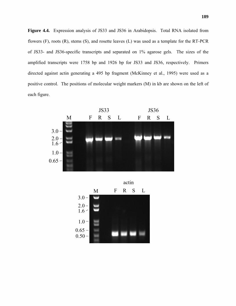
189
Figure 4.4. Expression analysis of JS33 and JS36 in Arabidopsis. Total RNA isolated from
flowers (F), roots (R), stems (S), and rosette leaves (L) was used as a template for the RT-PCR
of JS33- and JS36-specific transcripts and separated on 1% agarose gels. The sizes of the
amplified transcripts were 1758 bp and 1926 bp for JS33 and JS36, respectively. Primers
directed against actin generating a 495 bp fragment (McKinney et al., 1995) were used as a
positive control. The positions of molecular weight markers (M) in kb are shown on the left of
each figure.
JS33 JS36F R S L F R S L
2.01.6
1.00.65
3.0
M
F R S Lactin
2.01.6
1.00.65
3.0
0.50
M
190
signal sequence from Trypanosoma cruzi mannosidase, a poly-histidine tag, and two copies of
the hemagluttinin epitope (Figure 4.3, B) in the mammalian expression vector pEAK10. These
constructs were used to transiently transfect HEK293 cells that were 50% confluent (4 d old).
Media harvested from these cells 48 h post-transfection were tested for GalAT activity (Figure
4.5, A) and the data was analyzed for its significance compared to controls using a two sample,
one-tailed Student’s t test and an a priori α of 0.05 (95% confidence).
Media from cells transfected with the JS36∆1-41 construct were not able to promote the
significant incorporation of D-[14C]GalpA from UDP-D-[14C]GalpA into a radiolabeled product
compared to cells transfected with empty vector (n = 2, tJS36 = 1.589 [P ≈ 0.1]). In contrast,
media from JS36∆1-41-transfected cells that had been immunoprecipitated using anti-
hemagluttinin (anti-HA) antibodies and protein A-Sepharose gave 3 times greater incorporation
of radiolabel than that of the empty vector controls (n =2, tJS36 = 10.284 [P ≈ 0.005]). Neither
media (tJS33 = 0.057 [P > 0.25]) nor immunoprecipitates (tJS33 = 0.180 [P > 0.25]) of cells
transfected with the JS33∆1-43 construct produced incorporation levels that were significantly
different from cells transfected with empty vector. These results suggested that JS36 encoded an
enzyme capable of catalyzing the transfer of D-[14C]GalpA onto a product and based on these
results we proposed that JS36 encoded a putative GalAT.
Analysis of a second set of HEK293 cells transiently transfected with the same cDNA
constructs; however, did not result in GalAT activity in immunoprecipitated media (data not
shown). The second transient transfection experiment was conducted in a manner similar, but
not identical, to that of the first transfection experiment. Both experiments used HEK293 cells
that were 50% confluent and media and cell lysates were harvested 48 h post-transfection.
However, different buffers were used to prepare the media and cell lysates in the second transient
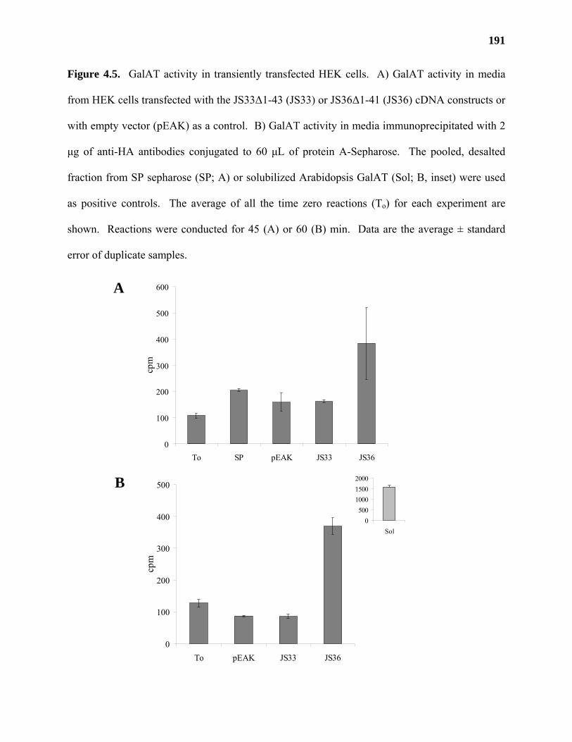
191
Figure 4.5. GalAT activity in transiently transfected HEK cells. A) GalAT activity in media
from HEK cells transfected with the JS33∆1-43 (JS33) or JS36∆1-41 (JS36) cDNA constructs or
with empty vector (pEAK) as a control. B) GalAT activity in media immunoprecipitated with 2
µg of anti-HA antibodies conjugated to 60 µL of protein A-Sepharose. The pooled, desalted
fraction from SP sepharose (SP; A) or solubilized Arabidopsis GalAT (Sol; B, inset) were used
as positive controls. The average of all the time zero reactions (To) for each experiment are
shown. Reactions were conducted for 45 (A) or 60 (B) min. Data are the average ± standard
error of duplicate samples.
A
B
0
100
200
300
400
500
To pEAK JS33 JS36
cpm
0500
100015002000
Sol
0
100
200
300
400
500
600
To SP pEAK JS33 JS36
cpm
192
experiment. Protease inhibitors were added to the harvested media and different washing buffers
were used for the immunoprecipitation of recombinant proteins from media and cell lysates. The
washing buffer used in the second experiment contained 50 mM HEPES, pH 7.3, 0.25 mM
MnCl2, 1% Triton X-100, 2 mM EDTA, and 25% [v/v] glycerol compared to 0.1% [w/v] TX-
100, 50 mM Tris/Cl, pH 7.4, and 300 mM NaCl used in the first experiment. Both treatments
were shown to be compatible with solubilized Arabidopsis GalAT activity. At this point, it is
unclear whether the lack of GalAT activity in the second transient transfection experiment was
due to the use of different buffers or due to other, as yet unknown, factors.
Western blot analysis of media and cell lysates from the second transient transfection
experiment indicated the presence of recombinant JS33 and JS36 proteins in the media of
transfected cells, and recombinant JS33 protein in cell lysates (Figure 4.6). The estimated
molecular weights of JS33 (86 kDa) and JS36 (88 kDa) were much higher than those calculated
from their amino acid sequences (73.2 kDa and 80.7 kDa for JS33 and JS36, respectively),
suggesting that both proteins were post-translationally modified.
The immunoblot results suggested that the cells lines were transfected with the JS cDNA
constructs and that they were capable of secreting the recombinant proteins into the media as
expected. The level of recombinant protein present in the media from cells transfected with JS36
was significantly lower than that of cells transfected with JS33. The reason for this disparity in
recombinant protein levels is unknown. In order to further study JS36 and JS33, and to increase
the level of recombinant JS36 protein made by HEK cell lines, we decided to create stable lines
expressing JS36 and JS33.
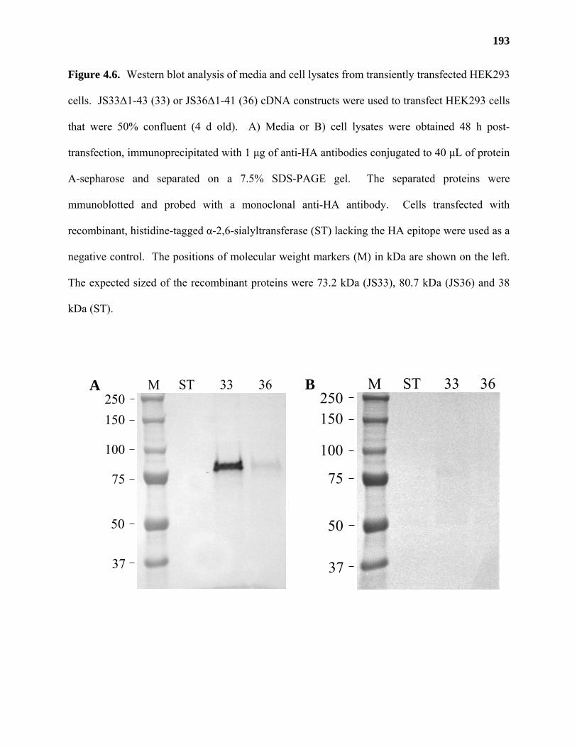
193
Figure 4.6. Western blot analysis of media and cell lysates from transiently transfected HEK293
cells. JS33∆1-43 (33) or JS36∆1-41 (36) cDNA constructs were used to transfect HEK293 cells
that were 50% confluent (4 d old). A) Media or B) cell lysates were obtained 48 h post-
transfection, immunoprecipitated with 1 µg of anti-HA antibodies conjugated to 40 µL of protein
A-sepharose and separated on a 7.5% SDS-PAGE gel. The separated proteins were
mmunoblotted and probed with a monoclonal anti-HA antibody. Cells transfected with
recombinant, histidine-tagged α-2,6-sialyltransferase (ST) lacking the HA epitope were used as a
negative control. The positions of molecular weight markers (M) in kDa are shown on the left.
The expected sized of the recombinant proteins were 73.2 kDa (JS33), 80.7 kDa (JS36) and 38
kDa (ST).
A B250150
100
75
50
37
M ST 33 36250150
100
75
50
37
M ST 33 36
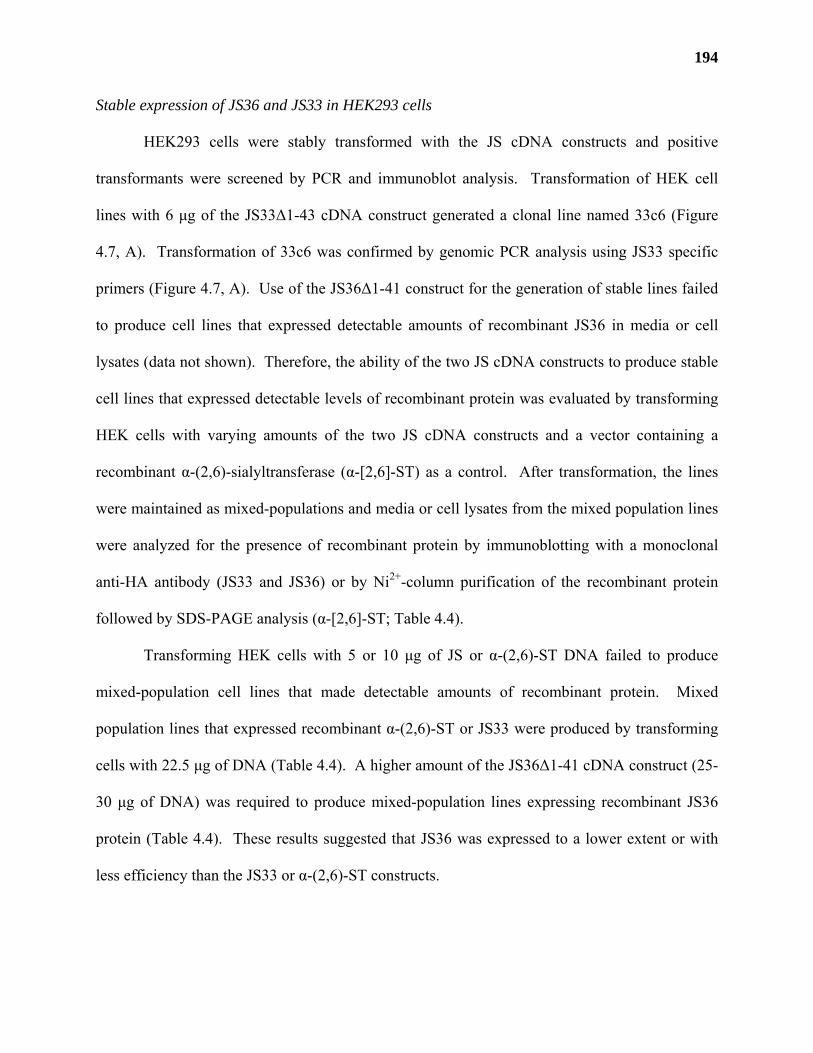
194
Stable expression of JS36 and JS33 in HEK293 cells
HEK293 cells were stably transformed with the JS cDNA constructs and positive
transformants were screened by PCR and immunoblot analysis. Transformation of HEK cell
lines with 6 µg of the JS33∆1-43 cDNA construct generated a clonal line named 33c6 (Figure
4.7, A). Transformation of 33c6 was confirmed by genomic PCR analysis using JS33 specific
primers (Figure 4.7, A). Use of the JS36∆1-41 construct for the generation of stable lines failed
to produce cell lines that expressed detectable amounts of recombinant JS36 in media or cell
lysates (data not shown). Therefore, the ability of the two JS cDNA constructs to produce stable
cell lines that expressed detectable levels of recombinant protein was evaluated by transforming
HEK cells with varying amounts of the two JS cDNA constructs and a vector containing a
recombinant α-(2,6)-sialyltransferase (α-[2,6]-ST) as a control. After transformation, the lines
were maintained as mixed-populations and media or cell lysates from the mixed population lines
were analyzed for the presence of recombinant protein by immunoblotting with a monoclonal
anti-HA antibody (JS33 and JS36) or by Ni2+-column purification of the recombinant protein
followed by SDS-PAGE analysis (α-[2,6]-ST; Table 4.4).
Transforming HEK cells with 5 or 10 µg of JS or α-(2,6)-ST DNA failed to produce
mixed-population cell lines that made detectable amounts of recombinant protein. Mixed
population lines that expressed recombinant α-(2,6)-ST or JS33 were produced by transforming
cells with 22.5 µg of DNA (Table 4.4). A higher amount of the JS36∆1-41 cDNA construct (25-
30 µg of DNA) was required to produce mixed-population lines expressing recombinant JS36
protein (Table 4.4). These results suggested that JS36 was expressed to a lower extent or with
less efficiency than the JS33 or α-(2,6)-ST constructs.
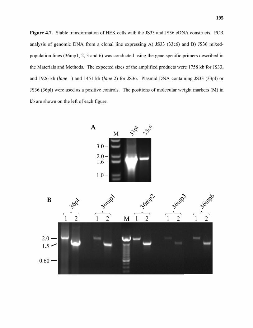
195
Figure 4.7. Stable transformation of HEK cells with the JS33 and JS36 cDNA constructs. PCR
analysis of genomic DNA from a clonal line expressing A) JS33 (33c6) and B) JS36 mixed-
population lines (36mp1, 2, 3 and 6) was conducted using the gene specific primers described in
the Materials and Methods. The expected sizes of the amplified products were 1758 kb for JS33,
and 1926 kb (lane 1) and 1451 kb (lane 2) for JS36. Plasmid DNA containing JS33 (33pl) or
JS36 (36pl) were used as a positive controls. The positions of molecular weight markers (M) in
kb are shown on the left of each figure.
A
B
36pl
1 2
36mp1
1 2
36mp2
1 2
36mp3
1 2M
36mp6
1 2
2.01.5
0.60
33pl
33c6
2.01.6
1.0
3.0
M
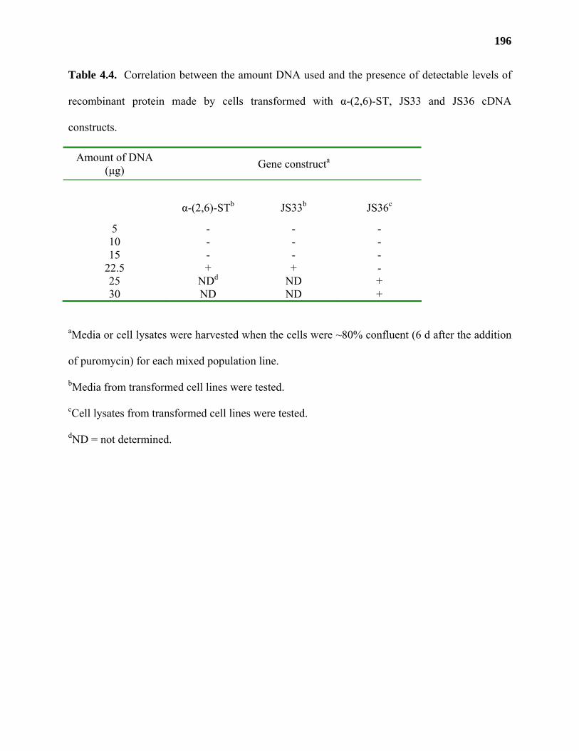
196
Table 4.4. Correlation between the amount DNA used and the presence of detectable levels of
recombinant protein made by cells transformed with α-(2,6)-ST, JS33 and JS36 cDNA
constructs.
Amount of DNA (µg) Gene constructa
α-(2,6)-STb JS33b JS36c
5 - - - 10 - - - 15 - - -
22.5 + + - 25 NDd ND + 30 ND ND +
aMedia or cell lysates were harvested when the cells were ~80% confluent (6 d after the addition
of puromycin) for each mixed population line.
bMedia from transformed cell lines were tested.
cCell lysates from transformed cell lines were tested.
dND = not determined.
197
The presence of JS36 in mixed-population lines generated with 25 or 30 µg of the
JS36∆1-41 cDNA construct was determined by PCR analysis of genomic DNA using JS36-
specific primers. Four mixed-population lines (36mp1-3, 6) were selected based on the presence
of PCR products (1.4 kb and 1.9 kb) of the expected size (Figure 4.7, B). The production of
mRNA transcripts in these mixed-population lines was confirmed by RT-PCR analysis using
JS36-specific primers (Figure 4.8). RT-PCR products of the expected sizes were generated in all
four of the JS36 mixed-population lines that were analyzed, suggesting that these lines all
produced mRNA transcripts encoding JS36. PCR reactions performed in the absence of reverse
transcriptase gave no product, which excluded the possibility of genomic DNA contamination
(Figure 4.8, lane 5). These results confirmed that we had successfully transformed HEK293
cells with gene constructs containing truncated versions of JS33 and JS36.
Stable lines expressing recombinant JS36 and JS33 proteins are detected in cell lysates
In contrast to the results obtained from the transient transfection experiments,
recombinant JS36 and JS33 proteins were not detected in media from stably transformed lines
(data not shown). High levels of recombinant protein were instead detected in cell lysates
(Figure 4.9). The estimated molecular weight of recombinant JS33 protein from the 33c6 cell
line (Figure 4.9, A) was similar to that detected in media from transiently transfected cells.
Recombinant JS36 protein from all 4 mixed-population lines migrated as a doublet, with
predicted molecular weights of 87 and 89 kDa, respectively (Figure 4.9, B). The predicted
molecular weights of both recombinant JS36 and JS33 were higher than that deduced by the
amino acid sequence, suggesting that both proteins were post-translationally modified in stable
and/or mixed population lines.
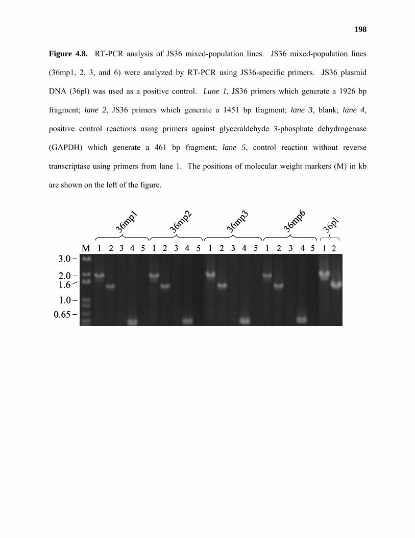
198
Figure 4.8. RT-PCR analysis of JS36 mixed-population lines. JS36 mixed-population lines
(36mp1, 2, 3, and 6) were analyzed by RT-PCR using JS36-specific primers. JS36 plasmid
DNA (36pl) was used as a positive control. Lane 1, JS36 primers which generate a 1926 bp
fragment; lane 2, JS36 primers which generate a 1451 bp fragment; lane 3, blank; lane 4,
positive control reactions using primers against glyceraldehyde 3-phosphate dehydrogenase
(GAPDH) which generate a 461 bp fragment; lane 5, control reaction without reverse
transcriptase using primers from lane 1. The positions of molecular weight markers (M) in kb
are shown on the left of the figure.
1 2 3 4 5 1 2 3 4 5 1 2 3 4 5 1 2 3 4 5
36mp1
36mp2
36mp3
36mp6
2.01.61.0
0.65
3.0M 1 2 3 4 5 1 2 3 4 5 1 2 3 4 5 1 2 3 4 5
36mp1
36mp2
36mp3
36mp6
2.01.61.0
0.65
3.0M 1 2
36pl
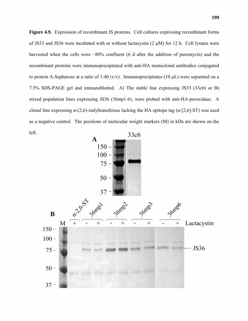
199
Figure 4.9. Expression of recombinant JS proteins. Cell cultures expressing recombinant forms
of JS33 and JS36 were incubated with or without lactacystin (2 µM) for 12 h. Cell lysates were
harvested when the cells were ~80% confluent (6 d after the addition of puromycin) and the
recombinant proteins were immunoprecipitated with anti-HA monoclonal antibodies conjugated
to protein A-Sepharose at a ratio of 1:40 (v/v). Immunoprecipitates (10 µL) were separated on a
7.5% SDS-PAGE gel and immunoblotted. A) The stable line expressing JS33 (33c6) or B)
mixed population lines expressing JS36 (36mp1-6), were probed with anti-HA-peroxidase. A
clonal line expressing α-(2,6)-sialyltransferase lacking the HA epitope tag (α-[2,6]-ST) was used
as a negative control. The positions of molecular weight markers (M) in kDa are shown on the
left.
A
B
150100
75
50
37
M - + - + Lactacystin- +
36mp1
36mp2
36mp3
- +
36mp6
α-2,6-
ST
+
JS36
150100
75
50
37
33c6
200
The fusion of the signal sequence from T. cruzi mannosidase with JS genes lacking their
predicted N-terminal transmembrane domain should have caused the recombinant JS proteins
from the stable lines to be secreted into the media. Therefore, the presence of recombinant JS
proteins in cell lysates indicated anomalous protein targeting in these stably transformed cell
lines. A growth curve conducted on 33c6 and 36mp2 showed that the level of recombinant
protein in cell lysates increased with increasing cell density (Figure 4.10). No recombinant
protein could be detected in media from either line at any stage of growth (data not shown),
indicating that the anomalous protein targeting was not affected by the age of the cells. The
anomalous protein targeting in JS36 mixed-population lines also did not seem to be due to
proteasome-driven degradation, since cells expressing recombinant JS36 protein grown in the
presence of 2 µM lactacystin, a potent proteasome inhibitor from Streptomyces lactacystinaeus,
had no effect on the amount of JS36 protein made, or on its migration on SDS-PAGE gels
(Figure 4.9, B). These results suggested that the stable lines that were generated were incapable
of secreting recombinant JS protein.
GalAT activity could not be detected in cell lysates from stably transformed lines
Cell lysates and immunoprecipitated cell lysates were prepared from stable lines
expressing recombinant JS33 and JS36 proteins and tested for GalAT activity (Figure 4.11, A
and B). Neither cell lysates (Figure 4.11, A) nor their immunoprecipitates (Figure 4.11, B)
catalyzed the transfer of D-[14C]GalpA from UDP-D-[14C]GalpA onto a radiolabeled product.
The results from these experiments suggested that recombinant JS proteins from cell lysates of
stable lines did not express GalAT activity.
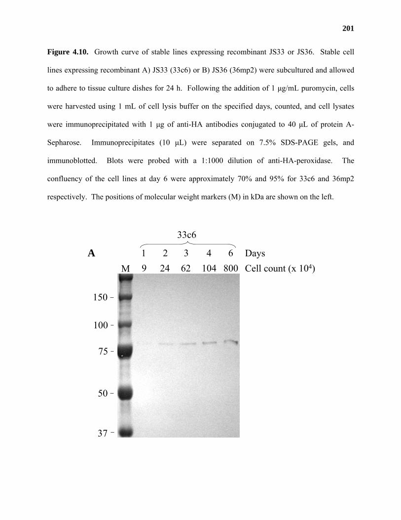
201
Figure 4.10. Growth curve of stable lines expressing recombinant JS33 or JS36. Stable cell
lines expressing recombinant A) JS33 (33c6) or B) JS36 (36mp2) were subcultured and allowed
to adhere to tissue culture dishes for 24 h. Following the addition of 1 µg/mL puromycin, cells
were harvested using 1 mL of cell lysis buffer on the specified days, counted, and cell lysates
were immunoprecipitated with 1 µg of anti-HA antibodies conjugated to 40 µL of protein A-
Sepharose. Immunoprecipitates (10 µL) were separated on 7.5% SDS-PAGE gels, and
immunoblotted. Blots were probed with a 1:1000 dilution of anti-HA-peroxidase. The
confluency of the cell lines at day 6 were approximately 70% and 95% for 33c6 and 36mp2
respectively. The positions of molecular weight markers (M) in kDa are shown on the left.
1 2 3 4 6 Days
150
100
75
50
37
M Cell count (x 104)9 24 62 104 800
33c6
A
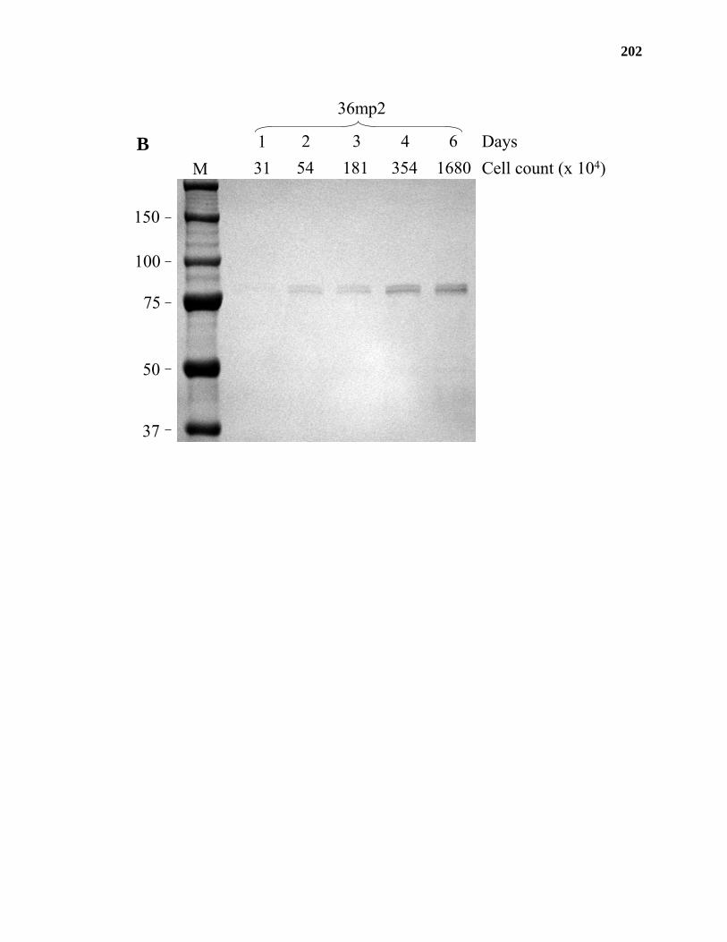
202
1 2 3 4 6 Days
150
100
75
50
37
M Cell count (x 104)31 54 181 354 1680
36mp2
B
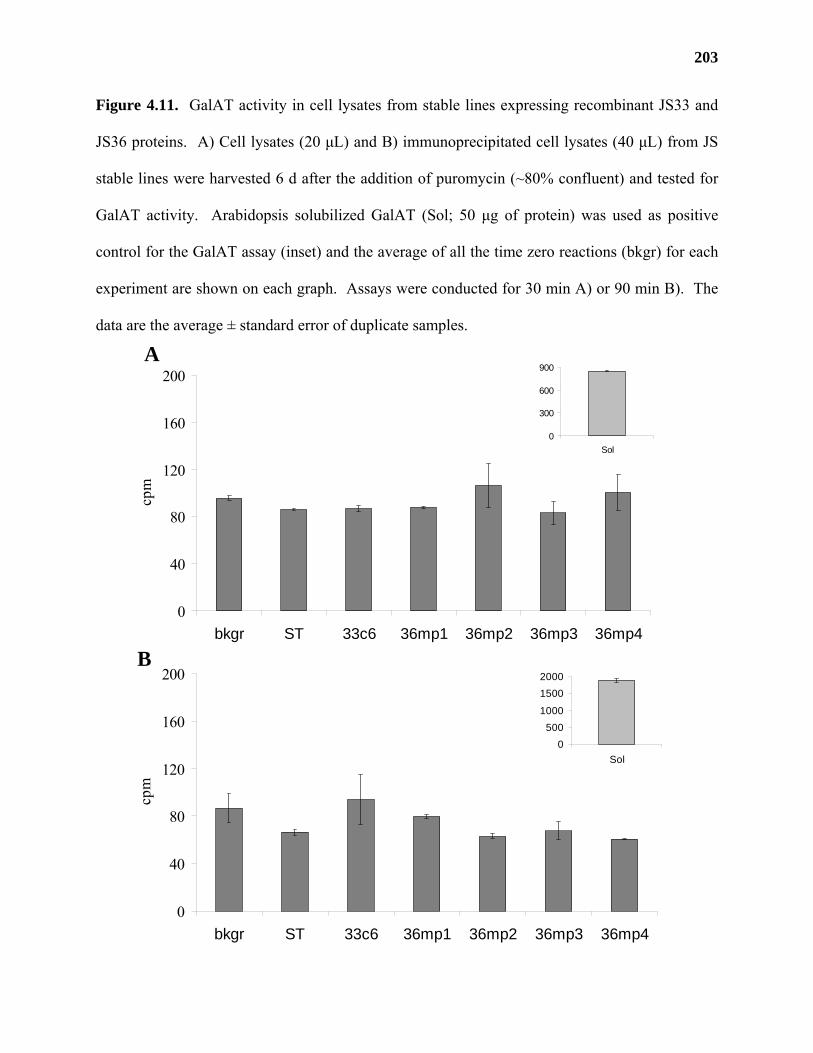
203
Figure 4.11. GalAT activity in cell lysates from stable lines expressing recombinant JS33 and
JS36 proteins. A) Cell lysates (20 µL) and B) immunoprecipitated cell lysates (40 µL) from JS
stable lines were harvested 6 d after the addition of puromycin (~80% confluent) and tested for
GalAT activity. Arabidopsis solubilized GalAT (Sol; 50 µg of protein) was used as positive
control for the GalAT assay (inset) and the average of all the time zero reactions (bkgr) for each
experiment are shown on each graph. Assays were conducted for 30 min A) or 90 min B). The
data are the average ± standard error of duplicate samples.
B
A
0
40
80
120
160
200
bkgr ST 33c6 36mp1 36mp2 36mp3 36mp4
cpm
0
300
600
900
Sol
0
40
80
120
160
200
bkgr ST 33c6 36mp1 36mp2 36mp3 36mp4
cpm
0500
1000
15002000
Sol
204
Construction and use of multiple antigenic peptide antibodies for the immunoprecipitation of
GalAT activity
As an additional means of proving the function of JS36 and JS33, selected regions of
JS36, JS33 and JS36L (At5g47780), an Arabidopsis protein with significant sequence identity to
JS36, were chosen for the production of multiple antigenic peptides (MAPs, Table 4.5). Each
MAP was used as an antigen for the immunization of two rabbits. Serum from each rabbit was
found to contain polyclonal antibodies that could specifically bind to the original MAP antigen
(data not shown). An initial experiment was conducted to determine whether any of the
polyclonal antibodies that were generated could immunoprecipitate GalAT activity from
solubilized Arabidopsis proteins. Polyclonal antibodies directed against JS36, JS33, and JS36L
were pooled and conjugated with protein A-Sepharose. The conjugates were then incubated with
an aliquot of solubilized proteins from Arabidopsis and the immunoprecipitated material was
washed and tested for GalAT activity using pooled preimmune serum as a negative control
(Figure 4.12).
The results from this experiment showed that none of the pooled antibodies were able to
immunoprecipitate significant amounts of GalAT activity (a priori α of 0.05) from solubilized
Arabidopsis proteins compared to their preimmune controls (Figure 4.12). The activity using the
pooled JS33 antibodies gave the greatest difference compared to the preimmune sera, but the
calculated t value (n =2, tJS33 = 2.483 [P > 0.05]) indicated that the activity was not significantly
different with 95% confidence. Our inability to obtain significant levels of immunoprecipitable
activity using pooled antibodies could be due to protein A-Sepharose, as its addition to
solubilized Arabidopsis proteins inhibited GalAT activity by approximately 40% (Figure 4.12).
Recent experiments demonstrated that some of the polyclonal antibodies generated against JS33
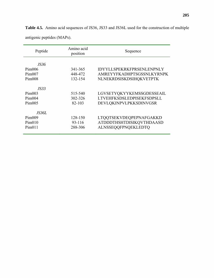
205
Table 4.5. Amino acid sequences of JS36, JS33 and JS36L used for the construction of multiple
antigenic peptides (MAPs).
Peptide Amino acid position Sequence
JS36
Pim006 341-365 IDYYLLSPEKRKFPRSENLENPNLY Pim007 448-472 AMREYYFKADHPTSGSSNLKYRNPK Pim008 132-154 NLNEKRDSISKDSIHQKVETPTK
JS33
Pim003 515-540 LGVSETYQKYYKEMSSGDESSEAIL Pim004 302-326 LTVEHFKSDSLEDPISEKFSDPSLL Pim005 82-103 DEVLQKINPVLPKKSDINVGSR
JS36L
Pim009 128-150 LTQQTSEKVDEQPEPNAFGAKKD Pim010 93-116 ATDDDTHSHTDISIKQVTHDAASD Pim011 288-306 ALNSSEQQFPNQEKLEDTQ
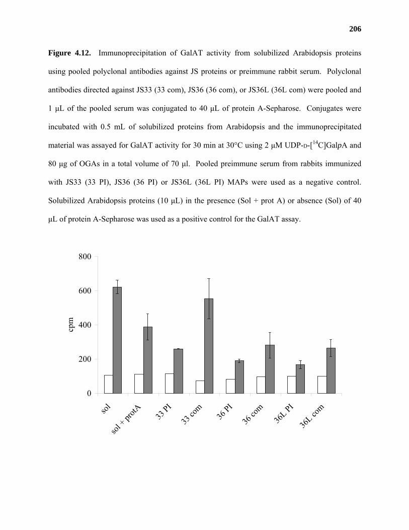
206
Figure 4.12. Immunoprecipitation of GalAT activity from solubilized Arabidopsis proteins
using pooled polyclonal antibodies against JS proteins or preimmune rabbit serum. Polyclonal
antibodies directed against JS33 (33 com), JS36 (36 com), or JS36L (36L com) were pooled and
1 µL of the pooled serum was conjugated to 40 µL of protein A-Sepharose. Conjugates were
incubated with 0.5 mL of solubilized proteins from Arabidopsis and the immunoprecipitated
material was assayed for GalAT activity for 30 min at 30°C using 2 µM UDP-D-[14C]GalpA and
80 µg of OGAs in a total volume of 70 µl. Pooled preimmune serum from rabbits immunized
with JS33 (33 PI), JS36 (36 PI) or JS36L (36L PI) MAPs were used as a negative control.
Solubilized Arabidopsis proteins (10 µL) in the presence (Sol + prot A) or absence (Sol) of 40
µL of protein A-Sepharose was used as a positive control for the GalAT assay.
0
200
400
600
800
sol
sol + pr
otA 33 PI
33 co
m36
PI
36 co
m36
L PI
36L co
m
cpm
207
or JS36 recognized the recombinant forms of these proteins made in cell lysates from 33c6 and
36mp2 cell lines (M. Atmodjo and D. Mohnen, unpublished results). New experiments are
currently being conducted using these specific antibodies which should enable us to
immunoprecipitate proteins from Arabidopsis extracts and determine whether they encode active
GalATs.
JS36 and JS33 are part of a putative multi-gene family in Arabidopsis
BLAST analysis of the Arabidopsis genome identified JS33 and an additional 13
GenBank sequences with between 36-68% sequence identity and 56-84% sequence similarity to
JS36 (Table 4.6). Further analysis revealed an additional 10 sequences with significant, but
reduced, sequence identity to JS36 (23-29% identity and 42-53% similarity). Similar to JS36
and JS33, all of these putative proteins belong to CAZy glycosyltransferase family 8 (Henrissat
et al., 2001).
Sequence alignment of all 25 amino acid sequences revealed the presence of highly
conserved domains, some of which were found in other glycosyltransferase families (Wiggins
and Munro, 1998), and include the putative DXD motif detected in JS33 and JS36 (Figure 4.13).
Based on the similarity of these sequences to JS36 and on the results obtained with the activity of
recombinant JS36 in transiently transfected HEK cells, we propose that all 25 genes are putative
GalATs and are part of a proposed 25-member GALAT (pGALAT) superfamily in Arabidopsis
(Table 4.6).
The 25 genes have been subdivided into 15 pGALAT and 10 pGALAT-like (pGALATL)
genes according to their sequence identity to JS36 (Table 4.6). Concurrent with this division,
many of the encoded proteins are predicted to contain a hypervariable N-terminal extension that
contains a putative transmembrane domain (http://www.ch.embnet.org/software/
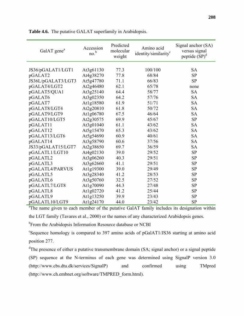
208
Table 4.6. The putative GALAT superfamily in Arabidopsis.
GalAT genea Accession no.b
Predicted molecular
weight
Amino acid identity/similarityc
Signal anchor (SA) versus signal peptide (SP)d
JS36/pGALAT1/LGT1 At3g61130 77.3 100/100 SA pGALAT2 At4g38270 77.8 68/84 SP JS36L/pGALAT3/LGT3 At5g47780 71.1 66/83 SP pGALAT4/LGT2 At2g46480 62.1 65/78 none pGALAT5/QUA1 At3g25140 64.4 58/77 SA pGALAT6 At3g02350 64.2 57/76 SA pGALAT7 At1g18580 61.9 51/71 SA pGALAT8/LGT4 At2g20810 61.8 50/72 SA pGALAT9/LGT9 At1g06780 67.5 46/64 SA pGALAT10/LGT5 At2g30575 69.9 45/67 SP pGALAT11 At3g01040 61.1 43/62 SA pGALAT12 At5g15470 65.3 43/62 SA pGALAT13/LGT6 At5g54690 60.9 40/61 SA pGALAT14 At3g58790 60.6 37/56 SA JS33/pGALAT15/LGT7 At2g38650 69.7 36/59 SA pGALATL1/LGT10 At4g02130 39.0 29/52 SP pGALATL2 At3g06260 40.3 29/51 SP pGALATL3 At3g62660 41.1 29/51 SP pGALATL4/PARVUS At1g19300 39.0 29/49 SP pGALATL5 At3g28340 41.2 28/53 SP pGALATL6 At3g50760 32.5 27/52 SP pGALATL7/LGT8 At1g70090 44.3 27/48 SP pGALATL8 At1g02720 41.2 25/44 SP pGALATL9 At1g13250 39.9 23/43 SP pGALATL10/LGT9 At1g24170 44.0 23/42 SP aThe name given to each member of the putative GalAT family includes its designation within
the LGT family (Tavares et al., 2000) or the names of any characterized Arabidopsis genes. bFrom the Arabidopsis Information Resource database or NCBI cSequence homology is compared to 397 amino acids of pGalAT1/JS36 starting at amino acid
position 277. dThe presence of either a putative transmembrane domain (SA; signal anchor) or a signal peptide
(SP) sequence at the N-terminus of each gene was determined using SignalP version 3.0
(http://www.cbs.dtu.dk/services/SignalP) and confirmed using TMpred
(http://www.ch.embnet.org/software/TMPRED_form.html).
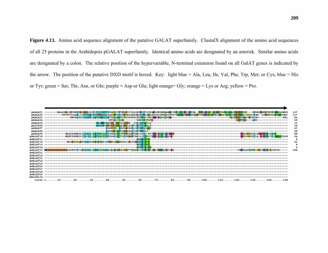
209
Figure 4.13. Amino acid sequence alignment of the putative GALAT superfamily. ClustalX alignment of the amino acid sequences
of all 25 proteins in the Arabidopsis pGALAT superfamily. Identical amino acids are designated by an asterisk. Similar amino acids
are designated by a colon. The relative position of the hypervariable, N-terminal extension found on all GalAT genes is indicated by
the arrow. The position of the putative DXD motif is boxed. Key: light blue = Ala, Leu, Ile, Val, Phe, Trp, Met, or Cys; blue = His
or Tyr; green = Ser, Thr, Asn, or Gln; purple = Asp or Glu; light orange= Gly; orange = Lys or Arg; yellow = Pro.
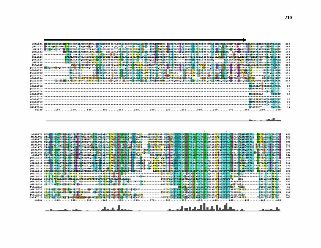
210
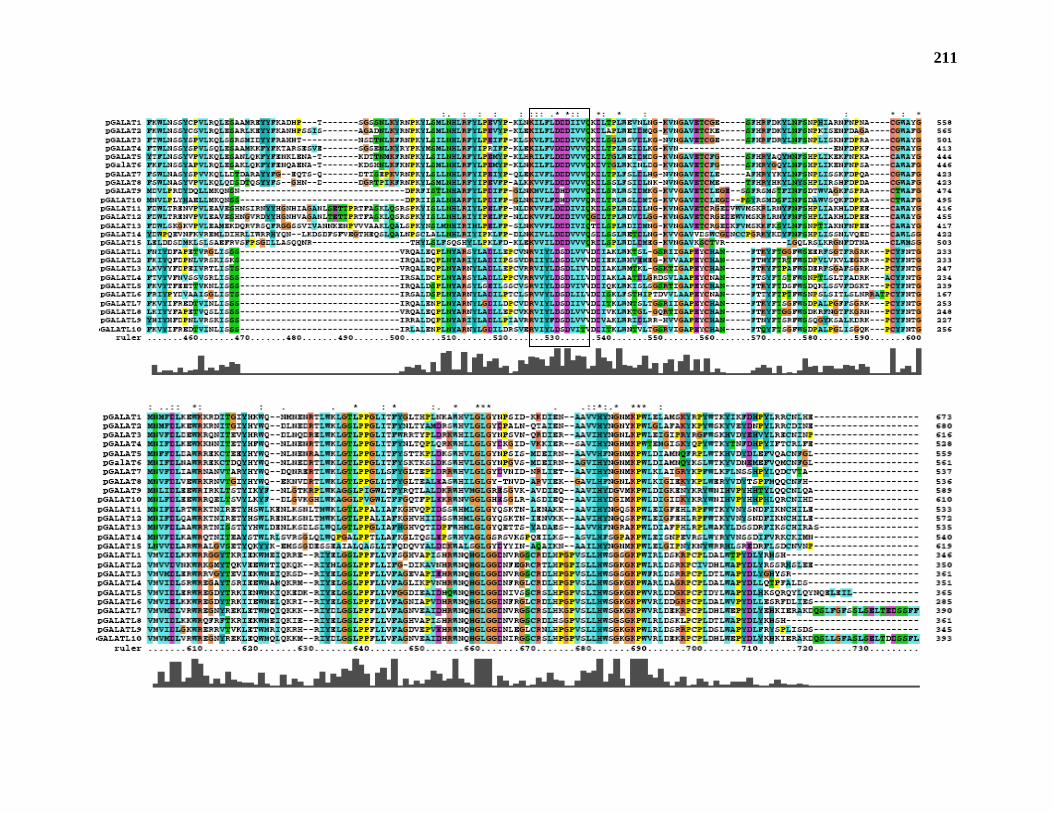
211
212
TMPRED_form.html; Figure 4.13) and are predicted type II membrane proteins (Paulson and
Colley, 1989; Keegstra and Raikhel, 2001). Conversely, pGalAT2, 3, 10 and all pGalATL
amino acid sequences lack this N-terminal extension and instead contain a putative signal
peptide (http://www.cbs.dtu.dk/services/SignalP; Figure 4.13). The only protein that was not
predicted to contain either an N-terminal transmembrane domain or a signal peptide was
pGalAT4. At present, the subcellular location and topology of the 25 pGALAT superfamily
proteins has not been elucidated and so the exact roles of the putative membrane-bound and
soluble pGalAT isoforms remain to be determined.
pGALAT1 (JS36) and 9 other members of the pGALAT superfamily (including
pGALAT3, 4, 8-10, 13, 15 and pGALATL1, 7, 10) were initially classified as putative
glycosyltransferases (Table 4.6) during the annotation of the Arabidopsis genome (Tavares et al.,
2000). This group of genes was called the LGT (like-glycosyl-transferase) family based on their
similarity to the LGTC gene from Neisseria gonorhoeae, a galactosyltransferase involved in
lipopolysaccharide biosynthesis (Gotschlich, 1994). Tavares et al. (2000) were able to identify
several conserved domains between members of the LGT family and the amino acid sequence of
LGTC, but did not speculate on the function of the LGT gene family in Arabidopsis.
pGALAT5 was previously identified as a T-DNA-insertion mutant of Arabidopsis named
qua1 (Bouton et al., 2002). Qua1 mutant plants are dwarfed, have reduced cell adhesion and
have 25% less D-GalpA in their leaves. Reduced cell adhesion and D-GalpA content are
phenotypes that would be expected for the mutation of a gene encoding a GalAT. Although
Bouton et al. (2002) were unable to provide enzymatic evidence proving the function of
pGALAT5/QUA1, the mutant phenotype provides further validity for our classification of the 25
Arabidopsis sequences into a putative GALAT super-family.
213
Recently, a mutant of a pGALATL subfamily member was isolated by transposon
mutatagenesis. Parvus (pgalatl4) was isolated as a semi-sterile mutant that exhibited a dwarf
phenotype and altered monosaccharide composition when grown under conditions of low
humidity (Lao et al., 2003). pGalatl4/parvus mutant plants had more α-L-arabinose and less β-D-
xylose in their cell walls, and linkage analysis showed an increase in 2-linked rhamnose and 3,4-
linked galactose compared to wild-type plants. Furthermore, galatl4/parvus plants showed a
difference in the developmental regulation of cell wall polysaccharide biosynthesis as older
leaves had a significant increase in the amount of 5-linked arabinose while younger leaves were
significantly reduced in the amount of 4-linked xylose.
Arabinose, galactose and rhamnose are components of the pectic polysaccharide RG-I.
Changes in the relative composition and linkage of these monosaccharides could be caused by
the plant’s attempt to cope with the mutation, an effect similar to that which occurs in plants with
the mur1 mutation (Zablackis et al., 1996; Reuhs et al., 2004). It is currently unknown whether
these changes in the galatl4/parvus mutant are caused by an alteration in D-GalpA content as the
amount of D-GalpA in galatl4/parvus mutant plants was not measured.
Phylogenetic analysis of the putative GALAT superfamily
Phylogenetic analysis of all 25 pGALAT superfamily members and selected amino acid
sequences from soybean (Glycine max), barrel medic (Medicago truncatula), rice (Oryza sativa),
tobacco (Nicotiana tobacum), and chickpea (Cicer arietinum), show that they cluster into 5
distinct groups with highly significant bootstrap values (Figure 4.14). Groups A and B contain 4
and 2 members, respectively, of the pGALAT subfamily. Group E contains pGALAT5/QUA1
and one other pGALAT subfamily member. The rest of the pGALAT subfamily, including
214
Figure 4.14. Phylogenetic analysis of the putative GALAT superfamily and its close homologs.
The amino acid sequences of all members of the pGALAT superfamily, along with EST
(GenBank accession no.) or tentative consensus (TC) sequences from Glycine max (Gm),
Medicago truncatula (Mt), Oryza sativa (Os), Nicotiana tobacum (Nt), and Cicer arietinum (Ca),
lacking their putative N-terminal transmembrane domains, were aligned using ClustalX (version
1.83). Weighted parsimony analysis of this alignment was conducted using PAUP software
(version 3.1). Bootstrap (100x) values are given in parentheses.
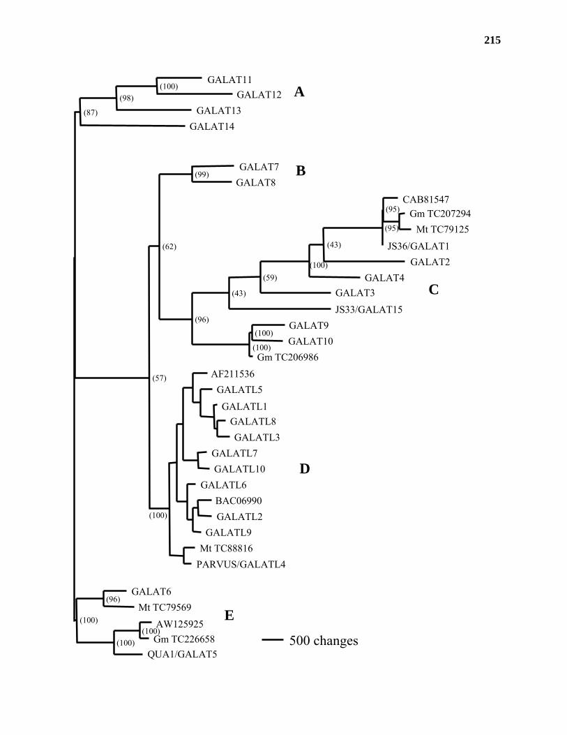
215
A
B
C
D
E
GALAT11GALAT12
GALAT13GALAT14
GALAT7GALAT8
CAB81547Gm TC207294
Mt TC79125
JS36/GALAT1GALAT2
GALAT4GALAT3JS33/GALAT15
GALAT9GALAT10
Gm TC206986AF211536
GALATL5
GALATL1GALATL8GALATL3
GALATL7GALATL10
GALATL6BAC06990GALATL2
GALATL9Mt TC88816
PARVUS/GALATL4
GALAT6Mt TC79569
AW125925Gm TC226658
QUA1/GALAT5500 changes(100)
(100)
(96)
(100)
(100)
(100)
(100)
(100)(98)
(87)
(57)
(99)
(62)
(96)(100)
(95)
(95)
(43)
(59)
(43)
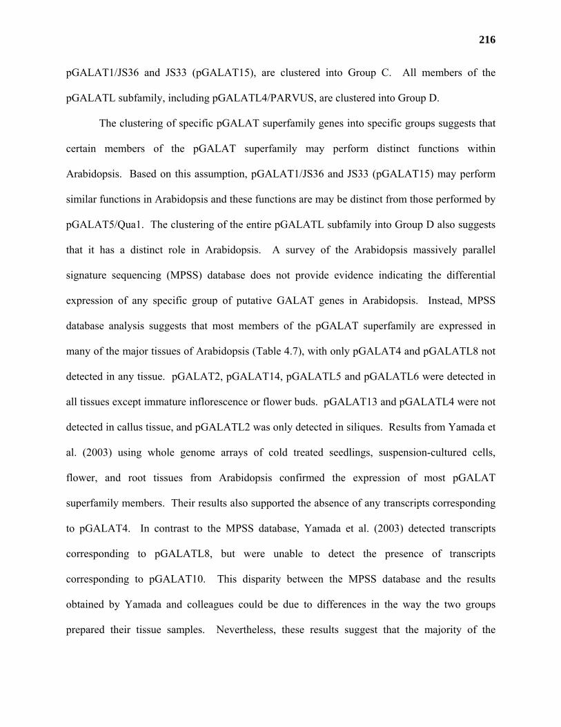
216
pGALAT1/JS36 and JS33 (pGALAT15), are clustered into Group C. All members of the
pGALATL subfamily, including pGALATL4/PARVUS, are clustered into Group D.
The clustering of specific pGALAT superfamily genes into specific groups suggests that
certain members of the pGALAT superfamily may perform distinct functions within
Arabidopsis. Based on this assumption, pGALAT1/JS36 and JS33 (pGALAT15) may perform
similar functions in Arabidopsis and these functions are may be distinct from those performed by
pGALAT5/Qua1. The clustering of the entire pGALATL subfamily into Group D also suggests
that it has a distinct role in Arabidopsis. A survey of the Arabidopsis massively parallel
signature sequencing (MPSS) database does not provide evidence indicating the differential
expression of any specific group of putative GALAT genes in Arabidopsis. Instead, MPSS
database analysis suggests that most members of the pGALAT superfamily are expressed in
many of the major tissues of Arabidopsis (Table 4.7), with only pGALAT4 and pGALATL8 not
detected in any tissue. pGALAT2, pGALAT14, pGALATL5 and pGALATL6 were detected in
all tissues except immature inflorescence or flower buds. pGALAT13 and pGALATL4 were not
detected in callus tissue, and pGALATL2 was only detected in siliques. Results from Yamada et
al. (2003) using whole genome arrays of cold treated seedlings, suspension-cultured cells,
flower, and root tissues from Arabidopsis confirmed the expression of most pGALAT
superfamily members. Their results also supported the absence of any transcripts corresponding
to pGALAT4. In contrast to the MPSS database, Yamada et al. (2003) detected transcripts
corresponding to pGALATL8, but were unable to detect the presence of transcripts
corresponding to pGALAT10. This disparity between the MPSS database and the results
obtained by Yamada and colleagues could be due to differences in the way the two groups
prepared their tissue samples. Nevertheless, these results suggest that the majority of the
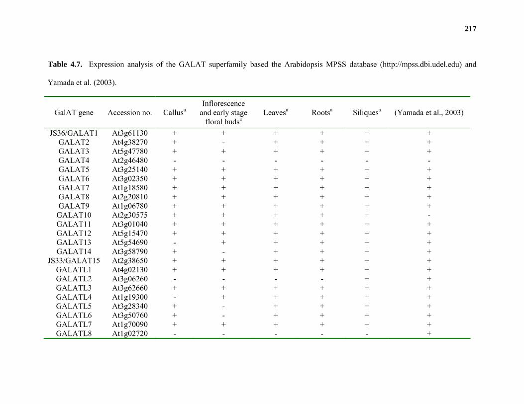
217
Table 4.7. Expression analysis of the GALAT superfamily based the Arabidopsis MPSS database (http://mpss.dbi.udel.edu) and
Yamada et al. (2003).
GalAT gene Accession no. Callusa Inflorescence
and early stage floral budsa
Leavesa Rootsa Siliquesa (Yamada et al., 2003)
JS36/GALAT1 At3g61130 + + + + + + GALAT2 At4g38270 + - + + + + GALAT3 At5g47780 + + + + + + GALAT4 At2g46480 - - - - - - GALAT5 At3g25140 + + + + + + GALAT6 At3g02350 + + + + + + GALAT7 At1g18580 + + + + + + GALAT8 At2g20810 + + + + + + GALAT9 At1g06780 + + + + + + GALAT10 At2g30575 + + + + + - GALAT11 At3g01040 + + + + + + GALAT12 At5g15470 + + + + + + GALAT13 At5g54690 - + + + + + GALAT14 At3g58790 + - + + + +
JS33/GALAT15 At2g38650 + + + + + + GALATL1 At4g02130 + + + + + + GALATL2 At3g06260 - - - - + + GALATL3 At3g62660 + + + + + + GALATL4 At1g19300 - + + + + + GALATL5 At3g28340 + - + + + + GALATL6 At3g50760 + - + + + + GALATL7 At1g70090 + + + + + + GALATL8 At1g02720 - - - - - +
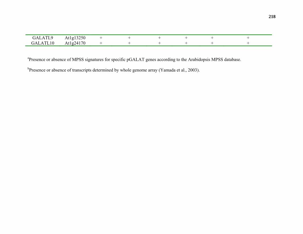
218
GALATL9 At1g13250 + + + + + + GALATL10 At1g24170 + + + + + +
aPresence or absence of MPSS signatures for specific pGALAT genes according to the Arabidopsis MPSS database.
bPresence or absence of transcripts determined by whole genome array (Yamada et al., 2003).
219
putative GALAT superfamily is widely expressed in Arabidopsis and therefore little information
on the possible roles of the distinct groups in the GALAT superfamily can be gleaned using this
database analysis. Mutation analysis and/or the characterization of members of the putative
GALAT superfamily will be required before the significance of these groupings can be fully
understood.
A BLAST search of the NCBI (http://www.ncbi.nlm.nih.gov/entrez/query.fcgi) and
TIGR (http://www.tigr.org) databases indicates the presence of several protein sequences from
other plant species with significant identity to pGalAT1/JS36 (Figure 4.14). Phylogenetic
analysis of selected sequences demonstrates that the proteins with the most similarity to
pGalAT1/JS36 are not from Arabidopsis, but are instead from soybean (TC207294), barrel
medic (TC79125), and chickpea (CAB81547). A recent search of the rice genome also revealed
a protein (BAD46265) with high sequence identity to pGalAT1/JS36. Other gene sequences (i.e.
BAC06990 from rice) cluster into specific groups within the phylogenetic tree. These results
strongly suggest that pGALAT1 and other members of the putative GALAT superfamily, are
highly conserved across several plant species.
A similar phylogenetic tree was constructed by the group that discovered the
galatl4/parvus mutant (Lao et al., 2003). Using conserved domains from pGALATL4/Parvus
and other CAZy family 8 glycosyltransferases from Arabidopsis, they identified all but two
members of the putative GALAT superfamily. Their tree clustered the pGALAT and pGALATL
subfamily sequences into two distinct branches. The similarity in the groupings of the pGALAT
and pGALATL genes by these two independent analyses strengthens the argument that they
represent two distinct subfamilies in Arabidopsis.
220
DISCUSSION
D-GalpA is the most abundant monosaccharide found in pectic polysaccharides. It has
been estimated that between 5-7 distinct GalATs are required for the incorporation of D-GalpA
into all of the known pectic structures of the primary cell wall (Mohnen, 2002). While GalAT
activity can be detected in tissue extracts from several different plant species (Villemez et al.,
1965; Lin et al., 1966; Doong et al., 1995; Sterling et al., 2001; Akita et al., 2002; Ishii, 2002),
previous attempts to identify GalAT genes from any plant source have not been successful.
Through the development of new solubilization conditions, we were able to solubilize
approximately 80% of the starting activity from Arabidopsis membranes. Instrumental in this
process was the elimination of the low-speed centrifugation step in the preparation of solubilized
proteins. The direct high-speed centrifugation of tobacco cell homogenates created a multi-
layered pellet in which approximately 80% of the total GalAT activity was found in the top-most
layer. The formation of this unique GalAT-containing layer was not found to be dependent on
the original plant source, as similar pellets could be created using suspension-cultured
Arabidopsis cells or radish roots (data not shown). Mixing of the three layers caused a
significant reduction in the amount of GalAT activity that could be solubilized, suggesting that
the other layers contained a potent inhibitor of GalAT. Our results showed that this layer was
enriched in subcellular membranes that contained GalAT activity and therefore could also be an
excellent starting material for the study of other pectin glycosyltransferases.
GalAT activity was purified a total of 17-fold through the combined use of SP-Sepharose,
RY3 and UDP-agarose chromatography columns. The purification achieved could be an
underestimation of the true purity of GalAT activity in the final fraction, as it was determined
that several of the enriched GalAT fractions contained a contaminating polygalacturonase
221
activity that degraded the radioactive product as it was being synthesized (data not shown).
Similar polygalacturonase activities have been detected in GalAT-containing membrane
fractions from pumpkin (Ishii, 2002) and from solubilized GalAT from petunia pollen tubes
(Akita et al., 2002). This degradative activity greatly decreased the amount of D-[14C]GalpA
incorporated into product, reducing both the specific and total activity. Attempts to remove this
contaminating activity by chromatography were unsuccessful, because the activity bound to all
chromatography resins that were negatively charged. The inclusion of low concentrations of
OGAs and UDP in the elution buffers used during RY3 and UDP-agarose chromatography was
found to aid in the selective removal of GalAT activity from the column, as well as inhibit some
of the polygalacturonase activity that had bound to these resins (data not shown).
A critical step in the purification of Arabidopsis GalAT was the use of the affinity resin
UDP-agarose. Previous elution schemes using this resin that were based solely on changes in
ionic strength were found to be ineffective for the purification of GalAT activity (data not
shown). By employing an elution scheme similar to one developed by Barker et al. (1972), we
were able to remove a substantial amount of contaminating proteins from the UDP-agarose
column and obtained a fraction that was significantly enriched in GalAT activity.
Proteolytic digestion and analysis of the resulting peptides contained within the most
purified chromatography fraction from the UDP-agarose column identified two protein
sequences, named JS33 and JS36, which contained conserved domains found in other
glycosyltransferases. Preliminary analysis of the two protein sequences indicated that they
encoded putative type II membrane proteins, were part of a family of retaining
glycosyltransferases (CAZy family 8), and were expressed ubiquitously in Arabidopsis. All of
these characteristics were properties expected for GalAT genes (Scheller et al., 1999; Sterling et
222
al., 2001). We successfully cloned and heterologously expressed N-terminally truncated and HA
epitope-tagged forms of JS33 and JS36 in mammalian HEK293 cells. The HA epitope tagged
proteins also contained the signal sequence from T. cruzi mannosidase for secretion of the
recombinant proteins into the media surrounding transfected (or transformed) HEK293 cells
(Vandersall-Nairn et al., 1998).
Two separate experiments were performed in which media from transiently transfected
HEK293 cells was harvested and analyzed for GalAT activity. In the first experiment,
immunoprecipitation of media from cells transfected with the JS36 cDNA construct using anti-
HA antibodies conjugated to protein A-Sepharose yielded GalAT activity. It was concluded that
JS36 encoded a putative GalAT. A second transient transfection experiment did not yield GalAT
activity, despite the presence of low levels of recombinant JS36 protein in the media.
There are several reasons why GalAT activity might not have been detected in the later
transient expression experiment. One reason may be the low level of recombinant protein
expressed in the transiently expressing cells. Our data show that more JS36 cDNA is required to
transform HEK293 cells than JS33 or α-(2,6)-ST. Therefore, higher than recommended amounts
of DNA (Jordan et al., 1996) may be required to obtain high levels of JS36 expression in HEK
cells. It is possible that in the second transient experiment, the transfection efficiency was lower
than the previous experiment, resulting in lower levels of recombinant protein and no detectable
GalAT activity. The use of higher amounts of cDNA may allow us to detect GalAT activity in
cells transfected with the JS36 constructs in future experiments.
Another possible explanation for the difficulty in detecting GalAT activity in the
transiently transfected cell lines expressing JS36 is the lack of essential cofactors and/or other
proteins that are required for optimal GalAT activity. It is well known that many
223
glycosyltransferases require additional proteins and/or cofactors for optimal activity and without
these proteins exhibit diminished abilities to catalyze their reactions. This phenomenon has been
demonstrated for the expression of ganglioside α-(2,8)-sialyltransferase II (STII) and β-(1,4)-N-
acetylgalactosaminyltransferase I (GalNAcTI) in mouse F-11A neuroblastoma cells (Bieberich et
al., 2002). Bieberich et al. (2002) were able to show that transfecting F-11A cells with STII
greatly enhanced the endogenous GalNAcTI activity in this cell line and vice versa.
Furthermore, they were able to show that STII and GalNAcTI formed protein complexes in the
Golgi, suggesting that Golgi glycosyltransferases involved in the production of common
glycoconjugates self-associate in vivo to form an activated complex that is more active than the
individual glycosyltransferases (Bieberich et al., 2002). Pectin biosynthesis is also thought to
involve the coordinated action of several glycosyltransferases (Mohnen, 2002) and so it is
possible that the co-expression of other pectin biosynthetic enzymes will be required to obtain
high levels of GalAT activity in HEK cell lines.
Stably transformed cell lines expressing recombinant JS33 or JS36 proteins also did not
possess any GalAT activity. Unlike the transiently transfected lines, media taken from the stable
lines expressing JS33 and JS36 did not contain any detectable levels of recombinant protein.
Instead, the recombinant proteins were found in cell lysates. The addition of 2 µM lactacystin to
stable cell lines expressing JS36 did not change the amount of recombinant protein made in cell
lysates or its migration on SDS-PAGE gels. These results suggest that the anomalous JS36
protein targeting is not due to proteasome-directed degradation of the recombinant protein, but is
due to some unknown event that occurs during or following protein expression.
The differential location of recombinant proteins in stable versus transient lines could be
caused by the retardation of these proteins in the secretory pathway. The retardation of
224
recombinantly expressed, Golgi-localized glycosyltransferases has been shown to occur during
the expression of a secreted form of GlcNAcTI in CHO cells (Opat et al., 2000). The authors
speculated that it was caused by the formation of high molecular weight protein complexes
within the Golgi apparatus. They also suggested that this property occurred only with
glycosyltransferases that were localized to the medial Golgi cisternae (Opat et al., 2000).
The specific subcellular localization of JS33 and JS36 protein in the stable HEK cell lines
has not been determined. It has been hypothesized that the bulk of the GalAT activity found in
plant cells is localized to the cis or medial Golgi cisternae (Staehelin and Moore, 1995; Vicre et
al., 1998). If JS33 and JS36 are GalATs that function in these early cisternae, then it is possible
that the recombinant forms of these proteins also create large molecular weight complexes that
are retained within the HEK cells.
The intracellular retention and/or production of inactive, recombinant protein could also
be caused by the abnormal N-glycosylation of recombinant JS33 and JS36. This phenomenon
has previously been demonstrated during the recombinant expression of rat α-(2,6)-
sialyltransferase in COS-1 cells (Chen and Colley, 2000). α-(2,6)-ST is a Golgi-localized protein
that is proteolytically processed in a downstream Golgi compartment into a smaller, secreted
isoform in mammalian cells (Chen et al., 2003b). α-(2,6)-ST was shown to be glycosylated at
two sites, Asn146 and Asn158. Site-directed mutagenesis showed that while both the
glycosylated and non-glycosylated forms of α-(2,6)-ST were correctly targeted to the Golgi, only
wildtype and the Asn146 mutant were proteolytically processed and properly secreted from the
COS-1 cells. Furthermore, only these isoforms were active in in vitro activity assays. The
authors hypothesized that the Asn158 and Asn146/Asn158 mutants of α-(2,6)-ST formed protein
aggregates upon cell lysis that inactivated the recombinant proteins. They also hypothesized that
225
the absence of the N-linked oligosaccharide at Asn158 caused the recombinant proteins to be
restricted to earlier Golgi compartments.
The difference between the calculated and observed molecular weights of recombinant
JS33 and JS36 (13 and 6-8 kDa, respectively) suggests that both proteins may be glycosylated in
HEK cells. The differential glycosylation of JS36 is also suggested by the appearance of two
recombinant isoforms of the protein in stably transformed lines. Both JS33 and JS36 each
possess 5 putative N-glycosylation sites; however, the nature and pattern of N-glycosylation was
not explored in this study. Differences in the N-glycosylation pattern of recombinant versus
native, Arabidopsis isoforms of JS33 and JS36 may be a contributing factor to the localization
and activity of the recombinant proteins made by HEK293 cells.
The putative GALAT superfamily constitutes a novel gene family in Arabidopsis. While
several members of this family have previously been described (Tavares et al., 2000; Bouton et
al., 2002; Lao et al., 2003), this is the first report that combines all 25 coding sequences from
Arabidopsis into one superfamily. Alignment of all the sequences within the pGALAT
superfamily indicates regions of high sequence conservation between superfamily members.
These regions include the Pfam domain PF01501 that is unique to CAZy family 8
glycosyltransferases (http://www.sanger.ac.uk/cgi-bin/Pfam/getacc?PF01501) and a putative
DXD motif that is conserved among several glycosyltransferase families (Wiggins and Munro,
1998).
The putative GALAT superfamily can be divided into two subfamilies according to their
sequence identity to pGalAT1/JS36. This subdivision is further justified by the clustering of all
pGALATL genes into one branch of our phylogenetic tree. All members of the pGalATL
subfamily and specific GalAT subfamily proteins (2, 3, and 10) are predicted to contain signal
226
peptides at their N-terminus, suggesting that they enter the secretory pathway and are either
retained within a subcellular compartment or secreted into the cell wall. The former location is
the most probable site of action for these genes as pectin biosynthesis occurs in the Golgi
apparatus (Zhang and Staehelin, 1992; Sterling et al., 2001) and no glycosyltransferases have
been detected in the extracellular matrix (Bacic et al., 1988; Brett and Waldron, 1990).
All other pGalAT subfamily proteins (except pGalAT4 and those mentioned above) have
an N-terminal extension containing a putative transmembrane domain and are predicted type II
membrane proteins. This topology has been associated with several Golgi-localized plant
(Edwards et al., 1999; Keegstra and Raikhel, 2001; Faik et al., 2002; Iwai et al., 2002) and
mammalian (Paulson and Colley, 1989; Munro, 1995; Chen et al., 2003b) glycosyltransferases
and provides evidence supporting the subcellular location of specific pGalAT proteins in
Arabidopsis.
The presence of putative soluble isoforms of pGalAT superfamily proteins in Arabidopsis
is unexpected. GalAT activity has not been detected in soluble fractions from any plant source
(Villemez et al., 1966; Doong et al., 1995; Takeuchi and Tsumuraya, 2001; Ishii, 2002). This
suggests that these soluble proteins are either inactivated during tissue homogenation or isolated
as highly associated protein complexes with their membrane-bound counterparts during
solubilization.
While the exact roles of the apparent soluble versus membrane-bound proteins are not
known, analysis of two putative GALAT superfamily mutants, pgalat5/qua1 and
pgalatl4/parvus, shows that they result in somewhat similar phenotypes in Arabidopsis (Bouton
et al., 2002; Lao et al., 2003). Both mutant plants are dwarfed and are altered in the relative
composition of sugars that are normally associated with pectic polysaccharides. The
227
pgalat5/qua1 mutant also exhibits a cell adhesion defect that is not seen in the pgalatl4/parvus
mutant, and the pgalatl4/parvus mutant is more sensitive to growth under conditions of low
humidity. These results suggest that pGALAT5/QUA1 and pGALATL4/PARVUS may have
similar, yet distinct, functions in Arabidopsis and also supports the separation of these genes to
different branches of the phylogenetic tree.
Analysis of the expression patterns of all the members of the putative GALAT
superfamily indicates that the majority of these genes are expressed in multiple tissues of
Arabidopsis. This result suggests that pGALAT genes are important components of all
Arabidopsis tissues; however, it provides no insight into the potential functions of pGALAT
genes such as pGALAT15/JS33 and pGALAT1/JS36. Of all the genes in the putative GALAT
superfamily, pGALAT4 is the only gene that does not contain a putative signal sequence or a
transmembrane domain at its N-terminus. Transcript analysis also suggests that this gene is not
expressed in any of the Arabidopsis tissues analyzed, indicating that it may be a pseudogene
(Tavares et al., 2000).
The generation of stable lines expressing recombinant forms of JS33 and JS36 represents
a step forward in the study of the proposed GalATs. Experiments aimed at determining enzyme
specificity, discovering associating factors and/or proteins, and developing new methods for
expression of JS33 and JS36 can now be carried out. The development of polyclonal antibodies
against specific GalAT genes will allow us to determine the subcellular location of these genes in
Arabidopsis and will greatly aid in determining the function of these genes. The importance of
the putative GALAT superfamily is made evident by the identification of close homologs of
specific pGALAT family members in other plant species. Through the use of stable lines
expressing pGALAT genes, the analysis of pGALAT mutant plants and the development of tools
228
such as polyclonal antibodies to specific pGalAT proteins, the specific roles of the putative
GALAT superfamily in pectin biosynthesis can be determined.
229
CONCLUSIONS
This research has significantly increased our understanding of pectin biosynthesis. The
localization of α-(1,4)-GalAT activity to the lumen of the Golgi apparatus in pea confirmed
previous, more indirect evidence suggesting the Golgi as the main site of pectic biosynthesis.
Proteinase K protection experiments indicated that the catalytic site of pea α-(1,4)-GalAT faced
the Golgi lumen. Golgi-localized α-(1,4)-GalAT activity also synthesized radiolabeled products
that were comparable to those generated by the tobacco enzyme. These results suggest that pea
α-(1,4)-GalAT is a Golgi-localized enzyme that synthesizes HGA in the lumen of the Golgi
apparatus. This is the first report of the subcellular location of a pectin glycosyltransferase in
any plant system.
One of the major problems with identifying pectin glycosyltransferases is the difficulty
associated with developing the appropriate assays for catalytic activity. Not only are these
assays important for detecting glycosyltransferase activity in plant extracts, but they are also
required for testing recombinantly expressed proteins for activity. Earlier methods used to detect
α-(1,4)-GalAT activity in microsomal membranes and/or solubilized protein fractions have
proven to be too expensive and time consuming to be useful for purification studies. The
development of a quick and simple assay for α-(1,4)-GalAT greatly facilitated the development
of new universal solubilization methods and aided in the partial purification of Arabidopsis α-
(1,4)-GalAT. The further development of this assay will allow its use to detect the activity of
other pectin glycosyltransferases.
The identification of the putative GalATs, JS36 and JS33, from partially purified protein
fractions from Arabidopsis represents a step forward towards the identification of pectin
230
biosynthetic glycosyltransferases. While the activity of JS36 and JS33 could not be
unequivocally demonstrated, analysis of several Arabidopsis mutants (K. Hosmer and D.
Mohnen, unpublished results) indicates that the disruption of several putative GALAT
superfamily genes causes alterations in glycosyl residues associated with pectic polysaccharides.
These results support the involvement of the putative GALAT superfamily in pectin
biosynthesis. The development of tools, such as MAP polyclonal antibodies, should enable the
elucidation of the specific roles that putative GALAT superfamily genes play in pectin
biosynthesis.
231
REFERENCES
Abdel-Massih R, Baydoun E, Brett C (2003) In vitro biosynthesis of (1,4)-α-galactan attached to a pectin-xyloglucan complex in pea. Planta 216: 502-511
Agius F, Gonzalez-Lamothe R, Caballero J, Munoz-Blanco J, Botella M, Valpuesta V (2002) Engineering increased vitamin C levels in plants by overexpression of a D-galacturonic acid reductase. Nat. Biotechnol. 21: 177-181
Akita K, Ishimizu T, Tsukamoto T, Ando T, Hase S (2002) Successive glycosyltransfer activity and enzymatic characterization of pectic polygalacturonate 4-α-galacturonosyltransferase solubilized from pollen tubes of Petunia axillaris using pyridylaminated oligogalacturonates as substrates. Plant Physiol. 130: 374-379
Albersheim P, Darvill A, O'Neill M, Schols H, Voragen A (1996) An hypothesis: The same six polysaccharides are components of the primary cell walls of all higher plants. In J Visser, AGJ Voragen, eds, Pectins and Pectinases. Elsevier Science, Amsterdam, pp 47-55
Allard S, Giraud M, Whitfield C, Graninger M, Messner P, Naismith J (2001) The crystal structure of dTDP-D-glucose 4,6-dehydratase (RmlB) from Salmonella enterica serovar typhimurium, the second enzyme in the dTDP--rhamnose pathway. J. Mol. Biol. 307: 283-295
Ames B (1966) Assay of inorganic phosphate, total phosphate and phosphatases. Methods Enzymol. 8: 115-118
An J, O'Neill M, Albersheim P, Darvill A (1994) Isolation and structural characterization of β-D-glucosyluronic acid and 4-O-methyl-β-D-glucosyluronic acid-containing oligosaccharides from the cell-wall pectic polysaccharide, rhamnogalacturonan I. Carbohydr. Res. 252: 235-243
An J, Zhang L, O'Neill M, Albersheim P, Darvill A (1994a) Isolation and structural characterization of endo-rhamnogalacturonase-generated fragments of the backbone of rhamnogalacturonan I. Carbohydr. Res. 264: 83-96
Ankel H, Feingold D (1965) Biosynthesis of uridine disphosphate D-xylose I. Uridine diphosphate glucuronate carboxyl-lyase of wheat germ. Biochemistry 4: 2468-2475
232
Aoki K, Ishida N, Kawakita M (2003) Substrate recognition by nucleotide sugar transporters: Further characterization of substrate recognition regions by analyses of UDP-galactose/CMP-sialic acid transporter chimerase and biochemical analysis of the substrate specificity of parental and chimeric transporters. J. Biol. Chem. 278: 22887-22893
Aspinall G (1980) Chemistry of cell wall polysaccharides. In J Preiss, ed, The biochemistry of plants, Vol 3. Academic Press, New York, pp 473-500
Aspinall G, Begbie R, Hamilton A, Whyte J (1967) Polysaccharides of soybeans. Part III. Extraction and fractionation of polysaccharides from cotyledon meal. J. Chem. Soc.: 1065-1070
Azadi P, O'Neill M, Bergmann C, Darvill A, Albersheim P (1995) The backbone of the pectic polysaccharide rhamnogalacturonan I is cleaved by an endohydrolase and an endolyase. Glycobiology 5: 783-789
Bacic T, Harris P, Stone B (1988) Structure and function of plant cell walls. In J Preiss, ed, The Biochemistry of Plants, Vol 14. Academic Press Inc., New York, pp 297-371
Bakker H, Routier F, Oelmann S, Jordi W, Lommen A, Gerardy-Schahn R, Bosch D (2004) Molecular cloning of two Arabidopsis UDP-galactose transporters by complementation of a deficient chinese hamster ovary cell line. Glycobiology Epub ahead of print
Baldwin T, Handford M, Yuseff M, Orellana A, Dupree P (2001) Identification and characterization of GONST1, a Golgi-localized GDP-mannose transporter in Arabidopsis. Plant Cell 13: 2283-2295
Barber G (1962) The enzymatic synthesis of uridine diphosphate L-rhamnose. Biochem. Biophys. Res. Commun. 8: 204-208
Barber G (1963) The formation of uridine diphosphate L-rhamnose by enzymes of the tobacco leaf. Arch. Biochem. Biophys. 103: 276-282
Barber G (1979) Observations on the mechanism of the reversible epimerization of GDP-D-mannose to GDP-L-galactose by an enzyme from Chlorella pyrenoidosa. J. Biol. Chem. 254: 7600-7603
233
Barber G, Chang M (1967) Synthesis of uridine diphosphate L-rhamnose by enzymes of Chlorella pyrenoidosa. Arch. Biochem. Biophys. 118: 659-663
Barker R, Olsen K, Shaper J, Hill R (1972) Agarose derivatives of uridine diphosphate and N-acetylglucosamine for the purification of a galactosyltransferase. J. Biol. Chem. 247: 7135-7147
Baron D, Streitberger U, Grisebach H (1973) Improved method for purification of UDP-apiose/UDP-xylose synthase from cell cultures of parsley. Biochim. Biophys. Acta. 293: 526-533
Bar-Peled M, Fluhr R, Gressel J (1993) Juvenile-specific localization and accumulation of a rhamnosyltransferase and its bitter flavonoid in foliage, flowers, and young citrus fruits. Plant Physiol. 103: 1377-1384
Bar-Peled M, Lewinsohn E, Fluhr R, Gressel J (1991) UDP-rhamnose:flavanone-7-O-glucoside-2''-O-rhamnosyltransferase. Purification and characterization of an enzyme catalyzing the production of bitter compounds in citrus. J. Biol. Chem. 266: 20953-20959
Basu S, Dotson G, Raetz C (2000) A facile enzymatic synthesis of uridine diphospho-[14C]galacturonic acid. Anal. Biochem. 280: 173-177
Baydoun E, Abdel-Massih R, Dani D, Rizk S, Brett C (2001) Galactosyl- and fucosyltransferases in etiolated pea epicotyls: Product identification and sub-cellular localisation. J. Plant Physiol. 158: 145-150
Berninsone P, Hwang H, Zemtseva I, Horvitz H, Hirschberg C (2001) SQV-7, a protein involved in Caenorhabditis elegans epithelial invagination and early embryogenesis, transports UDP-glucuronic acid, UDP-N- acetylgalactosamine, and UDP-galactose. Proc. Natl. Acad. Sci. 98: 3738-3743
Bieberich E, MacKinnon S, Silva J, Li D, Tencomnao T, Irwin L, Kapitonov D, Yu R (2002) Regulation of ganglioside biosynthesis by enzyme complex formation of glycosyltransferases. Biochemistry 41: 11479-11487
Blevins D, Lukaszewski K (1998) Boron in plant structure and function. Ann. Rev. Plant Physiol. Plant Mol. Biol. 49: 481-500
234
Blumenkrantz N, Asboe-Hansen G (1973) New method for quantitative determination of uronic acids. Anal. Biochem. 54: 484-489
Bolwell G, Dalessandro G, Northcote D (1985) Decrease of polygalacturonic acid synthase during xylem differentiation in sycamore. Phytochemistry 24: 699-702
Bolwell G, Northcote D (1983) Arabinan synthase and xylan synthase activities in Phaseolus vulgaris: Subcellular localization and possible mechanism of action. Biochem. J. 210: 497-507
Bonin C, Freshour G, Hahn M, Vanzin G, Reiter W (2003) The GMD1 and GMD2 genes of Arabidopsis encode isoforms of GDP-D-mannose 4,6-dehydratase with cell type-specific expression patterns. Plant Physiol. 132: 883-892
Bonin C, Potter I, Vanzin G, Reiter W (1997) The MUR1 gene of Arabidopsis thaliana encodes an isoform of GDP-D-mannose-4,6-dehydratase, catalyzing the first step in the de novo synthesis of GDP-L-fucose. Proc. Natl. Acad. Sci. 94: 2085-2090
Bonin C, Reiter W (2000) A bifunctional epimerase-reductase acts downstream of the MUR1 gene product and completes the de novo synthesis of GDP-L-fucose in Arabidopsis. Plant J. 21: 445-454
Bourlard T, Bruyant-Vannier M, Schaumann A, Bruyant P, Morvan C (2001) Purification of several pectin methyltransferases from cell suspension cultures of flax (Linum usitatissimum L.). C. R. Acad. Sci. III 324: 335-343
Bourlard T, Pellerin P, Morvan C (1997) Rhamnogalacturonans I and II are pectic substrates for flax-cell methyltransferases. Plant Physiol. Biochem. 35: 623-629
Bourlard T, Schaumann-Gaudinet A, Bruyant-Vannier M, Morvan C (1997a) Various pectin methyltransferases activities with affinity for low and highly methylated pectins. Plant Cell Physiol. 38: 259-267
Bouton S, Leboeuf E, Mouille G, Leydecker M, Talbotec J, Granier F, Lahaye M, Hofte H, Truong H (2002) QUASIMODO1 encodes a putative membrane-bound glycosyltransferase required for normal pectin synthesis and cell adhesion in Arabidopsis. Plant Cell 14: 2577-2590
Bouveng H (1965) Polysaccharides in pollen. Acta. Chim. Scand. 19: 953-963
235
Brabetz W, Wolter F, Brade H (2000) A cDNA encoding 3-deoxy-D-manno-oct-2-ulosonate-8-phosphate synthase of Pisum sativum L. (pea) functionally complements a kdsA mutant of the Gram-negative bacterium Salmonella enterica. Planta 212: 136-143
Braccini I, Grasso R, Perez S (1999) Conformational and configurational features of acidic polysaccharides and their interactions with calcium ions: A molecular modeling investigation. Carbohydr. Res. 317: 119-130
Braccini I, Perez S (2001) Molecular basis of Ca2+-induced gelation in alginates and pectins: The egg-box model revisited. Biomacromolecules 2: 1089-1096
Brandan E, Hirschberg C (1988) Purification of rat liver N-heparan-sulfate sulfotransferase. J. Biol. Chem. 263: 2417-2422
Breton C, Imberty A (1999) Structure/function studies of glycosyltransferases. Curr. Opin. Struct. Biol. 9: 563-571
Brett C, Waldron K (1990) Cell-wall formation. In The Physiology and Biochemistry of Plant Cell Walls. Unwin Hyman Ltd., London, pp 58-88
Brickell L, Reid J (1996) Biosynthesis in vitro of pectic (1,4)-β-D-galactan. In J Visser, AGJ Voragen, eds, Pectins and Pectinases. Elsevier Science, Amsterdam, pp 127-134
Briskin D, Leonard R, Hodges T (1987) Isolation of the plasma membrane: Membrane markers and general principals. Methods Enzymol. 148: 542-558
Brown J, Fry S (1993) Novel O-D-galacturonoyl esters in the pectic polysaccharides of suspension-cultured plant cells. Plant Physiol. 103: 993-999
Brummell D, Camirand A, Maclachlan G (1990) Differential distribution of xyloglucan glycosyltransferases in pea Golgi dictyosomes and secretory vesicles. J. Cell Sci. 96: 705-710
Brummell D, Harpster M (2001) Cell wall metabolism in fruit softening and quality and its manipulation in transgenic plants. Plant Mol. Biol. 47: 311-339
236
Bruyant-Vannier M, Gaudinet-Schaumann A, Bourlard T, Morvan C (1996) Solubilization and partial characterization of pectin methyltransferase from flax cells. Plant Physiol. Biochem. 34: 489-499
Burget E, Reiter W (1999) The mur4 mutant of Arabidopsis is partially defective in the de novo synthesis of uridine diphospho L-arabinose. Plant Physiol. 121: 383-390
Burget E, Verma R, Molhoj M, Reiter W (2003) The biosynthesis of L-arabinose in plants: Molecular cloning and characterization of a Golgi-localized UDP-D-xylose 4-epimerase encoded by the MUR4 gene of Arabidopsis. Plant Cell 15: 523-531
Bush M, Marry M, Huxham M, Jarvis M, McCann M (2001) Developmental regulation of pectic epitopes during potato tuberisation. Planta 213: 869-880
Cardoso S, Silva A, Coimbra M (2002) Structural characterisation of the olive pomace pectic polysaccharide arabinan side chains. Carbohydr.Res. 337: 917-924
Carpita N, Gibeaut D (1993) Structural models of primary cell walls in flowering plants: consistency of molecular structure with the physical properties of the walls during growth. Plant J. 3: 1-30
Carpita N, Tierney M, Campbell M (2001) Molecular biology of the plant cell wall: searching for the genes that define structure, architecture and dynamics. Plant Mol. Biol. 47: 1-5
Chen C, Colley K (2000) Minimal structural and glycosylation requirements for ST6Gal I activity and trafficking. Glycobiology 10: 531-583
Chen T, Chen C, Bergeron N, Close B, Bohrer T, Vertel B, Colley K (2003b) The two rat α-(2,6)-sialyltransferase (ST6Gal I) isoforms: Evaluation of catalytic activity and intra-Golgi localization. Glycobiology 13: 109-117
Chen W, Unligil U, Rini J, Stanley P (2001) Independent Lec1A CHO glycosylation mutants arise from point mutations in N-acetylglucosaminyltransferase I that reduce affinity for both substrates. Molecular consequences based on the crystal structure of GlcNAc-TI. Biochemistry 40: 8765-8772
Cheng L, Kindel P (1997) Detection and homogeneity of cell wall pectic polysaccharides of Lemna minor. Carbohydr. Res. 301: 205-212
237
Chou P, Fasman G (1978) Prediction of the secondary structure of proteins from their amino acid sequence. Adv. Enzymol. Relat. Areas Mol. Biol. 47: 45-148
Clausen M, Willats W, Knox J (2003) Synthetic methyl hexagalacturonate hapten inhibitors of anti-homogalacturonan monoclonal antibodies LM7, JIM5 and JIM7. Carbohydr. Res. 338: 1797-1800
Darvill A, Augur C, Bergmann C, Carlson R, Cheong J, Eberhard S, Hahn M, Lo V, Marfa V, Meyer B, Mohnen D, O'Neill M, Spiro M, Halbeek H, York W, Albersheim P (1992) Oligosaccharins: Oligosaccharides that regulate growth, development and defence responses in plants. Glycobiology 2: 181-198
Darvill A, McNeil M, Albersheim P (1978) Structure of plant cell walls VIII. A new pectic polysaccharide. Plant Physiol. 62: 418-422
Davies M, Dickinson D (1972) Properties of uridine diphosphoglucose dehydrogenase from pollen of Lilium longiflorum. Arch. Biochem. Biophys. 152: 53-61
Dean N, Zhang Y, Poster J (1997) The VRG4 gene is required for GDP-mannose transport into the lumen of the Golgi in the yeast, Saccharomyces cerevisiae. J. Biol. Chem. 272: 31908-31914
Delmas F, Petit J, Joubes J, Seveno M, Paccalet T, Hernould M, Lerouge P, Mouras A, Chevalier C (2003) The gene expression and enzyme activity of plant 3-deoxy-D-manno-2-octulosonic acid-8-phosphate synthase are preferentially associated with cell division in a cell cycle-dependent manner. Plant Physiol. 133: 348-360
Delmer D (1999) Cellulose biosynthesis: Exciting times for a difficult field of study. Ann. Rev. Plant Physiol. Plant Mol. Biol. 50: 245-276
Delmer D, Albersheim P (1970) The biosynthesis of sucrose and nucleoside diphosphate glucose in Phaseolus aureus. Plant Physiol. 45: 782-786
Deutscher S, Hirschberg C (1986) Mechanism of galactosylation in the Golgi apparatus. A Chinese hamster ovary cell mutant deficient in translocation of UDP-galactose across Golgi vesicle membranes. J. Biol. Chem. 261: 96-100
238
Dhugga K, Ray P (1994) Purification of (1,3)-β-D-glucan synthase activity from pea tissue. Two polypeptides of 55 kDa and 70 kDa copurify with enzyme activity. Eur. J. Biochem. 220: 943-953
Dolan L, Linstead P, Roberts K (1997) Developmental regulation of pectic polysaccharides in the root meristem of Arabidopsis. J. Exp. Bot. 48: 713-720
Dong C, Beis K, Giraud M, Blankenfeldt W, Allard S, Major L, Kerr I, Whitfield C, Naismith J (2003) A structural perspective on the enzymes that convert dTDP-D-glucose into dTDP-L-rhamnose. Biochem. Soc. Trans. 31: 532-536
Doong R, Ahmad S, Jensen R (1991) Higher plants express 3-deoxy-D-manno-octulosonate 8-phosphate synthase. Plant Cell Environ. 14: 113-120
Doong R, Gander J, Ganson R, Jensen R (1992) The cytosolic isoenzyme of 3-deoxy-D-arabino-heptulosonate synthase in Spinacia oleracea and other higher plants: Extreme substrate ambiguity and other properties. Physiol. Plant. 84: 351-360
Doong R, Liljebjelke K, Fralish G, Kumar A, Mohnen D (1995) Cell-free synthesis of pectin: Identification and partial characterization of polygalacturonate 4-α-galacturonosyltransferase and its products from membrane preparations of tobacco cell-suspension cultures. Plant Physiol. 109: 141-152
Doong R, Mohnen D (1998) Solubilization and characterization of a galacturonosyltransferase that synthesizes the pectic polysaccharide homogalacturonan. Plant J. 13: 363-374
Dormann P, Benning C (1996) Functional expression of uridine 5'-diphospho-glucose 4-epimerase (EC 5.1.3.2) from Arabidopsis thaliana in Saccharomyces cerevisiae and Escherichia coli. Arch. Biochem. Biophys. 327: 27-34
Dormann P, Benning C (1998) The role of UDP-glucose epimerase in carbohydrate metabolism of Arabidopsis. Plant J. 13: 641-652
Eberhard S, Doubrava N, Marfa V, Mohnen D, Southwick A, Darvill A, Albersheim P (1989) Pectic cell wall fragments regulate tobacco thin-cell-layer explant morphogenesis. Plant Cell 1: 747-755
Eda S, Miyabe K, Akiyama K, Ohnishi A, Kato K (1986) A pectic polysaccharide from cell walls of tobacco (Nicotiana tabacum) mesophyll. Carbohydr. Res. 158: 205-216
239
Edwards M, Dickson C, Chengappa S, Sidebottom C, Gidley M, Reid J (1999) Molecular characterisation of a membrane-bound galactosyltransferase of plant cell wall matrix polysaccharide biosynthesis. Plant J. 19: 691-697
Faik A, Price N, Raikhel N, Keegstra K (2002) An Arabidopsis gene encoding an α-xylosyltransferase involved in xyloglucan biosynthesis. Proc. Natl. Acad. Sci. 99: 7797-7802
Fan D, Feingold D (1969) Nucleoside diphosphate-sugar 4-epimerases I. Uridine diphosphate glucose 4-epimerase of wheat germ. Plant Physiol. 44: 599-604
Fan D, Feingold D (1970) Nucleoside diphosphate-sugar 4-epimerases II. Uridine diphosphate arabinose 4-epimerase of wheat germ. Plant Physiol. 46: 592-595
Feingold D, Avigad G (1980) Sugar nucleotide transformations in plants. In J Preiss, ed, The Biochemistry of Plants, Vol 3. Academic Press, New York, pp 101-170
Feingold D, Barber G (1990) Nucleotide sugars. In PM Dey, ed, Methods in Plant Biochemistry, Vol 2. Academic Press Ltd., London, pp 40-78
Feingold D, Neufeld E, Hassid W (1958) Enzymatic synthesis of uridine diphosphate glucuronic acid and uridine diphosphate galacturonic acid with extracts from Phaseolus aureus seedlings. Arch. Biochem. Biophys. 78: 401-406
Feingold D, Neufeld E, Hassid W (1960) The 4-epimerization and decarboxylation of uridine diphosphate D-glucuronic acid by extracts from Phaseolus aureus seedlings. J. Biol. Chem. 235: 910-913
Fleischer A, O'Neill M, Ehwald R (1999) The pore size of non-graminaceous plant cell walls is rapidly decreased by borate ester cross-linking of the pectic polysaccharide rhamnogalacturonan II. Plant Physiol. 121: 829-838
Franks P, Buckley T, Shope J, Mott K (2001) Guard cell volume and pressure measured concurrently by confocal microscopy and the cell pressure probe. Plant Physiol. 125: 1577-1584
Frederikson K, Larsson C (1989) Activation of (1,3)-β-glucan synthase by Ca2+, spermine, and cellobiose: Localization of activator sites using inside out plasma membrane vesicles. Physiol. Plantarum 77: 196-201
240
Fry S (1982) Phenolic components of the primary cell wall. Biochem. J. 203: 493-504
Fukuda H (1996) Xylogenesis: Initiation, progession, and cell death. Ann. Rev. Plant Physiol. Plant Mol. Biol. 47: 299-325
Gaunt M, Maitra U, Ankel H (1974) Uridine diphosphate galacturonate 4-epimerase from the blue-green alga Anabaena flos-aquae. J. Biol. Chem. 249: 2366-2372
Gebb C, Baron D, Grisebach H (1975) Spectroscopic evidence for the formation of a 4-keto intermediate in the UDP-apiose/UDP-xylose synthase reaction. Eur. J. Biochem. 54: 493-498
Geshi N, Jorgensen B, Scheller H, Ulvskov P (2000) In vitro biosynthesis of (1,4)-β-galactan attached to rhamnogalacturonan I. Planta 210: 622-629
Geshi N, Jorgensen B, Ulvskov P (2004) Subcellular localization and topology of β-(1,4)-galactosyltransferase that elongates β-(1,4)-galactan side chains in rhamnogalacturonan I in potato. Planta 218: 862-868
Geshi N, Pauly M, Ulvskov P (2002) Solubilization of galactosyltransferase that synthesizes (1,4)-β-galactan side chains in pectic rhamnogalacturonan I. Physiol. Plant. 114: 540-548
Gillaspy G, Keddie J, Oda K, Gruissem W (1995) Plant inositol monophosphatase is a lithium-sensitive enzyme encoded by a multigene family. Plant Cell 7: 2175-2185
Gleeson P (1998) Targeting of proteins to the Golgi apparatus. Histochem. Cell Biol. 109: 517-532
Glushka J, Terrell M, York W, O'Neill M, Gucwa A, Darvill A, Albersheim P, Prestegard J (2003) Primary structure of the 2-O-methyl-α-L-fucose-containing side chain of the pectic polysaccharide, rhamnogalacturonan II. Carbohydr. Res. 338: 341-352
Golovchenko VV, Ovodova RG, Shashkov AS, Ovodov YS (2002) Structural studies of the pectic polysaccharide from duckweed Lemna minor L. Phytochemistry 60: 89-97
Gotschlich E (1994) Genetic locus for the biosynthesis of the variable portion of Neisseria gonorrhoeae lipooligosaccharide. J. Exp. Med. 180: 2181-2190
241
Goubet F, Council L, Mohnen D (1998) Identification and partial characterization of the pectin methyltransferase "homogalacturonan-methyltransferase" from membranes of tobacco cell suspensions. Plant Physiol. 116: 337-347
Goubet F, Mohnen D (1999) Solubilization and partial characterization of homogalacturonan-methyltransferase from microsomal membranes of suspension-cultured tobacco cells. Plant Physiol. 121: 281-290
Goubet F, Mohnen D (1999a) Subcellular localization and topology of homogalacturonan methyltransferase in suspension-cultured Nicotiana tabacum cells. Planta 209: 112-117
Goubet F, Morriswood B, Dupree P (2003) Analysis of methylated and unmethylated polygalacturonic acid structure by polysaccharide analysis using carbohydrate gel electrophoresis. Anal. Biochem. 321: 174-182
Goubet F, Morvan C (1993) Evidence for several galactan synthases in flax (Linum usitassimum L.) suspension-cultured cells. Plant Cell Physiol. 34: 1297-1303
Goubet F, Morvan C (1994) Synthesis of cell wall galactans from flax (Linum usitatissimum L.) suspension-cultured cells. Plant Cell Physiol. 35: 719-727
Graninger M, Nidetzky B, Heinrichs D, Whitfield C, Messner P (1999) Characterization of dTDP-4-dehydrorhamnose 3,5-epimerase and dTDP-4-dehydrorhamnose reductase, required for dTDP-L-rhamnose biosynthesis in Salmonella enterica serovar Typhimurium LT2. J. Biol. Chem. 274: 25069-25077
Guillaumie F, Sterling J, Jensen K, Thomas O, Mohnen D (2003) Solid-supported enzymatic synthesis of pectic oligogalacturonides and their analysis by MALDI-TOF mass spectrometry. Carbohydr. Res. 338: 1951-1960
Hadfield K, Bennett A (1998) Polygalacturonases: Many genes in search of a function. Plant Physiol. 117: 337-343
Handford M, Sicilia F, Brandizzi F, Chung J, Dupree P (2004) Arabidopsis thaliana expresses multiple Golgi-localized nucleotide-sugar transporters related to GONST1. Mol. Genet. Genomics 272: 397-410
242
Hantus S, Pauly M, Darvill A, Albersheim P, York W (1997) Structural characterization of novel -galactose-containing oligosaccharide subunits of jojoba seed xyloglucans. Carbohydr. Res. 304: 11-20
Harper A, Bar-Peled M (2002) Biosynthesis of UDP-xylose. Cloning and characterization of a novel Arabidopsis gene family, UXS, encoding soluble and putative membrane-bound UDP-glucuronic acid decarboxylase isoforms. Plant Physiol. 130: 2188-2198
Harris P, Northcote D (1971) Polysaccharide formation in plant Golgi bodies. Biochim. Biophys. Acta. 237: 56-64
Hassid W (1967) Transformation of sugars in plants. Ann. Rev. Plant Physiol. 18: 253-280
Hassid W, Neufeld E, Feingold D (1959) Sugar nucleotides in the interconversion of carbohydrates in higher plants. Proc. Natl. Acad. Sci. 45: 905-915
Hayashi T, Koyama T, Matsuda K (1988) Formation of UDP-xylose and xyloglucan in soybean Golgi membranes. Plant Physiol. 87: 341-345
Hebda P, Behrman E, Barber G (1979) The guanosine 5'-diphosphate D-mannose:guanosine 5'-diphosphate L-galactose epimerase of Chlorella pyrenoidosa. Arch. Biochem. Biophys. 194: 496-502
Henrissat B, Coutinho P, Davies G (2001) A census of carbohydrate-active enzymes in the genome of Arabidopsis thaliana. Plant Mol. Biol. 47: 55-72
Heukeshoven J, Dernick R (1985) Simplified method for silver staining of proteins in polyacrylamide gels and the mechanism of silver staining. Electrophoresis 6: 103-112
Hirschberg C, Robbins P, Abeijon C (1998) Transporters of nucleotide sugars, ATP, and nucleotide sulfate in the endoplasmic reticulum and Golgi apparatus. Annu. Rev. Biochem. 67: 49-69
Hoeflich J, Berninsone P, Goebel C, Gravato-Nobre M, Libby B, Darby C, Politz S, Hodgkin J, Hirschberg C, Baumeister R (2004) Loss of srf-3 encoded nucleotide sugar transporter activity in Caenorhabditis elegans alters surface antigenicity and prevents bacterial adherence. J. Biol. Chem.: M402429200
243
Hong K, Ma D, Beverley S, Turco S (2000) The Leishmania GDP-mannose transporter is an autonomous, multi-specific, hexameric complex of LPG2 subunits. Biochemistry 39: 2013-2022
Huisman M, Brull L, Thomas-Oates J, Haverkamp J, Schols H, Voragen A (2001) The occurrence of internal (1,5)-linked arabinofuranose and arabinopyranose residues in arabinogalactan side chains from soybean pectic substances. Carbohydr. Res. 330: 103-114
Ishii T (1994) Feruloyl oligosaccharides from cell walls of suspension cultured spinach (Spinacia oleracea) cells and sugar beet (Beta vulgaris) pulp. Plant Cell Physiol. 35: 701-704
Ishii T (1997) O-Acetylated oligosaccharides from pectins of potato tuber cell walls. Plant Physiol. 113: 1265-1272
Ishii T (1997a) Structure and functions of feruloylated polysaccharides. Plant Sci. 127: 111-127
Ishii T (2002) A sensitive and rapid bioassay of homogalacturonan synthase using 2-aminobenzamide-labeled oligogalacturonides. Plant Cell Physiol. 43: 1386-1389
Ishii T, Matsunaga T (1996) Isolation and characterization of a boron-rhamnogalacturonan-II complex from cell walls of sugar beet pulp. Carbohydr. Res. 284: 1-9
Ishii T, Matsunaga T (2001) Pectic polysaccharide rhamnogalacturonan II is covalently linked to homogalacturonan. Phytochemistry 57: 969-974
Ishii T, Matsunaga T, Hayashi N (2001a) Formation of rhamnogalacturonan II-borate dimer in pectin determines cell wall thickness of pumpkin tissue. Plant Physiol. 126: 1698-1705
Ishii T, Matsunaga T, Pellerin P, O'Neill M, Darvill A, Albersheim P (1999) The plant cell wall polysaccharide rhamnogalacturonan II self-assembles into a covalently cross-linked dimer. J. Biol. Chem. 274: 13098-13104
Ishii T, Ohnishi-Kameyama M, Ono H (2004) Identification of elongating β-(1,4)-galactosyltransferase activity in mung bean (Vigna radiata) hypocotyls using 2-aminobenzaminated (1,4)-linked β-D-galactooligosaccharides as acceptor substrates. Planta 219: 310-318
244
Ishikawa M, Kuroyama H, Takeuchi Y, Tsumuraya Y (2000) Characterization of pectin methyltransferase from soybean hypocotyls. Planta 210: 782-791
Iwai H, Ishii T, Satoh S (2001) Absence of arabinan in the side chains of the pectic polysaccharides strongly associated with cell walls of Nicotiana plumbaginifolia non-organogenic callus with loosely attached constituent cells. Planta 213: 907-915
Iwai H, Masaoka N, Ishii T, Satoh S (2002) A pectin glucuronyltransferase gene is essential for intercellular attachment in the plant meristem. Proc. Natl. Acad. Sci. 99: 16319-16324
Jackson C, Dreaden T, Beal T, Eid M, Gao M, Kumar M, Mohnen D (2004) Pectin induces apoptosis in human prostrate cancer cells. in preparation
Jarvis M (1984) Structure and properties of pectin gels in plant cell walls. Plant Cell Environ. 7: 153-164
Jarvis M, Briggs S, Knox J (2003) Intercellular adhesion and cell separation in plants. Plant Cell Environ. 26: 977-989
Jeanmougin F, Thompson J, Gouy M, Higgins D, Gibson T (1998) Multiple sequence alignment with Clustal X. Trends Biochem. Sci. 23: 403-405
Joersbo M, Pedersen S, Nielsen J, Marcussen J, Brunstedt J (1999) Isolation and expression of two cDNA clones encoding UDP-galactose epimerase expressed in developing seeds of the endospermous legume guar. Plant Sci. 142: 147-154
Johansson H, Sterky F, Amini B, Lundeberg J, Kleczkowski L (2002) Molecular cloning and characterization of a cDNA encoding poplar UDP-glucose dehydrogenase, a key gene of hemicellulose/pectin formation. Biochim. Biophys. Acta. 1576: 53-58
John K, Schutzbach J, Ankel H (1977) Separation and allosteric properties of two forms of UDP-glucuronate carboxy-lyase. J. Biol. Chem. 252: 8013-8017
Jones L, Milne J, Ashford D, McQueen-Mason S (2003) Cell wall arabinan is essential for guard cell function. Proc. Natl. Acad. Sci. 100: 11783-11788
Jones L, Seymour G, Knox J (1997) Localization of pectic galactan in tomato cell walls using a monoclonal antibody specific to (1,4)-β-D-galactan. Plant Physiol. 113: 1405–1412
245
Jones P, Messner B, Nakajima J, Schaffner A, Saito K (2003) UGT73C6 and UGT78D1, glycosyltransferases involved in flavonol glycoside biosynthesis in Arabidopsis thaliana. J. Biol. Chem. 278: 43910-43918
Jordan M, Schallhorn A, Wurm F (1996) Transfecting mammalian cells: optimization of critical parameters affecting calcium-phosphate precipitate formation. Nucl. Acids. Res. 24: 596-601
Kamsteeg J, Van Brederode J, Van Nigtevecht G (1978) The formation of UDP-L-rhamnose from UDP-D-glucose by an enzyme preparation of red campion (Silene dioica (L) clairv) leaves. FEBS Lett. 91: 281-284
Kato H, Takeuchi Y, Tsumuraya Y, Hashimoto Y, Nakano H, Kovac P (2003) In vitro biosynthesis of galactans by membrane-bound galactosyltransferase from radish (Raphanus sativus L.) seedlings. Planta 217: 271-282
Kauss H, Hassid W (1967) Biosynthesis of the 4-O-methyl-D-glucuronic acid unit of hemicellulose B by transmethylation from S-adenosyl-L-methionine. J. Biol. Chem. 242: 1680-1684
Kauss H, Hassid W (1967a) Enzymic introduction of the methyl ester groups of pectin. J. Biol. Chem. 242: 3449-3453
Kauss H, Swanson A (1969) Cooperation of enzymes responsible for polymerization and methylation in pectin biosynthesis. Z. Naturforsch. 24: 28-33
Kauss H, Swanson A, Hassid W (1967b) Biosynthesis of the methyl ester groups of pectin by transmethylation from S-adenosyl-L-methionine. Biochem. Biophys. Res. Commun. 26: 234-240
Kawakita M, Ishida N, Miura N, Sun-Wada G, Yoshioka S (1998) Nucleotide sugar transporters: elucidation of their molecular identity and its implication for future studies. J. Biochem. 123: 777-785
Keegstra K, Raikhel N (2001) Plant glycosyltransferases. Curr. Opin. Plant Biol. 4: 219-224
Keegstra K, Talmadge K, Bauer W, Albersheim P (1973) The structure of plant cell walls III: A model of the walls of suspension-cultured sycamore cells based on the interconnections of the macromolecular components. Plant Physiol. 51: 188-196
246
Kikuchi A, Edashige Y, Ishii T, Satoh S (1996) A xylogalacturonan whose level is dependent on the size of cell clusters is present in the pectin from cultured carrot cells. Planta 200: 369-372
Kindel P, Watson R (1973) Synthesis, characterization and properties of uridine 5'-α-D-apio-D-furanosylpyrophosphate. Biochem. J. 133: 227-241
Kiyohara H, Yamada H, Otsuka Y (1987) Unit structure of the anti-complementary arabinogalactan from Angelica acutiloba Kitagawa,. Carbohydr. Res. 167: 221-237
Knox P, Linstead P, King J, Cooper C, Roberts K (1990) Pectin esterification is spatially regulated both within cell walls and between developing tissues of root apices. Planta 181: 512-521
Kobayashi M, Matoh T, Azuma J (1996) Two chains of rhamnogalacturonan II are cross-linked by borate-diol ester bonds in higher plant cell walls. Plant Physiol. 110: 1017-1020
Kobayashi M, Nakagawa H, Suda I, Miyagawa I, Matoh T (2002) Purification and cDNA cloning of UDP-D-glucuronate carboxy-lyase (UDP-D-xylose synthase) from pea seedlings. Plant Cell Physiol. 43: 1259-1265
Kohn R (1975) Ion binding on polyuronates, alginate and pectin. Pure Appl. Chem. 42: 371-397
Kojima N, Tachida Y, Yoshida Y, Tsuji S (1996) Characterization of mouse ST8Sia II (STX) as a neural cell adhesion molecule-specific polysialic acid synthase. Requirement of core α-1,6-linked fucose and a polypeptide chain for polysialylation. J. Biol. Chem. 271: 19457-19463
Komalavilas P, Mort A (1989) The acetylation at O-3 of galacturonic acid in the rhamnose-rich portion of pectins. Carbohydr. Res. 189: 261-272
Konishi T, Mitome T, Hatsushika H, Haque M, Kotake T, Tsumuraya Y (2004) Biosynthesis of pectic galactan by membrane-bound galactosyltransferase from soybean (Glycine max Merr.) seedlings. Planta 218: 833-842
Koplin R, Brisson J, Whitfield C (1997) UDP-galactofuranose precursor required for formation of the lipopolysaccharide O antigen of Klebsiella pneumoniae serotype O1 is synthesized by the product of the rfbDKPO1 gene. J. Biol. Chem. 272: 4121-4128
247
Kyte J, Doolittle R (1982) A simple method for displaying the hydrophobic character of a protein. J. Mol. Biol. 157: 105-132
Laemmli U (1970) Cleavage of structural proteins during the assembly of the head of bacteriophage T4. Nature 227: 680-685
Lake M, Williamson C, Slocurn R (1998) Molecular cloning and characterization of a UDP-glucose-4-epimerase gene (galE) and its expression in pea tissues. Plant Physiol. Biochem. 36: 555-562
Lao N, Long D, Kiang S, Coupland G, Shoue D, Carpita N, Kavanagh T (2003) Mutation of a family 8 glycosyltransferase gene alters cell wall carbohydrate composition and causes a humidity-sensitive semi-sterile dwarf phenotype in Arabidopsis. Plant Mol. Biol. 53: 647-661
Lau J, McNeil M, Darvill A, Albersheim P (1985) Structure of the backbone of rhamnogalacturonan I, a pectic polysaccharides in the primary cell walls of plants. Carbohydr. Res. 137: 111-125
Lau J, McNeil M, Darvill A, Albersheim P (1987) Treatment of rhamnogalacturonan I with lithium in ethylenediamine. Carbohydr. Res. 168: 245-274
Le Goff A, Renard C, Bonnin E, Thibault J (2001) Extraction, purification and chemical characterisation of xylogalacturonan from pea hulls. Carbohydr. Polym. 45: 325–334
Lee C, Wu M, Lee B, Jiang C, Chang H (2003) Changes in molecular weight of transacylated pectin catalyzed by tomato and citrus pectinesterases as determined by gel permeation chromatography. J. Agric. Food Chem. 51: 5455-5461
Lerouge P, O'Neill M, Darvill A, Albersheim P (1993) Structural characterization of endo-glycanase-generated oligoglycosyl side chains of rhamnogalacturonan I. Carbohydr. Res. 243: 359-371
Li Q, Reeves P (2000) Genetic variation of dTDP-L-rhamnose pathway genes in Salmonella enterica. Microbiology 146: 2291-2307
Liao T, Barber G (1972) Purification of guanosine 5'-diphosphate D-mannose oxidoreductase from Phaseolus vulgaris. Biochim. Biophys. Acta. 276: 85-93
248
Liljebjelke K, Adolphson R, Baker K, Doong R, Mohnen D (1995) Enzymatic synthesis and purification of uridine diphosphate [14C]galacturonic acid: A substrate for pectin biosynthesis. Anal. Biochem. 225: 296-304
Lin A, Mu J, Yang J, Roach P (1999) Self-glucosylation of glycogenin, the initiator of glycogen biosynthesis, involves an inter-subunit reaction. Arch. Biochem. Biophys. 363: 163-170
Lin T, Elbein A, Su J (1966) Substrate specificity in pectin synthesis. Biochem. Biophys. Res. Commun. 22: 650-657
Liners F, Gaspar T, Van Cutsem P (1994) Acetyl- and methylesterification of pectins of friable and compact sugar-beet calli: Consequences for intercellular adhesion. Planta 192: 545-556
Loewus F, Kelly S (1963) Inositol metabolism in plants I. Labeling patterns in cell wall polysaccharides from detached plants given myo-inositol-2-t or -2-14C. Arch. Biochem. Biophys. 102: 96-105
Loewus F, Loewus M (1983) myo-inositol: Its biosynthesis and metobolism. Ann. Rev. Plant Physiol. 34: 137-161
Lorence A, Chevone B, Mendes P, Nessler C (2004) myo-Inositol oxygenase offers a possible entry point into plant ascorbate biosynthesis. Plant Physiol. 134: 1200-1205
Lynch M, Staehelin L (1992) Domain-specific and cell type-specific localization of two types of cell wall matrix polysaccharides in the clover root tip. J. Cell Biol. 118: 467-479
Ma D, Russell D, Beverley S, Turco S (1997) Golgi GDP-mannose uptake requires Leishmania LPG2. A member of a eukaryotic family of putative nucleotide-sugar transporters. J. Biol. Chem. 272: 3799-3805
Majewska-Sawka A, Munster A, Rodriguez-Garcia M (2002) Guard cell wall: Immunocytochemical detection of polysaccharide components. J. Exp. Bot. 53: 1067-1079
Majewska-Sawka A, Munster A, Wisniewska E (2004) Temporal and spatial distribution of pectin epitopes in differentiating anthers and microspores of fertile and sterile sugar beet. Plant Cell Physiol. 45: 560-572
249
Matern U, Grisebach H (1977) UDP-apiose/UDP-xylose synthase. Subunit composition and binding studies. Eur. J. Biochem. 74: 303-312
Matsunaga T, Ishii T, Matsumoto S, Higuchi M, Darvill A, Albersheim P, O'Neill M (2004) Occurrence of the primary cell wall polysaccharide rhamnogalacturonan II in Pteridophytes, Lycophytes, and Bryophytes. Implications for the evolution of vascular plants. Plant Physiol. 134: 339-351
Matsuura K, Miyagawa I, Kobayashi M, Ohta D, Matoh T (2003) Arabidopsis 3-deoxy-D-manno-oct-2-ulosonate-8-phosphate synthase: cDNA cloning and expression analyses. J. Exp. Bot. 54: 1785-1787
McCann M, Roberts K (1991) Architecture of the primary cell wall. In CW Lloyd, ed, The cytoskeletal basis of plant growth and form. Academic Press Ltd., London, pp 109-129
McCartney L, Knox J (2002) Regulation of pectic polysaccharide domains in relation to cell development and cell properties in the pea testa. J. Exp. Bot. 53: 707-713
McCartney L, Steele-King C, Jordan E, Knox J (2003) Cell wall pectic β-galactan marks the acceleration of cell elongation in the Arabidopsis seedling root meristem. Plant J. 33: 447-454
McKinney E, Ali N, Traut A, Feldmann K, Belostotsky D, McDowell J, Meagher R (1995) Sequence-based identification of T-DNA insertion mutations in Arabidopsis actin mutants act2-1 and act4-1. Plant J. 8: 613-622
McNab J, Villemez C, Albersheim P (1968) Biosynthesis of galactan by a particulate enzyme preparation from Phaseolus aureus seedlings. Biochem. J. 106
McNeil M, Darvill A, Albersheim P (1980) Structure of plant cell walls: X. Rhamnogalacturonan I. A structurally complex pectic polysaccharide in the walls of suspension-cultured sycamore cells. Plant Physiol. 66: 1128-1134
McNeil M, Darvill A, Fry S, Albersheim P (1984) Structure and function of the primary cell walls of plants. Annu. Rev. Biochem. 53: 625-663
Mellerowicz E, Baucher M, Sundberg B, Boerjan W (2001) Unravelling cell wall formation in the woody dicot stem. Plant Mol. Biol. 47: 239-274
250
Micheli F (2001) Pectin methylesterases: Cell wall enzymes with important roles in plant physiology. Trends Plant Sci. 6: 414-419
Mohnen D (1999) Biosynthesis of pectins and galactomannans. In D Barton, K Nakanishi, O Meth-Cohn, eds, Comprehensive Natural Products Chemistry, Vol 3. Elsevier Science Ltd., Amsterdam, pp 497-527
Mohnen D (2002) Biosynthesis of pectins. In GB Seymour, JP Knox, eds, Pectins and their Manipulation. Blackwell Publishing and CRC Press, Oxford, pp 52-98
Mohnen D, Doong R, Liljebjelke K, Fralish G, Chan J (1996) Cell free synthesis of the pectic polysaccharide homogalacturonan. In J Visser, AGJ Voragen, eds, Pectins and Pectinases. Elsevier Science B. V., Amsterdam, pp 109-126
Mohnen D, Hahn M (1993) Cell wall carbohydrates as signals in plants. Semin. Cell Biol. 4: 93-102
Molhoj M, Verma R, Reiter W (2003) The biosynthesis of the branched-chain sugar D-apiose in plants: Functional cloning and characterization of a UDP-D-apiose/UDP-D-xylose synthase from Arabidopsis. Plant J. 35: 693-703
Molhoj M, Verma R, Reiter W (2004) The biosynthesis of D-galacturonate in plants. Functional cloning and characterization of a membrane-anchored UDP-D-glucuronate 4-epimerase from Arabidopsis. Plant Physiol.: pp.104.043745
Mollet J, Park S, Nothnagel E, Lord E (2000) A lily stylar pectin is necessary for pollen tube adhesion to an in vitro stylar matrix. Plant Cell 12: 1737-1750
Morre D, Brightman A, Sandelius A (1987) Membrane fractions from plant cells. In J Findlay, W Evans, eds, Biological Membranes. IRL Press, Oxford, pp 37-72
Mort A, Moerschbacher B, Pierce M, Maness N (1991) Problems encountered during the extraction, purification, and chromatography of pectic fragments, and some solutions to them. Carbohydr. Res. 215: 219-227
Mort A, Qui F, Maness N (1993) Determination of the pattern of methy esterification in pectin. Distribution of contiguous nonesterified residues. Carbohydr. Res. 247: 21-35
251
Munoz P, Norambuena L, Orellana A (1996) Evidence for a UDP-glucose transporter in Golgi apparatus-derived vesicles from pea and its possible role in polysaccharide biosynthesis. Plant Physiol. 112: 1585-1594
Munoz R, Lopez R, de Frutos M, Garcia E (1999) First molecular characterization of a uridine diphosphate galacturonate 4-epimerase: An enzyme required for capsular biosynthesis in Streptococcus pneumoniae type 1. Mol. Microbiol. 31: 703-713
Munro S (1995) An investigation of the role of transmembrane domains in Golgi protein retention. EMBO J. 14: 4695-4704
Nakamura A, Furuta H, Maeda H, Nagamatsu Y, Yoshimoto A (2001) Analysis of structural components and molecular construction of soybean soluble polysaccharides by stepwise enzymatic degradation. Biosci. Biotechnol. Biochem. 65: 2249-2258
Nakamura A, Furuta H, Maeda H, Takao T, Nagamatsu Y (2002) Analysis of the molecular construction of xylogalacturonan isolated from soluble soybean polysaccharides. Biosci. Biotechnol. Biochem. 66: 1155-1158
Nakamura A, Furuta H, Maeda H, Takao T, Nagamatsu Y (2002a) Structural studies by stepwise enzymatic degradation of the main backbone of soybean soluble polysaccharides consisting of galacturonan and rhamnogalacturonan. Biosci. Biotechnol. Biochem. 66: 1301-1313
Neckelmann G, Orellana A (1998) Metabolism of uridine 5'-diphosphate-glucose in Golgi vesicles from pea stems. Plant Physiol. 117: 1007-1014
Needs P, Rigby N, Colquhoun I, Ring S (1998) Conflicting evidence for non-methyl galacturonoyl esters in Daucus carota. Phytochemistry 48: 71-77
Nelsestuen G, Kirkwood S (1971) The mechanism of action of uridine diphosphoglucose dehydrogenase. Uridine diphosphohexodialdoses as intermediates. J. Biol. Chem. 246: 3828-3834
Neufeld E, Feingold D, Hassid W (1958) Enzymatic conversion of uridine diphosphate D-glucuronic acid to uridine diphosphate galacturonic acid, uridine diphosphate xylose, and uridine diphosphate arabinose. J. Am. Chem. Soc. 80: 4430-4431
252
Neufeld E, Feingold D, Ilves S, Kessler G, Hassid W (1961) Phosphorylation of D-galacturonic acid by extracts from germinating seeds of Phaseolus aureus. J. Biol. Chem. 236: 3102-3105
Norambuena L, Marchant L, Berninsone P, Hirschberg C, Silva H, Orellana A (2002) Transport of UDP-galactose in plants. Identification and functional characterization of AtUTr1, an Arabidopsis thaliana UDP-galactose/UDP-glucose transporter. J. Biol. Chem. 277: 32923-32929
Northcote D, Pickett-Heaps J (1966) A function of the Golgi apparatus in polysaccharide synthesis and transport in the root-cap cells of wheat. Biochem. J. 98: 159-167
Nothnagel E, McNeil M, Albersheim P, Dell A (1983) Host-pathogen interactions XXII: A galacturonic acid oligosaccharide from plant cell walls elicits phytoalexins. Plant Physiol. 71: 916-926
Nunan K, Scheller H (2003) Solubilization of an arabinan arabinosyltransferase activity from mung bean hypocotyls. Plant Physiol. 132: 331-342
Odzuck W, Kauss H (1972) Biosynthesis of pure arabinan and xylan. Phytochemistry 11: 2489-2494
O'Neill M, Albersheim P, Darvill A (1990) The pectic polysaccharides of primary cell walls. In PM Dey, ed, Methods Biochem., Vol 2. Academic Press, London, pp 415-441
O'Neill M, Eberhard S, Albersheim P, Darvill A (2001) Requirement of borate cross-linking of cell wall rhamnogalacturonan II for Arabidopsis growth. Science 294: 846-849
O'Neill M, Ishii T, Albersheim P, Darvill A (2004) Rhamnogalacturonan II: Structure and function of a borate cross-linked cell wall pectic polysaccharide. Ann. Rev. Plant Biol. 55: 109-139
O'Neill M, Warrenfeltz D, Kates K, Pellerin P, Doco T, Darvill A, Albersheim P (1996) Rhamnogalacturonan-II, a pectic polysaccharide in the walls of growing plant cell, forms a dimer that is covalently cross-linked by a borate ester. In vitro conditions for the formation and hydrolysis of the dimer. J. Biol. Chem. 271: 22923-22930
O'Neill M, Warrenfeltz D, Kates K, Pellerin P, Doco T, Darvill A, Albersheim P (1997) Additions and corrections to rhamnogalacturonan-II, a pectic polysaccharide in the walls
253
of growing plant cell, forms a dimer that is covalently cross-linked by a borate ester. In vitro conditions for the formation and hydrolysis of the dimer. J. Biol. Chem. 272: 3869-
Oomen R, Doeswijk-Voragen C, Bush M, Vincken J, Borkhardt B, van den Broek L, Corsar J, Ulvskov P, Voragen A, McCann M, Visser R (2002) In muro fragmentation of the rhamnogalacturonan I backbone in potato (Solanum tuberosum L.) results in a reduction and altered location of the galactan and arabinan side-chains and abnormal periderm development. Plant J 30: 403-413
Opat A, Houghton F, Gleeson P (2000) Medial Golgi but not late Golgi glycosyltransferases exist as high molecular weight complexes. Role of the luminal domain in complex formation and localization. J. Biol. Chem. 275: 11836-11845
Orellana A, Neckelmann G, Norambuena L (1997) Topography and function of Golgi uridine-5'-diphosphatase from pea stems. Plant Physiol. 114: 99-107
Orfila C, Knox J (2000) Spatial regulation of pectic polysaccharides in relation to pit fields in cell walls of tomato fruit pericarp. Plant Physiol. 122: 775-782
Orfila C, Seymour G, Willats W, Huxham I, Jarvis M, Dover C, Thompson A, Knox J (2001) Altered middle lamella homogalacturonan and disrupted deposition of (1,5)-α-L-arabinan in the pericarp of cnr, a ripening mutant of tomato. Plant Physiol. 126: 210-221
Oxenboll Sorensen S, Pauly M, Bush M, Skjot M, McCann M, Borkhardt B, Ulvskov P (2000) Pectin engineering: Modification of potato pectin by in vivo expression of an endo-(1,4)-β -D-galactanase. Proc. Natl. Acad. Sci. 97: 7639-7644
Pan Y, Kindel P (1977) Characterization of particulate D-apiosyl- and D-xylosyltransferase from Lemna minor. Arch. Biochem. Biophys. 183: 131-138
Paulson J, Colley K (1989) Glycosyltransferases. Structure, localization, and control of cell type-specific glycosylation. J. Biol. Chem. 264: 17615-17618
Pauly M, Scheller H (2000) O-Acetylation of plant cell wall polysaccharides: Identification and partial characterization of a rhamnogalacturonan O-acetyl-transferase from potato suspension-cultured cells. Planta 210: 659-667
Pellerin P, Doco T, Vida S, Williams P, Brillouet J, O'Neill M (1996) Structural characterization of red wine rhamnogalacturonan II. Carbohydr. Res. 290: 183-197
254
Perrin R, Wilkerson C, Keegstra K (2001) Golgi enzymes that synthesize plant cell wall polysaccharides: finding and evaluating candidates in the genomic era. Plant Mol. Biol. 47: 115-130
Peugnet I, Goubet F, Bruyant-Vannier M, Thoiron B, Morvan C, Schols H, Voragen A (2001) Solubilization of rhamnogalacturonan I galactosyltransferases from membranes of a flax cell suspension. Planta 213: 435-445
Popper Z, Sadler I, Fry S (2003) α-D-glucuronosyl-(1,3)-L-galactose, an unusual disaccharide from polysaccharides of the hornwort Anthoceros caucasicus. Phytochemistry 64: 325-335
Puvanesarajah V, Darvill A, Albersheim P (1991) Structural characterization of two oligosaccharide fragments formed by the selective cleavage of rhamnogalacturonan II: Evidence for the anomeric configuration and attachment sites of apiose and 3-deoxy-2-heptulosaric acid. Carbohydr. Res. 218: 211-222
Ray P (1967) Radioautographic study of cell wall deposition in growing plant cells. J. Cell Biol. 35: 659-674
Redgwell R, Hansen C (2000) Isolation and characterization of cell wall polysaccharides from cocoa (Theobroma cacao L.) beans. Planta 210: 823-830
Reiter W, Chapple C, Somerville C (1993) Altered growth and cell walls in a fucose-deficient mutant of Arabidopsis. Science 261: 1032-1035
Reiter W, Chapple C, Somerville C (1997) Mutants of Arabidopsis thaliana with altered cell wall polysaccharide composition. Plant J. 12: 335-345
Reiter W, Vanzin G (2001) Molecular genetics of nucleotide sugar interconversion pathways in plants. Plant Mol. Biol. 47: 95-113
Renard C, Crepeau M, Thibault J (1999) Glucuronic acid directly linked to galacturonic acid in the rhamnogalacturonan backbone of beet pectins. Eur. J. Biochem. 266: 566-574
Renard C, Jarvis M (1999a) Acetylation and methylation of homogalacturonans I: Optimisation of the reaction and characterisation of the products. Carbohydr. Polymers 39: 201-207
255
Reuhs B, Glenn J, Stephens S, Kim J, Christie D, Glushka J, Zablackis E, Albersheim P, Darvill A, O'Neill M (2004) L-galactose replaces L-fucose in the pectic polysaccharide rhamnogalacturonan II synthesized by the L-fucose-deficient mur1 Arabidopsis mutant. Planta 219: 147-157
Rhee S, Somerville C (1998) Tetrad pollen formation in quartet mutants of Arabidopsis thaliana is associated with persistence of pectic polysaccharides of the pollen mother cell wall. Plant J. 15: 79-88
Ridley B, O'Neill M, Mohnen D (2001) Pectins: structure, biosynthesis, and oligogalacturonide-related signaling. Phytochemistry 57: 929-967
Roberts J, Elliott K, Gonzalez-Carranza Z (2002) Abscission, dehiscence, and other cell separation processes. Ann. Rev. Plant Biol. 53: 131-158
Roberts R (1971) The formation of uridine diphosphate-glucuronic acid in plants. Uridine diphosphate-glucuronic acid pyrophosphorylase from barley seedlings. J. Biol. Chem. 246: 4995-5002
Robertson D, Smith C, Bolwell G (1996) Inducible UDP-glucose dehydrogenase from French bean (Phaseolus vulgaris L.) locates to vascular tissue and has alcohol dehydrogenase activity. Biochem. J. 313: 311-317
Rodgers M, Bolwell G (1992) Partial purification of Golgi-bound arabinosyltransferase and two isoforms of xylosyltransferase from French bean (Phaseolus vulgaris L.). Biochem. J. 288: 817-822
Rodriguez-Carvajal M, Herve du Penhoat C, Mazeau K, Doco T, Perez S (2003) The three-dimensional structure of the mega-oligosaccharide rhamnogalacturonan II monomer: A combined molecular modeling and NMR investigation. Carbohydr. Res. 338: 651-671
Ros J, Schols H, Voragen A (1998) Lemon albedo cell walls contain distinct populations of pectic hairy regions. Carbohydr. Polym. 37: 159-166
Rose J, Hadfield K, Labavitch J, Bennett A (1998) Temporal sequence of cell wall disassembly in rapidly ripening melon fruit. Plant Physiol. 117: 345-361
Royo J, Gomez E, Hueros G (2000) A maize homologue of the bacterial CMP-3-deoxy-D-manno-2-octulosonate (KDO) synthetases. Similar pathways operate in plants and
256
bacteria for the activation of KDO prior to its incorporation into outer cellular envelopes. J. Biol. Chem. 275: 24993-24999
Royo J, Gomez E, Hueros G (2000a) CMP-KDO synthetase, a plant gene borrowed from Gram-negative eubacteria. Trends Genet. 16: 432-433
Ryden P, Sugimoto-Shirasu K, Smith A, Findlay K, Reiter W, McCann M (2003) Tensile properties of Arabidopsis cell walls depend on both a xyloglucan cross-linked microfibrillar network and rhamnogalacturonan II-borate complexes. Plant Physiol. 132: 1033-1040
Salisbury F, Ross C (1992) The photosynthesis-transpiration compromise. In JC Carey, ed, Plant physiology. Wadsworth, Inc., Belmont, pp 66-92
Sandermann HJ, Tisue G, Grisebach H (1968) Biosynthesis of D-apiose IV. Formation of UDP-apiose from UDP-D-glucuronic acid in cell free extracts of parsley (Apium petroselinum L.) and Lemna minor. Biochim. Biophys. Acta. 165: 550-552
Saugy M, Farkas V, Maclachlan G (1988) Phosphatases and phosphodiesterases interfere with (1,3)-β-D-glucan synthase activity in pea epicotyl membrane preparations. Eur. J. Biochem. 177: 135-138
Saxena I, Brown R, Jr, Fevre M, Geremia R, Henrissat B (1995) Multidomain architecture of β-glycosyl transferases: implications for mechanism of action. J. Bacteriol. 177: 1419-1424
Scheller H, Doong R, Ridley B, Mohnen D (1999) Pectin biosynthesis: A solubilized α-(1,4)-galacturonosyltransferase from tobacco catalyzes the transfer of galacturonic acid from UDP-galacturonic acid onto the non-reducing end of homogalacturonan. Planta 207: 512-517
Schiefelbein J, Somerville C (1990) Genetic control of root hair development in Arabidopsis thaliana. Plant Cell 2: 235-243
Schols H, Bakx E, Schipper D, Voragen A (1995) A xylogalacturonan subunit present in the modified hairy regions of apple pectin. Carbohydr. Res. 279: 265-279
257
Schols H, Geraeds C, Searle-van Leeuwen M, Kormelink F, Voragen A (1990) Rhamnogalacturonase: A novel enzyme that degrades the hairy regions of pectins. Carbohydr. Res. 206: 105-115
Schols H, Vierhuis E, Bakx E, Voragen A (1995a) Different populations of pectic hairy regions occur in apple cell walls. Carbohydr. Res. 275: 343-360
Schols H, Voragen A (1994) Occurrence of pectic hairy regions in various plant cell wall materials and their degradability by rhamnogalacturonase. Carbohydr. Res. 256: 83-95
Schutzbach J, Feingold D (1970) Biosynthesis of uridine diphosphate D-xylose IV. Mechanism of action of uridine diphosphoglucuronate carboxy-lyase. J. Biol. Chem. 245: 2476-2482
Segawa H, Kawakita M, Ishida N (2002) Human and Drosophila UDP-galactose transporters transport UDP-N -acetylgalactosamine in addition to UDP-galactose. Eur. J. Biochem. 269: 128-138
Seifert G (2004) Nucleotide sugar interconversions and cell wall biosynthesis: How to bring the inside to the outside. Curr. Opin. Plant Biol. 7: 277-284
Seifert G, Barber C, Wells B, Dolan L, Roberts K (2002) Galactose biosynthesis in Arabidopsis: Genetic evidence for substrate channeling from UDP-D-galactose into cell wall polymers. Curr. Biol. 12: 1840-1845
Seifert G, Barber C, Wells B, Roberts K (2004a) Growth regulators and the control of nucleotide sugar flux. Plant Cell 16: 723-730
Seitz B, Klos C, Wurm M, Tenhaken R (2000) Matrix polysaccharide precursors in Arabidopsis cell walls are synthesized by alternate pathways with organ-specific expression patterns. Plant J. 21: 537-546
Skjot M, Pauly M, Bush M, Borkhardt B, McCann M, Ulvskov P (2002) Direct interference with rhamnogalacturonan I biosynthesis in Golgi vesicles. Plant Physiol. 129: 95-102
Smirnoff N, Conklin P, Loewus F (2001) Biosynthesis of ascorbic acid in plants: A Renaissance. Ann. Rev. Plant Physiol. Plant Mol. Biol. 52: 437-467
258
Spellman M, McNeil M, Darvill A, Albersheim P, Dell A (1983) Characterization of a structurally complex heptasaccharide isolated from the pectic polysaccharide rhamnogalacturonan II. Carbohydr. Res. 122: 131-153
Spellman M, McNeil M, Darvill A, Albersheim P, Henrick K (1983a) Isolation and characterization of 3-C-carboxy-5-deoxy-L-xylose, a naturally occurring, branched-chain, acidic monosaccharide. Carbohydr. Res. 122: 115-129
Spiro M, Kates K, Koller A, O'Neill M, Albersheim P, Darvill A (1993) Purification and characterization of biologically active (1,4)-linked-α-oligogalacturonides after partial digestion of polygalacturonic acid with endopolygalacturonase. Carbohydr. Res. 247: 9-20
Sprong H, Degroote S, Nilsson T, Kawakita M, Ishida N, van der Sluijs P, van Meer G (2003) Association of the Golgi UDP-galactose transporter with UDP-galactose:ceramide galactosyltransferase allows UDP-galactose import in the endoplasmic reticulum. Mol. Biol. Cell 14: 3482-3493
Staehelin LA, Moore I (1995) The plant Golgi apparatus: Structure, functional organization and trafficking mechanisms. Ann. Rev. Plant Physiol. Plant Mol. Biol. 46: 261-288
Stephenson M, Hawes M (1994) Correlation of pectin methylesterase activity in root caps of pea with root border cell separation. Plant Physiol. 106: 739-745
Sterling J, Quigley H, Orellana A, Mohnen D (2001) The catalytic site of the pectin biosynthetic enzyme α-(1,4)-galacturonosyltransferase is located in the lumen of the Golgi. Plant Physiol. 127: 360-371
Stevenson G, Neal B, Liu D, Hobbs M, Packer N, Batley M, Redmond J, Lindquist L, Reeves P (1994) Structure of the O antigen of Escherichia coli K-12 and the sequence of its rfb gene cluster. J. Bacteriol. 176: 4144-4156
Stevenson T, Darvill A, Albersheim P (1988) 3-deoxy-D-lyxo-2-heptulosaric acid, a component of the plant cell-wall polysaccharide rhamnogalacturonan-II. Carbohydr. Res. 179: 269-288
Stewart D, Copeland L (1998) Uridine 5'-diphosphate-glucose dehydrogenase from soybean nodules. Plant Physiol. 116: 349-355
259
Stewart D, Copeland L (1999) Kinetic properties of UDP-glucose dehydrogenase from soybean nodules. Plant Sci. 147: 119-125
Strominger J, Mapson L (1957) Uridine diphosphoglucose dehydrogenase of pea seedlings. Biochem. J. 66: 567-572
Swamy N, Salimath P (1991) Arabinans from Cajanus cajan cotyledon. Phytochemistry 30: 263-265
Swofford D (1998) PAUP. Phylogenetic analysis using parsimony and other methods. Sinauer Asso., Sunderland, MA
Takeuchi Y, Tsumuraya Y (2001) In vitro biosynthesis of homogalacturonan by a membrane-bound galacturonosyltransferase from epicotyls of azuki bean. Biosci. Biotechnol. Biochem. 65: 1519-1527
Tanford C, Reynolds J (1976) Characterization of membrane proteins in detergent solutions. Biochim. Biophys. Acta. 457: 133-170
Tavares R, Aubourg S, Lecharny A, Kreis M (2000) Organization and structural evolution of four multigene families in Arabidopsis thaliana: AtLCAD, AtLGT, AtMYST and AtHD-GL2. Plant Mol. Biol. 42: 703-717
Tenhaken R, Thulke O (1996) Cloning of an enzyme that synthesizes a key nucleotide-sugar precursor of hemicellulose biosynthesis from soybean: UDP-glucose dehydrogenase. Plant Physiol. 112: 1127-1134
Thakur B, Singh R, Handa A (1997) Chemistry and uses of pectin - a review. Crit. Rev. Food Sci. Nutr. 37: 47-73
Thomas J, Darvill A, Albersheim P (1989) Isolation and structural characterization of the pectic polysaccharide rhamnogalacturonan II from walls of suspension-cultured rice cells. Carbohydr. Res. 185: 261-277
Thompson J, Fry S (2000) Evidence for covalent linkage between xyloglucan and acidic pectins in suspension-cultured rose cells. Planta 211: 275-286
260
Tibbits C, MacDougall A, Ring S (1998) Calcium binding and swelling behaviour of a high methoxyl pectin gel. Carbohydr. Res. 310: 101-107
Tsuji S (1996) Molecular cloning and functional analysis of sialyltransferases. J. Biochem. 120: 1-13
Turner W, Botha F (2002) Purification and kinetic properties of UDP-glucose dehydrogenase from sugarcane. Arch. Biochem. Biophys. 407: 209-216
Usadel B, Kuschinsky A, Rosso M, Eckermann N, Pauly M (2004) RHM2 is involved in mucilage pectin synthesis and is required for the development of the seed coat in Arabidopsis. Plant Physiol. 134: 286-295
Vandersall-Nairn A, Merkle R, O'Brien K, Oeltmann T, Moremen K (1998) Cloning, expression, purification, and characterization of the acid α-mannosidase from Trypanosoma cruzi. Glycobiology 8: 1183-1194
Vannier M, Thoiron B, Morvan C, Demarty M (1992) Localization of methyltransferase activities throughout the endomembrane complex system of flaz (Linum usitatissimum L) hypocotyls. Biochem. J. 286: 863-868
Vicre M, Jauneau A, Knox J, Driouich A (1998) Immunolocalization of β-(1,4) and β-(1,6)-D-galactan epitopes in the cell wall and Golgi stacks of developing flax root tissues. Protoplasma 203: 26-34
Vidal S, Doco T, Williams P, Pellerin P, York W, O'Neill M, Glushka J, Darvill A, Albersheim P (2000) Structural characterization of the pectic polysaccharide rhamnogalacturonan II: Evidence for the backbone location of the aceric acid-containing oligoglycosyl side chain. Carbohydr. Res. 326: 277-294
Villemez C, Lin T, Hassid W (1965) Biosynthesis of the polygalacturonic acid chain of pectin by a particulate enzyme preparation from Phaseolus aureus seedlings. Proc. Natl. Acad. Sci. 54: 1626-1632
Villemez C, Swanson A, Hassid W (1966) Properties of a polygalacturonic acid-synthesizing enzyme system from Phaseolus aureus seedlings. Arch. Biochem. Biophys. 116: 446-452
261
Vincken J, Schols H, Oomen R, McCann M, Ulvskov P, Voragen A, Visser R (2003) If homogalacturonan were a side chain of rhamnogalacturonan I. Implications for cell wall architecture. Plant Physiol. 132: 1781-1789
Watson R, Orenstein N (1975) Chemistry and biochemistry of apiose. Adv. Carbohydr. Chem. Biochem. 31: 135-184
Watt G, Leoff C, Harper A, Bar-Peled M (2004) A bifunctional 3,5-epimerase/4-keto reductase for nucleotide-rhamnose synthesis in Arabidopsis. Plant Physiol. 134: 1337-1346
Weigel P, Hascal V, Tammi M (1997) Hyaluronan synthases. J. Biol. Chem. 272: 13997-14000
Wellmann E, Grisebach H (1971) Purification and properties of an enzyme preparation from Lemna minor L. catalyzing the synthesis of UDP-apiose and UDP-D-xylose from UDP-glucuronic acid. Biochim. Biophys. Acta. 235: 389-397
Western T, Burn J, Tan W, Skinner D, Martin-McCaffrey L, Moffatt B, Haughn G (2001) Isolation and characterization of mutants defective in seed coat mucilage secretory cell development in Arabidopsis. Plant Physiol. 127: 998-1011
Western T, Young D, Dean G, Tan W, Samuels A, Haughn G (2004) MUCILAGE-MODIFIED4 encodes a putative pectin biosynthetic enzyme developmentally regulated by APETALA2, TRANSPARENT TESTA GLABRA1, and GLABRA2 in the Arabidopsis seed coat. Plant Physiol. 134: 296-306
Wheeler G, Jones M, Smirnoff N (1998) The biosynthesis pathways of vitamin C in higher plants. Nature 393: 365-369
Whitcombe A, O'Neill M, Steffan W, Albersheim P, Darvill A (1995) Structural characterization of the pectic polysaccharide, rhamnogalacturonan-II. Carbohydr. Res. 271: 15-29
Wiggins C, Munro S (1998) Activity of the yeast MNN1 α-(1,3)-mannosyltransferase requires a motif conserved in many other families of glycosyltransferases. Proc. Natl. Acad. Sci. 95: 7945-7950
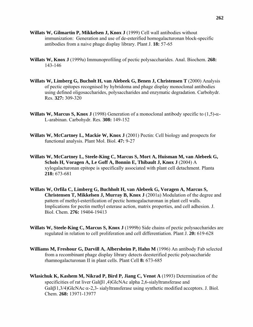
262
Willats W, Gilmartin P, Mikkelsen J, Knox J (1999) Cell wall antibodies without immunization: Generation and use of de-esterified homogalacturonan block-specific antibodies from a naive phage display library. Plant J. 18: 57-65
Willats W, Knox J (1999a) Immunoprofiling of pectic polysaccharides. Anal. Biochem. 268: 143-146
Willats W, Limberg G, Bucholt H, van Alebeek G, Benen J, Christensen T (2000) Analysis of pectic epitopes recognised by hybridoma and phage display monoclonal antibodies using defined oligosaccharides, polysaccharides and enzymatic degradation. Carbohydr. Res. 327: 309-320
Willats W, Marcus S, Knox J (1998) Generation of a monoclonal antibody specific to (1,5)-α-L-arabinan. Carbohydr. Res. 308: 149-152
Willats W, McCartney L, Mackie W, Knox J (2001) Pectin: Cell biology and prospects for functional analysis. Plant Mol. Biol. 47: 9-27
Willats W, McCartney L, Steele-King C, Marcus S, Mort A, Huisman M, van Alebeek G, Schols H, Voragen A, Le Goff A, Bonnin E, Thibault J, Knox J (2004) A xylogalacturonan epitope is specifically associated with plant cell detachment. Planta 218: 673-681
Willats W, Orfila C, Limberg G, Buchholt H, van Alebeek G, Voragen A, Marcus S, Christensen T, Mikkelsen J, Murray B, Knox J (2001a) Modulation of the degree and pattern of methyl-esterification of pectic homogalacturonan in plant cell walls. Implications for pectin methyl esterase action, matrix properties, and cell adhesion. J. Biol. Chem. 276: 19404-19413
Willats W, Steele-King C, Marcus S, Knox J (1999b) Side chains of pectic polysaccharides are regulated in relation to cell proliferation and cell differentiation. Plant J. 20: 619-628
Williams M, Freshour G, Darvill A, Albersheim P, Hahn M (1996) An antibody Fab selected from a recombinant phage display library detects deesterified pectic polysaccharide rhamnogalacturonan II in plant cells. Plant Cell 8: 673-685
Wlasichuk K, Kashem M, Nikrad P, Bird P, Jiang C, Venot A (1993) Determination of the specificities of rat liver Gal(β1,4)GlcNAc alpha 2,6-sialyltransferase and Gal(β1,3/4)GlcNAc α-2,3- sialyltransferase using synthetic modified acceptors. J. Biol. Chem. 268: 13971-13977
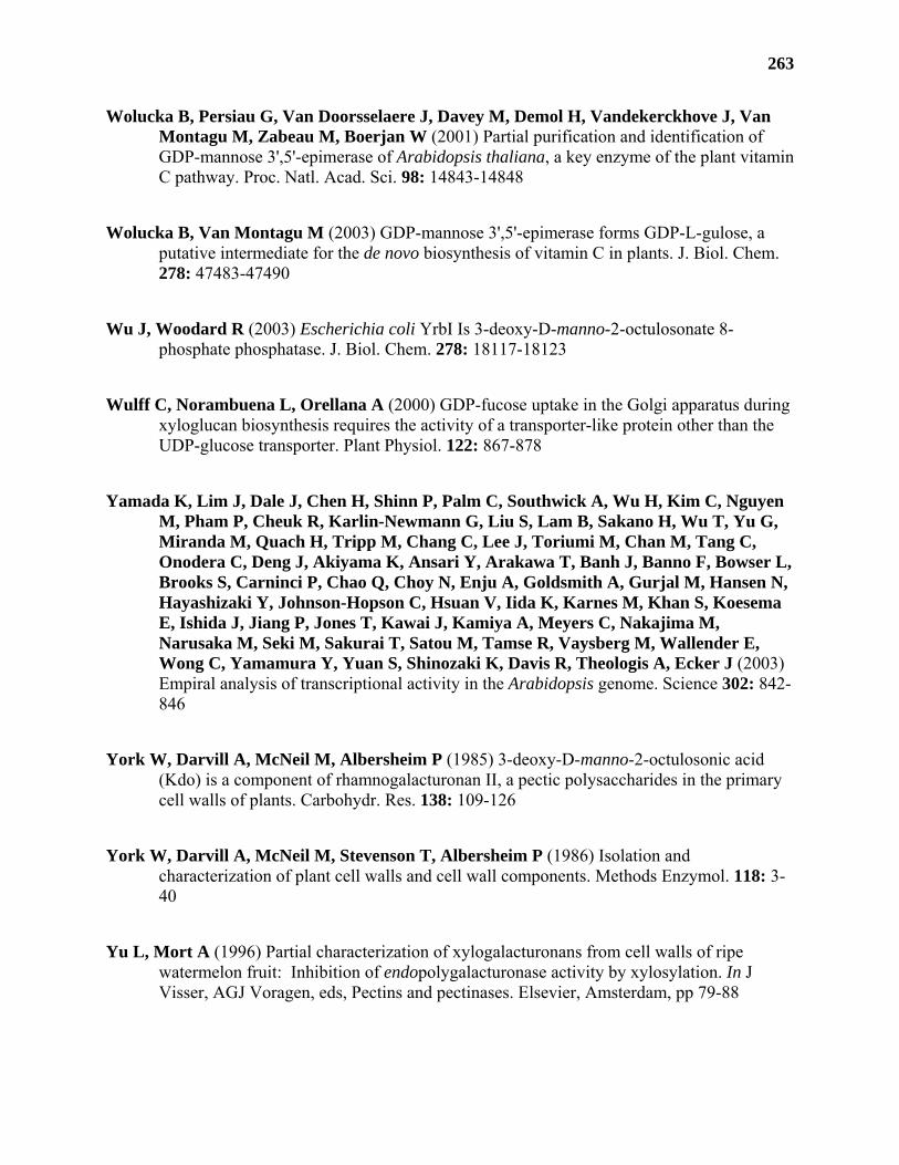
263
Wolucka B, Persiau G, Van Doorsselaere J, Davey M, Demol H, Vandekerckhove J, Van Montagu M, Zabeau M, Boerjan W (2001) Partial purification and identification of GDP-mannose 3',5'-epimerase of Arabidopsis thaliana, a key enzyme of the plant vitamin C pathway. Proc. Natl. Acad. Sci. 98: 14843-14848
Wolucka B, Van Montagu M (2003) GDP-mannose 3',5'-epimerase forms GDP-L-gulose, a putative intermediate for the de novo biosynthesis of vitamin C in plants. J. Biol. Chem. 278: 47483-47490
Wu J, Woodard R (2003) Escherichia coli YrbI Is 3-deoxy-D-manno-2-octulosonate 8-phosphate phosphatase. J. Biol. Chem. 278: 18117-18123
Wulff C, Norambuena L, Orellana A (2000) GDP-fucose uptake in the Golgi apparatus during xyloglucan biosynthesis requires the activity of a transporter-like protein other than the UDP-glucose transporter. Plant Physiol. 122: 867-878
Yamada K, Lim J, Dale J, Chen H, Shinn P, Palm C, Southwick A, Wu H, Kim C, Nguyen M, Pham P, Cheuk R, Karlin-Newmann G, Liu S, Lam B, Sakano H, Wu T, Yu G, Miranda M, Quach H, Tripp M, Chang C, Lee J, Toriumi M, Chan M, Tang C, Onodera C, Deng J, Akiyama K, Ansari Y, Arakawa T, Banh J, Banno F, Bowser L, Brooks S, Carninci P, Chao Q, Choy N, Enju A, Goldsmith A, Gurjal M, Hansen N, Hayashizaki Y, Johnson-Hopson C, Hsuan V, Iida K, Karnes M, Khan S, Koesema E, Ishida J, Jiang P, Jones T, Kawai J, Kamiya A, Meyers C, Nakajima M, Narusaka M, Seki M, Sakurai T, Satou M, Tamse R, Vaysberg M, Wallender E, Wong C, Yamamura Y, Yuan S, Shinozaki K, Davis R, Theologis A, Ecker J (2003) Empiral analysis of transcriptional activity in the Arabidopsis genome. Science 302: 842-846
York W, Darvill A, McNeil M, Albersheim P (1985) 3-deoxy-D-manno-2-octulosonic acid (Kdo) is a component of rhamnogalacturonan II, a pectic polysaccharides in the primary cell walls of plants. Carbohydr. Res. 138: 109-126
York W, Darvill A, McNeil M, Stevenson T, Albersheim P (1986) Isolation and characterization of plant cell walls and cell wall components. Methods Enzymol. 118: 3-40
Yu L, Mort A (1996) Partial characterization of xylogalacturonans from cell walls of ripe watermelon fruit: Inhibition of endopolygalacturonase activity by xylosylation. In J Visser, AGJ Voragen, eds, Pectins and pectinases. Elsevier, Amsterdam, pp 79-88
264
Zablackis E, Huang J, Muller B, Darvill A, Albersheim P (1995) Characterization of the cell-wall polysaccharides of Arabidopsis thaliana leaves. Plant Physiol. 107: 1129-1138
Zablackis E, York W, Pauly M, Hantus S, Reiter W, Chapple C, Albersheim P, Darvill A (1996) Substitution of L-fucose by L-galactose in cell walls of Arabidopsis mur1. Science 272: 1808-1810
Zar J (1999) Two-sample hypothesis. In T Ryu, ed, Biostatistical analysis, Ed Fourth. Simon and Schuster, New Jersey, pp 122-158
Zhang G, Staehelin L (1992) Functional compartmentation of the Golgi apparatus of plant cells: Immunocytochemical analysis of high-pressure frozen- and freeze-substituted sycamore maple suspension culture cells. Plant Physiol. 99: 1070-1083
Zhang Q, Liu H (2001) Chemical synthesis of UDP-β-L-arabinofuranose and its turnorver to UDP-β-L-arabinopyranose by UDP-galactopyranose mutase. Bioorg. Med. Chem. Lett. 11: 145-149