Targeting of nonlipidated, aggregated apoE with antibodies ...
Draft
Calpain inhibitor attenuates atherosclerosis and
inflammation in atherosclerotic rats through eNOS/NO/NF-κB pathway
Journal: Canadian Journal of Physiology and Pharmacology
Manuscript ID cjpp-2016-0652.R1
Manuscript Type: Article
Date Submitted by the Author: 07-Mar-2017
Complete List of Authors: Yu, Lan; Liaoning Medical University
Yin, Meihui; Liaoning Medical University, Yang, Xueyan; Liaoning Medical University, Lu, Meili; Liaoning Medical University, Tang, Futian; Liaoning Medical University Wang, Hongxin; Liaoning Medical University,
Is the invited manuscript for consideration in a Special
Issue?:
Keyword: calpain inhibitor CAI, atherosclerosis, nitric oxide, endothelial nitric oxide synthase, NF-κB
https://mc06.manuscriptcentral.com/cjpp-pubs
Canadian Journal of Physiology and Pharmacology
Draft
Calpain inhibitor attenuates atherosclerosis and inflammation in atherosclerotic
rats through eNOS/NO/NF-κB pathway
Lan Yu *ab
, Meihui Yin *a
, Xueyan Yang ac
, Meili Lu
a, Futian Tang
a, Hongxin Wang
a
a Key Laboratory of Cardiovascular and Cerebrovascular Drug Research of Liaoning
Province, Jinzhou Medical University, Jinzhou 121001, China
b Central Hospital of Yingkou Development Areas, Yingkou, 115007, China
c Internal Medicine-Cardiovascular Departments, the First Affiliated Hospital of Jinzhou
Medical University, Jinzhou 121001, China
Corresponding authors: Futian Tang (email: [email protected]) and Hongxin Wang (email:
*These authors contributed equally to this work.
Page 1 of 23
https://mc06.manuscriptcentral.com/cjpp-pubs
Canadian Journal of Physiology and Pharmacology
Draft
Abstract:
We previously reported that calpain, the Ca2+
-sensitive cysteine protease, gets involved in
the atherogenesis. This study was to investigate the effects of calpain inhibitor (CAI, 5
mg/kg/d) with or without NG-nitro-L-arginine-methyl ester (L-NAME) (100 mg/kg/d), the
inhibitor of nitric oxide synthase (NOS), on atherosclerosis and inflammation in rat
model induced by high cholesterol diet (HCD). The results demonstrated HCD increased
protein expression of calpain-1 but not calpain-2 in aortic tissue. In addition, CAI reduced
the thickness of atherosclerotic intima compared to HCD group, which was weakened by
the L-NAME combination. CAI with or without L-NAME decreased the activity of
calpain in the aorta. Besides, CAI decreased the expressions of vascular cell adhesion
molecule-1 (VCAM-1), intracellular cell adhesion molesul-1 (ICAM-1) and monocyte
chemoattractant protein-1 (MCP-1) in the aorta at the levels of both mRNA and protein.
Furthermore, CAI increased the activity and the protein expression of endothelial NOS
(eNOS) accompanied by increased content of NO and down-regulated the protein
expression of nuclear factor κB (NF-κB) of the nucleus in the aorta. However, the
above-mentioned effects were at least partly cancelled by L-NAME except for the protein
expression of eNOS. The results suggested that CAI attenuated atherosclerosis and
inflammation through eNOS/NO/NF-κB pathway.
Key Words: calpain inhibitor CAI, atherosclerosis; nitric oxide; endothelial nitric oxide
synthase; NF-κB
Introduction
Atherosclerosis has been reported to be the main cause of coronary artery disease
(CAD) and stroke (Peters et al. 2012). Vascular endothelial dysfunction-mediated
inflammatory response gets involved in the initiation and development of atherosclerosis.
Endothelial dysfunction contributes to atherosclerosis largely due to the decrease in
bioavailability of nitric oxide (NO) which is originated from endothelial NO synthase
(eNOS) (Sharma et al. 2015). Compared with apolipoprotein E knockout (ApoE KO)
mice, ApoE/eNOS double KO mice showed the increase in atherosclerotic lesion, which
suggested that eNOS protects the ApoE KO mice against the development of
Page 2 of 23
https://mc06.manuscriptcentral.com/cjpp-pubs
Canadian Journal of Physiology and Pharmacology
Draft
atherosclerosis (Kus et al. 2014). In addition, the combination of a NO-donating property
enhances the anti-atherogenic activity of atorvastatin (Momi et al. 2012). Endothelial
dysfunction-mediated inflammation contributes to the atherogenesis largely depending on
the induction of expression of inflammatory molecules including intercellular adhesion
molecule-1 (ICAM-1), vascular cell adhesion molecule-1 (VCAM-1) and monocyte
chemoattractant protein-1 (MCP-1) (Shah et al. 2015). Release impairment of NO from
endothelial cells leads to the up-regulation of ICAM-1, VCAM-1 and that NO synthesis
inhibition increases the gene expression of MCP-1 in endothelial cells (Kang et al. 2005;
Khan et al. 1996; Sohn et al. 2005a). Administration of NO donors to the animal deficient
in NO maintains the endothelial function and inhibits pathological interactions between
leukocytes in the circulation and the vascular endothelium (De Caterina et al. 1995; Sohn
et al. 2005b). In addition, the expressions of ICAM-1, VCAM-1 and MCP-1 are
associated with the activation of nuclear factor κB (NF-κB), a transcription protein
(Zeiher et al. 1995). In the physiological state, as a heterodimer of the p50 and p65
subunits, NF-κB stays in the cytoplasm. However, upon stimulation in the pathological
state, p50 and p65 can go to the nucleus, where affecting the expression of genes such as
ICAM-1, VCAM-1 and MCP-1 (Hall et al. 2006; Jones et al. 2003).
Calpains, the Ca2+
dependent cysteine proteases, strictly regulate their substrate
proteins by the limited proteolysis (Subramanian et al. 2012). Calpain family is
comprised of 15 homologues of catalytic subunits and 2 homologues of regulatory
subunits. Among the superfamily, Calpain-1 and calpain-2 are categorized as
“conventional calpain” (Daugherty et al. 2000). By using pharmacological inhibitors or
mice deficient in calpain-1 or calpain-2 or mice over-expressing calpastatin, the
endogenous inhibitor, many studies demonstrated that inactivation of calpain inhibited
atherosclerosis, suggesting the involvement of calpains in the development of
atherosclerosis (Clinkinbeard et al. 2013; Hua and Nair 2015; Miyazaki et al. 2013;
Ruetten and Thiemermann 1997; Takeshita et al. 2013). We previously reported that
calpain-1 gets involved in the atherogenesis by up-regulating the scavenger receptor
CD36 expression (Yang et al. 2016). In addition to conventional calpain, one of
unconventional calpain isozyme, calpain-6, appears to be induced in macrophages during
atherogenesis, thereby facilitating form cell formation (Miyazaki et al. 2016). Calpains
Page 3 of 23
https://mc06.manuscriptcentral.com/cjpp-pubs
Canadian Journal of Physiology and Pharmacology
Draft
have also been implicated to play a critical deleterious role in endothelial dysfunction
(Stalker et al. 2003). Activated calpain by Ca2+
damages the cells by the selective
degradation of the proteins such as eNOS and IκB (Averna et al. 2008; Dong et al. 2009).
Calpains also mediates the inflammatory processes via the NF-κB activation (Li et al.
2014). Taken together, these studies indicated that calpains get involved in the
atherosclerosis largely through endothelial dysfunction and inflammatory response.
Nevertheless, the molecular mechanisms underlying the involvement remain to be
investigated. This study was to evaluate the significance of NO in the inhibition of
atherosclerosis by calpain inhibitor CAI in rat model with early atherosclerosis induced
by high dose of vitamin D2 and diet with high content of cholesterol. The results
demonstrated that CAI inhibited the atherosclerosis through eNOS/NO/NF-κB pathway.
Materials and methods
Chemicals
Calpain inhibitor I (N-acetyl-leu-leu-norleucinal, CAI) was the product of Santa Cruz
Biotechnology (sc-29119, MW: 383.5). NG-nitro-L-arginine-methyl ester (L-NAME) was
purchased from Beyotime Institute of Biotechnology, Shanghai, China.
Atherosclerosis model of rats
The study was approved by the Committee on the Ethics of Animal Experiments of the
Jinzhou Medical University, China (Permit Number LNMU-2015-118) and performed in
accordance with the Guide for the Care and Use of Laboratory Animals of the National
Institutes of Health (Institute of Laboratory Animal Resources (U.S.). Committee on Care
and Use of Laboratory Animals.). Thirty-two six-week-old male Sprague–Dawley (SD)
rats (Animal Center, Jinzhou Medical University, China) were randomly and equally
divided into Control, HCD, CAI and CAI+ L-NAME groups. The atherosclerotic rat
model was established as described previously (Tang et al. 2006a). Briefly, vitamin D2
(300,000 IU/kg/day) (Sigma) was given to all rats by gavage for 4 days followed by fed
with animal chow in control group (Con) and high cholesterol diet (HCD) containing 2%
cholesterol in HCD, CAI and CAI+ L-NAME groups for 8 weeks. Rats in CAI and CAI+
Page 4 of 23
https://mc06.manuscriptcentral.com/cjpp-pubs
Canadian Journal of Physiology and Pharmacology
Draft
L-NAME groups were intraperitoneally injected CAI (0.5mg/kg/day) combined with or
without L-NAME (100 mg/kg/d). At the end of the experiment, the blood was collected
from eyes of rats under light anesthesia by ether from which serum was separated for
biochemical analysis. The rats were then sacrificed by peritoneal injection of over dose of
pentobarbital sodium (30 mg/kg) and the aortic issue was separated.
Morphological changes in aortic arch
Six-micrometer-thick section of aortic arch fixed in 10% formalin was stained with
hematoxylin and eosin as previously reported method (Tang et al. 2007) and the ratio of
thickness between intima (I) and intima plus media (I+M) was calculated.
Contents of cholesterol in aorta and lipid profiles in serum
Cholesterol content in lysate of aorta and the serum levels of total cholesterol (TC),
triacylglycerol (TG), low density lipoprotein (LDL) cholesterol and high density
lipoprotein (HDL) cholesterol were determined using kit from Beijing Zhongsheng
Bioengineering Company (China) as previously reported method (Tang et al. 2007; Tang
et al. 2006a).
Activity of calpain in aorta
Calpain activity in lysate of aorta was measured using a fluorescence substrate
N-succinyl-LLVY-AMC (Amyjet Scientific Inc, China) as previously reported method
(Tang et al. 2015).
Content of NO and eNOS activity in aorta
NO content and eNOS activity in lysate of aorta was determined using kit from
Nanjing Jiancheng Bioengineering Company (China) as previously reported method
(Tang et al. 2006b).
Immunohistochemical analysis of eNOS in vascular endothelia
Sections of aortic arch were used to determine the protein expression of eNOS as
previously reported method (Tang et al. 2007). The first and second antibody was rabbit
Page 5 of 23
https://mc06.manuscriptcentral.com/cjpp-pubs
Canadian Journal of Physiology and Pharmacology
Draft
anti-rat eNOS and sheep anti-rabbit IgG-peroxidase antibodies provided by Abcam
Company (Cambridge, MA, USA) respectively.
Contents of VCAM-1, ICAM-1 and MCP-1 in aorta
Contents of VCAM-1, ICAM-1 and MCP-1 in lysate of aorta was measured using
ELISA kits from R&D Systems (Minneapolis, MN, USA) as previously reported method
(Tang et al. 2016). The color absorbance at 450 nm was measured using a Bio-Rad
microplate reader.
mRNA expression of ICAM-1, VCAM-1 and MCP-1 in aorta
The mRNA expression of ICAM-1, VCAM-1 and MCP-1 in aorta was analyzed using
the BioRad iQ5 Real Time PCR system (BioRad Company) as previously reported
method (Tang et al. 2016). Primers used for qPCR are provided in Table 1.
Table 1. Primers used for qPCR.
Genes Forward primer(5’-3’) Reverse primer(5’-3’) GAPDH
MCP-1
ICAM-1
VCAM-1
CCACCCATGGCAAATTCCATGGCA
CTCACCTGCTGCTACTCATTCAC
GTGATGCTCAGGTATCCATCCA
GTTCCAGCGAGGGTCTACC
TCTAGACGGCAGGTCAGGTCCACC
ATGTCTGGACCCATTCCTTCTTG
CACAGTTCTCAAAGCACAGCG
AACTCTTGGCAAACATTAGGTGT
Protein expressions of calpain-1, calpain-2, CD68, eNOS and NF-κB in aorta
The protein expression of calpain-1, calpain-2, CD68, eNOS and p65 in aorta was
examined using Western blot as previously reported method (Luan et al. 2015; Tang et al.
2016). Nuclear proteins in aortic tissue were separated using Nuclear and Cytoplasm
Extraction Kit (Active Motif provided by Dakewe Biotech Limited, China) as previously
reported method (Tang et al. 2016). Antibodies against eNOS (1.1000), p65 (1:1500),
lamin B (1:1000) and GAPDH (1:500) were from Abcam (Cambridge, MA, USA).
Antibodies against calpain-1 (1:1000), calpain-2 (1:1000) and CD68 (1:2000) were from
Santa Cruz Biotechnology (Santa Cruz, CA, USA).
Statistical analysis
Data are shown as the mean ±SEM and analyzed by one-way analysis of variance
(ANOVA) and the t-test using SPSS 17.0 software. P<0.05 showed the statistically
Page 6 of 23
https://mc06.manuscriptcentral.com/cjpp-pubs
Canadian Journal of Physiology and Pharmacology
Draft
significant difference.
Results
CAI reduces the atherosclerosis and CD68 protein expression independent of the
serum lipid profiles
As shown in Figure 1, rats in HCD group demonstrated the significant increases in the
ratio of I to (I+M) (Figure 1A and 1B), the cholesterol content (Figure 1C) and CD68
protein expression (Figure 1D) in the aorta, all representing the formation of
atherosclerosis. However, these increases were attenuated by CAI, the specific calpain
inhibitor, suggesting that activation of calpain implies in the pathogenesis of
atherosclerosis. In addition, combination of L-NAME, the specific NOS inhibitor, with
CAI partly abolished the inhibitory effects of CAI on atherosclerosis. The results
indicated that CAI reduces the atherosclerosis at least partly through regulation of
NOS/NO pathway. Furthermore, both CAI and L-NAME did not affect the lipid profiles
in serum changed by HCD (Figure 1E), suggesting that the actions of CAI and L-NAME
on atherosclerosis are independent of serum lipid profiles.
CAI inactivates calpain without affecting the protein expressions of calpain-1 and
calpain-2
Calpain activity (Figure 2A) and calpain-1 protein expression (Figure 2B) in aortic
tissue of rats in HCD group increased compared with that of control rats. CAI reduced the
calpain activity without affecting the protein expression of calpain-1. Calpain activity
was comparable between CAI and CAI+ L-NAME groups. CAI in combination with or
without L-NAME had no effect on calpain-1 protein expression compared with HCD. In
addition, the protein expression of calpain-2 was unchanged among all four groups.
These results suggested that inactivation of calpain rather than down-regulation of
calpain-1 or calpain-2 protein expression by CAI contributes the inhibition of
atherosclerosis.
CAI increases the NO content and the activity and protein expression of eNOS in
Page 7 of 23
https://mc06.manuscriptcentral.com/cjpp-pubs
Canadian Journal of Physiology and Pharmacology
Draft
aorta
NO synthesized by eNOS exerts effect of anti-atherosclerosis. In order to investigate
the involvement of eNOS/NO in the inhibition of atherosclerosis by CAI, we examined
the content of NO, the activity and protein expression of eNOS in aorta. The results
showed the significant decreases in the NO content (Figure 3A) and eNOS activity
(Figure 3B) and protein expression (Figure 3C and 3D) in rat aorta of HCD group
compared with that in control group, suggesting that deficiency of NO production might
contribute to the atherosclerosis. However, these decreases were changed inversely by
CAI. Expectedly, combination of L-NAME with CAI partly reduced the effects of CAI on
NO content and eNOS activity without affecting the eNOS protein expression. Taken
together, the results further confirmed the hypothesis that enhancement of eNOS/NO
pathway gets involved in the inhibition of CAI on atherosclerosis.
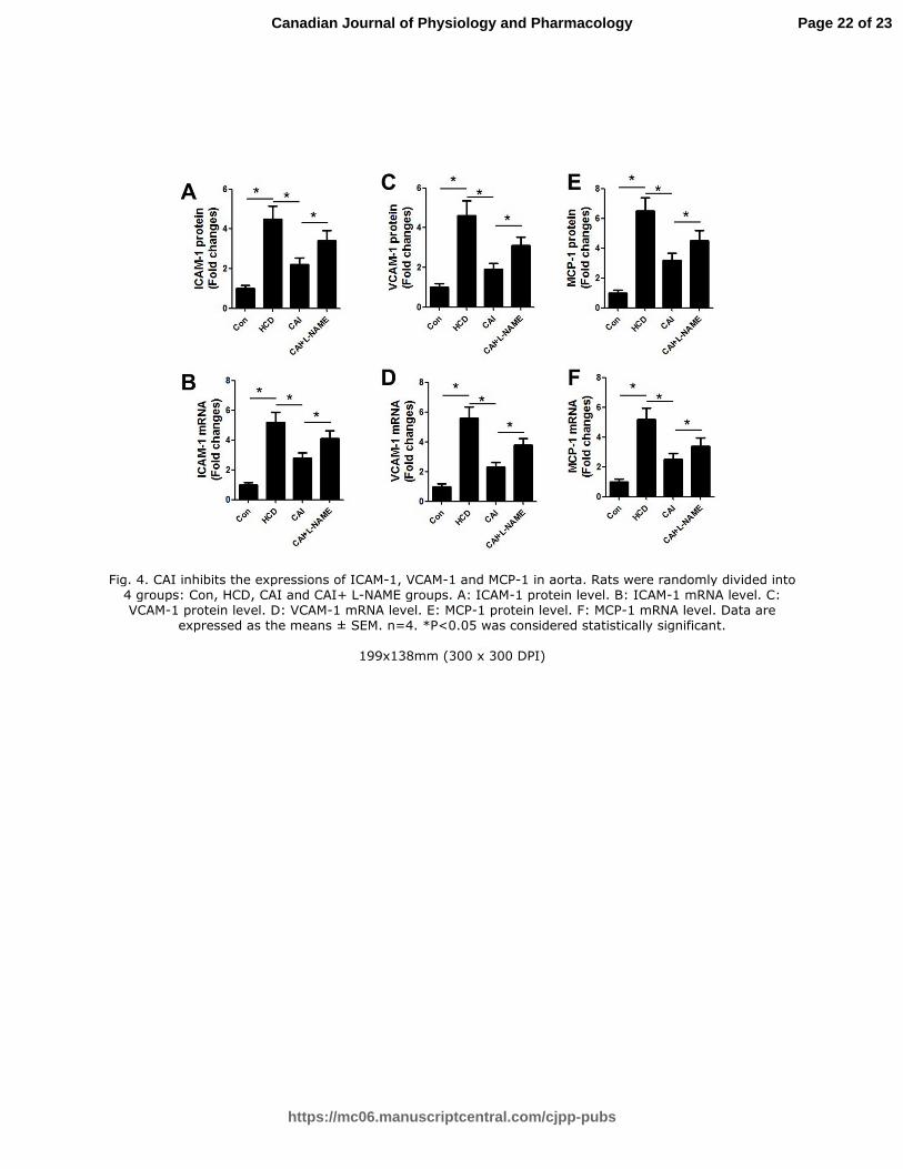
CAI inhibits the expressions of ICAM-1, VCAM-1 and MCP-1 in aorta
The results indicated that the mRNA and protein expressions of vascular inflammatory
molecules ICAM-1 (Figure. 4A and 4B), VCAM-1 (Figure. 4C and 4D) and MCP-1
(Figure. 4E and 4F) in HCD group significantly increased compared with that in control
group. However, these increases were reduced by CAI, which might at least partly
explain the mechanism underlying the inhibition of atherosclerosis by CAI. In addition,
combination of L-NAME with CAI partly abolished the effects of CAI on these
inflammatory molecules. Taken together, the results indicated that inhibition of
inflammation by CAI might be consequence of the enhancement of eNOS/NO pathway.
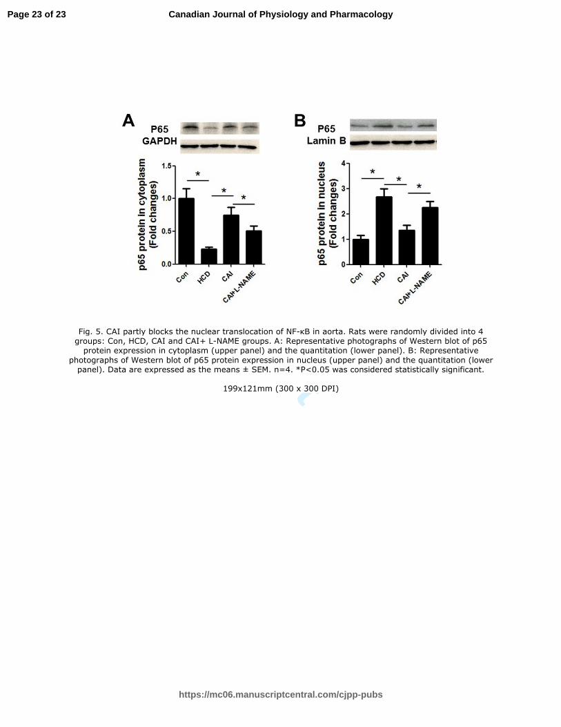
CAI reduces the NF-κB translocation into nucleus of aorta
NF-κB activation and nuclear translocation regulates the inflammation. Therefore, we
examined the protein expression of p65, the subunit of NF-κB in both cytoplasm and
nucleus of aorta to clarify the mechanisms underlying the regulation of inflammation by
CAI. The results showed that p65 protein expression significantly decreased in cytoplasm
(Figure. 5A), while increased in nucleus (Figure. 5B) in HCD group compared with that
in control group. However, the translocation was partly inhibited by CAI. Furthermore,
combination of L-NAME with CAI partly abolished the effects of CAI on nuclear
Page 8 of 23
https://mc06.manuscriptcentral.com/cjpp-pubs
Canadian Journal of Physiology and Pharmacology
Draft
translocation of NF-κB. Collectively, the results indicated that attenuation of
inflammation by CAI might be attributed to the inhibition of nuclear translocation of
NF-κB.
Discussion
We reported in this study that CAI, the specific inhibitor of calpain, attenuated the
atherosclerosis, increased the activity and the protein expression of eNOS, elevated the
content of NO, down-regulated the expression of ICAM-1, VCAM-1 and MCP-1 and
inhibited the nuclear translocation of NF-κB in aorta. However, all the beneficial effects
of CAI were partly abolished by L-NAME, the specific inhibitor of NOS, except for the
protein expression of eNOS. The results suggested that CAI inhibited the atherosclerosis
through eNOS/NO/NF-κB pathway.
By using pharmacological inhibitors or mice deficient in calpain-1 or calpain-2 or mice
over-expressing calpastatin, the endogenous inhibitor, many studies demonstrated that
inactivation of calpain inhibited the atherosclerosis, suggesting that calpains gets
involved in the pathogenesis of atherosclerosis (Clinkinbeard et al. 2013; Hua and Nair
2015; Miyazaki et al. 2013; Ruetten and Thiemermann 1997; Takeshita et al. 2013).
Expectedly, the present results demonstrated that CAI attenuated the atherosclerotic
lesion and reduced the vascular content of cholesterol in rats fed with high cholesterol
diet and excessive vitamin D. However, combination of CAI with L-NAME partly
abolished the inhibitory effect on atherosclerosis by CAI, suggesting that regulation of
endothelial NOS/NO pathway might contribute the inhibitory effect of CAI on
atherosclerotic lesions. Accumulative studies reported that the increase in production of
endothelial NO protected animals against atherosclerosis (De Caterina et al. 1995; Momi
et al. 2012; Zeiher et al. 1995). Regarding the roles of calpain-1 and calpain-2 in the
formation of atherosclerosis, both calpain-1 and calpain-2 are reportedly expressed in
macrophages in angiotensin II-infused athero-prone aorta, and are associated with their
inflammatory responses (Howatt et al. 2016). In addition, calpain-2 appears to be
upregulated in vascular endothelium during atherogenesis, and is contributed to
endothelial barrier dysfunction (Miyazaki et al. 2011). We examined the protein
expression of calpain-1 and calpain-2 in the aortic tissue and found that protein
Page 9 of 23
https://mc06.manuscriptcentral.com/cjpp-pubs
Canadian Journal of Physiology and Pharmacology
Draft
expression of calpain-1 rather than calpain-2 in atherosclerotic rat model increased
compared with that of control rats, suggesting that calpain-1 but not calpain-2 plays
important role in the formation of atherosclerosis of rat model. The present result was not
consistent with these reports and the inconsistency might be attributed to the different
animal models.
To further clarify the involvement of eNOS/NO pathway in the mechanism underlying
the inhibition of CAI on atherosclerosis, we examined the NO content, eNOS activity and
protein expression in aorta. As expected, CAI increased the eNOS activity and the
content of NO in aorta. In addition, CAI up-regulated eNOS protein expression in aortic
tissue assessed by both western blot and in endothelia evaluated by immunohistochemical
method. Consistently, combination of L-NAME weakened the effects of CAI on the
content of NO and eNOS activity without affecting the eNOS protein expression. The
results directly confirmed that the attenuation of the atherosclerosis by CAI was at least
partly mediated through eNOS/NO pathway. The present study was well in agreement
with the following reports. Kus et al. compared the atherosclerotic lesion between ApoE
KO mice and ApoE/eNOS double KO mice and found the increase in atherosclerotic
lesion in the latter mice, which suggested that eNOS plays the protective role against
atherosclerosis (Kus et al. 2014). Momi et al. reported that NO-donating property
enhances the anti-inflammatory and anti-atherogenic activity of atorvastatin (Momi et al.
2012). Regarding the possible mechanisms underlying the improvement of eNOS/NO
pathway by CAI, Averna et al. reported that incubation of endothelial cells in
Ca2+
-loading medium for a long time caused the degradation of eNOS through calpain
activation. The eNOS degradation further resulted in significant decrease in NO
production (Averna et al. 2008). Dong et al. also found that impaired endothelial function
was attributed to eNOS degradation mediated by calpain (Dong et al. 2009). Nevertheless,
the exact mechanisms need further investigated.
Inflammatory process is implied in atherosclerosis. To investigate the down-stream
response to the improvement of eNOS/NO pathway by CAI, we examined the
expressions of inflammatory molecules ICAM-1, MCP-1 and VCAM-1 in aorta at the
protein and mRNA levels. The results demonstrated that CAI lowered the expressions of
ICAM-1, VCAM-1 and MCP-1, which was partly reversed by combination of L-NAME.
Page 10 of 23
https://mc06.manuscriptcentral.com/cjpp-pubs
Canadian Journal of Physiology and Pharmacology
Draft
These results indicated that inhibition of inflammation by CAI was mediated by the
improvement of eNOS/NO pathway. Consistently, studies using atherosclerotic model
mice lacking of VCAM-1 or MCP gene or the mice over-expressing MCP-1 gene, have
showed the importance of these gene in atherogenesis (Cybulsky et al. 2001; Nelken et al.
1991). Furthermore, NO synthesis inhibition by high dosage of L-NAME to rats induces
vascular inflammation and atherosclerosis (Kataoka et al. 2004). These investigations
indicate that inflammation responses in atherogenesis are closely linked with the impaired
NO synthesis, and therefore improvement of NO production might be one of the
approaches for anti-inflammation, and anti-atherosclerosis. Regarding the mechanisms by
which calpain regulates the eNOS/NO function, subsequently inducing the inflammation,
there is study revealing that glucose induces loss of NO via a calpain-dependent decrease
in the association of heat shock protein 90 (hsp90) with eNOS (Stalker et al. 2003). In
addition, inhibition of calpain activity decreased endothelial cell surface expression of the
pro-inflammatory adhesion molecules ICAM-1 and VCAM-1 during hyperglycemia
(Stalker et al. 2003). These data demonstrate that calpains contribute to important
inflammatory events during hyperglycemia and that pharmacological inhibition of calpain
activity attenuates leukocyte endothelium interactions and preserves eNOS function.
The expressions of inflammatory molecules can be up-regulated by NF-κB activation.
In the physiological state, as a heterodimer of the p50 and p65 subunits, NF-κB stays in
the cytoplasm. However, upon stimulation in the pathological state, p50 and p65 can go
to the nucleus, where affecting the expression of genes such as ICAM-1, VCAM-1 and
MCP-1 (Chen et al. 1995). To examine the mechanism underlying the
eNOS/NO-mediated inhibition of inflammation by CAI, we observed the effect of CAI
on p65 translocation into nucleus. The study demonstrated that CAI attenuated the p65
nuclear translocation, which was partly abolished by combination of L-NAME. Part of
this result was supported by the finding that knockdown of endogenous P65 in HUVECs
through siRNA decreased the expressions of ICAM-1 and VCAM-1 at the mRNA and
protein levels (Wu et al. 2014). Taken together, these results indicated that CAI inhibited
the endothelial inflammation at least in part through eNOS/NO mediated attenuation of
p65 translocation into nucleus, subsequently reducing the atherosclerosis.
In summary, the results suggested that CAI attenuated the inflammation and
Page 11 of 23
https://mc06.manuscriptcentral.com/cjpp-pubs
Canadian Journal of Physiology and Pharmacology
Draft
atherosclerosis in rats through eNOS/NO/NF-κB pathway.
This study has several limitations. Firstly, HCD+L-NAME group should be assigned to
investigate the effect of L-NAME alone on the atherosclerosis, which might exclude the
direct effect of L-NAME on the atherosclerosis. Secondly, the use of non-selective
inhibitor of NOS, L-NAME can not exclude the facts that other isoforms of NOS, as well
as arginase might be also involved in pathogenesis, which requires the use of selective
inhibitor of NOS in the future study. Finally, the study lacks the mechanisms by which
calpain regulates the eNOS/NO pathway, which requires further investigation in the
future study, e.g. by checking the dissociation of HSP90 from eNOS.
Declarations
Conflict of interest
The authors declare that they have no competing interests.
Acknowledgment
This work was supported by National Natural Science Foundation of China
(No.81374008), Talent Fund of Liaoning Medical University (No. 2014-18) and Natural
Science Foundation of Liaoning Province (No. 2015020325). We thank Yingjie Zhang
for her critical suggestions in the revision of our manuscript.
Reference
Averna, M., Stifanese, R., De Tullio, R., Passalacqua, M., Salamino, F., Pontremoli, S.,
and Melloni, E. 2008. Functional role of HSP90 complexes with endothelial
nitric-oxide synthase (eNOS) and calpain on nitric oxide generation in endothelial cells.
J. Biol. Chem. 283(43): 29069-29076. doi: 10.1074/jbc.M803638200.
Chen, C.C., Rosenbloom, C.L., Anderson, D.C., and Manning, A.M. 1995. Selective
inhibition of E-selectin, vascular cell adhesion molecule-1, and intercellular adhesion
molecule-1 expression by inhibitors of I kappa B-alpha phosphorylation. J. Immunol.
155(7): 3538-3545.
Clinkinbeard, T., Ghoshal, S., Craddock, S., Creed Pettigrew, L., and Guttmann, R.P.
2013. Calpain cleaves methionine aminopeptidase-2 in a rat model of
ischemia/reperfusion. Brain Res. 1499: 129-135. doi: 10.1016/j.brainres.2012.12.039.
Cybulsky, M.I., Iiyama, K., Li, H., Zhu, S., Chen, M., Iiyama, M., Davis, V.,
Gutierrez-Ramos, J.C., Connelly, P.W., and Milstone, D.S. 2001. A major role for
Page 12 of 23
https://mc06.manuscriptcentral.com/cjpp-pubs
Canadian Journal of Physiology and Pharmacology
Draft
VCAM-1, but not ICAM-1, in early atherosclerosis. J. Clin. Invest. 107(10):
1255-1262. doi: 10.1172/JCI11871.
Daugherty, A., Manning, M.W., and Cassis, L.A. 2000. Angiotensin II promotes
atherosclerotic lesions and aneurysms in apolipoprotein E-deficient mice. J. Clin.
Invest. 105(11): 1605-1612. doi: 10.1172/JCI7818.
De Caterina, R., Libby, P., Peng, H.B., Thannickal, V.J., Rajavashisth, T.B., Gimbrone,
M.A., Jr., Shin, W.S., and Liao, J.K. 1995. Nitric oxide decreases cytokine-induced
endothelial activation. Nitric oxide selectively reduces endothelial expression of
adhesion molecules and proinflammatory cytokines. J. Clin. Invest. 96(1): 60-68. doi:
10.1172/JCI118074.
Dong, Y., Wu, Y., Wu, M., Wang, S., Zhang, J., Xie, Z., Xu, J., Song, P., Wilson, K., Zhao,
Z., Lyons, T., and Zou, M.H. 2009. Activation of protease calpain by oxidized and
glycated LDL increases the degradation of endothelial nitric oxide synthase. J. Cell
Mol. Med. 13(9A): 2899-2910. doi: 10.1111/j.1582-4934.2008.00416.x.
Hall, G., Hasday, J.D., and Rogers, T.B. 2006. Regulating the regulator: NF-kappaB
signaling in heart. J. Mol. Cell Cardiol. 41(4): 580-591. doi:
10.1016/j.yjmcc.2006.07.006.
Howatt, D.A., Balakrishnan, A., Moorleghen, J.J., Muniappan, L., Rateri, D.L., Uchida,
H.A., Takano, J., Saido, T.C., Chishti, A.H., Baud, L., and Subramanian, V. 2016.
Leukocyte Calpain Deficiency Reduces Angiotensin II-Induced Inflammation and
Atherosclerosis But Not Abdominal Aortic Aneurysms in Mice. Arterioscler. Thromb.
Vasc. Biol. 36(5): 835-845. doi: 10.1161/ATVBAHA.116.307285.
Hua, Y., and Nair, S. 2015. Proteases in cardiometabolic diseases: Pathophysiology,
molecular mechanisms and clinical applications. Biochim. Biophys. Acta 1852(2):
195-208. doi: 10.1016/j.bbadis.2014.04.032.
Institute of Laboratory Animal Resources (U.S.). Committee on Care and Use of
Laboratory Animals. Guide for the care and use of laboratory animals. In NIH
publication. U.S. Dept. of Health and Human Services, Public Health Service,
Bethesda, Md. p. v.
Jones, W.K., Brown, M., Ren, X., He, S., and McGuinness, M. 2003. NF-kappaB as an
integrator of diverse signaling pathways: the heart of myocardial signaling? Cardiovasc.
Toxicol. 3(3): 229-254.
Kang, D.G., Moon, M.K., Choi, D.H., Lee, J.K., Kwon, T.O., and Lee, H.S. 2005.
Vasodilatory and anti-inflammatory effects of the
1,2,3,4,6-penta-O-galloyl-beta-D-glucose (PGG) via a nitric oxide-cGMP pathway. Eur.
J. Pharmacol. 524(1-3): 111-119. doi: 10.1016/j.ejphar.2005.08.061.
Kataoka, C., Egashira, K., Ishibashi, M., Inoue, S., Ni, W., Hiasa, K., Kitamoto, S., Usui,
M., and Takeshita, A. 2004. Novel anti-inflammatory actions of amlodipine in a rat
model of arteriosclerosis induced by long-term inhibition of nitric oxide synthesis. Am.
J. Physiol. Heart Circ. Physiol. 286(2): H768-774. doi: 10.1152/ajpheart.00937.2002.
Khan, B.V., Harrison, D.G., Olbrych, M.T., Alexander, R.W., and Medford, R.M. 1996.
Nitric oxide regulates vascular cell adhesion molecule 1 gene expression and
redox-sensitive transcriptional events in human vascular endothelial cells. Proc. Natl.
Acad. Sci. U S A 93(17): 9114-9119.
Kus, K., Wisniewska, A., Toton-Zuranska, J., Olszanecki, R., Jawien, J., and Korbut, R.
2014. Significant deterioration of anti-atherogenic efficacy of nebivolol in a double
Page 13 of 23
https://mc06.manuscriptcentral.com/cjpp-pubs
Canadian Journal of Physiology and Pharmacology
Draft
(apolipoprotein E and endothelial nitric oxide synthase) knockout mouse model of
atherosclerosis in comparison to single (apolipoprotein E) knockout model. J. Physiol.
Pharmacol. 65(6): 877-881.
Li, X., Luo, R., Chen, R., Song, L., Zhang, S., Hua, W., and Chen, H. 2014. Cleavage of
IkappaBalpha by calpain induces myocardial NF-kappaB activation, TNF-alpha
expression, and cardiac dysfunction in septic mice. Am. J. Physiol. Heart Circ. Physiol.
306(6): H833-843. doi: 10.1152/ajpheart.00893.2012.
Luan, A., Tang, F., Yang, Y., Lu, M., Wang, H., and Zhang, Y. 2015. Astragalus
polysaccharide attenuates isoproterenol-induced cardiac hypertrophy by regulating
TNF-alpha/PGC-1alpha signaling mediated energy biosynthesis. Environ. Toxicol.
Pharmacol. 39(3): 1081-1090. doi: 10.1016/j.etap.2015.03.014.
Miyazaki, T., Koya, T., Kigawa, Y., Oguchi, T., Lei, X.F., Kim-Kaneyama, J.R., and
Miyazaki, A. 2013. Calpain and atherosclerosis. J. Atheroscler. Thromb. 20(3):
228-237.
Miyazaki, T., Taketomi, Y., Takimoto, M., Lei, X.F., Arita, S., Kim-Kaneyama, J.R., Arata,
S., Ohata, H., Ota, H., Murakami, M., and Miyazaki, A. 2011. m-Calpain induction in
vascular endothelial cells on human and mouse atheromas and its roles in VE-cadherin
disorganization and atherosclerosis. Circulation 124(23): 2522-2532. doi:
10.1161/CIRCULATIONAHA.111.021675.
Miyazaki, T., Tonami, K., Hata, S., Aiuchi, T., Ohnishi, K., Lei, X.F., Kim-Kaneyama,
J.R., Takeya, M., Itabe, H., Sorimachi, H., Kurihara, H., and Miyazaki, A. 2016.
Calpain-6 confers atherogenicity to macrophages by dysregulating pre-mRNA splicing.
J. Clin. Invest. 126(9): 3417-3432. doi: 10.1172/JCI85880.
Momi, S., Monopoli, A., Alberti, P.F., Falcinelli, E., Corazzi, T., Conti, V., Miglietta, D.,
Ongini, E., Minuz, P., and Gresele, P. 2012. Nitric oxide enhances the
anti-inflammatory and anti-atherogenic activity of atorvastatin in a mouse model of
accelerated atherosclerosis. Cardiovasc. Res. 94(3): 428-438. doi: 10.1093/cvr/cvs100.
Nelken, N.A., Coughlin, S.R., Gordon, D., and Wilcox, J.N. 1991. Monocyte
chemoattractant protein-1 in human atheromatous plaques. J. Clin. Invest. 88(4):
1121-1127. doi: 10.1172/JCI115411.
Peters, S.A., den Ruijter, H.M., Bots, M.L., and Moons, K.G. 2012. Improvements in risk
stratification for the occurrence of cardiovascular disease by imaging subclinical
atherosclerosis: a systematic review. Heart 98(3): 177-184. doi:
10.1136/heartjnl-2011-300747.
Ruetten, H., and Thiemermann, C. 1997. Effect of calpain inhibitor I, an inhibitor of the
proteolysis of I kappa B, on the circulatory failure and multiple organ dysfunction
caused by endotoxin in the rat. Br. J. Pharmacol. 121(4): 695-704. doi:
10.1038/sj.bjp.0701180.
Shah, P., Bajaj, S., Virk, H., Bikkina, M., and Shamoon, F. 2015. Rapid Progression of
Coronary Atherosclerosis: A Review. Thrombosis 2015: 634983. doi:
10.1155/2015/634983.
Sharma, A., Sellers, S., Stefanovic, N., Leung, C., Tan, S.M., Huet, O., Granville, D.J.,
Cooper, M.E., de Haan, J.B., and Bernatchez, P. 2015. Direct Endothelial Nitric Oxide
Synthase Activation Provides Atheroprotection in Diabetes-Accelerated
Atherosclerosis. Diabetes 64(11): 3937-3950. doi: 10.2337/db15-0472.
Sohn, E.J., Kang, D.G., Choi, D.H., Lee, A.S., Mun, Y.J., Woo, W.H., Kim, J.S., and Lee,
Page 14 of 23
https://mc06.manuscriptcentral.com/cjpp-pubs
Canadian Journal of Physiology and Pharmacology
Draft
H.S. 2005a. Effect of methanol extract of Sorbus cortex in a rat model of
L-NAME-induced atherosclerosis. Biol. Pharm. Bull 28(7): 1239-1243.
Sohn, E.J., Kang, D.G., Mun, Y.J., Woo, W.H., and Lee, H.S. 2005b. Anti-atherogenic
effects of the methanol extract of Sorbus cortex in atherogenic-diet rats. Biol. Pharm.
Bull 28(8): 1444-1449.
Stalker, T.J., Skvarka, C.B., and Scalia, R. 2003. A novel role for calpains in the
endothelial dysfunction of hyperglycemia. FASEB J. 17(11): 1511-1513. doi:
10.1096/fj.02-1213fje.
Subramanian, V., Uchida, H.A., Ijaz, T., Moorleghen, J.J., Howatt, D.A., and
Balakrishnan, A. 2012. Calpain inhibition attenuates angiotensin II-induced abdominal
aortic aneurysms and atherosclerosis in low-density lipoprotein receptor-deficient mice.
J. Cardiovasc. Pharmacol. 59(1): 66-76. doi: 10.1097/FJC.0b013e318235d5ea.
Takeshita, D., Tanaka, M., Mitsuyama, S., Yoshikawa, Y., Zhang, G.X., Obata, K., Ito, H.,
Taniguchi, S., and Takaki, M. 2013. A new calpain inhibitor protects left ventricular
dysfunction induced by mild ischemia-reperfusion in in situ rat hearts. J. Physiol. Sci.
63(2): 113-123. doi: 10.1007/s12576-012-0243-6.
Tang, F., Chan, E., Lu, M., Zhang, X., Dai, C., Mei, M., Zhang, S., Wang, H., and Song,
Q. 2015. Calpain-1 Mediated Disorder of Pyrophosphate Metabolism Contributes to
Vascular Calcification Induced by oxLDL. PLoS One 10(6): e0129128. doi:
10.1371/journal.pone.0129128.
Tang, F., Lu, M., Yu, L., Wang, Q., Mei, M., Xu, C., Han, R., Hu, J., Wang, H., and Zhang,
Y. 2016. Inhibition of TNF-alpha-mediated NF-kappaB activation by Ginsenoside Rg1
contributes the attenuation of cardiac hypertrophy induced by abdominal aorta
coarctation. J. Cardiovasc. Pharmacol. 68(4): 257-264. doi:
10.1097/FJC.0000000000000410.
Tang, F., Wu, X., Wang, T., Wang, P., Li, R., Zhang, H., Gao, J., Chen, S., Bao, L., Huang,
H., and Liu, P. 2007. Tanshinone II A attenuates atherosclerotic calcification in rat
model by inhibition of oxidative stress. Vascul. Pharmacol. 46(6): 427-438. doi:
10.1016/j.vph.2007.01.001.
Tang, F.T., Chen, S.R., Wu, X.Q., Wang, T.Q., Chen, J.W., Li, J., Bao, L.P., Huang, H.Q.,
and Liu, P.Q. 2006a. Hypercholesterolemia accelerates vascular calcification induced
by excessive vitamin D via oxidative stress. Calcif. Tissue Int. 79(5): 326-339. doi:
10.1007/s00223-006-0004-8.
Tang, F.T., Qian, Z.Y., Liu, P.Q., Zheng, S.G., He, S.Y., Bao, L.P., and Huang, H.Q. 2006b.
Crocetin improves endothelium-dependent relaxation of thoracic aorta in
hypercholesterolemic rabbit by increasing eNOS activity. Biochem. Pharmacol. 72(5):
558-565. doi: 10.1016/j.bcp.2006.05.018.
Wu, X.Y., Fan, W.D., Fang, R., and Wu, G.F. 2014. Regulation of microRNA-155 in
endothelial inflammation by targeting nuclear factor (NF)-kappaB P65. J. Cell
Biochem. 115(11): 1928-1936. doi: 10.1002/jcb.24864.
Yang, X., Yin, M., Yu, L., Lu, M., Wang, H., Tang, F., and Zhang, Y. 2016. Simvastatin
inhibited oxLDL-induced proatherogenic effects through
calpain-1-PPARgamma-CD36 pathway. Can. J. Physiol. Pharmacol. 94(12): 1336-1343.
doi: 10.1139/cjpp-2016-0295.
Zeiher, A.M., Fisslthaler, B., Schray-Utz, B., and Busse, R. 1995. Nitric oxide modulates
the expression of monocyte chemoattractant protein 1 in cultured human endothelial
Page 15 of 23
https://mc06.manuscriptcentral.com/cjpp-pubs
Canadian Journal of Physiology and Pharmacology
Draft
cells. Circ. Res. 76(6): 980-986.
Page 16 of 23
https://mc06.manuscriptcentral.com/cjpp-pubs
Canadian Journal of Physiology and Pharmacology
Draft
Figure legends
Fig. 1. CAI reduces the atherosclerotic lesion and CD68 expression without affecting the
lipid profiles in serum. Rats were randomly divided into 4 groups: Con, HCD, CAI and
CAI+ L-NAME groups. A: Histological changes of aortic arch. B: Ratio of I to (I+M), I:
thickness of intima, M: thickness of media. C: Cholesterol content in aorta. D: CD68
protein expression in aorta. E: Concentrations of TC, LDL, HDL and TG in serum. Data
are expressed as the means ± SEM. n=8. *P<0.05 was considered statistically significant.
Fig. 2. CAI reduces calpain activity without affecting the protein expressions of calpain-1
and calpain-2 in aorta. Rats were randomly divided into 4 groups: Con, HCD, CAI and
CAI+ L-NAME groups. A: Calpain activity. B: Calpain-1 protein expression. C: Calpain-2
protein expression. Data are expressed as the means ± SEM. n=8 for A; n=4 for B and C.
*P<0.05 was considered statistically significant.
Fig. 3. CAI increases the NO content and the activity and protein expression of eNOS in
aorta. Rats were randomly divided into 4 groups: Con, HCD, CAI and CAI+ L-NAME
groups. A: NO content in aorta. B: eNOS activity. C: Representative photographs of
Western blot of eNOS protein expression (upper panel) and the quantitation (lower panel).
D: Representative photographs of immunohistochemistry of eNOS protein expression
(left panel) and the quantitation (right panel). Data are expressed as the means ± SEM.
n=8 for A and B; n=4 for C and D. *P<0.05 was considered statistically significant.
Fig. 4. CAI inhibits the expressions of ICAM-1, VCAM-1 and MCP-1 in aorta. Rats were
randomly divided into 4 groups: Con, HCD, CAI and CAI+ L-NAME groups. A: ICAM-1
protein level. B: ICAM-1 mRNA level. C: VCAM-1 protein level. D: VCAM-1 mRNA
level. E: MCP-1 protein level. F: MCP-1 mRNA level. Data are expressed as the means ±
SEM. n=4. *P<0.05 was considered statistically significant.
Fig. 5. CAI partly blocks the nuclear translocation of NF-κB in aorta. Rats were
randomly divided into 4 groups: Con, HCD, CAI and CAI+ L-NAME groups. A:
Representative photographs of Western blot of p65 protein expression in cytoplasm
Page 17 of 23
https://mc06.manuscriptcentral.com/cjpp-pubs
Canadian Journal of Physiology and Pharmacology
Draft
(upper panel) and the quantitation (lower panel). B: Representative photographs of
Western blot of p65 protein expression in nucleus (upper panel) and the quantitation
(lower panel). Data are expressed as the means ± SEM. n=4. *P<0.05 was considered
statistically significant.
Page 18 of 23
https://mc06.manuscriptcentral.com/cjpp-pubs
Canadian Journal of Physiology and Pharmacology
Draft
Fig. 1. CAI reduces the atherosclerotic lesion and CD68 expression without affecting the lipid profiles in serum. Rats were randomly divided into 4 groups: Con, HCD, CAI and CAI+ L-NAME groups. A: Histological changes of aortic arch. B: Ratio of I to (I+M), I: thickness of intima, M: thickness of media. C: Cholesterol
content in aorta. D: CD68 protein expression in aorta. E: Concentrations of TC, LDL, HDL and TG in serum. Data are expressed as the means ± SEM. n=8. *P<0.05 was considered statistically significant.
199x152mm (300 x 300 DPI)
Page 19 of 23
https://mc06.manuscriptcentral.com/cjpp-pubs
Canadian Journal of Physiology and Pharmacology
Draft
Fig. 2. CAI reduces calpain activity without affecting the protein expressions of calpain-1 and calpain-2 in aorta. Rats were randomly divided into 4 groups: Con, HCD, CAI and CAI+ L-NAME groups. A: Calpain
activity. B: Calpain-1 protein expression. C: Calpain-2 protein expression. Data are expressed as the means ± SEM. n=8 for A; n=4 for B and C. *P<0.05 was considered statistically significant.
199x92mm (300 x 300 DPI)
Page 20 of 23
https://mc06.manuscriptcentral.com/cjpp-pubs
Canadian Journal of Physiology and Pharmacology
Draft
Fig. 3. CAI increases the NO content and the activity and protein expression of eNOS in aorta. Rats were randomly divided into 4 groups: Con, HCD, CAI and CAI+ L-NAME groups. A: NO content in aorta. B: eNOS activity. C: Representative photographs of Western blot of eNOS protein expression (upper panel) and the
quantitation (lower panel). D: Representative photographs of immunohistochemistry of eNOS protein expression (left panel) and the quantitation (right panel). Data are expressed as the means ± SEM. n=8 for
A and B; n=4 for C and D. *P<0.05 was considered statistically significant.
199x146mm (300 x 300 DPI)
Page 21 of 23
https://mc06.manuscriptcentral.com/cjpp-pubs
Canadian Journal of Physiology and Pharmacology
Draft
Fig. 4. CAI inhibits the expressions of ICAM-1, VCAM-1 and MCP-1 in aorta. Rats were randomly divided into 4 groups: Con, HCD, CAI and CAI+ L-NAME groups. A: ICAM-1 protein level. B: ICAM-1 mRNA level. C: VCAM-1 protein level. D: VCAM-1 mRNA level. E: MCP-1 protein level. F: MCP-1 mRNA level. Data are
expressed as the means ± SEM. n=4. *P<0.05 was considered statistically significant.
199x138mm (300 x 300 DPI)
Page 22 of 23
https://mc06.manuscriptcentral.com/cjpp-pubs
Canadian Journal of Physiology and Pharmacology
Draft
Fig. 5. CAI partly blocks the nuclear translocation of NF-κB in aorta. Rats were randomly divided into 4 groups: Con, HCD, CAI and CAI+ L-NAME groups. A: Representative photographs of Western blot of p65
protein expression in cytoplasm (upper panel) and the quantitation (lower panel). B: Representative
photographs of Western blot of p65 protein expression in nucleus (upper panel) and the quantitation (lower panel). Data are expressed as the means ± SEM. n=4. *P<0.05 was considered statistically significant.
199x121mm (300 x 300 DPI)
Page 23 of 23
https://mc06.manuscriptcentral.com/cjpp-pubs
Canadian Journal of Physiology and Pharmacology


![Differential associations of APOE-ε2 and APOE-ε4 alleles ...std [95%CI]:0.10[−0.02,0.18],p= 0.11), and this association was fully mediated by baseline Aβ. Conclusion Our data](https://static.fdocument.org/doc/165x107/613700be0ad5d20676485801/differential-associations-of-apoe-2-and-apoe-4-alleles-std-95ci010a002018p.jpg)