Activation of neurokinin-1 receptor exaggerates TRPV1 ... · The following drugs were used: PKCε...
24
Phosphorylation of TRPV1 by neurokinin-1 receptor agonist exaggerates the capsaicin-mediated substance P release from cultured rat dorsal root ganglion neurons He-Bin Tang a , Yu-Sang Li b , Kanako Miyano a , Yoshihiro Nakata a, * a Department of Pharmacology, Graduate School of Biomedical Sciences, Hiroshima University, Kasumi 1-2-3, Minami-ku, Hiroshima 734-8553, Japan b Department of Pathology, Graduate School of Biomedical Sciences, Hiroshima University, Kasumi 1-2-3, Minami-ku, Hiroshima 734-8551, Japan. * Correspondence should be addressed to: Y. Nakata, Department of Pharmacology, Graduate School of Biomedical Sciences, Hiroshima University, 1-2-3 Kasumi, Minami-ku, Hiroshima 734-8553, Japan E-mail: [email protected] Tel: +81 (0) 82 257 5310 Fax: +81 (0) 82 257 5314 1
Transcript of Activation of neurokinin-1 receptor exaggerates TRPV1 ... · The following drugs were used: PKCε...
Phosphorylation of TRPV1 by neurokinin-1 receptor agonist exaggerates the
capsaicin-mediated substance P release from cultured rat dorsal root ganglion
neurons
He-Bin Tanga, Yu-Sang Lib, Kanako Miyanoa, Yoshihiro Nakataa, *
aDepartment of Pharmacology, Graduate School of Biomedical Sciences,
Hiroshima University, Kasumi 1-2-3, Minami-ku, Hiroshima 734-8553, Japan
bDepartment of Pathology, Graduate School of Biomedical Sciences,
Hiroshima University, Kasumi 1-2-3, Minami-ku, Hiroshima 734-8551, Japan.
*Correspondence should be addressed to: Y. Nakata,
Department of Pharmacology, Graduate School of Biomedical Sciences,
Hiroshima University, 1-2-3 Kasumi, Minami-ku, Hiroshima 734-8553, Japan
E-mail: [email protected]
Tel: +81 (0) 82 257 5310
Fax: +81 (0) 82 257 5314
1
Abstract.
The present study was conducted to determine whether the activation of neurokinin-1
receptor (NK-1R) by its agonist (GR73632) enhances the capsaicin-evoked substance P
(SP) release using a radioimmunoassay. A pre-exposure to GR73632 enhanced the
capsaicin-evoked SP release in a time- and dose-dependent manner. The augmentation
of capsaicin-evoked SP release by GR73632 was completely inhibited by
pharmacological blockade of NK-1R or transient receptor potential vanilloid receptor
subtype 1 (TRPV1), and was partially attenuated by the inhibition of either protein
kinase C (PKC), cyclooxygenase (COX) or phospholipase C (PLC), p38 or p42/44
mitogen-activated protein (MAP) kinase, but not protein kinase A. This augmentation of
SP release was further increased by inhibition of c-Jun NH2-terminal kinase. A
short-term (10 min) exposure to GR73632 resulted in an increase in the TRPV1
phosphorylation. The increase in the TRPV1 phosphorylated forms induced by a 60-min
exposure to GR73632 was completely abolished by the inhibition of either PKC, COX
or PLC, p38 or p42/44 MAP kinases. Immunocytochemistry study demonstrated that the
NK-1R and TRPV1 were mainly co-expressed in the small-sized neurons. These
findings suggest that the activation of NK-1R by its agonist, by sensitizing the TRPV1
through the PKC phosphorylation of TRPV1, may play a role in the enhancement of the
capsaicin-evoked SP release from cultured rat DRG neurons.
Keywords: Dorsal root ganglion neuron; Neurokinin-1 receptor; Phosphorylation of
transient receptor potential vanilloid receptor subtype 1; Substance P release
2
1. Introduction
Recently, we have demonstrated that the activation of neurokinin-1 receptor
(NK-1R) by its agonists, substance P (SP) and GR73632, modulates the SP release from
cultured dorsal root ganglion (DRG) neurons accompanying NK-1R internalization
(Tang et al., 2007). In fact, the released SP from DRG neurons is not taken into these
cells again, it diffuses to the surrounding cells (including neuronal and non-neuronal
cells) and tissues, thereby achieves its functional roles, such as regulating the
biosynthesis and release of SP in DRG neurons and transmitting information about
various noxious stimuli from the periphery to the central nervous system. We have
therefore considered that the SP released from cultured DRG neurons contributes its
own release by activating the NK-1R.
NK-1R is a member of the Gq protein-coupled receptor family, which was widely
distributed in both central and peripheral nervous system (Li and Zhao, 1998; Harrison
and Geppetti, 2001). The main endogenous ligand for the NK-1R is substance P (SP), an
important neurotransmitter and primary afferent modulator belonging to the tachykinin
family (Hastrup and Schwartz, 1996). After binding with SP, the NK-1R caused the
activation of p38 and p42/44 mitogen-activated protein (MAP) kinases, nuclear
factor-kappa B and protein kinase C (PKC), and thereafter to increase the production of
prostaglandin E2 and the expression of cyclooxygenase (COX)-2 (Fiebich et al., 2000;
Ebner and Singewald, 2006; Koon et al., 2006). The NK-1R activation by SP also
involved the modulation of phospholipase C (PLC) and adenylate cyclase, thereby
increasing the consequent production of inositol trisphosphate (IP3) and cyclic-AMP
(Snijdelaar et al., 2000). Our latest findings demonstrated that the activation of NK-1R
by exogenously added SP triggered the SP release associating with the activation of
MAP kinases, PKC and COX-2 from cultured DRG neurons (Tang et al., 2007).
It is known that the activation of PLC, protein kinase A (PKA) and PKC, the
3
IP3-dependent calcium release and COX by various inflammatory mediators in several
signal transduction systems are involved in the sensitization and activation of transient
receptor potential vanilloid receptor subtype 1 (TRPV1) being a non-selective cation
channel with high calcium permeability (Premkumar and Ahern, 2000; Chuang et al.,
2001; Tang et al., 2004; Oshita et al., 2005; Tang et al., 2006; Tang and Nakata, 2008).
The sensitized TRPV1 can be further activated by capsaicin to evoke the SP release
from cultured DRG neurons (Hong and Wiley 2005; Tang et al, 2006; Tang and Nakata,
2006). In addition, the colocalization of SP and TRPV1 has been detected in rat DRG
neurons (Dinh et al., 2004; Price and Flores, 2007).
The present study has therefore examined the activation of NK-1R by its agonist
enhances the capsaicin-evoked SP release through the TRPV1 activation detected by its
phosphorylation.
2. Materials and Methods
2.1. Materials
The following drugs were used: PKCε translocation inhibitor peptide, Gö6976 and
bisindolylmaleimide I (Calbiochem, Darmstadt, Germany); CP-96,345 (Pfizer Central
Research, Groton, CT, USA); Dulbecco’s modified Eagle’s medium (DMEM; Nissui
Pharmaceutical, Tokyo, Japan); collagenase, phosphoramidon and bacitracin, captopril
and indomethacin, SB222200, GR94800 and capsaicin, capsazepine, U73122, GR73632
and SP600125 (Sigma Chemical, St Louis, MO, USA); H89 (Seikagaku, Tokyo, Japan);
[125I]Tyr8-substance P (81.4 TBq/mmol; New England Nuclear, Boston, MA, USA);
trypsin (Invitrogen, Burlington, ON, Canada); U0126 and SB203580 (Promega,
Madison, WI, USA).
2.2. Isolation and culture of rat DRG cells
4
DRGs of young adult Wistar rats (6–9 weeks of age) were dissociated into single
cells (3 DRGs/dish) by the treatment of enzymes (collagenase and trypsin) according to
a previously described method (Tang et al., 2004). All animal procedures were
performed in accordance with the Guide for Animal Experimentation, Hiroshima
University and the Committee of Research Facilities for Laboratory Animal Sciences,
Graduate School of Biomedical Sciences, Hiroshima University, Japan.
2.3. Measurement of SP content
Except for some cells treated by peptidase inhibitors alone (as a control), other cells
were exposed to GR73632 alone or together with various inhibitors in DMEM (serum
free) containing peptidase inhibitors (1 μM phosphoramidon, 4 μg/ml bacitracin and 1
μM captopril) for a designated period of time at 37°C in a water-saturated atmosphere
with 5% CO2. After being washed two times with 1 ml of Krebs-HEPES buffer, those
cells pretreated with GR73632 were continuously stimulated by capsaicin plus peptidase
inhibitors or by peptidase inhibitors alone in Krebs-HEPES buffer for 10 min at 37°C.
Thereafter, the SP content collected from Krebs-HEPES buffer was measured by a
highly sensitive radioimmunoassay (Tang et al., 2005).
2.4. Western blot analysis
At the end of the SP release experiments, the cell samples were processed for
Western blot analysis as previously described (Tang et al., 2006). Primary antibodies
were raised against phosphorylated TRPV1 (1:500 dilution; anti rat phospho-TRPV1
polyclonal antibody; Cosmo Bio) or β-actin (1:10,000 dilution; the mouse monoclonal
antibody for β-actin; Sigma). The horseradish peroxidase-conjugated anti-rabbit and
5
anti-mouse secondary antibodies (1:2,000 dilution; Cell Signaling, Beverly, MA) were
used for chemiluminescence detection according to the manufacturer’s instructions,
respectively.
2.5. Immunofluorescence staining
Cultured DRG neurons on coverglasses were treated with various stimuli for a
designated period of time, and rapidly fixed for 30 min at room temperature with 4%
paraformaldehyde (Sigma). Fixed cells were washed three times in 10 mM phosphate
buffered saline (PBS) containing 0.1% Triton X-100 (Sigma), and incubated overnight
at 4°C with mouse anti-capsaicin receptor monoclonal antibody (1:1000 dilution;
Chemicon, Temecula, CA) plus rabbit anti-SP receptor (1:2,000 dilution; Sigma) in PBS
containing 1% BSA and 0.1% Triton X-100 (Tang et al., 2007). Coverslips were then
incubated for 1 hour at room temperature with Alexa Fluor 488 goat anti-rabbit IgG
(1:1,000; Molecular Probes, Eugene, OR, USA) plus Alexa Fluor 568 goat anti-mouse
IgG (1:1,000; Molecular Probes), washed three times in PBS and visualized by a
fluorescent microscope (Biozero BZ-8000, Keyence, Osaka, Japan).
2.6. Statistics
The data are presented as the mean ± SEM. Statistical analyses were performed by
the multiple t-test with the Bonferroni correction following ANOVA. Significance was
set at a value of P < 0.05 (two-tailed).
3. Results
3.1. Effect of pretreatment with GR73632 on the capsaicin-induced SP release
To investigate whether a pretreatment with GR73632 causes the enhancement of SP
release evoked by capsaicin, the levels of capsaicin-induced SP release from cultured
6
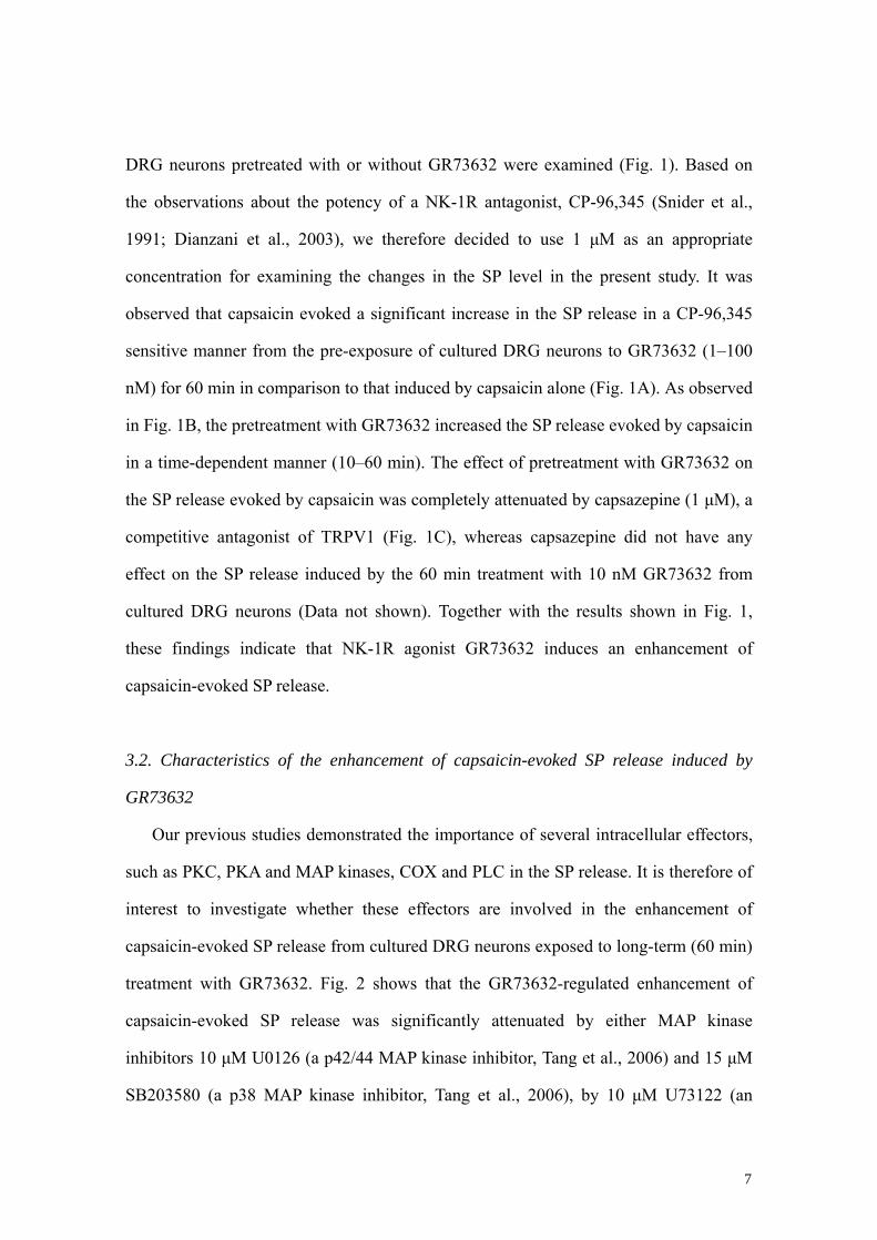
DRG neurons pretreated with or without GR73632 were examined (Fig. 1). Based on
the observations about the potency of a NK-1R antagonist, CP-96,345 (Snider et al.,
1991; Dianzani et al., 2003), we therefore decided to use 1 μM as an appropriate
concentration for examining the changes in the SP level in the present study. It was
observed that capsaicin evoked a significant increase in the SP release in a CP-96,345
sensitive manner from the pre-exposure of cultured DRG neurons to GR73632 (1–100
nM) for 60 min in comparison to that induced by capsaicin alone (Fig. 1A). As observed
in Fig. 1B, the pretreatment with GR73632 increased the SP release evoked by capsaicin
in a time-dependent manner (10–60 min). The effect of pretreatment with GR73632 on
the SP release evoked by capsaicin was completely attenuated by capsazepine (1 μM), a
competitive antagonist of TRPV1 (Fig. 1C), whereas capsazepine did not have any
effect on the SP release induced by the 60 min treatment with 10 nM GR73632 from
cultured DRG neurons (Data not shown). Together with the results shown in Fig. 1,
these findings indicate that NK-1R agonist GR73632 induces an enhancement of
capsaicin-evoked SP release.
3.2. Characteristics of the enhancement of capsaicin-evoked SP release induced by
GR73632
Our previous studies demonstrated the importance of several intracellular effectors,
such as PKC, PKA and MAP kinases, COX and PLC in the SP release. It is therefore of
interest to investigate whether these effectors are involved in the enhancement of
capsaicin-evoked SP release from cultured DRG neurons exposed to long-term (60 min)
treatment with GR73632. Fig. 2 shows that the GR73632-regulated enhancement of
capsaicin-evoked SP release was significantly attenuated by either MAP kinase
inhibitors 10 μM U0126 (a p42/44 MAP kinase inhibitor, Tang et al., 2006) and 15 μM
SB203580 (a p38 MAP kinase inhibitor, Tang et al., 2006), by 10 μM U73122 (an
7
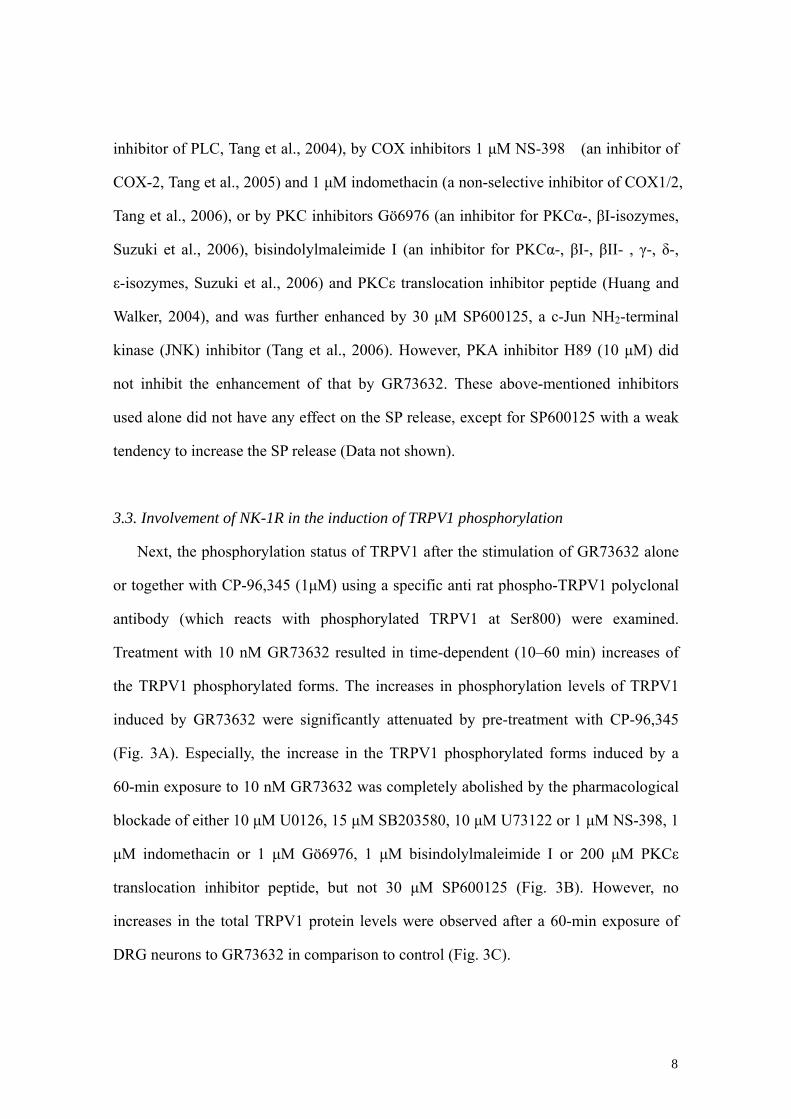
inhibitor of PLC, Tang et al., 2004), by COX inhibitors 1 μM NS-398 (an inhibitor of
COX-2, Tang et al., 2005) and 1 μM indomethacin (a non-selective inhibitor of COX1/2,
Tang et al., 2006), or by PKC inhibitors Gö6976 (an inhibitor for PKCα-, βI-isozymes,
Suzuki et al., 2006), bisindolylmaleimide I (an inhibitor for PKCα-, βI-, βII- , γ-, δ-,
ε-isozymes, Suzuki et al., 2006) and PKCε translocation inhibitor peptide (Huang and
Walker, 2004), and was further enhanced by 30 μM SP600125, a c-Jun NH2-terminal
kinase (JNK) inhibitor (Tang et al., 2006). However, PKA inhibitor H89 (10 μM) did
not inhibit the enhancement of that by GR73632. These above-mentioned inhibitors
used alone did not have any effect on the SP release, except for SP600125 with a weak
tendency to increase the SP release (Data not shown).
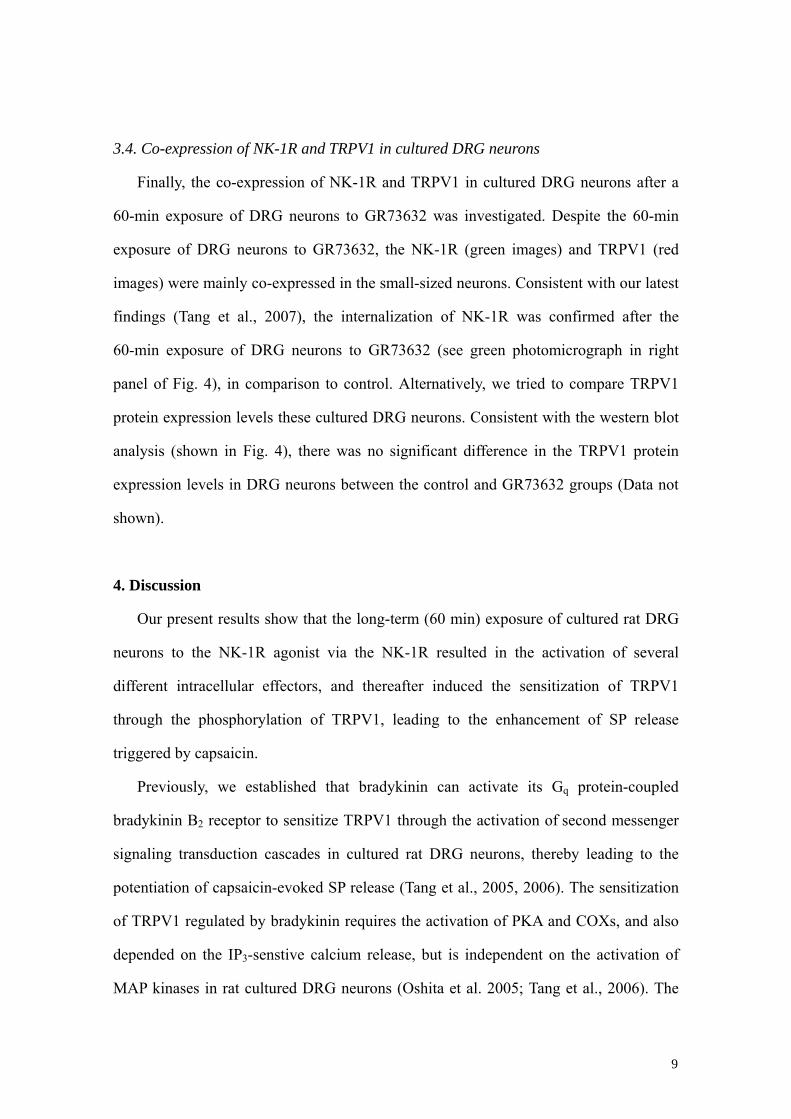
3.3. Involvement of NK-1R in the induction of TRPV1 phosphorylation
Next, the phosphorylation status of TRPV1 after the stimulation of GR73632 alone
or together with CP-96,345 (1μM) using a specific anti rat phospho-TRPV1 polyclonal
antibody (which reacts with phosphorylated TRPV1 at Ser800) were examined.
Treatment with 10 nM GR73632 resulted in time-dependent (10–60 min) increases of
the TRPV1 phosphorylated forms. The increases in phosphorylation levels of TRPV1
induced by GR73632 were significantly attenuated by pre-treatment with CP-96,345
(Fig. 3A). Especially, the increase in the TRPV1 phosphorylated forms induced by a
60-min exposure to 10 nM GR73632 was completely abolished by the pharmacological
blockade of either 10 μM U0126, 15 μM SB203580, 10 μM U73122 or 1 μM NS-398, 1
μM indomethacin or 1 μM Gö6976, 1 μM bisindolylmaleimide I or 200 μM PKCε
translocation inhibitor peptide, but not 30 μM SP600125 (Fig. 3B). However, no
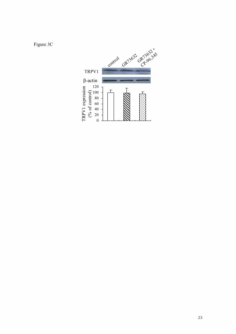
increases in the total TRPV1 protein levels were observed after a 60-min exposure of
DRG neurons to GR73632 in comparison to control (Fig. 3C).
8
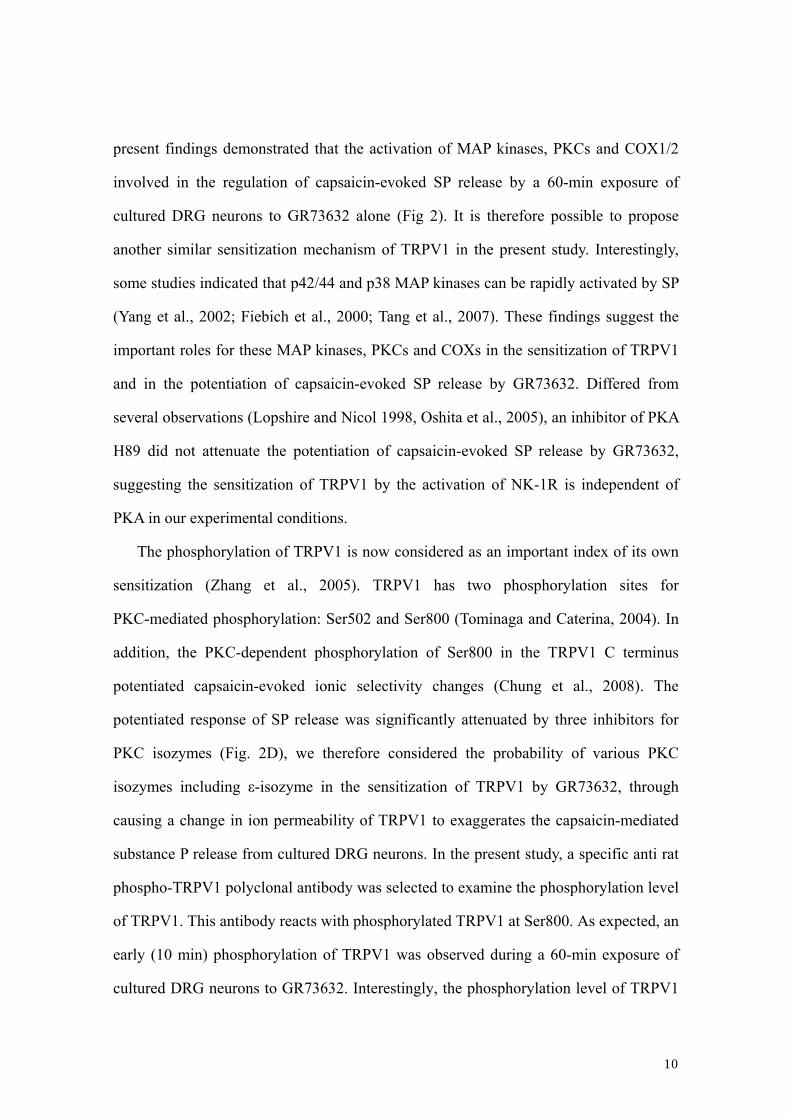
3.4. Co-expression of NK-1R and TRPV1 in cultured DRG neurons
Finally, the co-expression of NK-1R and TRPV1 in cultured DRG neurons after a
60-min exposure of DRG neurons to GR73632 was investigated. Despite the 60-min
exposure of DRG neurons to GR73632, the NK-1R (green images) and TRPV1 (red
images) were mainly co-expressed in the small-sized neurons. Consistent with our latest
findings (Tang et al., 2007), the internalization of NK-1R was confirmed after the
60-min exposure of DRG neurons to GR73632 (see green photomicrograph in right
panel of Fig. 4), in comparison to control. Alternatively, we tried to compare TRPV1
protein expression levels these cultured DRG neurons. Consistent with the western blot
analysis (shown in Fig. 4), there was no significant difference in the TRPV1 protein
expression levels in DRG neurons between the control and GR73632 groups (Data not
shown).
4. Discussion
Our present results show that the long-term (60 min) exposure of cultured rat DRG
neurons to the NK-1R agonist via the NK-1R resulted in the activation of several
different intracellular effectors, and thereafter induced the sensitization of TRPV1
through the phosphorylation of TRPV1, leading to the enhancement of SP release
triggered by capsaicin.
Previously, we established that bradykinin can activate its Gq protein-coupled
bradykinin B2 receptor to sensitize TRPV1 through the activation of second messenger
signaling transduction cascades in cultured rat DRG neurons, thereby leading to the
potentiation of capsaicin-evoked SP release (Tang et al., 2005, 2006). The sensitization
of TRPV1 regulated by bradykinin requires the activation of PKA and COXs, and also
depended on the IP3-senstive calcium release, but is independent on the activation of
MAP kinases in rat cultured DRG neurons (Oshita et al. 2005; Tang et al., 2006). The
9
present findings demonstrated that the activation of MAP kinases, PKCs and COX1/2
involved in the regulation of capsaicin-evoked SP release by a 60-min exposure of
cultured DRG neurons to GR73632 alone (Fig 2). It is therefore possible to propose
another similar sensitization mechanism of TRPV1 in the present study. Interestingly,
some studies indicated that p42/44 and p38 MAP kinases can be rapidly activated by SP
(Yang et al., 2002; Fiebich et al., 2000; Tang et al., 2007). These findings suggest the
important roles for these MAP kinases, PKCs and COXs in the sensitization of TRPV1
and in the potentiation of capsaicin-evoked SP release by GR73632. Differed from
several observations (Lopshire and Nicol 1998, Oshita et al., 2005), an inhibitor of PKA
H89 did not attenuate the potentiation of capsaicin-evoked SP release by GR73632,
suggesting the sensitization of TRPV1 by the activation of NK-1R is independent of
PKA in our experimental conditions.
The phosphorylation of TRPV1 is now considered as an important index of its own
sensitization (Zhang et al., 2005). TRPV1 has two phosphorylation sites for
PKC-mediated phosphorylation: Ser502 and Ser800 (Tominaga and Caterina, 2004). In
addition, the PKC-dependent phosphorylation of Ser800 in the TRPV1 C terminus
potentiated capsaicin-evoked ionic selectivity changes (Chung et al., 2008). The
potentiated response of SP release was significantly attenuated by three inhibitors for
PKC isozymes (Fig. 2D), we therefore considered the probability of various PKC
isozymes including ε-isozyme in the sensitization of TRPV1 by GR73632, through
causing a change in ion permeability of TRPV1 to exaggerates the capsaicin-mediated
substance P release from cultured DRG neurons. In the present study, a specific anti rat
phospho-TRPV1 polyclonal antibody was selected to examine the phosphorylation level
of TRPV1. This antibody reacts with phosphorylated TRPV1 at Ser800. As expected, an
early (10 min) phosphorylation of TRPV1 was observed during a 60-min exposure of
cultured DRG neurons to GR73632. Interestingly, the phosphorylation level of TRPV1
10
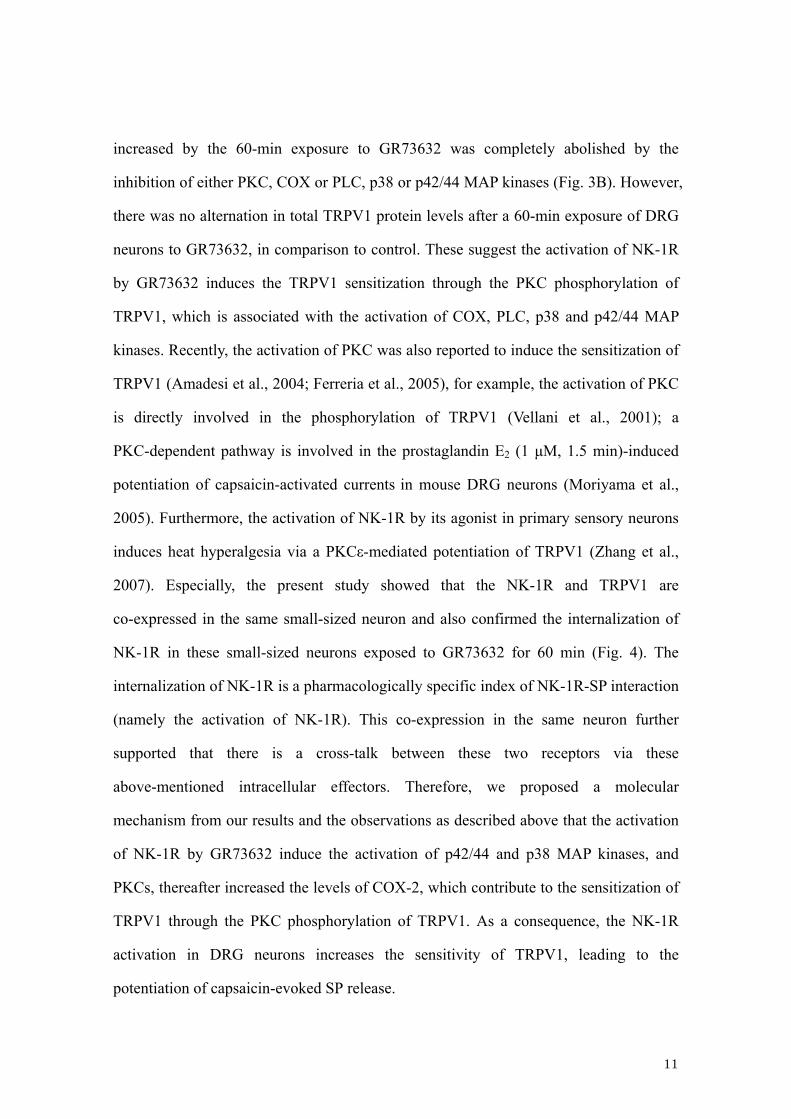
increased by the 60-min exposure to GR73632 was completely abolished by the
inhibition of either PKC, COX or PLC, p38 or p42/44 MAP kinases (Fig. 3B). However,
there was no alternation in total TRPV1 protein levels after a 60-min exposure of DRG
neurons to GR73632, in comparison to control. These suggest the activation of NK-1R
by GR73632 induces the TRPV1 sensitization through the PKC phosphorylation of
TRPV1, which is associated with the activation of COX, PLC, p38 and p42/44 MAP
kinases. Recently, the activation of PKC was also reported to induce the sensitization of
TRPV1 (Amadesi et al., 2004; Ferreria et al., 2005), for example, the activation of PKC
is directly involved in the phosphorylation of TRPV1 (Vellani et al., 2001); a
PKC-dependent pathway is involved in the prostaglandin E2 (1 μM, 1.5 min)-induced
potentiation of capsaicin-activated currents in mouse DRG neurons (Moriyama et al.,
2005). Furthermore, the activation of NK-1R by its agonist in primary sensory neurons
induces heat hyperalgesia via a PKCε-mediated potentiation of TRPV1 (Zhang et al.,
2007). Especially, the present study showed that the NK-1R and TRPV1 are
co-expressed in the same small-sized neuron and also confirmed the internalization of
NK-1R in these small-sized neurons exposed to GR73632 for 60 min (Fig. 4). The
internalization of NK-1R is a pharmacologically specific index of NK-1R-SP interaction
(namely the activation of NK-1R). This co-expression in the same neuron further
supported that there is a cross-talk between these two receptors via these
above-mentioned intracellular effectors. Therefore, we proposed a molecular
mechanism from our results and the observations as described above that the activation
of NK-1R by GR73632 induce the activation of p42/44 and p38 MAP kinases, and
PKCs, thereafter increased the levels of COX-2, which contribute to the sensitization of
TRPV1 through the PKC phosphorylation of TRPV1. As a consequence, the NK-1R
activation in DRG neurons increases the sensitivity of TRPV1, leading to the
potentiation of capsaicin-evoked SP release.
11
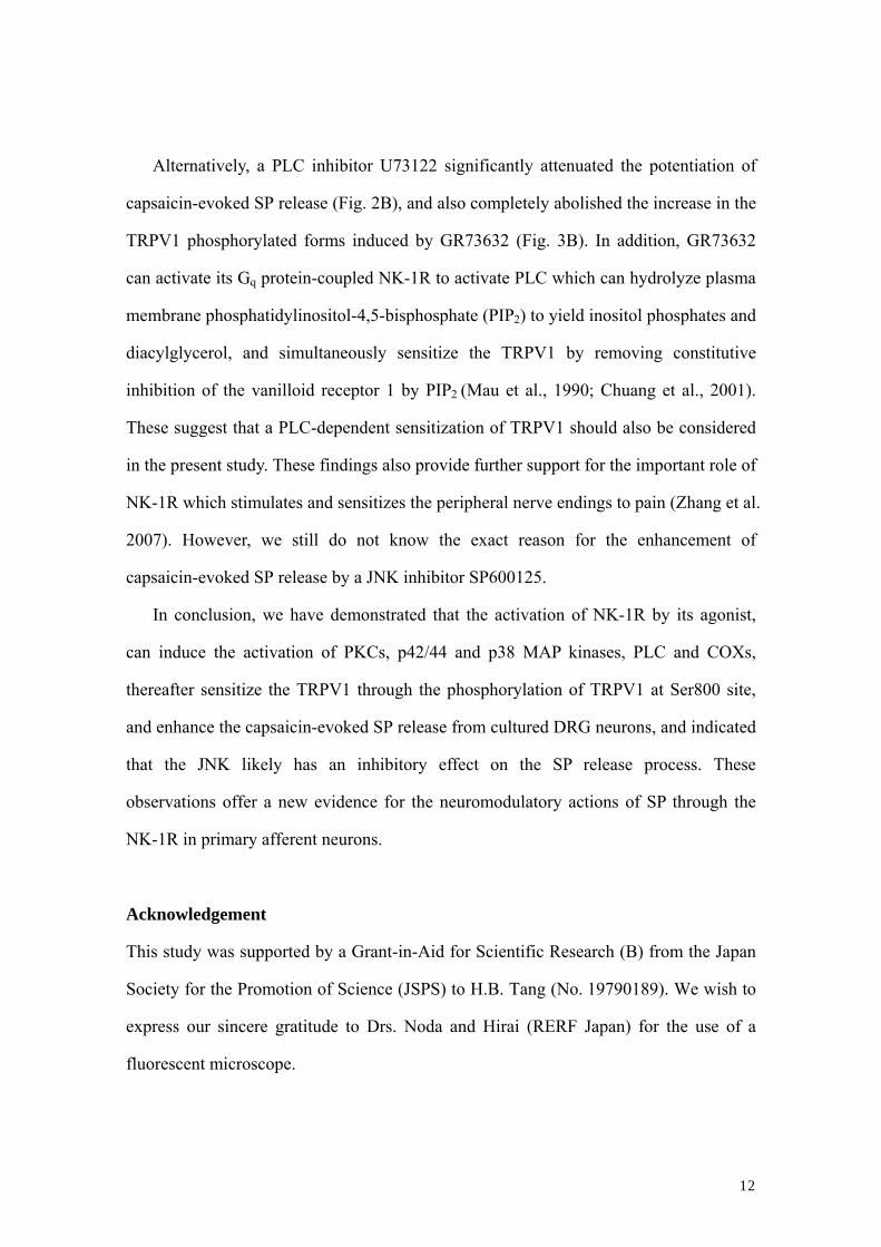
Alternatively, a PLC inhibitor U73122 significantly attenuated the potentiation of
capsaicin-evoked SP release (Fig. 2B), and also completely abolished the increase in the
TRPV1 phosphorylated forms induced by GR73632 (Fig. 3B). In addition, GR73632
can activate its Gq protein-coupled NK-1R to activate PLC which can hydrolyze plasma
membrane phosphatidylinositol-4,5-bisphosphate (PIP2) to yield inositol phosphates and
diacylglycerol, and simultaneously sensitize the TRPV1 by removing constitutive
inhibition of the vanilloid receptor 1 by PIP2 (Mau et al., 1990; Chuang et al., 2001).
These suggest that a PLC-dependent sensitization of TRPV1 should also be considered
in the present study. These findings also provide further support for the important role of
NK-1R which stimulates and sensitizes the peripheral nerve endings to pain (Zhang et al.
2007). However, we still do not know the exact reason for the enhancement of
capsaicin-evoked SP release by a JNK inhibitor SP600125.
In conclusion, we have demonstrated that the activation of NK-1R by its agonist,
can induce the activation of PKCs, p42/44 and p38 MAP kinases, PLC and COXs,
thereafter sensitize the TRPV1 through the phosphorylation of TRPV1 at Ser800 site,
and enhance the capsaicin-evoked SP release from cultured DRG neurons, and indicated
that the JNK likely has an inhibitory effect on the SP release process. These
observations offer a new evidence for the neuromodulatory actions of SP through the
NK-1R in primary afferent neurons.
Acknowledgement
This study was supported by a Grant-in-Aid for Scientific Research (B) from the Japan
Society for the Promotion of Science (JSPS) to H.B. Tang (No. 19790189). We wish to
express our sincere gratitude to Drs. Noda and Hirai (RERF Japan) for the use of a
fluorescent microscope.
12
References
Amadesi, S., Nie, J., Vergnolle, N., Cottrell, G.S., Grady, E.F., Trevisani, M., Manni, C.,
Geppetti, P., McRoberts, J.A., Ennes, H., Davis, J.B., Mayer, E.A., Bunnett, N.W., 2004.
Protease-activated receptor 2 sensitizes the capsaicin receptor transient receptor
potential vanilloid receptor 1 to induce hyperalgesia. J. Neurosci. 24, 4300–4312.
Chuang, H.H., Prescott, E.D., Kong, H., Shields, S., Jordt, S.E., Basbaum, A.I., Chao,
M.V., Julius, D., 2001. Bradykinin and nerve growth factor release the capsaicin
receptor from PtdIns(4,5)P2-mediated inhibition. Nature 411, 957–962.
Chung, M.K., Güler, A.D., Caterina, M.J., 2008. TRPV1 shows dynamic ionic
selectivity during agonist stimulation. Nat. Neurosci. 11, 555–564.
Dianzani, C., Collino, M., Lombardi, G., Garbarino, G., Fantozzi, R., 2003. Substance P
increases neutrophil adhesion to human umbilical vein endothelial cells. Br. J.
Pharmacol. 139, 1103–1110.
Dinh, Q.T., Groneberg, D.A., Peiser, C., Mingomataj, E., Joachim, R.A., Witt, C., Arck,
P.C., Klapp, B.F., Fischer, A., 2004. Substance P expression in TRPV1 and
trkA-positive dorsal root ganglion neurons innervating the mouse lung. Respir. Physiol.
Neurobiol. 144, 15–24.
Ebner, K., Singewald, N., 2006. The role of substance P in stress and anxiety responses.
Amino Acids 31, 251–272.
Ferreira, J., Triches, K.M., Medeiros, R., Calixto, J.B., 2005. Mechanisms involved in
the nociception produced by peripheral protein kinase c activation in mice. Pain 117,
171–181.
Fiebich, B.L., Schleicher, S., Butcher, R.D., Craig, A., Lieb, K., 2000. The neuropeptide
substance P activates p38 mitogen-activated protein kinase resulting in IL-6 expression
independently from NF-kappa B. J. Immunol. 65, 5606–5611.
Harrison, S., Geppetti, P., 2001. Substance P. Int J. Biochem. Cell Biol. 33, 555–576.
13
Hastrup, H., Schwartz, T.W., 1996. Septide and neurokinin A are high-affinity ligands
on the NK-1 receptor: evidence from homologous versus heterologous binding analysis.
FEBS. Lett. 399, 264–266.
Hong, S., Wiley, J.W., 2005. Early painful diabetic neuropathy is associated with
differential changes in the expression and function of vanilloid receptor 1. J. Biol. Chem.
280, 618–627.
Huang, X., Walker, J.W., 2004. Myofilament anchoring of protein kinase C-epsilon in
cardiac myocytes. J. Cell Sci. 117, 1971–1978.
Koon, H.W., Zhao, D., Zhan, Y., Rhee, S.H., Moyer, M.P., Pothoulakis, C., 2006.
Substance P stimulates cyclooxygenase-2 and prostaglandin E2 expression through
JAK-STAT activation in human colonic epithelial cells. J. Immunol. 176, 5050–5059.
Li, H.S., Zhao, Z.Q., 1998. Small sensory neurons in the rat dorsal root ganglia express
functional NK-1 tachykinin receptor. Eur. J. Neurosci. 10, 1292–1299.
Lopshire, J.C., Nicol, G.D., 1998. The cAMP transduction cascade mediates the
prostaglandin E2 enhancement of the capsaicin-elicited current in rat sensory neurons:
whole-cell and single-channel studies. J. Neurosci. 18, 6081–6092.
Mau, S.E., Larsen, P.J., Mikkelsen, J.A., Saermark, T., 1990. Substance P and related
tachykinins induce receptor-mediated hydrolysis of polyphosphoinositides in the rat
anterior pituitary. Mol. Cell. Endocrinol. 69, 69–78.
Moriyama, T., Higashi, T., Togashi, K., Iida, T., Segi, E., Sugimoto, Y., Tominaga, T.,
Narumiya, S., Tominaga, M., 2005. Sensitization of TRPV1 by EP1 and IP reveals
peripheral nociceptive mechanism of prostaglandins. Mol. Pain 1, 3.
Oshita, K., Inoue, A., Tang, H.B., Nakata, Y., Kawamoto, M., Yuge, O., 2005. CB(1)
cannabinoid receptor stimulation modulates transient receptor potential vanilloid
receptor 1 activities in calcium influx and substance P release in cultured rat dorsal root
ganglion cells. J. Pharmacol. Sci. 97, 377–385.
14
Premkumar, L.S., Ahern, G.P., 2000. Induction of vanilloid receptor channel activity by
protein kinase C. Nature 408, 985–990.
Price, T.J., Flores, C.M., 2007. Critical Evaluation of the Colocalization Between
Calcitonin Gene-Related Peptide, Substance P, Transient Receptor Potential Vanilloid
Subfamily Type 1 Immunoreactivities, and Isolectin B4 Binding in Primary Afferent
Neurons of the Rat and Mouse. J. Pain 8, 263–272.
Snijdelaar, D.G., Dirksen, R., Slappendel, R., Crul, B.J., 2000. Substance P. Eur. J. Pain
4, 121–135.
Suzuki, Y., Zhang, H., Saito, N., Kojima, I., Urano, T., Mogami, H., 2006.
Glucagon-like peptide 1 activates protein kinase C through Ca2+-dependent activation of
phospholipase C in insulin-secreting cells. J. Biol. Chem. 281, 28499–28507.
Tang, H.B., Inoue, A., Iwasa, M., Hide, I., Nakata, Y., 2006. Substance P release evoked
by capsaicin or potassium from rat cultured dorsal root ganglion neurons is conversely
modulated with bradykinin. J. Neurochem. 97, 1412–1418.
Tang, H.B., Inoue, A., Oshita, K., Nakata, Y., 2004. Sensitization of vanilloid receptor 1
induced by bradykinin via the activation of second messenger signaling cascades in rat
primary afferent neurons. Eur. J. Pharmacol. 498, 37–43.
Tang, H.B., Li, Y.S., Arihiro, K., Nakata, Y., 2007. Activation of the neurokinin-1
receptor by substance P triggers the release of substance P from cultured adult rat dorsal
root ganglion neurons. Mol. Pain 3, 42.
Tang, H.B., Inoue, A., Oshita, K., Hirate, K., Nakata, Y., 2005. Zaltoprofen inhibits
bradykinin-induced responses by blocking the activation of second messenger signaling
cascades in rat dorsal root ganglion cells. Neuropharmacology 48, 1035–1042.
Tang, H.B., Nakata, Y., 2006. Olopatadine attenuates the enhancement of
capsaicin-evoked substance P release by bradykinin from cultured dorsal root ganglion
neurons. Eur. J. Pharmacol. 552, 78–82.
15
Tang, H.B., Nakata, Y., 2008. The activation of transient receptor potential vanilloid
receptor subtype 1 by capsaicin without extracellular Ca2+ is involved in the mechanism
of distinct substance P release in cultured rat dorsal root ganglion neurons. Naunyn
Schmiedebergs Arch. Pharmacol. 377, 325–332
Tominaga, M, Caterina, M.J., 2004. Thermosensation and pain. J Neurobiol. 61, 3–12.
Yang, C.M., Hsiao, L.D., Chien, C.S., Lin, C.C., Luo, S.F., Wang, C.C., 2002.
Substance P-induced activation of p42/44 mitogen-activated protein kinase associated
with cell proliferation in human tracheal smooth muscle cells. Cell. Signal. 14,
913–923.
Vellani, V., Mapplebeck, S., Moriondo, A., Davis, J.B., McNaughton, P.A., 2001.
Protein kinase C activation potentiates gating of the vanilloid receptor VR1 by capsaicin,
protons, heat and anandamide. J Physiol. 534, 813–825.
Zhang, H., Cang, C.L., Kawasaki, Y., Liang, L.L., Zhang, Y.Q., Ji, R.R., Zhao, Z.Q.,
2007. Neurokinin-1 receptor enhances TRPV1 activity in primary sensory neurons via
PKCepsilon: a novel pathway for heat hyperalgesia. J. Neurosci. 27, 12067–12077.
Zhang, X, Huang, J, McNaughton, P.A., 2005. NGF rapidly increases membrane
expression of TRPV1 heat-gated ion channels. EMBO J. 24, 4211–4223.
16
Figure legends
Figure 1. Effect of pretreatment with GR73632 on the SP release evoked by capsaicin
from cultured DRG neurons.
Some cells were left untreated as a control, all other cells were pretreated with
GR73632 alone or together with CP-96,345 (1 μM) in serum-free DMEM for a
designated period of time at 37°C. Next, those cells were stimulated by capsaicin plus
peptidase inhibitors or by peptidase inhibitors only in Krebs-HEPES buffer for 10 min.
The data are obtained from 4 (A), 3–4 (B) or 3–4 (C) separate experiments. *, ** and
*** denote P < 0.05, 0.01 and 0.001, respectively.
Figure 2. Characteristics of the GR73632-enhanced release of SP induced by capsaicin
from cultured DRG neurons.
Some cells were left untreated as a control, all other cells were pretreated with
GR73632 alone or together with either 10 μM U0126, 15 μM SB203580 or 30 μM
SP600125, 10 μM U73122 or 1 μM NS-398, 1 μM indomethacin or 1μM Gö6976, 1 μM
bisindolymaleimide, 200 μM PKCε translocation inhibitor peptide or 10 μM H89 in
serum-free DMEM for 1 hour at 37°C. Next, those cells were stimulated by capsaicin
plus peptidase inhibitors or by peptidase inhibitors only in Krebs-HEPES buffer for 10
min. The data are obtained from 5 (A), 5–13 (B) or 8–9 (C), 4–9 (D) separate
experiments. *, ** and *** denote P < 0.05, 0.01 and 0.001, respectively.
Figure 3. The phosphorylation of TRPV1 induced by GR73632 in cultured adult rat
DRG neurons.
Representative blots of phosphorylated TRPV1 (P-TRPV1) expression in cultured
DRG neurons exposed to GR73632 alone or together with CP-96,345 for 10 to 60 min
(upside of A) or together with either 10 μM U0126, 15 μM SB203580 or 10 μM
17
U73122, 1 μM NS-398 or 1 μM indomethacin (IDM), Gö6976, bisindolylmaleimide I
(Bis) or PKCε translocation inhibitor peptide (PKCεI), 30 μM SP600125 for 60 min
(upside of B). Representative blots of TRPV1 expression in cultured DRG neurons
exposed to GR73632 alone or together with CP-96,345 for 60 min (upside of C). All
data have been quantified by normalizing the P-TRPV1 (downside of A; downside of B)
or TRPV1 (downside of C) expression level of control. The data are obtained from 3-5
(A, B) or 3 (C) separate experiments. #, ##, ### denote P < 0.05, 0.01, 0.001 in
comparison to the effect of respective GR73632 alone; *, **, *** denote P < 0.05, 0.01,
0.001 versus the effect of the untreated control.
Figure 4. Co-expression of NK-1R and TRPV1 in cultured adult rat DRG neurons.
Bright-field photomicrograph (top panels) of DRG neurons after the 60-min
exposure to GR73632; representative immunofluorescence photomicrographs for
NK-1R (green photomicrographs) and TRPV1 (red photomicrographs) in these DRG
neurons of control (left panels) and GR73632 (right panels) groups; digitally merged
photomicrographs (lowest panels). Bars: 15 µm. The red and white arrows indicate two
different size neurons.
18
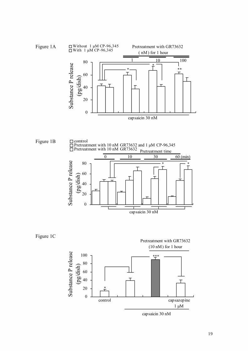
Figure 1A
0
20
40
60
80
Subs
tanc
e P
rele
ase
(pg/
dish
)
Without 1 μM CP-96,345With 1 μM CP-96,345
capsaicin 30 nM
Pretreatment with GR73632( nM) for 1 hour
1 10 100
** *
*
Figure 1B
0
20
40
60
80
Subs
tanc
e P
rele
ase
(pg/
dish
)
comtrolPretreatment with 10 nM GR73632 and 1 μM CP-96,345 Pretreatment with 10 nM GR73632
0 10 30 60 (min)
capsaicin 30 nM
Pretreatment time
* *
Figure 1C
0
20
40
60
80
100
Subs
tanc
e P
rele
ase
(pg/
dish
)
capsazepine1 μM
Pretreatment with GR73632(10 nM) for 1 hour
capsaicin 30 nM
control
***
*
19
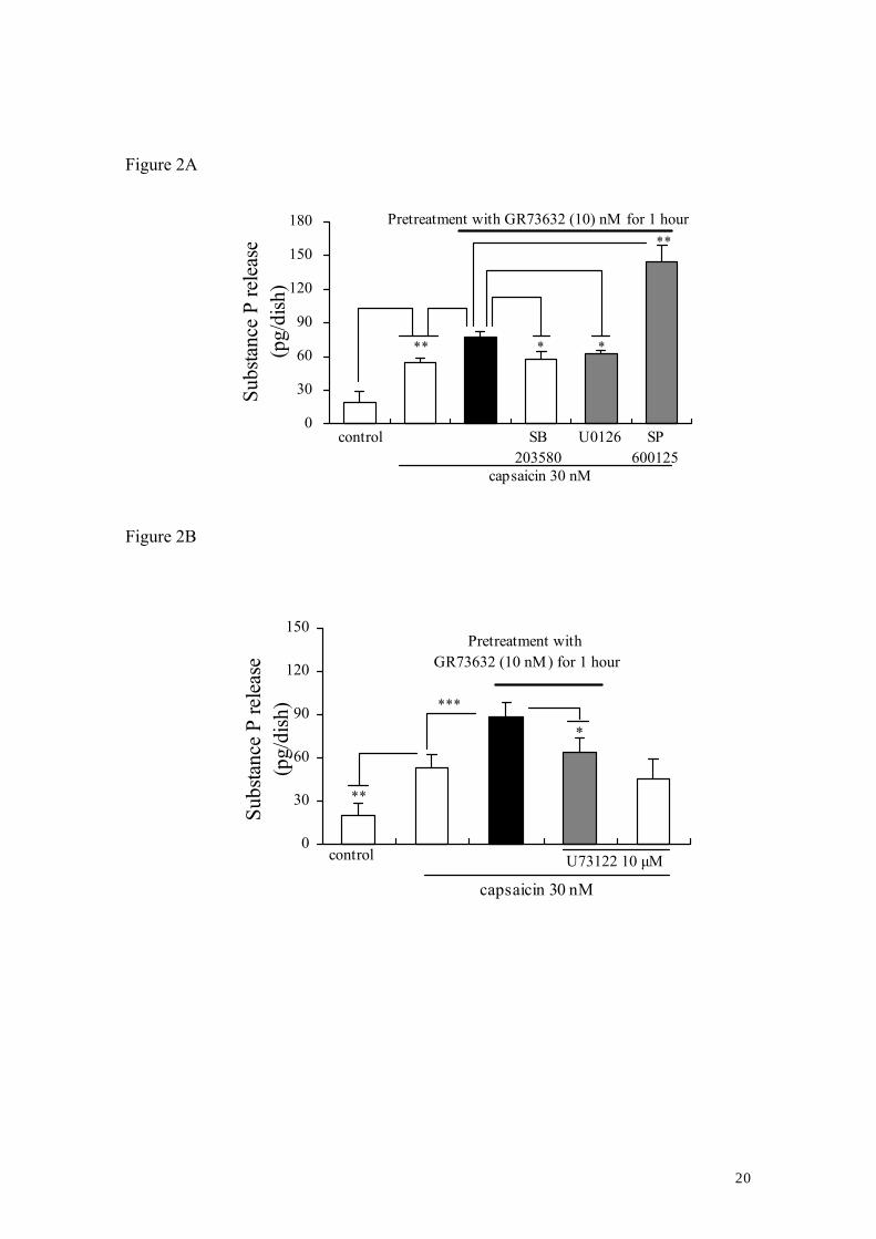
Figure 2A
0
30
60
90
120
150
180
Subs
tanc
e P
rele
ase
(pg/
dish
)
control SB203580
SP600125
U0126
Pretreatment with GR73632 (10) nM for 1 hour
capsaicin 30 nM
* **
**
*
Figure 2B
0
30
60
90
120
150
Subs
tanc
e P
rele
ase
(pg/
dish
)
control
capsaicin 30 nM
Pretreatment with GR73632 (10 nM) for 1 hour
U73122 10 μM
**
***
*
20
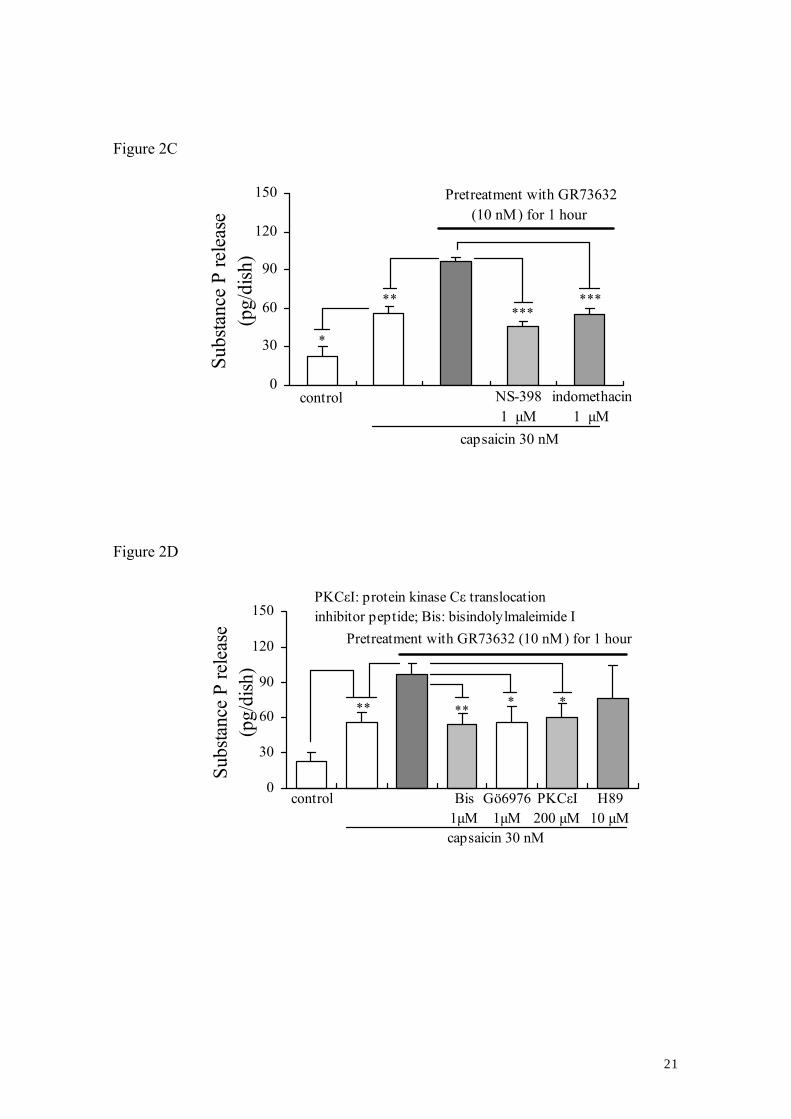
Figure 2C
0
30
60
90
120
150
Subs
tanc
e P
rele
ase
(pg/
dish
)
control
capsaicin 30 nM
Pretreatment with GR73632(10 nM) for 1 hour
NS-3981 μM
indomethacin1 μM
**
*
*** ***
Figure 2D
0
30
60
90
120
150
Subs
tanc
e P
rele
ase
(pg/
dish
)
control
capsaicin 30 nM
Pretreatment with GR73632 (10 nM) for 1 hour
H8910 μM
PKCεI: protein kinase Cε translocationinhibitor peptide; Bis: bisindolylmaleimide I
** * * **
Bis1μM
Gö69761μM
PKCεI200 μM
21
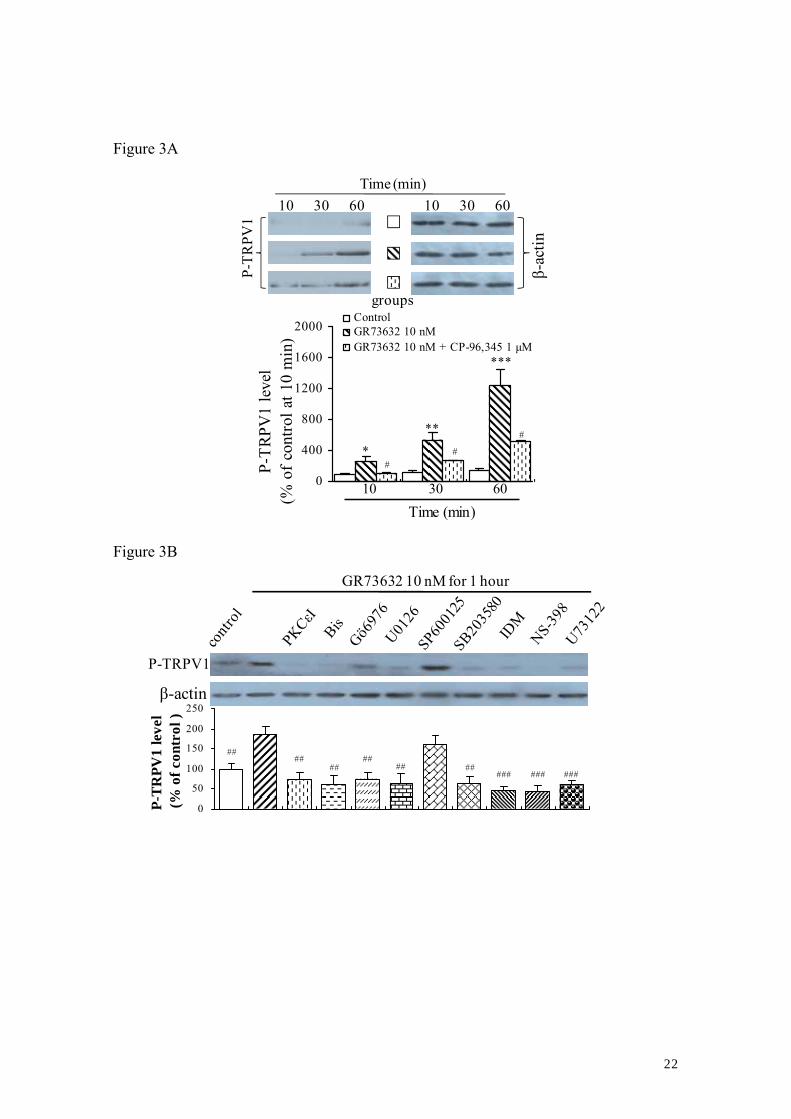
Figure 3A
0
400
800
1200
1600
2000
P-TR
PV1
leve
l (%
of c
ontro
l at 1
0 m
in)
ControlGR73632 10 nMGR73632 10 nM + CP-96,345 1 μM
10 60 30
*
**
***
#
# #
Time (min)
10 30 60
P-TR
PV1
β-ac
tin
Time (min)
groups
10 30 60
Figure 3B
0
50
100
150
200
250
P-T
RPV
1 le
vel
(% o
f con
trol
)
####
## ##
## ## ### ### ###
P-TRPV1
β-actin
GR73632 10 nM for 1 hour
22
TRPV1
β-actin
020406080
100120
TRPV
1 ex
pres
sion
(% o
f con
trol)
Figure 3C
23
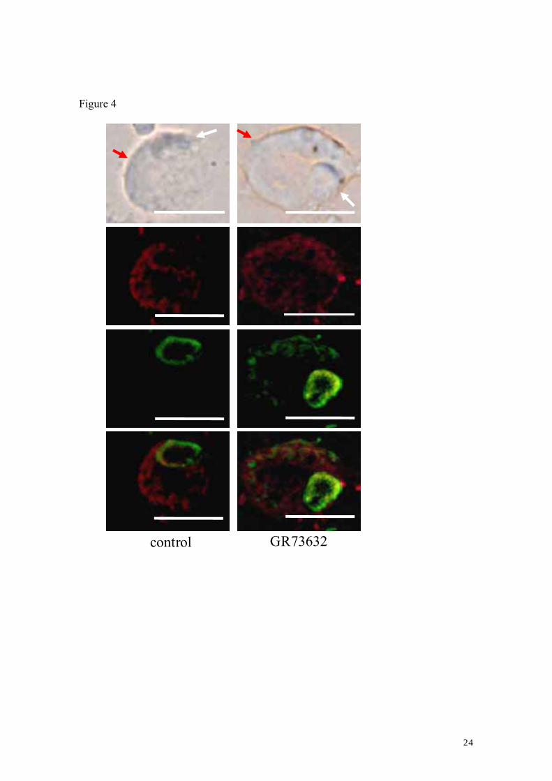
Figure 4
GR73632 control
24