Nuclear translocation of glutathione S-transferase
29
This document is downloaded at: 2018-12-20T22:58:30Z Title Nuclear translocation of glutathione S-transferase π is mediated by a non- classical localization signal. Author(s) Kawakatsu, Miho; Goto, Shinji; Yoshida, Takako; Urata, Yoshishige; Li, Tao-Sheng Citation Biochemical and Biophysical Research Communications, 411(4), pp.745- 750; 2011 Issue Date 2011-08-12 URL http://hdl.handle.net/10069/26096 Right Copyright © 2011 Elsevier Inc. All rights reserved. NAOSITE: Nagasaki University's Academic Output SITE http://naosite.lb.nagasaki-u.ac.jp
Transcript of Nuclear translocation of glutathione S-transferase
This document is downloaded at: 2018-12-20T22:58:30Z
Title Nuclear translocation of glutathione S-transferase π is mediated by a non-classical localization signal.
Author(s) Kawakatsu, Miho; Goto, Shinji; Yoshida, Takako; Urata, Yoshishige; Li,Tao-Sheng
Citation Biochemical and Biophysical Research Communications, 411(4), pp.745-750; 2011
Issue Date 2011-08-12
URL http://hdl.handle.net/10069/26096
Right Copyright © 2011 Elsevier Inc. All rights reserved.
NAOSITE: Nagasaki University's Academic Output SITE
http://naosite.lb.nagasaki-u.ac.jp
1
Nuclear translocation of glutathione S-transferase is mediated by a non-classical
localization signal
Miho Kawakatsu, Shinji Goto, Takako Yoshida, Yoshishige Urata and Tao-Sheng Li
Department of Stem Cell Biology, Atomic Bomb Disease Institute, Nagasaki University
Graduate School of Biomedical Sciences, Nagasaki 852-8523, Japan
Running Title: Nuclear localization signal of GST
Address correspondence to: Shinji Goto, Ph.D., Department of Stem Cell Biology,
Atomic Bomb Disease Institute, Nagasaki University Graduate School of Biomedical
Sciences, Nagasaki 852-8523, Japan
Tel. +81-95-819-7099; FAX. +81-95-819-7100
E-mail; [email protected]
Abbreviations: GSH, reduced form of glutathione; GST, glutathione S-transferase;
NLS, nuclear localization signal; MTS, mitochondrial targeting signal; GFP, green
fluorescent protein; PCR, polymerase chain reaction; HRP, horseradish peroxidase;
FITC, fluorescein isothiocyanate; RCC1, regulator of chromosome condensation 1;
Cu,Zn-SOD, Cu,Zn-superoxide dismutase; GPx, glutathione peroxidase
2
Abstract
Glutathione S-transferase (GST)a member of the GST family of
multifunctional enzymes, is highly expressed in human placenta and involved in the
protection of cellular components against electrophilic compounds or oxidative stress.
We have recently found that GST is expressed in the cytoplasm, mitochondria, and
nucleus in some cancer cells, and that the nuclear expression of GSTappears to
correlate with resistance to anti-cancer drugs. Although the mitochondrial targeting
signal of GST was previously identified in the amino-terminal region, the mechanism
of nuclear translocation remains completely unknown. In this study, we find that the
region of GST195-208 is critical for nuclear translocation, which is mediated by a
novel and non-classical nuclear localization signal. In addition, using an in vitro
transport assay, we demonstrate that the nuclear translocation of GST depends on the
cytosolic extract and ATP. Although further experiments are needed to understand in
depth the precise mechanism of nuclear translocation of GST, our results may help to
establish more efficient anti-cancer therapy, especially with respect to resistance to
anti-cancer drugs.
Key words: glutathione S-transferase , nuclear localization signal
3
Introduction
Glutathione S-transferases (GSTs) are multifunctional enzymes involved in the
protection of cellular components against electrophilic compounds or oxidative stress.
Glutathione S-transferase (EC 2.5.1.18) belongs to the family of GSTs involved in
cellular detoxification [1, 2]. The expression of GST increases in various
pre-cancerous and cancer tissues [3-5]. We have recently found that the mature form of
GST is distributed in the cytoplasm and mitochondria, and we have further identified
the mitochondrial targeting signal located in the amino-terminal (N-terminal) region of
GST6In other studies, nuclear translocation of GST was observed in some cancer
cells, and nuclear expression of GST seems to associate with resistance to anti-cancer
drugs [7-9]. However, the precise mechanism of nuclear translocation of GST remains
completely unclear
In general, the translocation of proteins from the cytoplasm to the nucleus is
dependent upon a specific sequence, which is called a nuclear localization signal (NLS).
The NLS occurs in both classical and non-classical types. The classical NLS is
composed of positively charged amino-acid cluster(s) [10-12] that can be recognized by
importin- (karyopherin-) and forms a transport complex with importin-
(karyopherin-) [13, 14]. However, a non-classical NLS lacks a contiguous stretch of
positively charged residues and bears no sequence similarity to the classical NLS [15].
Thus, a non-classical NLS is directly recognized by importin- family members without
the need for adapter proteins, such as importin- [16-18]. Because GST has no known
NLS in the amino acid sequence, the precise mechanism of the nuclear translocation of
GST has not yet been determined.
Considering the potential roles of nuclear GST in physiological and
pathological functions, we have tried to elucidate the mechanism of the nuclear
translocation of GST in this study. We have found that the carboxyl-terminal
4
(C-terminal) region of GST is critical for nuclear translocation, likely through a novel
mechanism that involves a non-classical NLS.
5
Materials and Methods
Materials
All materials for this study are listed in the supplementary file.
Cell culture
We used HCT8, COS-1, and HeLa cells for this study. The cells were maintained
in DMEM (COS-1 and HeLa) or RPMI 1640 (HCT8) basic medium, supplemented with
10% FBS at 37°C under a humidified atmosphere of 5% CO2.
Immunostaining to detect endogenous GST
To identify the intracellular localization of endogenous GST, the cells were
maintained with medium containing 10% FBS on glass coverslips in a six-well culture
plate (Nalge Nunc International, Naperville, IL). After 24 hrs of culture, the cells on the
glass coverslips were fixed with 3% paraformaldehyde for 20 min, treated with 1%
Triton-X 100 for 10 min, and blocked with 3% BSA for 30 min at room temperature.
The cells were incubated with a polyclonal antibody against human GST for 60 min
and were then treated with FITC-conjugated anti-rabbit IgG secondary antibody for 30
min, as described previously [6]. The nuclei were labeled with 10 M Hoechst 33342
for 15 min. The intracellular distribution of endogenous GSTwas observed under a
confocal laser scanning microscope (LSM5 pascal, Carl Zeiss, Jena, Germany).
Construction of vectors for protein expression
The GST cDNA was prepared by a reverse transcription polymerase chain
reaction (PCR) method using total RNA extracted from HCT8 cells as described
previously [7]. The cDNA of full-length GST was inserted into pcDNA3.1/NT-GFP or
pcDNA3.1/CT-GFP, using EcoRI-EcoRV restriction sites, for the construction of an
6
expression vector for GFP fused at the N-terminal (GFP-GST) or C-terminal
(GST-GFP) end of GST.
To create GFP-GST182-210, GFP-GST182-210 (K209G),
GFP-GST182-208, GFP-GST182-194, and GFP-GST195-208, GST fragments
were prepared by PCR using GST cDNA as a template and an appropriate set of
primers (Supplemental material). Following digestion with EcoRI and EcoRV, the
resulting PCR products were subcloned into the EcoRI-EcoRV restriction sites of
pcDNA3.1/NT-GFP.
To obtain FLAG-HA-tags at the N-terminal end of full-length or deletion
mutants of GST (FLAG-GST1-210, FLAG-GST1-181, and FLAG-GST1-194),
GST fragments were prepared by PCR using an appropriate set of primers
(Supplemental material). The PCR products were digested with NotI and ApaI and
subcloned into pcDNA3/FLAG-HA.
Evaluation of nuclear translocation of GSTby fusion proteins
To observe the nuclear translocation of GST, the expression vectors (2 g) for
various GFP-fused proteins were transfected into COS-1 cells or HCT8 cells with
Lipofectamine reagent or Lipofectamine 2000 according to the manufacturer's
instructions. The fusion proteins were expressed by cultivating the transfectants for 24
hrs at 37°C and were then fixed with 3% paraformaldehyde in PBS for 20 min. After
three washes with PBS, the nuclei were stained with Hoechst 33342. The intracellular
distribution of GFP-fused proteins was directly observed as green fluorescence under a
confocal laser scanning microscope.
To further confirm the nuclear translocation of GST obtained by the direct
detection of GFP-fused GST proteins, expression vectors (2 g) of FLAG-GST1-210,
FLAG-GST1-181 and FLAG-GST1-194 were also transfected into COS-1 cells or
7
HCT8 cells. After 24 hrs, the cells were stained by anti-FLAG primary antibody and
FITC-labeled anti-mouse IgG secondary antibody, as described above. The nuclei were
visualized by staining the cells with Hoechst 33342. The nuclear translocation of
GSTwas observed as positive staining of FLAG under a confocal laser scanning
microscope.
Preparation of cytosolic and nuclear proteins
The cytosolic and nuclear proteins were prepared as described previously [6].
Briefly, the cell pellets (1x10⁶ cells) were treated with 100 l of hypotonic buffer (10
mM HEPES at pH 7.8, 10 mM KCl, 0.1 mM EDTA, 1 mM dithiothreitol, 0.5 mM
phenylmethylsulfonylfluoride [PMSF], 2 g/ml pepstatin, and 2 g/ml leupeptin). After
centrifugation of the sample (1,800 x g, 4°C, 1 min), the supernatant and debris were
collected as rough cytosolic and nuclear fractions, respectively. The rough cytosolic
fractions were centrifuged at 15,000 x g for 20 min and 100,000 x g for 30 min at 4°C.
The final supernatant was collected as the cytosolic fraction for use in the following
study. The rough nuclear fractions were washed three times with hypotonic buffer and
treated with 100 l of 50 mM HEPES (pH 7.8), 420 mM KCl, 0.1 mM EDTA, 1 mM
dithiothreitol, 5 mM MgCl2, 0.5 mM PMSF, 2 g/ml pepstatin, and 2 g/ml leupeptin
and were then gently rotated with a rotator at 4°C for 30 min. The supernatant was
collected as the nuclear fraction.
Immunoblot analysis
Expression levels of GST, RCC1, Cu,Zn-SOD, GFP and FLAG-tagged
proteins in the cells were estimated by immunoblotting. Lysate (30 g of total protein)
was separated by sodium dodecyl sulfate-polyacrylamide gel electrophoresis
(SDS-PAGE) using a 12.5% or 15% gel, transferred to a nitrocellulose membrane, and
8
immunologically stained first with each appropriate primary antibody, then with
HRP-labeled anti-rabbit, anti-mouse or anti-goat IgG as the secondary antibody. Blots
were developed by enhanced chemiluminescence using an ECL kit. The protein
concentration was determined according to Redinbaugh and Turley [19], with bovine
serum albumin (BSA) as the standard.
Preparation of recombinant protein for in vitro nuclear transport assay
The cDNA of GFP or GFP-GST195-208 was inserted into the
pGEX-6P-1vector using SmaI-NotI restriction sites, resulting in an expression vector for
recombinant GFP or GFP-GST195-208, in which GFP is fused at the C-terminal end
of GST from Schistosoma japonicum. To create cDNA of GFP and GFP-GST195-208,
each fragment was prepared by PCR using pcDNA3.1/NT-GFP or
pcDNA3.1/NT-GFP-GST195-208 as a template and an appropriate set of primers
(Supplemental material). Following digestion with SmaI and NotI, the resulting PCR
products were subcloned into SmaI-NotI restriction sites of pGEX-6P-1. Expression in
the BL21 Escherichia coli host and purification of recombinant protein were performed
according to the manufacturer’s instructions.
The nucleotide sequences of vectors
All newly constructed vectors were transformed into a JM109 Escherichia coli
host and purified using a GeneElute Hp plasmid midiprep kit (Sigma Aldrich). The
nucleotide sequences of all constructs were determined using the CEQ DTCS-Quick
Start kit on a CEQ 8000 Genetic Analysis System (Beckman Coulter, Fullerton, CA).
9
In vitro nuclear transport assay
In vitro transport assays were performed according to the method described by
Adam et al. [20]. Briefly, HeLa cells were grown on glass coverslips in a 6-well plate.
The cells were washed twice on the glass coverslips with ice-cold transport buffer (20
mM Hepes-KOH, pH 7.3, 110 mM potassium acetate, 5 mM sodium acetate, 2 mM
DTT, 1.0 mM EGTA, and 1 μg/ml each aprotinin, leupeptin, and pepstatin) and
permeabilized for 5 min in ice-cold transport buffer containing 40 g/ml digitonin.
After washing with cold transport buffer, the permeabilized cells were incubated with
150 μl of import assay mixture containing recombinant GST-GFP or GST-GFP-GST
195-208 protein with or without cytosol extract or the ATP regeneration system (0.5
mM ATP, 0.5 mM GTP, 10 mM creatine phosphate, 50 μg/ml creatine kinase), for 30
min at either 30°C or 4°C. After three washes with import buffer and PBS, the cells
were fixed with 3% formaldehyde for 20 min, and then nuclei were labeled with 10 M
Hoechst 33342 for 10 min. Fluorescence was observed under a confocal laser scanning
microscope.
10
Results
The C-terminal region of GST is critical for nuclear translocation
Immunostaining with anti-GST antibody clearly showed the expression of
endogenous GST in the nuclei of HCT8 and COS-1 cells under steady-state conditions
(Fig.1A), although the expression of GST in the cytoplasm was stronger in HCT8 than
in COS-1 cells.
We constructed vectors encoding GFP at the N-terminal end (GFP-GST) or the
C-terminal end (GST-GFP) of GST, which were then transfected into HCT8 and
COS-1cells, respectively. The intracellular distribution of GFP-fused GSTproteins
(M.W. 52,000) was detected as GFP fluorescence under a confocal laser scanning
microscope 24 hrs after transfection. GFP fluorescence was observed in both the
cytoplasm and the nuclei of cells transfected with GFP-GST vectors (Fig. 1B),
indicating that the N-terminally GFP-fused GST translocated into the nuclei. However,
GFP fluorescence was observed only within the cytoplasm, not within the nuclei, in
cells transfected with GST-GFP vectors (Fig. 1C), which indicated a lack of
C-terminally GFP-fused GST in the nuclei. The treatment of cells with Leptomycin B,
a specific inhibitor of nuclear export of protein, did not change the nuclear distribution
of fusion proteins (data not shown), which eliminated the possibility that the fusion
proteins were exported from the nuclei in cells transfected with GST-GFP vectors.
These microscopy findings were corroborated by immunoblot analysis (Fig. 1D). Our
results suggest that artificial modification of GST by fusion with GFP may cause a
conformational change of the protein and that the C-terminal region, but not the
N-terminal region, of GSTis critical for nuclear translocation.
11
The C-terminal region of GST182-210 is required for nuclear translocation
Positively charged residues are known to be critical components of the classical-
NLS. As the C-terminal region of GST182-210 is rich in positively charged arginine
and lysine (bold-typed, Fig. 2A), this C-terminal region may mediate the nuclear
translocation of GST as a NLS To test this possibility, we constructed expression
vectors encoding N-terminally FLAG-tagged full-length GST(FLAG-GST1-210) or
a C-terminal deletion mutant GST (FLAG-GST1-181) (Fig. 2A). These vectors were
introduced into COS-1 cells, and the intracellular distribution of FLAG-tagged proteins
was detected by immunostaining with anti-FLAG antibody. As expected, the nuclear
localization of FLAG-tagged proteins was observed in cells transfected with the
FLAG-GST1-210 vector but was not observed in cells transfected with the
FLAG-GST1-181 vector (Fig. 2B), which suggests that the C-terminal region of
GSTresidues 182-210) is required for nuclear translocation.
Non-classical NLS mediates the nuclear translocation of GST
To further define the required C-terminal region of GST and understand the
relevant mechanism of nuclear translocation of this protein, expression vectors encoding
various N-terminally GFP-fused C-terminal fragments of GST were introduced into
the COS-1 cells (Supplementary Fig. 1A). Immunoblot analysis using anti-GFP
antibody showed the expression of GFP under all conditions (S. Fig. 1B). A clear band
corresponding to endogenous GSTwas also detected by the anti-GST antibody under
all conditions, but a band for GFP-fused GST was not detected in cells transfected
with GFP-GST182-194 vector. It is likely that the anti-GST antibody did not
recognize the GFP-fused GST protein that was expressed from GFP-GST182-194
vector, because a clear band corresponding to the fusion protein was detected by
anti-GFP antibody in these cells.
12
The intracellular distribution of GFP-fused GST was detected as GFP
fluorescence under confocal laser scanning microscopy (S. Fig. 1C). Nuclear
translocation of GST was detected in cells transfected with vectors of
GFP-GST182-208, GFP-GST195-208, GFP-GST182-210, and GFP-GST182-210
(K209G), in which lysine 209 is replaced with glycine. However, nuclear translocation
of GST was not detected in cells transfected with the GFP-GST182-194 vector. To
confirm the critical nature of the 195-208 sequence, COS-1 and HCT8 cells were
transfected with FLAG-GST1-210 and FLAG-GST1-194 vectors. Immunostaining
by anti-FLAG antibody clearly showed nuclear translocation of GST in cells
transfected with the FLAG-GST1-210 vector, but not with the FLAG-GST1-194
vector (Fig. 3). Our results suggest that the 195-208 region of GST is critical for
nuclear translocation. Considering the lack of a contiguous stretch of positively charged
amino acid residues within this region, nuclear translocation of GST is likely mediated
by a non-classical rather than a classical NLS.
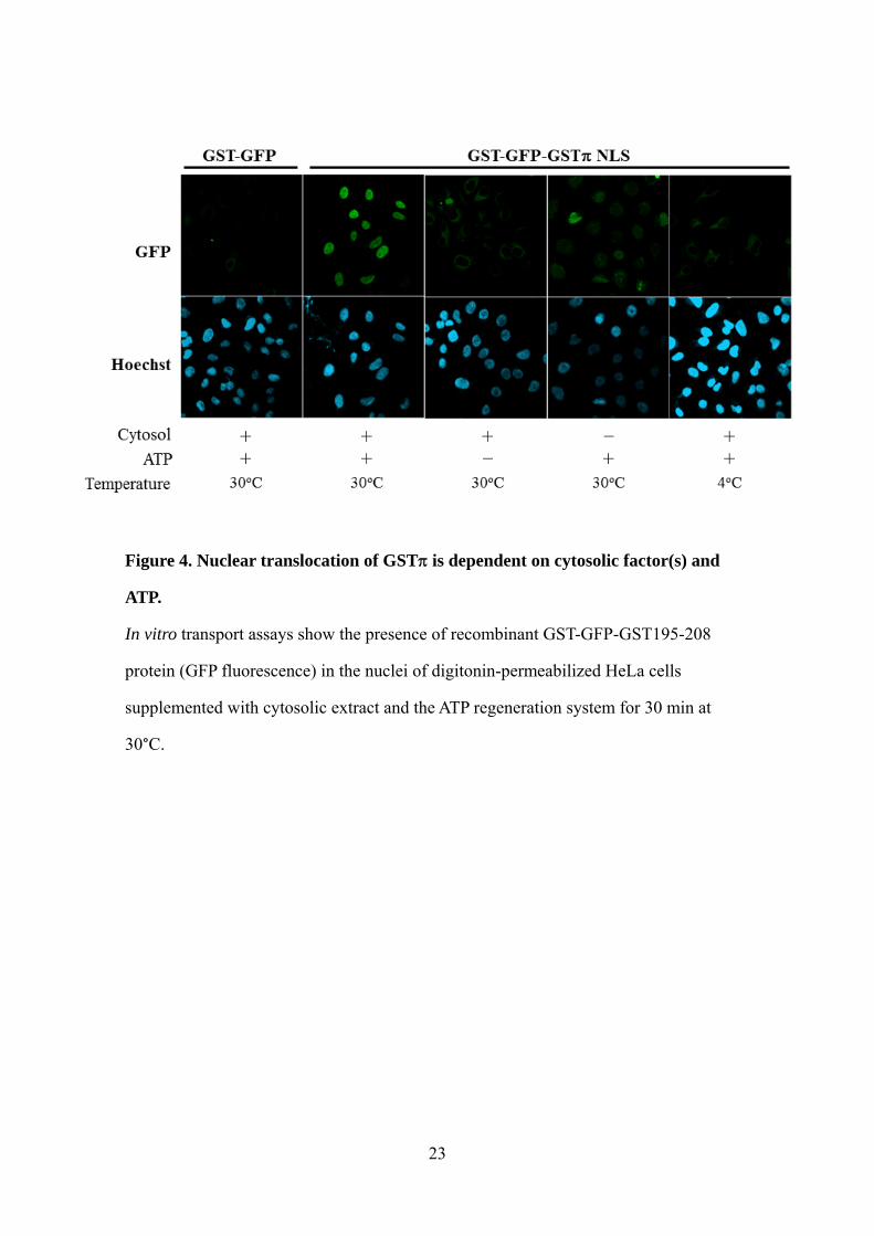
Nuclear translocation of GST is dependent on cytosolic factor(s) and ATP
Using the nuclear transport assay, we tried to further clarify how GST enters
the nucleus. We subjected recombinant GST-GFP or GST-GFP-GST195-208 proteins
to permeabilized HeLa cells under various conditions (Fig. 4). GFP fluorescence was
not observed in the nuclei following the addition of GST-GFP-GST195-208 proteins
alone to the permeabilized HeLa cells at 30°C or 4°C (data not shown). However, GFP
fluorescence was clearly observed in the nuclei of permeabilized HeLa cells following
the addition of GST-GFP-GST195-208 protein along with both cytosolic extract and
the ATP regeneration system at 30°C (but not with either of these components alone). In
contrast, GFP fluorescence was not observed in the nuclei of permeabilized HeLa cells
following the addition of GST-GFP protein, even in the presence of both cytosolic
13
extract and ATP regeneration system at 30°C. These results indicate that the nuclear
translocation of GST is dependent on cytosolic factor(s) and ATP. Although further
experiments are needed to understand the mechanism in depth, our results suggest that
different signals mediate the import of GSTinto the nucleus and mitochondria (S. Fig.
2).
14
Discussion
Proteins smaller than 40 kDa are believed to enter the nucleus by passive
diffusion through the nuclear pore complex. Although GST is a homodimer consisting
of two subunits each with a molecular mass of 24 kD in the cytosol, it is possible that
GST may form a monomer prior to passing through the nuclear pore complex by
passive diffusion. However, the present study and earlier results suggest that nuclear
translocation of GST likely does not result from passive diffusion from the cytoplasm,
as 1) nuclear translocation is selectively observed in N-terminal GFP-fused GST, the
molecular weight of which is about 52 kD (too large to diffuse through the nuclear pore
complex), but not in C-terminally GFP-fused GSTFig. 1B, C); and 2) a previous
study demonstrated nuclear translocation of GST in some but not all cancer cell lines
[7]. These observations suggest the existence of a specific mechanism for the nuclear
translocation of GST.
Various mechanisms have been proposed for the nuclear translocation of
proteins from the cytoplasm [10-18]. In the present study, we found that amino acid
residues 195-208 at the C-terminus were necessary for the nuclear localization of GST.
The GST NLS lacks the contiguous stretch of positively charged amino acid residues
characteristic of a classical NLS. In addition, we found that the GST NLS bears no
sequence similarity to other proteins, except for the same isoform of GST from other
species, using the Basic Local Alignment Search Tool (BLAST) provided by the
National Center for Biotechnology Information (USA). We also found no registered
motifs in the C-terminal region of GST in PROSITE, a database of protein families
and domains curated by the Swiss Institute of Bioinformatics (Switzerland). Therefore,
it is most likely that a non-classical rather than a classical NLS in the C-terminal region
mediates the nuclear translocation of GST. As the nuclear translocation of GST was
found to depend on cytosolic factor(s) and ATP, it is possible that GST forms complex
15
with other cytosolic protein(s) that recognize the NLS and facilitate nuclear
translocation.
Several previous studies have identified the relevant mechanisms of the nuclear
and mitochondrial localizations of proteins. For example, apurinic/apyrimidinic
endonuclease 1 (APE1), a key enzyme of DNA base excision repair, localizes in both
the nucleus and mitochondria. The nuclear form of APE1 is intact, whereas the
mitochondrial form lacks the N-terminal region (including the NLS), suggesting that the
cleavage of the intact form is a prerequisite for the mitochondrial import of APE1 [21].
Alternatively, phospholipid hydroperoxide glutathione peroxidase (PHGPx) is encoded
by a single gene, gpx-4, that has two distinct promoter regions. The upstream region
transcribes cytosolic PHGPx and mitochondrial PHGPx, while the downstream region
yields nuclear PHGPx [22]. However, we found that endogenous GST in the
cytoplasm, the mitochondria, and the nucleus has a similar molecular size [6]. This
means that the localization of GST in the nucleus and the mitochondria depends on
internal peptide signals without the need for alternative splicing or post-translational
modifications, such as proteolysis S.Fig. 2). Although our data suggest the existence of
specific mechanisms for the regulation of the intracellular distribution of GST, how
cells allow the translocation of GST to subcellular compartments will need to be the
subject of further studies. Our recent studies have shown that positive expression of
GSTin the nucleus seems to correlate with resistance to anti-cancer drugs [7-9].
Understanding the biochemical and physiological significance of the nuclear
translocation of GST may therefore help to establish more efficient anti-cancer
therapies.
16
Acknowledgements
This work was supported in part by a Grant-in-Aid for the Global Centers of
Excellence Program from the Ministry of Education, Science, Sports, Culture and
Technology of Japan, a grant from the President’s Discretionary Fund of Nagasaki
University (S.G.) and a fellowship from the Tsukushi Fellowship and Research
Foundation, Tokyo, Japan (M.K.).
17
References
[1] L. F. Chasseaud, The role of glutathione and glutathione S-transferases in the
metabolism of chemical carcinogens and other electrophilic agents, Adv. Cancer Res. 29
(1979) 175-274.
[2] D. J. Meyer, D. Beale, K. H. Tan, B. Coles and B. Ketterer, Glutathione transferases
in primary rat hepatomas: the isolation of a form with GSH peroxidase activity, FEBS
Lett. 184 (1985) 139-143.
[3] K. Satoh, A. Kitahara, Y. Soma, Y. Inaba, Y. I. Hatayama and K. Sato, Purification,
induction, and distribution of placental glutathione transferase: a new marker enzyme
for preneoplastic cells in the rat chemical hepatocarcinogenesis, Proc. Natl. Acad. Sci.
USA.82 (1985) 3964-3968.
[4] B. Mannervik, V. M. Castro, U. H. Danielson, M. K. Tahir, J. Hansson and U.
Ringborg, Expression of class Pi glutathione transferase in human malignant melanoma
cells, Carcinogenesis. 8 (1987) 1929-1932.
[5] A. F. Howie, L. M. Forrester, M. J. Glancey, J. J. Schlager, G. Powis, G. J. Beckett, J.
D. Hayes and C. R. Wolf, Glutathione S-transferase and glutathione peroxidase
expression in normal and tumour human tissues, Carcinogenesis. 11 (1990) 451-458.
[6] S. Goto, M. Kawakatsu, S. Izumi, Y. Urata., K. Kageyama, Y. Ihara, T. Koji, T.
Kondo, Glutathione S-transferase localizes in mitochondria and protects against
oxidative stress, FRBM. 46 (2009) 1392-403.
[7] S. Goto, Y. I hara, Y. Urata, S. Izumi, K. Abe, T. Koji and T. Kondo,
Doxorubicin-induced DNA intercalation and scavenging by nuclear glutathione
18
S-transferase, FASEB J. 15 (2001) 2702–2714.
[8] S. Goto, K. Kamada, Y. Soh, Y. Ihara and T. Kondo, Significance of nuclear
glutathione S-transferase in resistance to anti-cancer drugs, Jpn. J. Cancer Res. 93
(2002) 1047-1056.
[9] Y. Soh, S. Goto, M. Kitajima, S. Moriyama, K. Kotera, T. Nakayama, H. Nakajima,
T. Kondo and T. Ishimaru, Nuclear Localization of Glutathione S-Transferase is an
Evaluation Factor for Drug Resistance in Gynecological Cancers, Clinical Oncology
17(4) (2005) 264-270.
[10] C. Dingwall and R. A. Laskey, Nuclear targeting sequences--a consensus? Trends
Biochem. Sci.16 (1991) 478–481.
[11] K. Nakai, M. Kanehisa, A knowledge base for predicting protein localization sites
in eukaryotic cells, Genomics. 14 (1992) 897–911.
[12] F. Melchior and L. Gerace, Mechanisms of nuclear protein import, Curr. Opin. Cell
Biol. 7 (1995) 310–318.
[13] D. Görlich and I.W. Mattaj, Nucleocytoplasmic transport, Science .271 (1996)
1513–1518.
[14] E. A. Nigg, Nucleocytoplasmic transport: signals, mechanisms and regulation,
Nature. 386 (1997) 779–787.
[15] H. Siomi, G. Dreyfuss, A nuclear localization domain in the hnRNP A1 protein, J.
Cell .Biol. 129 (1995) 551-560.
[16] E. Nagoshi, N. Imamoto, R. Sato and Y. Yoneda, Nuclear import of sterol
regulatory element-binding protein-2, a basic helix-loop-helix-leucine zipper
19
(bHLH-Zip)-containing transcription factor, occurs through the direct interaction of
importin beta with HLH-Zip, Mol. Biol. Cell. 10 (1999) 2221–2233.
[17] Y. Yoneda, Nucleocytoplasmic protein traffic and its significance to cell function,
Genes Cell. 5 (2000) 777–787.
[18] S. J. Lee, T. Sekimoto, E. Yamashita, E. Nagoshi, A. Nakagawa, N. Imamoto, M.
Yoshimura, H. Sakai, K. T. Chong, T. Tsukihara and Y. Yoneda, The structure of
importin-beta bound to SREBP-2: nuclear import of a transcription factor, Science. 302
(2003) 1571–1575.
[19] M. G. Redinbaugh, R. B. Turley, Adaptation of the bicinchoninic acid protein assay
for use with microtiter plates and sucrose gradient fractions, Anal. Biochem. 153 (1986)
267-271.
[20] S. A. Adam, L. Gerace, Cytosolic proteins that specifically bind nuclear location
signals are receptors for nuclear import. Cell. 166(5) (1991) 837–847.
[21] R. Chattopadhyay, L. Wiederhold, B. Szczesny, I. Boldogh, T. K. Hazra, T. Izumi
and S. Mitra, Identification and characterization of mitochondrial abasic
(AP)-endonuclease in mammalian cells, Nucleic Acids Res. 34 (2006) 2067-2076.
[22] A. Borchert, N.E. Savaskan and H. Kuhn, Regulation of expression of the
phospholipid hydroperoxide/sperm nucleus glutathione peroxidase gene. Tissue-specific
expression pattern and identification of functional cis- and trans-regulatory elements, J.
Biol. Chem. 278 (2003) 2571-2580.
20
Figure legends
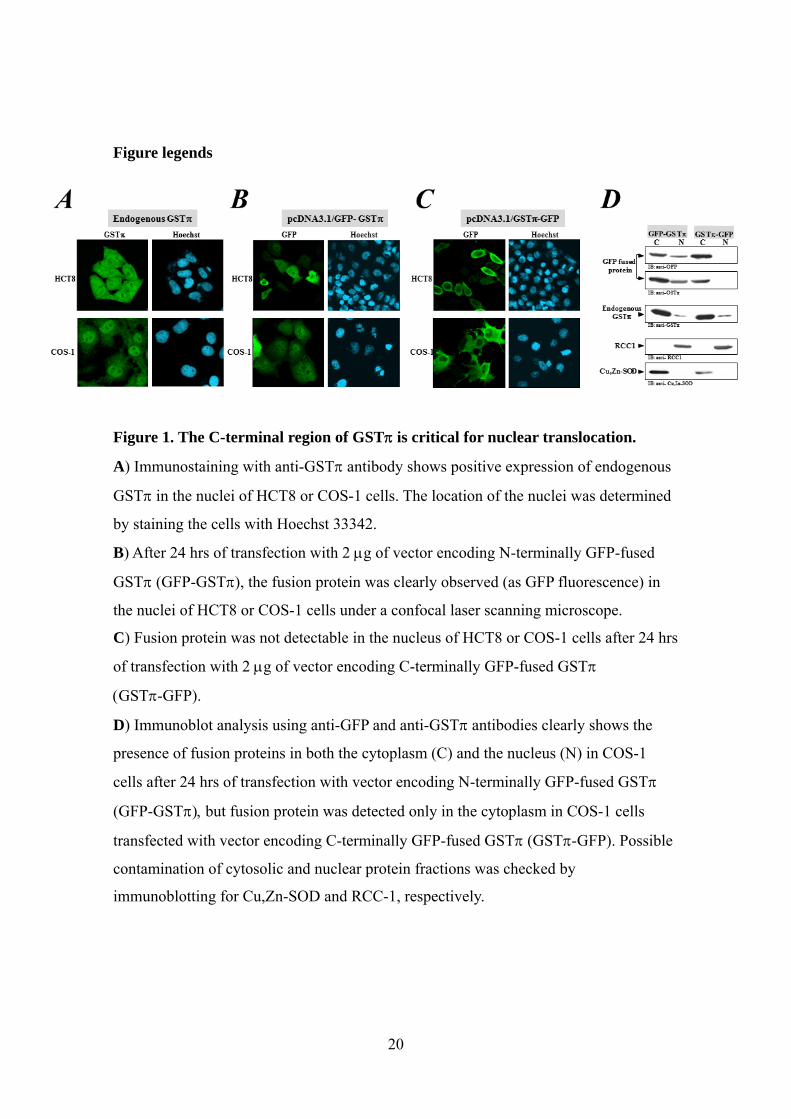
Figure 1. The C-terminal region of GST is critical for nuclear translocation.
A) Immunostaining with anti-GST antibody shows positive expression of endogenous
GST in the nuclei of HCT8 or COS-1 cells. The location of the nuclei was determined
by staining the cells with Hoechst 33342.
B) After 24 hrs of transfection with 2 g of vector encoding N-terminally GFP-fused
GST (GFP-GST, the fusion protein was clearly observed (as GFP fluorescence) in
the nuclei of HCT8 or COS-1 cells under a confocal laser scanning microscope.
C) Fusion protein was not detectable in the nucleus of HCT8 or COS-1 cells after 24 hrs
of transfection with 2 g of vector encoding C-terminally GFP-fused GST
GST-GFP).
D) Immunoblot analysis using anti-GFP and anti-GST antibodies clearly shows the
presence of fusion proteins in both the cytoplasm (C) and the nucleus (N) in COS-1
cells after 24 hrs of transfection with vector encoding N-terminally GFP-fused GST
(GFP-GSTbut fusion protein was detected only in the cytoplasm in COS-1 cells
transfected with vector encoding C-terminally GFP-fused GST (GST-GFP. Possible
contamination of cytosolic and nuclear protein fractions was checked by
immunoblotting for Cu,Zn-SOD and RCC-1, respectively.
21
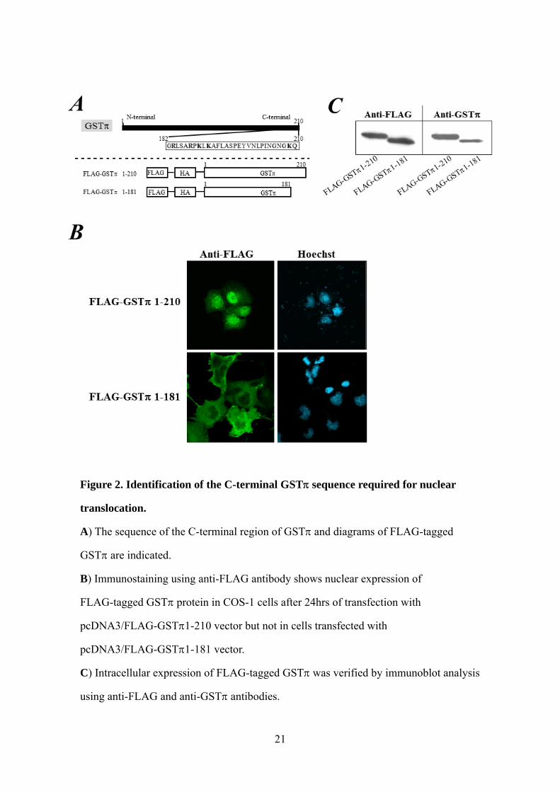
Figure 2. Identification of the C-terminal GST sequence required for nuclear
translocation.
A) The sequence of the C-terminal region of GST and diagrams of FLAG-tagged
GST are indicated.
B) Immunostaining using anti-FLAG antibody shows nuclear expression of
FLAG-tagged GST protein in COS-1 cells after 24hrs of transfection with
pcDNA3/FLAG-GST1-210 vector but not in cells transfected with
pcDNA3/FLAG-GST1-181 vector.
C) Intracellular expression of FLAG-tagged GST was verified by immunoblot analysis
using anti-FLAG and anti-GST antibodies.
22
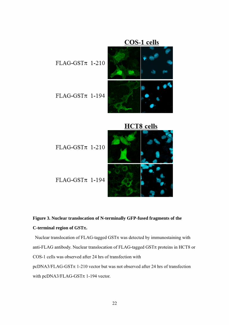
Figure 3. Nuclear translocation of N-terminally GFP-fused fragments of the
C-terminal region of GST.
Nuclear translocation of FLAG-tagged GST was detected by immunostaining with
anti-FLAG antibody. Nuclear translocation of FLAG-tagged GST proteins in HCT8 or
COS-1 cells was observed after 24 hrs of transfection with
pcDNA3/FLAG-GST1-210 vector but was not observed after 24 hrs of transfection
with pcDNA3/FLAG-GST1-194 vector.
23
Figure 4. Nuclear translocation of GST is dependent on cytosolic factor(s) and
ATP.
In vitro transport assays show the presence of recombinant GST-GFP-GST195-208
protein (GFP fluorescence) in the nuclei of digitonin-permeabilized HeLa cells
supplemented with cytosolic extract and the ATP regeneration system for 30 min at
30°C.
24
Supplementary file 1
Materials
RPMI 1640 medium and Dulbecco’s modified Eagle’s medium (DMEM) were
purchased from Sigma Aldrich (St. Louis, MO); fetal bovine serum (FBS) was from
Invitrogen Corp. (Carlsbad, CA); horseradish peroxidase (HRP)-labeled anti-mouse IgG,
HRP-labeled anti-rabbit IgG, and HRP-labeled anti-goat IgG were from DAKO A/S
(Glostrup, Denmark); fluorescein isothiocyanate (FITC)-conjugated anti-rabbit IgG and
FITC-conjugated anti-mouse IgG were from ICN Pharmaceuticals (Aurora, OH);
anti-green fluorescent protein (GFP) antibody was from Invitrogen Corp.; anti-FLAG
antibody was from Sigma Aldrich; anti-human regulator of chromosome condensation
(RCC1) antibody was from BD Biosciences Pharmingen (San Jose, CA); and
anti-human Cu,Zn-superoxide dismutase (Cu,Zn-SOD) antibody was a gift from Dr. K.
Suzuki (Hyogo College of Medicine, Nishinomiya, Japan). The Slow Fade Light
Antifade Kit, Lipofectamine reagent, Lipofectamine 2000, pcDNA3.1/NT-GFP vector,
and pcDNA3.1/CT-GFP vector were purchased from Invitrogen Corp. The
QuickChange Site-Directed Mutagenesis Kit was purchased from STRATAGENE (La
Jolla, CA). The FLAG-HA-tagged protein expression vector (pcDNA3/FLAG-HA
vector), constructed by introducing a BglII-Kozack-ATG-FLAG-HA-EcoRI fragment
into the BamHI-EcoRI restriction sites of pcDNA3 (Invitrogen), was a gift from Dr. J.
Yanagisawa (Institute of Applied Biochemistry, University of Tsukuba, Ibaraki, Japan).
The Enhanced Chemiluminescence (ECL) Kit, pGEX-6P-1 vector and Glutathione
25
Sepharose 4 Fast Flow were purchased from GE Healthcare Bio-Sciences (Little
Chalfont, UK). Hoechst 33342 and digitonin were from CALBIOCHEM (San Diego,
CA). The other chemicals and reagents were purchased from Sigma Aldrich.
PCR primers
To create GFP-GST182-210, GFP-GST182-210 (K209G), GFP-GST182-208,
GFP-GST182-194 and GFP-GST195-208, GST fragments were prepared by PCR
using GST cDNA as a template and an appropriate set of primers. The primers used
were as follows: GST182-210 sense, 5'-ACC GAA TTC TGG GGC GCC TCA GCG
CCC GGC CC; GST182-210 antisense, 5'-GCG GAT ATC TCA CTG TTT CCC GTT
GCC ATT GAT; GST182-210 (K209G) sense, 5'- ACC GAA TTC TGG GGC GCC
TCA GCG CCC GGC CC; GST182-210 (K209G) antisense, 5'- GCG GAT ATC TCA
CTG GGC CCC GTT GCC ATT GAT; GST182-208 sense, 5'-ACC GAA TTC TGG
GGC GCC TCA GCG CCC GGC CC; GST182-208 antisense, 5'- GCG GAT ATC
TCA CCC GTT GCC ATT GAT GGG GAG; GST182-194 sense, 5'- ACC GAA TTC
TGG GGC GCC TCA GCG CCC GGC CC; GST182-194 antisense, 5'- GCG GAT
ATC TCA CAG GAA GGC CTT GAG CTT GGG; GST195-208 sense, 5'- ACC GAA
TTC TGG CCT CCC CTG AGT ACG TGA AC; GST195-208 antisense, 5'- GCG
GAT ATC TCA CCC GTT GCC ATT GAT GGG GAG; GST1-194 sense, 5'-ACC
GAA TTC GCC ACC ATG CCG CCC TAC ACC GTG GTC; and GST1-194 antisense,
5'- GCG GAT ATC TCA CAG GAA GGC CTT GAG CTT GGG.
To obtain FLAG-HA-tagged at the N-terminal end of full-length or deletion mutants
of GST (FLAG-GST1-210, FLAG-GST1-181 and FLAG-GST1-194), GST
fragments were prepared by PCR using an appropriate set of primers. The following
primers were used: GST1-210 sense, 5'-CTC GCG GCC GCA AAT GCC GCC CTA
26
CAC CGT GGT C; GST1-210 antisense, 5'- ATA GGG CCC TCA CTG TTT CCC
GTT GCC ATT GAT; GST1-181 sense, 5'-CTC GCG GCC GCA AAT GCC GCC CTA
CAC CGT GGT C; GST1-181 antisense, 5'- ATA GGG CCC TCA CAC ATA TGC
TGA GAG CAG GGG; GST1-194 sense, 5'- CTC GCG GCC GCA AAT GCC GCC
CTA CAC CGT GGT C; and GST1-194 antisense, 5'-ATA GGG CCC TCA CAG GAA
GGC CTT GAG CTT GGG.
The cDNA of GFP or GFP-GST195-208 was inserted into the pGEX-6P-1vector
using the SmaI-NotI restriction sites to construct an expression vector for GFP or
GFP-GST195-208 ( in which GFP is fused at the C-terminal end of GST from
Schistosoma japonicum). To create cDNA of GFP and GFP-GST195-208, each
fragment was prepared by PCR using pcDNA3.1/NT-GFP or
pcDNA3.1/NT-GFP-GST195-208 as a template and an appropriate set of primers. The
primers used were as follows: GFP sense, 5'-ATA CCC GGG TAT GGC CAG CAA
AGG AGA AGA ACC T; GFP antisense, 5'-CTC GCG GCC GCT CAA CCA CAC
TGG ACT AGT GGA TC; GFP-GST195-208 sense, 5'-ATA CCC GGG TAT GGC
CAG CAA AGG AGA AGA ACC T; and GFP-GST195-208 antisense, 5'-CTC GCG
GCC GCT CAC TGT TTC CCG TTG CCA TTG AT.
27
Supplementary Figure legends
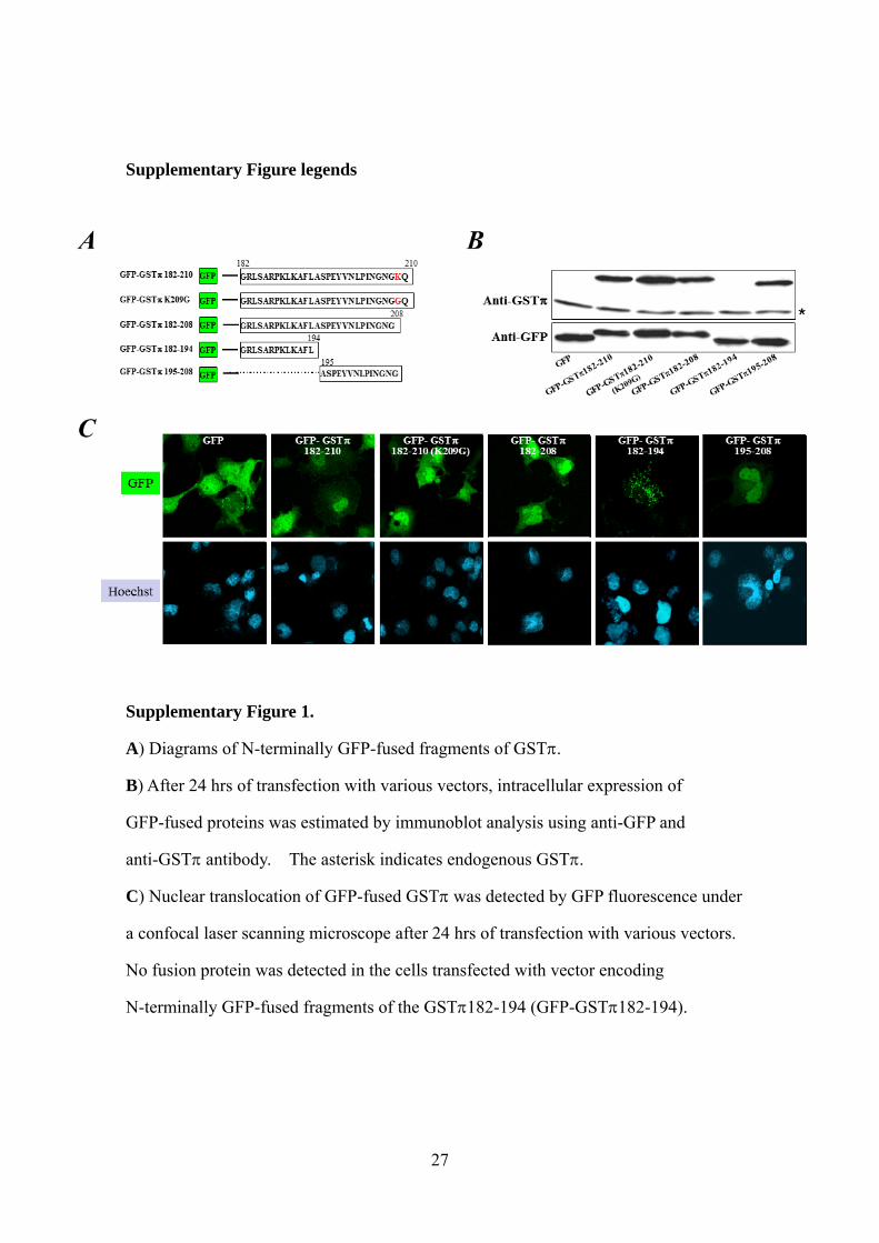
Supplementary Figure 1.
A) Diagrams of N-terminally GFP-fused fragments of GST.
B) After 24 hrs of transfection with various vectors, intracellular expression of
GFP-fused proteins was estimated by immunoblot analysis using anti-GFP and
anti-GST antibody. The asterisk indicates endogenous GST.
C) Nuclear translocation of GFP-fused GST was detected by GFP fluorescence under
a confocal laser scanning microscope after 24 hrs of transfection with various vectors.
No fusion protein was detected in the cells transfected with vector encoding
N-terminally GFP-fused fragments of the GST182-194 (GFP-GST182-194).
28
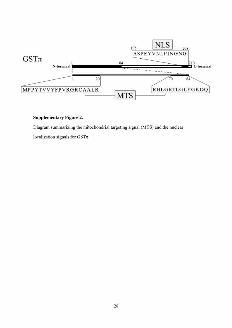
Supplementary Figure 2.
Diagram summarizing the mitochondrial targeting signal (MTS) and the nuclear
localization signals for GST.

![Medical Ozone Reduces the Risk of γ-Glutamyl Transferase ... · Previously, ozone’s protective effects against liver damage such as MTX-induced hepatotoxicity in rats [9], CCl](https://static.fdocument.org/doc/165x107/606bd1351d0ec53c2b5c31f0/medical-ozone-reduces-the-risk-of-glutamyl-transferase-previously-ozoneas.jpg)