3 - Connective tissue · , synthesized in the fibroblast, illustrated in Figure 3.3, steps 1–4....
26
© Copyright 2013 Elsevier, Ltd. All rights reserved. 3 Connective tissue CHAPTER CONTENTS Structural composition 29 Connective tissue cells . . . . . . . . . . . . . . . . 29 Extracellular matrix . . . . . . . . . . . . . . . . . . 29 Structures containing connective tissue 33 Trauma to soft connective tissue 40 Introduction . . . . . . . . . . . . . . . . . . . . . . 40 Inflammation . . . . . . . . . . . . . . . . . . . . . . 40 Repair . . . . . . . . . . . . . . . . . . . . . . . . . 41 Remodelling . . . . . . . . . . . . . . . . . . . . . . 41 Self-perpetuating inflammation . . . . . . . . . . . . 41 Effects of immobilization on healing . . . . . . . . . 42 Effects of mobilization on healing . . . . . . . . . . . 43 Treatment of traumatic soft connective tissue lesions 43 Muscular lesions . . . . . . . . . . . . . . . . . . . . 44 Tendinous lesions . . . . . . . . . . . . . . . . . . . 45 Ligamentous lesions . . . . . . . . . . . . . . . . . . 48 Capsular lesions . . . . . . . . . . . . . . . . . . . . 49 Structural composition Together with muscle, nerve and epithelium, connective tissue is one of the basic components in the human body. It binds structures together, helps in mechanical and chemical protection and also plays a principal role in reparative processes. Connective tissues are defined as those composed predomi- nantly of the extracellular matrix and connective tissue cells. The matrix is made up of fibrous proteins and a relatively amorphous ground substance. Many of the special properties of connective tissues are determined by the composition of the matrix, and their classification is also largely based on its characteristics. Connective tissue cells Cells of general connective tissues can be separated into the resident cell population (mainly fibroblasts) and a population of migrant cells with various defensive functions (macrophages, lymphocytes, mast cells, neutrophils and eosinophils), which may change in number and moderate their activities according to demand. Fibroblasts, the majority of cells in ordinary connective tissue, arise from the relevant undifferentiated mesenchymal stem cells 1 and are involved in the production of fibrous ele- ments and non-fibrous ground substance (Fig. 3.1). During wound repair they are particularly active and migrate along strands of fibrin by amoeboid movements to distribute them- selves through the healing area to start repair. Fibroblast activ- ity is influenced by various factors such as the partial pressure of oxygen, levels of steroid hormones, nutrition and the mechanical stress present in the tissue. 2 The other cell types are migrant cells and only occasionally present, such as: macrophages, lymphocytes, mast cells, and granulocutes (Table 3.1). 3,4 Extracellular matrix (ECM) The extracellular matrix is composed of insoluble protein fibres, the fibrillar matrix and a mixture of macromolecules, the interfibrillar matrix. The latter consists of adhesive glyco- proteins and soluble complexes composed of carbohydrate polymers linked to protein molecules (proteoglycans and glycosaminoglycans), which bind water. The extracellular matrix distributes the mechanical stresses on tissues and also provides the structural environment of the cells embedded
Transcript of 3 - Connective tissue · , synthesized in the fibroblast, illustrated in Figure 3.3, steps 1–4....
© Copyright 2013 Elsevier, Ltd. All rights reserved.
3 Connective tissue
CHAPTER CONTENTS
Structural composition 29
Connective tissue cells . . . . . . . . . . . . . . . . 29Extracellular matrix . . . . . . . . . . . . . . . . . . 29
Structures containing connective tissue 33
Trauma to soft connective tissue 40
Introduction . . . . . . . . . . . . . . . . . . . . . . 40Inflammation . . . . . . . . . . . . . . . . . . . . . . 40Repair . . . . . . . . . . . . . . . . . . . . . . . . . 41Remodelling . . . . . . . . . . . . . . . . . . . . . . 41Self-perpetuating inflammation . . . . . . . . . . . . 41Effects of immobilization on healing . . . . . . . . . 42Effects of mobilization on healing . . . . . . . . . . . 43
Treatment of traumatic soft connective tissue lesions 43
Muscular lesions . . . . . . . . . . . . . . . . . . . . 44Tendinous lesions . . . . . . . . . . . . . . . . . . . 45Ligamentous lesions . . . . . . . . . . . . . . . . . . 48Capsular lesions . . . . . . . . . . . . . . . . . . . . 49
Structural composition
Together with muscle, nerve and epithelium, connective tissue is one of the basic components in the human body. It binds structures together, helps in mechanical and chemical protection and also plays a principal role in reparative processes.
Connective tissues are defined as those composed predomi-nantly of the extracellular matrix and connective tissue cells. The matrix is made up of fibrous proteins and a relatively amorphous ground substance. Many of the special properties
of connective tissues are determined by the composition of the matrix, and their classification is also largely based on its characteristics.
Connective tissue cells
Cells of general connective tissues can be separated into the resident cell population (mainly fibroblasts) and a population of migrant cells with various defensive functions (macrophages, lymphocytes, mast cells, neutrophils and eosinophils), which may change in number and moderate their activities according to demand.
Fibroblasts, the majority of cells in ordinary connective tissue, arise from the relevant undifferentiated mesenchymal stem cells1 and are involved in the production of fibrous ele-ments and non-fibrous ground substance (Fig. 3.1). During wound repair they are particularly active and migrate along strands of fibrin by amoeboid movements to distribute them-selves through the healing area to start repair. Fibroblast activ-ity is influenced by various factors such as the partial pressure of oxygen, levels of steroid hormones, nutrition and the mechanical stress present in the tissue.2
The other cell types are migrant cells and only occasionally present, such as: macrophages, lymphocytes, mast cells, and granulocutes (Table 3.1).3,4
Extracellular matrix (ECM)
The extracellular matrix is composed of insoluble protein fibres, the fibrillar matrix and a mixture of macromolecules, the interfibrillar matrix. The latter consists of adhesive glyco-proteins and soluble complexes composed of carbohydrate polymers linked to protein molecules (proteoglycans and glycosaminoglycans), which bind water. The extracellular matrix distributes the mechanical stresses on tissues and also provides the structural environment of the cells embedded
General Principles
30
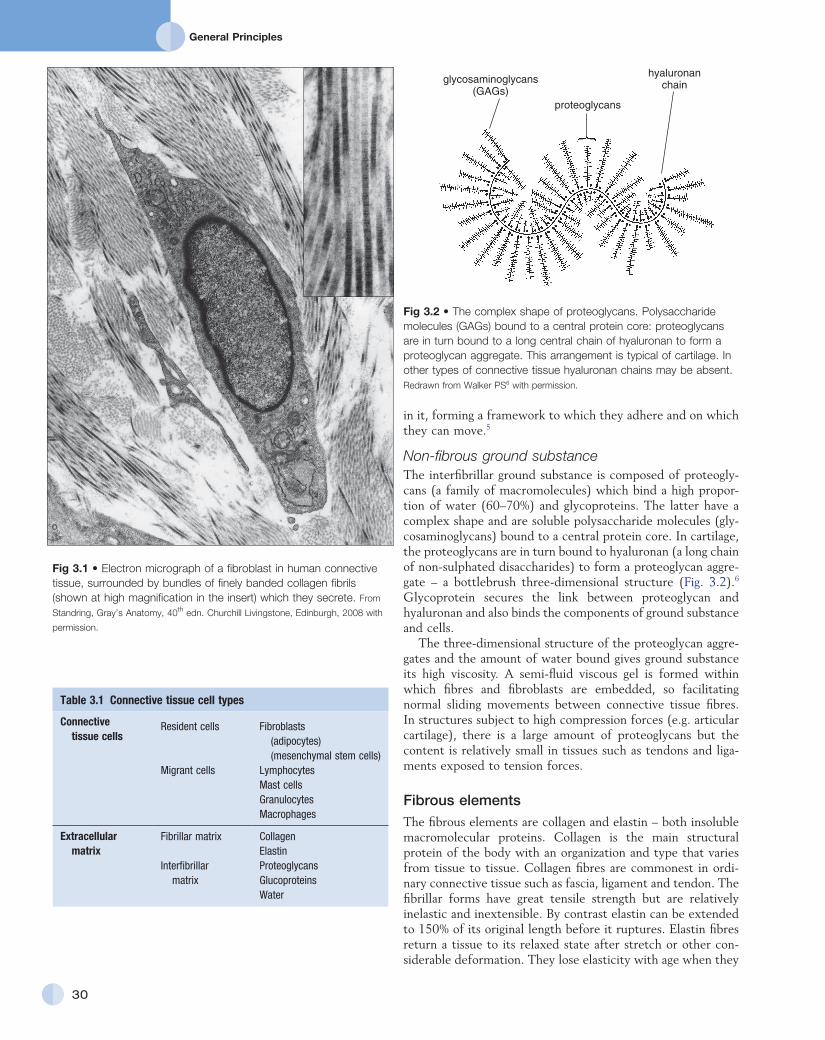
Fig 3.2 • The complex shape of proteoglycans. Polysaccharide molecules (GAGs) bound to a central protein core: proteoglycans are in turn bound to a long central chain of hyaluronan to form a proteoglycan aggregate. This arrangement is typical of cartilage. In other types of connective tissue hyaluronan chains may be absent. Redrawn from Walker PS6 with permission.
glycosaminoglycans(GAGs)
proteoglycans
hyaluronanchain
Fig 3.1 • Electron micrograph of a fibroblast in human connective tissue, surrounded by bundles of finely banded collagen fibrils (shown at high magnification in the insert) which they secrete. From
Standring, Gray’s Anatomy, 40th edn. Churchill Livingstone, Edinburgh, 2008 with
permission.
in it, forming a framework to which they adhere and on which they can move.5
Non-fibrous ground substanceThe interfibrillar ground substance is composed of proteogly-cans (a family of macromolecules) which bind a high propor-tion of water (60–70%) and glycoproteins. The latter have a complex shape and are soluble polysaccharide molecules (gly-cosaminoglycans) bound to a central protein core. In cartilage, the proteoglycans are in turn bound to hyaluronan (a long chain of non-sulphated disaccharides) to form a proteoglycan aggre-gate – a bottlebrush three-dimensional structure (Fig. 3.2).6 Glycoprotein secures the link between proteoglycan and hyaluronan and also binds the components of ground substance and cells.
The three-dimensional structure of the proteoglycan aggre-gates and the amount of water bound gives ground substance its high viscosity. A semi-fluid viscous gel is formed within which fibres and fibroblasts are embedded, so facilitating normal sliding movements between connective tissue fibres. In structures subject to high compression forces (e.g. articular cartilage), there is a large amount of proteoglycans but the content is relatively small in tissues such as tendons and liga-ments exposed to tension forces.
Fibrous elementsThe fibrous elements are collagen and elastin – both insoluble macromolecular proteins. Collagen is the main structural protein of the body with an organization and type that varies from tissue to tissue. Collagen fibres are commonest in ordi-nary connective tissue such as fascia, ligament and tendon. The fibrillar forms have great tensile strength but are relatively inelastic and inextensible. By contrast elastin can be extended to 150% of its original length before it ruptures. Elastin fibres return a tissue to its relaxed state after stretch or other con-siderable deformation. They lose elasticity with age when they
Table 3.1 Connective tissue cell types
Connective tissue cells
Resident cells Fibroblasts (adipocytes) (mesenchymal stem cells)
Migrant cells LymphocytesMast cellsGranulocytesMacrophages
Extracellular matrix
Fibrillar matrix CollagenElastin
Interfibrillar matrix
ProteoglycansGlucoproteinsWater
C H A P T E R 3Connective tissue
31
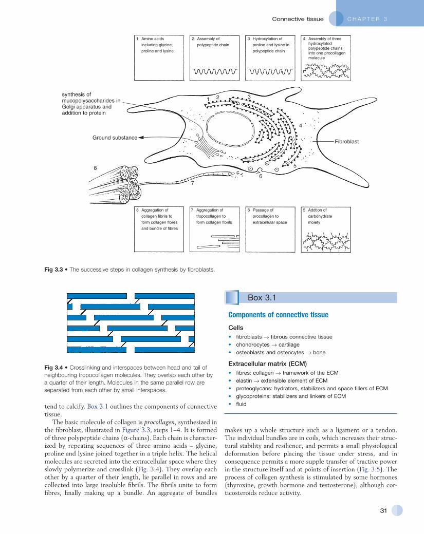
Fig 3.3 • The successive steps in collagen synthesis by fibroblasts.
synthesis ofmucopolysaccharides inGolgi apparatus andaddition to protein
Ground substance
4321
5678
8
76
5
4
321
Amino acids
including glycine,
proline and lysine
Assembly of
polypeptide chain
Hydroxylation of
proline and lysine in
polypeptide chain
Assembly of threehydroxylatedpolypeptide chainsinto one procollagenmolecule
Aggregation of
collagen fibrils to
form collagen fibres
and bundle of fibres
Aggregation of
tropocollagen to
form collagen fibrils
Passage of
procollagen to
extracellular space
Addtion of
carbohydrate
moiety
Fibroblast
Fig 3.4 • Crosslinking and interspaces between head and tail of neighbouring tropocollagen molecules. They overlap each other by a quarter of their length. Molecules in the same parallel row are separated from each other by small interspaces.
tend to calcify. Box 3.1 outlines the components of connective tissue.
The basic molecule of collagen is procollagen, synthesized in the fibroblast, illustrated in Figure 3.3, steps 1–4. It is formed of three polypeptide chains (α-chains). Each chain is character-ized by repeating sequences of three amino acids – glycine, proline and lysine joined together in a triple helix. The helical molecules are secreted into the extracellular space where they slowly polymerize and crosslink (Fig. 3.4). They overlap each other by a quarter of their length, lie parallel in rows and are collected into large insoluble fibrils. The fibrils unite to form fibres, finally making up a bundle. An aggregate of bundles
makes up a whole structure such as a ligament or a tendon. The individual bundles are in coils, which increases their struc-tural stability and resilience, and permits a small physiological deformation before placing the tissue under stress, and in consequence permits a more supple transfer of tractive power in the structure itself and at points of insertion (Fig. 3.5). The process of collagen synthesis is stimulated by some hormones (thyroxine, growth hormone and testosterone), although cor-ticosteroids reduce activity.
Box 3.1
Components of connective tissue
Cells• fibroblasts → fibrous connective tissue• chondrocytes → cartilage• osteoblasts and osteocytes → bone
Extracellular matrix (ECM)• fibres: collagen → framework of the ECM• elastin → extensible element of ECM• proteoglycans: hydrators, stabilizers and space fillers of ECM• glycoproteins: stabilizers and linkers of ECM• fluid
General Principles
32
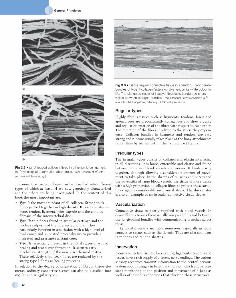
Regular typesHighly fibrous tissues such as ligaments, tendons, fascia and aponeuroses are predominantly collagenous and show a dense and regular orientation of the fibres with respect to each other. The direction of the fibres is related to the stress they experi-ence. Collagen bundles in ligaments and tendons are very strong and rupture usually takes place at the bony attachments rather than by tearing within their substance (Fig. 3.6).
Irregular typesThe irregular types consist of collagen and elastin interlacing in all directions. It is loose, extensible and elastic and found between muscles, blood vessels and nerves. It binds partly together, although allowing a considerable amount of move-ment to take place. In the sheaths of muscles and nerves and the adventitia of large blood vessels, the tissue is more dense with a high proportion of collagen fibres to protect these struc-tures against considerable mechanical stress. The dura mater is also an example of an irregular connective tissue sleeve.
VascularizationConnective tissue is poorly supplied with blood vessels. In dense fibrous tissues these usually run parallel to and between the longitudinal bundles with communicating branches across these.
Lymphatic vessels are more numerous, especially in loose connective tissues such as the dermis. They are also abundant in tendons and tendon sheaths.
InnervationDense connective tissues, for example, ligaments, tendons and fascia, have a rich supply of afferent nerve endings. The various sensory receptors transmit information to the central nervous system about changes in length and tension which allows con-stant monitoring of the position and movement of a joint as well as of injurious conditions that threaten these structures.
Connective tissue collagen can be classified into different types of which at least 14 are now genetically characterized and the others are being investigated. In the context of this book the most important are:
• Type I: the most abundant of all collagen. Strong thick fibres packed together in high density. It predominates in bone, tendon, ligament, joint capsule and the annulus fibrosus of the intervertebral disc.
• Type II: thin fibres found in articular cartilage and the nucleus pulposus of the intervertebral disc. They particularly function in association with a high level of hyaluronan and sulphated proteoglycans to provide a hydrated and pressure-resistant core.
• Type III: essentially present in the initial stages of wound healing and scar tissue formation. It secures early mechanical strength of the newly synthesized matrix. These relatively thin, weak fibres are replaced by the strong type I fibres as healing proceeds.
In relation to the degree of orientation of fibrous tissue ele-ments, ordinary connective tissues can also be classified into regular and irregular types.
Fig 3.5 • (a) Unloaded collagen fibres in a human knee ligament. (b) Physiological deformation after stress. From Kennedy et al7 with
permission (http://jbjs.org/).
(a)
(b)
Fig 3.6 • Dense regular connective tissue in a tendon. Thick parallel bundles of type 1 collagen (asterisks) give tendon its white colour in life. The elongated nuclei of inactive fibroblasts (tendon cells) are visible between collagen bundles. From Standring, Gray’s Anatomy, 40th
edn. Churchill Livingstone, Edinburgh, 2008 with permission.
* *
C H A P T E R 3Connective tissue
33
Structural and physiological studies8,9 have shown the pres-ence of at least four types of receptor. Three of these have encapsulated endings but the fourth consists of free unencap-sulated endings:
• Type 1 (Ruffini endings) are present in the superficial layers of a fibrous joint capsule. They respond to stretch and pressure within the capsule and are slow adapting with a low threshold. They signal joint position and movement.
• Type 2 are particularly located in the deep layers of the fibrous capsule. They respond to rapid movement, pressure change and vibration but adapt quickly. They have a low threshold and are inactive when the joint is at rest.
• Type 3 are found in ligaments. They transmit information on ligamentous tension so as to prevent excessive stress. Their threshold is relatively high and they adapt slowly. They are not active at rest.
• Type 4 are free unencapsulated nociceptor terminals which ramify within the fibrous capsule, around adjacent fat pads and blood vessels. They are thought to sense excessive joint movements and also to signal pain. They have a high threshold and are slow adapting. Synovial membrane is relatively insensitive to pain because of the absence of these nerve endings.
All these receptors influence muscle tone via spinal reflex arcs which are formed by the same nerves that supply the muscles which act on the joint. Parts of the joint capsule supplied by a given nerve correspond with the antagonist muscles. Tension on this part of the capsule produces reflex contraction of these muscles to prevent further overstretching of the capsule. In consequence all receptors have an important function in stabi-lization and protection of the joint. After rupture of a capsule, ligament perception is considerably disturbed because of the disruption of the transmission of afferent information. For example in a sprained ankle there is loss of control of locomo-tion. Even months after repair of ligamentous and capsular tissues has taken place, perception may still be distorted.
Structures containing connective tissue
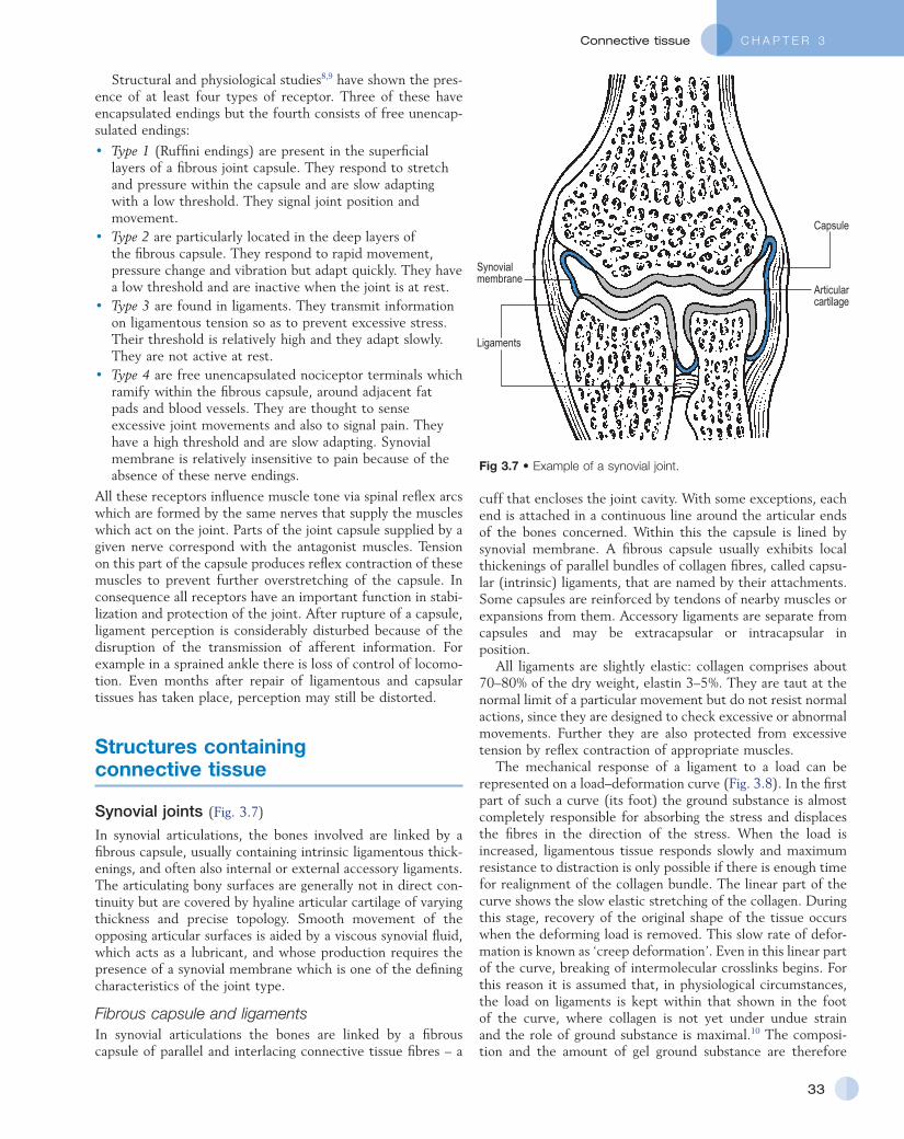
Synovial joints (Fig. 3.7)
In synovial articulations, the bones involved are linked by a fibrous capsule, usually containing intrinsic ligamentous thick-enings, and often also internal or external accessory ligaments. The articulating bony surfaces are generally not in direct con-tinuity but are covered by hyaline articular cartilage of varying thickness and precise topology. Smooth movement of the opposing articular surfaces is aided by a viscous synovial fluid, which acts as a lubricant, and whose production requires the presence of a synovial membrane which is one of the defining characteristics of the joint type.
Fibrous capsule and ligamentsIn synovial articulations the bones are linked by a fibrous capsule of parallel and interlacing connective tissue fibres – a
Fig 3.7 • Example of a synovial joint.
Synovialmembrane
Ligaments
Articularcartilage
Capsule
cuff that encloses the joint cavity. With some exceptions, each end is attached in a continuous line around the articular ends of the bones concerned. Within this the capsule is lined by synovial membrane. A fibrous capsule usually exhibits local thickenings of parallel bundles of collagen fibres, called capsu-lar (intrinsic) ligaments, that are named by their attachments. Some capsules are reinforced by tendons of nearby muscles or expansions from them. Accessory ligaments are separate from capsules and may be extracapsular or intracapsular in position.
All ligaments are slightly elastic: collagen comprises about 70–80% of the dry weight, elastin 3–5%. They are taut at the normal limit of a particular movement but do not resist normal actions, since they are designed to check excessive or abnormal movements. Further they are also protected from excessive tension by reflex contraction of appropriate muscles.
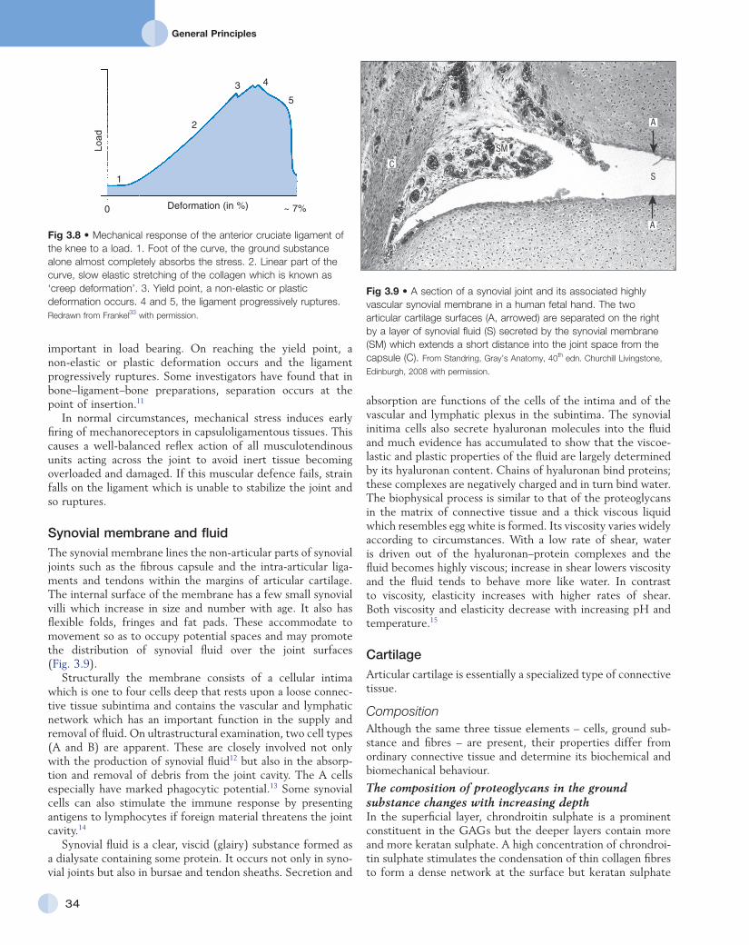
The mechanical response of a ligament to a load can be represented on a load–deformation curve (Fig. 3.8). In the first part of such a curve (its foot) the ground substance is almost completely responsible for absorbing the stress and displaces the fibres in the direction of the stress. When the load is increased, ligamentous tissue responds slowly and maximum resistance to distraction is only possible if there is enough time for realignment of the collagen bundle. The linear part of the curve shows the slow elastic stretching of the collagen. During this stage, recovery of the original shape of the tissue occurs when the deforming load is removed. This slow rate of defor-mation is known as ‘creep deformation’. Even in this linear part of the curve, breaking of intermolecular crosslinks begins. For this reason it is assumed that, in physiological circumstances, the load on ligaments is kept within that shown in the foot of the curve, where collagen is not yet under undue strain and the role of ground substance is maximal.10 The composi-tion and the amount of gel ground substance are therefore
General Principles
34
absorption are functions of the cells of the intima and of the vascular and lymphatic plexus in the subintima. The synovial initima cells also secrete hyaluronan molecules into the fluid and much evidence has accumulated to show that the viscoe-lastic and plastic properties of the fluid are largely determined by its hyaluronan content. Chains of hyaluronan bind proteins; these complexes are negatively charged and in turn bind water. The biophysical process is similar to that of the proteoglycans in the matrix of connective tissue and a thick viscous liquid which resembles egg white is formed. Its viscosity varies widely according to circumstances. With a low rate of shear, water is driven out of the hyaluronan–protein complexes and the fluid becomes highly viscous; increase in shear lowers viscosity and the fluid tends to behave more like water. In contrast to viscosity, elasticity increases with higher rates of shear. Both viscosity and elasticity decrease with increasing pH and temperature.15
CartilageArticular cartilage is essentially a specialized type of connective tissue.
CompositionAlthough the same three tissue elements – cells, ground sub-stance and fibres – are present, their properties differ from ordinary connective tissue and determine its biochemical and biomechanical behaviour.
The composition of proteoglycans in the ground substance changes with increasing depthIn the superficial layer, chrondroitin sulphate is a prominent constituent in the GAGs but the deeper layers contain more and more keratan sulphate. A high concentration of chrondroi-tin sulphate stimulates the condensation of thin collagen fibres to form a dense network at the surface but keratan sulphate
important in load bearing. On reaching the yield point, a non-elastic or plastic deformation occurs and the ligament progressively ruptures. Some investigators have found that in bone–ligament–bone preparations, separation occurs at the point of insertion.11
In normal circumstances, mechanical stress induces early firing of mechanoreceptors in capsuloligamentous tissues. This causes a well-balanced reflex action of all musculotendinous units acting across the joint to avoid inert tissue becoming overloaded and damaged. If this muscular defence fails, strain falls on the ligament which is unable to stabilize the joint and so ruptures.
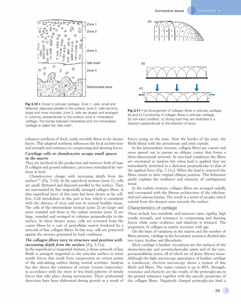
Synovial membrane and fluidThe synovial membrane lines the non-articular parts of synovial joints such as the fibrous capsule and the intra-articular liga-ments and tendons within the margins of articular cartilage. The internal surface of the membrane has a few small synovial villi which increase in size and number with age. It also has flexible folds, fringes and fat pads. These accommodate to movement so as to occupy potential spaces and may promote the distribution of synovial fluid over the joint surfaces (Fig. 3.9).
Structurally the membrane consists of a cellular intima which is one to four cells deep that rests upon a loose connec-tive tissue subintima and contains the vascular and lymphatic network which has an important function in the supply and removal of fluid. On ultrastructural examination, two cell types (A and B) are apparent. These are closely involved not only with the production of synovial fluid12 but also in the absorp-tion and removal of debris from the joint cavity. The A cells especially have marked phagocytic potential.13 Some synovial cells can also stimulate the immune response by presenting antigens to lymphocytes if foreign material threatens the joint cavity.14
Synovial fluid is a clear, viscid (glairy) substance formed as a dialysate containing some protein. It occurs not only in syno-vial joints but also in bursae and tendon sheaths. Secretion and
Fig 3.8 • Mechanical response of the anterior cruciate ligament of the knee to a load. 1. Foot of the curve, the ground substance alone almost completely absorbs the stress. 2. Linear part of the curve, slow elastic stretching of the collagen which is known as ‘creep deformation’. 3. Yield point, a non-elastic or plastic deformation occurs. 4 and 5, the ligament progressively ruptures. Redrawn from Frankel33 with permission.
4
5
3
2
1
0 ~ 7%
Load
Deformation (in %)
Fig 3.9 • A section of a synovial joint and its associated highly vascular synovial membrane in a human fetal hand. The two articular cartilage surfaces (A, arrowed) are separated on the right by a layer of synovial fluid (S) secreted by the synovial membrane (SM) which extends a short distance into the joint space from the capsule (C). From Standring, Gray’s Anatomy, 40th edn. Churchill Livingstone,
Edinburgh, 2008 with permission.
S
SM
C
A
A
C H A P T E R 3Connective tissue
35
enhances synthesis of thick, easily movable fibres in the deeper layers. This adapted synthesis influences the local architecture and strength and resistance to compressing and shearing forces.
Cartilage cells or chondrocytes occupy small spaces in the matrixThey are involved in the production and turnover both of type II collagen and ground substance, processes stimulated by vari-ation in load.
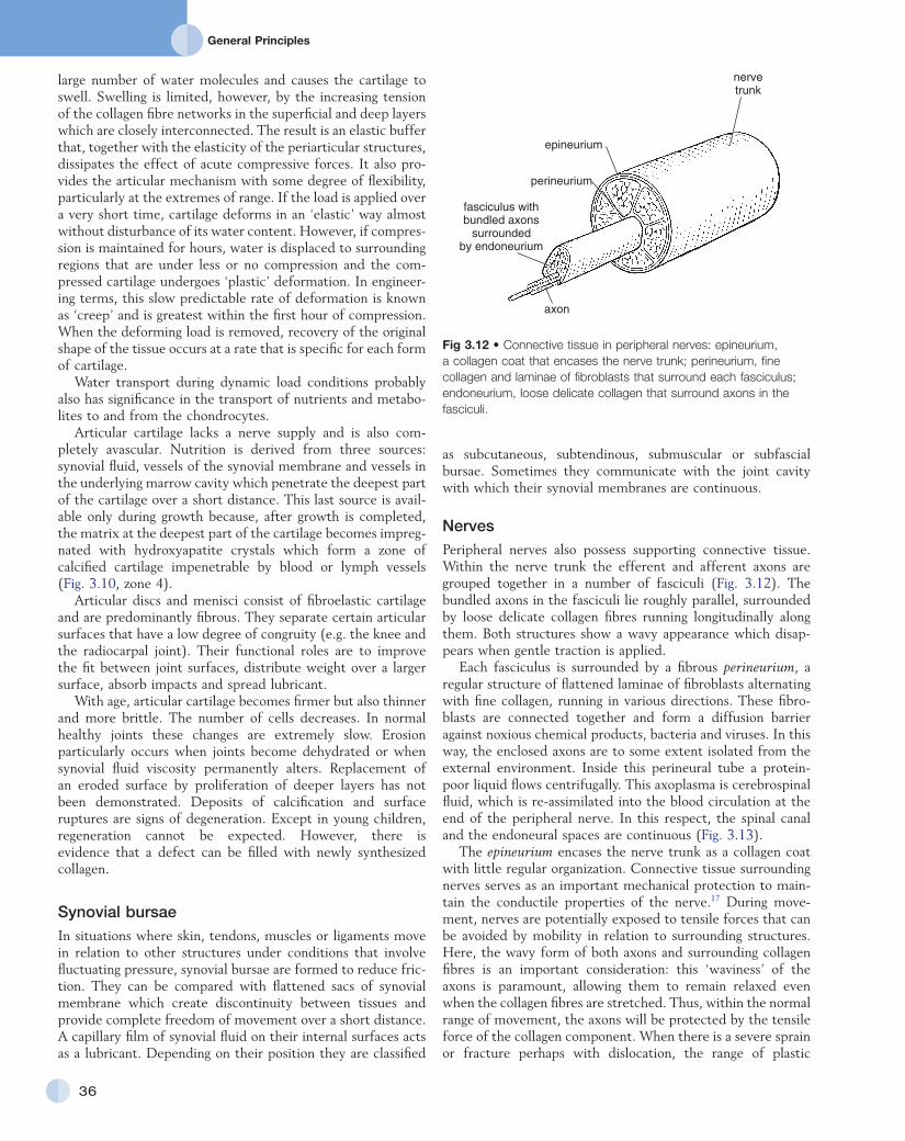
Chondrocytes change with increasing depth from the surface1,16 (Fig. 3.10). In the superficial stratum (zone 1), cells are small, flattened and disposed parallel to the surface. They are surrounded by fine tangentially arranged collagen fibres. A thin superficial layer of this zone has been shown to be cell-free. Cell metabolism in this part is low, which is consistent with the absence of wear and tear in normal healthy tissue. The cells of the intermediate stratum (zone 2) are larger and more rounded and those in the radiate stratum (zone 3) are large, rounded and arranged in columns perpendicular to the surface. In these deeper zones, cells are screened from the coarse fibres by a coat of pericellular matrix bordered by a network of fine collagen fibres. In this way, cells are protected against the stresses generated by load conditions.
The collagen fibres vary in structure and position with increasing depth from the surface (Fig. 3.11a)In the superficial or tangential stratum, a dense network of fine fibrils is arranged tangential to the articular surface to resist tensile forces that result from compression on certain points of the articulating surface during normal activities. Analysis has also shown the existence of certain ‘tension trajectories’ in accordance with the more or less fixed patterns of tensile forces that take place during movements. These preferential directions have been elaborated during growth as a result of
Fig 3.10 • Zones in articular cartilage. Zone 1, cells, small and flattened, disposed parallel to the surface; zone 2, cells become larger and more rounded; zone 3, cells are largest and arranged in columns, perpendicular to the surface; zone 4, mineralized cartilage. The border between mineralized and non-mineralized cartilage is called the ‘tide mark’.
Zone 1
Zone 2
‘tide mark’
Zone 3
Zone 4
Subchondral bone
Fig 3.11 • (a) Arrangement of collagen fibres in articular cartilage. (b) and (c) Functioning of collagen fibres in articular cartilage. (b) non-load condition, (c) during load they are stretched in a direction perpendicular to the direction of force.
(a)
(b)
(c)
forces acting on the joint. Near the border of the joint, the fibrils blend with the periosteum and joint capsule.
In the intermediate stratum, collagen fibres are coarser and more spread out to pursue an oblique course that forms a three-dimensional network. In non-load conditions the fibres are orientated at random but when load is applied they are immediately stretched in a direction perpendicular to that of the applied force (Fig. 3.11c). When the load is removed the fibres return to their original oblique position. This behaviour partly explains the resilience and elasticity of cartilagenous tissue.
In the radiate stratum, collagen fibres are arranged radially and correspond with the fibrous architecture of the subchon-dral and osseous lamina. The result is a series of arcades which extend from the deepest zone towards the surface.
Characteristics of cartilageThese include low metabolic and turnover rates, rigidity, high tensile strength, and resistance to compressing and shearing forces while some resilience and elasticity is retained. The proportion of collagen in matrix increases with age.
On the basis of variations in the matrix and the number of fibres present, cartilage in the locomotor system is divided into two types: hyaline and fibroelastic.
Most cartilage is hyaline: exceptions are the surfaces of the sternoclavicular and acromioclavicular joints and of the tem-poromandibular joints, all of which are of dense fibrous tissue. Although the light microscope appearance of hyaline cartilage is translucent, electron microscopy shows a system of fine fibrils and fibres. The water content is up to 80%. Strength, resistance and elasticity are the results of the proteoglycans in the ground substance together with the specific properties of the collagen fibres. Negatively charged proteoglycans bind a
General Principles
36
as subcutaneous, subtendinous, submuscular or subfascial bursae. Sometimes they communicate with the joint cavity with which their synovial membranes are continuous.
NervesPeripheral nerves also possess supporting connective tissue. Within the nerve trunk the efferent and afferent axons are grouped together in a number of fasciculi (Fig. 3.12). The bundled axons in the fasciculi lie roughly parallel, surrounded by loose delicate collagen fibres running longitudinally along them. Both structures show a wavy appearance which disap-pears when gentle traction is applied.
Each fasciculus is surrounded by a fibrous perineurium, a regular structure of flattened laminae of fibroblasts alternating with fine collagen, running in various directions. These fibro-blasts are connected together and form a diffusion barrier against noxious chemical products, bacteria and viruses. In this way, the enclosed axons are to some extent isolated from the external environment. Inside this perineural tube a protein-poor liquid flows centrifugally. This axoplasma is cerebrospinal fluid, which is re-assimilated into the blood circulation at the end of the peripheral nerve. In this respect, the spinal canal and the endoneural spaces are continuous (Fig. 3.13).
The epineurium encases the nerve trunk as a collagen coat with little regular organization. Connective tissue surrounding nerves serves as an important mechanical protection to main-tain the conductile properties of the nerve.17 During move-ment, nerves are potentially exposed to tensile forces that can be avoided by mobility in relation to surrounding structures. Here, the wavy form of both axons and surrounding collagen fibres is an important consideration: this ‘waviness’ of the axons is paramount, allowing them to remain relaxed even when the collagen fibres are stretched. Thus, within the normal range of movement, the axons will be protected by the tensile force of the collagen component. When there is a severe sprain or fracture perhaps with dislocation, the range of plastic
large number of water molecules and causes the cartilage to swell. Swelling is limited, however, by the increasing tension of the collagen fibre networks in the superficial and deep layers which are closely interconnected. The result is an elastic buffer that, together with the elasticity of the periarticular structures, dissipates the effect of acute compressive forces. It also pro-vides the articular mechanism with some degree of flexibility, particularly at the extremes of range. If the load is applied over a very short time, cartilage deforms in an ‘elastic’ way almost without disturbance of its water content. However, if compres-sion is maintained for hours, water is displaced to surrounding regions that are under less or no compression and the com-pressed cartilage undergoes ‘plastic’ deformation. In engineer-ing terms, this slow predictable rate of deformation is known as ‘creep’ and is greatest within the first hour of compression. When the deforming load is removed, recovery of the original shape of the tissue occurs at a rate that is specific for each form of cartilage.
Water transport during dynamic load conditions probably also has significance in the transport of nutrients and metabo-lites to and from the chondrocytes.
Articular cartilage lacks a nerve supply and is also com-pletely avascular. Nutrition is derived from three sources: synovial fluid, vessels of the synovial membrane and vessels in the underlying marrow cavity which penetrate the deepest part of the cartilage over a short distance. This last source is avail-able only during growth because, after growth is completed, the matrix at the deepest part of the cartilage becomes impreg-nated with hydroxyapatite crystals which form a zone of calcified cartilage impenetrable by blood or lymph vessels (Fig. 3.10, zone 4).
Articular discs and menisci consist of fibroelastic cartilage and are predominantly fibrous. They separate certain articular surfaces that have a low degree of congruity (e.g. the knee and the radiocarpal joint). Their functional roles are to improve the fit between joint surfaces, distribute weight over a larger surface, absorb impacts and spread lubricant.
With age, articular cartilage becomes firmer but also thinner and more brittle. The number of cells decreases. In normal healthy joints these changes are extremely slow. Erosion particularly occurs when joints become dehydrated or when synovial fluid viscosity permanently alters. Replacement of an eroded surface by proliferation of deeper layers has not been demonstrated. Deposits of calcification and surface ruptures are signs of degeneration. Except in young children, regeneration cannot be expected. However, there is evidence that a defect can be filled with newly synthesized collagen.
Synovial bursaeIn situations where skin, tendons, muscles or ligaments move in relation to other structures under conditions that involve fluctuating pressure, synovial bursae are formed to reduce fric-tion. They can be compared with flattened sacs of synovial membrane which create discontinuity between tissues and provide complete freedom of movement over a short distance. A capillary film of synovial fluid on their internal surfaces acts as a lubricant. Depending on their position they are classified
Fig 3.12 • Connective tissue in peripheral nerves: epineurium, a collagen coat that encases the nerve trunk; perineurium, fine collagen and laminae of fibroblasts that surround each fasciculus; endoneurium, loose delicate collagen that surround axons in the fasciculi.
nervetrunk
epineurium
perineurium
fasciculus withbundled axons
surroundedby endoneurium
axon
C H A P T E R 3Connective tissue
37
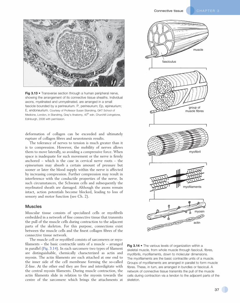
Fig 3.13 • Transverse section through a human peripheral nerve, showing the arrangement of its connective tissue sheaths. Individual axons, myelinated and unmyelinated, are arranged in a small fascicle bounded by a perineurium. P, perineurium; Ep, epineurium; E, endoneurium. Courtesy of Professor Susan Standring, GKT School of
Medicine, London, in Standring, Gray’s Anatomy, 40th edn. Churchill Livingstone,
Edinburgh, 2008 with permission.
E P
Ep
deformation of collagen can be exceeded and ultimately rupture of collagen fibres and neurotmesis results.
The tolerance of nerves to tension is much greater than it is to compression. However, the mobility of nerves allows them to move laterally, so avoiding a compressive force. When space is inadequate for such movement or the nerve is firmly anchored – which is the case in cervical nerve roots – the epineurium may absorb a certain amount of pressure but sooner or later the blood supply within the nerve is affected by increasing compression. Further compression may result in interference with the conductile properties of the nerve. In such circumstances, the Schwann cells and subsequently the myelinated sheath are damaged. Although the axons remain intact, action potentials become blocked, leading to loss of sensory and motor function (see Ch. 2).
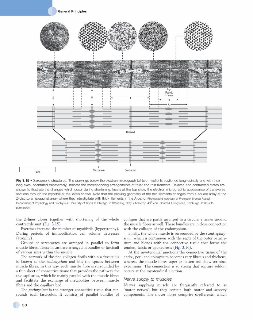
MusclesMuscular tissue consists of specialized cells or myofibrils embedded in a network of fine connective tissue that transmits the pull of the muscle cells during contraction to the adjacent parts of the skeleton. For this purpose, connections exist between the muscle cells and the finest collagen fibres of the connective tissue network.
The muscle cell or myofibril consists of sarcomeres or myo-filaments – the basic contractile units of a muscle – arranged in parallel (Fig. 3.14). In each sarcomere two types of filament are distinguishable, chemically characterized as actin and myosin. The actin filaments are each attached at one end to the inner side of the cell membrane forming the so-called Z-line. At the other end they are free and interdigitate with the central myosin filaments. During muscle contraction, the actin filaments slide in relation to the myosin towards the centre of the sarcomere which brings the attachments at
Fig 3.14 • The various levels of organization within a skeletal muscle, from whole muscle through fasciculi, fibres, myofibrils, myofilaments, down to molecular dimensions. The myofilaments are the basic contractile units of a muscle. Groups of myofilaments are arranged in parallel to form muscle fibres. These, in turn, are arranged in bundles or fasciculi. A network of connective tissue transmits the pull of the muscle cells during contraction via a tendon to the adjacent parts of the skeleton.
muscle
fasciculus
myofibril
myofilaments
group ofmuscle fibres
myosin
actin
General Principles
38
Contracted
Relaxed
1 µm
Z M IH
A
PseudoH zone
Sarcomere
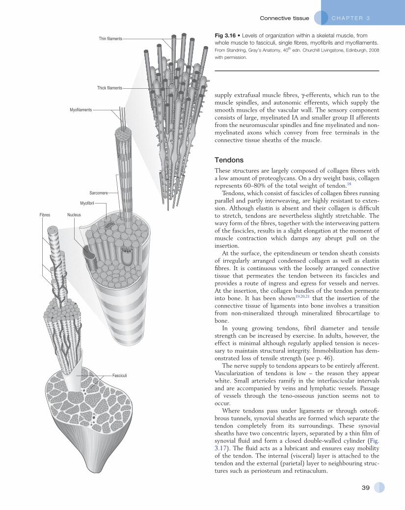
Fig 3.15 • Sarcomeric structures. The drawings below the electron micrograph (of two myofibrils sectioned longitudinally and with their long axes, orientated transversely) indicate the corresponding arrangements of thick and thin filaments. Relaxed and contracted states are shown to illustrate the changes which occur during shortening. Insets at the top show the electron micrographic appearance of transverse sections through the myofibril at the levels shown. Note that the packing geometry of the thin filaments changes from a square array at the Z-disc to a hexagonal array where they interdigitate with thick filaments in the A-band. Photographs courtesy of Professor Brenda Russell,
Department of Physiology and Biophysics, University of Illinois at Chicago, in Standring, Gray’s Anatomy, 40th edn. Churchill Livingstone, Edinburgh, 2008 with
permission.
the Z-lines closer together with shortening of the whole contractile unit (Fig. 3.15).
Exercises increase the number of myofibrils (hypertrophy). During periods of immobilization cell volume decreases (atrophy).
Groups of sarcomeres are arranged in parallel to form muscle fibres. These in turn are arranged in bundles or fasciculi of various sizes within the muscle.
The network of the fine collagen fibrils within a fasciculus is known as the endomysium and fills the spaces between muscle fibres. In this way, each muscle fibre is surrounded by a thin sheet of connective tissue that provides the pathway for the capillaries, which lie mainly parallel with the muscle fibres and facilitate the exchange of metabolites between muscle fibres and the capillary bed.
The perimysium is the stronger connective tissue that sur-rounds each fasciculus. It consists of parallel bundles of
collagen that are partly arranged in a circular manner around the muscle fibres as well. These bundles are in close connection with the collagen of the endomysium.
Finally, the whole muscle is surrounded by the stout epimy-sium, which is continuous with the septa of the outer perimy-sium and blends with the connective tissue that forms the tendon, fascia or aponeurosis (Fig. 3.16).
At the myotendinal junctions the connective tissue of the endo-, peri- and epimysium becomes very fibrous and thickens, whereas the muscle fibres taper or flatten and show terminal expansions. The connection is so strong that rupture seldom occurs at the myotendinal junction.
Nerve supply to musclesNerves supplying muscle are frequently referred to as ‘motor nerves’, but they contain both motor and sensory components. The motor fibres comprise α-efferents, which
C H A P T E R 3Connective tissue
39
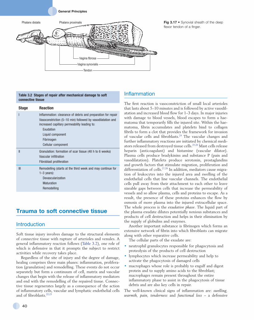
Fig 3.16 • Levels of organization within a skeletal muscle, from whole muscle to fasciculi, single fibres, myofibrils and myofilaments. From Standring, Gray’s Anatomy, 40th edn. Churchill Livingstone, Edinburgh, 2008
with permission.
Fasciculi
Fibres Nucleus
Myofilaments
Thin filaments
Thick filaments
Sarcomere
Myofibril
supply extrafusal muscle fibres, γ-efferents, which run to the muscle spindles, and autonomic efferents, which supply the smooth muscles of the vascular wall. The sensory component consists of large, myelinated IA and smaller group II afferents from the neuromuscular spindles and fine myelinated and non-myelinated axons which convey from free terminals in the connective tissue sheaths of the muscle.
TendonsThese structures are largely composed of collagen fibres with a low amount of proteoglycans. On a dry weight basis, collagen represents 60–80% of the total weight of tendon.18
Tendons, which consist of fascicles of collagen fibres running parallel and partly interweaving, are highly resistant to exten-sion. Although elastin is absent and their collagen is difficult to stretch, tendons are nevertheless slightly stretchable. The wavy form of the fibres, together with the interweaving pattern of the fascicles, results in a slight elongation at the moment of muscle contraction which damps any abrupt pull on the insertion.
At the surface, the epitendineum or tendon sheath consists of irregularly arranged condensed collagen as well as elastin fibres. It is continuous with the loosely arranged connective tissue that permeates the tendon between its fascicles and provides a route of ingress and egress for vessels and nerves. At the insertion, the collagen bundles of the tendon permeate into bone. It has been shown19,20,21 that the insertion of the connective tissue of ligaments into bone involves a transition from non-mineralized through mineralized fibrocartilage to bone.
In young growing tendons, fibril diameter and tensile strength can be increased by exercise. In adults, however, the effect is minimal although regularly applied tension is neces-sary to maintain structural integrity. Immobilization has dem-onstrated loss of tensile strength (see p. 46).
The nerve supply to tendons appears to be entirely afferent. Vascularization of tendons is low – the reason they appear white. Small arterioles ramify in the interfascicular intervals and are accompanied by veins and lymphatic vessels. Passage of vessels through the teno-osseous junction seems not to occur.
Where tendons pass under ligaments or through osteofi-brous tunnels, synovial sheaths are formed which separate the tendon completely from its surroundings. These synovial sheaths have two concentric layers, separated by a thin film of synovial fluid and form a closed double-walled cylinder (Fig. 3.17). The fluid acts as a lubricant and ensures easy mobility of the tendon. The internal (visceral) layer is attached to the tendon and the external (parietal) layer to neighbouring struc-tures such as periosteum and retinaculum.
General Principles
40
Fig 3.17 • Synovial sheath of the deep flexor tendon of a finger.
Vagina fibrosa
Vagina synovialis
Tendon
Phalanx proximalisPhalanx distalis
Table 3.2 Stages of repair after mechanical damage to soft connective tissue
Stage Reaction
I Inflammation: clearance of debris and preparation for repair
Vasoconstriction (5–10 min) followed by vasodilatation and increased capillary permeability leading to:
Exudation
Liquid component
Fibrinogen
Cellular component
II Granulation: formation of scar tissue (48 h to 6 weeks)
Vascular infiltration
Fibroblast proliferation
III Remodelling (starts at the third week and may continue for 1–3 years):
Devascularization
Maturation
Remodelling
Trauma to soft connective tissue
Introduction
Soft tissue injury involves damage to the structural elements of connective tissue with rupture of arterioles and venules. A general inflammatory reaction follows (Table 3.2), one role of which is defensive in that it prompts the subject to restrict activities while recovery takes place.
Regardless of the site of injury and the degree of damage, healing comprises three main phases: inflammation, prolifera-tion (granulation) and remodelling. These events do not occur separately but form a continuum of cell, matrix and vascular changes that begin with the release of inflammatory mediators and end with the remodelling of the repaired tissue. Connec-tive tissue regenerates largely as a consequence of the action of inflammatory cells, vascular and lymphatic endothelial cells and of fibroblasts.22,23
Inflammation
The first reaction is vasoconstriction of small local arterioles that lasts about 5–10 minutes and is followed by active vasodil-atation and increased blood flow for 1–3 days. In major injuries with damage to blood vessels, blood escapes to form a hae-matoma that temporarily fills the injured site. Within the hae-matoma, fibrin accumulates and platelets bind to collagen fibrils to form a clot that provides the framework for invasion of vascular cells and fibroblasts.24 The vascular changes and further inflammatory reactions are initiated by chemical medi-ators released from destroyed tissue cells.25,26 Mast cells release heparin (anticoagulant) and histamine (vascular dilator). Plasma cells produce bradykinins and substance P (pain and vasodilatation). Platelets produce serotonin, prostaglandins and growth factors that stimulate migration, proliferation and differentiation of cells.27,28 In addition, mediators cause migra-tion of leukocytes into the injured area and swelling of the endothelial cells that line vascular channels. The endothelial cells pull away from their attachment to each other to leave sizeable gaps between cells that increase the permeability of vessels and so allow plasma, cells and proteins to escape. As a result, the presence of these proteins enhances the flow by osmosis of more plasma into the injured extracellular space. The whole process is the exudative phase. The liquid part of the plasma exudate dilutes potentially noxious substances and products of cell destruction and helps in their elimination by the supply of globulins and enzymes.
Another important substance is fibrinogen which forms an extensive network of fibrin into which fibroblasts can migrate along with other reparative cells.
The cellular parts of the exudate are:
• neutrophil granulocytes responsible for phagocytosis and proteolysis of the products of cell destruction
• lymphocytes which increase permeability and help to activate the phagocytosis of damaged cells
• macrophages whose role is probably to engulf and digest protein and to supply amino acids to the fibroblast; macrophages remain present throughout the entire inflammatory phase to assist in the phagocytosis of tissue debris and are also key cells in repair.
The well-known clinical signs of inflammation are: swelling, warmth, pain, tenderness and functional loss – a defensive
C H A P T E R 3Connective tissue
41
• Second, the amount, form and strength of scar collagen changes: the immature and weak tissue with a random orientation of fibres in three planes is remodelled into linearly arranged bundles of connective tissue. The process is the result of a number of factors, including turnover of collagen, fibre linkage and increased intermolecular bonding.
It is now generally recognized that internal and external mechanical stress applied to the repair tissue is the main stimulus for remodelling. Tension by gentle movements in functional directions reorientates the collagen and breaks any weak or unnecessary crosslinks that may have formed. Mechanical stress thus has its greatest influence on remodelling at this time. Non-functional collagen is cleared away by phagocytosis.32–36
Remodelling may continue for years although more slowly as time passes. The tensile strength of replaced or repaired collagen in ligaments reaches 50% of normal by 6–25 months after injury and 100% only after 1–3 years.37 The strength of a scar formed in an injured muscle increases faster because of its superior vascular supply.25
Self-perpetuating inflammation
The linear sequence of events described above – an inflamma-tory reaction followed by repair and remodelling – is typical for acute wounds, either accidental or surgical. Orthopaedic medicine, however, also deals with chronic repetitive strains and tissue disruptions, overuse phenomena and excessive tension on devitalized tissues. Here the reaction of the tissues involved is often not linear; the inflammation may be pro-longed and the formation of scar tissue excessive and inappropriate.
Rest usually initiates adhesion formation in and around the healing breach. Oedema raises tissue tension and causes pain, so impeding functional movements that are extremely impor-tant in the early stage of regeneration. Without proper move-ment there is no balance between formation and lysis of the regenerating elements of the involved tissue. Proper alignment of collagen does not result and the final form of the scar tissue tends to remain ill-organized. Any small stress applied to an inappropriate tissue is sufficient to disrupt newly formed fibres in the healing breach. This in turn starts another inflammatory response and a vicious circle of chronic repetitive disruptions of inferior quality connective tissue will result. If such a state of chronic inflammation is maintained, the function of the affected area continues to deteriorate and leads to further tissue damage.
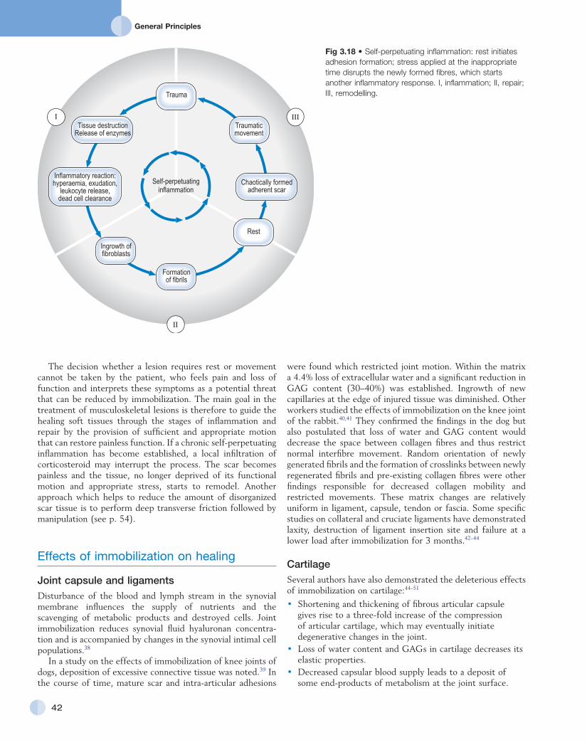
Cyriax drew attention to such chronic types of inflammation of soft tissues that began as a result of trauma but continued long after the cause had ceased to operate – self-perpetuating inflammation (see Fig. 3.18) – particularly prone to happen after a minor injury to a ligament. Occasionally it also occurs as an overuse phenomenon in a tendon. With knowledge of the inflammatory reaction in traumatized soft tissues, it is clear that lack of movement during the period of repair and remod-elling which leads to adhesive scar tissue formation can be responsible for some chronic lesions.
reaction of the body that prompts the subject to restrict activi-ties while recovery takes place.
Repair
It is worthwhile to mention that only the synovial capsules of the joints, skeletal muscle and bone are, to some degree, capable of regeneration. All other connective tissues heal by repair with the formation of collagen and thus scar tissue.
Once the exudative phase has cleared debris by dilution and phagocytosis, fibroblasts and capillaries migrate along the fibrin network. The process of vascular infiltration, fibroblast prolif-eration and the deposition of collagen usually begins within 48 hours of injury overlapping with the end of the exudative phase and the later remodelling phase. Repair is begun and directed by the release from macrophages of chemotactic agents which attract fibroblasts and endothelial cells, secrete growth factors which stimulate these cells to proliferate and produce lactic acid which enhances the synthesis of collagen by fibroblasts. High levels of corticosteroids prevent the migration of macrophages.
During their proliferation, fibroblasts develop into cells termed myofibroblasts that generate a traction-like activity on the matrix required for the reduction of any gap in the healing area.29
Capillaries, at the edge of the injured area, send forward buds which then turn and meet each other to form new capil-lary loops capable of maintaining a circulation that ensures oxygen and nutritional supply in the relatively hypoxic region where the healing tissues meet and, at the same time, enables the removal of metabolic waste products. These new capillaries are fragile and stay within the support of newly synthesized collagen which has already been deposited ahead of the forma-tion of the capillary loops. The highly vascular mass produced gives the surface of the tissue its granular appearance and hence its name – granulation tissue.
By the fourth or fifth day after injury the amount of collagen is significant and there is a progressive but gradually slower increase up to 6 weeks after injury. Corticosteroids decrease the number of fibroblasts and result in a diminished formation of collagen fibres and possibly a weaker fibrous scar. Normally the initial arrangement of collagen fibres is at random but after 6 weeks, tensile strength continues to increase because of orientation of fibres along the lines of stress in the injured tissue (remodelling).
Remodelling
Around the end of the third week maturation begins – the process of reshaping and strengthening the scar tissue by removing, reorganizing and replacing cells and matrix. A better structural orientation and increase in tensile strength result.30 The remodelling phase can be divided into a consolidation and maturation stage:31
• First, vascularization decreases and many of the new vessels atrophy and disappear as the blood supply becomes appropriately adjusted to the needs of the scar tissue.
General Principles
42
were found which restricted joint motion. Within the matrix a 4.4% loss of extracellular water and a significant reduction in GAG content (30–40%) was established. Ingrowth of new capillaries at the edge of injured tissue was diminished. Other workers studied the effects of immobilization on the knee joint of the rabbit.40,41 They confirmed the findings in the dog but also postulated that loss of water and GAG content would decrease the space between collagen fibres and thus restrict normal interfibre movement. Random orientation of newly generated fibrils and the formation of crosslinks between newly regenerated fibrils and pre-existing collagen fibres were other findings responsible for decreased collagen mobility and restricted movements. These matrix changes are relatively uniform in ligament, capsule, tendon or fascia. Some specific studies on collateral and cruciate ligaments have demonstrated laxity, destruction of ligament insertion site and failure at a lower load after immobilization for 3 months.42–44
CartilageSeveral authors have also demonstrated the deleterious effects of immobilization on cartilage:44–51
• Shortening and thickening of fibrous articular capsule gives rise to a three-fold increase of the compression of articular cartilage, which may eventually initiate degenerative changes in the joint.
• Loss of water content and GAGs in cartilage decreases its elastic properties.
• Decreased capsular blood supply leads to a deposit of some end-products of metabolism at the joint surface.
The decision whether a lesion requires rest or movement cannot be taken by the patient, who feels pain and loss of function and interprets these symptoms as a potential threat that can be reduced by immobilization. The main goal in the treatment of musculoskeletal lesions is therefore to guide the healing soft tissues through the stages of inflammation and repair by the provision of sufficient and appropriate motion that can restore painless function. If a chronic self-perpetuating inflammation has become established, a local infiltration of corticosteroid may interrupt the process. The scar becomes painless and the tissue, no longer deprived of its functional motion and appropriate stress, starts to remodel. Another approach which helps to reduce the amount of disorganized scar tissue is to perform deep transverse friction followed by manipulation (see p. 54).
Effects of immobilization on healing
Joint capsule and ligamentsDisturbance of the blood and lymph stream in the synovial membrane influences the supply of nutrients and the scavenging of metabolic products and destroyed cells. Joint immobilization reduces synovial fluid hyaluronan concentra-tion and is accompanied by changes in the synovial intimal cell populations.38
In a study on the effects of immobilization of knee joints of dogs, deposition of excessive connective tissue was noted.39 In the course of time, mature scar and intra-articular adhesions
Fig 3.18 • Self-perpetuating inflammation: rest initiates adhesion formation; stress applied at the inappropriate time disrupts the newly formed fibres, which starts another inflammatory response. I, inflammation; II, repair; III, remodelling.
Self-perpetuatinginflammation
Inflammatory reaction:hyperaemia, exudation,
leukocyte release,dead cell clearance
Chaotically formedadherent scar
Formationof fibrils
Trauma
Tissue destructionRelease of enzymes
Traumaticmovement
Rest
Ingrowth offibroblasts
IIII
II
C H A P T E R 3Connective tissue
43
circulation is increased (Table 3.3) which aids the supply of nutrients and elimination of cartilagenous debris. Physical joint movements have a beneficial effect on the assimilation of nutri-ents by the cartilage.54
Experimental findings11,55,56 on the influence of physical activity on ligaments and tendons support the view that the strength of connective tissue is increased with exercise training and decreased with immobilization, provided that the exercise programme is of an endurance nature. Trained animals have significantly heavier ligaments, stronger ligament–bone junctions and junction strength to body weight ratios. Similar effects pertain in repaired ligaments,42,57 which show significantly higher strength values after repair is complete if they have not been immobilized. Early mobilization also considerably influences the remodelling process and prevents formation of abnormal adhesions that may restrict joint movements.58
Another advantage of early mobilization is the positive effect on skeletal muscles,57,59,60 with increased circulation, muscle strength and endurance and maintenance of proprio-ceptive reflexes, which ensure the active stability of the joint.
Treatment of traumatic soft connective tissue lesions
The overall aim of treatment in orthopaedic medicine is to restore painless function of the connective tissue. During the last decades it has become clear that application of functional movement to healing connective tissue is extremely important and should be the first and principal objective for the therapist. Of course, the selection of the techniques will depend on several factors such as the stage of the lesion, the tissue involved, the severity of the lesion, the irritability of the tissue and the pain perception of the patient.
• Lysosomal enzymes released from dead chondrocytes lead to an autolysis of cartilage which is proportional to the time of immobilization.
MuscleMuscular reactions to immobilization have also been investi-gated.52,53 There is:
• decreased capillary density and muscle atrophy• decrease in muscle strength most dramatically during the
first week of immobilization. After 2 weeks in a plaster cast, there is 20% loss of maximum strength. Slow muscle fibres, with predominantly oxidative metabolism, are more susceptible to immobilization atrophy than are fast fibres
• an increased amount of connective tissue. Proliferation first takes place in the perimysial spaces, followed sometimes by the endomysial spaces. It is suggested53 that this may impair the vascular supply of muscle fibres and could facilitate degeneration and also could make regeneration more difficult. Although muscle structure, metabolism and function are severely impaired after immobilization, almost complete recovery is possible provided that the training programme starts with very moderate exercises and avoids maximum voluntary efforts of regenerating muscle fibres
• disturbance of neuromuscular coordination of muscle groups.
The same studies drew attention to the reactions of organ systems, such as the cardiovascular, respiratory, locomotor and autonomic, which may also become disturbed.
Effects of mobilization on healing
The benefit of early mobilization in most soft tissue lesions was advocated by Hippocrates more than 2400 years ago. Capsular
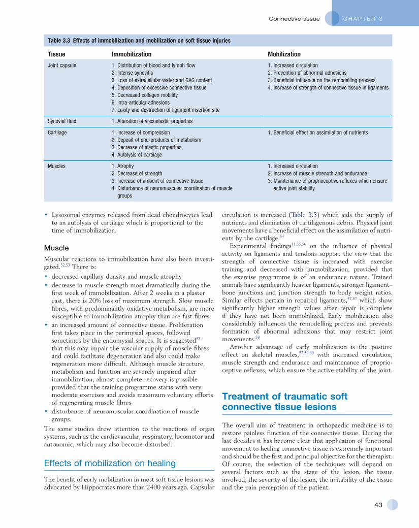
Table 3.3 Effects of immobilization and mobilization on soft tissue injuries
Tissue Immobilization Mobilization
Joint capsule 1. Distribution of blood and lymph flow 1. Increased circulation2. Intense synovitis 2. Prevention of abnormal adhesions3. Loss of extracellular water and GAG content 3. Beneficial influence on the remodelling process4. Deposition of excessive connective tissue 4. Increase of strength of connective tissue in ligaments5. Decreased collagen mobility6. Intra-articular adhesions7. Laxity and destruction of ligament insertion site
Synovial fluid 1. Alteration of viscoelastic properties
Cartilage 1. Increase of compression 1. Beneficial effect on assimilation of nutrients2. Deposit of end-products of metabolism3. Decrease of elastic properties4. Autolysis of cartilage
Muscles 1. Atrophy 1. Increased circulation2. Decrease of strength 2. Increase of muscle strength and endurance3. Increase of amount of connective tissue 3. Maintenance of proprioceptive reflexes which ensure
active joint stability4. Disturbance of neuromuscular coordination of muscle groups
General Principles
44
most often during unusual contractions,63 which produce sig-nificantly higher muscle force than when the muscle is held at the same length or is allowed to shorten. It has also been shown that muscles crossing two joints, such as hamstrings and gas-trocnemii, are particularly at risk. The vulnerable site appears to be near the muscle–tendon junction. The immediate response to the trauma is inflammation, associated oedema and localized haemorrhage. Excessive oedema and haemorrhage should be reduced as much as possible. Blood collections are not confined to the muscle proper but escape through the perimysium and fascia into the subcutaneous space. As a rule, the degree of pain is consistent with the extent of the rupture.
TreatmentThe immediate induction of local anaesthesia at the site of the lesion effectively blocks the nociceptive impulses which are responsible for muscle spasm at the site of damage. Cold therapy as an alternative has been criticized.64 Although it has a positive effect on pain threshold,65,66 physiological effects and procedures of application are still chiefly based on empirical and clinical findings. Van Wingerden suggests that cold therapy, especially when applied in the acute phase of injury, could lead to increased oedema, inhibition of the healing process and even to increased inflammatory reactions.64
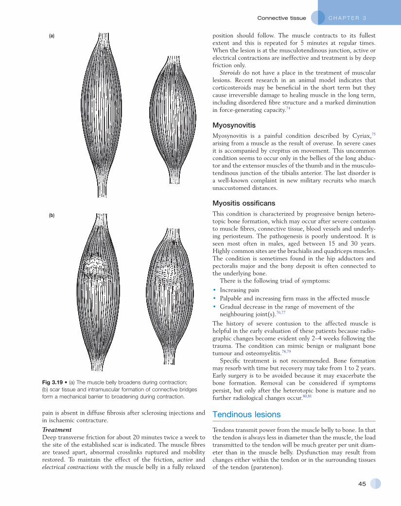
From the next day on, active or electrical contractions follow, with the muscle in a fully shortened position to maintain mobility by broadening the muscle belly. Meshwork of regen-erating fibrils in the healing breach may hinder the broadening capacity of the muscle fibres during contraction. In the later stages of healing (granulation and remodelling), intramuscular formation of abnormal crosslinks takes place and the inappro-priate scar tissue will form a mechanical barrier to broadening during contraction, a chief muscle function (Fig. 3.19).
Deep transverse friction also imitates broadening and pre-vents newly formed bonds from matting muscle fibres together. It should be started the day after injury, as it can be expected that repair has begun by this time. A gentle type of massage is performed daily for a short period of time (see Ch. 5). At this stage, it should not be done to an extent that interferes with the capillaries and fibrils consolidating themselves in the healing breach. Both intensive passive stretching and resisted move-ments may cause damage at the site of injury and must be avoided until recovery is well established. It has been demon-strated67,68 that contractile ability recovers rapidly.
Once the patient is free from pain and has a full range of mobility, repetitive stretching exercises seem to make an important contribution to the future prevention of these injuries.69–72 Return to sport can be allowed when the strength of the injured limb has been restored to within 10% of that of the unaffected limb (usually after 3–6 weeks).73
Chronic lesionsScarring takes place in chronic lesions, matting fibres together transversely. The range of broadening is impaired and contrac-tions against resistance are painful. A painful area can be pal-pated, although it may be difficult when the lesion is deeply situated, for example, the belly of biceps brachii or the exten-sor carpi radialis brevis. Pain probably results from overstretch-ing at the junction between normal and scar tissue, from local variations in tension. This explanation seems logical because
Muscular lesions
Delayed muscle soreness, contusion, (minor) rupture, myo-synovitis and myositis ossificans are different types of lesion which can occur in skeletal muscle.
Delayed muscle sorenessA delayed, specific soreness sometimes appearing 12–24 hours after intense exercise is well known in athletics and may be caused by the disturbance of metabolism, with a high concen-tration of lactic acid and the resulting inflammatory reactions: vasodilation, increased capillary permeability and intercellular oedema. Swelling and oxygen deficiency may irritate free nerve endings and lead to muscle spasm.61 Another, more recent, theory is that there is injury to sarcomeres and intramuscular collagen fibres and, in consequence, an inflammatory reaction.62
Passive stretching and active contraction cause discomfort. Pain lasts 3–4 days, gradually diminishing during the subse-quent days. Improper warm-up, unusual exertion, early season training and running before muscles are properly conditioned are some conditions that are thought to bring on the pain.
The best way to avoid this condition is to stay ‘in shape’ between seasons. Other precautions to be advised are:
• warm up before beginning an exercise programme• include stretching exercises in the warm-up• gradually increase the load and duration of the exercises• avoid excessive tension on muscles• allow the muscles to dissipate waste products
(warm-down).
Muscular contusionThis results from a direct blow on the muscle belly. There is a variable degree of severity, characterized by pain and intra- or intermuscular bleeding with extensive swelling. Intramuscular bleeding is more serious and lasts longer because of difficulty in dispersing the haematoma. Such a blow does not cause much discomfort or pain while the athlete is warm and actively taking part in sport; it is some hours afterwards that stiffness and disability set in. Active contractions against resistance cause discomfort and passive stretching of the muscle is painful and limited.
Treatment involves aspiration of the haematoma (within 3 days), after which compression is applied immediately, using an elastic wrap. A short period of rest may be prescribed but rest should never be total and prolonged.
After a few days, treatment with gentle deep transverse frictions and active contractions with the muscle in a fully shortened position can be started (see Minor muscular tears).
Minor muscular tears (or ‘muscle strain injuries’)
Acute lesionsThese are the common ‘muscle pull’ or ‘strain’ injuries that result from sudden and over-violent effort or movement in the muscle and usually cause immediate disability. Tears occur
C H A P T E R 3Connective tissue
45
position should follow. The muscle contracts to its fullest extent and this is repeated for 5 minutes at regular times. When the lesion is at the musculotendinous junction, active or electrical contractions are ineffective and treatment is by deep friction only.
Steroids do not have a place in the treatment of muscular lesions. Recent research in an animal model indicates that corticosteroids may be beneficial in the short term but they cause irreversible damage to healing muscle in the long term, including disordered fibre structure and a marked diminution in force-generating capacity.74
MyosynovitisMyosynovitis is a painful condition described by Cyriax,75 arising from a muscle as the result of overuse. In severe cases it is accompanied by crepitus on movement. This uncommon condition seems to occur only in the bellies of the long abduc-tor and the extensor muscles of the thumb and in the musculo-tendinous junction of the tibialis anterior. The last disorder is a well-known complaint in new military recruits who march unaccustomed distances.
Myositis ossificansThis condition is characterized by progressive benign hetero-topic bone formation, which may occur after severe contusion to muscle fibres, connective tissue, blood vessels and underly-ing periosteum. The pathogenesis is poorly understood. It is seen most often in males, aged between 15 and 30 years. Highly common sites are the brachialis and quadriceps muscles. The condition is sometimes found in the hip adductors and pectoralis major and the bony deposit is often connected to the underlying bone.
There is the following triad of symptoms:
• Increasing pain• Palpable and increasing firm mass in the affected muscle• Gradual decrease in the range of movement of the
neighbouring joint(s).76,77
The history of severe contusion to the affected muscle is helpful in the early evaluation of these patients because radio-graphic changes become evident only 2–4 weeks following the trauma. The condition can mimic benign or malignant bone tumour and osteomyelitis.78,79
Specific treatment is not recommended. Bone formation may resorb with time but recovery may take from 1 to 2 years. Early surgery is to be avoided because it may exacerbate the bone formation. Removal can be considered if symptoms persist, but only after the heterotopic bone is mature and no further radiological changes occur.80,81
Tendinous lesions
Tendons transmit power from the muscle belly to bone. In that the tendon is always less in diameter than the muscle, the load transmitted to the tendon will be much greater per unit diam-eter than in the muscle belly. Dysfunction may result from changes either within the tendon or in the surrounding tissues of the tendon (paratenon).
Fig 3.19 • (a) The muscle belly broadens during contraction; (b) scar tissue and intramuscular formation of connective bridges form a mechanical barrier to broadening during contraction.
(a)
(b)
pain is absent in diffuse fibrosis after sclerosing injections and in ischaemic contracture.
TreatmentDeep transverse friction for about 20 minutes twice a week to the site of the established scar is indicated. The muscle fibres are teased apart, abnormal crosslinks ruptured and mobility restored. To maintain the effect of the friction, active and electrical contractions with the muscle belly in a fully relaxed
General Principles
46
inflammatory reaction was excessive and the granulation and remodelling phases inadequate.
TreatmentTo prevent excessive inflammation and to stimulate better remodelling, gentle mobilization and transverse friction are used to orient the randomly distributed collagen in a functional direction. This also prevents the formation of abnormal adhe-sions. Treatment starts the day after the injury and massage should be superficial and light in order not to rupture newly generated fibrils. Treatment along these lines is continued daily for the first week and on alternate days for the second and third weeks. After 3 weeks, active unloaded contractions can be added. A tape or bandage to support the structure and prevent undue movement is advisable. Careful evaluation is necessary, gauging the degree of activity against the degree of local reaction.
The patient should be very cautious about returning to full activity until the lesion is completely pain free.
Chronic (overuse) lesionsTreatmentIn chronic tendinous lesions, transverse friction massage applied precisely to the exact area may be an extremely useful technique and can often bring lasting relief.
Modalities and effects of this treatment form are discussed in the online chapter Applied anatomy of the cervical spine but transverse friction aims to achieve transverse movement of the collagen structure of the connective tissue. In this way adhe-sion formation is prevented and existing adhesions are mobi-lized. The cyclic loading and motion of the healing connective tissues may also stimulate formation and remodelling of the collagen.
Another approach is to infiltrate locally a low dose of corticosteroid. This will quickly reduce the chronic inflamma-tory reaction but it also affects the proliferative and even the remodelling phase – the biosynthesis of collagen is altered by corticosteroids. For this reason, local steroid infiltrations should only be at the tenoperiosteal insertion and never in relation to the body of a tendon for fear of possible tendon rupture. A further disadvantage of steroid infiltration is the higher recur-rence rate (about 25%) after initial therapeutic success.
Persistent pain is thought to be caused by irreversible degen-erative changes within the tendon. Removal by surgical treat-ment may then be indicated. Again treatment may include sessions of transverse friction applied in a progressive way until the patient is free of symptoms. Thereafter, a programme of rehabilitation is undertaken.
TenosynovitisTenosynovitis is a well-known lesion at the ankle or the wrist where tendons possess a sheath. Roughening of the gliding surfaces of a tendon and the internal or visceral layer of the sheath results from an inflammatory reaction; commonly occu-pational overuse or strain. During movement, pain is evoked as the roughened surfaces move against each other. In severe cases fine crepitus is palpable and swelling obvious. Coarse crepitus is a warning sign that points to rheumatoid disease or tuberculosis.
TerminologyWithin the literature, there is much confusion about the ter-minology and over the last decades, numerous terms have been used to describe the pathology of tendons. The most common term is ‘tendinitis’ which focuses on clinical inflammatory signs. Puddu et al.82 proposed the term tendinosis as a histo-logical description of a degenerative pathology with a lack of inflammatory change. As these terms are often used inter-changeably and without precision83 it may be more appropriate to refer to a symptomatic primary tendon disorder as a tendi-nopathy as this makes no assumption as to the underlying pathological process.84
‘Tendinitis’When strain on a tendon tears some fibres, it always seems to occur in those parts of the tendon where vascularization is relatively poor. The insertion into bone and sometimes a spe-cific part of a tendon are such ‘critical’ vascularized zones. Good examples are a zone in the Achilles tendon about 2.5 cm above its insertion into the calcaneus85,86 and the supraspinatus tendon close to its insertion on the major tubercle.87
Above the age of 25, vascularization of tendinous tissues also decreases (ultimately by about 30%) which enhances their vulnerability.
Lesions result principally from overuse. However, the mechanical response of a tendon to a load not only depends on the amount of the externally applied force but also is closely bound up with the state of the tendon involved. Overuse may disturb the microcirculation which, especially in zones of hypovascularity, will negatively influence metabolic processes. If this continues, a process of degeneration starts, called ‘tendi-nosis’. In the beginning of the process this is a degenerative condition without accompanying inflammatory reactions and therefore clinical signs and symptoms may be totally absent.
From the moment that a normal load strains the tendon and tears some fibres, an inflammatory reaction begins. Long-standing treatment with corticosteroids or periods of immobi-lization will further negatively influence the condition of collagen and lead to further deterioration and decrease in the number of fibrocytes and fibres. In sports, a cold climate, bad equipment and wrong training procedures (e.g. lack of warm-up including absence of stretching, too progressive an increase of load and duration of the exercise programme, bouncing exer-cises and jerky muscle contractions) may all negatively influ-ence the condition.
The inflammatory reactions in tendinitis are located not only at but also around the ruptured fibres, in the areolar tissue between the fasciculi. Here oedema, growth of capillaries, migration of leukocytes and invasion with mesenchymal cells take place, which induces new generations of fibroblasts and leads to a large proliferation of connective tissue around the site of the lesion.
Acute traumatic lesionsIn acute traumatic lesions 48 hours after the injury, newly generated collagen fibrils start to close the tissue defect by the formation of scar tissue. Scars may remain lastingly painful and often have an inferior tensile strength, especially if the initial
C H A P T E R 3Connective tissue
47
Complete ruptureComplete rupture of a tendon usually results from indirect trauma. It always seems to occur at the midsubstance of the tendon. Acute tears are those occurring suddenly, usually as a result of a single injury. Chronic tears are those that are insidi-ous in onset and result from repetitive loading of a degenerated and weakened tendon (see above). Tendinous ruptures occur predominantly at the shoulder, wrist and heel. At the shoulder, the incidence of full thickness rotator cuff tears in cadaveric populations ranges from 30%90 to 60%.91
Ruptures of Achilles and tibialis posterior tendons are also quite common. The special anatomical situations of the flexor and extensor tendons of thumb and fingers subject them to high loads which may alter their histological structure and finally rupture them.
Healing of a ruptured tendon depends on the capabilities of both the tendon itself as well as on reactions from the sur-rounding tissues. Some tendons such as the Achilles tendon have a remarkable ability to heal after rupture, whereas others do not. Rotator cuff tears for instance usually do not heal and, if they do, a permanent weakening results. Healing of a rup-tured Achilles tendon does not differ from the general tissue reactions after mechanical damage: the initial stage of exuda-tion is followed by vascular granulation and fibroblast prolifera-tion. Collagen synthesis begins within the first week and reaches its maximum after about 4 weeks. It then continues for about 3 months. Maturation and remodelling begin at the end of the third week and continue for up to 1 year after the injury.
Mechanical strength of a healing tendon is related to the histological process of repair. During the second phase, strength increases but is still insufficient to prevent further stretching of the healing wound. For this reason, the region always has to be immobilized until the process of maturation has begun (about 3 weeks after injury). At that time the arrangement of collagen fibrils is not organized and remodelling depends entirely upon the presence of repetitive tensile forces applied to the scar tissue. Several studies support the concept that controlled cyclic passive loading of the healing tendon, per-formed after the initial healing phase (3 weeks) is effective in decreasing the formation of abnormal adhesions and increasing the tensile strength of the healing tissue.92–94
Local swelling of a tendonThis is not infrequently seen in the digital flexor tendons in the palm of the hand. When these swellings are localized at the level of the heads of the metacarpals, they engage and may become fixed in the distal part of the tendon sheath at the moment of flexion. Extension is often only possible with help. This symptom is known as ‘trigger’ finger or thumb.
TreatmentInfiltration of steroid, to influence the inflammatory compo-nent of the swelling, is sometimes effective. If this fails, surgi-cal removal of the swollen tissue (scar and/or calcium deposits) or slitting of the narrowed part of the sheath is the alternative.
An overview of the localization and treatment of musculo-tendinous lesions is given in Figure 3.20.
TreatmentThis consists of deep transverse friction whether the condition is recent or chronic but it is indicated only in posttraumatic lesions. Friction restores the smooth and painless movement of the tendon in relation to the internal layer of the sheath.
An injection of a small amount of steroid suspension between tendon and sheath is an alternative. Some lesions respond better to the massage and for others injection is the treatment of choice (see Ch. 5).
All activities that cause pain should be avoided until symp-toms have ceased.
TenovaginitisTenovaginitis is primarily a lesion of the tendon sheath itself and is often associated with considerable swelling and tender-ness but there is never crepitus. It may have a detectable cause in overuse but also occurs spontaneously. Non-specific types of tenovaginitis should be differentiated from those with a specific cause, such as bacterial infection, rheumatoid arthritis, gout and gonorrhoea.
TreatmentFor the non-specific types of tenovaginitis, treatment is the introduction of a small amount of steroid between tendon and sheath, which brings quick relief. In persistent cases, deep transverse friction can be tried but incision of the tendon sheath is often necessary.
In specific tenovaginitis, treatment is that of the underlying disease.
TendinosisThis is a degenerative condition of the tendon without accom-panying inflammatory reactions and therefore often without clinical symptoms. The lesion is characterized by visible dis-colouring of the tissue and loss of the mirror-like gloss of the tendon surface and is typically described as ‘mucoid degeneration’.88
Microscopically, tendinosis is characterized by a marked loss of collagen. The fibres show an irregular entangled course and an irregular waving pattern. Between the fibres, cavities filled with fluid loosen the tissue. The number of fibroblasts is decreased and their nuclei transformed.
Besides this local destruction, there are also signs of regen-eration as shown by slight fibroblast proliferation, formation of capillary sprouts and new mesenchymal cells, which synthe-size young collagen fibrils to fill up the tissue defect. If this process of granulation is insufficient, necrosis and calcification result.
Tendinosis and mucoid degeneration often lead to spontane-ous ruptures (long head of the biceps, Achilles tendon). It is suggested that the ‘typical’ histopathological changes charac-terized by tendon degeneration may not necessarily be directly linked to increased nociception giving the patients warning signals; while in painful ‘tendinitis’ the mechanically weaker tendons may be protected from ruptures due to decreased impact levels since painful activities will be avoided.89
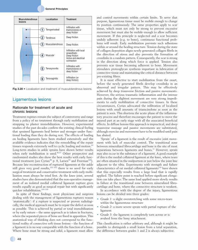
General Principles
48
and control movements within certain limits. To serve that purpose, ligamentous tissue must be mobile enough to change its position continuously. The same properties apply to scar tissue, which must not only be strong to prevent excessive movement but must also be mobile enough to allow sufficient movement. If this principle is neglected and a scar becomes unduly adherent (e.g. to bone), continuous functional prob-lems will result. Early mobilization prevents such adhesions within or around the healing structure. Tension during the state of collagen deposition aligns newly generated collagen fibrils in the direction of stress and also prevents the formation of crosslinks in a random pattern. Consequently, the scar is strong in the direction along which force is applied. Tension also prevents scar tissue becoming adherent to bone. Movement stimulates proteoglycan synthesis important in lubrication of connective tissue and maintaining the critical distance between pre-existing fibres.
It is most effective to start mobilization from the onset, before the newly generated fibrils develop crosslinks in an abnormal and irregular pattern. This may be effectively achieved by deep transverse friction and passive movements. However, the serious traumatic inflammation and the intense pain during the slightest movement are very strong impedi-ments to early mobilization of connective tissues. In these circumstances, Cyriax advocated the infiltration of localized lesions with small amounts of triamcinolone as soon as the patient is seen. This shortens the acute phase of the inflamma-tory process and therefore encourages the patient to move the injured part at an early stage with all the associated beneficial effects. In diffuse lesions this approach is impractical, and deep transverse massage and passive movements are substituted, although exercise and movement have to be modified until pain has abated.
‘Sprain’ of a ligament is the result of excessive joint move-ment with lack of muscular control. The transitional zone between mineralized fibrocartilage and bone is the site of most separations between ligaments and bones.11 However, sprain may also occur in the substance of a ligament. A good example of this is the medial collateral ligament at the knee, where tears are often situated in the midportion or just below the joint line adjacent to the tibia. Experiments with strength and failure characteristics of rat medial collateral ligaments110 have shown that this especially results from a large load that is rapidly applied. The failure point is reached before significant elonga-tion can take place. The same load applied more slowly results in failure at the transitional zone between mineralized fibro-cartilage and bone, where the connective structure is weakest.
In accordance with the degree of the injury, ligamentous lesions can be divided into three grades:
• Grade 1: a slight overstretching with some micro-tears within the ligamentous structure
• Grade 2: a more severe sprain with partial rupture of the ligament
• Grade 3: the ligament is completely torn across or is avulsed from the bony attachment.
This classification is rather arbitrary and, although it might be possible to distinguish a small lesion from a total separation, the difference between grades 1 and 2 is always subjective.
Ligamentous lesions
Rationale for treatment of acute and chronic lesionsTreatment regimes remain the subject of controversy and range from a policy of no treatment through early mobilization and strapping to plaster immobilization. However, experimental studies of the past decades confirm the existing clinical feeling that sprained ligaments heal better and stronger under func-tional loading than they do during rest. The effects of loading on healing ligaments have been studied extensively and the available evidence indicates that the remodelling of the repair tissues responds extremely well to cyclic loading and motion.95 Long-term studies in ankle sprains have shown better results when early mobilization is used.96,97 Other prospective and randomized studies also show the best results with early func-tional treatment (see Cyriax98 p. 8, Larsen99 and Freeman100). Because late reconstruction of ruptured ligaments at the ankle still gives very good results,101–103 there is no need for early surgical treatment and conservative treatment with early mobi-lization must always be tried first. At the knee joint, several studies have also demonstrated that the non-operative manage-ment of an isolated medial collateral ligament rupture gives results equally as good as surgical repair but with significantly quicker rehabilitation.104–109
In spite of these findings, most physicians and surgeons dealing with the management of ligamentous ruptures reason ‘anatomically’: if a rupture is suspected or proven radiologi-cally, the medical approach must be to repair the defect as soon as possible. This is achieved by partial or total immobilization or by early suture – the same approach as is taken in fractures, where the separated pieces of bone are fixed in apposition. This anatomical way of thinking does not correspond to the func-tional reality of connective soft tissue lesions – the function of a ligament is in no way comparable with the function of a bone. Where bone must be strong and solid, a ligament must allow
Fig 3.20 • Localization and treatment of musculotendinous lesions.
Musculotendinouslesions
Localization Treatment
TenoperiostealInfiltration withtriamcinolone ordeep friction
Tendinous Deep friction
Musculotendinous Deep friction
Muscular
Infiltration with localanaestheticDeep frictionActive and electricalcontraction
TenosynovitisInfiltration withtriamcinolone ordeep friction
Tenovaginitis Infiltration (ordeep friction)
1
2
3
4
5
6
C H A P T E R 3Connective tissue
49
An infiltration with a small amount of steroid at the site of the lesion is an alternative. The injection should be given during the first 48 hours – the initial and exudation stage. It reduces traumatic inflammation and prevents most structural and reflex changes. Pain also disappears, which enables the patient to move the joint in a normal way. Deep transverse friction then loses its efficacy.
Steroid injections during granulation and repair may lead to fewer fibroblasts, a diminished collagen fibre formation and a weaker scar.111 However, these effects are not seen after a single injection during the acute, inflammation stage.112
Overstretching a ligament often leads to permanent laxity with consequent instability of the joint. Cyriax emphasized the propensity of ligaments not controlled by muscles to develop such permanent laxity and cited the sternoclavicular, acromio-clavicular, sacroiliac and sacrococcygeal ligaments, the symphy-sis pubis, the cruciate ligaments at the knee and the inferior tibiofibular ligaments as examples.75 After trauma, reflex muscle spasm is not capable of stabilizing these joints. In int-racapsular ligaments (e.g. the cruciate ligaments), unsuccessful repair may also result from the synovial environment, limited fibroblast migration and reduced vascular ingrowth.
The subsequent traumatic inflammation can be reduced by the means already described. To prevent laxity, it is a principle of treatment to completely avoid movements which can be achieved by immobilization of the joint or by surgical repair. Such measures are best executed within 7 days.
After recovery, minor laxity is compatible with excellent function, whereas painful chronic laxity becomes painless in due course or can be converted into painless laxity by use of a local steroid infiltration.
In joints controlled by muscles, permanent laxity is much less likely to occur. Reflex muscle spasm efficiently stabilizes the joint. Grade I and II lesions treated on the lines set out will heal adequately. The results of conservative treatment in grade III lesions are also successful in almost all cases but it is essential that the lesion is isolated.106,113 It is then advisable to immobilize the joint partially so as to prevent any unwanted movement taking place during recovery. Lasting instability in these joints can be more or less compensated for by tautness of the muscles and tendons passing over the joint. Strength-building exercises are of great importance and must be on an exact and planned basis. If necessary, strapping or braces may provide added support.
In surgical repair, dense fibrous tissue is used to reconstruct the ligament – the fascia lata, part of the patellar tendon on other tendons. Following transplantation a graft heals to the recipient tissues but never approaches the strength it had before transplantation. Nevertheless grafts can significantly improve the stability of joints.
Capsular lesions
Traumatic arthritis, capsulitis or synovitis all have an identical meaning: inflammation of the entire capsule as a result of a more or less recent trauma. This is invisible on X-ray and therefore this investigation cannot exclude the diagnosis. If trauma has damaged the lymph and vascular network in the
Ligamentous lesions can also be classified according to the time that has elapsed since the causative accident:
• Acute: within 48 hours• Subacute: 48 hours to 6 weeks• Chronic: more than 6 weeks.
This classification is of importance in relation to treatment.Sprains with incomplete ligament rupture are often quite
painful and accompanied by muscle spasm and pseudolocking. This makes clinical examination difficult, and diazepam or general anaesthesia may be needed to complete a thorough examination.
In complete tears there is rarely pain of a significant degree and in knee and ankle sprain the patient can often walk without aid. It is a fear of ‘giving way’ that characterizes the lesion and prevents the patient from doing more strenuous activities such as going up or down stairs, jumping or doing full squats.
Clinical evaluation of an acutely injured joint should be carried out as soon as possible – within a few hours of the accident – otherwise pain, swelling and muscle spasm will often make it impossible to perform proper ligamentous tests. This is particularly necessary in first and second degree lesions, in which these symptoms are so evident. History and knowl-edge of the mechanism of the injury are important aids in diagnosis and point to the injured ligament(s). Tenderness and localized oedema indicate the anatomical site of the tear in most instances.
TreatmentMinor ligament sprain should be treated conservatively. Liga-ments will heal but it is essential not to strain them again during the first part of the granulation stage. For example, after damage to the medial collateral ligament of the knee joint, full extension should be prevented during the first 10 days.
In the acute stage, traumatic reactions such as pain and swelling should be kept to a minimum. Therefore, early appli-cation of compression and an elevated position of the extrem-ity are most important.
In a sprained ligament of the knee or ankle, crutches should be used if movement is necessary. The next day, physiotherapy is started. Effleurage diminishes swelling and pain, which are impediments to movement. Thereafter transverse friction is performed to move the damaged tissue to and fro over the subjacent bone in imitation of its normal behaviour. This pre-vents the random orientation of newly generated fibrils and formation of abnormal crosslinks between newly regenerated fibrils and pre-existing collagen fibres. In this acute stage, really deep transverse friction will take no more than 1 minute since there is no question of breaking down strong scars. However, it should still be as gentle as is compatible with securing ade-quate movement of the damaged tissue. Then the joint is moved passively through its greatest possible range without causing pain. The same movement(s) is repeated actively. There should be no attempt to increase this range in the acute or subacute stage. In a sprained joint of the lower limb, instruc-tion on gait follows. Strapping the joint to protect it from unwanted movements is a useful additional measure, especially if the patient seems anxious. Patients treated along these lines recover most rapidly.
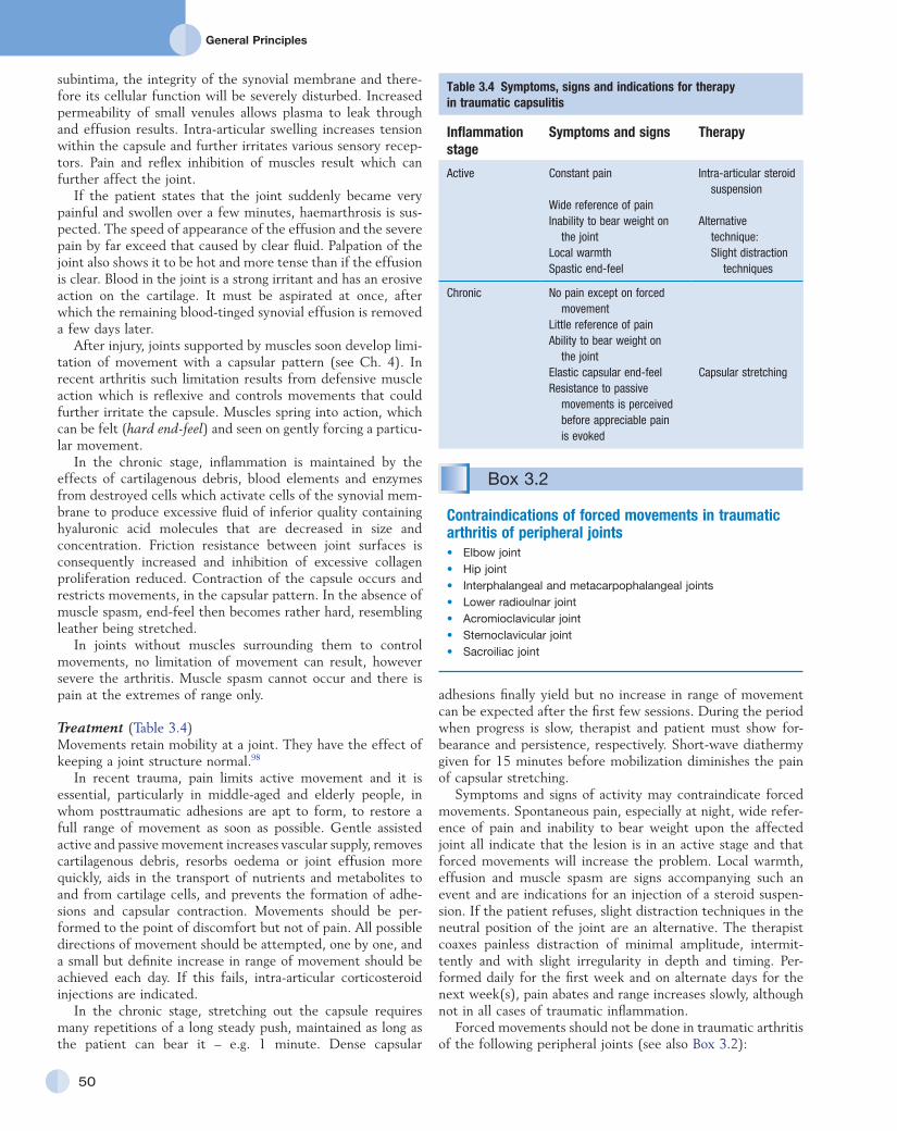
General Principles
50
subintima, the integrity of the synovial membrane and there-fore its cellular function will be severely disturbed. Increased permeability of small venules allows plasma to leak through and effusion results. Intra-articular swelling increases tension within the capsule and further irritates various sensory recep-tors. Pain and reflex inhibition of muscles result which can further affect the joint.
If the patient states that the joint suddenly became very painful and swollen over a few minutes, haemarthrosis is sus-pected. The speed of appearance of the effusion and the severe pain by far exceed that caused by clear fluid. Palpation of the joint also shows it to be hot and more tense than if the effusion is clear. Blood in the joint is a strong irritant and has an erosive action on the cartilage. It must be aspirated at once, after which the remaining blood-tinged synovial effusion is removed a few days later.
After injury, joints supported by muscles soon develop limi-tation of movement with a capsular pattern (see Ch. 4). In recent arthritis such limitation results from defensive muscle action which is reflexive and controls movements that could further irritate the capsule. Muscles spring into action, which can be felt (hard end-feel) and seen on gently forcing a particu-lar movement.
In the chronic stage, inflammation is maintained by the effects of cartilagenous debris, blood elements and enzymes from destroyed cells which activate cells of the synovial mem-brane to produce excessive fluid of inferior quality containing hyaluronic acid molecules that are decreased in size and concentration. Friction resistance between joint surfaces is consequently increased and inhibition of excessive collagen proliferation reduced. Contraction of the capsule occurs and restricts movements, in the capsular pattern. In the absence of muscle spasm, end-feel then becomes rather hard, resembling leather being stretched.
In joints without muscles surrounding them to control movements, no limitation of movement can result, however severe the arthritis. Muscle spasm cannot occur and there is pain at the extremes of range only.
Treatment (Table 3.4)Movements retain mobility at a joint. They have the effect of keeping a joint structure normal.98
In recent trauma, pain limits active movement and it is essential, particularly in middle-aged and elderly people, in whom posttraumatic adhesions are apt to form, to restore a full range of movement as soon as possible. Gentle assisted active and passive movement increases vascular supply, removes cartilagenous debris, resorbs oedema or joint effusion more quickly, aids in the transport of nutrients and metabolites to and from cartilage cells, and prevents the formation of adhe-sions and capsular contraction. Movements should be per-formed to the point of discomfort but not of pain. All possible directions of movement should be attempted, one by one, and a small but definite increase in range of movement should be achieved each day. If this fails, intra-articular corticosteroid injections are indicated.
In the chronic stage, stretching out the capsule requires many repetitions of a long steady push, maintained as long as the patient can bear it – e.g. 1 minute. Dense capsular
Table 3.4 Symptoms, signs and indications for therapy in traumatic capsulitis
Inflammation stage
Symptoms and signs Therapy
Active Constant pain Intra-articular steroid suspension
Wide reference of painInability to bear weight on
the jointAlternative
technique:Local warmth Slight distraction
techniquesSpastic end-feel
Chronic No pain except on forced movement
Little reference of painAbility to bear weight on
the jointElastic capsular end-feel Capsular stretchingResistance to passive
movements is perceived before appreciable pain is evoked
Box 3.2
Contraindications of forced movements in traumatic arthritis of peripheral joints• Elbow joint• Hip joint• Interphalangeal and metacarpophalangeal joints• Lower radioulnar joint• Acromioclavicular joint• Sternoclavicular joint• Sacroiliac joint
adhesions finally yield but no increase in range of movement can be expected after the first few sessions. During the period when progress is slow, therapist and patient must show for-bearance and persistence, respectively. Short-wave diathermy given for 15 minutes before mobilization diminishes the pain of capsular stretching.
Symptoms and signs of activity may contraindicate forced movements. Spontaneous pain, especially at night, wide refer-ence of pain and inability to bear weight upon the affected joint all indicate that the lesion is in an active stage and that forced movements will increase the problem. Local warmth, effusion and muscle spasm are signs accompanying such an event and are indications for an injection of a steroid suspen-sion. If the patient refuses, slight distraction techniques in the neutral position of the joint are an alternative. The therapist coaxes painless distraction of minimal amplitude, intermit-tently and with slight irregularity in depth and timing. Per-formed daily for the first week and on alternate days for the next week(s), pain abates and range increases slowly, although not in all cases of traumatic inflammation.
Forced movements should not be done in traumatic arthritis of the following peripheral joints (see also Box 3.2):
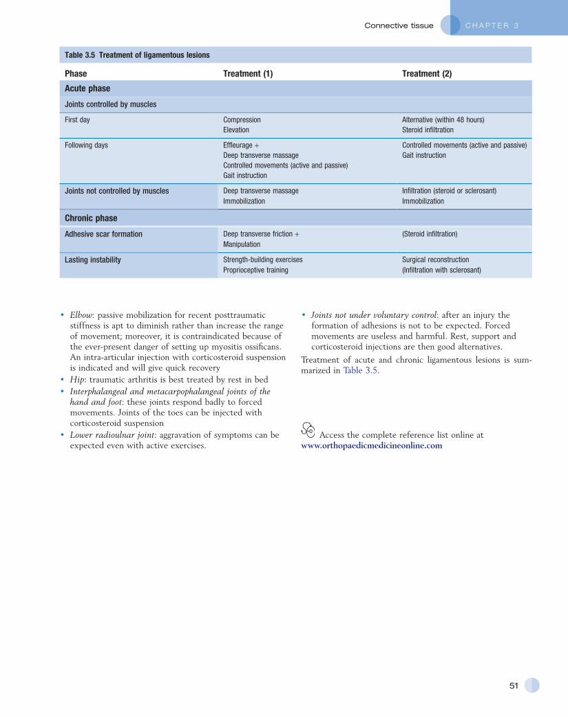
C H A P T E R 3Connective tissue
51
• Joints not under voluntary control: after an injury the formation of adhesions is not to be expected. Forced movements are useless and harmful. Rest, support and corticosteroid injections are then good alternatives.
Treatment of acute and chronic ligamentous lesions is sum-marized in Table 3.5.
• Elbow: passive mobilization for recent posttraumatic stiffness is apt to diminish rather than increase the range of movement; moreover, it is contraindicated because of the ever-present danger of setting up myositis ossificans. An intra-articular injection with corticosteroid suspension is indicated and will give quick recovery
• Hip: traumatic arthritis is best treated by rest in bed• Interphalangeal and metacarpophalangeal joints of the
hand and foot: these joints respond badly to forced movements. Joints of the toes can be injected with corticosteroid suspension
• Lower radioulnar joint: aggravation of symptoms can be expected even with active exercises.
Table 3.5 Treatment of ligamentous lesions
Phase Treatment (1) Treatment (2)
Acute phase
Joints controlled by muscles
First day Compression Alternative (within 48 hours)Elevation Steroid infiltration
Following days Effleurage + Controlled movements (active and passive)Deep transverse massage Gait instructionControlled movements (active and passive)Gait instruction
Joints not controlled by muscles Deep transverse massage Infiltration (steroid or sclerosant)Immobilization Immobilization
Chronic phase
Adhesive scar formation Deep transverse friction + (Steroid infiltration)Manipulation
Lasting instability Strength-building exercises Surgical reconstructionProprioceptive training (Infiltration with sclerosant)
Access the complete reference list online at www.orthopaedicmedicineonline.com
C H A P T E R 3Connective tissue
51.e1© Copyright 2013 Elsevier, Ltd. All rights reserved.
References
1. Warwick L, Williams PL. Gray’s Anatomy. 36th ed. Edinburgh: Churchill Livingstone; 1980.
2. McAnulty RJ. Fibroblasts and myofibroblasts: their source, function and role in disease. Int J Biochem & Cell Biol 2007;39:666–71.
3. Lewis CE, McGee JOD. The Macrophage. Oxford: IRL Press; 1992.
4. Holgate ST. Mast cells and their mediators. In: Holborrow EJ, Reeves WG, editors. Immunology in Medicine. 2nd ed. London: Academic Press; 1983. p. 979–94.
5. Pollard TD, Earnshaw WC. Section VIII, Cell adhesion and the extracellular matrix. In: Cell Biology. Philadelphia: Saunders; 2007.
6. Walker PS. Human Joints and their Artificial Replacements. Springfield: Thomas; 1977.
7. Kennedy JC, Hawkins RJ, Willis RB. Tension studies of human knee ligaments. J Bone Joint Surg 1976;50A:350–5.
8. Wyke B. The neurology of joints. Ann R Coll Surg 1967;41:25.
9. Rowinski M. Afferent neurobiology of the joint. In: Davies GJ, Gould JA, editors. Orthop and Sports: Physical Therapy. St Louis: Mosby, 1985. p. 50.
10. de Morree JJ. Dynamiek van het menselijk bindweefsel. Functie, beschadiging en herstel. Utrecht/Antwerp: Bohn, Scheltema & Holkema; 1989.
11. Tipton CM, Matthes RD, Maynard JA, Carey RA. The influence of physical activities on ligaments and tendons. Med Sci Sports 1975;7(3):165.
12. Henderson B, Pettipher ER. The synovial cell: biology and pathobiology. Sem Arthritis Rheum 1985;15:1–32.
13. Linck G, Porte A. Cytophysiology of the synovial membrane: distinction of two cell types of the intima revealed by their reaction with horseradish peroxidase and iron saccharate in the mouse. Biol Cell 1981;42:147–52.
14. Klareskog L, Forsum U, Kabelitz D, et al. Immune functions of human synovial cells. Phenotypic and T cell regulator properties of macrophage-like cells that express HLA-DR. Arthritis Rheum 1982;25:488–501.
15. Jay JD. Characterization of a bovine fluid lubricating factor. I. Chemical, surface activity and lubricating properties. Connect Tissue Res 1992;28:71–88.
16. Ghadially FN. Fine Structure of Synovial Joints. London: Butterworth; 1983.
17. Sunderland S. Nerves and Nerve Injuries. 2nd ed. London: Churchill Livingstone; 1978.
18. Harkness RD. Mechanical Properties of Collagenous Tissue. Treatise on Collagen. Gould (ed.). London: Academic Press; 1968.
19. Cooper RR, Misol S. Tendon and ligament insertion: a light and electron microscopy study. J Bone Joint Surg 1970;52A:1–21.
20. Laros GS, Tipton CM, Cooper RR. Influence of physical activity on ligament insertions in the knees of dogs. J Bone Joint Surg 1971;53A:275–86.
21. Benjamin M, Toumi H, Ralphs JR, et al. Where tendons and ligaments meet bone: attachment sites (‘entheses’) in relation to exercise and/or mechanical load. J Anat 2006;208:471–90.
22. Hunt ThK. Wound healing. In: Dunphy JL, Way LW, editors. Current Surgical Diagnosis and Treatment, chapter 9. Los Altos, California: Lange Medical; 1975. p. 97.
23. Peacock EE, van Winckle W. Surgery and Biology of Wound Repairs. Saunders; 1980.
24. Lehto M, Durance VC, Restall D. Collagen and fibronectin in a healing skeletal muscle injury. J Bone Joint Surg 1985;67B:820–7.
25. Zarins B. Soft tissue injury and repair: biomechanical aspects. Int J Sports Med 1982;3:9.
26. Kellett J. Acute soft tissue injuries: A review of the literature. Med Sci Sports Exerc 1986;18:5.
27. Banks AR. The role of growth factor in tissue repair. In: Clark RAF, Henson PM, editors. The Molecular and Cellular Biology of Wound Repair. New York: Plenum Press; 1988. p. 2059–79.
28. Fox GM. The role of growth factor in tissue repair. In: Clark RAF, Hensoon PM, editors. The Molecular and Cellular Biology of Wound Repair. New York: Plenum Press; 1988. p. 266–72.
29. Ehrlich HP, Rajaratnam JBM. Cell locomotion versus cell contraction forces for collagen lattice contraction: an in vitro model for wound contraction. Tissue Cell 1990;22:407–17.
30. Buckwalter JA, Crues R. Healing of musculoskeletal tissues. In: Rockwood CA, Green DP, editors. Fractures. Philadelphia: Lippincott; 1991.
31. Tillman LJ, Chasan NP. Properties of dense connective tissue and wound healing. In: Hertling D, Kessler RM, editors. Management of Common Musculoskeletal Disorders. Philadelphia: Lippincott; 1996. p. 8–21.
32. Stearns ML. Studies on development of connective tissue in transparant chambers in rabbit’s ear. Am J Anat 1940;67:55.
33. Frankel VH, Nordin M. Basic Biomechanics of the Skeletal System. Philadelphia: Lea & Febiger; 1980.
34. McGaw WT. The effect of tension on collagen remodelling by fibroblasts: a stereological ultrastructural study. Connect Tissue Res 1986;14:229.
35. van der Meulen JCH. Present state of knowledge on processes of healing in collagen structures. Int J Sports Med 1982;3:4.
36. Hardy MA. The biology of scar formation. Phys Ther 1989;69:1014–23.
37. Frank G, Woo SL-Y, Amiel D, et al. Medial collateral ligament healing. A
multidisciplinary assessment in rabbits. Am J Sports Med 1983;11:379.
38. Pitsillides AA, Skerry TM, Edwards JC. Joint immobilization reduces synovial fluid hyaluronan concentration and is accompanied by changes in the synovial intimal cell populations. Rheumatology 1999;38(11):1108.
39. Akeson WH, Amiel D, Woo S. Immobility effects of synovial joints; the pathomechanics of joint contracture. Biocheology 1980;17:95–110.
40. Akeson WH. An experimental study of joint stiffness. J Bone Joint Surg 1961;43A:1022.
41. Akeson WH, Ameil D, LaViolette D. The connective tissue response to immobility: an accelerated aging response. Exp Gerontol 1968;3:329.
42. Erikson E. Sports injuries of the knee ligaments; their diagnosis, treatment, rehabilitation and prevention. Medicine Science & Sports 1976;8:133–44.
43. Akeson WH, Amiel D, Abel MF. Effects of immobilization on joints. Clin Orthop 1987;219:33.
44. Matthiass AH. Immobilisation und Druckbelastung in ihrer Wirkung auf die Gelenke. Arch Orthop Unfall-Chir 1966;60:380.
45. Cotta H. Pathophysiologie des Knorpelschadens. Hefte Unfallheikunde 1976;127:1–22.
46. Dustmann HO. Knorpelveränderungen beim Hämarthros unter besonderer Berücksichtigung der Ruhigstellung. Arch Orthop Unfall-Chir 1971;71:148.
47. Giucciardi E. Some observations on the effect of blood and fibrinolytic enzyme on articular cartilage in the rabbit. J Bone Joint Surg 1967;49B:342–50.
48. Sood SC. A study on the effects of experimental immobilisation on rabbit articular cartilage. J Anat 1971;108:497.
49. Videman T. Connective tissue and immobilisation: key factors in musculoskeletal degeneration? Clin Orthop Rel Res 1987;221:26–32.
50. Ratcliffe A, Mow VC. Articular cartilage. In: Comper WD, editor. Extracellular Matrix, vol I, Tissue Function. Amsterdam: Harwood; 1996. p. 234–306.
51. Salter RB, Simmonds DF, Malcolm BW, et al. The biological effect of continuous passive motion on the healing of full-thickness defects in articular cartilage. J Bone Joint Surg 1980;62A:1232–51.
52. Gerber CH, Matter P, Chrisman OD, Langhans M. Funktionelle Rehabilitation nach komplexen Knieverletzungen. Wissenschaftliche Grundlagen und Praxis. Schweiz Z Sprtmed 1980;28:37–56.
53. Appell H-J. Muscular atrophy following immobilization. A review. Sports Med 1990;10(1):42–58.
54. Maroudas A. Distribution and diffusion of solutes in articular cartilage. Biophys J 1970;10:365.
General Principles
51.e2© Copyright 2013 Elsevier, Ltd. All rights reserved.
55. Tittel K. Zur Anpassungsfähigkeit einiger Gewebe des Haltungs- und Bewegungsapparates an Belastungen unterschiedlicher Intensität und Dauer. Med Sport 1973;13:147–56.
56. Robbins JR, Evanko SP, Vogel KG. Mechanical loading and TGF-beta regulate proteoglycan synthesis in tendon. Arch Biochem Biophys 1997;342(2):203–11.
57. Ehricht HG. Zur Diagnostik und Therapie der veralteten Bandruptur am oberen Sprunggelenk fibular. Med Sport 1978;18:274–80.
58. Smillie IS. Injuries to the Knee Joint. 5th ed. Edinburgh: Churchill Livingstone; 1978.
59. Cooper RR. Alterations during immobilization and regeneration of skeletal muscles in cats. J Bone Joint Surg 1972;54A:919–51.
60. Järvinen M. Healing of a crush injury in rat striated muscle with special reference to treatment by early mobilization or immobilization. Dissertation Turku, 1976.
61. Vries de HA. Quantitative EMG investigation of the spasm theory of muscle pain. Am J Phys Med 1966;45:119–34.
62. Bobbert MF, Hollander AP, Huijing PA. Factors in delayed onset muscular soreness of man. Med Sci Sports Exerc 1986;18:75–81.
63. Stauber WT. Eccentric action of muscles; physiology, injury and adaption. Exerc Sport Sci Rev 1989;17:157–85.
64. van Wingerden BAM. Ijstherapie in de sport – Indicatie of contraindicatie. Kine 2000, Eur Tijdschr Kinesither 1993:1.
65. Travell J. Ethyl chloride spray for painful muscle spasm. Arch Phys Med Rehab 1952;32:291–8.
66. Waylonis GW. The physiologic effects of ice massage. Arch Phys Med Rehab 1967;48:37–47.
67. Obremskey WT, Seaber AV, Ribbeck BM, Garrett WE Jr. Biomechanical and histological assessment of a controlled muscle strain injury treated with piroxicam. Trans Orthop Res Soc 1988;13:338.
68. Clanton TO, Coupe KJ. Hamstring strains in athletes: diagnosis and treatment. J Am Acad Orthop Surg 1998;6(4):237–48.
69. Beaulieu JE. Developing a stretching program. Physician Sportsmed 1981;9:59–65.
70. Stanish WD, Hubley-Kozey CL. Separating fact from fiction about a common sports activity: can stretching prevent athletic injuries? J Musculoskeletal Med 1984:25–32.
71. Wiktorsson-Moller M, Oberg B, Ekstrand J, Gillquist J. Effects of warming up, massage, and stretching on range of motion and muscle strength in the lower extremity. Am J Sports Med 1983;11:249–52.
72. Garrett WE Jr. Muscle strain injuries: clinical and basic aspects. Med Sci Sports Exerc 1990;22(4):436–43.
73. Ryan JB, Wheeler JH, Hopkinson WJ, Arciero RA, Kolakowski KR. Quadriceps
contusions. West Point update. Am J Sports Med 1991;19(3):299–304.
74. Beiner JM, Jokl P, Cholewicki J, Panjabi MM. The effect of anabolic steroids and corticosteroids on healing of muscle contusion injury. Am J Sports Med 1999;27(1):2–9.
75. Cyriax JH. Textbook of Orthopaedic Medicine, vol 1. London: Baillière Tindall; 1982.
76. Penniello MJ, Chapon F, Olivier D, et al. Myositis ossificans progressiva. Arch Pediatr 1995;2(1):34–8.
77. Traore O, Yiboudo J, Cisse R, et al. Non-traumatic circumscribed myositis ossificans. Apropos of a bilateral localization. Rev Chir Orthop Reparatrice Appar Mot 1998;84(1):79–83.
78. Howard CB, Porat S, Bar-On E, Nyska M, Segal D. Traumatic myositis ossificans of the quadriceps in infants. J Pediatr Orthop 1998;7(1):80–2.
79. Weinstein L, Fraerman S. Difficulties in early diagnosis of myositis ossificans. JAMA 1954;154:994.
80. Gilmer W, Anderson L. Reactions of the somatic tissue which progress to bone formation. South Med J 1959;52:1432.
81. Huss CD, Puhl JJ. Myosititis ossificans of the upper arm. Am J Sports Med 1980;8(6):419.
82. Puddu G, Ippolito E, Postacchini F. A classification of Achilles tendon disease. Am J Sports Med 1976;4:145–50.
83. Khan KM, Cook JL, Kannus P, et al. Time to abandon the ‘tendinitis’ myth. Br Med J 2002;324:626–67.
84. Maffulli N, Khan KM, Puddu G. Overuse tendon conditions: time to change a confusing terminology. Arthroscopy 1998;14(8):840–43.
85. Carr AJ, Norris SH. The blood supply of the calcanean tendon. J Bone Joint Surg 1989;71B:100–1.
86. Ahmed IM, Lagopoulos M, McConnell P, Soames RW, Sefton GK. The blood supply of the Achilles tendon. J Orthop Res 1998;16(5):591–6.
87. Katzer A, Wening JV, Becker-Manich HU, Lorke DE, Jungbluth KH. Rotator cuff rupture. Vascular supply and collagen fiber processes as pathogenetic factors. Unfallchirurgie 1997;23(2):52–9.
88. Kannus P, Józsa L. Histopathological changes preceding spontaneous rupture of a tendon. A controlled study of 891 patients. J Bone Joint Surg Am 1991;73(10):1507–25.
89. Tallon C, Maffulli N, Ewen SW. Ruptured Achilles tendons are significantly more degenerated than tendinopathic tendons. Med Sci Sports Exerc 2001;33(12):1983–90.
90. Kummer FJ, Zuckerman JD. The incidence of full thickness rotator cuff tears in a large cadaveric population. Bull Hosp Jt Dis 1995;54(1):30–1.
91. Milgrom C, Schaffler M, Gilbert S, van Holsbeeck M. Rotator cuff changes in asymptomatic adults. The effect of age, hand dominance and gender. J Bone Joint Surg 1994;77B(2):296–8.
92. Gelberman RH, Vande Berg JS, Lundborg GN, Akeson WH. Flexor tendon healing and restoration of the gliding surfaces. J Bone Joint Surg 1983;65A:70–80.
93. Takai S, Woo SL, Horibe S, Tung DK, Gelberman RH. The effects of frequency and duration of controlled passive mobilization on tendon healing. J Orthop Res 1991;9(5):705–13.
94. Stenho-Bittel L, Reddy GK, Gum S, Enwemeka CS. Biochemistry and biomechanics of healing tendon. Part I. Effects of rigid plaster casts and functional casts. Med Sci Sports Exerc 1998;30(6):788–93.
95. Buckwalter JA. Effects of early motion on healing of musculoskeletal tissues. Hand Clin 1996;12(1):113–24.
96. Peterson L, Althoff B, Renström P. Reconstruction of the lateral ligaments of the ankle joint. Proceedings of the First World Congress of Sports Medicine Applied to Football. Rome, Italy, February 1979:141.
97. Cass JR. Ankle instability: comparison of primary repair and delayed reconstruction after long-term follow-up study. Clin Orthop Rel Res 1985;198:110–7.
98. Cyriax JH. Textbook of Orthopaedic Medicine, vol II. 11th ed. London: Baillière Tindall; 1984.
99. Larsen E. Taping the ankle for chronic instability. Acta Orthop Scand 1984;55:551–3.
100. Freeman MAR. The etiology and prevention of functional instability of the foot. J Bone Joint Surg 1965;47B:678–85.
101. De Carlo MS, Talbot RW. Evaluation of ankle joint proprioception following injection of the anterior talofibular ligament. J Orthop Sports Phys Ther 1986;8:70–6.
102. Oostendorp RAB. Functionele instabiliteit na het inversie-trauma van enkel en voet: een effectonderzoek pleisterbandage versus pleisterbandage gecombineerd met fysiotherapie. Geneeskd Sport 1987;20(2):323–9.
103. Lynch SA, Renstrom PA. Treatment of acute lateral ankle ligament rupture in the athlete. Conservative versus surgical treatment. Sports Med 1999;27(1):61–71.
104. Fetto JF, Marshall JL. Medial collateral ligament injuries to the knee: a rationale for treatment. Clin Orthop Rel Res 1978;132:206.
105. Hastings DE. The non-operative management of collateral ligament injuries of the knee joint. Clin Orthop Rel Res 1980;147:22.
106. Indelicato PA. Non-operative treatment of complete tears of the medial collateral ligament of the knee. J Bone Joint Surg 1983;65A:323.
107. Jones RE, Henley B, Francis P. Non-operative management of isolated grade III collateral ligament injury in high school football players. Clin Orthop Rel Res 1986;213:137–40.
108. Indelicato PA, Hermansdorfer J, Huegel M. Non-operative management of

C H A P T E R 3Connective tissue
51.e3© Copyright 2013 Elsevier, Ltd. All rights reserved.
complete tears of the medial collateral ligament of the knee in intercollegiate football players. Clin Orthop Rel Res 1990;256:174–7.
109. Kannus P, Järvinen M. Non-operative treatment of acute knee ligament injuries. Sport Med 1990;9(4):244–60.
110. Crowninshield RD, Pope MH. Strength and failure characteristics of rat medial
collateral ligaments. J Trauma 1969;16:99.
111. Wiggins ME, Fadale PD, Ehrlich MG, Walsh WR. Effects of local injection of corticosteroids on the healing of ligaments. A follow-up report. J Bone Joint Surg 1995;77A(11):1682–91.
112. Campbell RB, Wiggins ME, Canistra LM, Fadale PD, Akelman E. Influence of
steroid injection on ligament healing in the rat. Clin Orthop 1996;332:242–53.
113. Hughston JC, Eilers AF. The role of the posterior oblique ligament in repairs of acute medial collateral ligament tears of the knee. J Joint Bone Surg 1973;55A:923–40.


![Quan%tave )Logics · Proofof Theorem • It)is)independent from)ini%al)state:) [Norris: Markov)chains. Thm.1.8.3] – Assume)there)are)two)Markov)chains)X,)Y) with)the)same)transi%on](https://static.fdocument.org/doc/165x107/60db574d66b400633d43abd5/quantave-logics-proofof-theorem-a-itisindependent-frominialstate-norris.jpg)

![Stromal fibroblast activation protein alpha promotes gastric … · 2018. 11. 12. · gional tumor progression majorly occurred in abdomen pelvic cavities [5, 6]. The underlying mechanisms](https://static.fdocument.org/doc/165x107/60dc1541981c0c65b612e293/stromal-fibroblast-activation-protein-alpha-promotes-gastric-2018-11-12-gional.jpg)