β-Hydroxysteroid Dehydrogenase Type 14 and Inhibitor ... · sowie die konservierte katalytische...
173
Structural Characterization of 17β-Hydroxysteroid Dehydrogenase Type 14 and Inhibitor Optimization Using Crystallography and Computational Techniques Dissertation zur Erlangung des Doktorgrades der Naturwissenschaften (Dr. rer. nat.) dem Fachbereich Pharmazie der Philipps-Universität Marburg vorgelegt von Nicole Bertoletti aus Sarzana Marburg/Lahn 2017
Transcript of β-Hydroxysteroid Dehydrogenase Type 14 and Inhibitor ... · sowie die konservierte katalytische...
Structural Characterization of17β-Hydroxysteroid Dehydrogenase Type 14
and Inhibitor Optimization Using Crystallographyand Computational Techniques
Dissertation
zur Erlangung des Doktorgradesder Naturwissenschaften
(Dr. rer. nat.)
demFachbereich Pharmazie der
Philipps-Universität Marburgvorgelegt
von
Nicole Bertolettiaus
Sarzana
Marburg/Lahn 2017
II
Erstgutachter Prof. Dr. Gerhard KlebeInstitut für Pharmazeutische ChemiePhilipps-Universität Marburg
Zweitgutachter Dr. Sarndrine Marchais-OberwinklerInstitut für Pharmazeutische ChemiePhilipps-Universität Marburg
Eingereicht am 22.8.2017
Tag der Mündlichen Prüfung am 4.10.2017
Hochschulkennziffer: 1180
III
Die Untersuchungen zur vorliegenden Arbeit wurden auf Anregung von Herrn Prof. Dr.Gerhard Klebe am Institut für Pharmazeutische Chemie des Fachbereichs Pharmazie derPhilipps-Universität Marburg in der Zeit von Januar 2014 bis August 2017 durchgeführt.

V
17β-Hydroxysteroid dehydrogenase type 14 (17β-HSD14) is the latest identified subtype of17β-HSDs. In vivo this enzyme oxidizes the hydroxyl group at position 17 of estradiol (E2)and 5-androstenediol (5-diol) in the presence of NAD+ as cofactor. Two isoforms of thiscytosolic protein exist that differ only in sequence position 205: S205 and T205. So far, theprotein has not been thoroughly investigated in detail and its physiological role remainsunknown. Prior to this thesis, the 17β-HSD14 apoenzyme (S205) had already beencrystallized. The determined structure revealed a very broad and open active site and theconserved catalytic triad and the Rossmann-fold motif. However, all C-terminal tails and forsome chains also amino acids in the flexible loop (189-212) were not defined in the electrondensity. Moreover, it is impossible to derive information regarding a potential substrate fromthis apo structure. Therefore, the renewed structural determination of the 17β-HSD14 apoprotein as well as in complex with its cofactor and substrate was of utmost importance.
After successful establishment of the expression and purification protocols for 17β-HSD14protein, the two enzyme isoforms (S205 and T205) were characterized biochemically. Thestructures of the S205 apoenzyme and the binary complexes with NAD+ of both isoformswere determined. In these complex structures the flexible loop adopts a unique closedconformation differing from the apo structure. Binding of the cofactor is accompanied by ashift of the flexible loop and of the C-terminal Tyr253’ of the adjacent monomer, therebyreducing the size of the active site. The ternary complex of the enzyme with estrone (E1) andNAD+ was also determined. E1 binds to the active site in an atypical fashion, in so far as itsA-ring and not the enzymatically modified position 17 close to the nicotinamide moiety ofNAD+.
Enzyme inhibitors are useful tools to study the consequences of enzyme inhibition in vivo.This allows to clarify whether this enzyme may be interesting as a new drug target for acertain disease. In addition, potent and selective 17β-HSD14 inhibitors may help understandthe selectivity issue with other 17β-HSDs. As no 17β-HSD14 inhibitor was known prior tothis study, the goal was to identify and optimize nonsteroidal 17β-HSD14 inhibitors. To that,a library of 17β-HSD1 and 17β-HSD2 inhibitors was screened against 17β-HSD14. The mostpromising hit was taken as the starting point for further chemical modification applying aligand-based approach. Newly designed compounds were synthesized and subsequently
VI | Summary
tested for their 17β-HSD14 inhibitory activity. Prior to this thesis, no human 17β-HSDstructure in complex with a nonsteroidal ligand was published. The crystal structuresconfirmed that the inhibitors bind to the substrate binding site and allowed to rationalize thestrong affinity of these inhibitors.
Subsequently, two different structure-based strategies were pursued for inhibitor design. Thefirst structure-based modifications of the initial pyridine-based scaffold led to a ten-foldmore potent inhibitor. The goal of the second structure-based optimization strategy was toextend the central pyridine core to interact with the empty binding pocket adjacent to thesteroid A and B-ring. The predicted binding mode was verified by co-crystal structures andthe low nanomolar potency was confirmed by biophysical characterization. The new crystalstructures revealed how small changes of the inhibitors affect the adopted binding mode. Thecharacterization of the most promising 17β-HSD14 inhibitors against 17β-HSD1, 17β-HSD2,and 17β-HSD10 revealed varying degrees of selectivity. In addition, some of these inhibitorsshowed very low cytotoxicity and did not interact with the multi-drug resistance protein Pgp,indicating these compounds might not be effluxed from the brain and that the risk ofpotential side effects is reduced. This suggests these inhibitors as tool compounds for furtherinvestigation in vivo.
To explain the selectivity profiles of the ligands towards 17β-HSD14 and other 17β-HSDs weconducted a structural comparison. The typical V-like shape of the binding pocket of17β-HSD14 is determined by His93 and Gln148, which are not present in 17β-HSD1,17β-HSD8 and 17β-HSD10. In addition, the latter three enzymes have a rather flat bindingpocket. This suggests that matching the characteristic three-dimensional requirements of17β-HSD14 and optionally addressing His93 and/or Gln148 will increase the selectivitytoward this target. Such inhibitors were predicted by docking a library of about 400 17β-HSD1 and 17β-HSD2 inhibitors with GOLD followed by in vitro screening of docking hitsand related compounds. Remarkably, predicted binding modes were in poor agreement withthe subsequently determined crystal structures due to the adaptability of the binding pocketcaused by the flexible loop.
Finally, a large fragment screening campaign by X-ray crystallography with the aim todiscover new inhibitor scaffolds bound to 17β-HSD14 was performed. This resulted in twofragments that could be clearly identified in the electron density. However, these fragmentsdid not significantly inhibit 17β-HSD14. In order to enhance affinity, fragment growing andfragment linking strategies were applied, resulting in two new inhibitors with better affinitythan the starting fragments.
In summary, both isoforms of 17β-HSD14, S205 and T205, were characterized biochemicallyand structurally resulting in four new crystal structures. The first two classes of inhibitor forthis enzyme were discovered and the ligands were thoroughly profiled. In addition, the
Summary | VII
structures of 12 nonsteroidal inhibitors in complex with the protein were elucidated for thefirst time for this protein family. The fragment screening by determining 96 fragment-soakedstructures, resulted in two fragment hits that were successfully optimize culminating in twoinhibitors more active than their precursor fragments.

IX
Die 17β-Hydroxysteroid Dehydrogenase Typ 14 (17β-HSD14) ist der zuletzt identifizierteSubtyp der 17β-HSDs. In vivo oxidiert dieses Enzym die Hydroxyl-Gruppe von Estradiol (E2)und 5-Androstendiol (5-Diol) an Position 17 in Gegenwart des Kofaktors NAD+. Esexistieren zwei Isoformen dieses zytosolischen Proteins, die sich ausschließlich inSequenzposition 205 unterscheiden: S205 und T205. Bis jetzt wurde das Protein noch nichtgründlich und im Detail untersucht und seine physiologische Rolle bleibt unbekannt. Vor derDurchführung dieser Doktorarbeit war das 17β-HSD14 Apoenzym (S205) bereitskristallisiert worden. Die gelöste Struktur zeigte ein sehr weites und offenes aktives Zentrumsowie die konservierte katalytische Triade und das Rossmann-Faltmotiv. Jedoch waren alleC-terminalen Enden und bei einigen Ketten auch Aminosäuren der flexiblen Schleife (189-212) nicht in der Elektronendichte definiert. Darüber hinaus ist es unmöglich, Informationenbezüglich eines potentiellen Substrats von dieser Apostruktur abzuleiten. Deshalb war dieerneute Strukturbestimmung des 17β-HSD14 Apoproteins sowie seiner Komplexe mitKofaktor und Substrat von größter Wichtigkeit.
Nach erfolgreicher Etablierung der Expressions- und Aufreinigungsprotokolle für 17β-HSD14 wurden die beiden Isoformen (S205 und T205) biochemisch charakterisiert. DieStrukturen des S205 Apoenzyms und der binären Komplexe beider Isoformen mit NAD+
wurden aufgeklärt. In diesen Strukturen nimmt die flexible Schleife eine einzigartigegeschlossene Konformation ein, die sich von der Apostruktur unterscheidet. Die Bindung desKofaktors geht einher mit einer Verschiebung der flexiblen Schleife und des C-terminalenTyr253’ des benachbarten Monomers, wodurch die Größe des aktiven Zentrums vermindertwird. Der ternäre Komplex des Enzyms mit Estron (E1) und NAD+ wurde ebenfallsaufgeklärt. E1 bindet auf untypische Weise in das aktive Zentrum, insofern als sein A-Ringund nicht die enzymatisch modifizierte Position 17 nahe dem Nikotinamid-Baustein desNAD+ positioniert ist.
Enzyminhibitoren sind nützliche Werkzeuge, um die Konsequenzen einer Enzymhemmungin vivo zu studieren. Dies erlaubt zu klären, ob dieses Enzym als neues Arzneistofftarget fürbestimmte Krankheiten interessant sein könnte. Außerdem könnten potente und selektive17β-HSD14 Inhibitoren auch helfen, das Selektivitätsproblem anderen 17β-HSDs zuverstehen. Da vor dieser Studie kein 17β-HSD14 Inhibitor bekannt war, war das Ziel die
X | Zusammenfassung
Identifizierung und Optimierung nicht-steroidaler 17β-HSD14 Inhibitoren. Dafür wurden17β-HSD1 und 17β-HSD2 Inhibitorbibliotheken gegen 17β-HSD14 gescreent. Dervielversprechendste Treffer wurde als Startpunkt für weitere chemische Modifizierung unterAnwendung eines ligandbasierten Ansatzes verwendet. Neu designte Verbindungen wurdensynthetisiert und anschließend auf ihre inhibitorische Aktivität gegen 17β-HSD14 getestet.Vor dieser Doktorarbeit waren keine Strukturen einer humanen 17β-HSD im Komplex miteinem nicht-steroidalen Liganden veröffentlicht. Die Kristallstrukturen bestätigten, dass dieInhibitoren an die Substratbindestelle binden und ermöglichten die hohe Affinität dieserInhibitoren zu erklären.
Anschließen wurden zwei unterschiedliche Strategien zum Inhibitordesign verfolgt. Dieersten struktur-basierten Modifikationen des ursprünglichen Pyridin-Grundgerüstes führtenzu 10-fach potenteren Inhibitoren. Das Ziel der zweiten struktur-basiertenOptimierungsstrategie war die Erweiterung des zentralen Pyridin Kerns, um eine Interaktionmit der leeren Tasche neben den Steroid-Ringen A und B zu gewährleisten. Dervorhergesagte Bindungsmodus wurde durch Kokristallstrukturen verifiziert und die niedrig-nanomolare Affinität durch biophysikalische Charakterisierung bestätigt. Die neuenKristallstrukturen offenbarten, wie kleine Änderungen der Inhibitoren den eingenommenenBindungsmodus beeinflussen. Die Charakterisierung der vielversprechendsten 17β-HSD14Inhibitoren bezüglich 17β-HSD1, 17β-HSD2 und 17β-HSD10 offenbarte unterschiedlicheGrade an Selektivität. Zusätzlich zeigten einige dieser Inhibitoren eine sehr niedrigeZytotoxizität und keine Wechselwirkung mit dem Multidrug-Resistance-Protein Pgp, wasdarauf hindeutet, dass diese Verbindungen nicht aus dem Gehirn ausgeschleust werden unddass das Risiko möglicher Nebenwirkungen erniedrigt ist. Dies legt die Nutzung dieserInhibitoren als Werkzeuge für weitere in vivo Untersuchungen nahe.
Um die Selektivitätsprofile dieser Liganden hinsichtlich 17β-HSD14 und anderen 17β-HSDszu erklären, führten wir einen strukturellen Vergleich durch. Die typische V-ähnliche Formder Bindetasche von 17β-HSD14 wird durch His93 und Gln148 bestimmt, welche in17β-HSD1, 17β-HSD8 and 17β-HSD10 fehlen. Zusätzlich haben diese drei Enzyme eine eherflache Bindetasche. Dies legt nahe, dass eine Anpassung an die charakteristischendreidimensionalen Anforderungen von 17β-HSD14 und wahlweise die Adressierung vonHis93 und/oder Gln148 die Selektivität für dieses Target erhöhen werden. Solche Inhibitorenwurden durch Docking einer Bibliothek von 400 17β-HSD1 und 17β-HSD2 Inhibitoren mitGOLD vorhergesagt, gefolgt von einem in vitro Screening der Docking Hits und verwandterVerbindungen. Bemerkenswerterweise waren die vorhergesagten Bindemoden in schlechterÜbereinstimmung mit den nachfolgend ermittelten Kristallstrukturen, bedingt durch dieAnpassungsfähigkeit der Bindetasche welche durch die flexible Schleife verursacht wird.
Zusammenfassung | XI
Schließlich wurde eine großangelegte röntgenkristallographische Fragment-ScreeningKampagne durchgeführt, mit dem Ziel neue Inhibitor-Grundgerüste die an 17β-HSD14binden zu entdecken. Dies führte zu zwei Fragmenten die deutlich in der Elektronendichteidentifiziert werden konnten. Jedoch zeigten diese Fragmente keine signifikante Inhibitionvon 17β-HSD14. Um die Affinität zu erhöhen, wurden Strategien zum Fragment-Wachstumund zur Fragment-Kopplung (growing und linking) angewendet, was zu zwei neuenInhibitoren mit gegenüber den Start-Fragmenten erhöhter Affinität führte.
Zusammengefasst wurden beide Isoformen von 17β-HSD14, S205 und T205, biochemischund strukturell charakterisiert, was zu vier neuen Kristallstrukturen führte. Die ersten zweiKlassen von Inhibitoren dieser Enzyme wurden entdeckt und gründlich charakterisiert.Zusätzlich wurden zum ersten Mal für diese Familie die Strukturen von 12 nicht-steroidalenInhibitoren im Komplex mit dem Protein ermittelt. Das Fragment-Screening durch dieBestimmung der Struktur von 96 mit Fragmenten getränkten Kristallen führte zu zweiFragment Hits, die erfolgreich optimiert und zu zwei Inhibitoren mit gegenüber denVorgänger-Fragmenten erhöhter Aktivität entwickelt werden konnten.

XIII
Summary .............................................................................................................................................. V
Zusammenfassung .............................................................................................................................. IV
Contents ........................................................................................................................................... XIII
Abbreviations ................................................................................................................................. XVII
1 Introduction............................................................................................................................. 1
1.1 Sex steroid hormones ............................................................................................................................. 21.2 17β-hydroxysteroid dehydrogenases family ....................................................................................... 21.2.1 Cofactor preference ................................................................................................................................ 61.2.2 Reducing 17β-HSDs enzymes ............................................................................................................... 71.2.3 Oxidizing 17β-HSDs enzymes .............................................................................................................. 81.2.4 17β-HSD14 .............................................................................................................................................. 91.3 Tools for the characterization of enzymes and their planned application in the current
study ........................................................................................................................................................ 111.4 Aim of the research project and thesis outline ................................................................................. 15
2 New Insights into Human β-Hydroxysteroid Dehydrogenase Type 14: First Crystal
Structures in Complex with a Steroidal Ligand .................................................................... 19
2.1 Introduction .......................................................................................................................................... 202.2 Results and discussion .......................................................................................................................... 212.2.1 Protein expression and purification ................................................................................................... 212.2.2 Protein stability and Thermal Shift Assay (TSA) experiment ........................................................ 212.2.3 Activity assay and biochemical characterization of both S205 and T205 ..................................... 232.2.4 Crystallization of 17β-HSD14 ............................................................................................................. 242.2.5 Description of the NAD+, estrone binding modes .......................................................................... 302.3 Conclusion ............................................................................................................................................. 322.4 Experimental section ............................................................................................................................ 322.4.1 Site directed mutagenesis ..................................................................................................................... 322.4.2 Expression of the 17β-HSD14 protein (S205 and T205 variants) and purification ..................... 33

XIV | Contents
2.4.3 Thermal shift Assay (TSA) .................................................................................................................. 342.4.4 Fluorimetric assay ................................................................................................................................. 342.4.5 Determination of kinetic constants .................................................................................................... 342.4.6 Protein crystallization .......................................................................................................................... 352.4.7 Data collection and processing ........................................................................................................... 352.4.8 Structure determination and refinement ........................................................................................... 36
3 First Structure-Activity Relationship of 17β-Hydroxysteroid Dehydrogenase Type 14:
Nonsteroidal Inhibitors and Crystal Structures in Complex with the Enzyme ................... 37
3.1 Introduction .......................................................................................................................................... 383.2 Results ..................................................................................................................................................... 393.2.1 Design of 17β-HSD14 inhibitor candidates ...................................................................................... 393.2.2 Calculation of physicochemical parameters ...................................................................................... 413.2.3 Inhibition of 17β-HSD14 determined with a fluorimetric assay .................................................... 443.2.4 Aggregation ........................................................................................................................................... 443.2.5 17β-HSD14 inhibitory activity ............................................................................................................ 443.2.6 Substituent variations on the C-ring .................................................................................................. 453.2.7 Substituent variations on the A-ring .................................................................................................. 463.2.8 Variation of the A-ring ........................................................................................................................ 473.2.9 Variations on the B-ring ...................................................................................................................... 483.2.10 Trisubstituted pyridines ....................................................................................................................... 493.2.11 Pan Assay Interference Compounds [168] ........................................................................................ 503.2.12 Crystal structure determination .......................................................................................................... 503.2.13 Description of the inhibitor binding site ........................................................................................... 513.2.14 Description of the binding mode of inhibitors in complex with 17β-HSD14 .............................. 553.2.15 MOE models .......................................................................................................................................... 583.2.16 Thermal shift assay ............................................................................................................................... 603.2.17 Comparison of the 17β-HSD1, 17β-HSD2 and 17β-HSD14 structures ........................................ 603.2.18 Selectivity ............................................................................................................................................... 623.3 Discussion .............................................................................................................................................. 633.3.1 Focus on the C-ring part ...................................................................................................................... 633.3.2 Focus on the A-ring part ...................................................................................................................... 653.3.3 Focus on the B-ring part ...................................................................................................................... 663.3.4 Second binding site for compound 3.9 .............................................................................................. 663.3.5 Comparison of the 17β-HSD1, 17β-HSD2 and 17β-HSD14 structures ........................................ 673.3.6 Basis for structure-based drug design ................................................................................................ 673.4 Conclusion ............................................................................................................................................. 673.5 Experimental section ............................................................................................................................ 683.5.1 17β-HSD14 inhibition assay ................................................................................................................ 68
Contents | XV
3.5.2 Enzyme expression ............................................................................................................................... 683.5.3 Radioactive assay using Procedure A ................................................................................................. 693.5.4 Enzyme Purification ............................................................................................................................. 693.5.5 Fluorimetric assay using Procedure B ................................................................................................ 703.5.6 Protein co-crystallization with inhibitors 3.1, 3.6, 3.9, 3.10, 3.12 ................................................... 703.5.7 Data collection and processing ........................................................................................................... 713.5.8 Structure determination and refinement ........................................................................................... 713.5.9 Thermal shift Assay (TSA) .................................................................................................................. 72
4 Structure-Based Design of 17β-HSD14 Inhibitors ............................................................... 73
4.1 Introduction .......................................................................................................................................... 744.2 Results ..................................................................................................................................................... 754.2.1 Structure design of the new inhibitors ............................................................................................... 754.2.2 Pan Assay Interference Compounds [168] ........................................................................................ 774.2.3 Physicochemical Parameters ............................................................................................................... 774.2.4 Biological Evaluation ............................................................................................................................ 784.2.5 17β-HSD14 Inhibitory Activity .......................................................................................................... 794.2.6 Crystal structure determination .......................................................................................................... 814.2.7 Description of the binding mode of the pyridine-based inhibitors in complex with
17β-HSD14 ............................................................................................................................................ 814.2.8 Description of the binding mode of three quinoline-based inhibitors in complex with
17β-HSD14 ............................................................................................................................................ 854.2.9 Comparison of the structures of 17β-HSD14, and 17β-HSD10. .................................................... 874.2.10 Selectivity ............................................................................................................................................... 874.2.11 Cytotoxicity evaluation ........................................................................................................................ 894.2.12 Pgp ATPase Activity Assays ................................................................................................................ 894.3 Discussion and conclusion .................................................................................................................. 914.3.1 SAR of pyridine derivatives ................................................................................................................. 914.3.2 Selectivity of pyridine derivatives ....................................................................................................... 924.3.3 SAR and selectivity of the quinoline/naphthalene derivatives ....................................................... 924.4 Experimental section ............................................................................................................................ 964.4.1 Enzyme expression and purification .................................................................................................. 964.4.2 Inhibition of 17β-HSD14 ..................................................................................................................... 974.4.3 Inhibition of 17β-HSD1 and 17β-HSD2 ............................................................................................ 974.4.4 Inhibition of 17β-HSD10 ..................................................................................................................... 974.4.5 Co-crystallization of the protein with inhibitors .............................................................................. 984.4.6 Crystallography ..................................................................................................................................... 984.4.7 MOE models .......................................................................................................................................... 994.4.8 Cytotoxicity Assay ................................................................................................................................ 99
XVI | Contents
4.4.9 Pgp ATPase Activity Assays ................................................................................................................ 99
5 Structural Comparison between 17β-HSD Enzymes and Virtual Screening of
Inhibitors ............................................................................................................................. 101
5.1 Introduction ........................................................................................................................................ 1025.2 Comparison of the crystal structures of 17β-HSD1 and 17β-HSD14.......................................... 1035.3 Comparison of the structures of 17β-HSD8, 17β-HSD10 and 17β-HSD14 ............................... 1085.4 Docking studies with 17β-HSD14 .................................................................................................... 110
6 X-ray Crystallographic Fragment Screening and Hit Optimization ................................. 113
6.1 Introduction ........................................................................................................................................ 1146.2 Results and Discussion ....................................................................................................................... 1156.2.1 Crystallization and Soaking experiments ........................................................................................ 1156.2.2 Binding mode of the fragment hits ................................................................................................... 1156.2.3 Fragment optimization applying fragment growing ...................................................................... 1176.2.4 Fragment optimization applying fragment linking ........................................................................ 1196.2.5 Inhibitory Activity validation ............................................................................................................ 1206.2.6 Binding mode confirmation of the optimized compounds .......................................................... 1226.3 Conclusion and Outlook .................................................................................................................... 1236.4 Experimental session .......................................................................................................................... 1246.4.1 Enzyme expression and purification ................................................................................................ 1246.4.2 Protein crystallization and soaking .................................................................................................. 1246.4.3 Co-crystallization of the protein with inhibitors ............................................................................ 1256.4.4 Data collection and processing ......................................................................................................... 1256.4.5 Structure determination and refinement ......................................................................................... 1256.4.6 Inhibition of 17β-HSD14 ................................................................................................................... 1266.4.7 MOE models ........................................................................................................................................ 126
7 Discussion and Conclusions ................................................................................................ 127
Bibliography ..................................................................................................................................... 133
Acknowledgements ........................................................................................................................... 149
Curriculum Vitae ............................................................................................................................. 151
Erklärung .......................................................................................................................................... 155
XVII
11β-HSD 11β-hydroxysteroid dehydrogenases17β-HSD 17β-hydroxysteroid dehydrogenases17β-HSD1 17β-hydroxysteroid dehydrogenases type 117β-HSD2 17β-hydroxysteroid dehydrogenases type217β-HSD8 17β-hydroxysteroid dehydrogenases type 817β-HSD10 17β-hydroxysteroid dehydrogenases type 1017β-HSD14 17β-hydroxysteroid dehydrogenasestype 154-dione 4-androstene-3,17-dione5-diol 5-androstene-3β, 17β-diolλ wavelengthÅ Ångström (1 Å = 10–10 m)AKR aldo-keto reductaseCHES N-Cyclohexyl-2-aminoethanesulfonic acidclogP calculated logarithm of the n-octanol/water partition coefficientCW capping waterDHEA dihydroepiandrostenedioneDMSO dimethyl sulfoxideDNA deoxyribonucleic acide– electronE1 estroneE2 estradiolFBLD fragment-based lead discoveryER Estrogen receptorGOL glycerolH-bond hydrogen bondHEPES 4-(2-hydroxyethyl)-1-piperazineethanesulfonic acidHTS high throughput screeningIC50 half maximal inhibitory concentrationIG50 concentration resulting in 50% inhibition of growth
XVIII | Abbreviations
Kd dissociation constant at equilibriumKi dissociation constant at equilibrium for an inhibitorKm substrate concentration at which the reaction rate is half of Vmax
kDa kilodaltonLE ligand efficiencyMe MethylMPD 2-methyl-2,4-pentanediolMW molecular weightMX macromolecular X-ray crystallographyNAD(H)+ nicotinamide adenine dinucleotideNADP(H)+ nicotinamide adenine dinucleotide phosphatePDB protein data bankPEG polyethylene glycolRMSD root-mean-square deviationSAR structure-activity relationshipSDR short-chain dehydrogenase/reductaseT testosteroneTm melting temperatureTLC thin-layer chromatographytris tris(hydroxymethyl)aminomethaneTSA thermal shift assayUV light UltravioletvdW van der WaalsVmax maximum velocity at saturating substrate concentrationW water molecule

1
Introduction
2 | Chapter 1
1.1 Sex steroid hormones
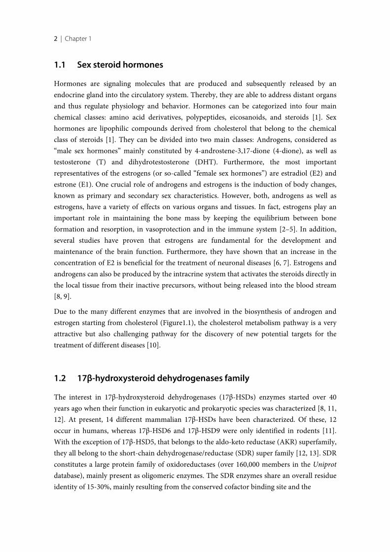
Hormones are signaling molecules that are produced and subsequently released by anendocrine gland into the circulatory system. Thereby, they are able to address distant organsand thus regulate physiology and behavior. Hormones can be categorized into four mainchemical classes: amino acid derivatives, polypeptides, eicosanoids, and steroids [1]. Sexhormones are lipophilic compounds derived from cholesterol that belong to the chemicalclass of steroids [1]. They can be divided into two main classes: Androgens, considered as“male sex hormones” mainly constituted by 4-androstene-3,17-dione (4-dione), as well astestosterone (T) and dihydrotestosterone (DHT). Furthermore, the most importantrepresentatives of the estrogens (or so-called “female sex hormones”) are estradiol (E2) andestrone (E1). One crucial role of androgens and estrogens is the induction of body changes,known as primary and secondary sex characteristics. However, both, androgens as well asestrogens, have a variety of effects on various organs and tissues. In fact, estrogens play animportant role in maintaining the bone mass by keeping the equilibrium between boneformation and resorption, in vasoprotection and in the immune system [2–5]. In addition,several studies have proven that estrogens are fundamental for the development andmaintenance of the brain function. Furthermore, they have shown that an increase in theconcentration of E2 is beneficial for the treatment of neuronal diseases [6, 7]. Estrogens andandrogens can also be produced by the intracrine system that activates the steroids directly inthe local tissue from their inactive precursors, without being released into the blood stream[8, 9].
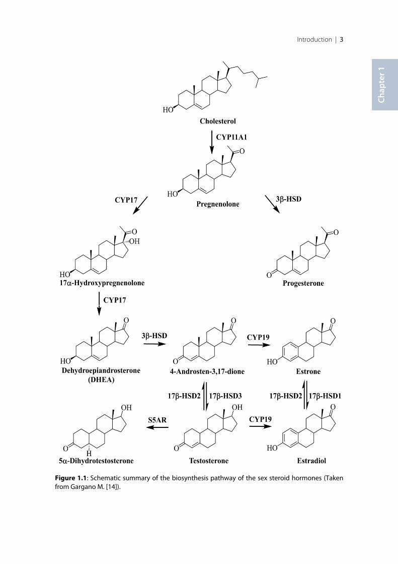
Due to the many different enzymes that are involved in the biosynthesis of androgen andestrogen starting from cholesterol (Figure1.1), the cholesterol metabolism pathway is a veryattractive but also challenging pathway for the discovery of new potential targets for thetreatment of different diseases [10].
1.2 17β-hydroxysteroid dehydrogenases family
The interest in 17β-hydroxysteroid dehydrogenases (17β-HSDs) enzymes started over 40years ago when their function in eukaryotic and prokaryotic species was characterized [8, 11,12]. At present, 14 different mammalian 17β-HSDs have been characterized. Of these, 12occur in humans, whereas 17β-HSD6 and 17β-HSD9 were only identified in rodents [11].With the exception of 17β-HSD5, that belongs to the aldo-keto reductase (AKR) superfamily,they all belong to the short-chain dehydrogenase/reductase (SDR) super family [12, 13]. SDRconstitutes a large protein family of oxidoreductases (over 160,000 members in the Uniprotdatabase), mainly present as oligomeric enzymes. The SDR enzymes share an overall residueidentity of 15-30%, mainly resulting from the conserved cofactor binding site and the
Introduction | 3
Chap
ter 1
Figure 1.1: Schematic summary of the biosynthesis pathway of the sex steroid hormones (Takenfrom Gargano M. [14]).
4 | Chapter 1
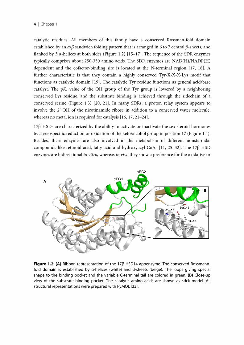
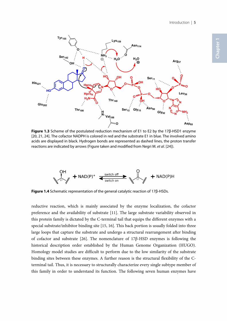
catalytic residues. All members of this family have a conserved Rossman-fold domainestablished by an α/β sandwich folding pattern that is arranged in 6 to 7 central β-sheets, andflanked by 3 α-helices at both sides (Figure 1.2) [15–17]. The sequence of the SDR enzymestypically comprises about 250-350 amino acids. The SDR enzymes are NAD(H)/NADP(H)dependent and the cofactor-binding site is located at the N-terminal region [17, 18]. Afurther characteristic is that they contain a highly conserved Tyr-X-X-X-Lys motif thatfunctions as catalytic domain [19]. The catalytic Tyr residue functions as general acid/basecatalyst. The pKa value of the OH group of the Tyr group is lowered by a neighboringconserved Lys residue, and the substrate binding is achieved through the sidechain of aconserved serine (Figure 1.3) [20, 21]. In many SDRs, a proton relay system appears toinvolve the 2’ OH of the nicotinamide ribose in addition to a conserved water molecule,whereas no metal ion is required for catalysis [16, 17, 21–24].
17β-HSDs are characterized by the ability to activate or inactivate the sex steroid hormonesby stereospecific reduction or oxidation of the keto/alcohol group in position 17 (Figure 1.4).Besides, these enzymes are also involved in the metabolism of different nonsteroidalcompounds like retinoid acid, fatty acid and hydroxyacyl CoAs [11, 25–32]. The 17β-HSDenzymes are bidirectional in vitro, whereas in vivo they show a preference for the oxidative or
Figure 1.2: (A) Ribbon representation of the 17β-HSD14 apoenzyme. The conserved Rossmann-fold domain is established by α-helices (white) and β-sheets (beige). The loops giving specialshape to the binding pocket and the variable C-terminal tail are colored in green. (B) Close-upview of the substrate binding pocket. The catalytic amino acids are shown as stick model. Allstructural representations were prepared with PyMOL [33].
Introduction | 5
Chap
ter 1
Figure 1.3 Scheme of the postulated reduction mechanism of E1 to E2 by the 17β-HSD1 enzyme[20, 21, 24]. The cofactor NADPH is colored in red and the substrate E1 in blue. The involved aminoacids are displayed in black. Hydrogen bonds are represented as dashed lines, the proton transferreactions are indicated by arrows (Figure taken and modified from Negri M. et al. [24]).
reductive reaction, which is mainly associated by the enzyme localization, the cofactorpreference and the availability of substrate [11]. The large substrate variability observed inthis protein family is dictated by the C-terminal tail that equips the different enzymes with aspecial substrate/inhibitor binding site [15, 16]. This back portion is usually folded into threelarge loops that capture the substrate and undergo a structural rearrangement after bindingof cofactor and substrate [26]. The nomenclature of 17β-HSD enzymes is following thehistorical description order established by the Human Genome Organization (HUGO).Homology model studies are difficult to perform due to the low similarity of the substratebinding sites between these enzymes. A further reason is the structural flexibility of the C-terminal tail. Thus, it is necessary to structurally characterize every single subtype member ofthis family in order to understand its function. The following seven human enzymes have
Figure 1.4 Schematic representation of the general catalytic reaction of 17β-HSDs.
6 | Chapter 1
already been structurally characterized: 17β-HSD1, 17β-HSD4, 17β-HSD5, 17β-HSD8, 17β-HSD10, 17β-HSD11 and 17β-HSD14 [27, 34].
The 17β-hydroxysteroid dehydrogenases (17β-HSDs) are essential for the last step of theformation and degradation of steroid hormones. They regulate the intracellular availabilityof steroid hormones and their potential activation of the nuclear receptors [11, 34]. Inaddition, these enzymes are specifically expressed in certain tissues. Consequently, thisenzyme family is of high interest as therapeutic targets for several steroid hormonedependent diseases [34], and for several types of 17β-HSDs a correlation with some humandiseases has already been found. In addition, the expression level of some of these enzymescan be used as prognostic marker in breast and prostate cancer [35, 36].
Cofactor preference
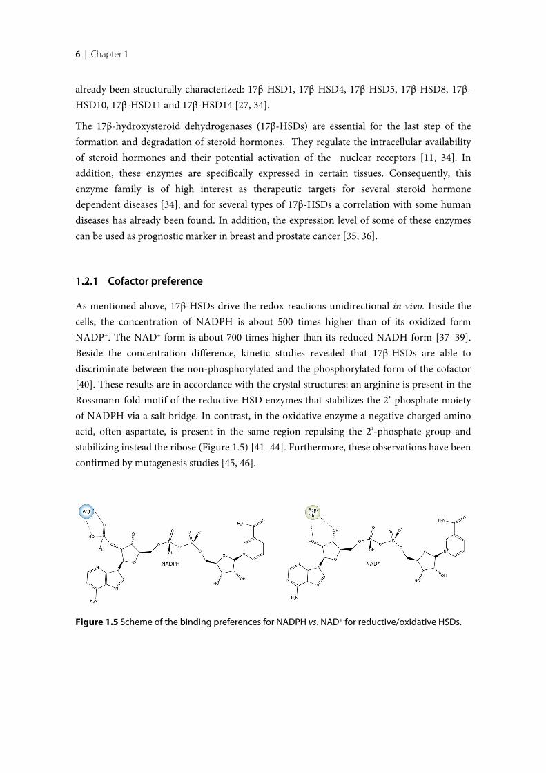
As mentioned above, 17β-HSDs drive the redox reactions unidirectional in vivo. Inside thecells, the concentration of NADPH is about 500 times higher than of its oxidized formNADP+. The NAD+ form is about 700 times higher than its reduced NADH form [37–39].Beside the concentration difference, kinetic studies revealed that 17β-HSDs are able todiscriminate between the non-phosphorylated and the phosphorylated form of the cofactor[40]. These results are in accordance with the crystal structures: an arginine is present in theRossmann-fold motif of the reductive HSD enzymes that stabilizes the 2’-phosphate moietyof NADPH via a salt bridge. In contrast, in the oxidative enzyme a negative charged aminoacid, often aspartate, is present in the same region repulsing the 2’-phosphate group andstabilizing instead the ribose (Figure 1.5) [41–44]. Furthermore, these observations have beenconfirmed by mutagenesis studies [45, 46].
Figure 1.5 Scheme of the binding preferences for NADPH vs. NAD+ for reductive/oxidative HSDs.
Introduction | 7
Chap
ter 1
Reducing 17β-HSDs enzymes
Six reductive 17β-HSDs enzymes are described in the literature [11, 24, 34]. They areactivating enzymes and responsible for the high level of active sex steroids in target tissues.
17β-HSD1 was the first enzyme in this family to be cloned and structurally characterized.This enzyme, which is active as a homodimer [47], is one of the most important enzymesinvolved in the last step of the activation of estradiol starting from estrone, resulting in a highconcentration of the sex hormone in the target tissue. This enzyme is estrogen specific. Aminor effect on the reduction of androgen was also identified [48–50]. 17β-HSD1 is acytosolic enzyme that is mainly expressed in breast, endometrium, ovary and placenta. Inminor concentration it is expressed in adipose tissue and skin. During the last decades, anincreasing number of inhibitors targeting 17β-HSD1 were discovered. The first inhibitors arebased on a steroidal scaffold, some are mixed inhibitors combining both, cofactor andsteroid, whereas the latter inhibitors show a nonsteroidal core [34, 51–54]. At present, severalcrystal structures of the apoenzyme, the cofactor-enzyme complex and the ternary complexeswith substrate or steroidal inhibitors are already resolved. However, no crystal structure ofthe enzyme in complex with a nonsteroidal inhibitor has been reported so far. 17β-HSD1 hasbeen proven to play a crucial role in several estrogen-dependent diseases such as breastcancer, ovarian tumor, endometriosis, and uterine leiomyoma [55–63] and it is a validateddrug target for estrogen dependent breast cancer.
17β-HSD3 is a microsomal membrane-bound enzyme that is mainly present in the testis thatit is bound to the endoplasmic reticulum through its N-terminal domain [11, 64]. 17β-HSD3has 310 amino acids and it catalyzes the reductive reaction of 4-dione and 5α-androstenedione to testosterone (T) and dihydrotestosterone (DHT), respectively [65, 66].This enzyme is overexpress in prostate cancer and, due its catalytic action, its inhibitionswould reduce the concentration of T and therefore it could be beneficial against tumorgrowth [67, 68]. The determination of the structure of this enzyme was unsuccessful due itshydrophobic nature.
17β-HSD5 is located in the cytosol. This enzyme is prevalently expressed in breast, liver andprostate. 17β-HSD5 shows a broad substrate specificity [69, 70]. As it is member of the aldo-ketoreductase (AKR) protein superfamily, it will not be further discussion.
17β-HSD7 is a microsomal enzyme bound to the endoplasmic reticulum. It is present inbreast, liver, testis, ovary, kidney, placenta as well as in neuronal tissue and lung [11, 71, 72].The enzyme is involved in the production of E2. Furthermore, it has been proven that itfulfills a main role in the synthesis of cholesterol [73]. No crystal structure is available so far.
17β-HSD12 is present in microsomes of especially kidney, liver, heart and skeletal muscleand in minor level in placenta, breast and ovary. This enzyme is involved mainly in the
8 | Chapter 1
regulation of the lipid biosynthesis and plays only a marginal role in the metabolism of E2[74–76].
17β-HSD13 is present in the liver but is also detected in ovary, bone marrow, kidney, brain,lung, skeletal muscle, bladder and testis. It is a cytosolic enzyme and it may be involved in thelipid metabolic pathways [77, 78].
Oxidizing 17β-HSDs enzymes
Characteristic for these enzymes is that they catalyze oxidation reactions and that they arefound ubiquitously in the body also in non-steroidogenic tissues. As they inactivate the sexhormones (oxidation of the potent estradiol and testosterone in estrone and 4-dione,respectively) and thus lower the concentration of the latter in the target tissues, it is assumedthat these enzymes play a protective role in vivo [11].
17β-HSD2 is widely expressed in tissues such as placenta, uterus, liver, bone, gastrointestinaland urinary tracts [79–82]. This enzyme is found to be bound to membranes of themicrosomal fraction. It catalyzes the conversion of E2, T and DHT to their less potent formsE1, 4-dione and 5α-androstenedione, respectively [29]. Due to the unspecific localization of17β-HSD2 and its physiological role in inactivating the sex hormones, it has been suggestedthat it plays a role in protecting tissues from excessive steroid concentrations [12]. Severalsteroidal and non-steroidal inhibitors have already been identified for this enzyme [34, 83,84]. The estrogen replacement therapy for the treatment of osteoporosis is proven to bebeneficial; however, this therapy is no longer recommended due the many side effects [34, 85,86]. 17β-HSD2 oxidizes E2 into E1, resulting in a decreased concentration of E2 in bone cells.Therefore, inhibition of this enzyme is a promising approach for the treatment ofosteoporosis [34, 87–92]. Unfortunately, the three-dimensional structure of this enzyme isstill unknown due to its hydrophobic nature that has proven to be a huge obstacle for thestructural elucidation.
17β-HSD4 is ubiquitously distributed and it is mainly involved in the inactivation of sexsteroids. The enzyme 17β-HSD4 is a much larger enzyme compared to the other 17β-HSDsand its tertiary structure can be divided into three domains [11, 18, 34].
17β-HSD8 is located in liver, placenta, gonads and kidney. 17β-HSD8 can catalyzes a widerange of substrates including estrogen, androgen and fatty acids and its three-dimensionalstructure is known [11, 34].
17β-HSD10 is a mitochondrial enzyme that is located in the central nervous system (CNS). Itis overexpressed in the amyloid plaques of patients suffering of Alzheimer’s disease. Theenzyme is involved in several substrate pathways, for instance in the inactivation of sex
Introduction | 9
Chap
ter 1
steroids and the catabolism of short hydroxyacyl CoAs [93–97]. One class of inhibitorsdescribed for this enzyme forms a covalent bond to the cofactor (NAD+) and typically has apeculiar chemical structure [94]. Few crystal structures of this protein are available asapoenzyme or in inhibitor-enzyme complexes.
17β-HSD11 is expressed in liver, lung, placenta and kidney. Its physiological role is notdisclosed yet; however, recent studies suggest that the enzyme might be involved in themetabolism of fatty acids rather than in the metabolism of sex steroids [98, 99].
17β-HSD14
Human 17β-hydroxysteroid dehydrogenase type 14 (17β-HSD14) — also called retSDR3,DHRS10 or SDR47C19 — is the latest enzyme identified that belongs to the 17β-HSD family[11, 16, 32, 34]. Initially, its gene was isolated from the retina by Haeseleer and Palczewski[100]. Subsequently, a second version of the gene was isolated from a melanotic melanomacell in the framework of a genome sequencing campaign [101, 102]. Both genes are identicalwith the exception of a single point mutation of the amino acid at position 205: The geneisolated from the retina encodes at this position for a serine (17β-HSD14 S205), whereas thegene isolated from a melanotic melanoma encodes for a threonine (17β-HSD14 T205). Sinceonly the S205 variant was characterized so far, the reason for this protein polymorphism isnot yet clear. However, it is hard to believe that the single point mutation could give rise to asignificant difference in activity, as the structural difference is limited to a single methylgroup. Furthermore, it cannot be excluded that a spontaneous mutation occurred during theisolation of the second gene from the cancer tissue. Nevertheless, it would be of high interestto characterize also the T205 protein variant.
As the gene was first isolated from a retina cDNA library, it was hypothesized that thisenzyme would be involved in the retinoid metabolism. However, this function could not beproven [100]. Northern blot analyses has revealed that the S205 hHSD17B14 gene is mainlyexpressed in brain, liver, placenta [32], and in the kidneys [100]. However, Sivik et al [103]applied immunochemical based methods to demonstrate that the protein is also expressed inadrenals and testis as well as in the eyes, heart, kidney, esophagus, liver, rectum, salivaryglands, skeletal muscles and in breast cancer tissue [35]. The striking discrepancy between theenzyme-containing tissues reported in the two studies can be explained by the differences inthe specificity of the applied antibodies. Thus, further investigation is required before aconclusion can be drawn [104].
Although the in vitro reaction of 17β-HSD14 was investigated, its physiological role in vivo isstill unclear. About 50 ligands binding to SDR enzymes were tested on 17β-HSD14, but onlysome sex steroids showed significant affinity to the enzyme. These results suggest that the
10 | Chapter 1
enzyme is potentially involved in the sex steroid metabolic pathway [32]. 17β-HSD14catalyzes the oxidation of the alcohol function at position 17 of E2, 5-androstene-3β, 17β-diol(5-diol), and T — using NAD+ as a cofactor — and transforms them into their less activeforms E1, dihydroepiandrostenedione (DHEA), and 4-dione, respectively [32]. However, thelow turnover rate for these steroids and the not saturatable kinetics of T suggest thehypothesis that in vivo the enzyme might play a role also in other metabolic pathways (Km=5.6 µM ± 1.7 for E2; Km= 13.6 µM ± 1.6 for 5-diol) [32].
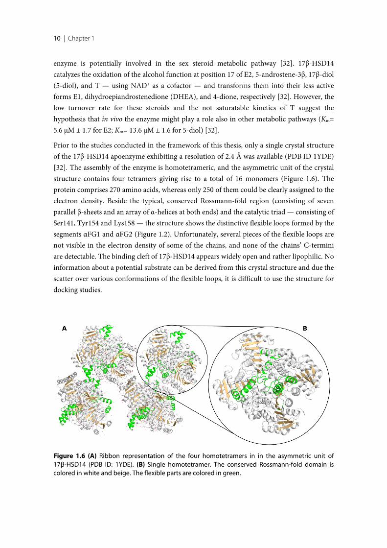
Prior to the studies conducted in the framework of this thesis, only a single crystal structureof the 17β-HSD14 apoenzyme exhibiting a resolution of 2.4 Å was available (PDB ID 1YDE)[32]. The assembly of the enzyme is homotetrameric, and the asymmetric unit of the crystalstructure contains four tetramers giving rise to a total of 16 monomers (Figure 1.6). Theprotein comprises 270 amino acids, whereas only 250 of them could be clearly assigned to theelectron density. Beside the typical, conserved Rossmann-fold region (consisting of sevenparallel β-sheets and an array of α-helices at both ends) and the catalytic triad — consisting ofSer141, Tyr154 and Lys158 — the structure shows the distinctive flexible loops formed by thesegments αFG1 and αFG2 (Figure 1.2). Unfortunately, several pieces of the flexible loops arenot visible in the electron density of some of the chains, and none of the chains’ C-terminiare detectable. The binding cleft of 17β-HSD14 appears widely open and rather lipophilic. Noinformation about a potential substrate can be derived from this crystal structure and due thescatter over various conformations of the flexible loops, it is difficult to use the structure fordocking studies.
Figure 1.6 (A) Ribbon representation of the four homotetramers in in the asymmetric unit of17β-HSD14 (PDB ID: 1YDE). (B) Single homotetramer. The conserved Rossmann-fold domain iscolored in white and beige. The flexible parts are colored in green.
Introduction | 11
Chap
ter 1
1.3 Tools for the characterization of enzymes and their planned
application in the current study
Different techniques are available for the characterization of enzymes and for studyingligand-protein interactions. The different methods are often complementary to each other,resulting in a more complete and reliable picture of the studied effects in operation. However,during the planning of the experiment to characterize the enzyme structure it is important totake the limitations especially into account. For example, for well-characterized enzymes insilico approaches can successfully identify compounds during drug development [104];however, as mention earlier, it can be rather challenging to identify binders based onhomology models derived from of sequence data showing low identity and for proteinsexhibiting highly flexible parts. Several attempts to dock ligands into 17β-HSD14 wereperformed in the course of some preliminary studies of this project. However, thesubsequntly determined crystal structures revealed that the predicted binding modes wereincorrect.
One of the techniques on which the current thesis is strongly based is macromolecular X-raycrystallography. This technique is a diffraction method for the determination of structuralinformation up to the atomic level [105–108]. Since the 17β-HSD enzyme family does notshare a high sequence homology, especially across the binding-site region, crystal structurescan provide important insights into the peculiarities of the binding sites. As mention above,one crystals structure for 17β-HSD14 had already been described in literature prior to thisstudy [32]. Even though this crystal structure already revealed some details about thearchitecture of this protein, there still remained many open questions. The electron density ofhighly variable regions of the protein were ill-defined (flexible loops and the C-terminal tail),however they are of utmost importance because they contribute to ligand binding. Thisdeficiency could resulted from several effects, for instance the relative low resolution of thedataset (2.4 Å) could have prevented to properly resolve these mobile regions. Another aspectthat makes this structure not ideal as a starting point for a rational drug discovery endeavor isthat 16 monomeric units (four tetramers) form the asymmetric unit. These chains of the 16units all differ in the arrangement of the flexible loops resulting in binding pockets ofdeviating shape and volume. It is therefore difficult to predict which of the chains representsthe relevant conformation of the active binding site competent to accommodate a ligand.Furthermore, the question remains whether the observed flexibility of the protein also occurswhile the protein is in complex with a ligand and/or the cofactor. It is obvious that a higherquality of the apoenzyme crystal structure and the availability of multiple crystal structures ofthe protein in complex with cofactor and ligand would be tremendously beneficial for theintended drug design studies.
12 | Chapter 1
Even though the X-ray diffraction technology underwent an immense improvement withinthe last decades, for instance the development of more powerful light sources at synchrotronsthat are equipped with faster detectors, one important factor limiting the quality of the crystalstructure is the quality of the protein crystal itself [106, 108]. In this study, in order toproduce well-diffracting three dimensional crystals, extensive crystallization screenings wereperformed.
Crystal structures are also essential for the rational design of ligands. Crystals ofprotein-ligand complexes can be prepared following two different strategies: Soaking orcocrystallization [109, 110]. The strength of the crystal soaking approach is that it can beperformed very fast, since protein-ligand complexes are simply prepared by exposingpremanufactured crystals with known diffraction quality to the ligand of interest. Usuallysoaking of fragments or small ligands is unproblematic; however, more bulky ligands can beincompatible with the crystal packing. Due to their high affinity, they can forcibly squeezeinto the pre-shaped active site and thereby adopting themselves unrealistic conformation orinducing conformational changes of protein sidechains/loops, or even interfere with thepacking in the crystal. This frequently results in a decrease in crystal quality (increasedmosaicity) or even a complete destruction of the exposed crystal. Furthermore, cases havebeen reported where soaking seemingly results in a different binding mode thancocrystallization [106, 110, 111]. This observation suggests that conformational changes ofthe protein upon ligand binding are already established in solution prior crystallization — asreflected by the co-crystallized structures — will prevented false conclusions that might occurif premanufactured crystals are subjected to soaking experiments [110, 111].Cocrystallization is a viable alternative to soaking protocols. In this case, a solution of proteinand ligand is prepared that is subsequently used to grow crystals. Thereby, crystals areformed in periodic arrangements of the pre-assembled protein-ligand complexes of interest.Since the ligands bind to the protein already in solution, this induces protein rearrangementsand thus reflects better the conformation of the protein-ligand complex in solution andhopefully the biologically relevant conditions — and thus will be less biased by putativeimposed crystal packing effects of the apoenzyme. In addition, co-crystallization could evenresult in a qualitatively better crystal structure. The downside of this technique is that it ismore demanding with respect to protein material and can potentially result in a new crystalform that requires new crystallization conditions [109–111].
Through an extensice examination of the protein-ligand complex crystal structure ligandportions that do not achieve interactions to the protein can be identified. This knowledge canbe used to rationally improve the chemical structure of the ligand in the next design cycle[112].
Introduction | 13
Chap
ter 1
If it is intended to study especially the function of the protein, one limitation ofmacromolecular X-ray crystallography is that hydrogen atoms are usually not detected.Consequently, it is not possible to directly determine the protonation state of ligands andamino acids. Thus, the protonation state can only be rationalized on the basis of consensusinteraction patterns and distance and angle between atoms.
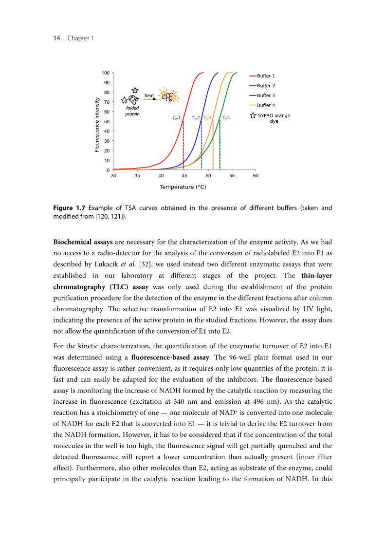
Another technique used in this work is the thermal shift assay (TSA) also called differentialscanning fluorimetry (DSF). The stability of a protein is temperature-dependent [113, 114],and this method detects the differences of the melting temperature of a protein under variousconditions. Stable, correctly folded protein tends to have the hydrophilic amino acidsexposed to the surface and the hydrophobic ones are buried within the core. At a definedtemperature — specific for each macromolecule and dependent on the buffer composition —the protein will partially or completely unfold and, as a consequence, the hydrophobic aminoacids will get exposed to the solvent. This assay detects at which temperature this unfoldingevent occurs. It is usually performed using a real-time PCR devise [115], and is dependent ona special dye (SYPRO orange) that begins to fluorescent upon binding to exposedhydrophobic portions of the protein. No or very low florescence is detected while theglobular protein is correctly folded. However, with increasing temperature the protein willstart to unfold and thus expose hydrophobic residues to the solvent phase, thereby getting incontact with the dye, that in consequence starts to fluorescent. Recording the intensity of thefluorescence signal over a temperature range results in a sigmoidal curve, where the meltingtemperature Tm of the protein is described by the inflection point (Figure 1.7). When theprotein is surrounded by molecules that help to stabilize its tertiary structure, a shift to ahigher melting point will occur. The TSA is extremely useful for the screening of differentadditives, for instance different salts and buffers at different pH values, in order to find abuffer composition that shows an optimal stabilizing effect on the protein. The application ofbuffers that optimally stabilize the protein has the advantage that the yield during proteinexpression as well as the success rate during crystallization screenings can drastically increase[116]. This assay also allows fast and efficient screening for binding ligands. The principle isthat upon binding of a ligand to the protein, the ligand stabilizes or destabilizes throughbinding the protein architecture and the observed shift of the melting temperature isproportional to the strength of the formed complex (i.e. in close series even to the affinity ofthe ligand) as well as proportional to the concentration of the ligand. The amount ofstabilization due to the complexation with different ligands results in shifts of varying extendof the melting temperature Tm compared to the melting point of the uncomplexed enzyme[116–119]. However, the magnitude of the shift is not reflecting the affinity of the ligand butit is primarily proportional to the change in the entropy of binding upon formation of thecomplex [119]. Thus, the TSA can be used to discriminate binders from non-binders, but notfor the determination and comparison of compound affinities.
14 | Chapter 1
Figure 1.7 Example of TSA curves obtained in the presence of different buffers (taken andmodified from [120, 121]).
Biochemical assays are necessary for the characterization of the enzyme activity. As we hadno access to a radio-detector for the analysis of the conversion of radiolabeled E2 into E1 asdescribed by Lukacik et al. [32], we used instead two different enzymatic assays that wereestablished in our laboratory at different stages of the project. The thin-layerchromatography (TLC) assay was only used during the establishment of the proteinpurification procedure for the detection of the enzyme in the different fractions after columnchromatography. The selective transformation of E2 into E1 was visualized by UV light,indicating the presence of the active protein in the studied fractions. However, the assay doesnot allow the quantification of the conversion of E1 into E2.
For the kinetic characterization, the quantification of the enzymatic turnover of E2 into E1was determined using a fluorescence-based assay. The 96-well plate format used in ourfluorescence assay is rather convenient, as it requires only low quantities of the protein, it isfast and can easily be adapted for the evaluation of the inhibitors. The fluorescence-basedassay is monitoring the increase of NADH formed by the catalytic reaction by measuring theincrease in fluorescence (excitation at 340 nm and emission at 496 nm). As the catalyticreaction has a stoichiometry of one — one molecule of NAD+ is converted into one moleculeof NADH for each E2 that is converted into E1 — it is trivial to derive the E2 turnover fromthe NADH formation. However, it has to be considered that if the concentration of the totalmolecules in the well is too high, the fluorescence signal will get partially quenched and thedetected fluorescence will report a lower concentration than actually present (inner filtereffect). Furthermore, also other molecules than E2, acting as substrate of the enzyme, couldprincipally participate in the catalytic reaction leading to the formation of NADH. In this
Introduction | 15
Chap
ter 1
case, the increasing florescence would not exclusively represent the formation of E1.Therefore, it is important to perform a negative control of the reaction in parallel, forexample containing the enzyme and NAD+, but without the substrate E2. One of the largestlimitations of this assay are intrinsically self-fluorescent inhibitors. If an inhibitor isfluorescent at a similar wavelength (λ) as NADH, the read out of the florescence signalbecomes inaccurate and the interpretation of the results is rather difficult or even impossible.
1.4 Aim of the research project and thesis outline
Although the sequencing of the human genome has been solved and all genes are accessible,the physiological role of more than half of all SDR members remains unknown or poorlyexamined. It is of utmost importance to deorphanize and characterize these enzymes as abasis to explore their physiological functions and thereby identify new potential drug targetsfor the treatment of human diseases [122].
17β-HSD14 has been suggested to play a role in neuromodulation [32] and in inflammationprocesses [123]. The availability of a potent and selective enzyme inhibitor would fosterresearch in this direction and potentially support the collection of data to proof theinvolvement of this enzyme in neuronal diseases. Furthermore, such an inhibitor is alsoprerequisite for the conduction of proteomic or metabolic studies in vivo. In addition, potentand selective enzyme inhibitors are also useful tool compounds to study the consequence offull enzyme inhibition, comparable to the change of the phenotype of a knockout mouse.Having access to such a potent inhibitor allowing the detailed characterization in vivo, thisprotein could prove to be an attractive drug target as it is already the case for 17β-HSD1 [34,124, 125] and 17β-HSD2 [87, 90–92]. Potent and selective enzyme inhibitors are also neededto address the selectivity issues of inhibitors with respect to other 17β-HSDs.
Taking all these considerations into account, the main research objective of this thesis is tostructurally characterize the active site of 17β-HSD14 in order to facilitate the developmentof highly active inhibitors. Newly discovered and optimized inhibitors can then be applied astools to further elucidate the structure and function of the enzyme, and to gain insights intothe possible functional roles of this enzyme in vivo.
Chapter 2 of this thesis (publication [126]) describes the chemical and biologicalcharacterization of both S205 and T205 isoforms of 17β-HSD14. To obtain both variants ofthe recombinant protein in high yield, an expression and purification protocol had to beestablished. As it turned out that the protein was quite challenging to handle, specialattention was attributed to on the different approaches followed to overcome issues duringthe purification procedure to obtain the protein in crystallization and assay-pure quality, inparticular protein stability. An extensive crystallization screening enabled the determination
16 | Chapter 1
of four novel crystal structures of the human 17β-HSD14, as apoenzyme, in binary complexwith NAD+ and in ternary complex with NAD+ and the catalytic product of the enzymereaction (E1). These crystal structures were the basis to obtain new insights into the enzyme’sproperties. Since we had access to the S205 as well as to the T205 isoform of the protein, weperformed the biochemical characterization of both.
Chapter 3 (publications [126] and [127]) describes our first ligand-based drug discoveryapproach. The goal of this chapter was to identify and to optimize the first class of 17β-HSD14 inhibitors. In a preliminary study a library of 17β-HSD1 and 17β-HSD2 inhibitors —selected to guarantee scaffold diversity — was tested on potential inhibitory activity for17β-HSD14. The most interesting hit was taken as a starting point for further chemicaloptimization. As matter of fact, this investigation was performed before the first structure ofthe ternary complex (protein-cofactor-ligand) could be determined. Therefore, theoptimization of the inhibitor was performed at the beginning following a ligand-basedapproach. The newly designed compounds were synthesized and tested for 17β-HSD14inhibitory activity. The best inhibitors identified in this study showed a very high affinitytoward the enzyme with a Ki of about 10 nM. In this chapter, the first five crystal structures ofthe protein in its ternary complex with the cofactor and highly potent nonsteroidal inhibitorswere further elucidated. It is striking that until now no human SDR 17β-HSD enzymestructure has ever been reported in complex with a nonsteroidal compound. It is known thatseveral attempts have been conducted with 17β-HSD1; however, they all failed, possiblyowing to the lipophilicity of the active site or the flexibility of the compounds.
Chapter 4 (manuscript in preparation) describes our strategies to optimize the previouslyreported class of 2,6-pyridine ketone inhibitors (Chapter 3). The availability of the crystalstructures of the enzyme in complex with an inhibitor enabled us to pursue a rationalstructure-based approach. A special focus was placed on scaffold diversity with the aim tofurther characterize the binding pocket of the target protein and thereby to create inhibitorswith different pharmacokinetic properties. Seven new crystal structures of inhibitors incomplex with the protein were determined. This was necessary to understand the inhibitors’structure-activity relationship (SAR) as a basis for their further optimization. In fact, thesesystematic studies revealed how small structural changes of the substituents on the inhibitorscan lead to surprising variation of their binding mode. Furthermore, this chapter describesconsiderations regarding the selectivity profile of the inhibitors toward the different closelyrelated 17β-HSD enzymes as well as in silico determined physicochemical properties of thenew inhibitors.
In Chapter 5 the structural differences between different HSD enzymes are address. X-raycrystal structure models of proteins provide unvaluable structural information about bindingsites and therefore enable to chemically tailor ligands to bind to the target. Crystal structures
Introduction | 17
Chap
ter 1
also allow the comparison between the three-dimensional arrangements of the amino acidsdetermining the active sites of different crystallized members of the family. This informationcan be useful for modelling of the three-dimensional structural arrangement of othernoncrystallizable 17β-HSDs. Structural differences between 17β-HSD14 and three related17β-HSDs (h17β-HSD1, h17β-HSD8, and 17β-HSD10) are discussed, with a focus on eachenzyme’s active site.
As the starting point for the design of the ligands was taken from an already existing libraryof 17β-HSD1/2 inhibitors, it was the aim to discover also a new scaffold in order to possiblyovercome the selectivity issue toward other HSDs. Thus, in Chapter 6 (manuscript inpreparation), we initiated a fragment-based lead discovery (FBLD) campaign with the goal todiscover new inhibitor scaffolds. Therefore, a 96-entry fragment library assembled applyingselection criteria following a slightly extended “Rule of 3” was screened. The crystallographicfragment screening approach comprises the promising perspective that more novel hits areidentified and structurally characterized than by any other biophysical screening technique,especially for ligands that show a low binding affinity. Nevertheless, such ligands can exhibithigh ligand efficiency and the structural information about their binding modes is of utmostimportance for further optimization.

19
New Insights into Human β-Hydroxysteroid Dehydrogenase Type 14:
First Crystal Structures in Complex with a Steroidal Ligand
Introductory remarks
Parts of the following chapter have been published in the Journal of Medicinal Chemistry in2016. The cloning of the plasmid for the 17β-HSD14 T205 variant was done by Dr. GabrieleMöller. TLC and fluorescence based assay were designed and performed by Dr. SandrineMarchais-Oberwinkler in collaboration with the author of the thesis. The expression and thepurification of the 17β-HSD14, the crystallization study, the elucidation of the crystalstructures and the TSA assay were established and performed by the author of this thesis.Furthermore, the author significantly contributed to the writing of the manuscript incollaboration with Dr. Sandrine Marchais-Oberwinkler and Florian Braun.
20 | Chapter 2
2.1 Introduction
17β-Hydroxysteroid dehydrogenase type 14 (17β-HSD14), also called retSDR3, DHRS10 orSDR47C19, is the latest 17β-HSD which has been identified [11, 32, 34]. It belongs to theshort-chain dehydrogenase-reductase (SDR) family and its physiological role is yet unknown.Estradiol (E2), 5-androstene-3β,17β-diol (5-diol) and testosterone (T) have been identified assubstrates in vitro [32]. 17β-HSD14 catalyzes the alcohol oxidation, NAD+ dependent, of theaforementioned estrogens and androgens at their position 17 giving rise to estrone (E1),dehydroepiandrosterone (DHEA), and 4-androstene-3,17-dione (4-dione), respectively [32].A library of 50 ligands of SDR enzymes were tested at 17β-HSD14 but only theaforementioned steroids showed significant enzyme affinity, indicating that this enzymemight be involved in steroid metabolism [32].
The gene coding for 17β-HSD14 was first isolated from the human retinal epithelium byHaeseleer et al. [100] and contains a serine at position 205 (S205). An alternate version of thegene was subsequently isolated from a melanotic melanoma cell during a genome sequencingcampaign [101]. This allelic variant, termed T205, carries a threonine at position 205. Themeaning of the observed polymorphism has not been analyzed until now and the T205variant has also never been characterized to date. In this study, the structural and thebiochemical characterization of the T205 will be addressed as well as its comparison to theS205 enzyme.
Concerning its localization, northern blot analyses have shown that the human HSD17B14gene is dominantly expressed in the brain, liver, placenta [32], and in the kidney [100]. Inanother study, using an immunochemical based method Sivik et al.[103] demonstrated thatthe protein is also expressed in adrenals and testis as well as in eye, heart, kidney, esophagus,liver, rectum, salivary glands and skeletal muscle. 17β-HSD14 has also been identified inbreast cancer tissue [35, 103]. 17β-HSD14 is a cytosolic enzyme [32].
The S205 variant of 17β-HSD14 has been previously crystallized and the 3D-structure of theapoenzyme determined was by Lukacik et al. [32]. Crystal structures of a target proteinprovide important structural insights into binding sites. However, from the existingstructure, no information about the protein/ligand interaction, either with the cofactor orwith the substrate, can be extracted.
In this study, the characterization of the new T205 variant and four new crystal structures ofthe protein as apoenzyme (S205), holoenzyme (T205 and S205) and as inhibitor-enzymecomplex (T205) are presented. These results provided further insights for thecharacterization of this enzyme.
Structural Characterization of 17ß-HSD14 | 21
Chap
ter 2
2.2 Results and discussion
Protein expression and purification
Both recombinant 17β-HSD14 protein variants (S205 and T205) were overexpressed in E.coliBL21 pLysS via transformation with the corresponding N-6His-tag plasmid, followingLukacik’s procedure [32], applying minor modifications. Pure enzyme was obtained with ayield between 8-15 mg of protein per liter of bacterial culture. During the expression andpurification process, protein content was followed either by a TLC plate activity assay or by afluorimetric assay, based on the detection of the formed NADH.
During the establishment of the expression protocol several E.coli bacteria lines were tested.It turned out that only the E.coli BL21 (DE3) pLysS cells were able to overexpress the enzymein satisfactory amount. The enzyme showed a particular tendency to aggregate and toprecipitate with the pellet during the first centrifugation step of the purification of thebacteria homogenate suspension. The problem was resolved by resuspending the pelletderiving from four liters of culture with more buffer (about 400 mL vs 120 mL used byLukacik et al. [32]) and by the addition of 0.5% of Triton X-100, a detergent that helped tokeep the protein in solution. To avoid protein precipitation, it was beneficial to lowercentrifugation (from 30000g to 17700g). Another issue was the constant contamination withDNA in the fraction containing the recombinant enzyme. DNA contamination could beavoided by using a DE-52 column. As such column is rather expensive, we directly appliedthe supernatant to a 5 mL Ni-NTA column and we removed DNA with a first washing stepusing a buffer composed of 50 mM Tris and 1.5 M NaCl. Such high salt concentrationremoved any nonspecific bound DNA. Unfortunately, the enzyme was still fairly unstable insolution. It was necessary to discover additives to add to the different buffers duringpurification having the capacity to keep the enzyme in solution. This issue is discussed in thefollowing paragraph.
Protein stability and Thermal Shift Assay (TSA) experiment
The main challenge encountered during protein purification was the low stability of 17β-HSD14. In the absence of any buffer additives, no or only minor amounts of protein could beisolated. Glycerol is known to stabilize proteins by compacting their structures to a globularshape [128, 129] and its addition substantially increased the efficiency of protein purification.However, it must be emphasized that glycerol in presence of NAD+ and the enzyme, withoutsubstrate, induces the production of a fluorescent substance which, after investigation, turnedout to have the same fluorescence fingerprint as NADH. We concluded that glycerol isrecognized as a substrate by 17β-HSD14, thereby transforming NAD+ into NADH. Ethylene
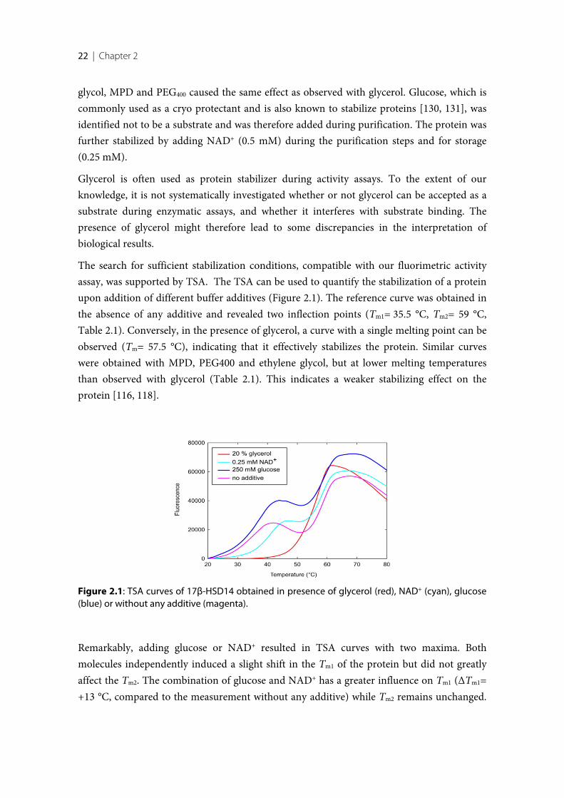
22 | Chapter 2
glycol, MPD and PEG400 caused the same effect as observed with glycerol. Glucose, which iscommonly used as a cryo protectant and is also known to stabilize proteins [130, 131], wasidentified not to be a substrate and was therefore added during purification. The protein wasfurther stabilized by adding NAD+ (0.5 mM) during the purification steps and for storage(0.25 mM).
Glycerol is often used as protein stabilizer during activity assays. To the extent of ourknowledge, it is not systematically investigated whether or not glycerol can be accepted as asubstrate during enzymatic assays, and whether it interferes with substrate binding. Thepresence of glycerol might therefore lead to some discrepancies in the interpretation ofbiological results.
The search for sufficient stabilization conditions, compatible with our fluorimetric activityassay, was supported by TSA. The TSA can be used to quantify the stabilization of a proteinupon addition of different buffer additives (Figure 2.1). The reference curve was obtained inthe absence of any additive and revealed two inflection points (Tm1= 35.5 °C, Tm2= 59 °C,Table 2.1). Conversely, in the presence of glycerol, a curve with a single melting point can beobserved (Tm= 57.5 °C), indicating that it effectively stabilizes the protein. Similar curveswere obtained with MPD, PEG400 and ethylene glycol, but at lower melting temperaturesthan observed with glycerol (Table 2.1). This indicates a weaker stabilizing effect on theprotein [116, 118].
Remarkably, adding glucose or NAD+ resulted in TSA curves with two maxima. Bothmolecules independently induced a slight shift in the Tm1 of the protein but did not greatlyaffect the Tm2. The combination of glucose and NAD+ has a greater influence on Tm1 (ΔTm1=+13 °C, compared to the measurement without any additive) while Tm2 remains unchanged.
Figure 2.1: TSA curves of 17β-HSD14 obtained in presence of glycerol (red), NAD+ (cyan), glucose(blue) or without any additive (magenta).
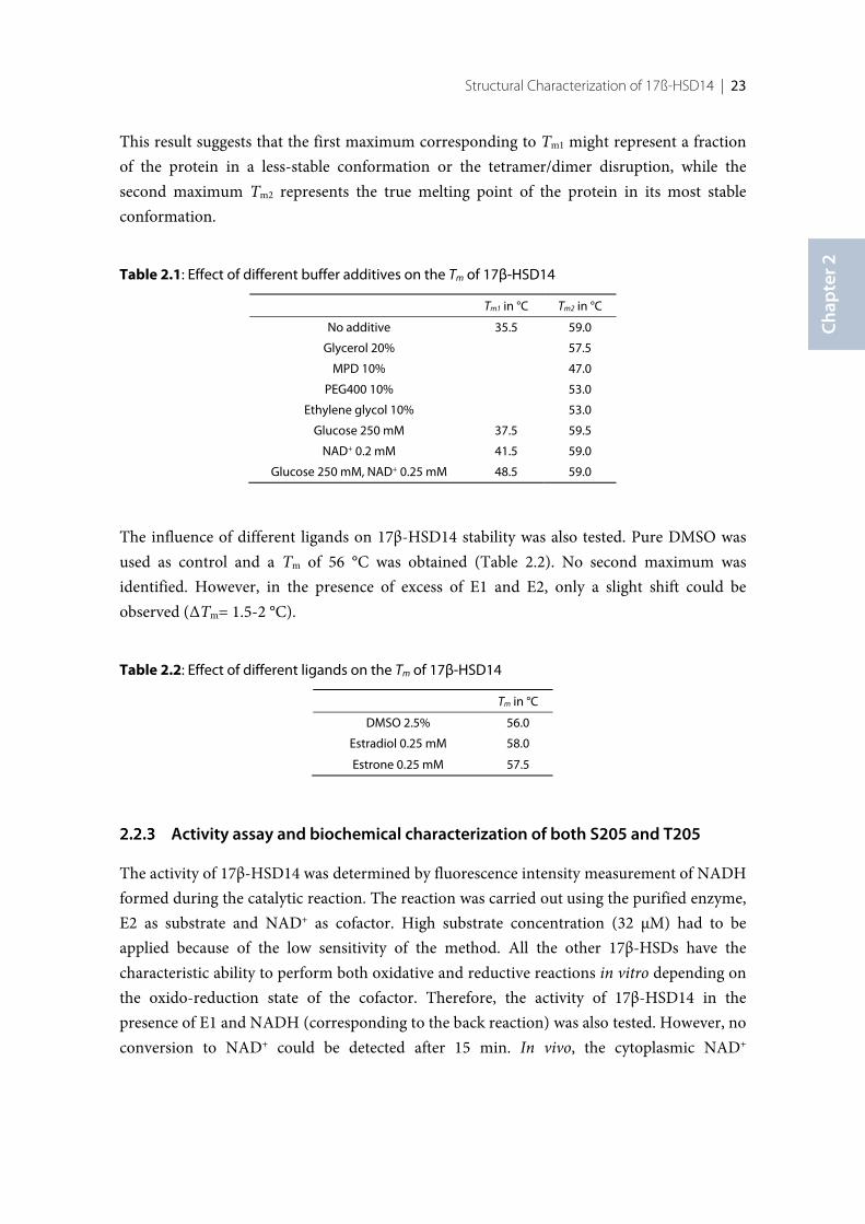
Structural Characterization of 17ß-HSD14 | 23
Chap
ter 2
This result suggests that the first maximum corresponding to Tm1 might represent a fractionof the protein in a less-stable conformation or the tetramer/dimer disruption, while thesecond maximum Tm2 represents the true melting point of the protein in its most stableconformation.
Table 2.1: Effect of different buffer additives on the Tm of 17β-HSD14
Tm1 in °C Tm2 in °C
No additive 35.5 59.0
Glycerol 20% 57.5
MPD 10% 47.0
PEG400 10% 53.0
Ethylene glycol 10% 53.0
Glucose 250 mM 37.5 59.5
NAD+ 0.2 mM 41.5 59.0
Glucose 250 mM, NAD+ 0.25 mM 48.5 59.0
The influence of different ligands on 17β-HSD14 stability was also tested. Pure DMSO wasused as control and a Tm of 56 °C was obtained (Table 2.2). No second maximum wasidentified. However, in the presence of excess of E1 and E2, only a slight shift could beobserved (ΔTm= 1.5-2 °C).
Table 2.2: Effect of different ligands on the Tm of 17β-HSD14
Tm in °C
DMSO 2.5% 56.0
Estradiol 0.25 mM 58.0
Estrone 0.25 mM 57.5
Activity assay and biochemical characterization of both S205 and T205
The activity of 17β-HSD14 was determined by fluorescence intensity measurement of NADHformed during the catalytic reaction. The reaction was carried out using the purified enzyme,E2 as substrate and NAD+ as cofactor. High substrate concentration (32 µM) had to beapplied because of the low sensitivity of the method. All the other 17β-HSDs have thecharacteristic ability to perform both oxidative and reductive reactions in vitro depending onthe oxido-reduction state of the cofactor. Therefore, the activity of 17β-HSD14 in thepresence of E1 and NADH (corresponding to the back reaction) was also tested. However, noconversion to NAD+ could be detected after 15 min. In vivo, the cytoplasmic NAD+
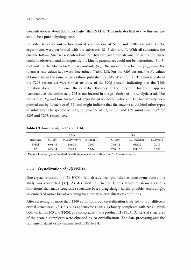
24 | Chapter 2
concentration is about 500 times higher than NADH. This indicates that in vivo this enzymeshould be a pure dehydrogenase.
In order to carry out a biochemical comparison of S205 and T205 variants, kineticexperiments were performed with the substrates E2, 5-diol and T. With all substrates, theenzyme follows Michaelis-Menten kinetics. However, with testosterone, no saturation curvecould be observed, and consequently the kinetic parameters could not be determined. For 5-diol and E2 the Michaelis-Menten constants (Km), the maximum velocities (Vmax) and theturnover rate values (kcat) were determined (Table 2.3). For the S205 variant, the Km valuesobtained are in the same range as those published by Lukacik et al. [32]. The kinetic data ofthe T205 variant are very similar to those of the S205 protein, indicating that the T205mutation does not influence the catalytic efficiency of the enzyme. This result appearsreasonable as the amino acid 205 is not located in the proximity of the catalytic triad. Therather high Km and low turnover of 17β-HSD14 for both, 5-diol and E2, had already beenpointed out by Lukacik et al.[32] and might indicate that the enzyme could bind other typesof substrates. The specific activity, in presence of E2, is 1.35 and 1.21 nmol.min-1.mg-1 forS205 and T205, respectively.
Table 2.3: Kinetic analysis of 17β-HSD14
S205 T205
Substrate Km (µM) Vmax (nM·min-1) kcat (min-1) Km (µM) Vmax (nM·min-1) kcat (min-1)
5-diol 6.6±1.5 58±0.4 0.017 7.8±1.2 68±0.3 0.019
E2 6.2±1.4 82±0.1 0.024 7.9±1.7 114±0.3 0.033
Mean values and given standard deviations were calculated based on 5 - 7 measurements
Crystallization of 17β-HSD14
One crystal structure for 17β-HSD14 had already been published as apoenzyme before thisstudy was conducted [32]. As described in Chapter 1, this structure showed variouslimitations that made conclusive structure-based drug design hardly possible. Accordingly,we embarked onto a broad screening for alternative crystallization conditions.
After screening of more than 1200 conditions, our crystallization trials led to four differentcrystal structures: 17β-HSD14 as apoenzyme (S205), as binary complexes with NAD+ (withboth variants S205 and T205), as a complex with the product E1 (T205). All crystal structuresof the protein complexes were obtained by co-crystallization. The data processing and therefinement statistics are summarized in Table 2.4.
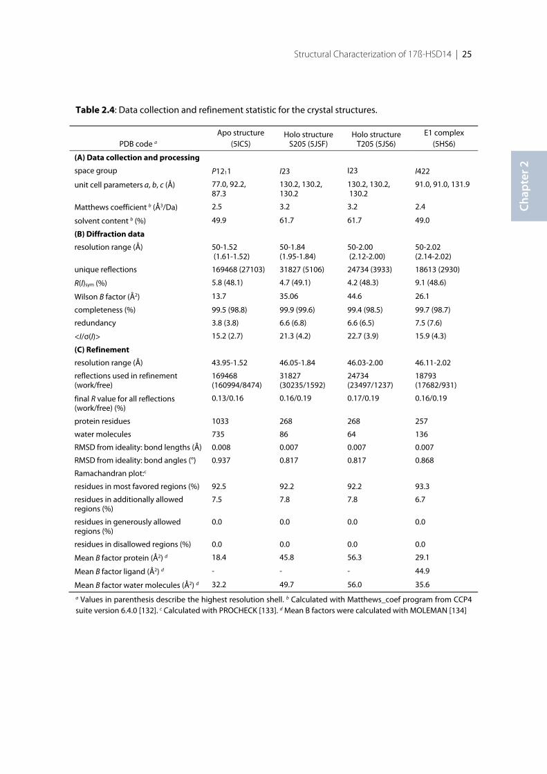
Structural Characterization of 17ß-HSD14 | 25
Chap
ter 2
Table 2.4: Data collection and refinement statistic for the crystal structures.
a Values in parenthesis describe the highest resolution shell. b Calculated with Matthews_coef program from CCP4suite version 6.4.0 [132]. c Calculated with PROCHECK [133]. d Mean B factors were calculated with MOLEMAN [134]
PDB code a
Apo structure(5ICS)
Holo structureS205 (5JSF)
Holo structureT205 (5JS6)
E1 complex(5HS6)
(A) Data collection and processing
space group P1211 I23 I23 I422
unit cell parameters a, b, c (Å) 77.0, 92.2,87.3
130.2, 130.2,130.2
130.2, 130.2, 130.2
91.0, 91.0, 131.9
Matthews coefficient b (Å3/Da) 2.5 3.2 3.2 2.4
solvent content b (%) 49.9 61.7 61.7 49.0
(B) Diffraction data
resolution range (Å) 50-1.52 (1.61-1.52)
50-1.84(1.95-1.84)
50-2.00 (2.12-2.00)
50-2.02(2.14-2.02)
unique reflections 169468 (27103) 31827 (5106) 24734 (3933) 18613 (2930)
R(I)sym (%) 5.8 (48.1) 4.7 (49.1) 4.2 (48.3) 9.1 (48.6)
Wilson B factor (Å2) 13.7 35.06 44.6 26.1
completeness (%) 99.5 (98.8) 99.9 (99.6) 99.4 (98.5) 99.7 (98.7)
redundancy 3.8 (3.8) 6.6 (6.8) 6.6 (6.5) 7.5 (7.6)
<I/σ(I)> 15.2 (2.7) 21.3 (4.2) 22.7 (3.9) 15.9 (4.3)
(C) Refinement
resolution range (Å) 43.95-1.52 46.05-1.84 46.03-2.00 46.11-2.02
reflections used in refinement(work/free)
169468(160994/8474)
31827(30235/1592)
24734(23497/1237)
18793(17682/931)
final R value for all reflections(work/free) (%)
0.13/0.16 0.16/0.19 0.17/0.19 0.16/0.19
protein residues 1033 268 268 257
water molecules 735 86 64 136
RMSD from ideality: bond lengths (Å) 0.008 0.007 0.007 0.007
RMSD from ideality: bond angles (°) 0.937 0.817 0.817 0.868
Ramachandran plot:c
residues in most favored regions (%) 92.5 92.2 92.2 93.3
residues in additionally allowedregions (%)
7.5 7.8 7.8 6.7
residues in generously allowedregions (%)
0.0 0.0 0.0 0.0
residues in disallowed regions (%) 0.0 0.0 0.0 0.0
Mean B factor protein (Å2) d 18.4 45.8 56.3 29.1
Mean B factor ligand (Å2) d - - - 44.9
Mean B factor water molecules (Å2) d 32.2 49.7 56.0 35.6
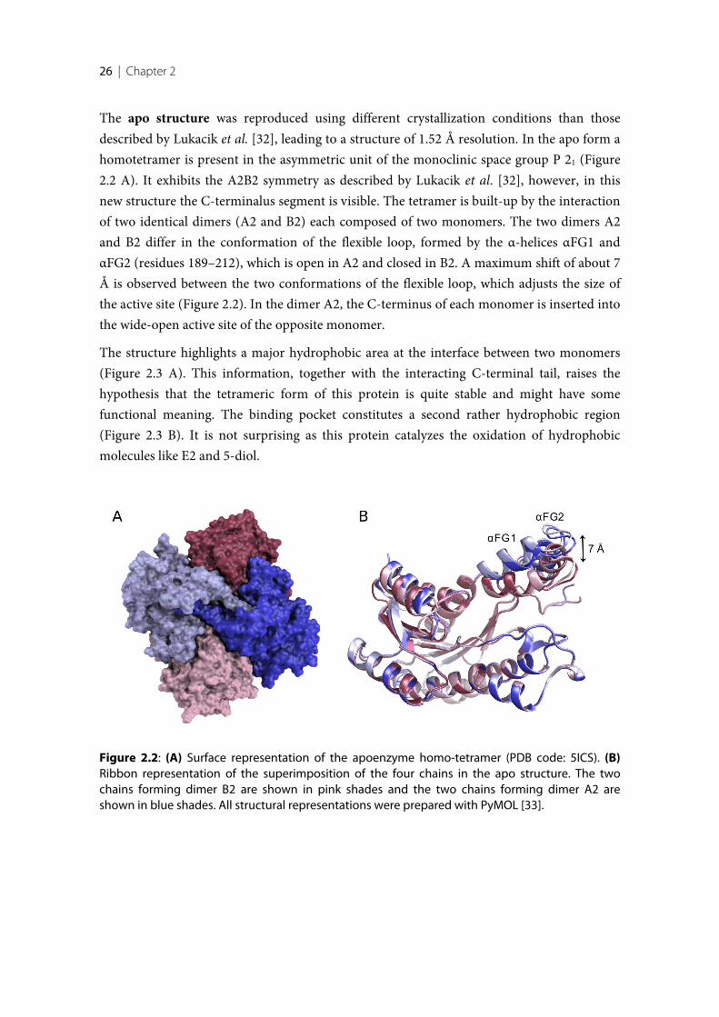
26 | Chapter 2
The apo structure was reproduced using different crystallization conditions than thosedescribed by Lukacik et al. [32], leading to a structure of 1.52 Å resolution. In the apo form ahomotetramer is present in the asymmetric unit of the monoclinic space group P 21 (Figure2.2 A). It exhibits the A2B2 symmetry as described by Lukacik et al. [32], however, in thisnew structure the C-terminalus segment is visible. The tetramer is built-up by the interactionof two identical dimers (A2 and B2) each composed of two monomers. The two dimers A2and B2 differ in the conformation of the flexible loop, formed by the α-helices αFG1 andαFG2 (residues 189–212), which is open in A2 and closed in B2. A maximum shift of about 7Å is observed between the two conformations of the flexible loop, which adjusts the size ofthe active site (Figure 2.2). In the dimer A2, the C-terminus of each monomer is inserted intothe wide-open active site of the opposite monomer.
The structure highlights a major hydrophobic area at the interface between two monomers(Figure 2.3 A). This information, together with the interacting C-terminal tail, raises thehypothesis that the tetrameric form of this protein is quite stable and might have somefunctional meaning. The binding pocket constitutes a second rather hydrophobic region(Figure 2.3 B). It is not surprising as this protein catalyzes the oxidation of hydrophobicmolecules like E2 and 5-diol.
Figure 2.2: (A) Surface representation of the apoenzyme homo-tetramer (PDB code: 5ICS). (B)Ribbon representation of the superimposition of the four chains in the apo structure. The twochains forming dimer B2 are shown in pink shades and the two chains forming dimer A2 areshown in blue shades. All structural representations were prepared with PyMOL [33].
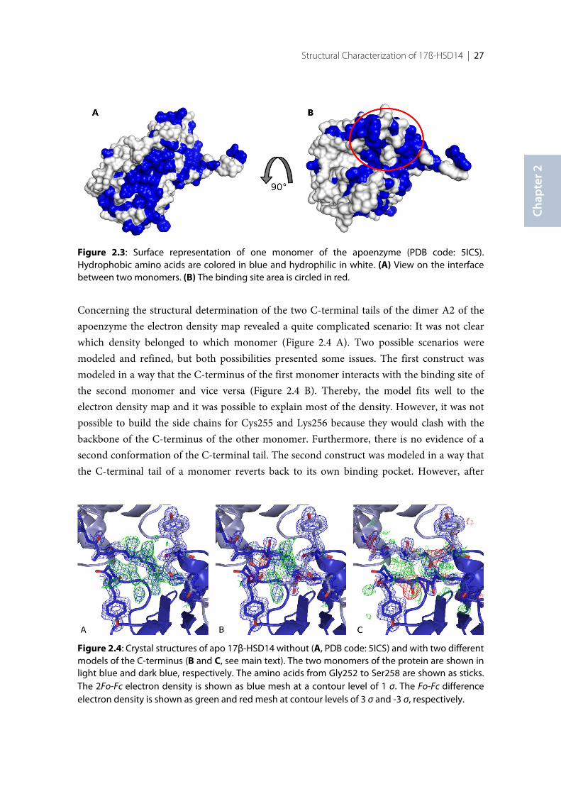
Structural Characterization of 17ß-HSD14 | 27
Chap
ter 2
Concerning the structural determination of the two C-terminal tails of the dimer A2 of theapoenzyme the electron density map revealed a quite complicated scenario: It was not clearwhich density belonged to which monomer (Figure 2.4 A). Two possible scenarios weremodeled and refined, but both possibilities presented some issues. The first construct wasmodeled in a way that the C-terminus of the first monomer interacts with the binding site ofthe second monomer and vice versa (Figure 2.4 B). Thereby, the model fits well to theelectron density map and it was possible to explain most of the density. However, it was notpossible to build the side chains for Cys255 and Lys256 because they would clash with thebackbone of the C-terminus of the other monomer. Furthermore, there is no evidence of asecond conformation of the C-terminal tail. The second construct was modeled in a way thatthe C-terminal tail of a monomer reverts back to its own binding pocket. However, after
Figure 2.3: Surface representation of one monomer of the apoenzyme (PDB code: 5ICS).Hydrophobic amino acids are colored in blue and hydrophilic in white. (A) View on the interfacebetween two monomers. (B) The binding site area is circled in red.
Figure 2.4: Crystal structures of apo 17β-HSD14 without (A, PDB code: 5ICS) and with two differentmodels of the C-terminus (B and C, see main text). The two monomers of the protein are shown inlight blue and dark blue, respectively. The amino acids from Gly252 to Ser258 are shown as sticks.The 2Fo-Fc electron density is shown as blue mesh at a contour level of 1 σ. The Fo-Fc differenceelectron density is shown as green and red mesh at contour levels of 3 σ and -3 σ, respectively.
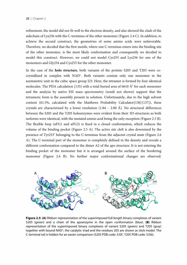
28 | Chapter 2
refinement, the model did not fit well to the electron density, and also showed the clash of thesidechain of Lys256 with the C-terminus of the other monomer (Figure 2.4 C). In addition, toachieve the second construct, the geometries of some amino acids were unfavorable.Therefore, we decided that the first motife, where one C-terminus enters into the binding siteof the other monomer, is the most likely conformation and consequently we decided tomodel this construct. However, we could not model Cys255 and Lys256 for one of themonomers and Gly254 and Cys255 for the other monomer.
In the case of the holo structure, both variants of the protein S205 and T205 were co-crystallized in complex with NAD+. Both variants contain only one monomer in theasymmetric unit in the cubic space group I23. Here, the tetramer is formed by four identicalmolecules. The PISA calculation [135] with a total buried area of 6610 Å2 for each monomerand the analysis by native ESI mass spectrometry (result not shown) support that thetetrameric form is the assembly present in solution. Unfortunately, due to the high solventcontent (61.5%, calculated with the Matthews Probability Calculator[136],[137]), thesecrystals are characterized by a lower resolution (1.84 – 2.00 Å). No structural differencesbetween the S205 and the T205 holoenzymes were evident from their 3D-structures as bothisoforms were identical, with the mutated amino acid being the only exception (Figure 2.5 B).The flexible loop (αFG1 and αFG2) is fixed in a closed conformation, which reduces thevolume of the binding pocket (Figure 2.5 A). The active site cleft is also downsized by thepresence of Tyr253’ belonging to the C-terminus from the adjacent crystal mate (Figure 2.6A). The C-terminal part of the monomer is completely defined in the density and reveals adifferent conformation compared to the dimer A2 of the apo structure: It is not entering thebinding pocket of the monomer but it is arranged around the surface of the borderingmonomer (Figure 2.6 B). No further major conformational changes are observed.
Figure 2.5: (A) Ribbon representation of the superimposed full length binary complexes of variantS205 (green) and a chain of the apoenzyme in the open conformation (blue). (B) Ribbonrepresentation of the superimposed binary complexes of variant S205 (green) and T205 (gray)together with bound NAD+; the catalytic triad and the residues 205 are shown as stick model. TheC-terminal tail is hidden for an easier comparison (S205 PDB code: 5JSF, T205 PDB code: 5JS6).
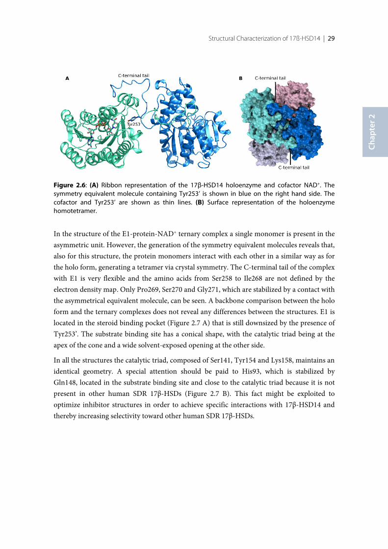
Structural Characterization of 17ß-HSD14 | 29
Chap
ter 2
In the structure of the E1-protein-NAD+ ternary complex a single monomer is present in theasymmetric unit. However, the generation of the symmetry equivalent molecules reveals that,also for this structure, the protein monomers interact with each other in a similar way as forthe holo form, generating a tetramer via crystal symmetry. The C-terminal tail of the complexwith E1 is very flexible and the amino acids from Ser258 to Ile268 are not defined by theelectron density map. Only Pro269, Ser270 and Gly271, which are stabilized by a contact withthe asymmetrical equivalent molecule, can be seen. A backbone comparison between the holoform and the ternary complexes does not reveal any differences between the structures. E1 islocated in the steroid binding pocket (Figure 2.7 A) that is still downsized by the presence ofTyr253’. The substrate binding site has a conical shape, with the catalytic triad being at theapex of the cone and a wide solvent-exposed opening at the other side.
In all the structures the catalytic triad, composed of Ser141, Tyr154 and Lys158, maintains anidentical geometry. A special attention should be paid to His93, which is stabilized byGln148, located in the substrate binding site and close to the catalytic triad because it is notpresent in other human SDR 17β-HSDs (Figure 2.7 B). This fact might be exploited tooptimize inhibitor structures in order to achieve specific interactions with 17β-HSD14 andthereby increasing selectivity toward other human SDR 17β-HSDs.
Figure 2.6: (A) Ribbon representation of the 17β-HSD14 holoenzyme and cofactor NAD+. Thesymmetry equivalent molecule containing Tyr253’ is shown in blue on the right hand side. Thecofactor and Tyr253’ are shown as thin lines. (B) Surface representation of the holoenzymehomotetramer.
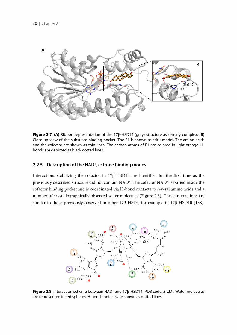
30 | Chapter 2
Description of the NAD+, estrone binding modes
Interactions stabilizing the cofactor in 17β-HSD14 are identified for the first time as thepreviously described structure did not contain NAD+. The cofactor NAD+ is buried inside thecofactor binding pocket and is coordinated via H-bond contacts to several amino acids and anumber of crystallographically observed water molecules (Figure 2.8). These interactions aresimilar to those previously observed in other 17β-HSDs, for example in 17β-HSD10 [138].
Figure 2.7: (A) Ribbon representation of the 17β-HSD14 (gray) structure as ternary complex. (B)Close-up view of the substrate binding pocket. The E1 is shown as stick model. The amino acidsand the cofactor are shown as thin lines. The carbon atoms of E1 are colored in light orange. H-bonds are depicted as black dotted lines.
Figure 2.8: Interaction scheme between NAD+ and 17β-HSD14 (PDB code: 5ICM). Water moleculesare represented in red spheres. H-bond contacts are shown as dotted lines.
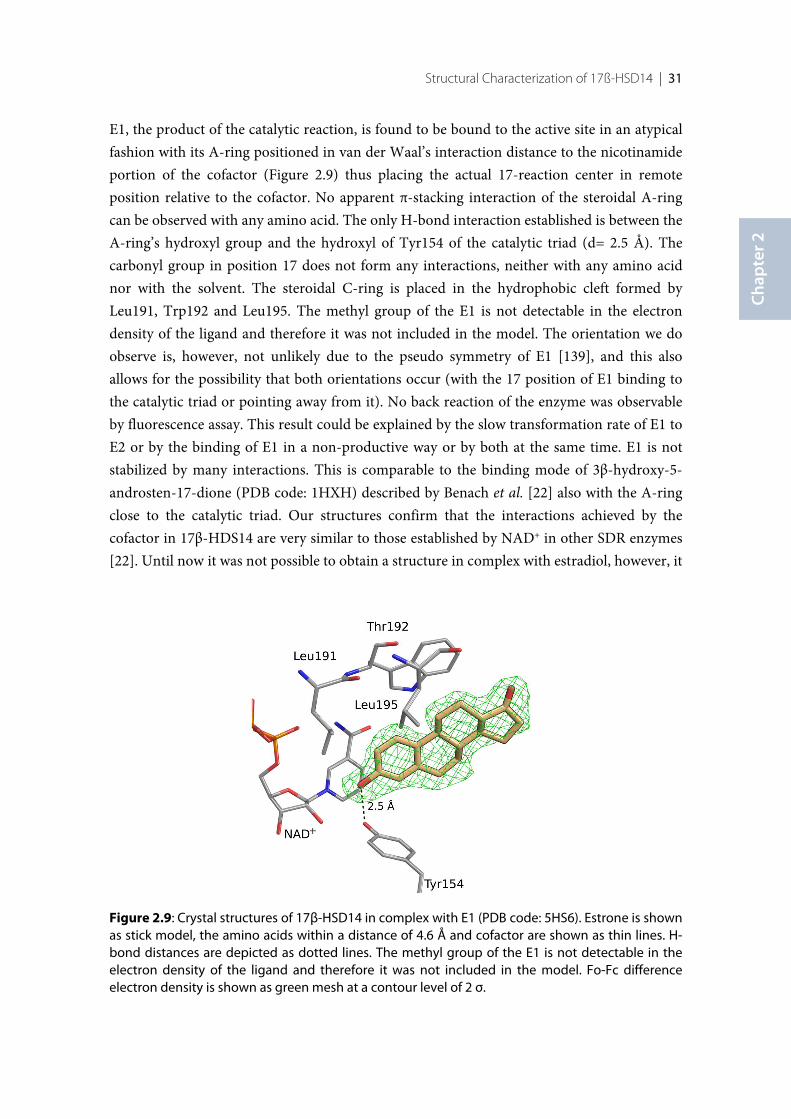
Structural Characterization of 17ß-HSD14 | 31
Chap
ter 2
E1, the product of the catalytic reaction, is found to be bound to the active site in an atypicalfashion with its A-ring positioned in van der Waal’s interaction distance to the nicotinamideportion of the cofactor (Figure 2.9) thus placing the actual 17-reaction center in remoteposition relative to the cofactor. No apparent π-stacking interaction of the steroidal A-ringcan be observed with any amino acid. The only H-bond interaction established is between theA-ring’s hydroxyl group and the hydroxyl of Tyr154 of the catalytic triad (d= 2.5 Å). Thecarbonyl group in position 17 does not form any interactions, neither with any amino acidnor with the solvent. The steroidal C-ring is placed in the hydrophobic cleft formed byLeu191, Trp192 and Leu195. The methyl group of the E1 is not detectable in the electrondensity of the ligand and therefore it was not included in the model. The orientation we doobserve is, however, not unlikely due to the pseudo symmetry of E1 [139], and this alsoallows for the possibility that both orientations occur (with the 17 position of E1 binding tothe catalytic triad or pointing away from it). No back reaction of the enzyme was observableby fluorescence assay. This result could be explained by the slow transformation rate of E1 toE2 or by the binding of E1 in a non-productive way or by both at the same time. E1 is notstabilized by many interactions. This is comparable to the binding mode of 3β-hydroxy-5-androsten-17-dione (PDB code: 1HXH) described by Benach et al. [22] also with the A-ringclose to the catalytic triad. Our structures confirm that the interactions achieved by thecofactor in 17β-HDS14 are very similar to those established by NAD+ in other SDR enzymes[22]. Until now it was not possible to obtain a structure in complex with estradiol, however, it
Figure 2.9: Crystal structures of 17β-HSD14 in complex with E1 (PDB code: 5HS6). Estrone is shownas stick model, the amino acids within a distance of 4.6 Å and cofactor are shown as thin lines. H-bond distances are depicted as dotted lines. The methyl group of the E1 is not detectable in theelectron density of the ligand and therefore it was not included in the model. Fo-Fc differenceelectron density is shown as green mesh at a contour level of 2 σ.

32 | Chapter 2
is predictable that E2, like E1, may not bind so tight in the active site. Together with the weakbinding affinities and low-turnovers measured for E2 and 5-diol, it is questionable if thepreferred substrates of this enzyme are steroids.
2.3 Conclusion
In this work, we have elucidated the first crystal structures of 17β-HSD14 in complex withligands. The structure of the binary complex with the cofactor NAD+ differs from theapoenzyme with respect to the flexible loop (αFG1 and αFG2 segment) which adopts aunique closed conformation in the presence of NAD+: It is found in either open (A2 dimer)or closed (B2 dimer) conformation without cofactor. His93 and Gln148, that stabilized eachother in the substrate binding site close to the catalytic triad, are not present in other human17β-HSDs and might be involved to achieve specific interactions with 17β-HSD14. Bindingof the cofactor appears to induce a shift of the flexible loop, which reduces the size of theactive site and thereby produces the active conformation of the enzyme. The overall structureand the active site geometry in the ternary complexes with E1 is very similar to those of thebinary complex, with the flexible loop in the same closed state delimiting an elongated andconical active site. However, further studies are still necessary to investigate the endogenoussubstrate and the physiological role of the enzyme in vivo.
2.4 Experimental section
Site directed mutagenesis
The plasmid p11-HSD17B14 coding for the S205 variant of human 17β-HSD14 (sequenceaccording to NCBI data bank entry Q9BPX1) was obtained from Udo Oppermann,Structural Genomics Consortium (SGC) Oxford.
The point mutation in order to gain the T205 variant was introduced into the plasmid byusing the QuikChange Lightning Site-Directed Mutagenesis Kit (Agilent Technologies) withthe forward primer 5’-CGGACCCGCGTGCGACCATTCGTGAAGGTATGC-3’ and thereverse primer 5’-GCATACCTTCACGAATGGTCGCACGCGGGTCCG-3’ and followingthe manufacturer’s instructions. Sanger sequencing confirmed the correct mutation.
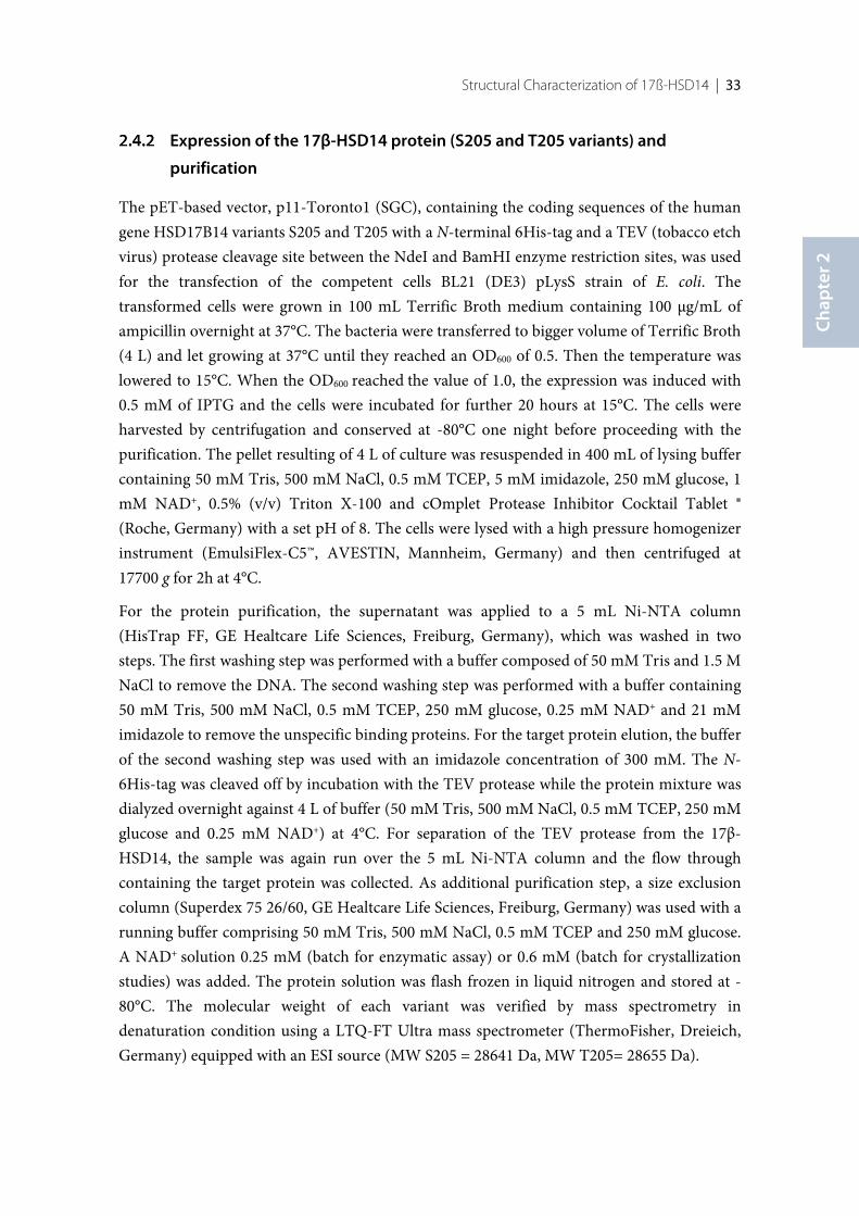
Structural Characterization of 17ß-HSD14 | 33
Chap
ter 2
Expression of the 17β-HSD14 protein (S205 and T205 variants) and
purification
The pET-based vector, p11-Toronto1 (SGC), containing the coding sequences of the humangene HSD17B14 variants S205 and T205 with a N-terminal 6His-tag and a TEV (tobacco etchvirus) protease cleavage site between the NdeI and BamHI enzyme restriction sites, was usedfor the transfection of the competent cells BL21 (DE3) pLysS strain of E. coli. Thetransformed cells were grown in 100 mL Terrific Broth medium containing 100 µg/mL ofampicillin overnight at 37°C. The bacteria were transferred to bigger volume of Terrific Broth(4 L) and let growing at 37°C until they reached an OD600 of 0.5. Then the temperature waslowered to 15°C. When the OD600 reached the value of 1.0, the expression was induced with0.5 mM of IPTG and the cells were incubated for further 20 hours at 15°C. The cells wereharvested by centrifugation and conserved at -80°C one night before proceeding with thepurification. The pellet resulting of 4 L of culture was resuspended in 400 mL of lysing buffercontaining 50 mM Tris, 500 mM NaCl, 0.5 mM TCEP, 5 mM imidazole, 250 mM glucose, 1mM NAD+, 0.5% (v/v) Triton X-100 and cOmplet Protease Inhibitor Cocktail Tablet ®(Roche, Germany) with a set pH of 8. The cells were lysed with a high pressure homogenizerinstrument (EmulsiFlex-C5™, AVESTIN, Mannheim, Germany) and then centrifuged at17700 g for 2h at 4°C.
For the protein purification, the supernatant was applied to a 5 mL Ni-NTA column(HisTrap FF, GE Healtcare Life Sciences, Freiburg, Germany), which was washed in twosteps. The first washing step was performed with a buffer composed of 50 mM Tris and 1.5 MNaCl to remove the DNA. The second washing step was performed with a buffer containing50 mM Tris, 500 mM NaCl, 0.5 mM TCEP, 250 mM glucose, 0.25 mM NAD+ and 21 mMimidazole to remove the unspecific binding proteins. For the target protein elution, the bufferof the second washing step was used with an imidazole concentration of 300 mM. The N-6His-tag was cleaved off by incubation with the TEV protease while the protein mixture wasdialyzed overnight against 4 L of buffer (50 mM Tris, 500 mM NaCl, 0.5 mM TCEP, 250 mMglucose and 0.25 mM NAD+) at 4°C. For separation of the TEV protease from the 17β-HSD14, the sample was again run over the 5 mL Ni-NTA column and the flow throughcontaining the target protein was collected. As additional purification step, a size exclusioncolumn (Superdex 75 26/60, GE Healtcare Life Sciences, Freiburg, Germany) was used with arunning buffer comprising 50 mM Tris, 500 mM NaCl, 0.5 mM TCEP and 250 mM glucose.A NAD+ solution 0.25 mM (batch for enzymatic assay) or 0.6 mM (batch for crystallizationstudies) was added. The protein solution was flash frozen in liquid nitrogen and stored at -80°C. The molecular weight of each variant was verified by mass spectrometry indenaturation condition using a LTQ-FT Ultra mass spectrometer (ThermoFisher, Dreieich,Germany) equipped with an ESI source (MW S205 = 28641 Da, MW T205= 28655 Da).
34 | Chapter 2
Thermal shift Assay (TSA)
The thermal shift assay was conducted in a 96 well PCR plate (Bio-Rad, Munich, Germany)using a real-time PCR instrument (iCycler5, Bio-Rad, Munich, Germany). The runningbuffer was obtained by the dilution (1:1000) of SYPRO orange dye (Invitrogen, ThemoFisherLife Technologies, Darmstadt, Germany) in a buffer composed of 50 mM Tris, 0.5 M NaCland 0.5 mM TCEP, pH 8.0. For the protein stability study, the running buffer was mixed withdifferent additives (20% glycerol, 10% MPD, 10% PEG 400, 10% ethylene glycol, 250 mMglucose or 0.25 mM NAD+). For the protein complexes, a concentration of 2.5% of DMSO(reference measurement) or a stock solution in pure DMSO of compound 2, estrone, orestradiol was added to the running buffer containing 250 mM glucose and 0.25 mM NAD+
(final ligand concentration of 0.25 mM and final DMSO concentration of 2.5%). A proteinconcentration of 2.6 µM was used (3 µL) and the different solutions were added to a finalvolume of 40 µL.
Fluorimetric assay
The activity assay was based on measuring the fluorescence of the NADH formed. Theoxidation of E2 into E1 in presence of NAD+ and purified 17β-HSD14 was followed.Fluorescence spectra were recorded on a Tecan Saphir 2. NADH fluorescence emission wasmeasured at 496 nm following excitation at 340 nm. The slit width for excitation was 7 nmwhile that for emission was 15 nm. The assay was performed in 100 mM phosphate buffer atpH 8.0, mixed with estradiol in DMSO (32 µM) and NAD+ (1.5 mM) to reach a final DMSOconcentration of 0.05%. The enzymatic reaction was started by addition of the pure protein(1 mg/mL, 3.0 µM) and measured continuously for 15 min at 25°C. Reactions wereperformed in 200 µL volumes. The assay was run in 96 well-plates in duplicate in threeindependent experiments. A linear relationship between product formation and reactiontime was established. The slope of the reaction, representing the velocity of the NADHformation, was quantified. For each new batch of protein, a control experiment was runwithout E2 to assess the absence of activity in absence of substrate.
Determination of kinetic constants
For the determination of the kinetic parameters the substrate concentration was varied atleast between 12% and 250% of the estimated Km value. Kinetic constants were calculatedfrom the initial velocity data by direct fitting curves as Hanes Woolf plot.
Structural Characterization of 17ß-HSD14 | 35
Chap
ter 2
Protein crystallization
The activity of the protein was proven before use in each crystallization study. Thecrystallization of the apo form as well as the cocrystallization of the holoenzyme and of 17-HSD14 in complex with E1 were performed by sitting drop vapor diffusion technique.Initially, an automatized crystallization screening of 1248 conditions was performed(MarXtal, University of Marburg).
For the apo structure (S205), 0.1 µL of protein solution (10 mg/mL) were combined with anequal volume of mother liquor having the composition: 0.1 M sodium acetate, 30% (w/v)PEG 4000, 0.2 M ammonium acetate, 20% glycerol, pH 4.60. After growing for 5 weeks at18°C, crystals were harvested, exposed to the mother liquor solution with the addition of 20%glycerol for 30 seconds and finally flash frozen with liquid nitrogen.
The two isoforms (S205 and T205) of the holoenzyme were crystallized by combining 2 µL ofa solution containing protein (9.5 mg/mL) and 0.6 mM NAD+ with 2 µL of a buffercomposed of 0.1 M HEPES and 3.3 M sodium formate, pH 7.00. Over a time period of twoweeks at 18°C, the crystals were grown, harvested and flash frozen using a cryo buffer madeof mother liquor with 20% (w/v) PEG400.
A solution containing 0.1 M CHES, 1 M tri-sodium citrate, 0.4 mM E1 and 1% DMSO, pH9.5, was mixed with the protein solution (9.5 mg/mL, T205), containing 0.6 mM NAD+, in aratio 1:1 to obtain the protein-estrone complex. After growing for 4 weeks at 18°C, thecrystals were exposed to a cryo buffer for 3 sec composed of 0.1 M CHES, 1 M tri-sodiumcitrate, 1% DMSO, 20% (w/v) glucose, pH 9.5, containing 0.4 mM of estrone, andsubsequently flash frozen in liquid nitrogen.
Data collection and processing
The data collection of protein-estrone (PDB code: 5HS6) was achieved at Elettra beamlineXRD1 in Trieste, Italy at a wavelength of 1Å and a temperature of 100 K on a silicon Pilatus2M pixel detector. For the holo structure of both variants (S205 PDB code: 5JSF; T205 PDBcode: 5JS6), the data collection was performed at beamline ID23-1 of the EuropeanSynchrotron Radiation Facility (ESRF) [140] in Grenoble, France at a wavelength of 0.97242Å and a temperature of 100K on a silicon Pilatus 6M pixel detector. The diffraction data ofapo form dataset (PDB code: 5ICS) was collected on BL 14.1 operated by the Helmholtz-Zentrum Berlin (HZB) at the BESSY II electron storage ring (Berlin-Adlershof, Germany)[141] at a wavelength of 0.9184 Å and a temperature of 100 K on a silicon Pilatus 6M pixeldetector. All datasets were indexed, processed and scaled with XDS [142].
36 | Chapter 2
Structure determination and refinement
All structures were determined by molecular replacement with the program PHASER MR[143] from the CCP4 suite [132]. The structure 5EN4 was used as a search model. In therefinement, a subset corresponding to 5% of all reflections were omitted during refinementand used for the calculation of Rfree. Model building was achieved in COOT [144] andrefinement using PHENIX.refine version 1.10.1-2155 [145]. Ligands SMILE codes werecreated with Molinspiration v2014.11[146] and built with the Grade Web Server [147], whichwas also used for energetically minimization and restraint generation. Cartesian simulatedannealing, applying default parameters, was used as a first refinement step. Subsequently,refinement of XYZ coordinates, occupancies and individual B-factors were alternated withstructural adaption in COOT [144] until the model was readily built and gave the bestpossible explanation of the electron density. For the apo structure (PDB code: 5ICS), thetemperature factors of all atoms, except hydrogen atoms, were refined anisotropically withadditional NCS restraints. TLS refinement was performed for the protein-complex structures(estrone complex PDB code: 5HS6) and the both variants of the holoenzyme (S205 PDBcode: 5JSF, T205 PDB code: 5JS6) with appropriate TLS groups from the TLSMD webserver[148],[149]. The model of the apo- and holo structure a were refined with hydrogensautomatically added by PHENIX.refine.
37
First Structure-Activity Relationship of 17β-Hydroxysteroid
Dehydrogenase Type 14: Nonsteroidal Inhibitors and
Crystal Structures in Complex with the Enzyme
Introductory remarks
Parts of the following chapter have been published in the Journal of Medicinal Chemistry in2016. The preliminary inhibition assay was done by Dr. Gabriele Möller. Fluorescence-basedassays were designed and performed by Dr. Sandrine Marchais-Oberwinkler in collaborationwith Florian Braun. The synthesis of the compounds and their characterization was carriedout by Florian Braun. The expression and the purification of the 17β-HSD14, thecomputational modeling studies, the TSA assay and the crystallization studies and theelucidation of the first crystal structures of the protein-inhibitor complexes were establishedand performed by the author of this thesis. Furthermore, the author significantly contributedto the discussion and to the writing of the manuscript in collaboration with Florian Braunand Dr. Sandrine Marchais-Oberwinkler.
38 | Chapter 3
3.1 Introduction
Human 17β-hydroxysteroid dehydrogenase type 14 (17β-HSD14), also called DHRS10 andretSDR3, is an oxido-reductase belonging to the SDR (Short-chain Dehydrogenase-Reductase) family [11, 32]. In vitro, the enzyme oxidizes the hydroxyl group at position 17 ofestradiol (E2) and 5-androsten-3β,17β-diol (5-diol) in presence of the cofactor NAD+,however, in vivo, its natural substrate is still unknown.
While Sivik et al. [103] described a broad distribution pattern of 17β-HSD14 across varioustissues based on immunohistochemistry studies, Northern blotting experiments showed thatthe enzyme is predominantly expressed in the brain, liver and placenta [32] as well as in thekidney [100]. Furthermore, immunofluorescence studies revealed a cytosolic localization[32].
In order to understand the function of 17β-HSD14, the enzyme needs to be furthercharacterized. Inhibitors are useful chemical tools, which can be used not only to characterizethe binding site of an enzyme but also to get insight into the physiological role of the latterupon in vivo administration. No inhibitor has been reported for this enzyme.
The crystal structure of the human 17β-HSD14 has been determined recently as holoenzyme(PDB code: 5JS6 and 5JSF) and as ternary complex with estrone (E1) (PDB code: 5HS6). Insolution, 17β-HSD14 is a tetramer.[126] The binding cavity was shown to be rather lipophilicwith a conical shape. The substrate active site is narrow in the vicinity of the catalytic triadand is solvent exposed at the other end. Tyr253’ from the C-terminal chain of the adjacentunit in the tetramer reduces the size of the active site.
Up to now 14 different 17β-HSD subtypes have been reported [34]. 17β-HSD1 and 17β-HSD2, the two best characterized subtypes, predominantly catalyze the oxidation andreduction of estrogens and androgens. Inhibitors of these two enzymes have already beenreported [10, 52, 124, 150–160]. While 17β-HSD1 is a cytosolic enzyme and shows areductive activity in vivo (activation of estrogens), 17β-HSD2 is membrane-bound andcatalyzes the oxidation of estrogens and androgens to their less potent analogues similarly to17β-HSD14. However, 17β-HSD2 and 17β-HSD14 differ in their tissue distribution patternsince in contrary to 17β-HSD14, 17β-HSD2 is not present in the human brain temporallobe [161]. The presence of 17β-HSD14 in the brain might indicate that this enzyme isinvolved in the regulation of active estrogens and androgens in this organ [32].
The goal of this work was to identify the first 17β-HSD14 nonsteroidal inhibitors and tooptimize their structures, which led to highly active compounds. The inhibitor optimizationwas performed following a ligand-based approach. The synthesis and biological evaluation ofhighly active compounds with a nonsteroidal scaffold together with five new crystalstructures of the ternary complexes are reported. Analysis of the crystal structures of the
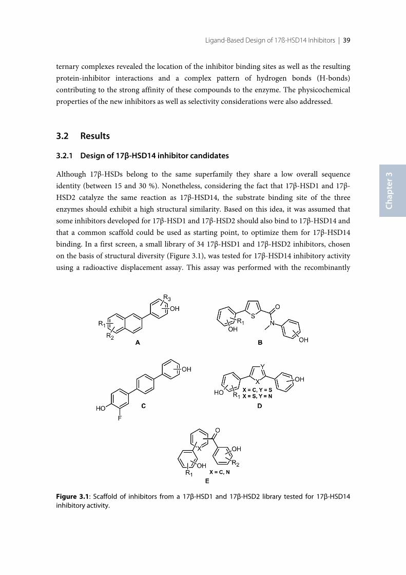
Ligand-Based Design of 17ß-HSD14 Inhibitors | 39
Chap
ter 3
ternary complexes revealed the location of the inhibitor binding sites as well as the resultingprotein-inhibitor interactions and a complex pattern of hydrogen bonds (H-bonds)contributing to the strong affinity of these compounds to the enzyme. The physicochemicalproperties of the new inhibitors as well as selectivity considerations were also addressed.
3.2 Results
Design of 17β-HSD14 inhibitor candidates
Although 17β-HSDs belong to the same superfamily they share a low overall sequenceidentity (between 15 and 30 %). Nonetheless, considering the fact that 17β-HSD1 and 17β-HSD2 catalyze the same reaction as 17β-HSD14, the substrate binding site of the threeenzymes should exhibit a high structural similarity. Based on this idea, it was assumed thatsome inhibitors developed for 17β-HSD1 and 17β-HSD2 should also bind to 17β-HSD14 andthat a common scaffold could be used as starting point, to optimize them for 17β-HSD14binding. In a first screen, a small library of 34 17β-HSD1 and 17β-HSD2 inhibitors, chosenon the basis of structural diversity (Figure 3.1), was tested for 17β-HSD14 inhibitory activityusing a radioactive displacement assay. This assay was performed with the recombinantly
Figure 3.1: Scaffold of inhibitors from a 17β-HSD1 and 17β-HSD2 library tested for 17β-HSD14inhibitory activity.
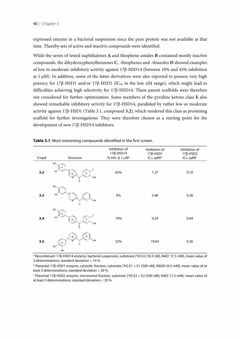
40 | Chapter 3
expressed enzyme in a bacterial suspension since the pure protein was not available at thattime. Thereby sets of active and inactive compounds were identified.
While the series of tested naphthalenes A and thiophene amides B contained mostly inactivecompounds, the dihydroxyphenylbenzenes C, -thiophenes and -thiazoles D showed examplesof low to moderate inhibitory activity against 17β-HSD14 (between 10% and 45% inhibitionat 1 µM). In addition, some of the latter derivatives were also reported to possess very highpotency for 17β-HSD1 and/or 17β-HSD2 (IC50 in the low nM range), which might lead todifficulties achieving high selectivity for 17β-HSD14. These parent scaffolds were thereforenot considered for further optimization. Some members of the pyridine ketone class E alsoshowed remarkable inhibitory activity for 17β-HSD14, paralleled by rather low or moderateactivity against 17β-HSD1 (Table 3.1, compound 3.2), which rendered this class as promisingscaffold for further investigations. They were therefore chosen as a starting point for thedevelopment of new 17β-HSD14 inhibitors.
Table 3.1: Most interesting compounds identified in the first screen.
Cmpd Structure
Inhibition of17β-HSD14
% Inh. @ 1 mMa
Inhibition of17β-HSD1IC50 (µM)b
Inhibition of17β-HSD2IC50 (µM)c
3.2 62% 1.27 0.10
3.3 0% 5.48 0.26
3.4 19% 0.29 0.04
3.5 32% 19.65 0.26
a Recombinant 17β-HSD14 enzyme, bacterial suspension, substrate [3H]-E2 [18.3 nM], NAD+ [7.5 mM], mean value of3 determinations; standard deviation < 10 %.b Placental 17β-HSD1 enzyme, cytosolic fraction, substrate [3H]-E1 + E1 [500 nM], NADH [0.5 mM], mean value of atleast 3 determinations; standard deviation < 20 %.c Placental 17β-HSD2 enzyme, microsomal fraction, substrate [3H]-E2 + E2 [500 nM], NAD+ [1.5 mM], mean value ofat least 3 determinations; standard deviation < 20 %
Ligand-Based Design of 17ß-HSD14 Inhibitors | 41
Chap
ter 3
The most interesting hits identified in the preliminary screen are listed in Table 3.1. Thecompounds can be categorized into 2,5- (3.2, 3.3) and 2,6-substituted (3.5) pyridine ketonesand the 1,4-substituted phenyl (3.4). In the 2,5-compound class, 3.2 and 3.3 (62% inhibitionat 1 µM vs 0%) differ by the presence/absence of a fluorine atom in ortho position to the OHgroup of the C-ring, suggesting the importance of this atom for inhibitory activity.Furthermore, comparison of 3.2 with 3.4 (62% inhibition at 1 µM vs 19%) shows that thepyridine core B is more potent than the phenyl analog, pointing toward the importance of thenitrogen atom in the B-ring for activity. Concerning the 2,6-compound class, comparison of3.5 with 2,5-substituted 3.3 (32% inhibition at 1 µM vs 0%) shows that moving the A-ringfrom 5- to 6-position leads to a gain in activity.
Furthermore, considering the selectivity aspect, the poor inhibitory activity of the 2,6-substituted 3.5 toward 17β-HSD1 (IC50= 19.65 µM), compared to the 2,5-substituted analog3.3 (IC50= 5.48 µM) suggests that the 2,6-substitution pattern might improve selectivity for17β-HSD14 toward 17β-HSD1. Selectivity against 17β-HSD2 does not become obvious withthe set of studied test compounds. Consequently, the 2,6-pyridine ketone class was selectedfor optimization and the derivatives, with modification at the A-ring as well as at the C-ringby substituents with different properties. Overall, 35 compounds were synthesized and theirsynthesis pathways are described in the thesis of Florian Braun. The obtained compounds arelisted in Table 3.2. Special attention was paid to the physicochemical properties of thedesigned compounds in order to focus on compounds which should have a promisingbioavailability profile according to the Veber rules [162] and the Lipinski rule of 5 [163].
Calculation of physicochemical parameters
For each synthesized compound (Table 3.2), the molecular weight (MW) was calculated to bein the range of 300 to 400 g/mol, aside from the trisubstituted compounds 3.31-3.33 with aslightly higher MW. The clogP was calculated in silico (using Molinspiration [146]) andturned out to be below 5 except for 3.31-3.33. The total polar surface area (tPSA), thenumber of rotational bonds as well as the number of H-bond donors and acceptors fulfill theVeber rules [162] and the Lipinski rule of 5 [163]. In addition, considering the potential roleof the enzyme in the brain, the capability of the inhibitors to cross the blood–brain barrier(BBB) should also be taken into account. The physicochemical properties to be met bycompounds showing a good BBB penetration are described by Pajouhesh and Lenz [164].These criteria are matched for most of the synthesized compounds especially for 3.17 and3.28 as examples. The solubility range of most of the compounds was also determined bymixing several concentrations of the studied inhibitors in 100 mM phosphate buffer at pH7.4 and analyzing its precipitation status at different time points (0, 1, 2 and 24 h). The pKa
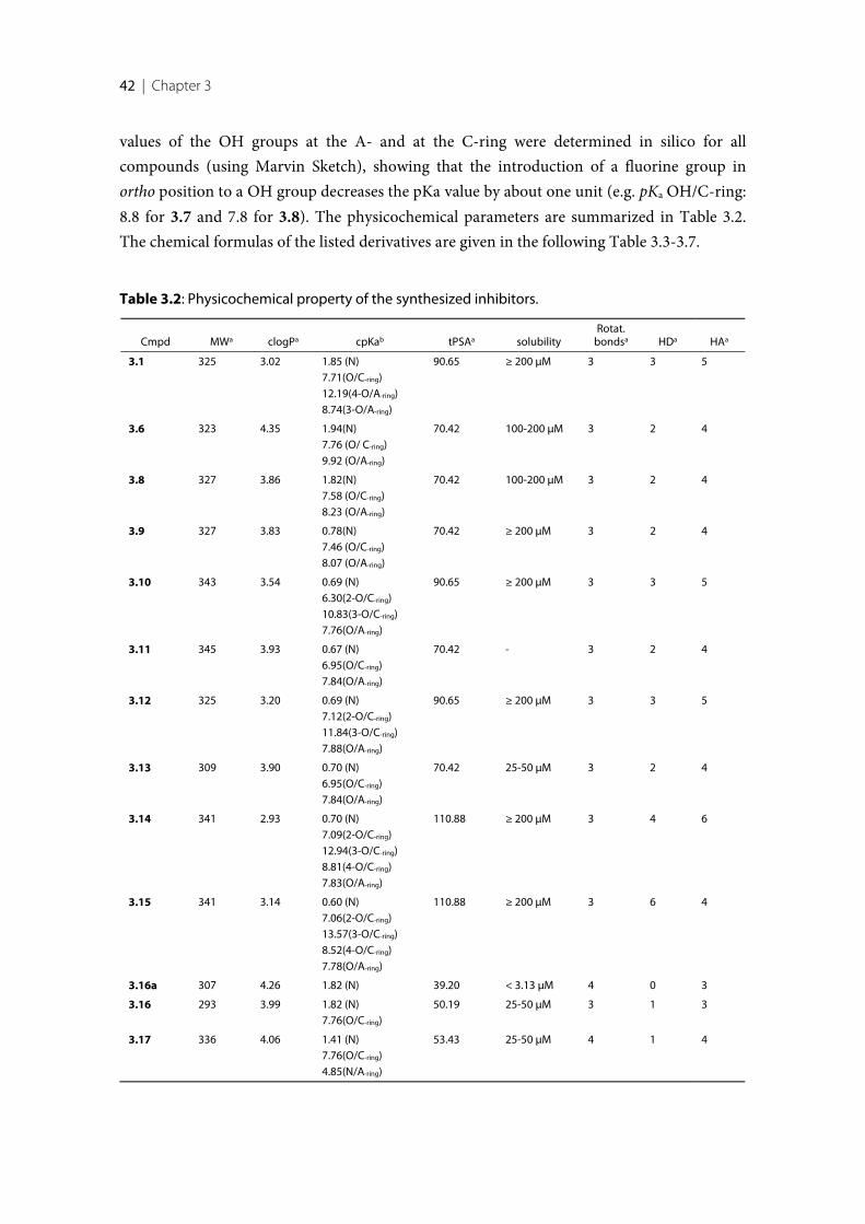
42 | Chapter 3
values of the OH groups at the A- and at the C-ring were determined in silico for allcompounds (using Marvin Sketch), showing that the introduction of a fluorine group inortho position to a OH group decreases the pKa value by about one unit (e.g. pKa OH/C-ring:8.8 for 3.7 and 7.8 for 3.8). The physicochemical parameters are summarized in Table 3.2.The chemical formulas of the listed derivatives are given in the following Table 3.3-3.7.
Table 3.2: Physicochemical property of the synthesized inhibitors.
Cmpd MWa clogPa cpKab tPSAa solubilityRotat.
bondsa HDa HAa
3.1 325 3.02 1.85 (N)7.71(O/C-ring)12.19(4-O/A-ring)8.74(3-O/A-ring)
90.65 ≥ 200 µM 3 3 5
3.6 323 4.35 1.94(N)7.76 (O/ C-ring)9.92 (O/A-ring)
70.42 100-200 µM 3 2 4
3.8 327 3.86 1.82(N)7.58 (O/C-ring)8.23 (O/A-ring)
70.42 100-200 µM 3 2 4
3.9 327 3.83 0.78(N)7.46 (O/C-ring)8.07 (O/A-ring)
70.42 ≥ 200 µM 3 2 4
3.10 343 3.54 0.69 (N)6.30(2-O/C-ring)10.83(3-O/C-ring)7.76(O/A-ring)
90.65 ≥ 200 µM 3 3 5
3.11 345 3.93 0.67 (N)6.95(O/C-ring)7.84(O/A-ring)
70.42 - 3 2 4
3.12 325 3.20 0.69 (N)7.12(2-O/C-ring)11.84(3-O/C-ring)7.88(O/A-ring)
90.65 ≥ 200 µM 3 3 5
3.13 309 3.90 0.70 (N)6.95(O/C-ring)7.84(O/A-ring)
70.42 25-50 µM 3 2 4
3.14 341 2.93 0.70 (N)7.09(2-O/C-ring)12.94(3-O/C-ring)8.81(4-O/C-ring)7.83(O/A-ring)
110.88 ≥ 200 µM 3 4 6
3.15 341 3.14 0.60 (N)7.06(2-O/C-ring)13.57(3-O/C-ring)8.52(4-O/C-ring)7.78(O/A-ring)
110.88 ≥ 200 µM 3 6 4
3.16a 307 4.26 1.82 (N) 39.20 < 3.13 µM 4 0 3
3.16 293 3.99 1.82 (N)7.76(O/C-ring)
50.19 25-50 µM 3 1 3
3.17 336 4.06 1.41 (N)7.76(O/C-ring)4.85(N/A-ring)
53.43 25-50 µM 4 1 4
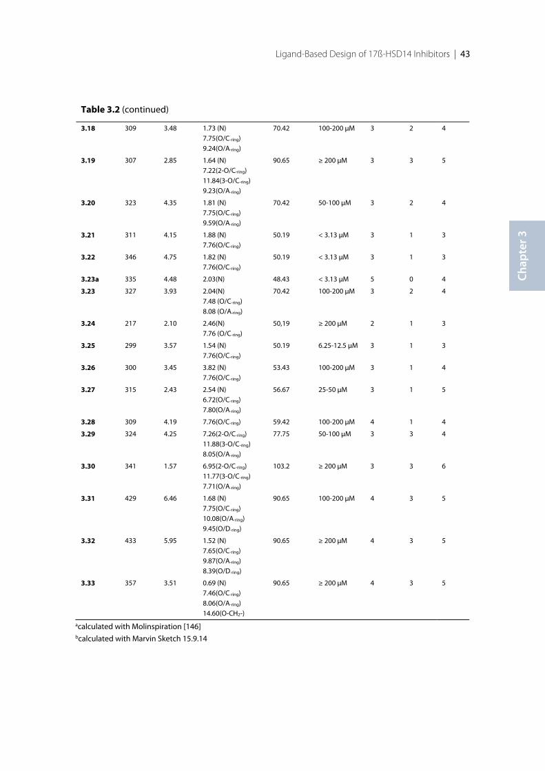
Ligand-Based Design of 17ß-HSD14 Inhibitors | 43
Chap
ter 3
Table 3.2 (continued)
3.18 309 3.48 1.73 (N)7.75(O/C-ring)9.24(O/A-ring)
70.42 100-200 µM 3 2 4
3.19 307 2.85 1.64 (N)7.22(2-O/C-ring)11.84(3-O/C-ring)9.23(O/A-ring)
90.65 ≥ 200 µM 3 3 5
3.20 323 4.35 1.81 (N)7.75(O/C-ring)9.59(O/A-ring)
70.42 50-100 µM 3 2 4
3.21 311 4.15 1.88 (N)7.76(O/C-ring)
50.19 < 3.13 µM 3 1 3
3.22 346 4.75 1.82 (N)7.76(O/C-ring)
50.19 < 3.13 µM 3 1 3
3.23a 335 4.48 2.03(N) 48.43 < 3.13 µM 5 0 4
3.23 327 3.93 2.04(N)7.48 (O/C-ring)8.08 (O/A-ring)
70.42 100-200 µM 3 2 4
3.24 217 2.10 2.46(N)7.76 (O/C-ring)
50,19 ≥ 200 µM 2 1 3
3.25 299 3.57 1.54 (N)7.76(O/C-ring)
50.19 6.25-12.5 µM 3 1 3
3.26 300 3.45 3.82 (N)7.76(O/C-ring)
53.43 100-200 µM 3 1 4
3.27 315 2.43 2.54 (N)6.72(O/C-ring)7.80(O/A-ring)
56.67 25-50 µM 3 1 5
3.28 309 4.19 7.76(O/C-ring) 59.42 100-200 µM 4 1 4
3.29 324 4.25 7.26(2-O/C-ring)11.88(3-O/C-ring)8.05(O/A-ring)
77.75 50-100 µM 3 3 4
3.30 341 1.57 6.95(2-O/C-ring)11.77(3-O/C-ring)7.71(O/A-ring)
103.2 ≥ 200 µM 3 3 6
3.31 429 6.46 1.68 (N)7.75(O/C-ring)10.08(O/A-ring)9.45(O/D-ring)
90.65 100-200 µM 4 3 5
3.32 433 5.95 1.52 (N)7.65(O/C-ring)9.87(O/A-ring)8.39(O/D-ring)
90.65 ≥ 200 µM 4 3 5
3.33 357 3.51 0.69 (N)7.46(O/C-ring)8.06(O/A-ring)14.60(O-CH2-)
90.65 ≥ 200 µM 4 3 5
acalculated with Molinspiration [146]bcalculated with Marvin Sketch 15.9.14
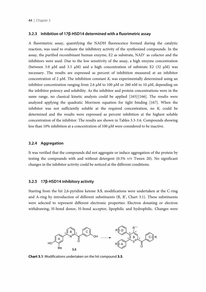
44 | Chapter 3
Inhibition of 17β-HSD14 determined with a fluorimetric assay
A fluorimetric assay, quantifying the NADH fluorescence formed during the catalyticreaction, was used to evaluate the inhibitory activity of the synthesized compounds. In theassay, the purified recombinant human enzyme, E2 as substrate, NAD+ as cofactor and theinhibitors were used. Due to the low sensitivity of the assay, a high enzyme concentration(between 3.0 µM and 3.5 µM) and a high concentration of substrate E2 (32 µM) wasnecessary. The results are expressed as percent of inhibition measured at an inhibitorconcentration of 2 µM. The inhibition constant Ki was experimentally determined using aninhibitor concentration ranging from 2.6 µM to 100 µM or 260 nM to 10 µM, depending onthe inhibitor potency and solubility. As the inhibitor and protein concentrations were in thesame range, no classical kinetic analysis could be applied [165],[166]. The results wereanalyzed applying the quadratic Morrison equation for tight binding [167]. When theinhibitor was not sufficiently soluble at the required concentration, no Ki could bedetermined and the results were expressed as percent inhibition at the highest solubleconcentration of the inhibitor. The results are shown in Tables 3.3-3.6. Compounds showingless than 10% inhibition at a concentration of 100 µM were considered to be inactive.
Aggregation
It was verified that the compounds did not aggregate or induce aggregation of the protein bytesting the compounds with and without detergent (0.5% v/v Tween 20). No significantchanges in the inhibitor activity could be noticed at the different conditions.
17β-HSD14 inhibitory activity
Starting from the hit 2,6-pyridine ketone 3.5, modifications were undertaken at the C-ringand A-ring by introduction of different substituents (R, R’, Chart 3.1). These substituentswere selected to represent different electronic properties: Electron donating or electronwithdrawing, H-bond donor, H-bond acceptor, lipophilic and hydrophilic. Changes were
Chart 3.1: Modifications undertaken on the hit compound 3.5.
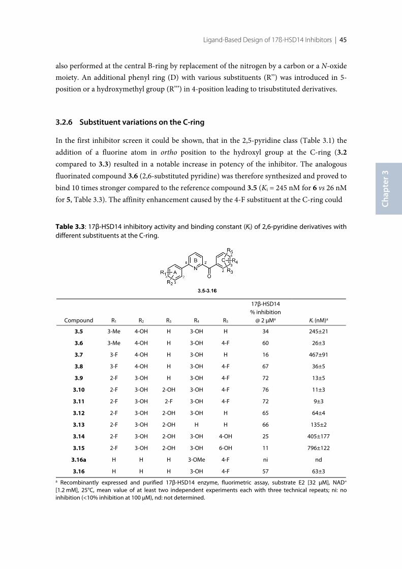
Ligand-Based Design of 17ß-HSD14 Inhibitors | 45
Chap
ter 3
also performed at the central B-ring by replacement of the nitrogen by a carbon or a N-oxidemoiety. An additional phenyl ring (D) with various substituents (R’’) was introduced in 5-position or a hydroxymethyl group (R’’’) in 4-position leading to trisubstituted derivatives.
Substituent variations on the C-ring
In the first inhibitor screen it could be shown, that in the 2,5-pyridine class (Table 3.1) theaddition of a fluorine atom in ortho position to the hydroxyl group at the C-ring (3.2compared to 3.3) resulted in a notable increase in potency of the inhibitor. The analogousfluorinated compound 3.6 (2,6-substituted pyridine) was therefore synthesized and proved tobind 10 times stronger compared to the reference compound 3.5 (Ki = 245 nM for 6 vs 26 nMfor 5, Table 3.3). The affinity enhancement caused by the 4-F substituent at the C-ring could
Table 3.3: 17β-HSD14 inhibitory activity and binding constant (Ki) of 2,6-pyridine derivatives withdifferent substituents at the C-ring.
Compound R1 R2 R3 R4 R5
17β-HSD14% inhibition
@ 2 µMa Ki (nM)a
3.5 3-Me 4-OH H 3-OH H 34 245±21
3.6 3-Me 4-OH H 3-OH 4-F 60 26±3
3.7 3-F 4-OH H 3-OH H 16 467±91
3.8 3-F 4-OH H 3-OH 4-F 67 36±5
3.9 2-F 3-OH H 3-OH 4-F 72 13±5
3.10 2-F 3-OH 2-OH 3-OH 4-F 76 11±3
3.11 2-F 3-OH 2-F 3-OH 4-F 72 9±3
3.12 2-F 3-OH 2-OH 3-OH H 65 64±4
3.13 2-F 3-OH 2-OH H H 66 135±2
3.14 2-F 3-OH 2-OH 3-OH 4-OH 25 405±177
3.15 2-F 3-OH 2-OH 3-OH 6-OH 11 796±122
3.16a H H H 3-OMe 4-F ni nd
3.16 H H H 3-OH 4-F 57 63±3
a Recombinantly expressed and purified 17β-HSD14 enzyme, fluorimetric assay, substrate E2 [32 µM], NAD+
[1.2 mM], 25°C, mean value of at least two independent experiments each with three technical repeats; ni: noinhibition (<10% inhibition at 100 µM), nd: not determined.
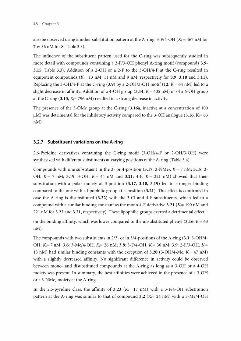
46 | Chapter 3
also be observed using another substitution pattern at the A-ring: 3-F/4-OH (Ki = 467 nM for7 vs 36 nM for 8, Table 3.3).
The influence of the substituent pattern used for the C-ring was subsequently studied inmore detail with compounds containing a 2-F/3-OH phenyl A-ring motif (compounds 3.9-3.15, Table 3.3). Addition of a 2-OH or a 2-F to the 3-OH/4-F at the C-ring resulted inequipotent compounds (Ki= 13 nM; 11 nM and 9 nM, respectively for 3.9, 3.10 and 3.11).Replacing the 3-OH/4-F at the C-ring (3.9) by a 2-OH/3-OH motif (12; Ki= 64 nM) led to aslight decrease in affinity. Addition of a 4-OH group (3.14, Ki= 405 nM) or of a 6-OH groupat the C-ring (3.15, Ki= 796 nM) resulted in a strong decrease in activity.
The presence of the 3-OMe group at the C-ring (3.16a, inactive at a concentration of 100µM) was detrimental for the inhibitory activity compared to the 3-OH analogue (3.16, Ki= 63nM).
Substituent variations on the A-ring
2,6-Pyridine derivatives containing the C-ring motif (3-OH/4-F or 2-OH/3-OH) weresynthesized with different substituents at varying positions of the A-ring (Table 3.4).
Compounds with one substituent in the 3- or 4-position (3.17: 3-NMe2, Ki= 7 nM; 3.18: 3-OH, Ki= 7 nM; 3.19: 3-OH, Ki= 44 nM and 3.21: 4-F, Ki= 221 nM) showed that theirsubstitution with a polar moiety at 3-position (3.17, 3.18, 3.19) led to stronger bindingcompared to the one with a lipophilic group at 4-position (3.21). This effect is confirmed incase the A-ring is disubstituted (3.22) with the 3-Cl and 4-F substituents, which led to acompound with a similar binding constant as the mono 4-F derivative 3.21 (Ki= 190 nM and221 nM for 3.22 and 3.21, respectively). These lipophilic groups exerted a detrimental effect
on the binding affinity, which was lower compared to the unsubstituted phenyl (3.16, Ki= 63nM).
The compounds with two substituents in 2/3- or in 3/4-positions of the A-ring (3.1: 3-OH/4-OH, Ki= 7 nM; 3.6: 3-Me/4-OH, Ki= 26 nM; 3.8: 3-F/4-OH, Ki= 36 nM; 3.9: 2-F/3-OH, Ki=13 nM) had similar binding constants with the exception of 3.20 (3-OH/4-Me, Ki= 47 nM)with a slightly decreased affinity. No significant difference in activity could be observedbetween mono- and disubstituted compounds at the A-ring as long as a 3-OH or a 4-OHmoiety was present. In summary, the best affinities were achieved in the presence of a 3-OHor a 3-NMe2 moiety at the A-ring.
In the 2,5-pyridine class, the affinity of 3.23 (Ki= 17 nM) with a 3-F/4-OH substitutionpattern at the A-ring was similar to that of compound 3.2 (Ki= 24 nM) with a 3-Me/4-OH
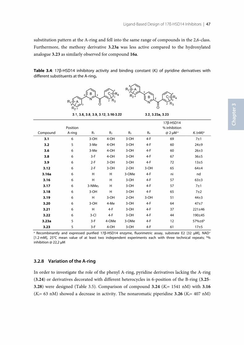
Ligand-Based Design of 17ß-HSD14 Inhibitors | 47
Chap
ter 3
substitution pattern at the A-ring and fell into the same range of compounds in the 2,6-class.Furthermore, the methoxy derivative 3.23a was less active compared to the hydroxylatedanalogue 3.23 as similarly observed for compound 16a.
Table 3.4: 17β-HSD14 inhibitory activity and binding constant (Ki) of pyridine derivatives withdifferent substituents at the A-ring.
CompoundPositionA-ring R1 R2 R3 R4
17β-HSD14% inhibition
@ 2 µM a Ki (nM)a
3.1 6 3-OH 4-OH 3-OH 4-F 69 7±1
3.2 5 3-Me 4-OH 3-OH 4-F 60 24±9
3.6 6 3-Me 4-OH 3-OH 4-F 60 26±3
3.8 6 3-F 4-OH 3-OH 4-F 67 36±5
3.9 6 2-F 3-OH 3-OH 4-F 72 13±5
3.12 6 2-F 3-OH 2-OH 3-OH 65 64±4
3.16a 6 H H 3-OMe 4-F ni nd
3.16 6 H H 3-OH 4-F 57 63±3
3.17 6 3-NMe2 H 3-OH 4-F 57 7±1
3.18 6 3-OH H 3-OH 4-F 65 7±2
3.19 6 H 3-OH 2-OH 3-OH 51 44±3
3.20 6 3-OH 4-Me 3-OH 4-F 64 47±7
3.21 6 H 4-F 3-OH 4-F 37 221±46
3.22 6 3-Cl 4-F 3-OH 4-F 44 190±45
3.23a 5 3-F 4-OMe 3-OMe 4-F 12 57%±6b
3.23 5 3-F 4-OH 3-OH 4-F 61 17±5a Recombinantly and expressed purified 17β-HSD14 enzyme, fluorimetric assay, substrate E2 [32 µM], NAD+
[1.2 mM], 25°C mean value of at least two independent experiments each with three technical repeats; b%inhibition @ 22,2 µM
Variation of the A-ring
In order to investigate the role of the phenyl A-ring, pyridine derivatives lacking the A-ring(3.24) or derivatives decorated with different heterocycles in 6-position of the B-ring (3.25-3.28) were designed (Table 3.5). Comparison of compound 3.24 (Ki= 1541 nM) with 3.16(Ki= 63 nM) showed a decrease in activity. The nonaromatic piperidine 3.26 (Ki= 407 nM)
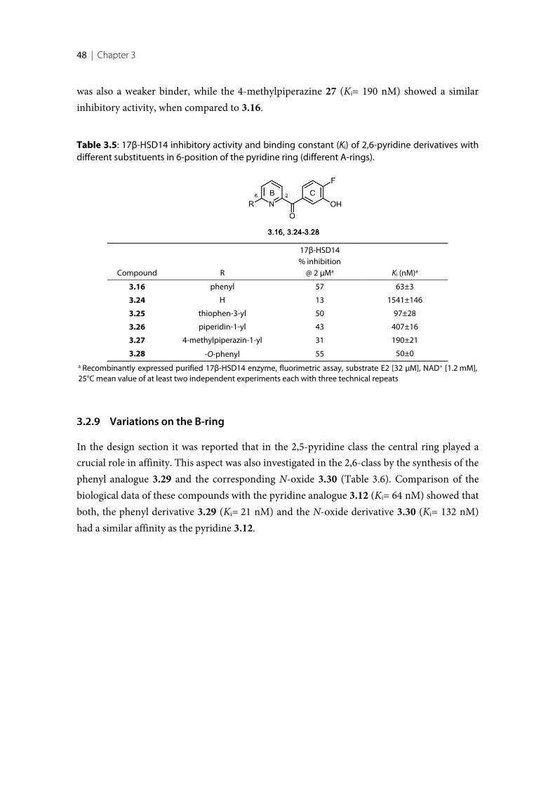
48 | Chapter 3
was also a weaker binder, while the 4-methylpiperazine 27 (Ki= 190 nM) showed a similarinhibitory activity, when compared to 3.16.
Table 3.5: 17β-HSD14 inhibitory activity and binding constant (Ki) of 2,6-pyridine derivatives withdifferent substituents in 6-position of the pyridine ring (different A-rings).
Compound R
17β-HSD14% inhibition
@ 2 µMa Ki (nM)a
3.16 phenyl 57 63±3
3.24 H 13 1541±146
3.25 thiophen-3-yl 50 97±28
3.26 piperidin-1-yl 43 407±16
3.27 4-methylpiperazin-1-yl 31 190±21
3.28 -O-phenyl 55 50±0a Recombinantly expressed purified 17β-HSD14 enzyme, fluorimetric assay, substrate E2 [32 µM], NAD+ [1.2 mM],25°C mean value of at least two independent experiments each with three technical repeats
Variations on the B-ring
In the design section it was reported that in the 2,5-pyridine class the central ring played acrucial role in affinity. This aspect was also investigated in the 2,6-class by the synthesis of thephenyl analogue 3.29 and the corresponding N-oxide 3.30 (Table 3.6). Comparison of thebiological data of these compounds with the pyridine analogue 3.12 (Ki= 64 nM) showed thatboth, the phenyl derivative 3.29 (Ki= 21 nM) and the N-oxide derivative 3.30 (Ki= 132 nM)had a similar affinity as the pyridine 3.12.
Ligand-Based Design of 17ß-HSD14 Inhibitors | 49
Chap
ter 3
Table 3.6: 17β-HSD14 inhibitory activity and binding constant (Ki) for compounds with different B-rings.
Compound X
17β-HSD14% inhibition
@ 2 µMa Ki (nM)a
12 N 77 64±4
29 C 60 21±2
30 N+-O- 47 132±13a Recombinantly expressed and purified 17β-HSD14 enzyme, fluorimetric assay, substrate E2 [32 µM], NAD+ [1.2mM], mean value of at least two independent experiments each with three technical repeats
Trisubstituted pyridines
Based on the hypothesis that the C-ring and the carbonyl group of both the 2,5- and 2,6-pyridines achieve the same interaction as a consequence of binding in the same area, thesimilarity in affinity of the 2,5-derivative 3.23 (Ki= 17 nM) and the 2,6-derivative 3.8 (Ki= 36nM, Table 3) suggested that the available space for binding the additional ring must be large,even tolerating an A-ring attached to the 5- or 6-position. Trisubstituted pyridinescombining substitutions at the 5- and 6-positions were therefore synthesized (3.31 and 3.32,Table 3.7). These trisubstituted compounds turned out to be equipotent (3.31, Ki= 9 nM;3.32, Ki= 15 nM) with a similar affinity compared to the disubstituted derivatives 3.6 and3.23.
Introducing a hydroxymethyl group at the 4-position of the B-ring led to a slight decrease inactivity (3.33 Ki= 86 nM compared to 3.9 Ki= 13 nM, Table 3.7).
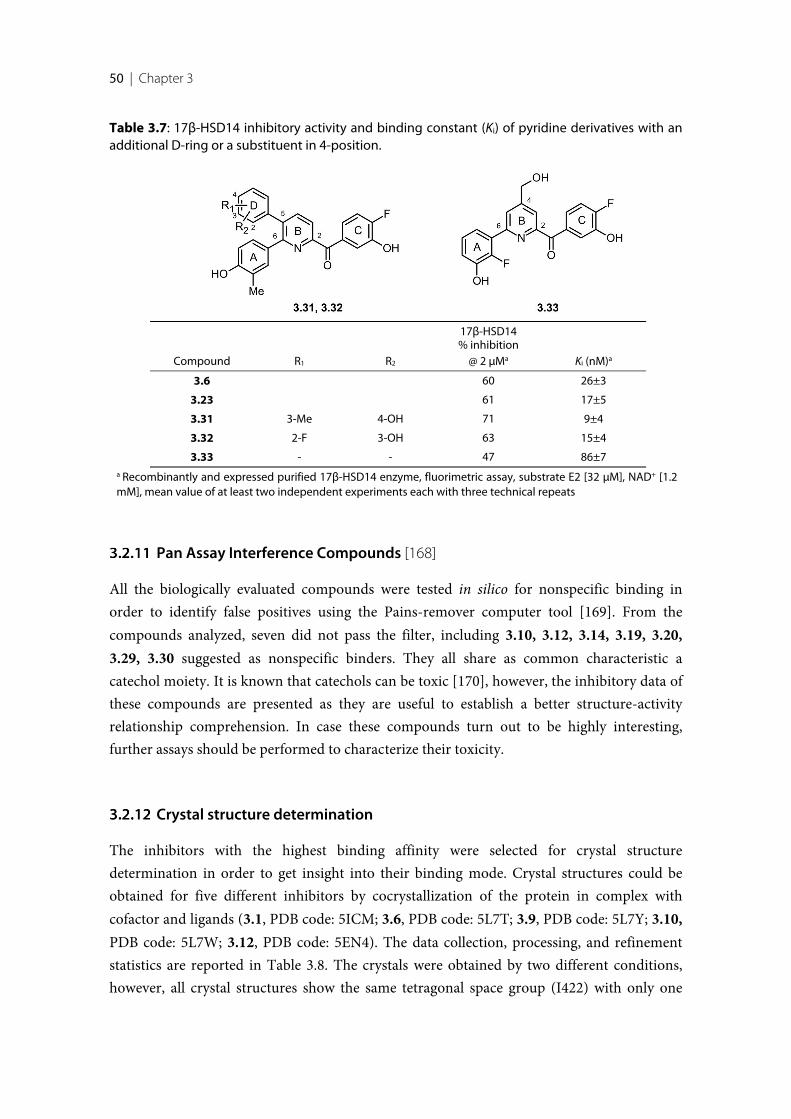
50 | Chapter 3
Table 3.7: 17β-HSD14 inhibitory activity and binding constant (Ki) of pyridine derivatives with anadditional D-ring or a substituent in 4-position.
Compound R1 R2
17β-HSD14% inhibition
@ 2 µMa Ki (nM)a
3.6 60 26±3
3.23 61 17±5
3.31 3-Me 4-OH 71 9±4
3.32 2-F 3-OH 63 15±4
3.33 - - 47 86±7a Recombinantly and expressed purified 17β-HSD14 enzyme, fluorimetric assay, substrate E2 [32 µM], NAD+ [1.2mM], mean value of at least two independent experiments each with three technical repeats
Pan Assay Interference Compounds [168]
All the biologically evaluated compounds were tested in silico for nonspecific binding inorder to identify false positives using the Pains-remover computer tool [169]. From thecompounds analyzed, seven did not pass the filter, including 3.10, 3.12, 3.14, 3.19, 3.20,3.29, 3.30 suggested as nonspecific binders. They all share as common characteristic acatechol moiety. It is known that catechols can be toxic [170], however, the inhibitory data ofthese compounds are presented as they are useful to establish a better structure-activityrelationship comprehension. In case these compounds turn out to be highly interesting,further assays should be performed to characterize their toxicity.
Crystal structure determination
The inhibitors with the highest binding affinity were selected for crystal structuredetermination in order to get insight into their binding mode. Crystal structures could beobtained for five different inhibitors by cocrystallization of the protein in complex withcofactor and ligands (3.1, PDB code: 5ICM; 3.6, PDB code: 5L7T; 3.9, PDB code: 5L7Y; 3.10,PDB code: 5L7W; 3.12, PDB code: 5EN4). The data collection, processing, and refinementstatistics are reported in Table 3.8. The crystals were obtained by two different conditions,however, all crystal structures show the same tetragonal space group (I422) with only one
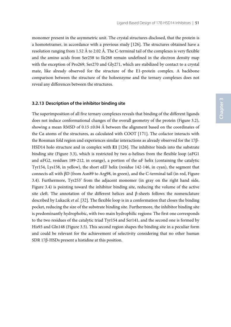
Ligand-Based Design of 17ß-HSD14 Inhibitors | 51
Chap
ter 3
monomer present in the asymmetric unit. The crystal structures disclosed, that the protein isa homotetramer, in accordance with a previous study [126]. The structures obtained have aresolution ranging from 1.52 Å to 2.02 Å. The C-terminal tail of the complexes is very flexibleand the amino acids from Ser258 to Ile268 remain undefined in the electron density mapwith the exception of Pro269, Ser270 and Gly271, which are stabilized by contact to a crystalmate, like already observed for the structure of the E1-protein complex. A backbonecomparison between the structure of the holoenzyme and the ternary complexes does notreveal any differences between the structures.
Description of the inhibitor binding site
The superimposition of all five ternary complexes reveals that binding of the different ligandsdoes not induce conformational changes of the overall geometry of the protein (Figure 3.2),showing a mean RMSD of 0.15 ±0.04 Å between the alignment based on the coordinates ofthe Cα atoms of the structures, as calculated with COOT [171]. The cofactor interacts withthe Rossman fold region and experiences similar interactions as already observed for the 17β-HSD14 holo structure and in complex with E1 [126]. The inhibitor binds into the substratebinding site (Figure 3.3), which is restricted by two α-helixes from the flexible loop (αFG1and αFG2, residues 189-212, in orange), a portion of the αF helix (containing the catalyticTyr154, Lys158, in yellow), the short αEF helix (residue 142-146, in cyan), the segment thatconnects αE with βD (from Asn89 to Arg98, in green), and the C-terminal tail (in red, Figure3.4). Furthermore, Tyr253’ from the adjacent monomer (in gray on the right hand side,Figure 3.4) is pointing toward the inhibitor binding site, reducing the volume of the activesite cleft. The annotation of the different helices and β-sheets follows the nomenclaturedescribed by Lukacik et al. [32]. The flexible loop is in a conformation that closes the bindingpocket, reducing the size of the substrate binding site. Furthermore, the inhibitor binding siteis predominantly hydrophobic, with two main hydrophilic regions: The first one correspondsto the two residues of the catalytic triad Tyr154 and Ser141, and the second one is formed byHis93 and Gln148 (Figure 3.5). This second region shapes the binding site in a peculiar formand could be relevant for the achievement of selectivity considering that no other humanSDR 17β-HSDs present a histidine at this position.
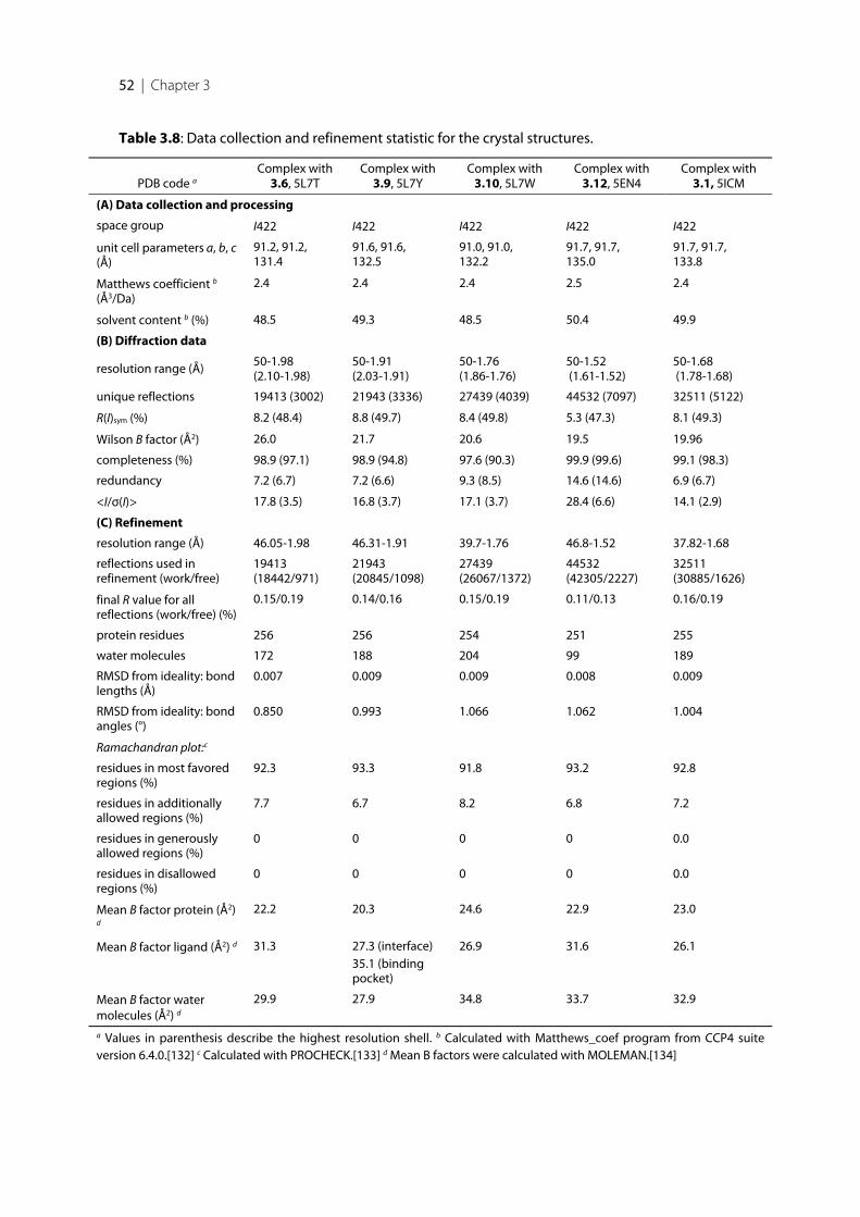
52 | Chapter 3
Table 3.8: Data collection and refinement statistic for the crystal structures.
PDB code aComplex with
3.6, 5L7TComplex with
3.9, 5L7YComplex with
3.10, 5L7WComplex with
3.12, 5EN4Complex with
3.1, 5ICM
(A) Data collection and processing
space group I422 I422 I422 I422 I422
unit cell parameters a, b, c(Å)
91.2, 91.2,131.4
91.6, 91.6,132.5
91.0, 91.0,132.2
91.7, 91.7,135.0
91.7, 91.7,133.8
Matthews coefficient b
(Å3/Da)2.4 2.4 2.4 2.5 2.4
solvent content b (%) 48.5 49.3 48.5 50.4 49.9
(B) Diffraction data
resolution range (Å) 50-1.98(2.10-1.98)
50-1.91(2.03-1.91)
50-1.76(1.86-1.76)
50-1.52 (1.61-1.52)
50-1.68 (1.78-1.68)
unique reflections 19413 (3002) 21943 (3336) 27439 (4039) 44532 (7097) 32511 (5122)
R(I)sym (%) 8.2 (48.4) 8.8 (49.7) 8.4 (49.8) 5.3 (47.3) 8.1 (49.3)
Wilson B factor (Å2) 26.0 21.7 20.6 19.5 19.96
completeness (%) 98.9 (97.1) 98.9 (94.8) 97.6 (90.3) 99.9 (99.6) 99.1 (98.3)
redundancy 7.2 (6.7) 7.2 (6.6) 9.3 (8.5) 14.6 (14.6) 6.9 (6.7)
<I/σ(I)> 17.8 (3.5) 16.8 (3.7) 17.1 (3.7) 28.4 (6.6) 14.1 (2.9)
(C) Refinement
resolution range (Å) 46.05-1.98 46.31-1.91 39.7-1.76 46.8-1.52 37.82-1.68
reflections used inrefinement (work/free)
19413(18442/971)
21943(20845/1098)
27439(26067/1372)
44532(42305/2227)
32511(30885/1626)
final R value for allreflections (work/free) (%)
0.15/0.19 0.14/0.16 0.15/0.19 0.11/0.13 0.16/0.19
protein residues 256 256 254 251 255
water molecules 172 188 204 99 189
RMSD from ideality: bondlengths (Å)
0.007 0.009 0.009 0.008 0.009
RMSD from ideality: bondangles (°)
0.850 0.993 1.066 1.062 1.004
Ramachandran plot:c
residues in most favoredregions (%)
92.3 93.3 91.8 93.2 92.8
residues in additionallyallowed regions (%)
7.7 6.7 8.2 6.8 7.2
residues in generouslyallowed regions (%)
0 0 0 0 0.0
residues in disallowedregions (%)
0 0 0 0 0.0
Mean B factor protein (Å2)d
22.2 20.3 24.6 22.9 23.0
Mean B factor ligand (Å2) d 31.3 27.3 (interface)35.1 (bindingpocket)
26.9 31.6 26.1
Mean B factor watermolecules (Å2) d
29.9 27.9 34.8 33.7 32.9
a Values in parenthesis describe the highest resolution shell. b Calculated with Matthews_coef program from CCP4 suiteversion 6.4.0.[132] c Calculated with PROCHECK.[133] d Mean B factors were calculated with MOLEMAN.[134]
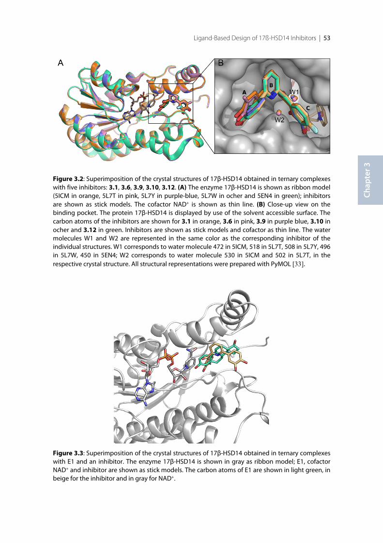
Ligand-Based Design of 17ß-HSD14 Inhibitors | 53
Chap
ter 3
Figure 3.2: Superimposition of the crystal structures of 17β-HSD14 obtained in ternary complexeswith five inhibitors: 3.1, 3.6, 3.9, 3.10, 3.12. (A) The enzyme 17β-HSD14 is shown as ribbon model(5ICM in orange, 5L7T in pink, 5L7Y in purple-blue, 5L7W in ocher and 5EN4 in green); inhibitorsare shown as stick models. The cofactor NAD+ is shown as thin line. (B) Close-up view on thebinding pocket. The protein 17β-HSD14 is displayed by use of the solvent accessible surface. Thecarbon atoms of the inhibitors are shown for 3.1 in orange, 3.6 in pink, 3.9 in purple blue, 3.10 inocher and 3.12 in green. Inhibitors are shown as stick models and cofactor as thin line. The watermolecules W1 and W2 are represented in the same color as the corresponding inhibitor of theindividual structures. W1 corresponds to water molecule 472 in 5ICM, 518 in 5L7T, 508 in 5L7Y, 496in 5L7W, 450 in 5EN4; W2 corresponds to water molecule 530 in 5ICM and 502 in 5L7T, in therespective crystal structure. All structural representations were prepared with PyMOL [33].
Figure 3.3: Superimposition of the crystal structures of 17β-HSD14 obtained in ternary complexeswith E1 and an inhibitor. The enzyme 17β-HSD14 is shown in gray as ribbon model; E1, cofactorNAD+ and inhibitor are shown as stick models. The carbon atoms of E1 are shown in light green, inbeige for the inhibitor and in gray for NAD+.
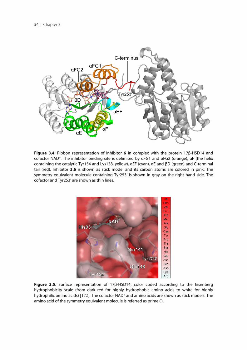
54 | Chapter 3
Figure 3.5: Surface representation of 17β-HSD14; color coded according to the Eisenberghydrophobicity scale (from dark red for highly hydrophobic amino acids to white for highlyhydrophilic amino acids) [172]. The cofactor NAD+ and amino acids are shown as stick models. Theamino acid of the symmetry equivalent molecule is referred as prime (’).
Figure 3.4: Ribbon representation of inhibitor 6 in complex with the protein 17β-HSD14 andcofactor NAD+. The inhibitor binding site is delimited by αFG1 and αFG2 (orange), αF (the helixcontaining the catalytic Tyr154 and Lys158, yellow), αEF (cyan), αE and βD (green) and C-terminaltail (red). Inhibitor 3.6 is shown as stick model and its carbon atoms are colored in pink. Thesymmetry equivalent molecule containing Tyr253’ is shown in gray on the right hand side. Thecofactor and Tyr253’ are shown as thin lines.
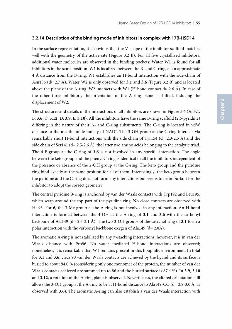
Ligand-Based Design of 17ß-HSD14 Inhibitors | 55
Chap
ter 3
Description of the binding mode of inhibitors in complex with 17β-HSD14
In the surface representation, it is obvious that the V-shape of the inhibitor scaffold matcheswell with the geometry of the active site (Figure 3.2 B). For all five crystallized inhibitors,additional water molecules are observed in the binding pockets: Water W1 is found for allinhibitors in the same position. W1 is localized between the B- and C-ring, at an approximate4 Å distance from the B-ring. W1 establishes an H-bond interaction with the side-chain ofAsn186 (d» 2.7 Å). Water W2 is only observed for 3.1 and 3.6 (Figure 3.2 B) and is locatedabove the plane of the A-ring. W2 interacts with W1 (H-bond contact d» 2.6 Å). In case ofthe other three inhibitors, the orientation of the A-ring plane is shifted, inducing thedisplacement of W2.
The structures and details of the interactions of all inhibitors are shown in Figure 3.6 (A: 3.1,B: 3.6; C: 3.12; D: 3.9; E: 3.10). All the inhibitors have the same B-ring scaffold (2,6-pyridine)differing in the nature of their A- and C-ring substituents. The C-ring is located in vdWdistance to the nicotinamide moiety of NAD+. The 3-OH group at the C-ring interacts viaremarkably short H-bond interactions with the side chain of Tyr154 (d= 2.3-2.5 Å) and theside chain of Ser141 (d= 2.5-2.6 Å), the latter two amino acids belonging to the catalytic triad.The 4-F group at the C-ring of 3.6 is not involved in any specific interaction. The anglebetween the keto group and the phenyl C-ring is identical in all the inhibitors independent ofthe presence or absence of the 2-OH group at the C-ring. The keto group and the pyridinering bind exactly at the same position for all of them. Interestingly, the keto group betweenthe pyridine and the C-ring does not form any interactions but seems to be important for theinhibitor to adopt the correct geometry.
The central pyridine B-ring is anchored by van der Waals contacts with Trp192 and Leu195,which wrap around the top part of the pyridine ring. No close contacts are observed withHis93. For 6, the 3-Me group at the A-ring is not involved in any interaction. An H-bondinteraction is formed between the 4-OH at the A-ring of 3.1 and 3.6 with the carbonylbackbone of Ala149 (d= 2.7-3.1 Å). The two 3-OH groups of the catechol ring of 3.1 form apolar interaction with the carbonyl backbone oxygen of Ala149 (d= 2.8Å).
The aromatic A-ring is not stabilized by any π-stacking interactions, however, it is in van derWaals distance with Pro96. No water mediated H-bond interactions are observed;nonetheless, it is remarkable that W1 remains present in this lipophilic environment. In totalfor 3.1 and 3.6, circa 90 van der Waals contacts are achieved by the ligand and its surface isburied to about 94.0 % (considering only one monomer of the protein, the number of van derWaals contacts achieved are summed up to 86 and the buried surface is 87.4 %). In 3.9, 3.10and 3.12, a rotation of the A-ring plane is observed. Nevertheless, the altered orientation stillallows the 3-OH group at the A-ring to be at H-bond distance to Ala149-CO (d= 2.8-3.0 Å, asobserved with 3.6). The aromatic A-ring can also establish a van der Waals interaction with
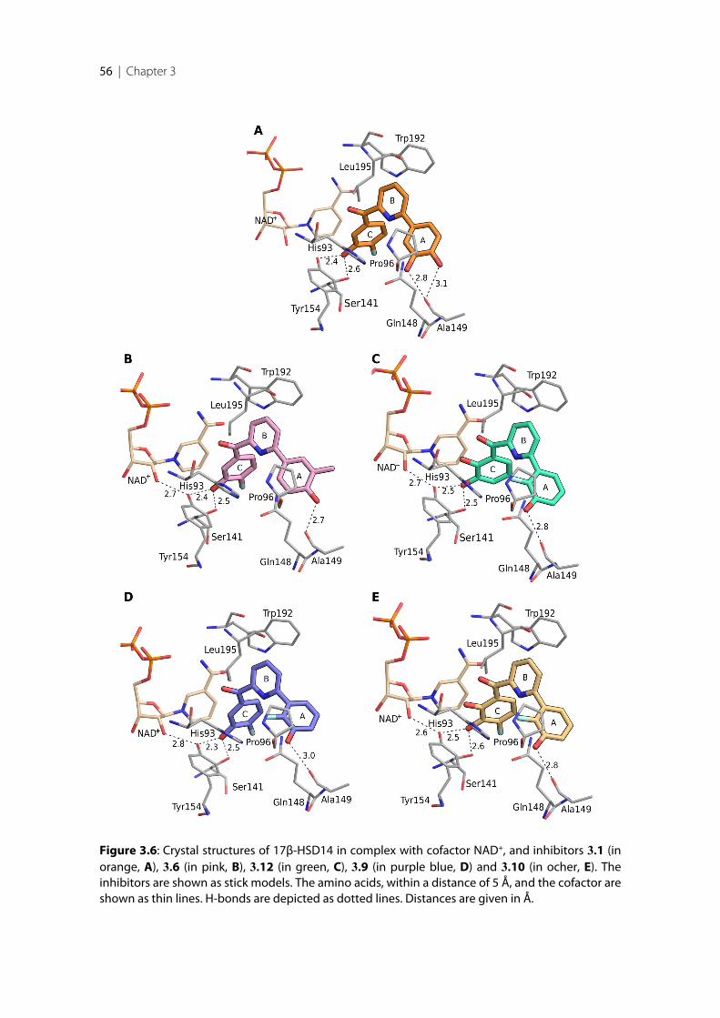
56 | Chapter 3
Figure 3.6: Crystal structures of 17β-HSD14 in complex with cofactor NAD+, and inhibitors 3.1 (inorange, A), 3.6 (in pink, B), 3.12 (in green, C), 3.9 (in purple blue, D) and 3.10 (in ocher, E). Theinhibitors are shown as stick models. The amino acids, within a distance of 5 Å, and the cofactor areshown as thin lines. H-bonds are depicted as dotted lines. Distances are given in Å.
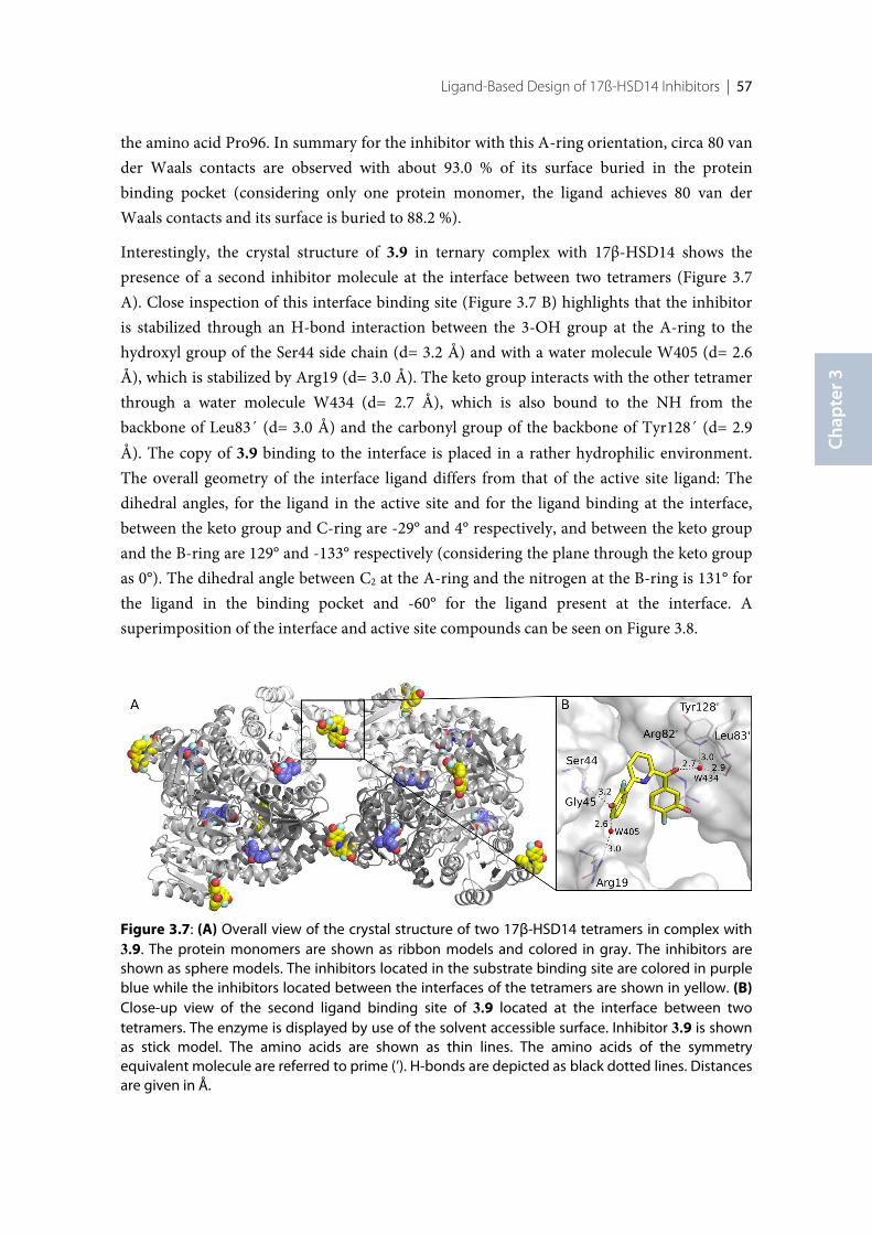
Ligand-Based Design of 17ß-HSD14 Inhibitors | 57
Chap
ter 3
the amino acid Pro96. In summary for the inhibitor with this A-ring orientation, circa 80 vander Waals contacts are observed with about 93.0 % of its surface buried in the proteinbinding pocket (considering only one protein monomer, the ligand achieves 80 van derWaals contacts and its surface is buried to 88.2 %).
Interestingly, the crystal structure of 3.9 in ternary complex with 17β-HSD14 shows thepresence of a second inhibitor molecule at the interface between two tetramers (Figure 3.7A). Close inspection of this interface binding site (Figure 3.7 B) highlights that the inhibitoris stabilized through an H-bond interaction between the 3-OH group at the A-ring to thehydroxyl group of the Ser44 side chain (d= 3.2 Å) and with a water molecule W405 (d= 2.6Å), which is stabilized by Arg19 (d= 3.0 Å). The keto group interacts with the other tetramerthrough a water molecule W434 (d= 2.7 Å), which is also bound to the NH from thebackbone of Leu83´ (d= 3.0 Å) and the carbonyl group of the backbone of Tyr128´ (d= 2.9Å). The copy of 3.9 binding to the interface is placed in a rather hydrophilic environment.The overall geometry of the interface ligand differs from that of the active site ligand: Thedihedral angles, for the ligand in the active site and for the ligand binding at the interface,between the keto group and C-ring are -29° and 4° respectively, and between the keto groupand the B-ring are 129° and -133° respectively (considering the plane through the keto groupas 0°). The dihedral angle between C2 at the A-ring and the nitrogen at the B-ring is 131° forthe ligand in the binding pocket and -60° for the ligand present at the interface. Asuperimposition of the interface and active site compounds can be seen on Figure 3.8.
Figure 3.7: (A) Overall view of the crystal structure of two 17β-HSD14 tetramers in complex with3.9. The protein monomers are shown as ribbon models and colored in gray. The inhibitors areshown as sphere models. The inhibitors located in the substrate binding site are colored in purpleblue while the inhibitors located between the interfaces of the tetramers are shown in yellow. (B)Close-up view of the second ligand binding site of 3.9 located at the interface between twotetramers. The enzyme is displayed by use of the solvent accessible surface. Inhibitor 3.9 is shownas stick model. The amino acids are shown as thin lines. The amino acids of the symmetryequivalent molecule are referred to prime (’). H-bonds are depicted as black dotted lines. Distancesare given in Å.
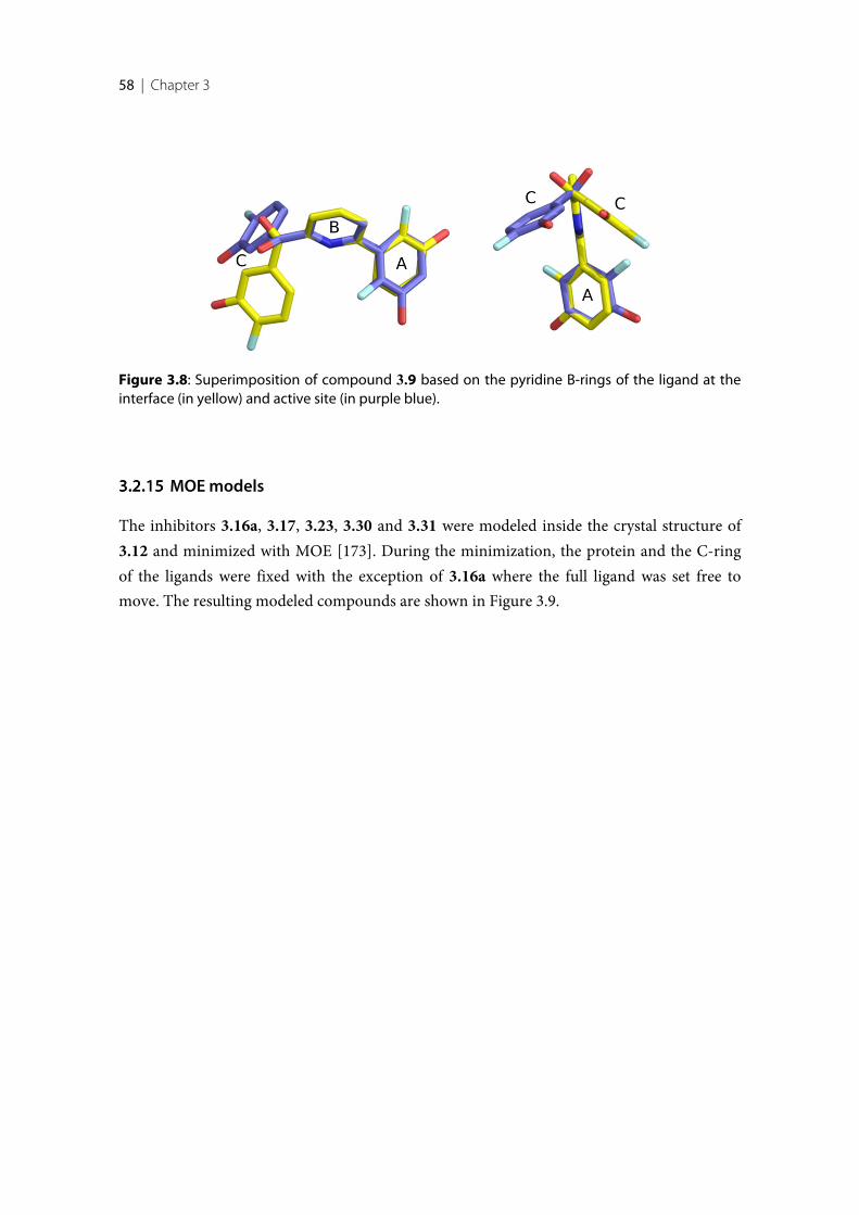
58 | Chapter 3
MOE models
The inhibitors 3.16a, 3.17, 3.23, 3.30 and 3.31 were modeled inside the crystal structure of3.12 and minimized with MOE [173]. During the minimization, the protein and the C-ringof the ligands were fixed with the exception of 3.16a where the full ligand was set free tomove. The resulting modeled compounds are shown in Figure 3.9.
Figure 3.8: Superimposition of compound 3.9 based on the pyridine B-rings of the ligand at theinterface (in yellow) and active site (in purple blue).
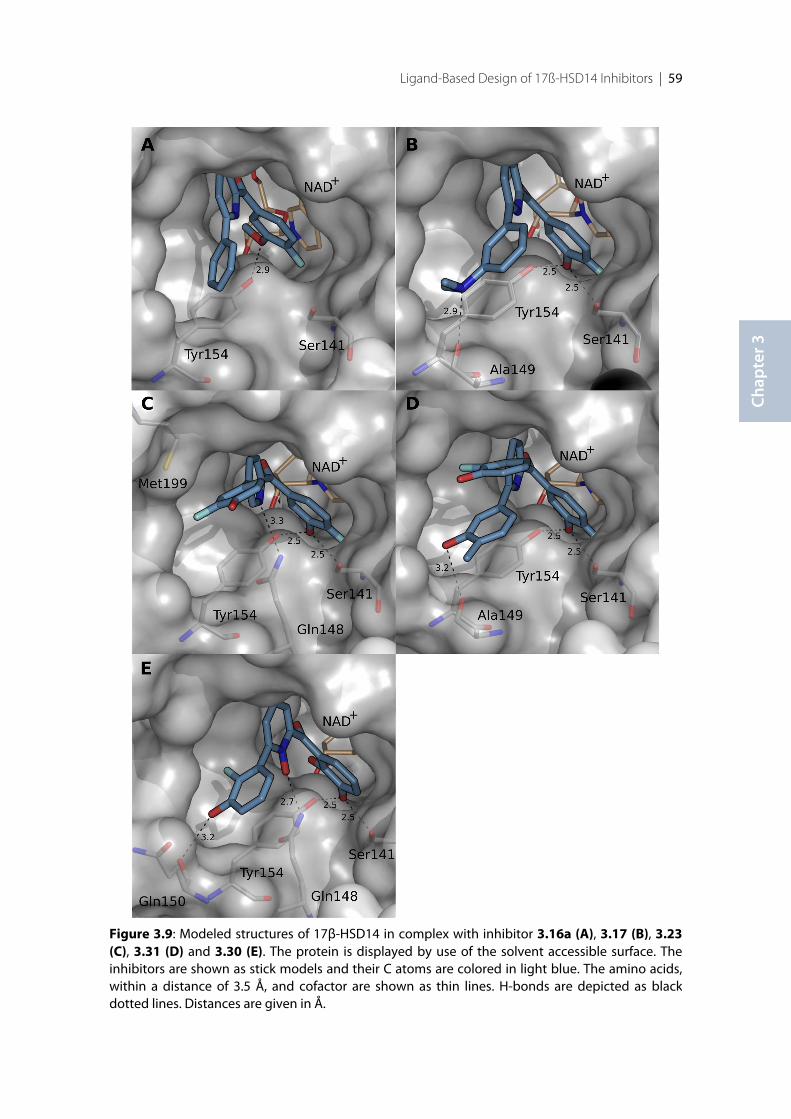
Ligand-Based Design of 17ß-HSD14 Inhibitors | 59
Chap
ter 3
Figure 3.9: Modeled structures of 17β-HSD14 in complex with inhibitor 3.16a (A), 3.17 (B), 3.23(C), 3.31 (D) and 3.30 (E). The protein is displayed by use of the solvent accessible surface. Theinhibitors are shown as stick models and their C atoms are colored in light blue. The amino acids,within a distance of 3.5 Å, and cofactor are shown as thin lines. H-bonds are depicted as blackdotted lines. Distances are given in Å.
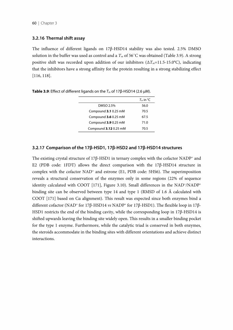
60 | Chapter 3
Thermal shift assay
The influence of different ligands on 17β-HSD14 stability was also tested. 2.5% DMSOsolution in the buffer was used as control and a Tm of 56˚C was obtained (Table 3.9). A strongpositive shift was recorded upon addition of our inhibitors (ΔTm=11.5-15.0°C), indicatingthat the inhibitors have a strong affinity for the protein resulting in a strong stabilizing effect[116, 118].
Table 3.9: Effect of different ligands on the Tm of 17β-HSD14 (2.6 µM).
Tm in °C
DMSO 2.5% 56.0
Compound 3.1 0.25 mM 70.5
Compound 3.6 0.25 mM 67.5
Compound 3.9 0.25 mM 71.0
Compound 3.12 0.25 mM 70.5
Comparison of the 17β-HSD1, 17β-HSD2 and 17β-HSD14 structures
The existing crystal structure of 17β-HSD1 in ternary complex with the cofactor NADP+ andE2 (PDB code: 1FDT) allows the direct comparison with the 17β-HSD14 structure incomplex with the cofactor NAD+ and estrone (E1, PDB code: 5HS6). The superimpositionreveals a structural conservation of the enzymes only in some regions (22% of sequenceidentity calculated with COOT [171], Figure 3.10). Small differences in the NAD+/NADP+
binding site can be observed between type 14 and type 1 (RMSD of 1.6 Å calculated withCOOT [171] based on Cα alignment). This result was expected since both enzymes bind adifferent cofactor (NAD+ for 17β-HSD14 vs NADP+ for 17β-HSD1). The flexible loop in 17β-HSD1 restricts the end of the binding cavity, while the corresponding loop in 17β-HSD14 isshifted upwards leaving the binding site widely open. This results in a smaller binding pocketfor the type 1 enzyme. Furthermore, while the catalytic triad is conserved in both enzymes,the steroids accommodate in the binding sites with different orientations and achieve distinctinteractions.
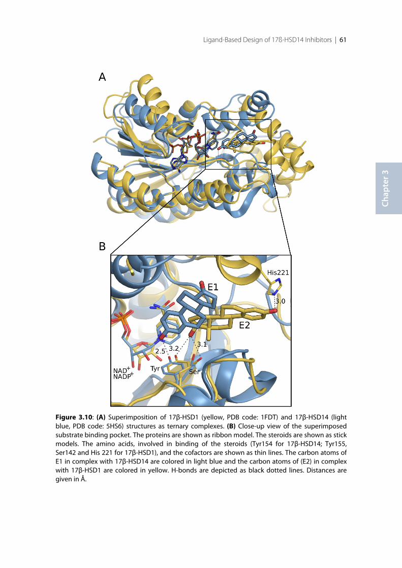
Ligand-Based Design of 17ß-HSD14 Inhibitors | 61
Chap
ter 3
Figure 3.10: (A) Superimposition of 17β-HSD1 (yellow, PDB code: 1FDT) and 17β-HSD14 (lightblue, PDB code: 5HS6) structures as ternary complexes. (B) Close-up view of the superimposedsubstrate binding pocket. The proteins are shown as ribbon model. The steroids are shown as stickmodels. The amino acids, involved in binding of the steroids (Tyr154 for 17β-HSD14; Tyr155,Ser142 and His 221 for 17β-HSD1), and the cofactors are shown as thin lines. The carbon atoms ofE1 in complex with 17β-HSD14 are colored in light blue and the carbon atoms of (E2) in complexwith 17β-HSD1 are colored in yellow. H-bonds are depicted as black dotted lines. Distances aregiven in Å.
62 | Chapter 3
Selectivity
Taking into account that the parent scaffold of the new 17β-HSD14 inhibitors was derivedfrom 17β-HSD1 and 17β-HSD2 inhibitors, it was of utmost importance to study theirselectivity profile with respect to 17β-HSD1 and 17β-HSD2 binding.
The 17β-HSD1 and 17β-HSD2 inhibition assay was performed using a radioactive assay,quantifying the amount of [3H]-labeled E2 (for type 1) and [3H]-labeled E1 (for type 2)formed after incubation with protein, cofactor and the inhibitor as previously described[174]. The results are shown in Table 3.10, expressed as percent inhibition when tested at aninhibitor concentration of 1 µM.
As expected the 2,5-pyridine 3.23 showed the highest affinity for 17β-HSD1 (47% inhibition)compared to the 2,6-pyridines (3.6-3.20, 9-23% inhibition), which were all nearly inactive in17β-HSD1.
Inhibition of 17β-HSD2 was slightly higher than that of 17β-HSD1 for the compounds with a2,6-substitution pattern (between 30% and 62% inhibition at 1µM concentration for 3.6-3.20) and much higher for the 2,5-pyridine ketones (64% and 85% when tested at 1 µMconcentration for 3.23 and 3.2, respectively).
A direct comparison of the 17β-HSD1 and 17β-HSD2 inhibitory activities with those of 17β-HSD14 is however problematic as different conditions were used in the assays.
However, under the applied condition in the 17β-HSD2 inhibition assay, using the Cheng-Prusoff equation for competitive inhibition, a calculated Ki (cKi) could be estimated: For acompound with an IC50 of around 1 µM (50% inhibition at 1 µM) a cKi of about 450 nM wasexpected (with Km 17β-HSD2 = 400 nM in this assay [175] and [S]= 500 nM).
With 48% and 43% inhibition of 17β-HSD2, compounds 3.9 and 3.10 showed a cKi ≥450 nM.Comparison of their Ki values for 17β-HSD2 and 17β-HSD14 binding allowed to calculate aselectivity factor (ratio of Ki(HSD2)/Ki(HSD14)), which could be estimated to be around 35 for 9and ≥41 for 3.10.Compounds 3.9 and 3.10 are relatively selective 17β-HSD14 inhibitors. Theselectivity profile of 3.12 toward 17β-HSD2 (with only 30% inhibition at 1 µM) should beeven better.
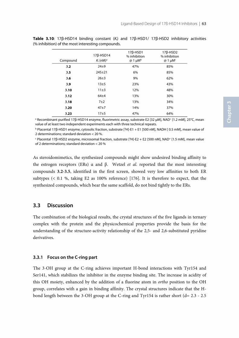
Ligand-Based Design of 17ß-HSD14 Inhibitors | 63
Chap
ter 3
Table 3.10: 17β-HSD14 binding constant (Ki) and 17β-HSD1/ 17β-HSD2 inhibitory activities(% inhibition) of the most interesting compounds.
Compound
17β-HSD14
Ki (nM)a
17β-HSD1% inhibition
@ 1 μMb
17β-HSD2% inhibition
@ 1 μMc
3.2 24±9 47% 85%
3.5 245±21 6% 85%
3.6 26±3 9% 62%
3.9 13±5 23% 43%
3.10 11±3 12% 48%
3.12 64±4 13% 30%
3.18 7±2 13% 34%
3.20 47±7 14% 37%
3.23 17±5 47% 64%a Recombinant purified 17β-HSD14 enzyme, fluorimetric assay, substrate E2 [32 µM], NAD+ [1.2 mM], 25°C, meanvalue of at least two independent experiments each with three technical repeats.b Placental 17β-HSD1 enzyme, cytosolic fraction, substrate [3H]-E1 + E1 [500 nM], NADH [ 0.5 mM], mean value of2 determinations; standard deviation < 20 %.c Placental 17β-HSD2 enzyme, microsomal fraction, substrate [3H]-E2 + E2 [500 nM], NAD+ [1.5 mM], mean valueof 2 determinations; standard deviation < 20 %
As steroidomimetics, the synthesized compounds might show undesired binding affinity tothe estrogen receptors (ERs) α and β. Wetzel et al. reported that the most interestingcompounds 3.2-3.5, identified in the first screen, showed very low affinities to both ERsubtypes (< 0.1 %, taking E2 as 100% reference) [176]. It is therefore to expect, that thesynthesized compounds, which bear the same scaffold, do not bind tightly to the ERs.
3.3 Discussion
The combination of the biological results, the crystal structures of the five ligands in ternarycomplex with the protein and the physicochemical properties provide the basis for theunderstanding of the structure-activity relationship of the 2,5- and 2,6-substituted pyridinederivatives.
Focus on the C-ring part
The 3-OH group at the C-ring achieves important H-bond interactions with Tyr154 andSer141, which stabilizes the inhibitor in the enzyme binding site. The increase in acidity ofthis OH moiety, enhanced by the addition of a fluorine atom in ortho position to the OHgroup, correlates with a gain in binding affinity. The crystal structures indicate that the H-bond length between the 3-OH group at the C-ring and Tyr154 is rather short (d= 2.3 - 2.5
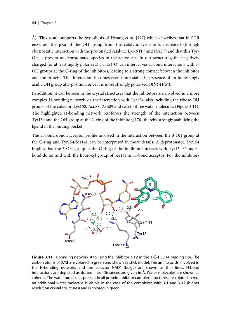
64 | Chapter 3
Å). This result supports the hypothesis of Hwang et al. [177] which describes that in SDRenzymes, the pKa of the OH group from the catalytic tyrosine is decreased (throughelectrostatic interaction with the protonated catalytic Lys-NH3
+ and NAD+) and that this Tyr-OH is present as deprotonated species in the active site. In our structures, the negativelycharged (or at least highly polarized) Tyr154-O- can interact via H-bond interactions with 3-OH groups at the C-ring of the inhibitors, leading to a strong contact between the inhibitorand the protein. This interaction becomes even more stable in presence of an increasinglyacidic OH group at 3-position, once it is more strongly polarized O(δ-)-H(δ+).
In addition, it can be seen in the crystal structures that the inhibitors are involved in a morecomplex H-bonding network via the interaction with Tyr154, also including the ribose-OHgroups of the cofactor, Lys158, Asn88, Asn89 and two to three water molecules (Figure 3.11).The highlighted H-bonding network reinforces the strength of the interaction betweenTyr154 and the OH group at the C-ring of the inhibitor,[178] thereby strongly stabilizing theligand in the binding pocket.
The H-bond donor/acceptor profile involved in the interaction between the 3-OH group atthe C-ring and Tyr154/Ser141 can be interpreted in more details: A deprotonated Tyr154implies that the 3-OH group at the C-ring of the inhibitor interacts with Tyr154-O- as H-bond donor and with the hydroxyl group of Ser141 as H-bond acceptor. For the inhibitors
Figure 3.11: H-bonding network stabilizing the inhibitor 3.12 in the 17β-HSD14 binding site. Thecarbon atoms of 3.12 are colored in green and shown as stick model. The amino acids, involved inthe H-bonding network, and the cofactor NAD+ (beige) are shown as thin lines. H-bondinteractions are depicted as dotted lines. Distances are given in Å. Water molecules are shown asspheres. The water molecules present in all protein-inhibitor complex structures are colored in red,an additional water molecule is visible in the case of the complexes with 3.1 and 3.12 (higherresolution crystal structures) and is colored in green.
Ligand-Based Design of 17ß-HSD14 Inhibitors | 65
Chap
ter 3
also bearing a 2-OH group at the C-ring moiety, an intramolecular H-bond with their ketogroups can be formed and no interaction with Tyr154 is expected (the OH group of Tyr 154is already involved in a contact with the ribose-OH and the 3-OH group at the C-ringinhibitor, and is not available for an additional interaction). In addition, it can be remarkedthat the rigidification induced by the intramolecular H-bond does not correlate with anincrease in binding affinity (comparison of compounds 3.9 and 3.10).
Replacing the 3-OH group at the C-ring by a 3-OMe moiety was shown to be detrimental forinhibitory activity (3.16a: no inhibition @100 µM compared to 3.16: Ki= 63 nM). Whencompound 16a was modeled into the crystal structure of 17β-HSD14 (Figure 3.9 A), itresulted in a shift of the binding pose (compared to the compounds observed in the crystalstructures, see Figure 3.6), which could allow an H-bond interaction between the oxygen ofthe 3-OMe group and the OH group of Tyr154 of the catalytic triad, however, the H-bondcontact is only possible when the 3-OMe group acts as an acceptor (Figure 3.9 A). Theinactivity of this compound confirms the hypothesis that an H-bond donor in 3-position ofthe C-ring is necessary for the inhibitory activity of the compound. In addition, nointeraction with Ser141 could be observed in the presence of the 3-OMe group in the modelstructure.
Focus on the A-ring part
Based on the biological results, it is evident that the removal of the A-ring leads to a strongdecrease in activity (3.16, Ki= 63 nM compared to 3.24 Ki= 1541 nM), indicating animportant interaction of the A-ring with the protein. The crystal structures show that noamino acid is able to achieve a π-stacking interaction in the vicinity of the aromatic A-ring.This result suggests that the aromatic A-ring must be stabilized by hydrophobic interactionswith Pro96.
Furthermore, it can be observed that the introduction of a 3-OH or 4-OH group on the A-ring induces a slight increase in affinity compared to the unsubstituted A-ring. Either thesehydroxyl moieties interact with the carbonyl backbone of Ala149 via a weak H-bondinteraction or the benefit of a strong interaction with Ala149 is decreased by the desolvationcost. It is also striking that neither an electron donating nor an electron withdrawing grouphas an influence on the potency.
The significant affinity enhancement of the dimethylamino derivative 3.17 was investigatedusing the modeled structure (Figure 3.9 B). Here, the NMe2 moiety comes into close contactwith the carbonyl group of Ala149. A strong interaction would only be possible between theNMe2 group and the carbonyl oxygen when the tertiary amine is protonated. However,taking into account the pKa value of the NMe2 group it is unlikely that this group is charged.
66 | Chapter 3
The gain in affinity might come from additional hydrophobic interactions between the lattergroup and Pro96.
In the complex structure with 3.6 the 3-Me group at the A-ring interacts neither with theprotein nor with the solvent. Instead, it is pointing toward an empty cavity, while the 3-OHgroup of 3.12 is oriented in the opposite direction interacting with Ala149. The swappedorientation of the 3-Me group at the A-ring of 3.6 might result from electrostatic repulsionsbetween the methyl group and the carbonyl moiety of Ala149.
Regarding the almost equipotent 2,5-substituted (3.23, Ki= 17 nM) and the 2,6-substitutedderivative (3.8, Ki= 36 nM), it was previously suggested, that the C-rings of both compoundsachieve the same interaction and that the space available for accommodating the A-ring ateither 5- or 6-position must be large to host a ring at both positions. From the modeledstructure of 3.23 (Figure 3.9 C), it can be assumed that the A-ring in 5-position fits well intothe binding pocket, interacting through hydrophobic contacts with Met199. Furthermore, themodeled 2,5,6-trisubstituted derivative 3.31 (Ki= 9 nM) also suggests that compoundsbearing rings at 5- and 6-position can fit in the cavity and may achieve an intramolecular π-stacking interaction (d= 4 Å, Figure 3.9 D).
Focus on the B-ring part
Investigating the importance of the nitrogen at the B-ring indicates that inhibitor 3.12 (Ki=64 nM) and its phenyl analogue 3.29 (Ki= 21 nM) show similar affinities. Obviously, thenitrogen does not achieve any specific interaction with the protein, as confirmed by thecrystal structure. However, the pyridine moiety enhances the solubility for the compounds ofthis inhibitor class. Concerning the N-oxide derivative 3.30 (Ki= 132 nM), the previouslyobserved interaction between the 3-OH group at the A-ring of compound 3.12 with Ala149can no longer be accomplished, however, this OH-group can now address the backbonecarbonyl group of Gln150, as suggested by the modeled structure (Figure 3.9 E). Anadditional H-bond interaction of N+-O- with Gln148 could be gained, but the highdesolvation penalty induced by the introduction of the charges might not be compensated,overall resulting in no increase in affinity.
Second binding site for compound 3.9
For compound 3.9, a second binding pose at the tetramer interface was observed. Theinhibitor was refined to occupancy of 80% at this additional binding site, suggesting that thisligand exhibits a lower affinity for this region compared to the active site. The binding of theinhibitor in this position might be irrelevant for the inhibitory process, while it might be an
Ligand-Based Design of 17ß-HSD14 Inhibitors | 67
Chap
ter 3
artefact resulting from crystal packing as the binding at the interface does not induce anyconformational changes of the ternary structure and of the ligand binding site. However, it isstriking that Michiels et al. [179] reported in their NMR studies that phytoestrogens mightalso bind at the dimer interface of 17β-HSD1.
Comparison of the 17β-HSD1, 17β-HSD2 and 17β-HSD14 structures
A positive influence of a fluorine atom in ortho position to a phenolic hydroxyl group wasalready reported during the development of 17β-HSD1 dihydroxyphenylthiophenesinhibitors [150] and 17β-HSD2 hydroxyphenyl-N-methylsulfonamide thiophenes inhibitors[89]. As no crystal structures of 17β-HSD1 and 17β-HSD2 in complex with thesenonsteroidal compounds are available, their binding modes remain unclear. As 17β-HSDtype 1, type 2 and type 14 belong to the SDR superfamily, they share similarities in the regionin the vicinity of the catalytic triad. The presence of the complex H-bonding network wasalso identified in the type 1 enzyme after analysis of its crystal structure (PDB code: 1FDT). Itcould be expected that the 17β-HSD1 and 17β-HSD2 inhibitors with an acidic OH-phenylgroup have a similar binding mode, interacting with the catalytic triad as observed in thecrystal structure of type 14. Therefore, these new 17β-HSD14 structures in complex withnonsteroidal inhibitors can represent a useful comparative data for 17β-HSD1 dockingstudies and 17β-HSD2 homology modelling.
Basis for structure-based drug design
Based on these results, it should be possible to optimize the current ligands using structure-based drug design. In this compound class the interactions with the catalytic Tyr154 andSer141, as well as with Ala149 are very important to anchor the ligand scaffold in the correctposition. Specific interactions involving His93 and Gln148 in inhibitor binding should resultin an improved activity and selectivity, particularly as His93 is not present in other humanSDR 17β-HSDs. Addressing the water molecule W1 should also lead to an additionalinteraction with the protein. As the active site is open and solvent exposed, the polar aminoacids which are in close neighborhood to the active site next to the surface could also betargeted by specific interactions.
3.4 Conclusion
Nonsteroidal 17β-HSD14 inhibitors have been identified. The initial hits identified in apreliminary screen in the 2,6-susbtituted pyridine class showed a Ki around 250 nM/300 nM
68 | Chapter 3
which was optimized to result in six highly active compounds with Ki< 15 nM and two with aKi of 7 nM. The considerations of substituent effects applied during optimization weresuccessful. It appears that at the C-ring, an acidic 3-OH group is essential to achieve highpotency, interacting via strong H-bond contacts to Tyr154 and Ser141 thereby stabilizing theinteraction through an extensive H-bonding network. The structure-activity relationshipfound for the A-ring shows, that a 3-OH or a 4-OH group increases the potency of theinhibition by interacting with Ala149. The crystal structures in complex with the inhibitorsconfirm the rather large active site, reduced by the C-terminal chain from an adjacentmonomer. The new 17β-HSD14 inhibitors show good physicochemical properties, whichshould be associated with a good bioavailability profile. They also present a good selectivityprofile toward both closely related subtypes 17β-HSD1 and 17β-HSD2. The determinedcrystal structures give important insights not only to characterize the novel protein target butalso to understand the binding poses of these nonsteroidal inhibitors and provide the basisfor their further structure-based optimization.
3.5 Experimental section
17β-HSD14 inhibition assay
In a preliminary study compounds 3.1-3.5 were tested using a radioactive displacement assay(Procedure A, % of inhibition at a concentration of 1 µM). The newly synthesizedcompounds were tested using a fluorimetric assay (Procedure B, as percent inhibition at 2µM, percent inhibition at the concentration of highest solubility of the compound and Ki).Procedure A and B use the same enzyme source, obtained from a bacterial culture. Inprocedure A, the assay is performed with a bacterial suspension, and in procedure B with thepurified form of the enzyme. Due to differences in assay conditions only results within thesame assay can be compared.
Enzyme expression
The pET based plasmid containing the coding sequences of the human gene HSD17B14(using the T205 variant), with a N-terminal 6His-tag and a TEV (Tobacco Etch Virus)protease cleavage site was used for the transfection of E. coli BL21 (DE3) pLysS competentcells. The transformed bacteria cells were grown overnight in 100 mL of Terrific Brothmedium containing 100 µg/mL of ampicillin at 37°C. Subsequently, 25 mL of the overnightculture were transferred in 1 L of the aforementioned medium and allowed to grow at 37 °Cuntil an OD600 of 0.4 was reached. Then the temperature was lowered to 15° C. When theculture reached the OD600 of 1.0 the cells were induced with 0.5 mM of IPTG. The bacteria
Ligand-Based Design of 17ß-HSD14 Inhibitors | 69
Chap
ter 3
cells were harvested by centrifugation and conserved at -80° C overnight before proceedingwith the purification.
Radioactive assay using Procedure A
The bacterial pellet obtained was resuspended in 100 mM phosphate buffer pH 7.7. Thebacterial suspension was incubated with [3H]-E2 (final concentration: 18.3 nM) in thepresence of the potential inhibitor in DMSO (final concentration in assay: 1µM, final DMSOconcentration: 1%) at 37°C. The enzymatic reaction was started by addition of NAD+ (7.5mM) and incubated for 2 h. The reaction was stopped by addition of 0.21 M ascorbic acid ina methanol/acetic acid mixture (99:1, v/v). Substrate and product were extracted from thereaction mixture by SPE (Strata C18-E columns from Phenomenex on a vacuum device).Separation and quantification of the radioactive steroids was performed with HPLC (Luna5µm C18(2), 125 x 4.00 mm from Phenomenex, with an acetonitrile/water mixture (43:57,v.v), flow rate 1mL/min). Substrate conversion in % was calculated after integration of theproduct and substrate peaks. Inhibition was calculated based on conversion without potentialinhibitor (DMSO only) which was set to 0% inhibition.
Enzyme Purification
The cell pellet, previously obtained after IPTG induction, was resuspended in a buffercontaining 50 mM Tris, 500 mM NaCl, 0.5 mM TCEP, 250 mM glucose, 1 mM NAD+, 0.5%(v/v) Triton X-100 and cOmplet Protease Inhibitor Cocktail Tablet (Roche, Germany)adjusted at a pH of 8. The cells were disrupted with a high pressure homogenizer(EmulsiFlex-C5™, AVESTIN, Mannheim, Germany) and the obtained homogenate wascentrifuged at 17700 g for 2h at 4°C. The supernatant was applied to a Ni-NTA column (5 mLHisTrap FF, GE Healtcare Life Sciences, Freiburg, Germany). Two washing steps wereapplied: in the first, to remove the DNA, a buffer composed of 50 mM Tris and 1.5 M NaClwas run against the Ni-column. The second washing step was then performed with a buffercontaining 50 mM Tris, 500 mM NaCl, 0.5 mM TCEP, 250 mM glucose, 0.25 mM NAD+ and21 mM imidazole to remove the unspecific binding proteins. The target protein was eluted byincreasing the imidazole concentration in the buffer to 300 mM. TEV protease was added tothe protein mixture to cleave the N-6His-tag and the product solution was dialyzed overnightat 4°C to reduce the imidazole concentration in the sample (50 mM Tris, 500 mM NaCl, 0.5mM TCEP, 250 mM glucose and 0.25 mM NAD+). A second Ni-NTA column was used forseparation of the TEV protease from the 17β-HSD14. In this step the protein was collectedfrom the flow through of the column, while the TEV protease remained on the column. Withthe goal to increase the purity of the protein, an additional purification step, using a size
70 | Chapter 3
exclusion chromatography (Superdex 75 26/60, GE Healtcare Life Sciences, Freiburg,Germany) was performed with a running buffer comprising 50 mM Tris, 500 mM NaCl, 0.5mM TCEP and 250 mM glucose. To the isolated target protein, an NAD+ solution 0.25 mM(batch for enzymatic assay) or 0.6 mM (batch for crystallization studies) was added. Theprotein solution was flash-frozen in liquid nitrogen and stored at -80°C.
Fluorimetric assay using Procedure B
The potential inhibitor (in DMSO, final DMSO concentration in assay: 1%) was added to amixture of NAD+ (1.2 mM) and E2 (32 µM) in 100 mM phosphate buffer pH 8. Theenzymatic reaction was started by addition of the purified enzyme (1 mg/ml) and theproduction of the fluorescent NADH formed was measured continuously for 15 min at 25°C.The fluorimetric assays were recorded on a Tecan Saphire 2 (λex at 340 nm and λem at 496nm). The slit width for excitation was 7 nm and for emission 15 nm. Reactions wereperformed in 200 µL volumes. The assay was run in 96 well-plates in duplicate, eachexperiment resulting from three technical repeats. A linear relationship between productformation and reaction time was obtained. The slope of the progress curves was calculated bylinear regression. The inhibitors do not show fluorescence at the concentrations used in theassay.
The Ki values were calculated using the Morrison equation [167]. For calculation, threeconstants were necessary: the substrate concentration (32 µM), the Km for 17β-HSD14 withthis substrate E2 (6.18 µM [126]) and the concentration in active protein, which wasdetermined experimentally for each experiment (3.2 or 3.3 µM), using the procedure detailedby Copeland [167]. The fitting and data analysis was performed using GraphPad Prism 7.
Protein co-crystallization with inhibitors 3.1, 3.6, 3.9, 3.10, 3.12
Protein activity was verified before performing the crystallization studies for each inhibitor.The co-crystallization of 17β-HSD14 in complex with the four inhibitors 3.1, 3.6, 3.9, 3.10and 3.12 was performed by sitting drop vapor diffusion technique.
For the crystallization of inhibitors 3.6 and 3.9 in complex with the protein an inhibitor stocksolution in pure DMSO was added to the protein solution (9.5 mg/mL) containing 0.6 mMNAD+ with a final inhibitor concentration of 0.8 mM and a DMSO concentration of 1%. 2 µLof the mother liquor containing 0.1 M CHES, 1 M tri-sodium citrate, pH 9.5, was mixed with2 µL of the protein solution. After growing for 4 weeks at 18°C, the crystals were exposed to acryo buffer composed of the mother liquor with the addition of 20% (w/v) glucose and 0.4mM of either 3.6 or 3.9, and subsequently flash-frozen in liquid nitrogen.
Ligand-Based Design of 17ß-HSD14 Inhibitors | 71
Chap
ter 3
The inhibitors 3.1, 3.10 and 3.12 were crystallized under different conditions. The sameconcentration of the protein containing 0.6 mM of NAD+, was mixed with inhibitor andDMSO, to the final concentration of 4 mM of inhibitor and 5% DMSO. Afterwards, 2 µL ofthis protein-inhibitor solution were mixed with 2 µL of mother liquor composed of 0.1 MHEPES, 20% (w/v) PEG6000 and 5% (v/v) DMSO, adjusted to pH 7.0. Crystals were grown ata temperature of 18°C for four weeks. The crystals obtained with 3.10 were exposed to a cryobuffer obtained by the combination of mother liquor with the addition of 20% glucose andsuccessively flash-frozen with liquid nitrogen. The crystals resulting from the complex with3.12 were kept at room temperature.
Data collection and processing
The data for the protein-inhibitor complex structure containing 3.1 (PDB code: 5ICM) and3.10 (PDB code: 5L7W) were collected at Elettra Sincrotrone at beamline XRD1 in Trieste,Italy. The data collection was achieved at a wavelength of 1Å and a temperature of 100 K on aDECTRIS Pilatus 2M pixel detector. The diffraction data of 3.6 and 3.9, were obtained atbeamline P13 at a wavelength of 0.8 Å and a temperature of 100 K (PDB code: 5L7T and PDBcode: 5L7Y, respectively) and 3.12 at beamline P14 at a wavelength of 0.9763 Å and atemperature of 291 K (PDB code: 5EN4) at PETRA III (EMBL/DESY) in Hamburg,Germany, on a silicon Pilatus 6M pixel detector. All datasets were indexed, processed andscaled with XDS [142].
Structure determination and refinement
The structures were determined by molecular replacement with the program PHASER MR[143] from the CCP4 suite [132]. The structure 5EN4 was determined using 1YDE [32] asstarting point for the molecular replacement search. For the remaining structures, 5EN4 wasused as a search model. A subset that corresponds to 5% of the reflections was used for thecalculation of Rfree and consequently was omitted from the refinement. The model was builtin Coot [171] and refined using PHENIX.refine version 1.10.1-2155 [145]. The ligands wereenergetically minimized and their restraints were generated using Grade Web Server [147]and eLBOW [145] based on the SMILEs codes obtained with Molinspiration v2014.11 [146].As first refinement step, for all the structures, a Cartesian simulated annealing was performedusing default parameters. The final models were built in Coot with alternating structuralmodification and refinement of XYZ coordinates, occupancies and individual B-factors inPhenix. With exception of 5L7T, hydrogens were included in the models of the structures.For 5EN4, the temperature factors of all atoms, except hydrogen atoms and solvent
72 | Chapter 3
molecules, were refined anisotropically. For 5L7W and 5ICM, TLS refinement wasperformed with TLS groups selected from the TLSMD web server [148, 149]. For 5L7T and5L7Y, the temperature factors were refined isotropically.
Hydrogen bonds and van der Waals contacts between the protein 17β-HSD14 and theinhibitors were assigned with the aid of the program CONTACTSYM [180, 181]. The buriedsurfaces of the inhibitors in the binding pocket were calculated using the program MS [182,183].
Thermal shift Assay (TSA)
The thermal shift assay was conducted in a 96 wells PCR plate (Bio-Rad, Munich, Germany)using a real-time PCR instrument (iCycler5, Bio-Rad, Munich, Germany). The runningbuffer was obtained by the dilution (1:1000) of SYPRO orange dye (Invitrogen,ThermoFisher Life Technologies, Darmstadt, Germany) in a buffer composed of 50 mM Tris,0.5 M NaCl and 0.5 mM TCEP, pH 8.0. For the protein complexes, a concentration of 2.5%of DMSO (reference measurement) or a stock solution in pure DMSO of compound 3.2,estrone, or estradiol was added to the running buffer containing 250 mM glucose and 0.25mM NAD+ (final ligand concentration of 0.25 mM and final DMSO concentration of 2.5%).A protein concentration of 2.6 µM was used (3 µL) and the different solutions were added toa final volume of 40 µL.
73
Structure-Based Design of 17β-HSD14 Inhibitors
Introductory remarks
The following chapter is part of a manuscript in preparation. Fluorescence-based assays weredesigned and performed by Dr. Sandrine Marchais-Oberwinkler in collaboration withFlorian Braun. The synthesis of the compounds, their characterization and the Pgp effluxpumps assay were carried out by Florian Braun. The expression and the purification of the17β-HSD14, the crystallization studies and the elucidation of the crystal structures of theprotein-inhibitor complexes were established and performed by the author of this thesis. Theselectivity assay was done by Dr. Gabriele Möller (Helmholtz Zentrum Munich) and Dr.Martin Frotscher (Saarland University). The cytotoxicity study was performed by NathalieGuragossian and Prof. Dr. Marc Le Borgne (University of Lyon, France). Furthermore, theauthor significantly contributed to the discussion and to the writing of the manuscript incollaboration with Florian Braun and Dr. Sandrine Marchais-Oberwinkler.
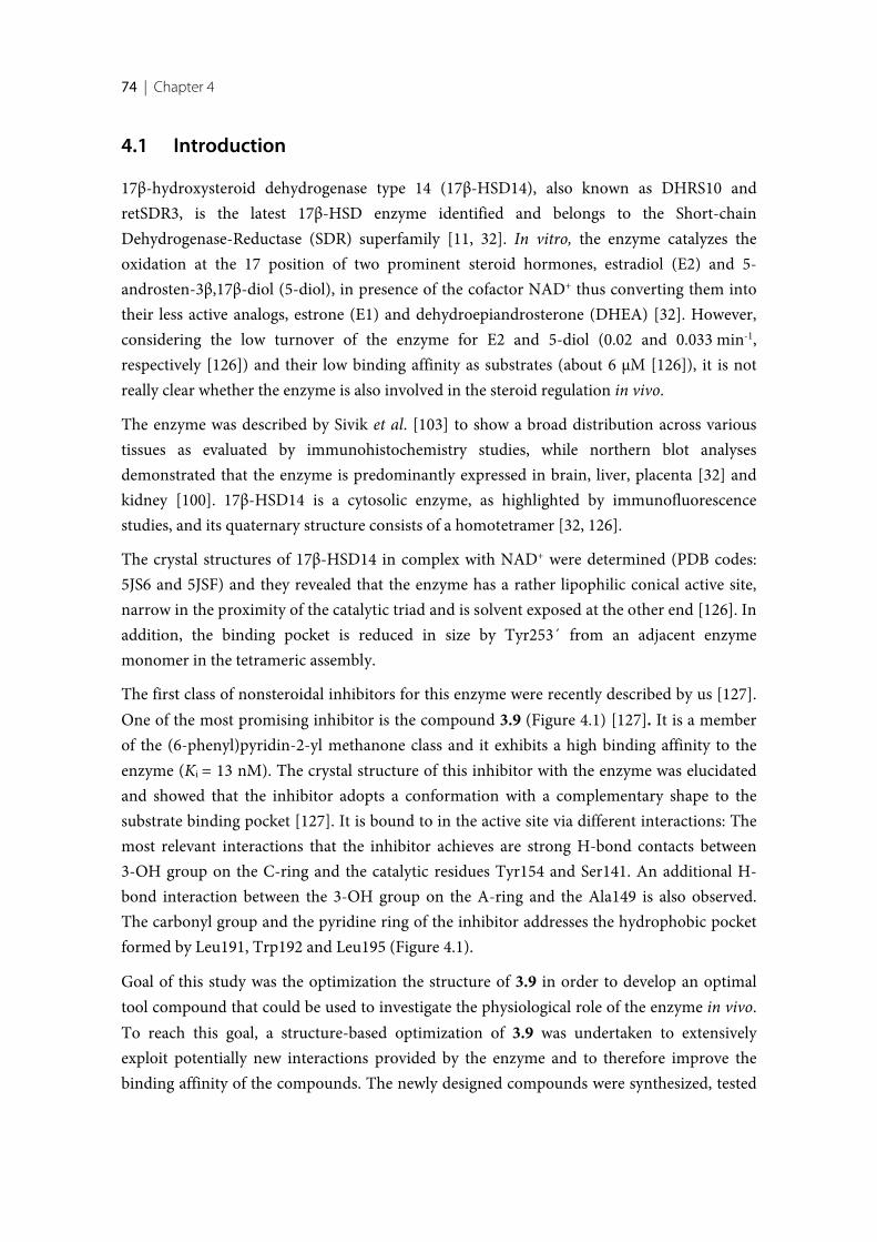
74 | Chapter 4
4.1 Introduction
17β-hydroxysteroid dehydrogenase type 14 (17β-HSD14), also known as DHRS10 andretSDR3, is the latest 17β-HSD enzyme identified and belongs to the Short-chainDehydrogenase-Reductase (SDR) superfamily [11, 32]. In vitro, the enzyme catalyzes theoxidation at the 17 position of two prominent steroid hormones, estradiol (E2) and 5-androsten-3β,17β-diol (5-diol), in presence of the cofactor NAD+ thus converting them intotheir less active analogs, estrone (E1) and dehydroepiandrosterone (DHEA) [32]. However,considering the low turnover of the enzyme for E2 and 5-diol (0.02 and 0.033 min-1,respectively [126]) and their low binding affinity as substrates (about 6 µM [126]), it is notreally clear whether the enzyme is also involved in the steroid regulation in vivo.
The enzyme was described by Sivik et al. [103] to show a broad distribution across varioustissues as evaluated by immunohistochemistry studies, while northern blot analysesdemonstrated that the enzyme is predominantly expressed in brain, liver, placenta [32] andkidney [100]. 17β-HSD14 is a cytosolic enzyme, as highlighted by immunofluorescencestudies, and its quaternary structure consists of a homotetramer [32, 126].
The crystal structures of 17β-HSD14 in complex with NAD+ were determined (PDB codes:5JS6 and 5JSF) and they revealed that the enzyme has a rather lipophilic conical active site,narrow in the proximity of the catalytic triad and is solvent exposed at the other end [126]. Inaddition, the binding pocket is reduced in size by Tyr253´ from an adjacent enzymemonomer in the tetrameric assembly.
The first class of nonsteroidal inhibitors for this enzyme were recently described by us [127].One of the most promising inhibitor is the compound 3.9 (Figure 4.1) [127]. It is a memberof the (6-phenyl)pyridin-2-yl methanone class and it exhibits a high binding affinity to theenzyme (Ki = 13 nM). The crystal structure of this inhibitor with the enzyme was elucidatedand showed that the inhibitor adopts a conformation with a complementary shape to thesubstrate binding pocket [127]. It is bound to in the active site via different interactions: Themost relevant interactions that the inhibitor achieves are strong H-bond contacts between3-OH group on the C-ring and the catalytic residues Tyr154 and Ser141. An additional H-bond interaction between the 3-OH group on the A-ring and the Ala149 is also observed.The carbonyl group and the pyridine ring of the inhibitor addresses the hydrophobic pocketformed by Leu191, Trp192 and Leu195 (Figure 4.1).
Goal of this study was the optimization the structure of 3.9 in order to develop an optimaltool compound that could be used to investigate the physiological role of the enzyme in vivo.To reach this goal, a structure-based optimization of 3.9 was undertaken to extensivelyexploit potentially new interactions provided by the enzyme and to therefore improve thebinding affinity of the compounds. The newly designed compounds were synthesized, tested
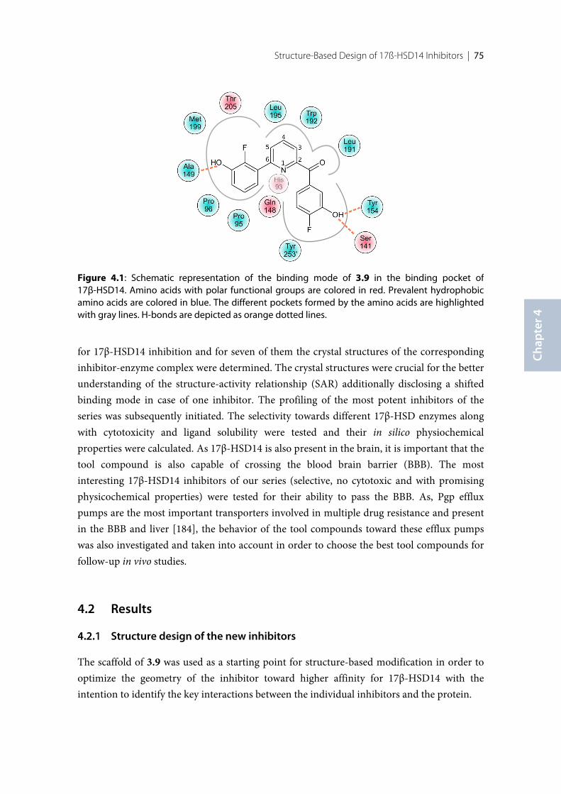
Structure-Based Design of 17ß-HSD14 Inhibitors | 75
Chap
ter 4
for 17β-HSD14 inhibition and for seven of them the crystal structures of the correspondinginhibitor-enzyme complex were determined. The crystal structures were crucial for the betterunderstanding of the structure-activity relationship (SAR) additionally disclosing a shiftedbinding mode in case of one inhibitor. The profiling of the most potent inhibitors of theseries was subsequently initiated. The selectivity towards different 17β-HSD enzymes alongwith cytotoxicity and ligand solubility were tested and their in silico physiochemicalproperties were calculated. As 17β-HSD14 is also present in the brain, it is important that thetool compound is also capable of crossing the blood brain barrier (BBB). The mostinteresting 17β-HSD14 inhibitors of our series (selective, no cytotoxic and with promisingphysicochemical properties) were tested for their ability to pass the BBB. As, Pgp effluxpumps are the most important transporters involved in multiple drug resistance and presentin the BBB and liver [184], the behavior of the tool compounds toward these efflux pumpswas also investigated and taken into account in order to choose the best tool compounds forfollow-up in vivo studies.
4.2 Results
Structure design of the new inhibitors
The scaffold of 3.9 was used as a starting point for structure-based modification in order tooptimize the geometry of the inhibitor toward higher affinity for 17β-HSD14 with theintention to identify the key interactions between the individual inhibitors and the protein.
Figure 4.1: Schematic representation of the binding mode of 3.9 in the binding pocket of17β-HSD14. Amino acids with polar functional groups are colored in red. Prevalent hydrophobicamino acids are colored in blue. The different pockets formed by the amino acids are highlightedwith gray lines. H-bonds are depicted as orange dotted lines.
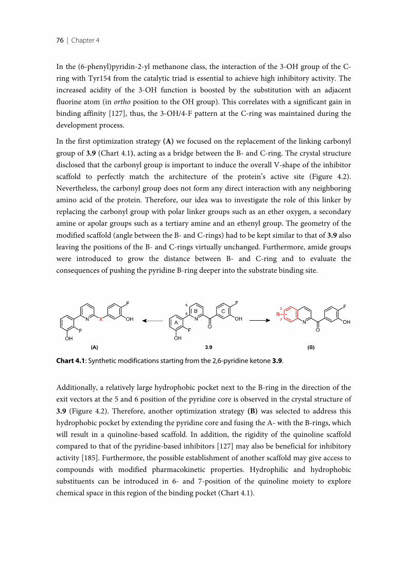
76 | Chapter 4
In the (6-phenyl)pyridin-2-yl methanone class, the interaction of the 3-OH group of the C-ring with Tyr154 from the catalytic triad is essential to achieve high inhibitory activity. Theincreased acidity of the 3-OH function is boosted by the substitution with an adjacentfluorine atom (in ortho position to the OH group). This correlates with a significant gain inbinding affinity [127], thus, the 3-OH/4-F pattern at the C-ring was maintained during thedevelopment process.
In the first optimization strategy (A) we focused on the replacement of the linking carbonylgroup of 3.9 (Chart 4.1), acting as a bridge between the B- and C-ring. The crystal structuredisclosed that the carbonyl group is important to induce the overall V-shape of the inhibitorscaffold to perfectly match the architecture of the protein’s active site (Figure 4.2).Nevertheless, the carbonyl group does not form any direct interaction with any neighboringamino acid of the protein. Therefore, our idea was to investigate the role of this linker byreplacing the carbonyl group with polar linker groups such as an ether oxygen, a secondaryamine or apolar groups such as a tertiary amine and an ethenyl group. The geometry of themodified scaffold (angle between the B- and C-rings) had to be kept similar to that of 3.9 alsoleaving the positions of the B- and C-rings virtually unchanged. Furthermore, amide groupswere introduced to grow the distance between B- and C-ring and to evaluate theconsequences of pushing the pyridine B-ring deeper into the substrate binding site.
Additionally, a relatively large hydrophobic pocket next to the B-ring in the direction of theexit vectors at the 5 and 6 position of the pyridine core is observed in the crystal structure of3.9 (Figure 4.2). Therefore, another optimization strategy (B) was selected to address thishydrophobic pocket by extending the pyridine core and fusing the A- with the B-rings, whichwill result in a quinoline-based scaffold. In addition, the rigidity of the quinoline scaffoldcompared to that of the pyridine-based inhibitors [127] may also be beneficial for inhibitoryactivity [185]. Furthermore, the possible establishment of another scaffold may give access tocompounds with modified pharmacokinetic properties. Hydrophilic and hydrophobicsubstituents can be introduced in 6- and 7-position of the quinoline moiety to explorechemical space in this region of the binding pocket (Chart 4.1).
Chart 4.1: Synthetic modifications starting from the 2,6-pyridine ketone 3.9.
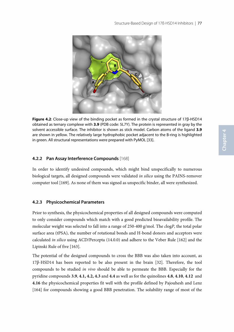
Structure-Based Design of 17ß-HSD14 Inhibitors | 77
Chap
ter 4
Figure 4.2: Close-up view of the binding pocket as formed in the crystal structure of 17β-HSD14obtained as ternary complexe with 3.9 (PDB code: 5L7Y). The protein is represented in gray by thesolvent accessible surface. The inhibitor is shown as stick model. Carbon atoms of the ligand 3.9are shown in yellow. The relatively large hydrophobic pocket adjacent to the B-ring is highlightedin green. All structural representations were prepared with PyMOL [33].
Pan Assay Interference Compounds [168]
In order to identify undesired compounds, which might bind unspecifically to numerousbiological targets, all designed compounds were validated in silico using the PAINS-removercomputer tool [169]. As none of them was signed as unspecific binder, all were synthesized.
Physicochemical Parameters
Prior to synthesis, the physicochemical properties of all designed compounds were computedto only consider compounds which match with a good predicted bioavailability profile. Themolecular weight was selected to fall into a range of 250-400 g/mol. The clogP, the total polarsurface area (tPSA), the number of rotational bonds and H-bond donors and acceptors werecalculated in silico using ACD/Percepta (14.0.0) and adhere to the Veber Rule [162] and theLipinski Rule of five [163].
The potential of the designed compounds to cross the BBB was also taken into account, as17β-HSD14 has been reported to be also present in the brain [32]. Therefore, the toolcompounds to be studied in vivo should be able to permeate the BBB. Especially for thepyridine compounds 3.9, 4.1, 4.2, 4.3 and 4.4 as well as for the quinolines 4.8, 4.10, 4.12 and4.16 the physicochemical properties fit well with the profile defined by Pajouhesh and Lenz[164] for compounds showing a good BBB penetration. The solubility range of most of the
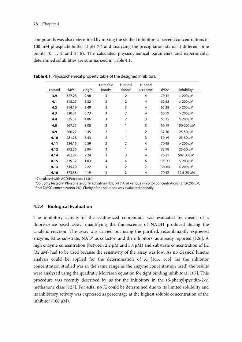
78 | Chapter 4
compounds was also determined by mixing the studied inhibitors at several concentrations in100 mM phosphate buffer at pH 7.4 and analyzing the precipitation status at different timepoints (0, 1, 2 and 24 h). The calculated physicochemical parameters and experimentaldetermined solubilities are summarized in Table 4.1.
Table 4.1: Physicochemical property table of the designed inhibitors.
compd MWa clogPa
rotatablebondsa
H-bonddonora
H-bondacceptora tPSAa Solubilityb
3.9 327.28 2.98 3 2 4 70.42 > 200 µM
4.1 315.27 3.32 3 2 4 62.58 > 200 µM
4.2 314.19 3.49 3 3 4 65.38 > 200 µM
4.3 328.31 3.73 3 2 4 56.59 > 200 µM
4.4 325.31 4.06 3 2 3 53.35 > 200 µM
4.8 267.25 3.08 2 1 3 50.19 100-200 µM
4.9 266.27 4.45 2 1 2 37.30 25-50 µM
4.10 281.28 3.43 2 1 3 50.19 25-50 µM
4.11 284.15 2.59 2 2 4 70.42 > 200 µM
4.12 292.26 2.86 3 1 4 73.98 25-50 µM
4.14 282.27 2.24 2 3 4 76.21 50-100 µM
4.15 339.32 1.03 4 4 6 105.31 > 200 µM
4.13 335.29 2.22 3 2 7 104.65 > 200 µM
4.16 373.38 4.19 3 2 4 70.42 12.5-25 µMaCalculated with ACD/Percepta 14.0.0bSolubilty tested in Phosphate-Buffered Saline (PBS, pH 7.4) at various inhibitor concentrations (3.13-200 µM,final DMSO concentration 2%). Clarity of the solutions was evaluated optically.
Biological Evaluation
The inhibitory activity of the synthesized compounds was evaluated by means of afluorescence-based assay, quantifying the fluorescence of NADH produced during thecatalytic reaction. The assay was carried out using the purified, recombinantly expressedenzyme, E2 as substrate, NAD+ as cofactor, and the inhibitors, as already reported [126]. Ahigh enzyme concentration (between 2.2 µM and 3.4 µM) and substrate concentration of E2(32 µM) had to be used because the sensitivity of the assay was low. As no classical kineticanalysis could be applied for the determination of Ki [165, 166] (as the inhibitorconcentration studied was in the same range as the enzyme concentration used) the resultswere analyzed using the quadratic Morrison equation for tight binding inhibitors [167]. Thisprocedure was recently described by us for the inhibitors in the (6-phenyl)pyridin-2-ylmethanone class [127]. For 4.8a, no Ki could be determined due to its limited solubility andits inhibitory activity was expressed as percentage at the highest soluble concentration of theinhibitor (100 µM).
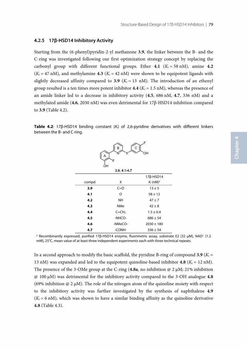
Structure-Based Design of 17ß-HSD14 Inhibitors | 79
Chap
ter 4
17β-HSD14 Inhibitory Activity
Starting from the (6-phenyl)pyridin-2-yl methanone 3.9, the linker between the B- and theC-ring was investigated following our first optimization strategy concept by replacing thecarbonyl group with different functional groups. Ether 4.1 (Ki = 58 nM), amine 4.2(Ki = 47 nM), and methylamine 4.3 (Ki = 42 nM) were shown to be equipotent ligands withslightly decreased affinity compared to 3.9 (Ki = 13 nM). The introduction of an ethenylgroup resulted is a ten times more potent inhibitor 4.4 (Ki = 1.5 nM), whereas the presence ofan amide linker led to a decrease in inhibitory activity (4.5, 686 nM, 4.7, 336 nM) and amethylated amide (4.6, 2030 nM) was even detrimental for 17β-HSD14 inhibition comparedto 3.9 (Table 4.2).
Table 4.2: 17β-HSD14 binding constant (Ki) of 2,6-pyridine derivatives with different linkersbetween the B- and C-ring.
compd X
17β-HSD14
Ki (nM)a
3.9 C=O 13 ± 5
4.1 O 58 ± 12
4.2 NH 47 ± 7
4.3 NMe 42 ± 8
4.4 C=CH2 1.5 ± 0.4
4.5 -NHCO- 686 ± 54
4.6 -NMeCO- 2030 ± 180
4.7 -CONH- 336 ± 54a Recombinantly expressed, purified 17β-HSD14 enzyme, fluorimetric assay, substrate E2 [32 µM], NAD+ [1.2mM], 25°C, mean value of at least three independent experiments each with three technical repeats.
In a second approach to modify the basic scaffold, the pyridine B-ring of compound 3.9 (Ki =13 nM) was expanded and led to the equipotent quinoline-based inhibitor 4.8 (Ki = 12 nM).The presence of the 3-OMe group at the C-ring (4.8a, no inhibition @ 2 µM; 21% inhibition@ 100 µM) was detrimental for the inhibitory activity compared to the 3-OH analogue 4.8(69% inhibition @ 2 µM). The role of the nitrogen atom of the quinoline moiety with respectto the inhibitory activity was further investigated by the synthesis of naphthalene 4.9(Ki = 6 nM), which was shown to have a similar binding affinity as the quinoline derivative4.8 (Table 4.3).
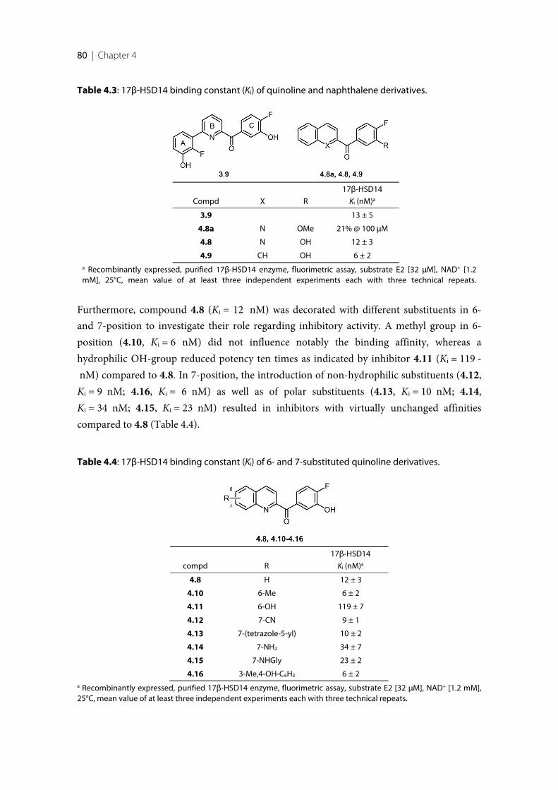
80 | Chapter 4
Table 4.3: 17β-HSD14 binding constant (Ki) of quinoline and naphthalene derivatives.
Compd X R
17β-HSD14
Ki (nM)a
3.9 13 ± 5
4.8a N OMe 21% @ 100 µM
4.8 N OH 12 ± 3
4.9 CH OH 6 ± 2a Recombinantly expressed, purified 17β-HSD14 enzyme, fluorimetric assay, substrate E2 [32 µM], NAD+ [1.2mM], 25°C, mean value of at least three independent experiments each with three technical repeats.
Furthermore, compound 4.8 (Ki = 12 nM) was decorated with different substituents in 6-and 7-position to investigate their role regarding inhibitory activity. A methyl group in 6-position (4.10, Ki = 6 nM) did not influence notably the binding affinity, whereas ahydrophilic OH-group reduced potency ten times as indicated by inhibitor 4.11 (Ki = 119 - nM) compared to 4.8. In 7-position, the introduction of non-hydrophilic substituents (4.12,Ki = 9 nM; 4.16, Ki = 6 nM) as well as of polar substituents (4.13, Ki = 10 nM; 4.14,Ki = 34 nM; 4.15, Ki = 23 nM) resulted in inhibitors with virtually unchanged affinitiescompared to 4.8 (Table 4.4).
Table 4.4: 17β-HSD14 binding constant (Ki) of 6- and 7-substituted quinoline derivatives.
compd R
17β-HSD14
Ki (nM)a
4.8 H 12 ± 3
4.10 6-Me 6 ± 2
4.11 6-OH 119 ± 7
4.12 7-CN 9 ± 1
4.13 7-(tetrazole-5-yl) 10 ± 2
4.14 7-NH2 34 ± 7
4.15 7-NHGly 23 ± 2
4.16 3-Me,4-OH-C6H3 6 ± 2a Recombinantly expressed, purified 17β-HSD14 enzyme, fluorimetric assay, substrate E2 [32 µM], NAD+ [1.2 mM],25°C, mean value of at least three independent experiments each with three technical repeats.
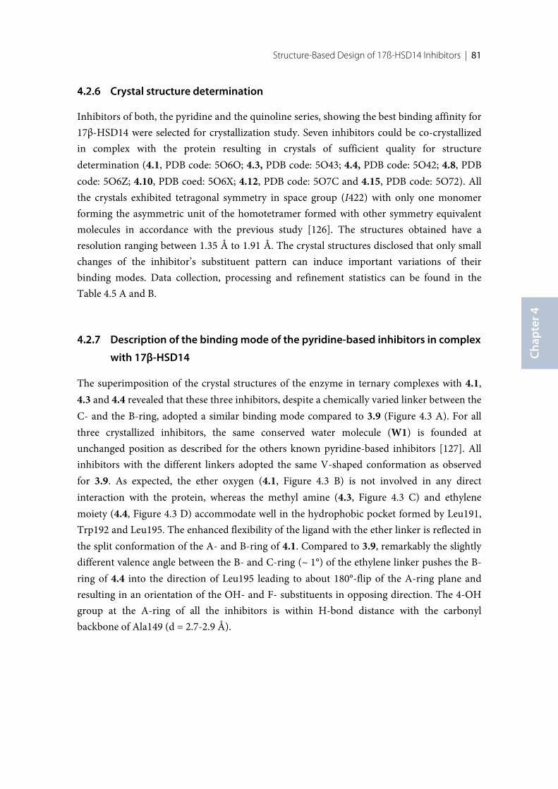
Structure-Based Design of 17ß-HSD14 Inhibitors | 81
Chap
ter 4
Crystal structure determination
Inhibitors of both, the pyridine and the quinoline series, showing the best binding affinity for17β-HSD14 were selected for crystallization study. Seven inhibitors could be co-crystallizedin complex with the protein resulting in crystals of sufficient quality for structuredetermination (4.1, PDB code: 5O6O; 4.3, PDB code: 5O43; 4.4, PDB code: 5O42; 4.8, PDBcode: 5O6Z; 4.10, PDB coed: 5O6X; 4.12, PDB code: 5O7C and 4.15, PDB code: 5O72). Allthe crystals exhibited tetragonal symmetry in space group (I422) with only one monomerforming the asymmetric unit of the homotetramer formed with other symmetry equivalentmolecules in accordance with the previous study [126]. The structures obtained have aresolution ranging between 1.35 Å to 1.91 Å. The crystal structures disclosed that only smallchanges of the inhibitor’s substituent pattern can induce important variations of theirbinding modes. Data collection, processing and refinement statistics can be found in theTable 4.5 A and B.
Description of the binding mode of the pyridine-based inhibitors in complex
with 17β-HSD14
The superimposition of the crystal structures of the enzyme in ternary complexes with 4.1,4.3 and 4.4 revealed that these three inhibitors, despite a chemically varied linker between theC- and the B-ring, adopted a similar binding mode compared to 3.9 (Figure 4.3 A). For allthree crystallized inhibitors, the same conserved water molecule (W1) is founded atunchanged position as described for the others known pyridine-based inhibitors [127]. Allinhibitors with the different linkers adopted the same V-shaped conformation as observedfor 3.9. As expected, the ether oxygen (4.1, Figure 4.3 B) is not involved in any directinteraction with the protein, whereas the methyl amine (4.3, Figure 4.3 C) and ethylenemoiety (4.4, Figure 4.3 D) accommodate well in the hydrophobic pocket formed by Leu191,Trp192 and Leu195. The enhanced flexibility of the ligand with the ether linker is reflected inthe split conformation of the A- and B-ring of 4.1. Compared to 3.9, remarkably the slightlydifferent valence angle between the B- and C-ring (~ 1°) of the ethylene linker pushes the B-ring of 4.4 into the direction of Leu195 leading to about 180°-flip of the A-ring plane andresulting in an orientation of the OH- and F- substituents in opposing direction. The 4-OHgroup at the A-ring of all the inhibitors is within H-bond distance with the carbonylbackbone of Ala149 (d = 2.7-2.9 Å).
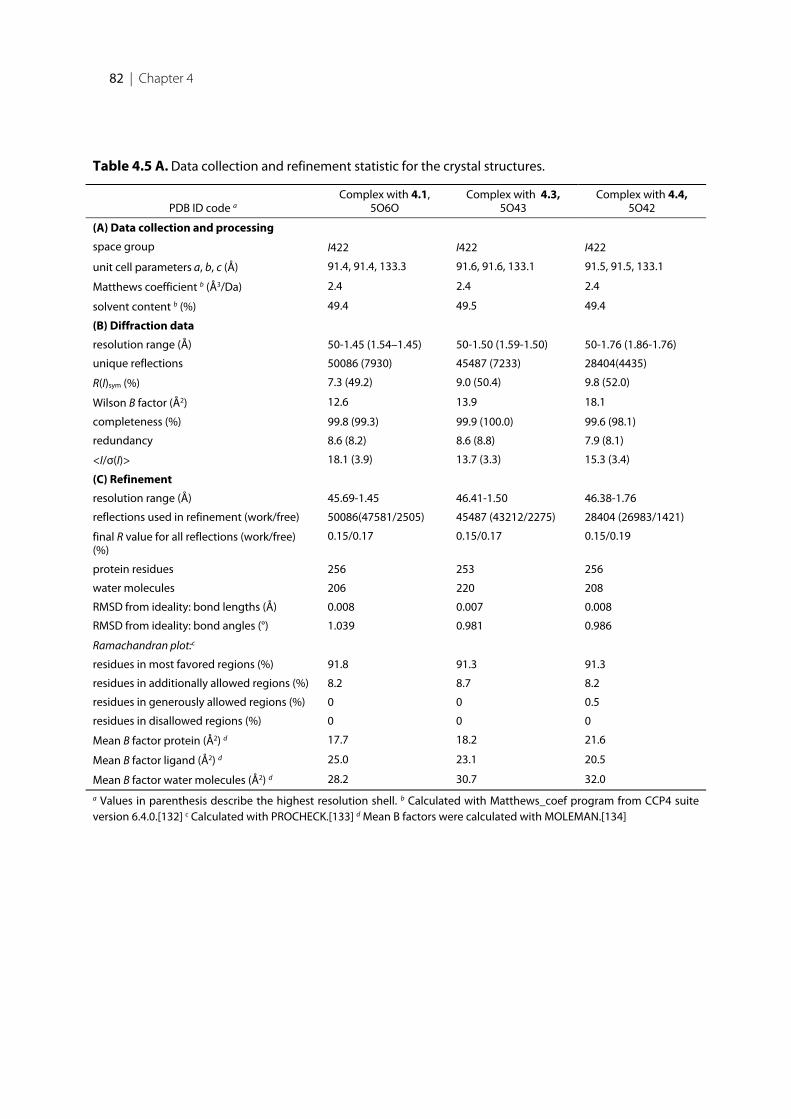
82 | Chapter 4
Table 4.5 A. Data collection and refinement statistic for the crystal structures.
PDB ID code aComplex with 4.1,
5O6OComplex with 4.3,
5O43Complex with 4.4,
5O42
(A) Data collection and processing
space group I422 I422 I422
unit cell parameters a, b, c (Å) 91.4, 91.4, 133.3 91.6, 91.6, 133.1 91.5, 91.5, 133.1
Matthews coefficient b (Å3/Da) 2.4 2.4 2.4
solvent content b (%) 49.4 49.5 49.4
(B) Diffraction data
resolution range (Å) 50-1.45 (1.54–1.45) 50-1.50 (1.59-1.50) 50-1.76 (1.86-1.76)
unique reflections 50086 (7930) 45487 (7233) 28404(4435)
R(I)sym (%) 7.3 (49.2) 9.0 (50.4) 9.8 (52.0)
Wilson B factor (Å2) 12.6 13.9 18.1
completeness (%) 99.8 (99.3) 99.9 (100.0) 99.6 (98.1)
redundancy 8.6 (8.2) 8.6 (8.8) 7.9 (8.1)
<I/σ(I)> 18.1 (3.9) 13.7 (3.3) 15.3 (3.4)
(C) Refinement
resolution range (Å) 45.69-1.45 46.41-1.50 46.38-1.76
reflections used in refinement (work/free) 50086(47581/2505) 45487 (43212/2275) 28404 (26983/1421)
final R value for all reflections (work/free)(%)
0.15/0.17 0.15/0.17 0.15/0.19
protein residues 256 253 256
water molecules 206 220 208
RMSD from ideality: bond lengths (Å) 0.008 0.007 0.008
RMSD from ideality: bond angles (°) 1.039 0.981 0.986
Ramachandran plot:c
residues in most favored regions (%) 91.8 91.3 91.3
residues in additionally allowed regions (%) 8.2 8.7 8.2
residues in generously allowed regions (%) 0 0 0.5
residues in disallowed regions (%) 0 0 0
Mean B factor protein (Å2) d 17.7 18.2 21.6
Mean B factor ligand (Å2) d 25.0 23.1 20.5
Mean B factor water molecules (Å2) d 28.2 30.7 32.0
a Values in parenthesis describe the highest resolution shell. b Calculated with Matthews_coef program from CCP4 suiteversion 6.4.0.[132] c Calculated with PROCHECK.[133] d Mean B factors were calculated with MOLEMAN.[134]
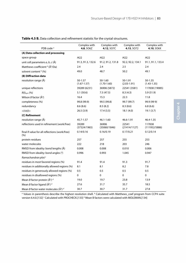
Structure-Based Design of 17ß-HSD14 Inhibitors | 83
Chap
ter 4
Table 4.5 B. Data collection and refinement statistic for the crystal structures.
PDB code aComplex with
4.8, 5O6ZComplex with
4.12, 5O7CComplex with
4.15, 5O72Complex with
4.10, 5O6X
(A) Data collection and processing
space group I422 I422 I422 I422
unit cell parameters a, b, c (Å) 91.3, 91.3, 132.6 91.2, 91.2, 131.8 92.2, 92.2, 134.1 91.1, 91.1, 133.4
Matthews coefficient b (Å3/Da) 2.4 2.4 2.5 2.4
solvent content b (%) 49.0 48.7 50.2 49.1
(B) Diffraction data
resolution range (Å) 50-1.57(1.67-1.57)
50-1.60(1.70-1.60)
50-1.91(2.03-1.91)
50-1.35(1.43-1.35)
unique reflections 39289 (6231) 36906 (5872) 22541 (3581) 117838 (19085)
R(I)sym (%) 5.1 (50.6) 7.5 (47.5) 8.3 (4.3) 5.9 (51.9)
Wilson B factor (Å2) 16.4 15.3 22.3 11.8
completeness (%) 99.8 (99.9) 99.5 (99.8) 99.7 (99.7) 99.9 (99 9)
redundancy 8.6 (8.6) 8.5 (8.2) 8.3 (8.6) 6.8 (6.6)
<I/σ(I)> 26.5 (3.9) 17.4 (3.5) 18.1 (4.0) 19.1 (3.7)
(C) Refinement
resolution range (Å) 45.7-1.57 46.1-1.60 46.6-1.91 46.4-1.35
reflections used in refinement (work/free) 39289(37324/1965)
36906(35060/1846)
22541(21414/1127)
117838(111952/5886)
final R value for all reflections (work/free)(%)
0.14/0.16 0.16/0.19 0.17/0.21 0.12/0.14
protein residues 257 257 255 253
water molecules 222 218 203 246
RMSD from ideality: bond lengths (Å) 0.008 0.008 0.010 0.006
RMSD from ideality: bond angles (°) 0.996 0.993 1.045 0.947
Ramachandran plot:c
residues in most favored regions (%) 91.4 91.4 91.3 91.7
residues in additionally allowed regions (%) 8.1 8.1 8.2 7.8
residues in generously allowed regions (%) 0.5 0.5 0.5 0.5
residues in disallowed regions (%) 0 0 0 0
Mean B factor protein (Å2) d 19.0 19.7 23.8 13.9
Mean B factor ligand (Å2) d 27.6 31.7 35.7 18.5
Mean B factor water molecules (Å2) d 30.7 30.7 31.7 27.8
a Values in parenthesis describe the highest resolution shell. b Calculated with Matthews_coef program from CCP4 suiteversion 6.4.0.[132] c Calculated with PROCHECK.[133] d Mean B factors were calculated with MOLEMAN.[134]
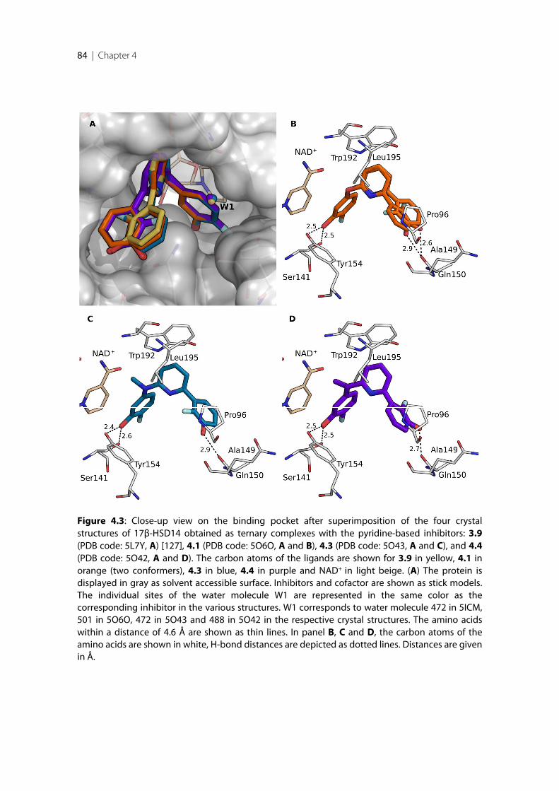
84 | Chapter 4
Figure 4.3: Close-up view on the binding pocket after superimposition of the four crystalstructures of 17β-HSD14 obtained as ternary complexes with the pyridine-based inhibitors: 3.9(PDB code: 5L7Y, A) [127], 4.1 (PDB code: 5O6O, A and B), 4.3 (PDB code: 5O43, A and C), and 4.4(PDB code: 5O42, A and D). The carbon atoms of the ligands are shown for 3.9 in yellow, 4.1 inorange (two conformers), 4.3 in blue, 4.4 in purple and NAD+ in light beige. (A) The protein isdisplayed in gray as solvent accessible surface. Inhibitors and cofactor are shown as stick models.The individual sites of the water molecule W1 are represented in the same color as thecorresponding inhibitor in the various structures. W1 corresponds to water molecule 472 in 5ICM,501 in 5O6O, 472 in 5O43 and 488 in 5O42 in the respective crystal structures. The amino acidswithin a distance of 4.6 Å are shown as thin lines. In panel B, C and D, the carbon atoms of theamino acids are shown in white, H-bond distances are depicted as dotted lines. Distances are givenin Å.
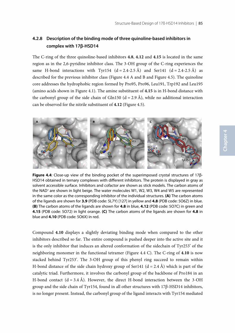
Structure-Based Design of 17ß-HSD14 Inhibitors | 85
Chap
ter 4
Description of the binding mode of three quinoline-based inhibitors in
complex with 17β-HSD14
The C-ring of the three quinoline-based inhibitors 4.8, 4.12 and 4.15 is located in the sameregion as in the 2,6-pyridine inhibitor class. The 3-OH group of the C-ring experiences thesame H-bond interactions with Tyr154 (d = 2.4-2.5 Å) and Ser141 (d = 2.4-2.5 Å) asdescribed for the previous inhibitor class (Figure 4.4 A and B and Figure 4.5). The quinolinecore addresses the hydrophobic region formed by Pro95, Pro96, Leu191, Trp192 and Leu195(amino acids shown in Figure 4.1). The amine substituent of 4.15 is in H-bond distance withthe carbonyl group of the side chain of Gln150 (d = 2.9 Å), while no additional interactioncan be observed for the nitrile substituent of 4.12 (Figure 4.5).
Figure 4.4: Close-up view of the binding pocket of the superimposed crystal structures of 17β-HSD14 obtained in ternary complexes with different inhibitors. The protein is displayed in gray assolvent accessible surface. Inhibitors and cofactor are shown as stick models. The carbon atoms ofthe NAD+ are shown in light beige. The water molecules W1, W2, W3, W4 and W5 are representedin the same color as the corresponding inhibitor of the individual structures. (A) The carbon atomsof the ligands are shown for 3.9 (PDB code: 5L7Y) [127] in yellow and 4.8 (PDB code: 5O6Z) in blue.(B) The carbon atoms of the ligands are shown for 4.8 in blue, 4.12 (PDB code: 5O7C) in green and4.15 (PDB code: 5O72) in light orange. (C) The carbon atoms of the ligands are shown for 4.8 inblue and 4.10 (PDB code: 5O6X) in red.
Compound 4.10 displays a slightly deviating binding mode when compared to the otherinhibitors described so far. The entire compound is pushed deeper into the active site and itis the only inhibitor that induces an altered conformation of the sidechain of Tyr253’ of theneighboring monomer in the functional tetramer (Figure 4.4 C). The C-ring of 4.10 is nowstacked behind Tyr253’. The 3-OH group of this phenyl ring succeed to remain withinH-bond distance of the side chain hydroxy group of Ser141 (d = 2.4 Å) which is part of thecatalytic triad. Furthermore, it involves the carbonyl group of the backbone of Pro184 in anH-bond contact (d = 3.4 Å). However, the direct H-bond interaction between the 3-OHgroup and the side chain of Tyr154, found in all other structures with 17β-HSD14 inhibitors,is no longer present. Instead, the carbonyl group of the ligand interacts with Tyr154 mediated
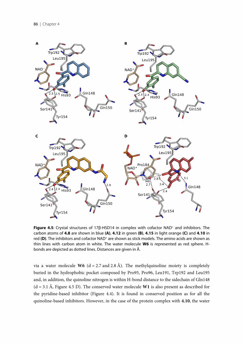
86 | Chapter 4
via a water molecule W6 (d = 2.7 and 2.8 Å). The methylquinoline moiety is completelyburied in the hydrophobic pocket composed by Pro95, Pro96, Leu191, Trp192 and Leu195and, in addition, the quinoline nitrogen is within H-bond distance to the sidechain of Gln148(d = 3.1 Å, Figure 4.5 D). The conserved water molecule W1 is also present as described forthe pyridine-based inhibitor (Figure 4.4). It is found in conserved position as for all thequinoline-based inhibitors. However, in the case of the protein complex with 4.10, the water
Figure 4.5: Crystal structures of 17β-HSD14 in complex with cofactor NAD+ and inhibitors. Thecarbon atoms of 4.8 are shown in blue (A), 4.12 in green (B), 4.15 in light orange (C) and 4.10 inred (D). The inhibitors and cofactor NAD+ are shown as stick models. The amino acids are shown asthin lines with carbon atom in white. The water molecule W6 is represented as red sphere. H-bonds are depicted as dotted lines. Distances are given in Å.
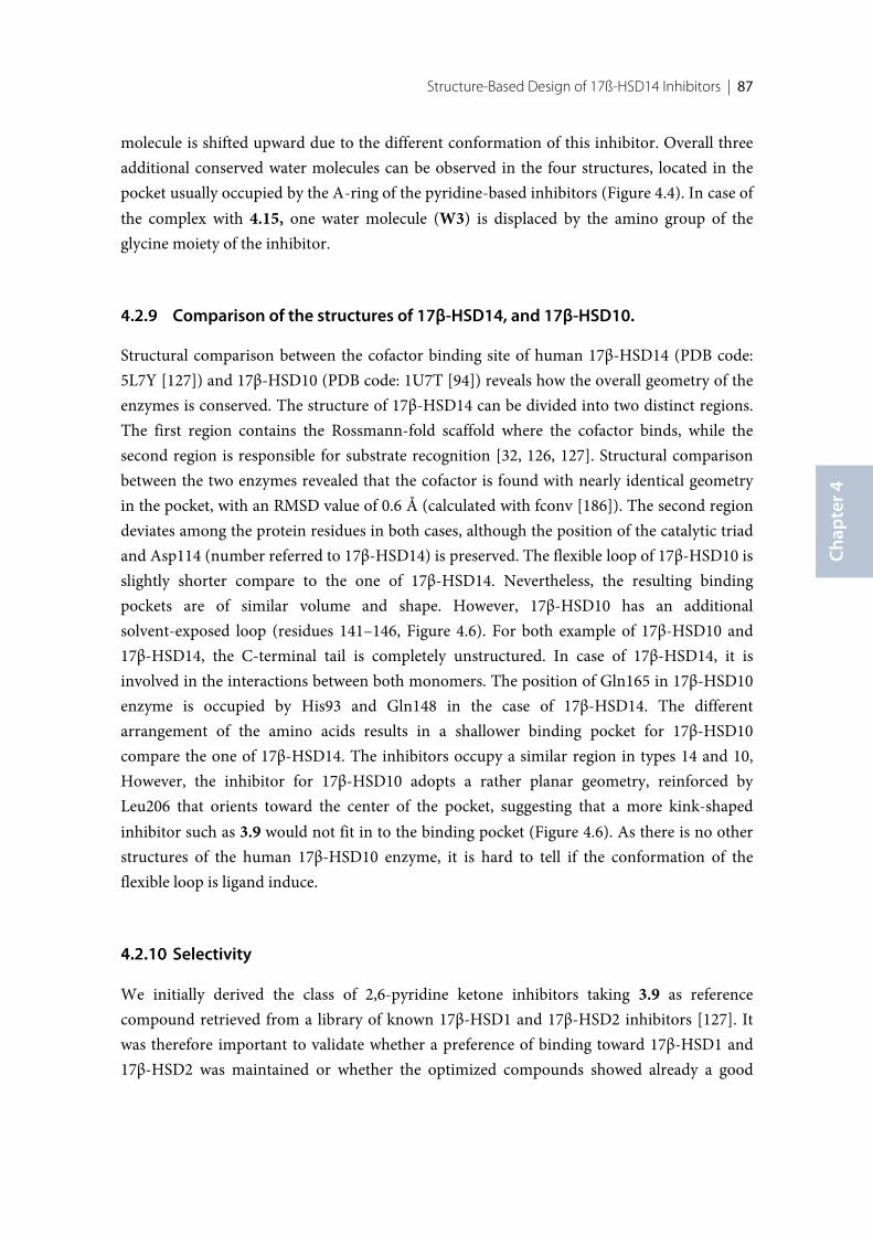
Structure-Based Design of 17ß-HSD14 Inhibitors | 87
Chap
ter 4
molecule is shifted upward due to the different conformation of this inhibitor. Overall threeadditional conserved water molecules can be observed in the four structures, located in thepocket usually occupied by the A-ring of the pyridine-based inhibitors (Figure 4.4). In case ofthe complex with 4.15, one water molecule (W3) is displaced by the amino group of theglycine moiety of the inhibitor.
Comparison of the structures of 17β-HSD14, and 17β-HSD10.
Structural comparison between the cofactor binding site of human 17β-HSD14 (PDB code:5L7Y [127]) and 17β-HSD10 (PDB code: 1U7T [94]) reveals how the overall geometry of theenzymes is conserved. The structure of 17β-HSD14 can be divided into two distinct regions.The first region contains the Rossmann-fold scaffold where the cofactor binds, while thesecond region is responsible for substrate recognition [32, 126, 127]. Structural comparisonbetween the two enzymes revealed that the cofactor is found with nearly identical geometryin the pocket, with an RMSD value of 0.6 Å (calculated with fconv [186]). The second regiondeviates among the protein residues in both cases, although the position of the catalytic triadand Asp114 (number referred to 17β-HSD14) is preserved. The flexible loop of 17β-HSD10 isslightly shorter compare to the one of 17β-HSD14. Nevertheless, the resulting bindingpockets are of similar volume and shape. However, 17β-HSD10 has an additionalsolvent-exposed loop (residues 141–146, Figure 4.6). For both example of 17β-HSD10 and17β-HSD14, the C-terminal tail is completely unstructured. In case of 17β-HSD14, it isinvolved in the interactions between both monomers. The position of Gln165 in 17β-HSD10enzyme is occupied by His93 and Gln148 in the case of 17β-HSD14. The differentarrangement of the amino acids results in a shallower binding pocket for 17β-HSD10compare the one of 17β-HSD14. The inhibitors occupy a similar region in types 14 and 10,However, the inhibitor for 17β-HSD10 adopts a rather planar geometry, reinforced byLeu206 that orients toward the center of the pocket, suggesting that a more kink-shapedinhibitor such as 3.9 would not fit in to the binding pocket (Figure 4.6). As there is no otherstructures of the human 17β-HSD10 enzyme, it is hard to tell if the conformation of theflexible loop is ligand induce.
Selectivity
We initially derived the class of 2,6-pyridine ketone inhibitors taking 3.9 as referencecompound retrieved from a library of known 17β-HSD1 and 17β-HSD2 inhibitors [127]. Itwas therefore important to validate whether a preference of binding toward 17β-HSD1 and17β-HSD2 was maintained or whether the optimized compounds showed already a good
88 | Chapter 4
selectivity profile toward 17β-HSD14. In addition, as mentioned previously, the investigationof the selectivity profile with respect to binding toward 17β-HSD10 is of high concernconsidering the given high similarity of 17β-HSD14 with the latter enzyme.
We selected inhibitors that showed good inhibition of 17β-HSD14 (Ki < 100 nM) to validatethe selectivity profile towards 17β-HSD1, 17β-HSD2 and 17β-HSD10. The potential of theselected 14 inhibitors to inhibit 17β-HSD1, 17β-HSD2 and 17β-HSD10 was studied using acompetitive radioactive assay, by quantifying the formation of of [3H]-labeled E2 (for 17β-HSD type 1) and [3H]-labeled estrone (E1, for 17β-HSD type 2 and 10) after incubation of theprotein with the cofactor and the inhibitor [174]. The results of the assay are shown in Table4.6 and expressed as percentage of inhibition.
The first approach to modify the scaffold of the pyridine derivatives resulted in theproduction of less selective compounds (4.1, 4.2, 4.3 and 4.4), which showed even higheraffinity for 17β-HSD1 and 17β-HSD2, (57-99% 17β-HSD1 inhibition @ 1 µM and 82-100%
Figure 4.6: Superimposition of 17β-HSD14 (yellow, PDB code: 5L7Y) and 17β-HSD10 (orange, PDBcode: 1U7T) structures. The proteins are shown as ribbon model. The ligands and cofactors areshown as stick models and their carbon atoms are colored as the corresponding protein structure.
Structure-Based Design of 17ß-HSD14 Inhibitors | 89
Chap
ter 4
17β-HSD2 inhibition @ 1 µM) compared to the initial compound 3.9 (8% 17β-HSD1inhibition and 56% 17β-HSD2 inhibition @ 1 µM).
The second strategy to optimize the scaffold, which led to the quinoline-based compounds4.8-4.16, resulted in inhibitors which showed no significant inhibitory activity toward17β-HSD1, only the naphthalene derivative 4.9 falls out of this profile and exhibits high 17β-HSD1 inhibitory activity (64% inhibition @ 1 µM).
In our previous study [127], we could show that 3.9 shows a 17β-HSD2 inhibition of 50% at1µM, corresponding to an IC50 of 1 µM. Taking into account the kinetic constant of the assay(Km= 400 nM, substrate concentration = 500 nM), a Ki for 3.9 can be estimated to be about450 nM. The determined Ki for 3.9 in the 17β-HSD14 assay was 13 nM. 3.9 is therefore about35 fold more selective on 17β-HSD14 compared to 17β-HSD2. As compounds 4.12, 4.13 and4.15 exhibit a 17β-HSD2 inhibition in the same range as 3.9 (52-63% inhibition @ 1 µM), itcan be expected that they will be as selective as 3.9 toward 17β-HSD2, thus underlining thesatisfactory selectivity discrimination of the quinoline-based inhibitors against 17β-HSD1and 17β-HSD2.
With respect to 17β-HSD10, only compounds 4.1 and 4.3 showed a weak inhibitory activity(17% and 19% inhibition @ 1 µM, respectively) while all the other synthesized derivatives areinactive suggesting an overall very convincing selectivity profile toward 17β-HSD10 (Table4.6).
Cytotoxicity evaluation
Seven compounds, selected on the basis of their 17β-HSD14 inhibition profile and selectivitywere evaluated for their cytotoxicity using an MTT assay based on the HEK293 cell line. Alltested compounds, 3.9, 4.4, 4.11, 4.13 and 4.15 displayed a very low cytotoxicity (IG50 >100 µM). Due to insufficient solubility, 4.11 and 4.12 could not be tested at 100 µM, however,the cells showed 80% survival at a concentration of 20 µM, the maximally possibleconcentration tested.
Pgp ATPase Activity Assays
Pgp, also termed MDR1 and ABCB1, is an integral plasma membrane protein expressed atthe luminal surface of the BBB [187]. The enzyme is an ATP-dependent drug efflux pump,which plays an important role in multi-drug resistance [184]. As mentioned, 17β-HSD14 wasdescribed to be also expressed in the brain and liver [100]. Goal of the project is thedevelopment of tool compounds to study the physiological role of 17β-HSD14 in vivo, it was
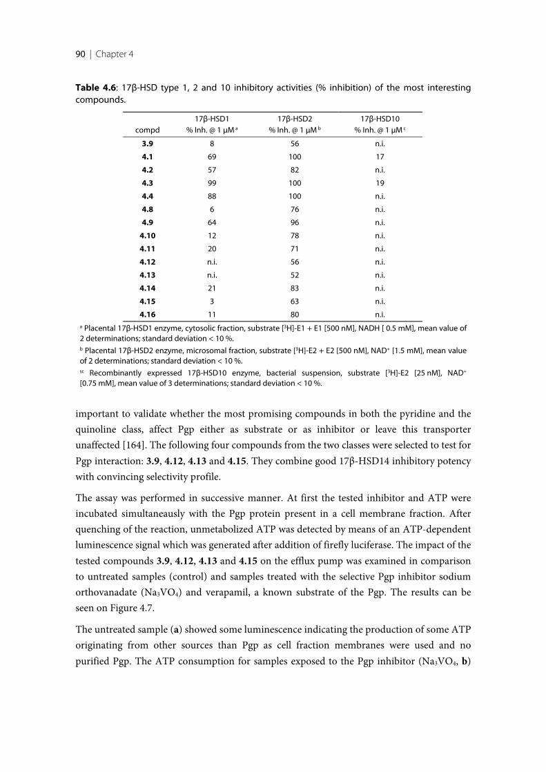
90 | Chapter 4
Table 4.6: 17β-HSD type 1, 2 and 10 inhibitory activities (% inhibition) of the most interestingcompounds.
compd17β-HSD1
% Inh. @ 1 µM a
17β-HSD2% Inh. @ 1 µM b
17β-HSD10% Inh. @ 1 µM c
3.9 8 56 n.i.
4.1 69 100 17
4.2 57 82 n.i.
4.3 99 100 19
4.4 88 100 n.i.
4.8 6 76 n.i.
4.9 64 96 n.i.
4.10 12 78 n.i.
4.11 20 71 n.i.
4.12 n.i. 56 n.i.
4.13 n.i. 52 n.i.
4.14 21 83 n.i.
4.15 3 63 n.i.
4.16 11 80 n.i.a Placental 17β-HSD1 enzyme, cytosolic fraction, substrate [3H]-E1 + E1 [500 nM], NADH [ 0.5 mM], mean value of2 determinations; standard deviation < 10 %.b Placental 17β-HSD2 enzyme, microsomal fraction, substrate [3H]-E2 + E2 [500 nM], NAD+ [1.5 mM], mean valueof 2 determinations; standard deviation < 10 %.sc Recombinantly expressed 17β-HSD10 enzyme, bacterial suspension, substrate [3H]-E2 [25 nM], NAD+
[0.75 mM], mean value of 3 determinations; standard deviation < 10 %.
important to validate whether the most promising compounds in both the pyridine and thequinoline class, affect Pgp either as substrate or as inhibitor or leave this transporterunaffected [164]. The following four compounds from the two classes were selected to test forPgp interaction: 3.9, 4.12, 4.13 and 4.15. They combine good 17β-HSD14 inhibitory potencywith convincing selectivity profile.
The assay was performed in successive manner. At first the tested inhibitor and ATP wereincubated simultaneausly with the Pgp protein present in a cell membrane fraction. Afterquenching of the reaction, unmetabolized ATP was detected by means of an ATP-dependentluminescence signal which was generated after addition of firefly luciferase. The impact of thetested compounds 3.9, 4.12, 4.13 and 4.15 on the efflux pump was examined in comparisonto untreated samples (control) and samples treated with the selective Pgp inhibitor sodiumorthovanadate (Na3VO4) and verapamil, a known substrate of the Pgp. The results can beseen on Figure 4.7.
The untreated sample (a) showed some luminescence indicating the production of some ATPoriginating from other sources than Pgp as cell fraction membranes were used and nopurified Pgp. The ATP consumption for samples exposed to the Pgp inhibitor (Na3VO4, b)
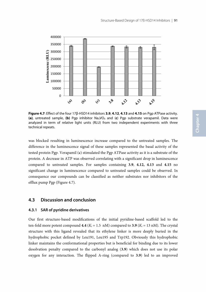
Structure-Based Design of 17ß-HSD14 Inhibitors | 91
Chap
ter 4
was blocked resulting in luminescence increase compared to the untreated samples. Thedifference in the luminescence signal of these samples represented the basal activity of thetested protein Pgp. Verapamil (c) stimulated the Pgp ATPase activity as it is a substrate of theprotein. A decrease in ATP was observed correlating with a significant drop in luminescencecompared to untreated samples. For samples containing 3.9, 4.12, 4.13 and 4.15 nosignificant change in luminescence compared to untreated samples could be observed. Inconsequence our compounds can be classified as neither substrates nor inhibitors of theefflux pump Pgp (Figure 4.7).
4.3 Discussion and conclusion
SAR of pyridine derivatives
Our first structure-based modifications of the initial pyridine-based scaffold led to theten-fold more potent compound 4.4 (Ki = 1.5 nM) compared to 3.9 (Ki = 13 nM). The crystalstructure with this ligand revealed that its ethylene linker is more deeply buried in thehydrophobic pocket defined by Leu191, Leu195 and Trp192. Obviously this hydrophobiclinker maintains the conformational properties but is beneficial for binding due to its lowerdesolvation penalty compared to the carbonyl analog (3.9) which does not use its polaroxygen for any interaction. The flipped A-ring (compared to 3.9) led to an improved
Figure 4.7: Effect of the four 17β-HSD14 inhibitors 3.9, 4.12, 4.13 and 4.15 on Pgp ATPase activity.(a), untreated sample, (b) Pgp inhibitor Na3VO4 and (c) Pgp substrate verapamil. Data wereanalyzed in term of relative light units (RLU) from two independent experiments with threetechnical repeats.
0
50000
100000
150000
200000
250000
300000
350000
400000
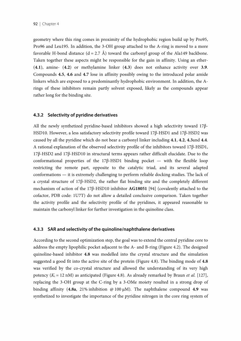
92 | Chapter 4
geometry where this ring comes in proximity of the hydrophobic region build up by Pro95,Pro96 and Leu195. In addition, the 3-OH group attached to the A-ring is moved to a morefavorable H-bond distance (d = 2.7 Å) toward the carbonyl group of the Ala149 backbone.Taken together these aspects might be responsible for the gain in affinity. Using an ether-(4.1), amine- (4.2) or methylamine linker (4.3) does not enhance activity over 3.9.Compounds 4.5, 4.6 and 4.7 lose in affinity possibly owing to the introduced polar amidelinkers which are exposed to a predominantly hydrophobic environment. In addition, the A-rings of these inhibitors remain partly solvent exposed, likely as the compounds appearrather long for the binding site.
Selectivity of pyridine derivatives
All the newly synthetized pyridine-based inhibitors showed a high selectivity toward 17β-HSD10. However, a less satisfactory selectivity profile toward 17β-HSD1 and 17β-HSD2 wascaused by all the pyridine which do not bear a carbonyl linker including 4.1, 4.2, 4.3and 4.4.A rational explanation of the observed selectivity profile of the inhibitors toward 17β-HSD1,17β-HSD2 and 17β-HSD10 in structural terms appears rather difficult elucidate. Due to theconformational properties of the 17β-HSD1 binding pocket — with the flexible looprestricting the remote part, opposite to the catalytic triad, and its several adaptedconformations — it is extremely challenging to perform reliable docking studies. The lack ofa crystal structure of 17β-HSD2, the rather flat binding site and the completely differentmechanism of action of the 17β-HSD10 inhibitor AG18051 [94] (covalently attached to thecofactor, PDB code: 1U7T) do not allow a detailed conclusive comparison. Taken togetherthe activity profile and the selectivity profile of the pyridines, it appeared reasonable tomaintain the carbonyl linker for further investigation in the quinoline class.
SAR and selectivity of the quinoline/naphthalene derivatives
According to the second optimization step, the goal was to extend the central pyridine core toaddress the empty lipophilic pocket adjacent to the A- and B-ring (Figure 4.2). The designedquinoline-based inhibitor 4.8 was modelled into the crystal structure and the simulationsuggested a good fit into the active site of the protein (Figure 4.8). The binding mode of 4.8was verified by the co-crystal structure and allowed the understanding of its very highpotency (Ki = 12 nM) as anticipated (Figure 4.8). As already remarked by Braun et al. [127],replacing the 3-OH group at the C-ring by a 3-OMe moiety resulted in a strong drop ofbinding affinity (4.8a, 21% inhibition @ 100 µM). The naphthalene compound 4.9 wassynthetized to investigate the importance of the pyridine nitrogen in the core ring system of
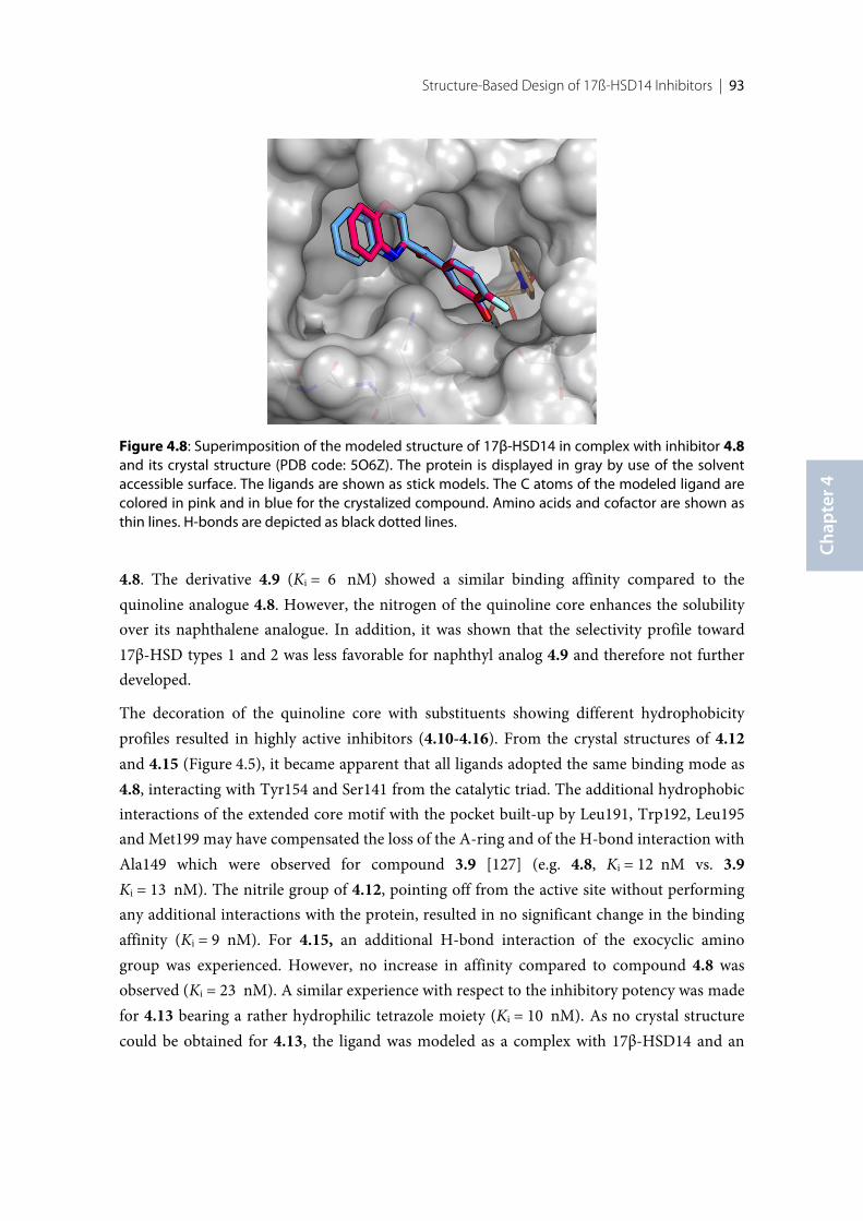
Structure-Based Design of 17ß-HSD14 Inhibitors | 93
Chap
ter 4
4.8. The derivative 4.9 (Ki = 6 nM) showed a similar binding affinity compared to thequinoline analogue 4.8. However, the nitrogen of the quinoline core enhances the solubilityover its naphthalene analogue. In addition, it was shown that the selectivity profile toward17β-HSD types 1 and 2 was less favorable for naphthyl analog 4.9 and therefore not furtherdeveloped.
The decoration of the quinoline core with substituents showing different hydrophobicityprofiles resulted in highly active inhibitors (4.10-4.16). From the crystal structures of 4.12and 4.15 (Figure 4.5), it became apparent that all ligands adopted the same binding mode as4.8, interacting with Tyr154 and Ser141 from the catalytic triad. The additional hydrophobicinteractions of the extended core motif with the pocket built-up by Leu191, Trp192, Leu195and Met199 may have compensated the loss of the A-ring and of the H-bond interaction withAla149 which were observed for compound 3.9 [127] (e.g. 4.8, Ki = 12 nM vs. 3.9Ki = 13 nM). The nitrile group of 4.12, pointing off from the active site without performingany additional interactions with the protein, resulted in no significant change in the bindingaffinity (Ki = 9 nM). For 4.15, an additional H-bond interaction of the exocyclic aminogroup was experienced. However, no increase in affinity compared to compound 4.8 wasobserved (Ki = 23 nM). A similar experience with respect to the inhibitory potency was madefor 4.13 bearing a rather hydrophilic tetrazole moiety (Ki = 10 nM). As no crystal structurecould be obtained for 4.13, the ligand was modeled as a complex with 17β-HSD14 and an
Figure 4.8: Superimposition of the modeled structure of 17β-HSD14 in complex with inhibitor 4.8and its crystal structure (PDB code: 5O6Z). The protein is displayed in gray by use of the solventaccessible surface. The ligands are shown as stick models. The C atoms of the modeled ligand arecolored in pink and in blue for the crystalized compound. Amino acids and cofactor are shown asthin lines. H-bonds are depicted as black dotted lines.
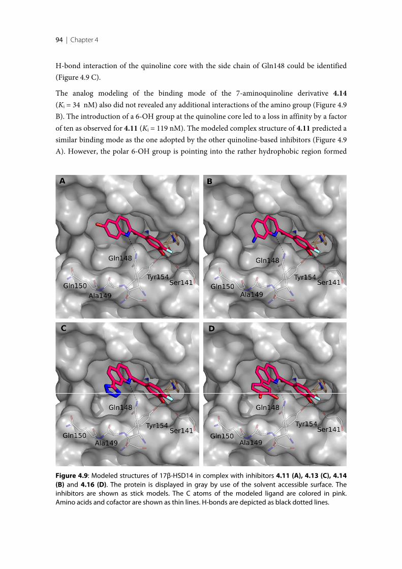
94 | Chapter 4
H-bond interaction of the quinoline core with the side chain of Gln148 could be identified(Figure 4.9 C).
The analog modeling of the binding mode of the 7-aminoquinoline derivative 4.14(Ki = 34 nM) also did not revealed any additional interactions of the amino group (Figure 4.9B). The introduction of a 6-OH group at the quinoline core led to a loss in affinity by a factorof ten as observed for 4.11 (Ki = 119 nM). The modeled complex structure of 4.11 predicted asimilar binding mode as the one adopted by the other quinoline-based inhibitors (Figure 4.9A). However, the polar 6-OH group is pointing into the rather hydrophobic region formed
Figure 4.9: Modeled structures of 17β-HSD14 in complex with inhibitors 4.11 (A), 4.13 (C), 4.14(B) and 4.16 (D). The protein is displayed in gray by use of the solvent accessible surface. Theinhibitors are shown as stick models. The C atoms of the modeled ligand are colored in pink.Amino acids and cofactor are shown as thin lines. H-bonds are depicted as black dotted lines.
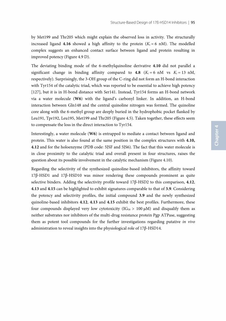
Structure-Based Design of 17ß-HSD14 Inhibitors | 95
Chap
ter 4
by Met199 and Thr205 which might explain the observed loss in activity. The structurallyincreased ligand 4.16 showed a high affinity to the protein (Ki = 6 nM). The modelledcomplex suggests an enhanced contact surface between ligand and protein resulting inimproved potency (Figure 4.9 D).
The deviating binding mode of the 6-methylquinoline derivative 4.10 did not parallel asignificant change in binding affinity compared to 4.8 (Ki = 6 nM vs Ki = 13 nM,respectively). Surprisingly, the 3-OH group of the C-ring did not form an H-bond interactionwith Tyr154 of the catalytic triad, which was reported to be essential to achieve high potency[127], but it is in H-bond distance with Ser141. Instead, Tyr154 forms an H-bond networkvia a water molecule (W6) with the ligand’s carbonyl linker. In addition, an H-bondinteraction between Gln148 and the central quinoline nitrogen was formed. The quinolinecore along with the 6-methyl group are deeply buried in the hydrophobic pocket flanked byLeu191, Tpr192, Leu195, Met199 and Thr205 (Figure 4.5). Taken together, these effects seemto compensate the loss in the direct interaction to Tyr154.
Interestingly, a water molecule (W6) is entrapped to mediate a contact between ligand andprotein. This water is also found at the same position in the complex structures with 4.10,4.12 and for the holoenzyme (PDB code: 5JSF and 5JS6). The fact that this water molecule isin close proximity to the catalytic triad and overall present in four structures, raises thequestion about its possible involvement in the catalytic mechanism (Figure 4.10).
Regarding the selectivity of the synthesized quinoline-based inhibitors, the affinity toward17β-HSD1 and 17β-HSD10 was minor rendering these compounds prominent as quiteselective binders. Adding the selectivity profile toward 17β-HSD2 to this comparison, 4.12,4.13 and 4.15 can be highlighted to exhibit signatures comparable to that of 3.9. Consideringthe potency and selectivity profiles, the initial compound 3.9 and the newly synthesizedquinoline-based inhibitors 4.12, 4.13 and 4.15 exhibit the best profiles. Furthermore, thesefour compounds displayed very low cytotoxicity (IG50 > 100 µM) and disqualify them asneither substrates nor inhibitors of the multi-drug resistance protein Pgp ATPase, suggestingthem as potent tool compounds for the further investigations regarding putative in vivoadministration to reveal insights into the physiological role of 17β-HSD14.
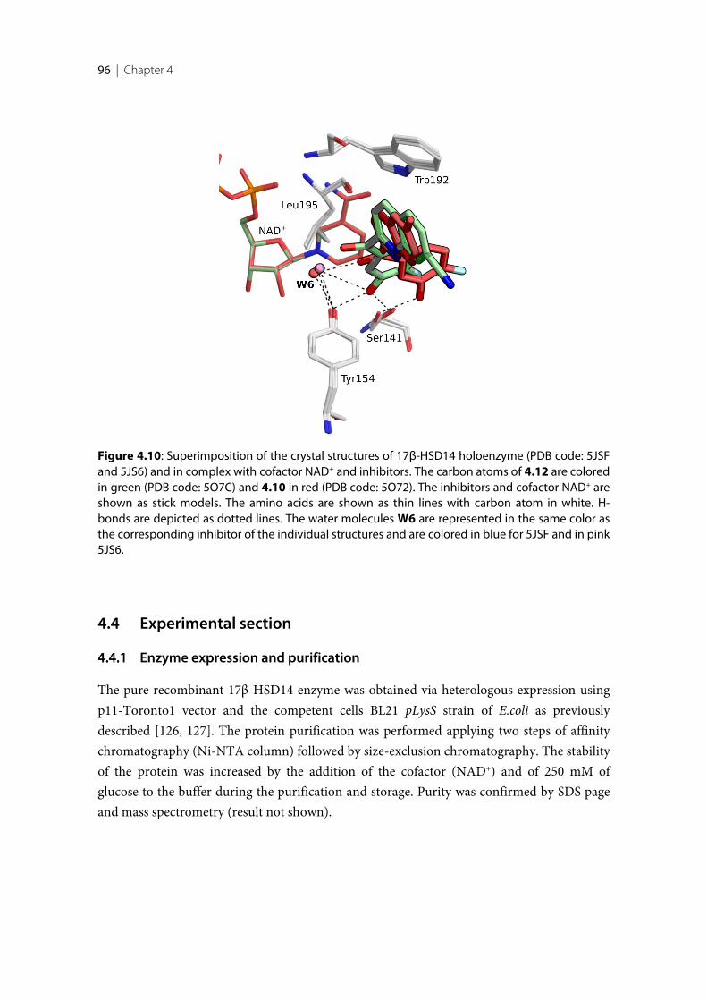
96 | Chapter 4
4.4 Experimental section
Enzyme expression and purification
The pure recombinant 17β-HSD14 enzyme was obtained via heterologous expression usingp11-Toronto1 vector and the competent cells BL21 pLysS strain of E.coli as previouslydescribed [126, 127]. The protein purification was performed applying two steps of affinitychromatography (Ni-NTA column) followed by size-exclusion chromatography. The stabilityof the protein was increased by the addition of the cofactor (NAD+) and of 250 mM ofglucose to the buffer during the purification and storage. Purity was confirmed by SDS pageand mass spectrometry (result not shown).
Figure 4.10: Superimposition of the crystal structures of 17β-HSD14 holoenzyme (PDB code: 5JSFand 5JS6) and in complex with cofactor NAD+ and inhibitors. The carbon atoms of 4.12 are coloredin green (PDB code: 5O7C) and 4.10 in red (PDB code: 5O72). The inhibitors and cofactor NAD+ areshown as stick models. The amino acids are shown as thin lines with carbon atom in white. H-bonds are depicted as dotted lines. The water molecules W6 are represented in the same color asthe corresponding inhibitor of the individual structures and are colored in blue for 5JSF and in pink5JS6.
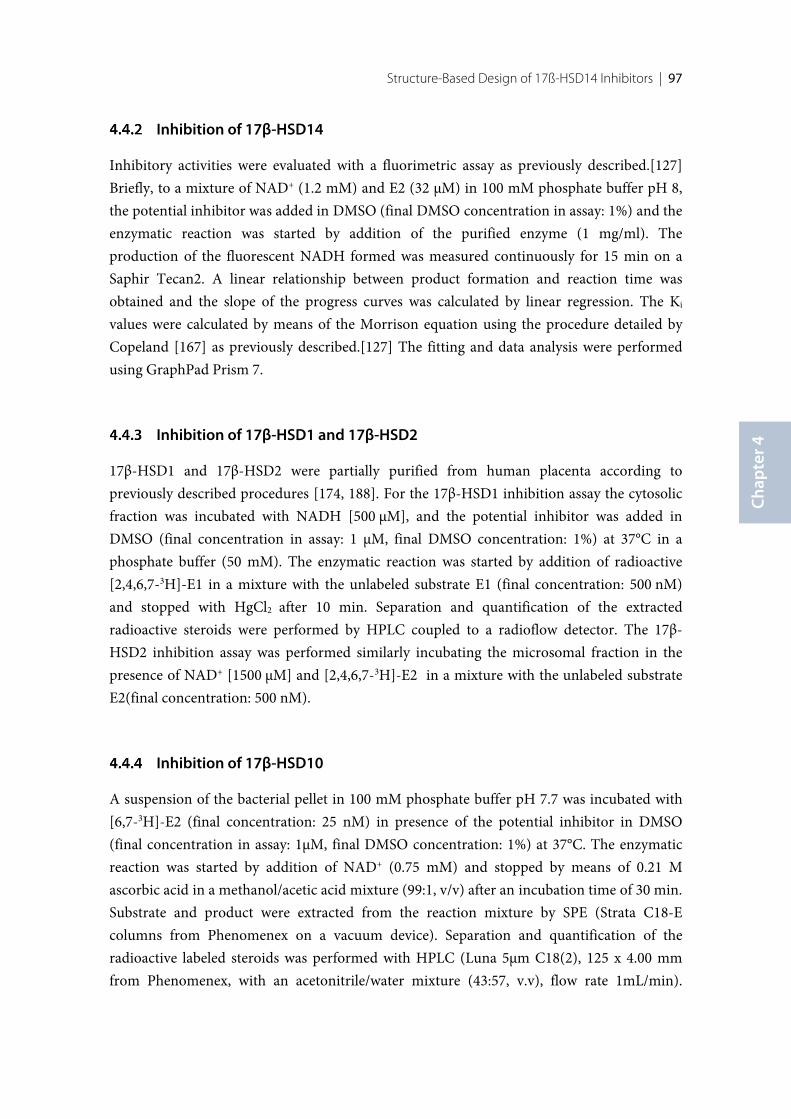
Structure-Based Design of 17ß-HSD14 Inhibitors | 97
Chap
ter 4
Inhibition of 17β-HSD14
Inhibitory activities were evaluated with a fluorimetric assay as previously described.[127]Briefly, to a mixture of NAD+ (1.2 mM) and E2 (32 µM) in 100 mM phosphate buffer pH 8,the potential inhibitor was added in DMSO (final DMSO concentration in assay: 1%) and theenzymatic reaction was started by addition of the purified enzyme (1 mg/ml). Theproduction of the fluorescent NADH formed was measured continuously for 15 min on aSaphir Tecan2. A linear relationship between product formation and reaction time wasobtained and the slope of the progress curves was calculated by linear regression. The Ki
values were calculated by means of the Morrison equation using the procedure detailed byCopeland [167] as previously described.[127] The fitting and data analysis were performedusing GraphPad Prism 7.
Inhibition of 17β-HSD1 and 17β-HSD2
17β-HSD1 and 17β-HSD2 were partially purified from human placenta according topreviously described procedures [174, 188]. For the 17β-HSD1 inhibition assay the cytosolicfraction was incubated with NADH [500 µM], and the potential inhibitor was added inDMSO (final concentration in assay: 1 µM, final DMSO concentration: 1%) at 37°C in aphosphate buffer (50 mM). The enzymatic reaction was started by addition of radioactive[2,4,6,7-3H]-E1 in a mixture with the unlabeled substrate E1 (final concentration: 500 nM)and stopped with HgCl2 after 10 min. Separation and quantification of the extractedradioactive steroids were performed by HPLC coupled to a radioflow detector. The 17β-HSD2 inhibition assay was performed similarly incubating the microsomal fraction in thepresence of NAD+ [1500 µM] and [2,4,6,7-3H]-E2 in a mixture with the unlabeled substrateE2(final concentration: 500 nM).
Inhibition of 17β-HSD10
A suspension of the bacterial pellet in 100 mM phosphate buffer pH 7.7 was incubated with[6,7-3H]-E2 (final concentration: 25 nM) in presence of the potential inhibitor in DMSO(final concentration in assay: 1µM, final DMSO concentration: 1%) at 37°C. The enzymaticreaction was started by addition of NAD+ (0.75 mM) and stopped by means of 0.21 Mascorbic acid in a methanol/acetic acid mixture (99:1, v/v) after an incubation time of 30 min.Substrate and product were extracted from the reaction mixture by SPE (Strata C18-Ecolumns from Phenomenex on a vacuum device). Separation and quantification of theradioactive labeled steroids was performed with HPLC (Luna 5µm C18(2), 125 x 4.00 mmfrom Phenomenex, with an acetonitrile/water mixture (43:57, v.v), flow rate 1mL/min).
98 | Chapter 4
Substrate conversion is given in % as calculated after integration of the product and substratepeaks. Inhibition was calculated based on conversion without potential inhibitor (DMSOonly) which was set to 0% inhibition.
Co-crystallization of the protein with inhibitors
The co-crystallization of 17β-HSD14 in complex with the different classes of inhibitors wasperformed following the already described slightly optimized protocols [126, 127].
The final inhibitor concentration of 4 mM was incubated with the protein solution (9.5mg/mL) containing 0.6 mM NAD+ and 5% DMSO for 30 minutes. The mixture was thencentrifuged at 10000 g for 20 minutes in order to separate the precipitated. Afterwards, 2 µLof the supernatant was mixed with 2 µL of mother liquor composed of 0.1 M HEPES, 20%(w/v) PEG6000 and 5% (v/v) DMSO, adjusted to pH 7.0. Crystals of sufficient quality for datacollection were grown at a temperature of 18°C for 10 days and then exposed for a fewseconds to a cryo buffer obtained by the combination of mother liquor with the addition of20% glucose. Finally the specimen were successively flash-frozen in liquid nitrogen.
Crystallography
Data collection and processing
All the datasets of the structures of the protein-inhibitor complexes were collected atbeamline 14.1 at BESSY II in Berlin, Germany. The data collection was achieved with awavelength of 0.9184 Å and a temperature of 100 K on a DECTRIS Pilatus 6M pixel detector.All datasets were indexed, processed and scaled with XDS [142].
Structure determination and refinement
The structures were determined by molecular replacement with the program PHASERMR[143] from the CCP4 suit.[132] In all cases, coordinates of the PDB entry 5EN4 were usedas a search model. A subset, corresponding to 5% of the reflections, was used for thecalculation of Rfree and consequently was omitted from the refinement. The model was builtin COOT [171] and refined using PHENIX.refine version 1.10.1-2155 [145]. The ligands wereenergetically minimized and their restraints were generated using Grade Web Server [147]and eLBOW [145] based on the SMILEs codes obtained with Molinspiration v2013.91 [146].As first refinement step for all structures, a Cartesian simulated annealing was performedusing default parameters. The final models were built in COOT with alternating structural
Structure-Based Design of 17ß-HSD14 Inhibitors | 99
Chap
ter 4
modification and refinement of XYZ coordinates, occupancies and individual B-factors inPHENIX. Hydrogens were included in the models of the structures. For 5O6X, thetemperature factors of all atoms, except hydrogen atoms and solvent molecules, were refinedanisotropically. For 5O72, the temperature factors were refined isotropically. For the otherfive structures, TLS refinement was performed with nine TLS groups selected from theTLSMD web server [148, 149].
MOE models
The inhibitor 4.8, 4.11, 4.13, 4.14 and 4.16 were model inside the crystal structure PDB code:5L7Y [126] and minimized with MOE [173]. During minimization, the AMBER12 force fieldwas applied. The heavy atoms of the protein and the 3-OH group at the C-ring of theinhibitors were fixed. In order to give to the ligand sufficient freedom to move also theresidues His93, Gln148 and Met199 where allowed unrestricted to move duringminimization.
Cytotoxicity Assay
Potential cytotoxicity of inhibitors 3.9, 5.12, 5.13 and 5.15 toward HEK293 cells wasdetermined by an MTT assay [189] as previously described [190]. The cells were seeded into96-well plates overnight at a 1 x 10⁴ cells/well density. Series of inhibitor concentrations(from 0 to 100 μM) were added to the well and incubated for 72 h, at 37 °C under 5% CO2.The cell viability was subsequent evaluated by the MTT colorimetric assay.
Pgp ATPase Activity Assays
The effect of inhibitors 3.9, 5.12, 5.13 and 5.15 on Pgp ATPase activity was evaluated bymeans of the Pgp-GloTM assay system (Promega, Madison, WI, USA).In a white 96-well plate,the inhibitors 3.9, 5.12, 5.13 and 5.15 were incubated in DMSO (final concentration in assay:10 µM) with 25 μg of recombinant human Pgp membrane. The Pgp-glo assay buffer was usedas the untreated sample control. The positive control of drug induced Pgp ATPase activitywas obtained by addition of 200 μM verapamil to the assay buffer. As selective inhibitor ofPgp ATPase activity, 100 μM sodium orthovanadate in the assay buffer was used. All thesamples contained a final DMSO concentration of 1%. The Pgp ATPase activity was initiatedby the addition of 5 mM MgATP and incubated at 37 °C for 60 min. Luminescence wasrecorded after adding 50 μL of ATP detection reagent. After 25 min of incubation at roomtemperature, the total luminescence signal was detected on a Tecan Saphire 2.

Structural Comparison between 17β-HSD Enzymes
and Virtual Screening of Inhibitors
Introductory remarks
This chapter is based on the results preliminary addressed in Chapters 2 and 3. In thefollowing a detailed discussion of the structural differences between 17β-HSD14 and theother 17β-HSD proteins will be provided. Furthermore, docking studies performed duringthe early stage of this project will be discussed.
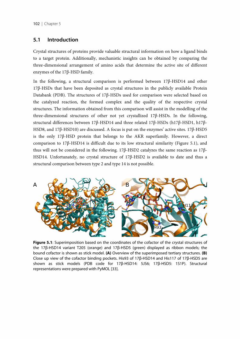
102 | Chapter 5
5.1 Introduction
Crystal structures of proteins provide valuable structural information on how a ligand bindsto a target protein. Additionally, mechanistic insights can be obtained by comparing thethree-dimensional arrangement of amino acids that determine the active site of differentenzymes of the 17β-HSD family.
In the following, a structural comparison is performed between 17β-HSD14 and other17β-HSDs that have been deposited as crystal structures in the publicly available ProteinDatabank (PDB). The structures of 17β-HSDs used for comparison were selected based onthe catalyzed reaction, the formed complex and the quality of the respective crystalstructures. The information obtained from this comparison will assist in the modelling of thethree-dimensional structures of other not yet crystallized 17β-HSDs. In the following,structural differences between 17β-HSD14 and three related 17β-HSDs (h17β-HSD1, h17β-HSD8, and 17β-HSD10) are discussed. A focus is put on the enzymes’ active sites. 17β-HSD5is the only 17β-HSD protein that belongs to the AKR superfamily. However, a directcomparison to 17β-HSD14 is difficult due to its low structural similarity (Figure 5.1), andthus will not be considered in the following. 17β-HSD2 catalyzes the same reaction as 17β-HSD14. Unfortunately, no crystal structure of 17β-HSD2 is available to date and thus astructural comparison between type 2 and type 14 is not possible.
Figure 5.1: Superimposition based on the coordinates of the cofactor of the crystal structures ofthe 17β-HSD14 variant T205 (orange) and 17β-HSD5 (green) displayed as ribbon models; thebound cofactor is shown as stick model. (A) Overview of the superimposed tertiary structures. (B)Close up view of the cofactor binding pockets. His93 of 17β-HSD14 and His117 of 17β-HSD5 areshown as stick models (PDB code for 17β-HSD14: 5JS6; 17β-HSD5: 1S1P). Structuralrepresentations were prepared with PyMOL [33].
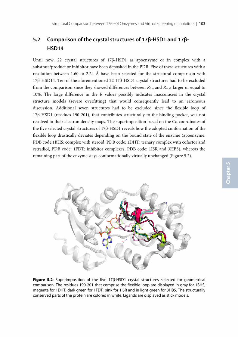
Structural Comparison between 17ß-HSD Enzymes and Virtual Screening of Inhibitors | 103
Chap
ter 5
5.2 Comparison of the crystal structures of 17β-HSD1 and 17β-
HSD14
Until now, 22 crystal structures of 17β-HSD1 as apoenzyme or in complex with asubstrate/product or inhibitor have been deposited in the PDB. Five of these structures with aresolution between 1.60 to 2.24 Å have been selected for the structural comparison with17β-HSD14. Ten of the aforementioned 22 17β-HSD1 crystal structures had to be excludedfrom the comparison since they showed differences between Rfree and Rwork larger or equal to10%. The large difference in the R values possibly indicates inaccuracies in the crystalstructure models (severe overfitting) that would consequently lead to an erroneousdiscussion. Additional seven structures had to be excluded since the flexible loop of17β-HSD1 (residues 190-201), that contributes structurally to the binding pocket, was notresolved in their electron density maps. The superimposition based on the Cα coordinates ofthe five selected crystal structures of 17β-HSD1 reveals how the adopted conformation of theflexible loop drastically deviates depending on the bound state of the enzyme (apoenzyme,PDB code:1BHS; complex with steroid, PDB code: 1DHT; ternary complex with cofactor andestradiol, PDB code: 1FDT; inhibitor complexes, PDB code: 1I5R and 3HB5), whereas theremaining part of the enzyme stays conformationally virtually unchanged (Figure 5.2).
Figure 5.2: Superimposition of the five 17β-HSD1 crystal structures selected for geometricalcomparison. The residues 190-201 that comprise the flexible loop are displayed in gray for 1BHS,magenta for 1DHT, dark green for 1FDT, pink for 1I5R and in light green for 3HB5. The structurallyconserved parts of the protein are colored in white. Ligands are displayed as stick models.
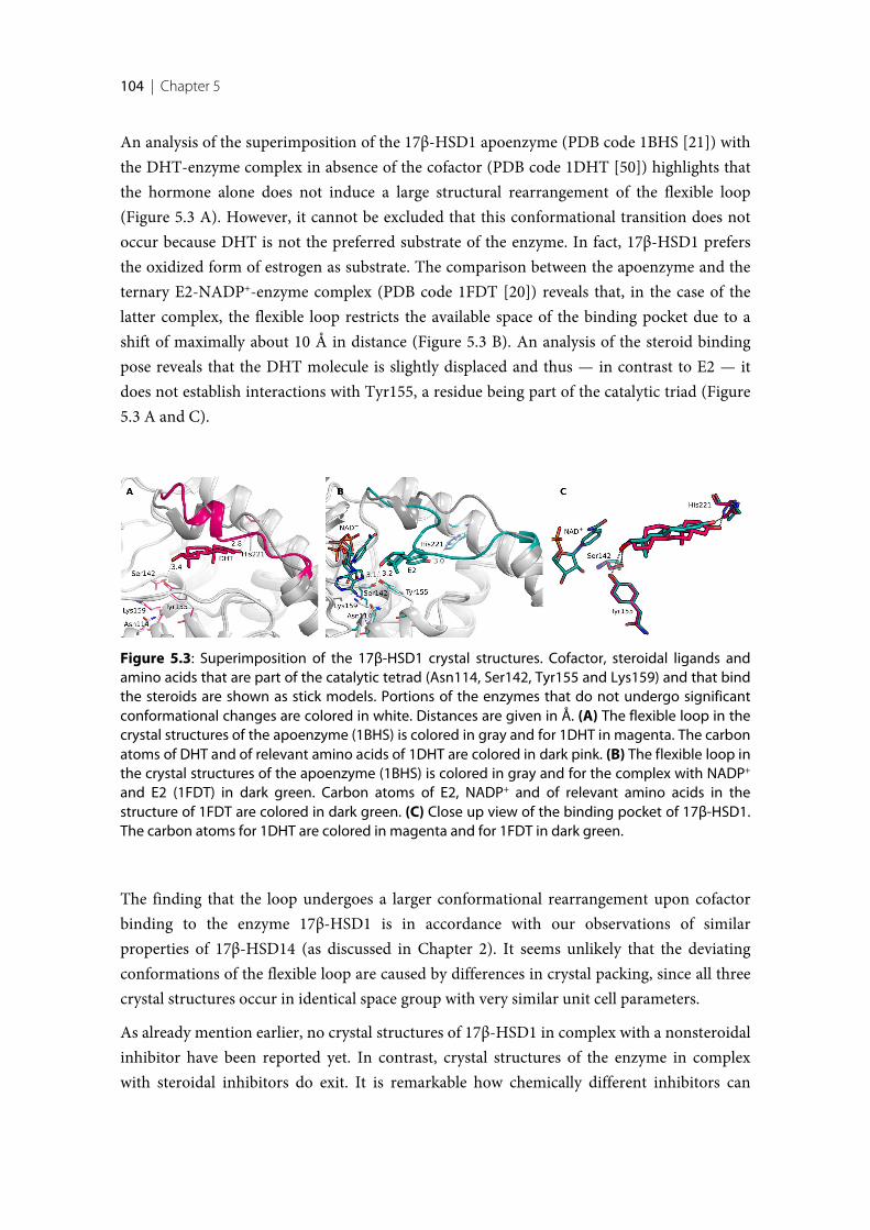
104 | Chapter 5
An analysis of the superimposition of the 17β-HSD1 apoenzyme (PDB code 1BHS [21]) withthe DHT-enzyme complex in absence of the cofactor (PDB code 1DHT [50]) highlights thatthe hormone alone does not induce a large structural rearrangement of the flexible loop(Figure 5.3 A). However, it cannot be excluded that this conformational transition does notoccur because DHT is not the preferred substrate of the enzyme. In fact, 17β-HSD1 prefersthe oxidized form of estrogen as substrate. The comparison between the apoenzyme and theternary E2-NADP+-enzyme complex (PDB code 1FDT [20]) reveals that, in the case of thelatter complex, the flexible loop restricts the available space of the binding pocket due to ashift of maximally about 10 Å in distance (Figure 5.3 B). An analysis of the steroid bindingpose reveals that the DHT molecule is slightly displaced and thus — in contrast to E2 — itdoes not establish interactions with Tyr155, a residue being part of the catalytic triad (Figure5.3 A and C).
The finding that the loop undergoes a larger conformational rearrangement upon cofactorbinding to the enzyme 17β-HSD1 is in accordance with our observations of similarproperties of 17β-HSD14 (as discussed in Chapter 2). It seems unlikely that the deviatingconformations of the flexible loop are caused by differences in crystal packing, since all threecrystal structures occur in identical space group with very similar unit cell parameters.
As already mention earlier, no crystal structures of 17β-HSD1 in complex with a nonsteroidalinhibitor have been reported yet. In contrast, crystal structures of the enzyme in complexwith steroidal inhibitors do exit. It is remarkable how chemically different inhibitors can
Figure 5.3: Superimposition of the 17β-HSD1 crystal structures. Cofactor, steroidal ligands andamino acids that are part of the catalytic tetrad (Asn114, Ser142, Tyr155 and Lys159) and that bindthe steroids are shown as stick models. Portions of the enzymes that do not undergo significantconformational changes are colored in white. Distances are given in Å. (A) The flexible loop in thecrystal structures of the apoenzyme (1BHS) is colored in gray and for 1DHT in magenta. The carbonatoms of DHT and of relevant amino acids of 1DHT are colored in dark pink. (B) The flexible loop inthe crystal structures of the apoenzyme (1BHS) is colored in gray and for the complex with NADP+
and E2 (1FDT) in dark green. Carbon atoms of E2, NADP+ and of relevant amino acids in thestructure of 1FDT are colored in dark green. (C) Close up view of the binding pocket of 17β-HSD1.The carbon atoms for 1DHT are colored in magenta and for 1FDT in dark green.
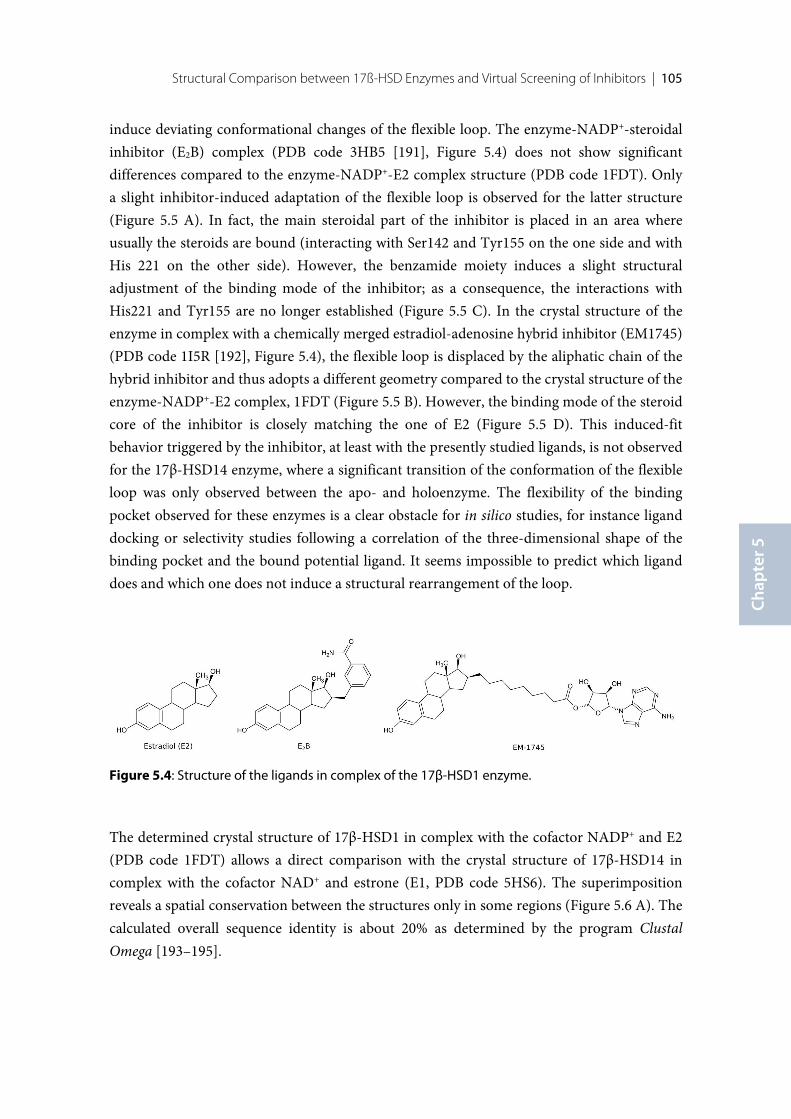
Structural Comparison between 17ß-HSD Enzymes and Virtual Screening of Inhibitors | 105
Chap
ter 5
induce deviating conformational changes of the flexible loop. The enzyme-NADP+-steroidalinhibitor (E2B) complex (PDB code 3HB5 [191], Figure 5.4) does not show significantdifferences compared to the enzyme-NADP+-E2 complex structure (PDB code 1FDT). Onlya slight inhibitor-induced adaptation of the flexible loop is observed for the latter structure(Figure 5.5 A). In fact, the main steroidal part of the inhibitor is placed in an area whereusually the steroids are bound (interacting with Ser142 and Tyr155 on the one side and withHis 221 on the other side). However, the benzamide moiety induces a slight structuraladjustment of the binding mode of the inhibitor; as a consequence, the interactions withHis221 and Tyr155 are no longer established (Figure 5.5 C). In the crystal structure of theenzyme in complex with a chemically merged estradiol-adenosine hybrid inhibitor (EM1745)(PDB code 1I5R [192], Figure 5.4), the flexible loop is displaced by the aliphatic chain of thehybrid inhibitor and thus adopts a different geometry compared to the crystal structure of theenzyme-NADP+-E2 complex, 1FDT (Figure 5.5 B). However, the binding mode of the steroidcore of the inhibitor is closely matching the one of E2 (Figure 5.5 D). This induced-fitbehavior triggered by the inhibitor, at least with the presently studied ligands, is not observedfor the 17β-HSD14 enzyme, where a significant transition of the conformation of the flexibleloop was only observed between the apo- and holoenzyme. The flexibility of the bindingpocket observed for these enzymes is a clear obstacle for in silico studies, for instance liganddocking or selectivity studies following a correlation of the three-dimensional shape of thebinding pocket and the bound potential ligand. It seems impossible to predict which liganddoes and which one does not induce a structural rearrangement of the loop.
The determined crystal structure of 17β-HSD1 in complex with the cofactor NADP+ and E2(PDB code 1FDT) allows a direct comparison with the crystal structure of 17β-HSD14 incomplex with the cofactor NAD+ and estrone (E1, PDB code 5HS6). The superimpositionreveals a spatial conservation between the structures only in some regions (Figure 5.6 A). Thecalculated overall sequence identity is about 20% as determined by the program ClustalOmega [193–195].
Figure 5.4: Structure of the ligands in complex of the 17β-HSD1 enzyme.
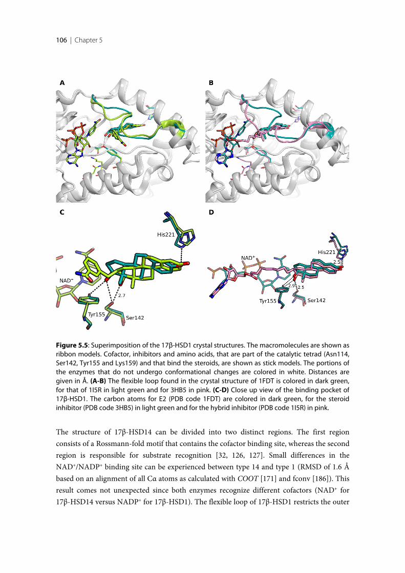
106 | Chapter 5
The structure of 17β-HSD14 can be divided into two distinct regions. The first regionconsists of a Rossmann-fold motif that contains the cofactor binding site, whereas the secondregion is responsible for substrate recognition [32, 126, 127]. Small differences in theNAD+/NADP+ binding site can be experienced between type 14 and type 1 (RMSD of 1.6 Åbased on an alignment of all Cα atoms as calculated with COOT [171] and fconv [186]). Thisresult comes not unexpected since both enzymes recognize different cofactors (NAD+ for17β-HSD14 versus NADP+ for 17β-HSD1). The flexible loop of 17β-HSD1 restricts the outer
Figure 5.5: Superimposition of the 17β-HSD1 crystal structures. The macromolecules are shown asribbon models. Cofactor, inhibitors and amino acids, that are part of the catalytic tetrad (Asn114,Ser142, Tyr155 and Lys159) and that bind the steroids, are shown as stick models. The portions ofthe enzymes that do not undergo conformational changes are colored in white. Distances aregiven in Å. (A-B) The flexible loop found in the crystal structure of 1FDT is colored in dark green,for that of 1I5R in light green and for 3HB5 in pink. (C-D) Close up view of the binding pocket of17β-HSD1. The carbon atoms for E2 (PDB code 1FDT) are colored in dark green, for the steroidinhibitor (PDB code 3HB5) in light green and for the hybrid inhibitor (PDB code 1I5R) in pink.
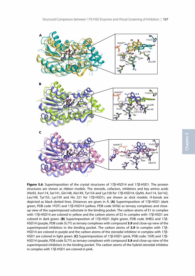
Structural Comparison between 17ß-HSD Enzymes and Virtual Screening of Inhibitors | 107
Chap
ter 5
Figure 5.6: Superimposition of the crystal structures of 17β-HSD14 and 17β-HSD1. The proteinstructures are shown as ribbon models. The steroids, cofactors, inhibitors and key amino acids(His93, Asn114, Ser141, Gln148, Ala149, Tyr154 and Lys158 for 17β-HSD14; Gly94, Asn114, Ser142,Leu149, Tyr155, Lys159 and His 221 for 17β-HSD1), are shown as stick models. H-bonds aredepicted as black dotted lines. Distances are given in Å. (A) Superimposition of 17β-HSD1 (darkgreen, PDB code 1FDT) and 17β-HSD14 (yellow, PDB code 5HS6) as ternary complexes and close-up view of the superimposed substrate in the binding pocket. The carbon atoms of E1 in complexwith 17β-HSD14 are colored in yellow and the carbon atoms of E2 in complex with 17β-HSD1 arecolored in dark green. (B) Superimposition of 17β-HSD1 (light green, PDB code 3HB5) and 17β-HSD14 (purple, PDB code 5L7Y) as ternary complexes with compound 3.9 and close-up view of thesuperimposed inhibitors in the binding pocket. The carbon atoms of 3.9 in complex with 17β-HSD14 are colored in purple and the carbon atoms of the steroidal inhibitor in complex with 17β-HSD1 are colored in light green. (C) Superimposition of 17β-HSD1 (pink, PDB code: 1I5R) and 17β-HSD14 (purple, PDB code 5L7Y) as ternary complexes with compound 3.9 and close-up view of thesuperimposed inhibitors in the binding pocket. The carbon atoms of the hybrid steroidal inhibitorin complex with 17β-HSD1 are colored in pink.
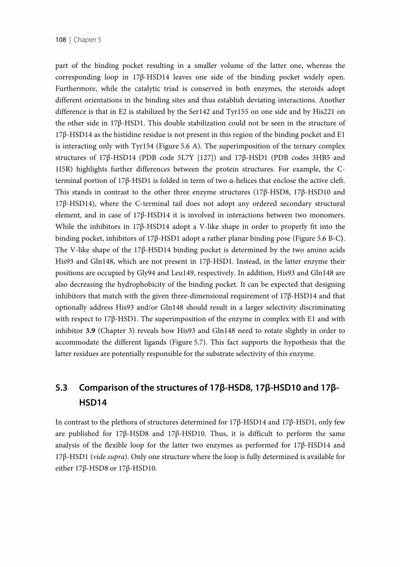
108 | Chapter 5
part of the binding pocket resulting in a smaller volume of the latter one, whereas thecorresponding loop in 17β-HSD14 leaves one side of the binding pocket widely open.Furthermore, while the catalytic triad is conserved in both enzymes, the steroids adoptdifferent orientations in the binding sites and thus establish deviating interactions. Anotherdifference is that in E2 is stabilized by the Ser142 and Tyr155 on one side and by His221 onthe other side in 17β-HSD1. This double stabilization could not be seen in the structure of17β-HSD14 as the histidine residue is not present in this region of the binding pocket and E1is interacting only with Tyr154 (Figure 5.6 A). The superimposition of the ternary complexstructures of 17β-HSD14 (PDB code 5L7Y [127]) and 17β-HSD1 (PDB codes 3HB5 and1I5R) highlights further differences between the protein structures. For example, the C-terminal portion of 17β-HSD1 is folded in term of two α-helices that enclose the active cleft.This stands in contrast to the other three enzyme structures (17β-HSD8, 17β-HSD10 and17β-HSD14), where the C-terminal tail does not adopt any ordered secondary structuralelement, and in case of 17β-HSD14 it is involved in interactions between two monomers.While the inhibitors in 17β-HSD14 adopt a V-like shape in order to properly fit into thebinding pocket, inhibitors of 17β-HSD1 adopt a rather planar binding pose (Figure 5.6 B-C).The V-like shape of the 17β-HSD14 binding pocket is determined by the two amino acidsHis93 and Gln148, which are not present in 17β-HSD1. Instead, in the latter enzyme theirpositions are occupied by Gly94 and Leu149, respectively. In addition, His93 and Gln148 arealso decreasing the hydrophobicity of the binding pocket. It can be expected that designinginhibitors that match with the given three-dimensional requirement of 17β-HSD14 and thatoptionally address His93 and/or Gln148 should result in a larger selectivity discriminatingwith respect to 17β-HSD1. The superimposition of the enzyme in complex with E1 and withinhibitor 3.9 (Chapter 3) reveals how His93 and Gln148 need to rotate slightly in order toaccommodate the different ligands (Figure 5.7). This fact supports the hypothesis that thelatter residues are potentially responsible for the substrate selectivity of this enzyme.
5.3 Comparison of the structures of 17β-HSD8, 17β-HSD10 and 17β-
HSD14
In contrast to the plethora of structures determined for 17β-HSD14 and 17β-HSD1, only feware published for 17β-HSD8 and 17β-HSD10. Thus, it is difficult to perform the sameanalysis of the flexible loop for the latter two enzymes as performed for 17β-HSD14 and17β-HSD1 (vide supra). Only one structure where the loop is fully determined is available foreither 17β-HSD8 or 17β-HSD10.
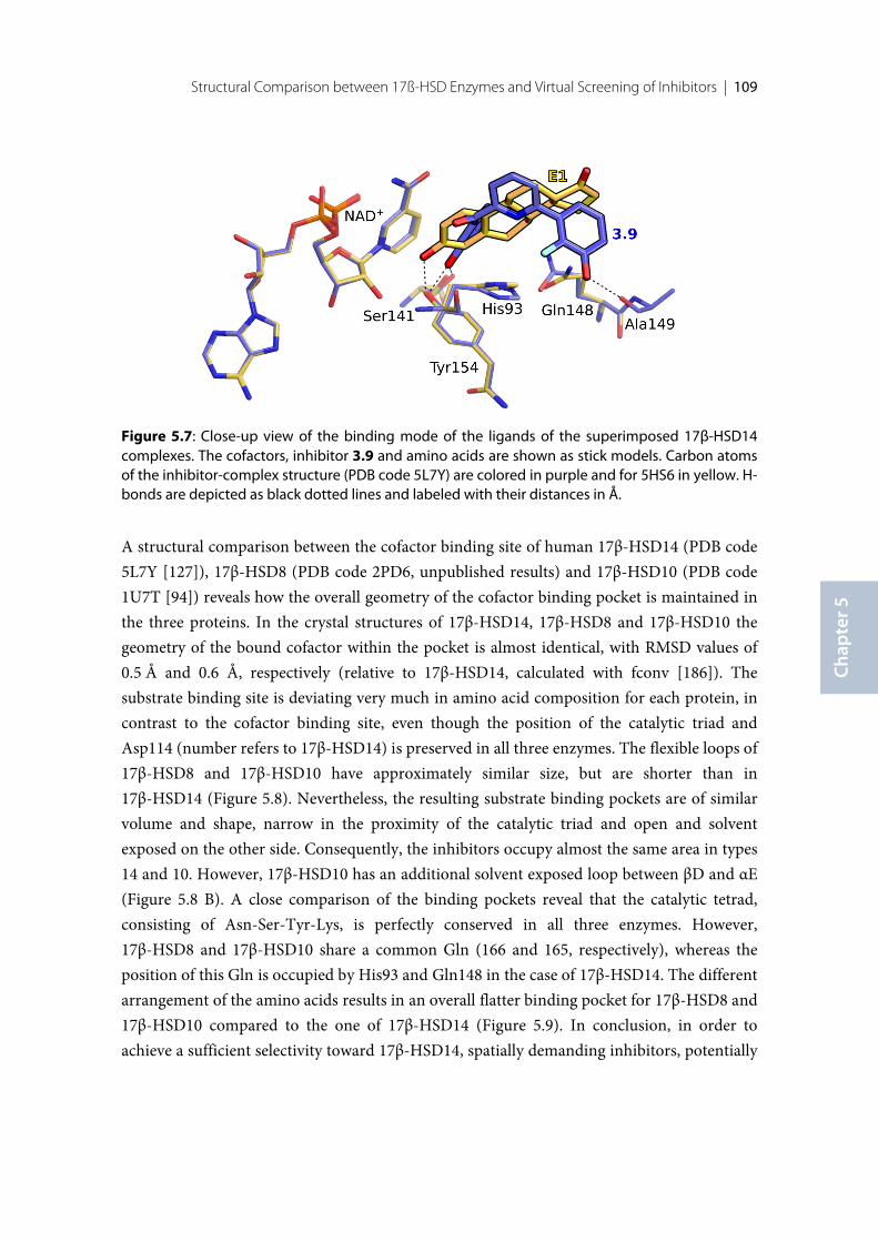
Structural Comparison between 17ß-HSD Enzymes and Virtual Screening of Inhibitors | 109
Chap
ter 5
A structural comparison between the cofactor binding site of human 17β-HSD14 (PDB code5L7Y [127]), 17β-HSD8 (PDB code 2PD6, unpublished results) and 17β-HSD10 (PDB code1U7T [94]) reveals how the overall geometry of the cofactor binding pocket is maintained inthe three proteins. In the crystal structures of 17β-HSD14, 17β-HSD8 and 17β-HSD10 thegeometry of the bound cofactor within the pocket is almost identical, with RMSD values of0.5 Å and 0.6 Å, respectively (relative to 17β-HSD14, calculated with fconv [186]). Thesubstrate binding site is deviating very much in amino acid composition for each protein, incontrast to the cofactor binding site, even though the position of the catalytic triad andAsp114 (number refers to 17β-HSD14) is preserved in all three enzymes. The flexible loops of17β-HSD8 and 17β-HSD10 have approximately similar size, but are shorter than in17β-HSD14 (Figure 5.8). Nevertheless, the resulting substrate binding pockets are of similarvolume and shape, narrow in the proximity of the catalytic triad and open and solventexposed on the other side. Consequently, the inhibitors occupy almost the same area in types14 and 10. However, 17β-HSD10 has an additional solvent exposed loop between βD and αE(Figure 5.8 B). A close comparison of the binding pockets reveal that the catalytic tetrad,consisting of Asn-Ser-Tyr-Lys, is perfectly conserved in all three enzymes. However,17β-HSD8 and 17β-HSD10 share a common Gln (166 and 165, respectively), whereas theposition of this Gln is occupied by His93 and Gln148 in the case of 17β-HSD14. The differentarrangement of the amino acids results in an overall flatter binding pocket for 17β-HSD8 and17β-HSD10 compared to the one of 17β-HSD14 (Figure 5.9). In conclusion, in order toachieve a sufficient selectivity toward 17β-HSD14, spatially demanding inhibitors, potentially
Figure 5.7: Close-up view of the binding mode of the ligands of the superimposed 17β-HSD14complexes. The cofactors, inhibitor 3.9 and amino acids are shown as stick models. Carbon atomsof the inhibitor-complex structure (PDB code 5L7Y) are colored in purple and for 5HS6 in yellow. H-bonds are depicted as black dotted lines and labeled with their distances in Å.
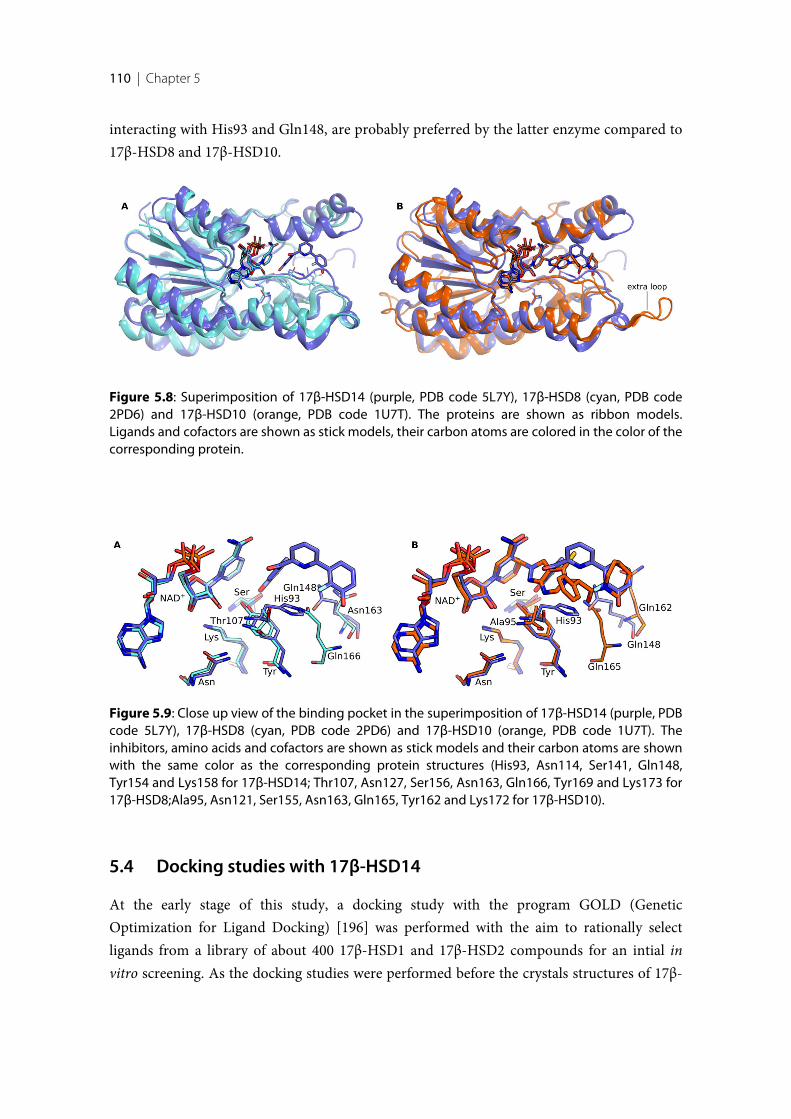
110 | Chapter 5
interacting with His93 and Gln148, are probably preferred by the latter enzyme compared to17β-HSD8 and 17β-HSD10.
5.4 Docking studies with 17β-HSD14
At the early stage of this study, a docking study with the program GOLD (GeneticOptimization for Ligand Docking) [196] was performed with the aim to rationally selectligands from a library of about 400 17β-HSD1 and 17β-HSD2 compounds for an intial invitro screening. As the docking studies were performed before the crystals structures of 17β-
Figure 5.8: Superimposition of 17β-HSD14 (purple, PDB code 5L7Y), 17β-HSD8 (cyan, PDB code2PD6) and 17β-HSD10 (orange, PDB code 1U7T). The proteins are shown as ribbon models.Ligands and cofactors are shown as stick models, their carbon atoms are colored in the color of thecorresponding protein.
Figure 5.9: Close up view of the binding pocket in the superimposition of 17β-HSD14 (purple, PDBcode 5L7Y), 17β-HSD8 (cyan, PDB code 2PD6) and 17β-HSD10 (orange, PDB code 1U7T). Theinhibitors, amino acids and cofactors are shown as stick models and their carbon atoms are shownwith the same color as the corresponding protein structures (His93, Asn114, Ser141, Gln148,Tyr154 and Lys158 for 17β-HSD14; Thr107, Asn127, Ser156, Asn163, Gln166, Tyr169 and Lys173 for17β-HSD8;Ala95, Asn121, Ser155, Asn163, Gln165, Tyr162 and Lys172 for 17β-HSD10).
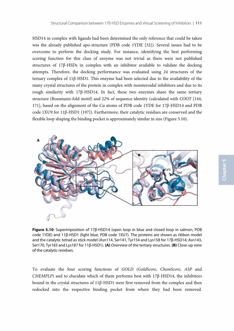
Structural Comparison between 17ß-HSD Enzymes and Virtual Screening of Inhibitors | 111
Chap
ter 5
HSD14 in complex with ligands had been determined the only reference that could be takenwas the already published apo-structure (PDB code 1YDE [32]). Several issues had to beovercome to perform the docking study. For instance, identifying the best performingscoring function for this class of enzyme was not trivial as there were not publishedstructures of 17β-HSDs in complex with an inhibitor available to validate the dockingattempts. Therefore, the docking performance was evaluated using 24 structures of theternary complex of 11β-HSD1. This enzyme had been selected due to the availability of themany crystal structures of the protein in complex with nonsteroidal inhibitors and due to itsrough similarity with 17β-HSD14. In fact, these two enzymes share the same tertiarystructure (Rossmann-fold motif) and 22% of sequence identity (calculated with COOT [144,171], based on the alignment of the Cα atoms of PDB code 1YDE for 17β-HSD14 and PDBcode 1XU9 for 11β-HSD1 [197]). Furthermore, their catalytic residues are conserved and theflexible loop shaping the binding pocket is approximately similar in size (Figure 5.10).
To evaluate the four scoring functions of GOLD (GoldScore, ChemScore, ASP andCHEMPLP) and to elucidate which of them performs best with 17β-HSD14, the inhibitorsbound in the crystal structures of 11β-HSD1 were first removed from the complex and thenredocked into the respective binding pocket from where they had been removed.
Figure 5.10: Superimposition of 17β-HSD14 (open loop in blue and closed loop in salmon, PDBcode 1YDE) and 11β-HSD1 (light blue, PDB code 1XU7). The proteins are shown as ribbon modeland the catalytic tetrad as stick model (Asn114, Ser141, Tyr154 and Lys158 for 17β-HSD14; Asn143,Ser170, Tyr183 and Lys187 for 11β-HSD1). (A) Overview of the tertiary structures. (B) Close-up viewof the catalytic residues.
112 | Chapter 5
Subsequently, every inhibitor was docked into the binding pockets of each of the individual24 crystal structures of 11β-HSD1 (cross-docking attempt). The docking was set in a way togenerate only three poses for each ligand. A visual inspection of the docking results revealedthat ChemScore and CHEMPLP produced the best fit of the ligands into the binding pocketand generated the closest binding pose with respect to the one observed in the crystalstructures. Before the validated settings could be applied to 17β-HSD14, a model of theenzyme in complex with its cofactor had to be generated with MOE [173]. The cofactormodel was derived from the crystal structure of 17β-HSD8 (PDB code 2PD6), the mostsimilar enzyme to 17β-HSD14 of this protein family. The NAD+ was manually inserted intothe cofactor binding pocket followed by a minimization with MOE using the MMFF94Xforce-field. During minimization, protein atoms except for H atoms were kept rigid. Theenzyme-cofactor model was generated using the monomer of the protein with the flexibleloop in the open as well as in the closed conformation leading to two final models.
A preliminary study to dock E2 (the substrate of 17β-HSD14) into the binding pocket wasperformed using both generated models. However, only the model with the openconformation resulted in a satisfying result. Therefore, the model with the loop in the openconformation was selected to proceed further with the simulation. To additionally refine thedocking settings, a set of 15 molecules consisting of 5 active compounds and 10 decoys foundby an initial enzymatic testing was docked into the model (Chapter 3). ChemScore ranked the5 active compounds within the top 8 scored molecules and thereby confirmed that thisscoring function performs best for this enzyme. Consequently, the model with the open loopconformation and ChemScore, as scoring function, were selected to perform the docking ofthe 400 entries from the 17β-HSD1 and 17β-HSD2 nonsteroidal inhibitor library. Accordingto the visual inspection of the top 100 generated poses, 15 molecules where selected to betested experimentally for their inhibitory activity against the enzyme 17β-HSD14.Unfortunately, a reliable hit rate for this study could not be determined for two reasons:Firstly, only about half of the selected molecules were available for testing. Secondly, withsuccessfully determination of the crystal structure of 17β-HSD14 it became obvious that onlythe closed conformation of the flexible loop is adopted upon ligand binding and not, asanticipated in this initial docking runs, the open conformation of the loop. Thus, all bindingposes predicted by docking are significantly shifted relative to their binding poses in thecrystal structure. This study shows the limitation of a virtual screening attempt for a ratherflexible protein in the absence of crystal structures. Even if some of the ligands would becorrectly selected, the results would be based, to some extent, on incorrect assumptions.
113
X-ray Crystallographic Fragment Screening
and Hit Optimization
Introductory remarks
A library of 96 fragments was assembled in the framework of the BMBF project Frag2Xtal.Protein expression and purification, crystallization experiments, soaking experiment andstructure determination were designed and carry out by the author of this thesis. Part of thesoaking experiments was performed by Lorena Zara under the supervision of the author ofthis thesis. The optimized ligands were designed by the author of this thesis in collaborationwith Florian Braun. The synthesis of the optimized ligands and the determination of theirbinding affinity were performed by Florian Braun.
114 | Chapter 7
6.1 Introduction
Within the last decade, fragment-based lead discovery (FBLD) has become a promisingapproach for the discovery of new lead structures. Since fragments are significantly smaller(<300 Da) than typical ligands discovered in high-throughput screening (HTS) campaign —which focus on screening candidates usually comprising already drug-like propertiesparticularly in term of size and molecular weight — fragments are smaller and weakerbinding with respect to the interaction features provided by a binding pocket. Consequently,fragment screening usually results in a significantly higher hit rate than a HTS screening, andtherefore a fragment library usually contain much less entries than HTS libraries to cover alarge variety of chemical functional groups to map the properties of a protein binding site(~1000 entries of a fragment library compared to 100,000 up to millions entries for a HTSlibrary) [198]. Hence, the design of a fragment library and its screening on a given target isalso feasible in an academic setting which usually cannot afford the resources necessary foran HTS campaign. Fragments usually contain multiple heteroatoms and even though theaffinity of fragments is low, the established interactions are usually of high quality andindicate the hot spots of binding. Therefore, fragment hits are good starting points for thefurther lead optimization [199]. Furthermore, since starting fragments are designed for lowlipophilicity, also the resulting drug-like molecule will usually show lower lipophilic than anHTS hit giving rise to superior pharmacokinetic properties of the evolved lead candidates[200]. However, since the fragment can, due to its limited size, establish only a fewinteractions with the residues of the binding pocket of the target protein, fragments usuallyhave a very low affinity. Thus, their successful identification is very challenging. Thedetection of fragment binders commonly follows a screening cascade, starting with abiophysical screening assay (e.g. SPR, NMR, thermal shift assay), and subsequentlyconfirmed hits are characterized by X-ray crystallography. However, in a recentcomprehensive case study [201, 202], it could be shown that the preselection of a on firstsight promising subset of fragments by the biophysical assays followed by a subsequentcrystallographic characterization misses a fairly large amount of putative crystallographic hits(nearly 50%), and does not even achieve the expected enrichment rate making the subsequentcrystallographic analysis more efficient [198, 203, 204]. We therefore decide to perform thescreening of the entire 96-entry in-house fragment library directly on protein crystals withoutapplication of any pre-filter on the target protein 17β-HSD14. So far, the reportednonsteroidal 17β-HSD14 inhibitors all comprise a very similar and highly lipophilic scaffold.Consequently, the discovery of a novel scaffold using a fragment-based lead discoveryapproach could result in the development of new lead candidates with improvedphysicochemical properties.
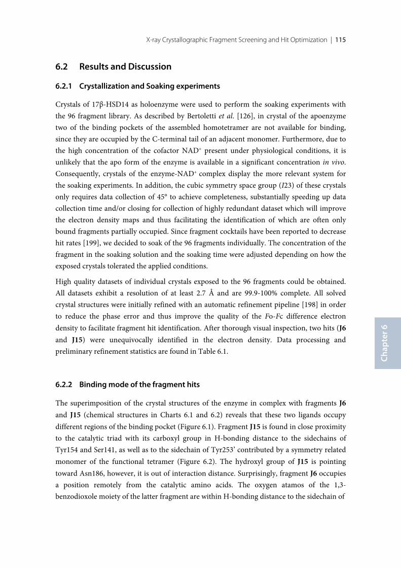
X-ray Crystallographic Fragment Screening and Hit Optimization | 115
Chap
ter 6
6.2 Results and Discussion
Crystallization and Soaking experiments
Crystals of 17β-HSD14 as holoenzyme were used to perform the soaking experiments withthe 96 fragment library. As described by Bertoletti et al. [126], in crystal of the apoenzymetwo of the binding pockets of the assembled homotetramer are not available for binding,since they are occupied by the C-terminal tail of an adjacent monomer. Furthermore, due tothe high concentration of the cofactor NAD+ present under physiological conditions, it isunlikely that the apo form of the enzyme is available in a significant concentration in vivo.Consequently, crystals of the enzyme-NAD+ complex display the more relevant system forthe soaking experiments. In addition, the cubic symmetry space group (I23) of these crystalsonly requires data collection of 45° to achieve completeness, substantially speeding up datacollection time and/or closing for collection of highly redundant dataset which will improvethe electron density maps and thus facilitating the identification of which are often onlybound fragments partially occupied. Since fragment cocktails have been reported to decreasehit rates [199], we decided to soak of the 96 fragments individually. The concentration of thefragment in the soaking solution and the soaking time were adjusted depending on how theexposed crystals tolerated the applied conditions.
High quality datasets of individual crystals exposed to the 96 fragments could be obtained.All datasets exhibit a resolution of at least 2.7 Å and are 99.9-100% complete. All solvedcrystal structures were initially refined with an automatic refinement pipeline [198] in orderto reduce the phase error and thus improve the quality of the Fo-Fc difference electrondensity to facilitate fragment hit identification. After thorough visual inspection, two hits (J6and J15) were unequivocally identified in the electron density. Data processing andpreliminary refinement statistics are found in Table 6.1.
Binding mode of the fragment hits
The superimposition of the crystal structures of the enzyme in complex with fragments J6and J15 (chemical structures in Charts 6.1 and 6.2) reveals that these two ligands occupydifferent regions of the binding pocket (Figure 6.1). Fragment J15 is found in close proximityto the catalytic triad with its carboxyl group in H-bonding distance to the sidechains ofTyr154 and Ser141, as well as to the sidechain of Tyr253’ contributed by a symmetry relatedmonomer of the functional tetramer (Figure 6.2). The hydroxyl group of J15 is pointingtoward Asn186, however, it is out of interaction distance. Surprisingly, fragment J6 occupiesa position remotely from the catalytic amino acids. The oxygen atamos of the 1,3-benzodioxole moiety of the latter fragment are within H-bonding distance to the sidechain of
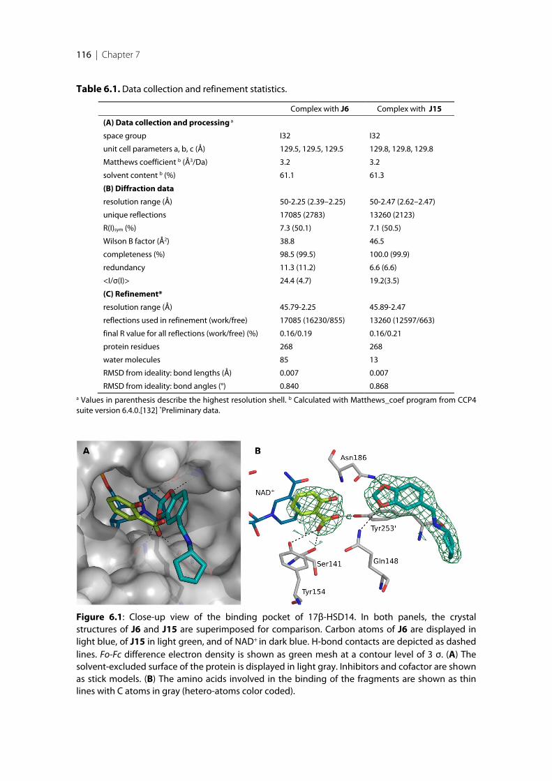
116 | Chapter 7
Table 6.1. Data collection and refinement statistics.
Complex with J6 Complex with J15
(A) Data collection and processing a
space group I32 I32
unit cell parameters a, b, c (Å) 129.5, 129.5, 129.5 129.8, 129.8, 129.8
Matthews coefficient b (Å3/Da) 3.2 3.2
solvent content b (%) 61.1 61.3
(B) Diffraction data
resolution range (Å) 50-2.25 (2.39–2.25) 50-2.47 (2.62–2.47)
unique reflections 17085 (2783) 13260 (2123)
R(I)sym (%) 7.3 (50.1) 7.1 (50.5)
Wilson B factor (Å2) 38.8 46.5
completeness (%) 98.5 (99.5) 100.0 (99.9)
redundancy 11.3 (11.2) 6.6 (6.6)
<I/σ(I)> 24.4 (4.7) 19.2(3.5)
(C) Refinement*
resolution range (Å) 45.79-2.25 45.89-2.47
reflections used in refinement (work/free) 17085 (16230/855) 13260 (12597/663)
final R value for all reflections (work/free) (%) 0.16/0.19 0.16/0.21
protein residues 268 268
water molecules 85 13
RMSD from ideality: bond lengths (Å) 0.007 0.007
RMSD from ideality: bond angles (°) 0.840 0.868a Values in parenthesis describe the highest resolution shell. b Calculated with Matthews_coef program from CCP4suite version 6.4.0.[132] *Preliminary data.
Figure 6.1: Close-up view of the binding pocket of 17β-HSD14. In both panels, the crystalstructures of J6 and J15 are superimposed for comparison. Carbon atoms of J6 are displayed inlight blue, of J15 in light green, and of NAD+ in dark blue. H-bond contacts are depicted as dashedlines. Fo-Fc difference electron density is shown as green mesh at a contour level of 3 σ. (A) Thesolvent-excluded surface of the protein is displayed in light gray. Inhibitors and cofactor are shownas stick models. (B) The amino acids involved in the binding of the fragments are shown as thinlines with C atoms in gray (hetero-atoms color coded).
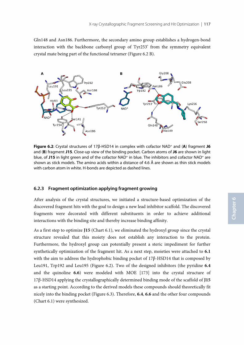
X-ray Crystallographic Fragment Screening and Hit Optimization | 117
Chap
ter 6
Gln148 and Asn186. Furthermore, the secondary amino group establishes a hydrogen-bondinteraction with the backbone carbonyl group of Tyr253’ from the symmetry equivalentcrystal mate being part of the functional tetramer (Figure 6.2 B).
Fragment optimization applying fragment growing
After analysis of the crystal structures, we initiated a structure-based optimization of thediscovered fragment hits with the goal to design a new lead inhibitor scaffold. The discoveredfragments were decorated with different substituents in order to achieve additionalinteractions with the binding site and thereby increase binding affinity.
As a first step to optimize J15 (Chart 6.1), we eliminated the hydroxyl group since the crystalstructure revealed that this moiety does not establish any interaction to the protein.Furthermore, the hydroxyl group can potentially present a steric impediment for furthersynthetically optimization of the fragment hit. As a next step, moieties were attached to 6.1with the aim to address the hydrophobic binding pocket of 17β-HSD14 that is composed byLeu191, Trp192 and Leu195 (Figure 6.2). Two of the designed inhibitors (the pyridine 6.4and the quinoline 6.6) were modeled with MOE [173] into the crystal structure of17β-HSD14 applying the crystallographically determined binding mode of the scaffold of J15as a starting point. According to the derived models these compounds should theoretically fitnicely into the binding pocket (Figure 6.3). Therefore, 6.4, 6.6 and the other four compounds(Chart 6.1) were synthesized.
Figure 6.2: Crystal structures of 17β-HSD14 in complex with cofactor NAD+ and (A) fragment J6and (B) fragment J15. Close-up view of the binding pocket. Carbon atoms of J6 are shown in lightblue, of J15 in light green and of the cofactor NAD+ in blue. The inhibitors and cofactor NAD+ areshown as stick models. The amino acids within a distance of 4.6 Å are shown as thin stick modelswith carbon atom in white. H-bonds are depicted as dashed lines.
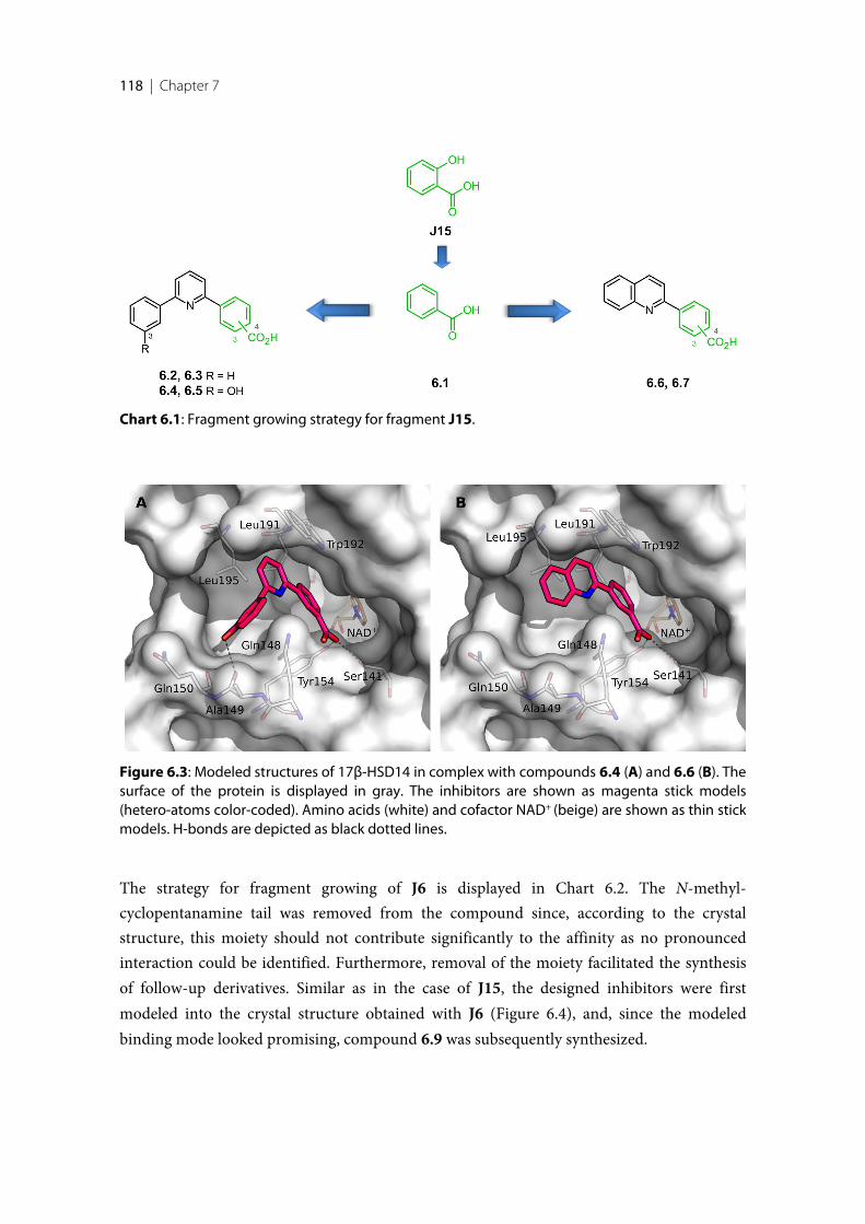
118 | Chapter 7
The strategy for fragment growing of J6 is displayed in Chart 6.2. The N-methyl-cyclopentanamine tail was removed from the compound since, according to the crystalstructure, this moiety should not contribute significantly to the affinity as no pronouncedinteraction could be identified. Furthermore, removal of the moiety facilitated the synthesisof follow-up derivatives. Similar as in the case of J15, the designed inhibitors were firstmodeled into the crystal structure obtained with J6 (Figure 6.4), and, since the modeledbinding mode looked promising, compound 6.9 was subsequently synthesized.
Chart 6.1: Fragment growing strategy for fragment J15.
Figure 6.3: Modeled structures of 17β-HSD14 in complex with compounds 6.4 (A) and 6.6 (B). Thesurface of the protein is displayed in gray. The inhibitors are shown as magenta stick models(hetero-atoms color-coded). Amino acids (white) and cofactor NAD+ (beige) are shown as thin stickmodels. H-bonds are depicted as black dotted lines.
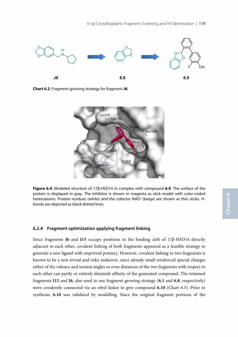
X-ray Crystallographic Fragment Screening and Hit Optimization | 119
Chap
ter 6
Fragment optimization applying fragment linking
Since fragments J6 and J15 occupy positions in the binding cleft of 17β-HSD14 directlyadjacent to each other, covalent linking of both fragments appeared as a feasible strategy togenerate a new ligand with improved potency. However, covalent linking to two fragments isknown to be a non-trivial and risky endeavor, since already small reinforced special changeseither of the valence and torsion angles or even distances of the two fragments with respect toeach other can partly or entirely diminish affinity of the generated compound. The trimmedfragments J15 and J6, also used in our fragment growing strategy (6.1 and 6.8, respectively)were covalently connected via an ethyl linker to give compound 6.10 (Chart 6.3). Prior tosynthesis, 6.10 was validated by modelling. Since the original fragment portions of the
Chart 6.2: Fragment growing strategy for fragment J6.
Figure 6.4: Modeled structure of 17β-HSD14 in complex with compound 6.9. The surface of theprotein is displayed in gray. The inhibitor is shown in magenta as stick model with color-codedheteroatoms. Protein residues (white) and the cofactor NAD+ (beige) are shown as thin sticks. H-bonds are depicted as black dotted lines.
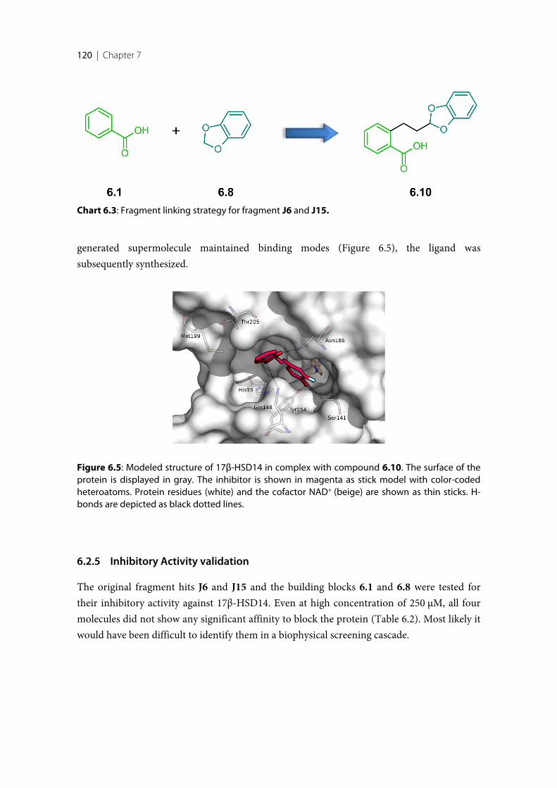
120 | Chapter 7
generated supermolecule maintained binding modes (Figure 6.5), the ligand wassubsequently synthesized.
Inhibitory Activity validation
The original fragment hits J6 and J15 and the building blocks 6.1 and 6.8 were tested fortheir inhibitory activity against 17β-HSD14. Even at high concentration of 250 μM, all fourmolecules did not show any significant affinity to block the protein (Table 6.2). Most likely itwould have been difficult to identify them in a biophysical screening cascade.
Chart 6.3: Fragment linking strategy for fragment J6 and J15.
Figure 6.5: Modeled structure of 17β-HSD14 in complex with compound 6.10. The surface of theprotein is displayed in gray. The inhibitor is shown in magenta as stick model with color-codedheteroatoms. Protein residues (white) and the cofactor NAD+ (beige) are shown as thin sticks. H-bonds are depicted as black dotted lines.
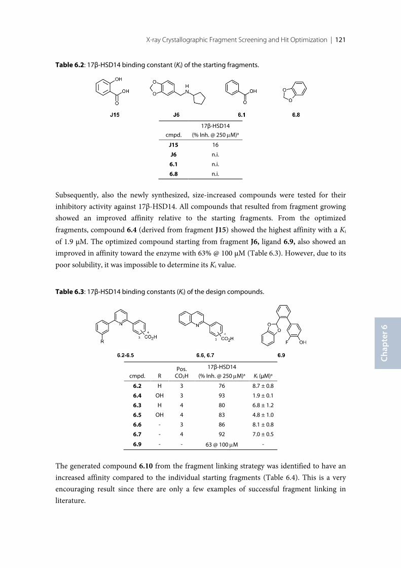
X-ray Crystallographic Fragment Screening and Hit Optimization | 121
Chap
ter 6
Table 6.2: 17β-HSD14 binding constant (Ki) of the starting fragments.
cmpd.
17β-HSD14
(% Inh. @ 250 mM)a
J15 16
J6 n.i.
6.1 n.i.
6.8 n.i.
Subsequently, also the newly synthesized, size-increased compounds were tested for theirinhibitory activity against 17β-HSD14. All compounds that resulted from fragment growingshowed an improved affinity relative to the starting fragments. From the optimizedfragments, compound 6.4 (derived from fragment J15) showed the highest affinity with a Ki
of 1.9 µM. The optimized compound starting from fragment J6, ligand 6.9, also showed animproved in affinity toward the enzyme with 63% @ 100 µM (Table 6.3). However, due to itspoor solubility, it was impossible to determine its Ki value.
Table 6.3: 17β-HSD14 binding constants (Ki) of the design compounds.
cmpd. RPos.
CO2H
17β-HSD14
(% Inh. @ 250 mM)a Ki (µM)a
6.2 H 3 76 8.7 ± 0.8
6.4 OH 3 93 1.9 ± 0.1
6.3 H 4 80 6.8 ± 1.2
6.5 OH 4 83 4.8 ± 1.0
6.6 - 3 86 8.1 ± 0.8
6.7 - 4 92 7.0 ± 0.5
6.9 - - 63 @ 100 mM -
The generated compound 6.10 from the fragment linking strategy was identified to have anincreased affinity compared to the individual starting fragments (Table 6.4). This is a veryencouraging result since there are only a few examples of successful fragment linking inliterature.
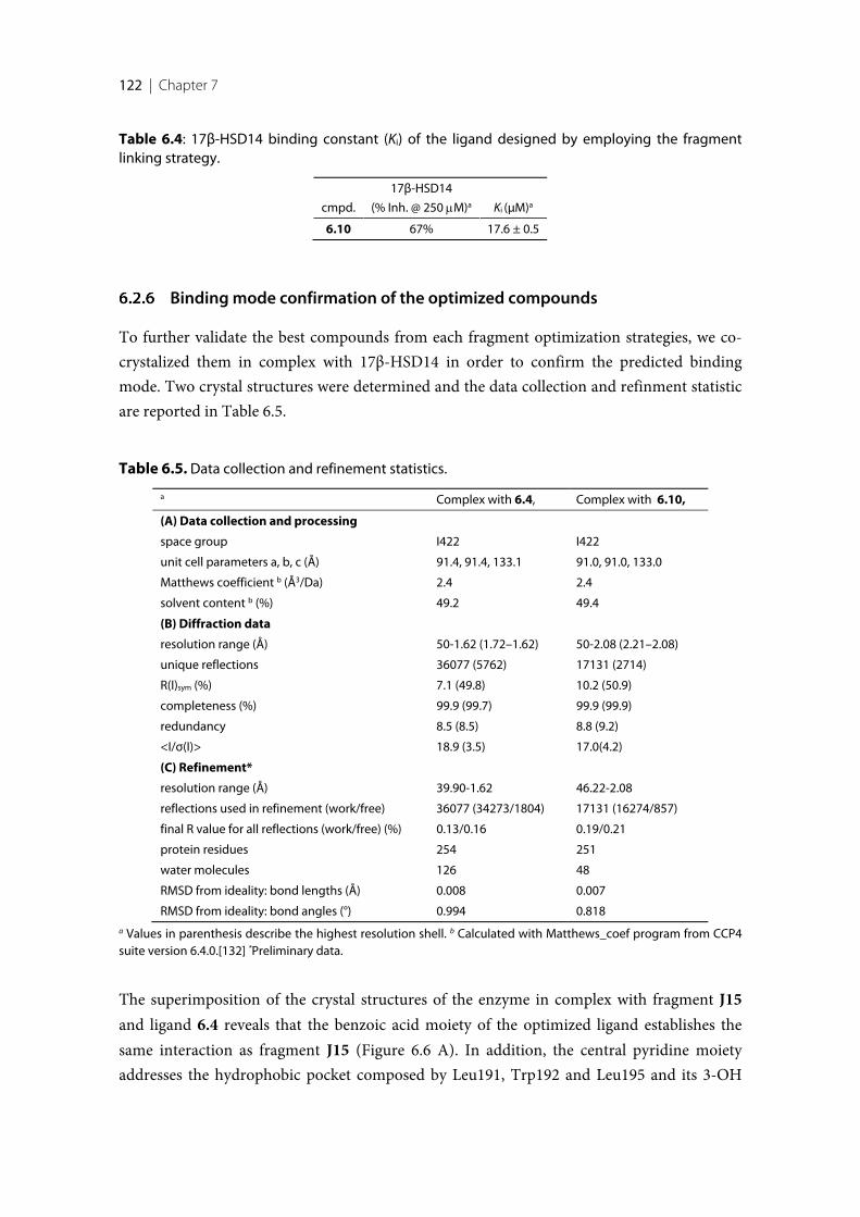
122 | Chapter 7
Table 6.4: 17β-HSD14 binding constant (Ki) of the ligand designed by employing the fragmentlinking strategy.
cmpd.
17β-HSD14
(% Inh. @ 250 mM)a Ki (µM)a
6.10 67% 17.6 ± 0.5
Binding mode confirmation of the optimized compounds
To further validate the best compounds from each fragment optimization strategies, we co-crystalized them in complex with 17β-HSD14 in order to confirm the predicted bindingmode. Two crystal structures were determined and the data collection and refinment statisticare reported in Table 6.5.
Table 6.5. Data collection and refinement statistics.
a Complex with 6.4, Complex with 6.10,
(A) Data collection and processing
space group I422 I422
unit cell parameters a, b, c (Å) 91.4, 91.4, 133.1 91.0, 91.0, 133.0
Matthews coefficient b (Å3/Da) 2.4 2.4
solvent content b (%) 49.2 49.4
(B) Diffraction data
resolution range (Å) 50-1.62 (1.72–1.62) 50-2.08 (2.21–2.08)
unique reflections 36077 (5762) 17131 (2714)
R(I)sym (%) 7.1 (49.8) 10.2 (50.9)
completeness (%) 99.9 (99.7) 99.9 (99.9)
redundancy 8.5 (8.5) 8.8 (9.2)
<I/σ(I)> 18.9 (3.5) 17.0(4.2)
(C) Refinement*
resolution range (Å) 39.90-1.62 46.22-2.08
reflections used in refinement (work/free) 36077 (34273/1804) 17131 (16274/857)
final R value for all reflections (work/free) (%) 0.13/0.16 0.19/0.21
protein residues 254 251
water molecules 126 48
RMSD from ideality: bond lengths (Å) 0.008 0.007
RMSD from ideality: bond angles (°) 0.994 0.818a Values in parenthesis describe the highest resolution shell. b Calculated with Matthews_coef program from CCP4suite version 6.4.0.[132] *Preliminary data.
The superimposition of the crystal structures of the enzyme in complex with fragment J15and ligand 6.4 reveals that the benzoic acid moiety of the optimized ligand establishes thesame interaction as fragment J15 (Figure 6.6 A). In addition, the central pyridine moietyaddresses the hydrophobic pocket composed by Leu191, Trp192 and Leu195 and its 3-OH
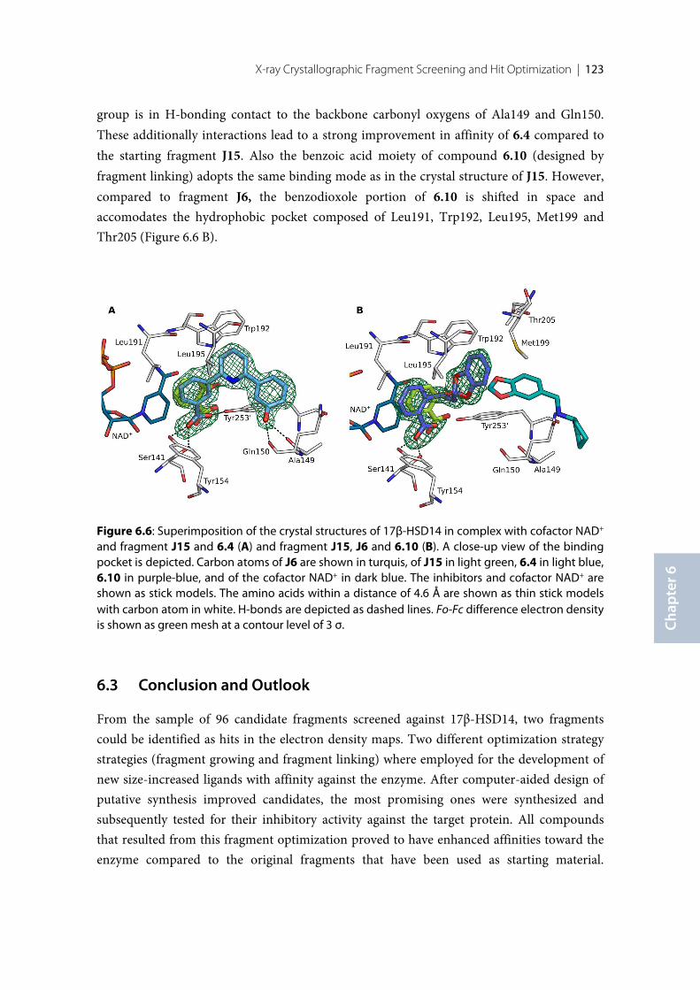
X-ray Crystallographic Fragment Screening and Hit Optimization | 123
Chap
ter 6
group is in H-bonding contact to the backbone carbonyl oxygens of Ala149 and Gln150.These additionally interactions lead to a strong improvement in affinity of 6.4 compared tothe starting fragment J15. Also the benzoic acid moiety of compound 6.10 (designed byfragment linking) adopts the same binding mode as in the crystal structure of J15. However,compared to fragment J6, the benzodioxole portion of 6.10 is shifted in space andaccomodates the hydrophobic pocket composed of Leu191, Trp192, Leu195, Met199 andThr205 (Figure 6.6 B).
6.3 Conclusion and Outlook
From the sample of 96 candidate fragments screened against 17β-HSD14, two fragmentscould be identified as hits in the electron density maps. Two different optimization strategystrategies (fragment growing and fragment linking) where employed for the development ofnew size-increased ligands with affinity against the enzyme. After computer-aided design ofputative synthesis improved candidates, the most promising ones were synthesized andsubsequently tested for their inhibitory activity against the target protein. All compoundsthat resulted from this fragment optimization proved to have enhanced affinities toward theenzyme compared to the original fragments that have been used as starting material.
Figure 6.6: Superimposition of the crystal structures of 17β-HSD14 in complex with cofactor NAD+
and fragment J15 and 6.4 (A) and fragment J15, J6 and 6.10 (B). A close-up view of the bindingpocket is depicted. Carbon atoms of J6 are shown in turquis, of J15 in light green, 6.4 in light blue,6.10 in purple-blue, and of the cofactor NAD+ in dark blue. The inhibitors and cofactor NAD+ areshown as stick models. The amino acids within a distance of 4.6 Å are shown as thin stick modelswith carbon atom in white. H-bonds are depicted as dashed lines. Fo-Fc difference electron densityis shown as green mesh at a contour level of 3 σ.
124 | Chapter 7
Compounds 6.4 and 6.10 could be successfully crystallized in complex with 17β-HSD14.Whereas the part from the starting fragment of compound 6.4 showed a conserved bindingmode, the observed pose of 6.10 was only partially conserved as the benzodioxole moietyoccupied a shifted position relative to its position found for the starting fragment J6. Theshifted position of the benzodioxole moiety is possibly caused by the introduction of the ethyllinker and thus it is worthwhile to test linkers of deviating length and different chemicalcomposition to find one that allows the benzodioxole moiety to adapt an orientation bettermatching that observed for fragment J6. In order to exclude that, besides the two fragmentsthat have been clearly identified, any additional fragments are still bound to 17β-HSD14 andhave been overlooked due to insufficiently defined electron density, the Fo-Fc differenceelectron density maps should be reinspected applying the PanDDA program. The latterprogram enables a sharpening of the electron density features of a bound fragment andthereby facilitates its identification.
6.4 Experimental session
Enzyme expression and purification
The pure recombinant 17β-HSD14 enzyme was obtained via heterologous expression aspreviously described [126, 127]. The protein purification was accomplished by two steps ofaffinity chromatography (Ni-NTA column) followed by size-exclusion chromatography. Thestability of the protein was maintained by the addition of 0.6 mM cofactor NAD+ prior to ourcrystallization trials with the soaking technique. The purity was confirmed by SDS page.
Protein crystallization and soaking
The crystallization of 17β-HSD14 in complex with cofactor NAD+ was carry out as alreadydescribed [126]. In summary, 2 µL of a solution containing 0.1 M HEPES and 3.3 M sodiumformate, pH 7.00, was mixed with 2 µL of the protein solution (9.5 mg/mL, T205), containing0.6 mM NAD+. After growing for 2 weeks at 18°C, appropriated crystals were exposed for atime span of 1 min up to 20 hours to the soaking solution containing 0.2M NaCl, 0.1MHEPES set at pH 7.00, 17.5% PEG3350, 100-50 mM fragment with a final DMSOconcentration of 10% and 25.0% PEG 400 as cryo-protectant. The crystals obtained weresubsequently flash frozen in liquid nitrogen.
X-ray Crystallographic Fragment Screening and Hit Optimization | 125
Chap
ter 6
Co-crystallization of the protein with inhibitors
The co-crystallization of 17β-HSD14 in complex with the different types of inhibitors wasperformed following the already described procedure [126, 127]. The final inhibitorconcentration of 4 mM was incubated with the protein solution (9.5 mg/mL) containing 0.6mM NAD+ and 5% DMSO for 30 minutes. The mixture was then centrifuged at 10000 g for20 minutes in order to separate the precipitates. Afterwards, 2 µL of the supernatant wasmixed with 2 µL of mother liquor composed of 0.1 M HEPES, 20% (w/v) PEG6000 and 5%(v/v) DMSO and adjusted to pH 7.0. Crystals of sufficient quality for data collection weregrown at a temperature of 18°C for 10 days and then exposed for a few seconds to a cryobuffer solution obtained by the combination of mother liquor with the addition of 20%glucose and then flash-frozen in liquid nitrogen.
Data collection and processing
The data collection of crystals exposed to soaking trials with the 96 probe fragments and ofthe two co-crystalized inhibitor complexes was achieved at beamlines , Elettra beamlineXRD1 in Trieste, Italy at a wavelength of 1Å and a temperature of 100 K on a Pilatus 2Mpixel detector, ID23-1 beamline at the European Synchrotron Radiation Facility (ESRF)[140] in Grenoble, France at a wavelength of 0.97242 Å and a temperature of 100K on asilicon Pilatus 6M pixel detector, BL 14.1 operated by the Helmholtz-Zentrum Berlin (HZB)at the BESSY II electron storage ring (Berlin-Adlershof, Germany) [141] at a wavelength of0.9184 Å and at a temperature of 100 K on a Pilatus 6M pixel detector and at beamline P13 atPETRA III (EMBL/DESY) in Hamburg, Germany, on a silicon Pilatus 6M pixel detector at atemperature of 100K. All datasets were indexed, processed and scaled with XDS [142].
Structure determination and refinement
All structures were determined by molecular replacement with the program PHASER MR[143] from the CCP4 suite [132]. The structure 5JS6 was used as a search model. In therefinement, a subset corresponding to 5% of all reflections were omitted during refinementand used for the calculation of Rfree. Model building was achieved in COOT [144] andrefinement using the PHENIX.refine version 1.10.1-2829 [145]. Ligand SMILE codes werecreated with Molinspiration v2014.11[146] and built as 3D-models with the Grade WebServer [147], which was also used for energetically minimization and restraint generation.Cartesian simulated annealing, applying default parameters, was used as a first refinementstep. Subsequently, refinement of XYZ coordinates, occupancies and individual B-factors
126 | Chapter 7
were alternated with structural adaption in COOT [144] until the model was readily built andgave the best possible explanation of the electron density.
Inhibition of 17β-HSD14
Inhibitory activities were evaluated with a fluorimetric assay as previously described [126,127]. Briefly, the potential inhibitor was added in DMSO (final DMSO concentration inassay: 1%) to a mixture of NAD+ (1.2 mM) and estradiol (32 µM) in 100 mM phosphatebuffer pH 8, and the enzymatic reaction was started with the addition of the purified enzyme(1 mg/ml). The production of the fluorescent NADH formed was measured continuously for15 min on a Saphir Tecan2. A linear relationship between product formation and reactiontime was obtained and the slope of the progress curves was determined by linear regression.The Ki values were calculated by means of the Morrison equation using the proceduredetailed by Copeland [167] as previously described.[127] The fitting and data analysis wereperformed using GraphPad Prism 7.
MOE models
The inhibitors 6.2-6.7 and 6.9-6.10 were modeled inside the crystal structure of the proteinin complex with J15 and minimized with MOE [173]. During minimization, the AMBER12force field was applied. The heavy atoms of the protein and the oxygen atom of the carboxylicacid group of the inhibitors involved in H-bond to Tyr154 were kept fix. In order to give tothe ligand sufficient freedom to move also the residues His93, Gln148 and Met199 wereallowed to move unrestrictedly during minimization.

127
Discussion and Conclusions
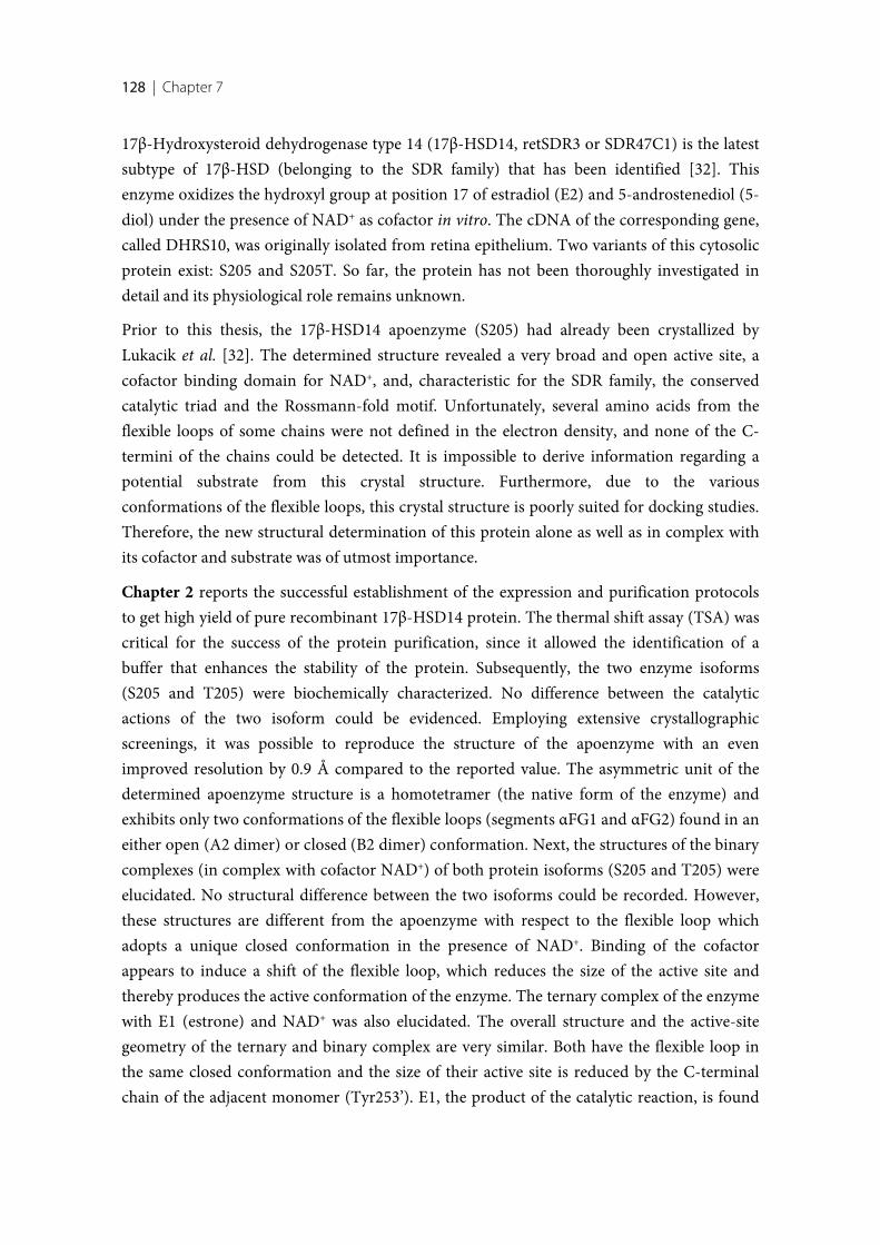
128 | Chapter 7
17β-Hydroxysteroid dehydrogenase type 14 (17β-HSD14, retSDR3 or SDR47C1) is the latestsubtype of 17β-HSD (belonging to the SDR family) that has been identified [32]. Thisenzyme oxidizes the hydroxyl group at position 17 of estradiol (E2) and 5-androstenediol (5-diol) under the presence of NAD+ as cofactor in vitro. The cDNA of the corresponding gene,called DHRS10, was originally isolated from retina epithelium. Two variants of this cytosolicprotein exist: S205 and S205T. So far, the protein has not been thoroughly investigated indetail and its physiological role remains unknown.
Prior to this thesis, the 17β-HSD14 apoenzyme (S205) had already been crystallized byLukacik et al. [32]. The determined structure revealed a very broad and open active site, acofactor binding domain for NAD+, and, characteristic for the SDR family, the conservedcatalytic triad and the Rossmann-fold motif. Unfortunately, several amino acids from theflexible loops of some chains were not defined in the electron density, and none of the C-termini of the chains could be detected. It is impossible to derive information regarding apotential substrate from this crystal structure. Furthermore, due to the variousconformations of the flexible loops, this crystal structure is poorly suited for docking studies.Therefore, the new structural determination of this protein alone as well as in complex withits cofactor and substrate was of utmost importance.
Chapter 2 reports the successful establishment of the expression and purification protocolsto get high yield of pure recombinant 17β-HSD14 protein. The thermal shift assay (TSA) wascritical for the success of the protein purification, since it allowed the identification of abuffer that enhances the stability of the protein. Subsequently, the two enzyme isoforms(S205 and T205) were biochemically characterized. No difference between the catalyticactions of the two isoform could be evidenced. Employing extensive crystallographicscreenings, it was possible to reproduce the structure of the apoenzyme with an evenimproved resolution by 0.9 Å compared to the reported value. The asymmetric unit of thedetermined apoenzyme structure is a homotetramer (the native form of the enzyme) andexhibits only two conformations of the flexible loops (segments αFG1 and αFG2) found in aneither open (A2 dimer) or closed (B2 dimer) conformation. Next, the structures of the binarycomplexes (in complex with cofactor NAD+) of both protein isoforms (S205 and T205) wereelucidated. No structural difference between the two isoforms could be recorded. However,these structures are different from the apoenzyme with respect to the flexible loop whichadopts a unique closed conformation in the presence of NAD+. Binding of the cofactorappears to induce a shift of the flexible loop, which reduces the size of the active site andthereby produces the active conformation of the enzyme. The ternary complex of the enzymewith E1 (estrone) and NAD+ was also elucidated. The overall structure and the active-sitegeometry of the ternary and binary complex are very similar. Both have the flexible loop inthe same closed conformation and the size of their active site is reduced by the C-terminalchain of the adjacent monomer (Tyr253’). E1, the product of the catalytic reaction, is found
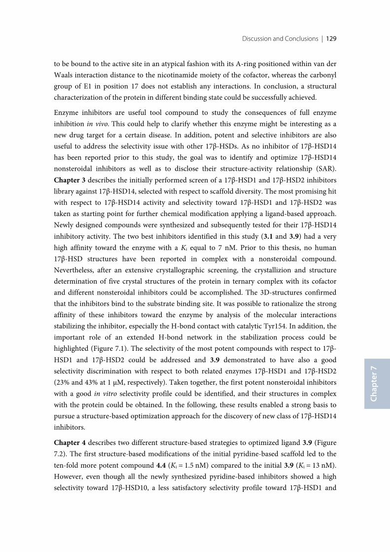
Discussion and Conclusions | 129
Chap
ter 7
to be bound to the active site in an atypical fashion with its A-ring positioned within van derWaals interaction distance to the nicotinamide moiety of the cofactor, whereas the carbonylgroup of E1 in position 17 does not establish any interactions. In conclusion, a structuralcharacterization of the protein in different binding state could be successfully achieved.
Enzyme inhibitors are useful tool compound to study the consequences of full enzymeinhibition in vivo. This could help to clarify whether this enzyme might be interesting as anew drug target for a certain disease. In addition, potent and selective inhibitors are alsouseful to address the selectivity issue with other 17β-HSDs. As no inhibitor of 17β-HSD14has been reported prior to this study, the goal was to identify and optimize 17β-HSD14nonsteroidal inhibitors as well as to disclose their structure-activity relationship (SAR).Chapter 3 describes the initially performed screen of a 17β-HSD1 and 17β-HSD2 inhibitorslibrary against 17β-HSD14, selected with respect to scaffold diversity. The most promising hitwith respect to 17β-HSD14 activity and selectivity toward 17β-HSD1 and 17β-HSD2 wastaken as starting point for further chemical modification applying a ligand-based approach.Newly designed compounds were synthesized and subsequently tested for their 17β-HSD14inhibitory activity. The two best inhibitors identified in this study (3.1 and 3.9) had a veryhigh affinity toward the enzyme with a Ki equal to 7 nM. Prior to this thesis, no human17β-HSD structures have been reported in complex with a nonsteroidal compound.Nevertheless, after an extensive crystallographic screening, the crystallizion and structuredetermination of five crystal structures of the protein in ternary complex with its cofactorand different nonsteroidal inhibitors could be accomplished. The 3D-structures confirmedthat the inhibitors bind to the substrate binding site. It was possible to rationalize the strongaffinity of these inhibitors toward the enzyme by analysis of the molecular interactionsstabilizing the inhibitor, especially the H-bond contact with catalytic Tyr154. In addition, theimportant role of an extended H-bond network in the stabilization process could behighlighted (Figure 7.1). The selectivity of the most potent compounds with respect to 17β-HSD1 and 17β-HSD2 could be addressed and 3.9 demonstrated to have also a goodselectivity discrimination with respect to both related enzymes 17β-HSD1 and 17β-HSD2(23% and 43% at 1 μM, respectively). Taken together, the first potent nonsteroidal inhibitorswith a good in vitro selectivity profile could be identified, and their structures in complexwith the protein could be obtained. In the following, these results enabled a strong basis topursue a structure-based optimization approach for the discovery of new class of 17β-HSD14inhibitors.
Chapter 4 describes two different structure-based strategies to optimized ligand 3.9 (Figure7.2). The first structure-based modifications of the initial pyridine-based scaffold led to theten-fold more potent compound 4.4 (Ki = 1.5 nM) compared to the initial 3.9 (Ki = 13 nM).However, even though all the newly synthesized pyridine-based inhibitors showed a highselectivity toward 17β-HSD10, a less satisfactory selectivity profile toward 17β-HSD1 and
130 | Chapter 7
17β-HSD2 was observed. The optimization of the cocrystallization conditions resulted in thedetermination of three new crystal structures of the inhibitor-enzyme complex. The analysisof the crystal structure of 4.4 in complex with the protein revealed how this ligand achievesits high affinity through a complementary geometry with the enzyme.
The goal of the second structure-based optimization strategy was to extend the centralpyridine core to interact with the empty binding pocket adjacent to the A and B-ring. Thedesigned quinoline-based inhibitors were first modelled into the crystal structure and, as thesimulation suggested a good fit to the active site of the protein, they were subsequentlysynthesized. The predicted binding mode of 4.8 was confirmed by the co-crystal structureand, as anticipated, the very high potency (Ki = 12 nM) was subsequently determined by
Figure 7.1: Schematic summary of Chapter 3. On the left, the preliminary screening of known17β-HSD14 inhibitors is shown leading to the first optimization candidates. On the right, the H-bond network stabilizes the inhibitor bound to the active site of 17β-HSD14. Carbon atoms of theinhibitor are colored in gray and displayed as sticks. Protein residues involved in establishing theH-bonding network and the cofactor NAD+ (beige) are shown as thin sticks. H-bond interactionsare depicted as dotted lines. Water molecules are shown as red spheres.
Figure 7.2: Schematic summary of Chapter 4. On the left and on the right, synthetic modificationsscheme starting from the 2,6-pyridine ketone 3.9 are indicated. The image in the center shows aclose-up view of the binding pocket of 17β-HSD14 in complex with 3.9. The inhibitor is shown asstick model with carbon atoms colored in yellow. Protein residues (white) and the cofactor NAD+
(beige) are shown as thin sticks. H-bond interactions are depicted as dotted lines. The Fo-Fc
difference electron density is shown as green mesh at a contour level of 3σ.
Discussion and Conclusions | 131
Chap
ter 7
biophysical characterization. Four co-crystal structures were obtained of the enzyme incomplex with the quinoline-based inhibitors. Crystal structures of the protein-ligandcomplexes were prerequisite for understanding the inhibitor’s SAR and for their furtheroptimization. The determined crystal structures revealed how small chemical modificationsof the inhibitor’s substituents can affect the adopted binding mode. In fact, the deviatingbinding mode of 4.10 (that differs from 4.8 by only a single methyl group) did not result in asignificant change of the binding affinity compared to 4.8 (Ki = 6 nM vs Ki = 13 nM,respectively). Surprisingly, the 3-OH group of the C-ring did not form a direct H-bondinteraction with Tyr154 of the catalytic triad. Instead, it establishes an H-bond only withSer141. Nevertheless, Tyr154 establishes an H-bonding contact through a bridging watermolecule (W6) with the ligand’s carbonyl linker. The fact that this water molecule is found inclose proximity to the catalytic triad and that so far its position was found conserved in fourstructures, raises the question about a possible involvement in the catalytic mechanism.
The selectivity profile of three compounds (4.12, 4.13 and 4.15) between 17β-HSD14 and thethree other proteins (17β-HSD1, 17β-HSD2, and 17β-HSD10) was comparable to theselectivity profile of 3.9. Thus, these four compounds exhibit the most favorable selectivityprofile as well as the highest potency. In addition, 3.9, 4.12, 4.13 and 4.15 showed a very lowcytotoxicity (IG50 > 100 µM) and were not appointed either as substrates or as inhibitors ofthe multi-drug resistance protein Pgp, indicating that these compounds might not be effluxfrom the brain and at the same time the risk of potential side effects would be reduced. Thissuggests them as potent tool compounds for the further investigation of a putative in vivoadministration to get insights into the physiological role of 17β-HSD14.
In Chapter 5, a structural comparison with the aim to rationally explain the selectivityprofiles of the ligands between 17β-HSD14 and other 17β-HSDs was conducted. Thesuperimposition based on the Cα coordinates of the crystal structures of 17β-HSD1 revealshow the adopted conformation of the flexible loop drastically deviates depending on thebound state of the enzyme. Furthermore, the comparison revealed that different inhibitorclasses induce different conformational geometries for this protein region. The variablecharacter of the flexible loop is also observed for 17β-HSD14. As only one crystal structurefor 17β-HSD8 and 17β-HSD10 has been published yet, it is difficult to perform the sameanalysis of the flexible loop for the latter two enzymes. The typical V-like shape of thebinding pocket of 17β-HSD14 is determined by the two amino acids His93 and Gln148,which are not present in 17β-HSD1, 17β-HSD8 and 17β-HSD10. In addition, the latter threeenzymes also exhibit a rather flat binding pocket. In conclusion, it has to be expected that thedesign of inhibitors that match with the characteristically three-dimensional requirements of17β-HSD14 (and optionally address His93 and/or Gln148) should increase the selectivitytoward the latter target. A docking study with the program GOLD (Genetic Optimization forLigand Docking) [196] was performed with the aim to rationally select ligands for an initial
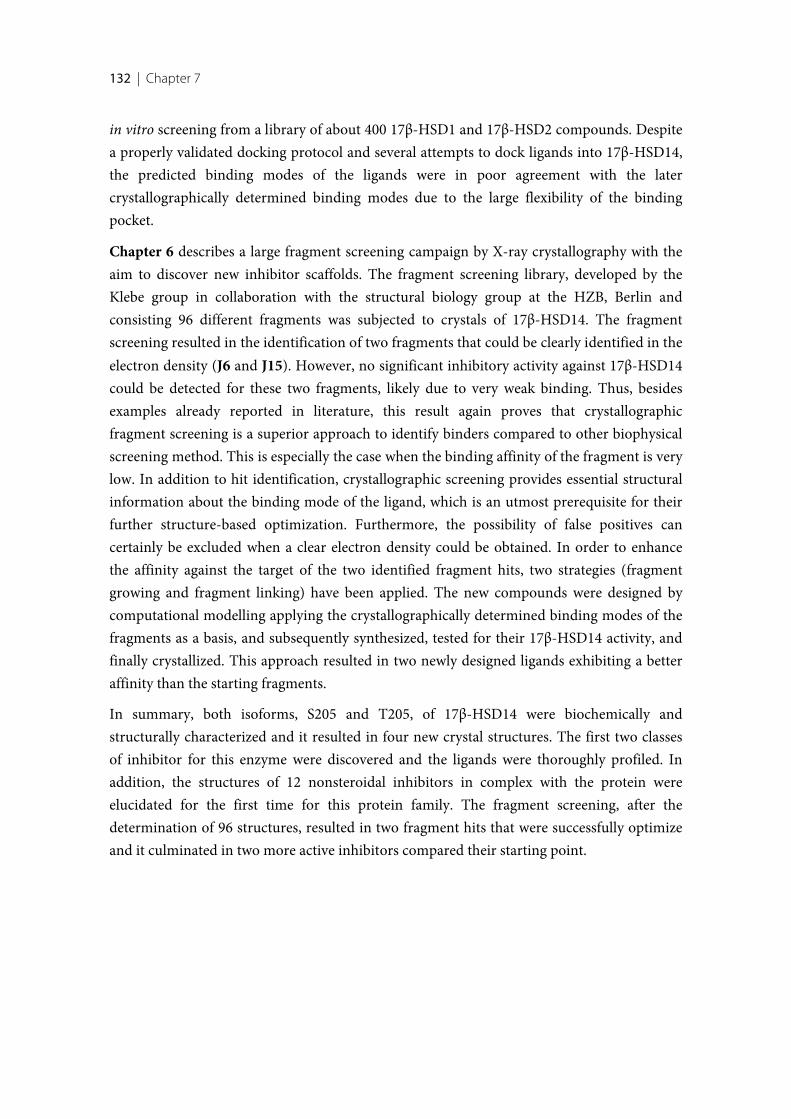
132 | Chapter 7
in vitro screening from a library of about 400 17β-HSD1 and 17β-HSD2 compounds. Despitea properly validated docking protocol and several attempts to dock ligands into 17β-HSD14,the predicted binding modes of the ligands were in poor agreement with the latercrystallographically determined binding modes due to the large flexibility of the bindingpocket.
Chapter 6 describes a large fragment screening campaign by X-ray crystallography with theaim to discover new inhibitor scaffolds. The fragment screening library, developed by theKlebe group in collaboration with the structural biology group at the HZB, Berlin andconsisting 96 different fragments was subjected to crystals of 17β-HSD14. The fragmentscreening resulted in the identification of two fragments that could be clearly identified in theelectron density (J6 and J15). However, no significant inhibitory activity against 17β-HSD14could be detected for these two fragments, likely due to very weak binding. Thus, besidesexamples already reported in literature, this result again proves that crystallographicfragment screening is a superior approach to identify binders compared to other biophysicalscreening method. This is especially the case when the binding affinity of the fragment is verylow. In addition to hit identification, crystallographic screening provides essential structuralinformation about the binding mode of the ligand, which is an utmost prerequisite for theirfurther structure-based optimization. Furthermore, the possibility of false positives cancertainly be excluded when a clear electron density could be obtained. In order to enhancethe affinity against the target of the two identified fragment hits, two strategies (fragmentgrowing and fragment linking) have been applied. The new compounds were designed bycomputational modelling applying the crystallographically determined binding modes of thefragments as a basis, and subsequently synthesized, tested for their 17β-HSD14 activity, andfinally crystallized. This approach resulted in two newly designed ligands exhibiting a betteraffinity than the starting fragments.
In summary, both isoforms, S205 and T205, of 17β-HSD14 were biochemically andstructurally characterized and it resulted in four new crystal structures. The first two classesof inhibitor for this enzyme were discovered and the ligands were thoroughly profiled. Inaddition, the structures of 12 nonsteroidal inhibitors in complex with the protein wereelucidated for the first time for this protein family. The fragment screening, after thedetermination of 96 structures, resulted in two fragment hits that were successfully optimizeand it culminated in two more active inhibitors compared their starting point.
133
[1] Nussey S, Whitehead S (2001) Principles of endocrinology. An Integrated Approach. edPublishers BS (BIOS Scientific Publishers, Oxford) Available at:https://www.ncbi.nlm.nih.gov/books/NBK20/#A10
[2] Carr MC (2003) The Emergence of the Metabolic Syndrome with Menopause. J ClinEndocrinol Metab 88: 2404–2411.
[3] Husen B, Huhtinen K, Poutanen M, Kangas L, Messinger J, Thole H (2006) Evaluation ofinhibitors for 17β-hydroxysteroid dehydrogenase type 1 in vivo in immunodeficient miceinoculated with MCF-7 cells stably expressing the recombinant human enzyme. Mol CellEndocrinol 248: 109–113.
[4] Weitzmann MN, Pacifici R, Parslow TG, Katz DR, Fuller K (2006) Estrogen deficiency andbone loss: an inflammatory tale. J Clin Invest 116: 1186–94.
[5] Gunnarsson C, Hellqvist E, Stål O, Group2 and the SSBC (2005) 17beta-Hydroxysteroiddehydrogenases involved in local oestrogen synthesis have prognostic significance in breastcancer. Br J Cancer 92: 547–52.
[6] Birge SJ (1997) The role of estrogen in the treatment of Alzheimer’s disease. Neurology 48:36–41.
[7] Gillies GE, McArthur S (2010) Estrogen Actions in the Brain and the Basis for DifferentialAction in Men and Women: A Case for Sex-Specific Medicines. Pharmacol Rev 62. Availableat: http://pharmrev.aspetjournals.org/content/62/2/155.long
[8] Labrie F, Luu-The V, Lin S-XX, Simard J, Labrie C (2000) Role of 17β-hydroxysteroiddehydrogenases in sex steroid formation in peripheral intracrine tissues. Trends EndocrinolMetab 11: 421–7.
[9] Labrie F (1991) Intracrinology. Mol Cell Endocrinol 78: 113–18.
[10] Poirier D (2003) Inhibitors of 17β-hydroxysteroid dehydrogenases. Curr Med Chem 10: 453–77.
[11] Lukacik P, Kavanagh KL, Oppermann U (2006) Structure and function of human 17β-hydroxysteroid dehydrogenases. Mol Cell Endocrinol 248: 61–71.
[12] Peltoketo H, Luu-The V, Simard J, Adamski J (1999) 17beta-hydroxysteroid dehydrogenase(HSD)/17-ketosteroid reductase (KSR) family; nomenclature and main characteristics of the17HSD/KSR enzymes. J Mol Endocrinol 23: 1–11.
134 | Bibliography
[13] Labrie F, Luu-The V, Lin SX, Simard J, Labrie C, El-Alfy M, Pelletier G, Bélanger A (2000)Intracrinology: role of the family of 17 beta-hydroxysteroid dehydrogenases in humanphysiology and disease. J Mol Endocrinol 25: 1–16.
[14] Gargano EM (2015) Development of 17 β -Hydroxysteroid Dehydrogenase Type 2 and Type1 Inhibitors for the Treatment of Osteoporosis and Estrogen Dependent Diseases.Dissertation (Universität des Saarlandes).
[15] Jörnvall H, Persson B, Krook M, Atrian S, Gonzàlez-Duarte R, Jeffery J, Ghosh D (1995)Short-chain dehydrogenases/reductases (SDR). Biochemistry 34: 6003–13.
[16] Oppermann U, Filling C, Hult M, Shafqat N, Wu X, Lindh M, Shafqat J, Nordling E, KallbergY, Persson B, Jörnvall H (2003) Short-chain dehydrogenases/reductases (SDR): the 2002update. Chem Biol Interact 143–144: 247–253.
[17] Persson B, Kallberg Y (2013) Classification and nomenclature of the superfamily of short-chain dehydrogenases/reductases (SDRs). Chem Biol Interact 202: 111–5.
[18] Moeller G, Adamski J (2006) Multifunctionality of human 17β-hydroxysteroiddehydrogenases. Mol Cell Endocrinol 248: 47–55.
[19] Puranen TJ, Poutanen MH, Peltoketo HE, Vihko PT, Vihko RK (1994) Site-directedmutagenesis of the putative active site of human 17β-hydroxysteroid dehydrogenase type 1.Biochem J 304: 289–293.
[20] Breton R, Housset D, Mazza C, Fontecilla-Camps JC (1996) The structure of a complex ofhuman 17β-hydroxysteroid dehydrogenase with estradiol and NADP+ identifies twoprincipal targets for the design of inhibitors. Structure 4: 905–915.
[21] Ghosh D, Pletnev VZ, Zhu DW, Wawrzak Z, Duax WL, Pangborn W, Labrie F, Lin SX (1995)Structure of human estrogenic 17 β-hydroxysteroid dehydrogenase at 2.20 A resolution.Structure 3: 503–13.
[22] Filling C, Berndt KD, Benach J, Knapp S, Prozorovski T, Nordling E, Ladenstein R, JörnvallH, Oppermann U (2002) Critical Residues for Structure and Catalysis in Short-chainDehydrogenases/Reductases. J Biol Chem 277: 25677–25684.
[23] Hedlund J, Jörnvall H, Persson B (2010) Subdivision of the MDR superfamily of medium-chain dehydrogenases/reductases through iterative hidden Markov model refinement. BMCBioinformatics 11: 534.
[24] Negri M, Recanatini M, Hartmann RW, Kollman P, Case D (2010) Insights in 17β-HSD1Enzyme Kinetics and Ligand Binding by Dynamic Motion Investigation. PLoS One 5: e12026.
[25] Penning TM (1996) 17β-Hydroxysteroid dehydrogenase: inhibitors and inhibitor design.Endocr Relat Cancer 3: 41–56.
[26] Penning TM (2011) Human hydroxysteroid dehydrogenases and pre-receptor regulation:Insights into inhibitor design and evaluation. J Steroid Biochem Mol Biol 125: 46–56.
[27] Moeller G, Adamski J (2009) Integrated view on 17β-hydroxysteroid dehydrogenases. MolCell Endocrinol 301: 7–19.
[28] Meier M, Möller G, Adamski J (2009) Perspectives in Understanding the Role of Human 17β-Hydroxysteroid Dehydrogenases in Health and Disease. Ann N Y Acad Sci 1155: 15–24.
Bibliography | 135
[29] Luu-The V (2001) Analysis and characteristics of multiple types of human 17β-hydroxysteroid dehydrogenase. J Steroid Biochem Mol Biol 76: 143–151.
[30] Haller F, Moman E, Hartmann RW, Adamski J, Mindnich R (2010) Molecular Framework ofSteroid/Retinoid Discrimination in 17β-Hydroxysteroid Dehydrogenase Type 1 andPhotoreceptor-associated Retinol Dehydrogenase. J Mol Biol 399: 255–267.
[31] Zhongyi S, Rantakari P, Lamminen T, Toppari J, Poutanen M (2007) Transgenic Male MiceExpressing Human Hydroxysteroid Dehydrogenase 2 Indicate a Role for the EnzymeIndependent of Its Action on Sex Steroids. Endocrinology 148: 3827–3836.
[32] Lukacik P, Keller B, Bunkoczi G, Kavanagh KL, Hwa Lee W, Adamski J, Oppermann U, LeeWH, Hwa Lee W, Adamski J, Oppermann U (2007) Structural and biochemicalcharacterization of human orphan DHRS10 reveals a novel cytosolic enzyme with steroiddehydrogenase activity. Biochem J 402: 419–427.
[33] The PyMOL Molecular Graphics System, Version 1.7.x Schrödinger, LLC.
[34] Marchais-Oberwinkler S, Henn C, Möller G, Klein T, Negri M, Oster A, Spadaro A, Werth R,Wetzel M, Xu K, Frotscher M, Hartmann RW, Adamski J (2011) 17β-Hydroxysteroiddehydrogenases (17β-HSDs) as therapeutic targets: Protein structures, functions, and recentprogress in inhibitor development. J Steroid Biochem Mol Biol 125: 66–82.
[35] Sivik T, Gunnarsson C, Fornander T, Nordenskjöld B, Skoog L, Stål O, Jansson A (2012) 17β-Hydroxysteroid Dehydrogenase Type 14 Is a Predictive Marker for Tamoxifen Response inOestrogen Receptor Positive Breast Cancer. PLoS One 7: e40568.
[36] Jansson AK, Gunnarsson C, Cohen M, Sivik T, Stål O (2006) 17β-HydroxysteroidDehydrogenase 14 Affects Estradiol Levels in Breast Cancer Cells and Is a Prognostic Markerin Estrogen Receptor-Positive Breast Cancer. Cancer Res 66: 11471–11477.
[37] Sherbet DP, Papari-Zareei M, Khan N, Sharma KK, Brandmaier A, Rambally S,Chattopadhyay A, Andersson S, Agarwal AK, Auchus RJ (2007) Cofactors, redox state, anddirectional preferences of hydroxysteroid dehydrogenases. Mol Cell Endocrinol 265266: 83–88.
[38] D Voet and J G Voet (1990) Biochemistry (Headington Hill Hall, New York)
[39] Agarwal AK, Auchus RJ (2005) Minireview: Cellular Redox State Regulates HydroxysteroidDehydrogenase Activity and Intracellular Hormone Potency. Endocrinology 146: 2531–2538.
[40] Jin J-Z, Lin S-X (1999) Human Estrogenic 17β-Hydroxysteroid Dehydrogenase:Predominance of Estrone Reduction and Its Induction by NADPH. Biochem Biophys ResCommun 259: 489–493.
[41] Duax WL, Pletnev V, Addlagatta A, Bruenn J, Weeks CM (2003) Rational Proteomics I.Fingerprint Identification and Cofactor Specificity in the Short-Chain Oxidoreductase(SCOR) Enzyme Family. Proteins Struct Funct Genet 53: 931–943.
[42] Pletnev VZ, Weeks CM, Duax WL (2004) Rational proteomics II: Electrostatic nature ofcofactor preference in the short-chain oxidoreductase (SCOR) enzyme family. Proteins StructFunct Bioinforma 57: 294–301.
136 | Bibliography
[43] Mazza C, Breton R, Housset D, Fontecilla-Camps JC (1998) Unusual charge stabilization ofNADP+ in 17β-hydroxysteroid dehydrogenase. J Biol Chem 273: 8145–52.
[44] Shi R, Lin S-X (2004) Cofactor hydrogen bonding onto the protein main chain is conserved inthe short chain dehydrogenase/reductase family and contributes to nicotinamide orientation.J Biol Chem 279: 16778–16785.
[45] Chen Z, Tsigelny I, Lee WR, Baker ME, Chang SH (1994) Adding a positive charge at residue46 of Drosophila alcohol dehydrogenase increases cofactor specificity for NADP+. FEBS Lett356: 81–85.
[46] Scrutton NS, Berry A, Perham RN (1990) Redesign of the coenzyme specificity of adehydrogenase by protein engineering. Nature 343: 38–43.
[47] Lin SX, Yang F, Jin JZ, Breton R, Zhu DW, Luu-The V, Labrie F (1992) Subunit identity of thedimeric 17β-hydroxysteroid dehydrogenase from human placenta. J Biol Chem 267: 16182–16187.
[48] Luu-The V, Zhang Y, Poirier D, Labrie F (1995) Characteristics of human types 1, 2 and 317β-hydroxysteroid dehydrogenase activities: oxidation/reduction and inhibition. J SteroidBiochem Mol Biol 55: 581–587.
[49] Poutanen M, Miettinen M, Vihko R (1993) Differential estrogen substrate specificities fortransiently expressed human placental 17β-hydroxysteroid dehydrogenase and anendogenous enzyme expressed in cultured COS-m6 cells. Endocrinology 133: 2639–2644.
[50] Han Q, Campbell RL, Gangloff A, Huang YW, Lin SX (2000) Dehydroepiandrosterone anddihydrotestosterone recognition by human estrogenic 17β-hydroxysteroid dehydrogenase. C-18/c-19 steroid discrimination and enzyme-induced strain. J Biol Chem 275: 1105–1111.
[51] Messinger J, Hirvelä L, Husen B, Kangas L, Koskimies P, Pentikäinen O, Saarenketo P, TholeH (2006) New inhibitors of 17β-hydroxysteroid dehydrogenase type 1. Mol Cell Endocrinol248: 192–198.
[52] Brozic P, Lanisnik Risner T, Gobec S (2008) Inhibitors of 17β-hydroxysteroid dehydrogenasetype 1. Curr Med Chem 15: 137–150.
[53] Bey E, Marchais-Oberwinkler S, Werth R, Negri M, Al-Soud YA, Kruchten P, Oster A,Frotscher M, Birk B, Hartmann RW (2008) Design, Synthesis, Biological Evaluation andPharmacokinetics of Bis(hydroxyphenyl) substituted Azoles, Thiophenes, Benzenes, and Aza-Benzenes as Potent and Selective Nonsteroidal Inhibitors of 17β-HydroxysteroidDehydrogenase Type 1 (17β-HSD1). J Med Chem 51: 6725–6739.
[54] Oster A, Klein T, Werth R, Kruchten P, Bey E, Negri M, Marchais-Oberwinkler S, FrotscherM, Hartmann RW (2010) Novel estrone mimetics with high 17β-HSD1 inhibitory activity.Bioorg Med Chem 18: 3494–3505.
[55] Hilborn E, Stål O, Jansson A (2017) Estrogen and androgen-converting enzymes 17β-hydroxysteroid dehydrogenase and their involvement in cancer: with a special focus on 17β-hydroxysteroid dehydrogenase type 1, 2, and breast cancer. Oncotarget 8: 30552–30562.
[56] Miyoshi Y, Ando A, Shiba E, Taguchi T, Tamaki Y, Noguchi S (2001) Involvement of up-regulation of 17β-hydroxysteroid dehydrogenase type 1 in maintenance of intratumoral highestradiol levels in postmenopausal breast cancers. Int J Cancer 94: 685–689.
Bibliography | 137
[57] Jansson A (2009) 17β-hydroxysteroid dehydrogenase enzymes and breast cancer. J SteroidBiochem Mol Biol 114: 64–67.
[58] Blomquist CH, Bonenfant M, McGinley DM, Posalaky Z, Lakatua DJ, Tuli-Puri S, Bealka DG,Tremblay Y (2002) Androgenic and estrogenic 17β-hydroxysteroid dehydrogenase/17-ketosteroid reductase in human ovarian epithelial tumors: evidence for the type 1, 2 and 5isoforms. J Steroid Biochem Mol Biol 81: 343–351.
[59] Šmuc T, Pucelj MR, Šinkovec J, Husen B, Thole H, Rižner TL (2007) Expression analysis ofthe genes involved in estradiol and progesterone action in human ovarian endometriosis.Gynecol Endocrinol 23: 105–111.
[60] Saloniemi T, Järvensivu P, Koskimies P, Jokela H, Lamminen T, Ghaem-Maghami S, Dina R,Damdimopoulou P, Mäkelä S, Perheentupa A, Kujari H, Brosens J, Poutanen M (2010) NovelHydroxysteroid (17β) Dehydrogenase 1 Inhibitors Reverse Estrogen-Induced EndometrialHyperplasia in Transgenic Mice. Am J Pathol 176: 1443–1451.
[61] Kasai T, Shozu M, Murakami K, Segawa T, Shinohara K, Nomura K, Inoue M (2004)Increased Expression of Type I 17β-Hydroxysteroid Dehydrogenase Enhances in SituProduction of Estradiol in Uterine Leiomyoma. J Clin Endocrinol Metab 89: 5661–5668.
[62] Vihko P, Herrala A, Härkönen P, Isomaa V, Kaija H, Kurkela R, Pulkka A (2006) Control ofcell proliferation by steroids: The role of 17HSDs. Mol Cell Endocrinol 248: 141–148.
[63] Vihko P, Herrala A, Härkönen P, Isomaa V, Kaija H, Kurkela R, Li Y, Patrikainen L, PulkkaA, Soronen P, Törn S (2005) Enzymes as modulators in malignant transformation. J SteroidBiochem Mol Biol 93: 277–283.
[64] Azzi A, Rehse PH, Zhu D-W, Campbell RL, Labrie F, Lin S-X (1996) Crystal structure ofhuman estrogenic 17β-hydroxysteroid dehydrogenase complexed with 17β-estradiol. NatStruct Mol Biol 3: 665–668.
[65] Laplante Y, Poirier D (2008) Proliferative effect of androst-4-ene-3,17-dione and itsmetabolites in the androgen-sensitive LNCaP cell line. Steroids 73: 266–271.
[66] Moghrabi, Nabil and Andersson S (1998) 17β-Hydroxysteroid Dehydrogenases: PhysiologicalRoles in Health and Disease. TEM 7: 265–270.
[67] Vicker N, Sharland CM, Heaton WB, Ramos Gonzalez AM, Bailey H V, Smith A, Springall JS,
Day JM, Tutill HJ, Reed MJ, Purohit A, Potter BVL (2009) The design of novel 17-hydroxysteroid dehydrogenase type 3 inhibitors. Mol Cell Endocrinol 301: 259–265.
[68] Koh E, Noda T, Kanaya J, Namiki M (2002) Differential expression of 17β-hydroxysteroiddehydrogenase isozyme genes in prostate cancer and noncancer tissues. Prostate 53: 154–159.
[69] Lin H-K, Jez JM, Schlegel BP, Peehl DM, Pachter JA, Penning TM (1997) Expression andCharacterization of Recombinant Type 2 3α-Hydroxysteroid Dehydrogenase (HSD) fromHuman Prostate: Demonstration of Bifunctional 3α/17β-HSD Activity and CellularDistribution. Mol Endocrinol 11: 1971–1984.
[70] Matsuura K, Shiraishi H, Hara A, Sato K, Deyashiki Y, Ninomiya M SS (1998) Identificationof a Principal mRNA Species for Human 3α-Hydroxysteroid Dehydrogenase Isoform
138 | Bibliography
(AKR1C3) That Exhibits High Prostaglandin D2 11-Ketoreductase Activity. J Biochem 124:940–946.
[71] Song D, Liu G, Luu-The V, Zhao D, Wang L, Zhang H, Xueling G, Li S, Désy L, Labrie F,Pelletier G (2006) Expression of aromatase and 17β-hydroxysteroid dehydrogenase types 1, 7and 12 in breast cancer An immunocytochemical study. J Steroid Biochem Mol Biol 101: 136–144.
[72] Liu H, Robert A, Luu-The V (2005) Cloning and characterization of human form 2 type 717β-hydroxysteroid dehydrogenase, a primarily 3β-keto reductase and estrogen activatingand androgen inactivating enzyme. J Steroid Biochem Mol Biol 94: 173–179.
[73] Ohnesorg T, Keller B, Hrabé de Angelis M, Adamski J (2006) Transcriptional regulation ofhuman and murine 17beta-hydroxysteroid dehydrogenase type-7 confers its participation incholesterol biosynthesis. J Mol Endocrinol 37: 185–97.
[74] Sakurai N, Miki Y, Suzuki T, Watanabe K, Narita T, Ando K, Yung TMCC, Aoki D, SasanoH, Handa H (2006) Systemic distribution and tissue localizations of human 17beta-hydroxysteroid dehydrogenase type 12. J Steroid Biochem Mol Biol 99: 174–181.
[75] Moon Y-A, Horton JD (2003) Identification of two mammalian reductases involved in thetwo-carbon fatty acyl elongation cascade. J Biol Chem 278: 7335–7343.
[76] Luu-The V, Tremblay P, Labrie F (2006) Characterization of Type 12 17β-HydroxysteroidDehydrogenase, an Isoform of Type 3 17β-Hydroxysteroid Dehydrogenase Responsible forEstradiol Formation in Women. Mol Endocrinol 20: 437–443.
[77] Liu S, Huang C, Li D, Ren W, Zhang H, Qi M, Li X, Yu L (2007) Molecular cloning andexpression analysis of a new gene for short- chain dehydrogenase/reductase 9. Acta BiochimPlonica Commun 54: 213–218.
[78] Su W, Wang Y, Jia X, Wu W, Li L, Tian X, Li S, Wang C, Xu H, Cao J, Han Q, Xu S, Chen Y,Zhong Y, Zhang X, Liu P, Gustafsson J-A, Guan Y (2014) Comparative proteomic studyreveals 17β-HSD13 as a pathogenic protein in nonalcoholic fatty liver disease. Proc Natl AcadSci 111: 11437–11442.
[79] Casey ML, MacDonald PC, Andersson S (1994) 17β-Hydroxysteroid dehydrogenase type 2:chromosomal assignment and progestin regulation of gene expression in humanendometrium. J Clin Invest 94: 2135–2141.
[80] Moghrabi N, Head JR, Andersson S (1997) Cell Type-Specific Expression of 17β-Hydroxysteroid Dehydrogenase Type 2 in Human Placenta and Fetal Liver. J Clin EndocrinolMetab 82: 3872–3878.
[81] Mustonen M V, Poutanen MH, Kellokumpu S, de Launoit Y, Isomaa V V, Vihko RK, VihkoPT (1998) Mouse 17β-hydroxysteroid dehydrogenase type 2 mRNA is predominantlyexpressed in hepatocytes and in surface epithelial cells of the gastrointestinal and urinarytracts. J Mol Endocrinol 20: 67–74.
[82] Mustonen M, Poutanen M, Chotteau-Lelievre A, De Launoit Y, Isomaa V, Vainio S, Vihko R,Vihko P (1997) Ontogeny of 17β-hydroxysteroid dehydrogenase type 2 mRNA expression inthe developing mouse placenta and fetus. Mol Cell Endocrinol 134: 33–40.
Bibliography | 139
[83] Poirier D, Bydal P, Tremblay MR, Sam K-M, Luu-The V (2001) Inhibitors of type II 17β-hydroxysteroid dehydrogenase. Mol Cell Endocrinol 171: 119–128.
[84] Day J. M., Tutill H. J. PA (2010) 17β-Hydroxysteroid dehydrogenase inhibitors. MinervaEndocrinol 35: 87–108.
[85] Felson DT, Zhang Y, Hannan MT, Kiel DP, Wilson P, Anderson JJ (1993) The Effect ofPostmenopausal Estrogen Therapy on Bone Density in Elderly Women. N Engl J Med 329:1141–1146.
[86] Chen C-L, Weiss NS, Newcomb P, Barlow W, White E (2002) Hormone ReplacementTherapy in Relation to Breast Cancer. JAMA 287: 734.
[87] Gargano EM, Allegretta G, Perspicace E, Carotti A, Van Koppen C, Frotscher M, Marchais-Oberwinkler S, Hartmann RW (2015) 17β-Hydroxysteroid Dehydrogenase Type 2 Inhibition:Discovery of Selective and Metabolically Stable Compounds Inhibiting Both the HumanEnzyme and Its Murine Ortholog. PLoS One 10: e0134754.
[88] Spadaro A, Negri M, Marchais-Oberwinkler S, Bey E, Frotscher M (2012)Hydroxybenzothiazoles as New Nonsteroidal Inhibitors of 17β-HydroxysteroidDehydrogenase Type 1 (17β-HSD1). PLoS One 7. doi:10.1371/journal.pone.0029252
[89] Perspicace E, Giorgio A, Carotti A, Marchais-Oberwinkler S, Hartmann RW (2013) Novel N-methylsulfonamide and retro-N-methylsulfonamide derivatives as 17β-hydroxysteroiddehydrogenase type 2 (17β-HSD2) inhibitors with good ADME-related physicochemicalparameters. Eur J Med Chem 69: 201–215.
[90] Marchais-Oberwinkler S, Xu K, Wetzel M, Perspicace E, Negri M, Meyer A, Odermatt A,Möller G, Adamski J, Hartmann RW (2013) Structural optimization of 2,5-thiophene amidesas highly potent and selective 17β-hydroxysteroid dehydrogenase type 2 inhibitors for thetreatment of osteoporosis. J Med Chem 56: 167–181.
[91] Gargano EM, Perspicace E, Hanke N, Carotti A, Marchais-Oberwinkler S, Hartmann RW(2014) Metabolic stability optimization and metabolite identification of 2,5-thiophene amide17β-hydroxysteroid dehydrogenase type 2 inhibitors. Eur J Med Chem 87: 203–219.
[92] Gargano EM, Perspicace E, Carotti A, Marchais-Oberwinkler S, Hartmann RW (2016)Addressing cytotoxicity of 1,4-biphenyl amide derivatives: Discovery of new potent andselective 17β-hydroxysteroid dehydrogenase type 2 inhibitors. Bioorg Med Chem Lett 26: 21–24.
[93] Du Yan S, Fu J, Soto C, Chen X, Zhu H, Al-Mohanna F, Collison K, Zhu A, Stern E, Saido T,Tohyama M, Ogawa S, Roher A, Stern D (1997) An intracellular protein that binds amyloid-βpeptide and mediates neurotoxicity in Alzheimer’s disease. Nature 389: 689–695.
[94] Kissinger CR, Rejto PA, Pelletier LA, Thomson JA, Showalter RE, Abreo MA, Agree CS,Margosiak S, Meng JJ, Aust RM, Vanderpool D, Li B, Tempczyk-Russell A, Villafranca JE(2004) Crystal Structure of Human ABAD/HSD10 with a Bound Inhibitor: Implications forDesign of Alzheimer’s Disease Therapeutics. J Mol Biol 342: 943–952.
[95] Oppermann UC, Salim S, Tjernberg LO, Terenius L, Jörnvall H (1999) Binding of amyloidbeta-peptide to mitochondrial hydroxyacyl-CoA dehydrogenase (ERAB): regulation of an
140 | Bibliography
SDR enzyme activity with implications for apoptosis in Alzheimer’s disease. FEBS Lett 451:238–242.
[96] He XY, Merz G, Yang YZ, Mehta P, Schulz H, Yang SY (2001) Characterization andlocalization of human type10 17beta-hydroxysteroid dehydrogenase. Eur J Biochem 268:4899–4907.
[97] Yang S-Y, He X-Y, Schulz H (2005) Multiple functions of type 10 17β-hydroxysteroiddehydrogenase. Trends Endocrinol Metab 16: 167–175.
[98] Chai Z, Brereton P, Suzuki T, Sasano H, Obeyesekere V, Escher G, Saffery R, Fuller P,Enriquez C, Krozowski Z (2003) 17β-Hydroxysteroid Dehydrogenase Type XI Localizes toHuman Steroidogenic Cells. Endocrinology 144: 2084–2091.
[99] Motojima K (2004) 17β-Hydroxysteroid dehydrogenase type 11 is a major peroxisomeproliferator-activated receptor α-regulated gene in mouse intestine. Eur J Biochem 271: 4141–4146.
[100] Haeseleer F, Palczewski K (2000) Short-chain dehydrogenases/reductases in retina. MethodsEnzymol 316: 372–383.
[101] Strausberg RL, Feingold EA, Grouse LH, Derge JG, Klausner RD, Collins FS, Wagner L,Shenmen CM, Schuler GD, Altschul SF, Zeeberg B, Buetow KH, Schaefer CF, Bhat NK,Hopkins RF, Jordan H, Moore T, Max SI, Wang J, et al. (2002) Generation and initial analysisof more than 15,000 full-length human and mouse cDNA sequences. Proc Natl Acad Sci 99:16899–16903.
[102] Homo sapiens hydroxysteroid (17-beta) dehydrogenase 14, mRNA (cDNA clone MGC:10685IMAGE:4026397), complete cds CN - BC006283.2 (2006) Available at:http://www.ncbi.nlm.nih.gov/nuccore/BC006283.2
[103] Sivik T, Vikingsson S, Gréen H, Jansson A (2012) Expression Patterns of 17β-HydroxysteroidDehydrogenase 14 in Human Tissues. Horm Metab Res 44: 949–956.
[104] Beck KR, Kaserer T, Schuster D, Odermatt A (2017) Virtual screening applications in short-chain dehydrogenase/reductase research. J Steroid Biochem Mol Biol 171: 157–177.
[105] Halle B (2004) Biomolecular cryocrystallography: structural changes during flash-cooling.Proc Natl Acad Sci USA 101: 4793–4798.
[106] Wlodawer A, Minor W, Dauter Z, Jaskolski M (2013) Protein crystallography for aspiringcrystallographers or how to avoid pitfalls and traps in macromolecular structuredetermination. FEBS J 280: 5705–5736.
[107] Shi Y (2014) A glimpse of structural biology through X-ray crystallography. Cell 159: 995–1014.
[108] Wlodawer A, Minor W, Dauter Z, Jaskolski M (2008) Protein crystallography for non-crystallographers, or how to get the best (but not more) from published macromolecularstructures. FEBS J 275: 1–21.
[109] Hassell AM, An G, Bledsoe RK, Bynum JM, Luke H, Iii C, Deng S-JJ, Gampe RT, Grisard TE,Madauss KP, Nolte RT, Rocque WJ, Wang L, Weaver KL, Williams SP, Wisely GB, Xu R,Shewchuk LM (2007) Crystallization of protein–ligand complexes. Acta Cryst 63: 72–79.
Bibliography | 141
[110] Danley DE (2006) Crystallization to obtain protein–ligand complexes for structure-aideddrug design. Acta Cryst 62: 569–575.
[111] Ehrmann FR, Stojko J, Metz A, Ois Debaene F, Barandun LJ, Heine A, Diederich FO,Cianférani S, Reuter K, Klebe G (2017) Soaking suggests “alternative facts”: Only co-crystallization discloses major ligand-induced interface rearrangements of a homodimerictRNA-binding protein indicating a novel mode-of-inhibition. PLoS One 12: e0175723.
[112] Zheng H, Hou J, Zimmerman MD, Wlodawer A, Minor W (2014) The future ofcrystallography in drug discovery. Expert Opin Drug Discov 9: 125–137.
[113] Scheilman JA (1997) Temperature, Stability, and the Hydrophobic Interaction. Biophys J 73:2960–2964.
[114] Privalov PL (1979) Stability of Proteins Small Globular Proteins. Adv Protein Chem 33: 167–241.
[115] Lo M-C, Aulabaugh A, Jin G, Cowling R, Bard J, Malamas M, Ellestad G (2004) Evaluation offluorescence-based thermal shift assays for hit identification in drug discovery. Anal Biochem332: 153–159.
[116] Vedadi M, Niesen FH, Allali-Hassani A, Fedorov OY, Finerty PJ, Wasney GA, Yeung R,Arrowsmith C, Ball LJ, Berglund H, Hui R, Marsden BD, Nordlund PR, Sundstrom M,Weigelt J, Edwards AM, Thornton JM (2006) Chemical screening methods to identify ligandsthat promote protein stability, protein crystallization, and structure determination. Proc NatlAcad Sci 103: 15835–15840.
[117] Senisterra GA, Markin E, Yamazaki K, Hui R, Vedadi M, Awrey DE (2006) Screening forLigands Using a Generic and High-Throughput Light-Scattering-Based Assay. J BiomolScreen 11: 940–948.
[118] Matulis D, Kranz JK, Salemme FR, Todd MJ (2005) Thermodynamic Stability of CarbonicAnhydrase: Measurements of Binding Affinity and Stoichiometry Using ThermoFluor.Biochemistry 44: 5258–5266.
[119] Niesen FH, Berglund H, Vedadi M (2007) The use of differential scanning fluorimetry todetect ligand interactions that promote protein stability. Nat Protoc 2: 2212–2221.
[120] Lavinder JJ, Hari SB, Sullivan BJ, Magliery TJ (2009) High-Throughput Thermal Scanning: AGeneral, Rapid Dye-Binding Thermal Shift Screen for Protein Engineering. JACS Commun131: 3794–3795.
[121] Lucet IS, Hildebrand JM, Czabotar PE, Zhang J-G, Nicola NA, Silke J, Babon JJ, Murphy JM(2005) Determination of Pseudokinase-ligand Interaction by a Fluorescence-based ThermalShift Assay. bio-protol 4. Available at: http://www.bio-protocol.org/e1135
[122] Cummings MD, Farnum MA, Nelen MI (2006) Universal Screening Methods andApplications of ThermoFluor®. J Biomol Screen 11: 854–863.
[123] Saijo K, Collier JG, Li AC, Katzenellenbogen JA, Glass CK (2011) An ADIOL-ERβ-CtBPtransrepression pathway negatively regulates microglia-mediated inflammation. Cell 145:584–595.
142 | Bibliography
[124] Frotscher M, Ziegler E, Marchais-Oberwinkler S, Kruchten P, Neugebauer A, Fetzer L,Scherer C, Müller-Vieira U, Messinger J, Thole H, Hartmann RW (2008) Design, synthesis,and biological evaluation of (hydroxyphenyl)naphthalene and -quinoline derivatives: potentand selective nonsteroidal inhibitors of 17β-hydroxysteroid dehydrogenase type 1 (17β-HSD1) for the treatment of estrogen-dependent disease. J Med Chem 51: 2158–2169.
[125] Kruchten P, Werth R, Bey E, Oster A, Marchais-Oberwinkler S, Frotscher M, Hartmann RW(2009) Selective inhibition of 17β-hydroxysteroid dehydrogenase type 1 (17β-HSD1) reducesestrogen responsive cell growth of T47-D breast cancer cells. J Steroid Biochem Mol Biol 114:200–206.
[126] Bertoletti N, Braun F, Lepage M, Möller G, Adamski J, Heine A, Klebe G, Marchais-Oberwinkler S (2016) New Insights into Human 17β-Hydroxysteroid Dehydrogenase Type14: First Crystal Structures in Complex with a Steroidal Ligand and with a PotentNonsteroidal Inhibitor. J Med Chem 59: 6961–6967.
[127] Braun F, Bertoletti N, Möller G, Adamski J, Steinmetzer T, Salah M, Abdelsamie AS, vanKoppen CJ, Heine A, Klebe G, Marchais-Oberwinkler S (2016) First Structure–ActivityRelationship of 17β-Hydroxysteroid Dehydrogenase Type 14 Nonsteroidal Inhibitors andCrystal Structures in Complex with the Enzyme. J Med Chem 59: 10719–10737.
[128] Gekko K, Timasheff SN (1981) Mechanism of protein stabilization by glycerol: preferentialhydration in glycerol-water mixtures. Biochemistry 20: 4667–4676.
[129] Myers JS, Jakoby WB (1975) Glycerol as an agent eliciting small conformational changes inalcohol dehydrogenase. J Biol Chem 250: 3785–3789.
[130] Bondos SE, Bicknell A (2003) Detection and prevention of protein aggregation before, during,and after purification. Anal Biochem 316: 223–231.
[131] Rodgers DW (1994) Cryocrystallography. Structure 2: 1135–1140.
[132] Collaborative Computational Project N 4 (1994) The CCP4 suite: programs for proteincrystallography. Acta Crystallogr D Biol Crystallogr 50: 760–763.
[133] Laskowski R a., MacArthur MW, Moss DS, Thornton JM (1993) PROCHECK: a program tocheck the stereochemical quality of protein structures. J Appl Crystallogr 26: 283–291.
[134] Kleywegt GJ, Zou JY, Kjeldgaard M, Jones TA (2001) International Tables forCrystallography, Volume F. Crystallography of Biological Macromolecules (Rossmann, M.G.and Arnold, E.,)
[135] Krissinel E, Henrick K (2007) Inference of Macromolecular Assemblies from CrystallineState. J Mol Biol 372: 774–797.
[136] Kantardjieff KA, Rupp B (2003) Matthews coefficient probabilities: Improved estimates forunit cell contents of proteins, DNA, and protein–nucleic acid complex crystals. Protein Sci 12:1865–1871.
[137] Weichenberger CX, Rupp B (2014) Ten years of probabilistic estimates of biocrystal solventcontent: new insights via nonparametric kernel density estimate. Acta Crystallogr Sect D BiolCrystallogr 70: 1579–1588.
Bibliography | 143
[138] Powell AJ, Read JA, Banfield MJ, Gunn-Moore F, Yan SD, Lustbader J, Stern AR, Stern DM,Brady RL (2000) Recognition of structurally diverse substrates by type II 3-hydroxyacyl-CoAdehydrogenase (HADH II)/Amyloid-β binding alcohol dehydrogenase (ABAD)1. J Mol Biol303: 311–327.
[139] Gangloff A, Shi R, Nahoum V, Lin S-X (2003) Pseudo-symmetry of C19 steroids, alternativebinding orientations, and multispecificity in human estrogenic 17β-hydroxysteroiddehydrogenase. FASEB J Off Publ Fed Am Soc Exp Biol 17: 274–276.
[140] Gabadinho J, Beteva A, Guijarro M, Rey-Bakaikoa V, Spruce D, Bowler MW, Brockhauser S,Flot D, Gordon EJ, Hall DR, Lavault B, McCarthy AA, McCarthy J, Mitchell E, Monaco S,Mueller-Dieckmann C, Nurizzo D, Ravelli RBG, Thibault X, et al. (2010) MxCuBE: asynchrotron beamline control environment customized for macromolecular crystallographyexperiments. J Synchrotron Radiat 17: 700–707.
[141] Mueller U, Darowski N, Fuchs MR, Förster R, Hellmig M, Paithankar KS, Pühringer S,Steffien M, Zocher G, Weiss MS (2012) Facilities for macromolecular crystallography at theHelmholtz-Zentrum Berlin. J Synchrotron Radiat 19: 442–449.
[142] Kabsch W (2010) XDS. Acta Crystallogr Sect D Biol Crystallogr 66: 125–132.
[143] McCoy AJ, Grosse-Kunstleve RW, Adams PD, Winn MD, Storoni LC, Read RJ (2007) Phasercrystallographic software. J Appl Crystallogr 40: 658–674.
[144] Emsley P, Cowtan K (2004) Coot: model-building tools for molecular graphics. ActaCrystallogr Sect D Biol Crystallogr 60: 2126–2132.
[145] Adams PD, Afonine P V., Bunkóczi G, Chen VB, Davis IW, Echols N, Headd JJ, Hung LW,Kapral GJ, Grosse-Kunstleve RW, McCoy AJ, Moriarty NW, Oeffner R, Read RJ, RichardsonDC, Richardson JS, Terwilliger TC, Zwart PH (2010) PHENIX: A comprehensive Python-based system for macromolecular structure solution. Acta Crystallogr Sect D Biol Crystallogr66: 213–221.
[146] http://www.molinspiration.com/
[147] Smart OS, Womack TO, Sharff A, Flensburg C, Keller P, Paciorek W, Vonrhein C, BricogneG (2011) grade, version v1.102. http://www.globalphasing.com.
[148] Painter J, Merritt EA (2006) TLSMD web server for the generation of multi-group TLSmodels. J Appl Crystallogr 39: 109–111.
[149] Painter J, Merritt EA (2006) Optimal description of a protein structure in terms of multiplegroups undergoing TLS motion. Acta Crystallogr Sect D Biol Crystallogr 62: 439–450.
[150] Bey E, Marchais-Oberwinkler S, Negri M, Kruchten P, Oster A, Klein T, Spadaro A, Werth R,Frotscher M, Birk B, Hartmann RW (2009) New insights into the SAR and binding modes ofbis(hydroxyphenyl)thiophenes and -benzenes: influence of additional substituents on 17β-hydroxysteroid dehydrogenase type 1 (17β-HSD1) inhibitory activity and selectivity. J MedChem 52: 6724–6743.
[151] Poirier D, Boivin RP, Tremblay MR, Bérubé M, Qiu W, Lin S-X (2005) Estradiol-adenosinehybrid compounds designed to inhibit type 1 17β-hydroxysteroid dehydrogenase. J MedChem 48: 8134–8147.
144 | Bibliography
[152] Maltais R, Ayan D, Trottier A, Barbeau X, Lagüe P, Bouchard J-E, Poirier D (2014) Discoveryof a non-estrogenic irreversible inhibitor of 17β-hydroxysteroid dehydrogenase type 1 from3-substituted-16β-(m-carbamoylbenzyl)-estradiol derivatives. J Med Chem 57: 204–222.
[153] Maltais R, Trottier A, Barbeau X, Lagüe P, Perreault M, Thériault J-F, Lin S-X, Poirier D(2016) Impact of structural modifications at positions 13, 16 and 17 of 16β-(m-carbamoylbenzyl)-estradiol on 17β-hydroxysteroid dehydrogenase type 1 inhibition andestrogenic activity. J Steroid Biochem Mol Biol 161: 24–35.
[154] Lilienkampf A, Karkola S, Alho-Richmond S, Koskimies P, Johansson N, Huhtinen K, VihkoK, Wähälä K (2009) Synthesis and biological evaluation of 17β-hydroxysteroid dehydrogenasetype 1 (17β-HSD1) inhibitors based on a thieno[2,3-d]pyrimidin-4(3H)-one core. J MedChem 52: 6660–6671.
[155] Allan GM, Lawrence HR, Cornet J, Bubert C, Fischer DS, Vicker N, Smith A, Tutill HJ,Purohit A, Day JM, Mahon MF, Reed MJ, Potter BVL (2006) Modification of estrone at the 6,16, and 17 positions: novel potent inhibitors of 17beta-hydroxysteroid dehydrogenase type 1.J Med Chem 49: 1325–1345.
[156] Allan GM, Vicker N, Lawrence HR, Tutill HJ, Day JM, Huchet M, Ferrandis E, Reed MJ,Purohit A, Potter BVL (2008) Novel inhibitors of 17beta-hydroxysteroid dehydrogenase type1: templates for design. Bioorg Med Chem 16: 4438–4456.
[157] Schuster D, Nashev LG, Kirchmair J, Laggner C, Wolber G, Langer T, Odermatt A (2008)Discovery of nonsteroidal 17beta-hydroxysteroid dehydrogenase 1 inhibitors bypharmacophore-based screening of virtual compound libraries. J Med Chem 51: 4188–4199.
[158] Starcević S, Brozic P, Turk S, Cesar J, Rizner TL, Gobec S (2011) Synthesis and biologicalevaluation of (6- and 7-phenyl) coumarin derivatives as selective nonsteroidal inhibitors of17β-hydroxysteroid dehydrogenase type 1. J Med Chem 54: 248–261.
[159] Wetzel M, Marchais-Oberwinkler S, Hartmann RW (2011) 17β-HSD2 inhibitors for thetreatment of osteoporosis: Identification of a promising scaffold. Bioorg Med Chem 19: 807–815.
[160] Xu K, Al-Soud YA, Wetzel M, Hartmann RW, Marchais-Oberwinkler S (2011) Triazole ring-opening leads to the discovery of potent nonsteroidal 17β-hydroxysteroid dehydrogenase type2 inhibitors. Eur J Med Chem 46: 5978–5990.
[161] Stoffel-Wagner B, Watzka M, Steckelbroeck S, Schramm J, Bidlingmaier JF, Klingmüller D(1999) Expression of 17β-hydroxysteroid dehydrogenase types 1, 2, 3 and 4 in the humantemporal lobe. J Endocrinol 160: 119–126.
[162] Veber DF, Johnson SR, Cheng H-Y, Smith BR, Ward KW, Kopple KD (2002) MolecularProperties That Influence the Oral Bioavailability of Drug Candidates. J Med Chem 45: 2615–2623.
[163] Lipinski CA, Lombardo F, Dominy BW, Feeney PJ (2001) Experimental and computationalapproaches to estimate solubility and permeability in drug discovery and developmentsettings. Adv Drug Deliv Rev 46: 3–26.
[164] Pajouhesh H, Lenz GR (2005) Medicinal Chemical Properties of Successful Central NervousSystem Drugs. NeuroRx 2: 541–553.
Bibliography | 145
[165] Morrison JF (1982) The slow-binding and slow, tight-binding inhibition of enzyme-catalysedreactions. Trends Biochem Sci 7: 102–105.
[166] Morrison JF (1969) Kinetics of the reversible inhibition of enzyme-catalysed reactions bytight-binding inhibitors. Biochim Biophys Acta 185: 269–286.
[167] Copeland RA (2000) Enzymes: A Practical Introduction to Structure, Mechanism, and DataAnalysis (Wiley-VCH, New York). 2nd Ed.
[168] Baell JB, Holloway GA (2010) New substructure filters for removal of pan assay interferencecompounds (PAINS) from screening libraries and for their exclusion in bioassays. J MedChem 53: 2719–2740.
[169] http://cbligand.org/PAINS/search_struct.php
[170] Schweigert N, Zehnder AJB, Eggen RIL (2001) Chemical properties of catechols and theirmolecular modes of toxic action in cells, from microorganisms to mammals. EnvironMicrobiol 3: 81–91.
[171] Emsley P, Lohkamp B, Scott WG, Cowtan K (2010) Features and development of Coot. ActaCrystallogr Sect D Biol Crystallogr 66: 486–501.
[172] Eisenberg D, Schwarz E, Komaromy M, Wall R (1984) Analysis of membrane and surfaceprotein sequences with the hydrophobic moment plot. J Mol Biol 179: 125–142.
[173] Molecular Operating Environment (MOE), 2013.08; Chemical Computing Group Inc., 1010Sherbooke St. West, Suite #910, Montreal, QC, Canada, H3A 2R7, 2016.
[174] Kruchten P, Werth R, Marchais-Oberwinkler S, Frotscher M, Hartmann RW (2009)Development of a biological screening system for the evaluation of highly active and selective17β-HSD1-inhibitors as potential therapeutic agents. Mol Cell Endocrinol 301: 154–157.
[175] Kruchten P (2009) Entwicklung eines Screeningsystems zur Identifizierung hochaktiver undselektiver Hemmstoffe der 17β-Hydroxysteroid-Dehydrogenase Typ 1 (17β-HSD1).Dissertation (Saarland University, Saarbrücken).
[176] Wetzel M, Gargano EM, Hinsberger S, Marchais-Oberwinkler S, Hartmann RW (2012)Discovery of a new class of bicyclic substituted hydroxyphenylmethanones as 17β-hydroxysteroid dehydrogenase type 2 (17β-HSD2) inhibitors for the treatment ofosteoporosis. Eur J Med Chem 47: 1–17.
[177] Hwang C-C, Chang Y-H, Hsu C-N, Hsu H-H, Li C-W, Pon H-I (2005) Mechanistic roles ofSer-114, Tyr-155, and Lys-159 in 3alpha-hydroxysteroid dehydrogenase/carbonyl reductasefrom Comamonas testosteroni. J Biol Chem 280: 3522–3528.
[178] Bissantz C, Kuhn B, Stahl M (2010) A medicinal chemist’s guide to molecular interactions. JMed Chem 53: 5061–5084.
[179] Michiels PJA, Ludwig C, Stephan M, Fischer C, Möller G, Messinger J, van Dongen M, TholeH, Adamski J, Günther UL (2009) Ligand-based NMR spectra demonstrate an additionalphytoestrogen binding site for 17β-hydroxysteroid dehydrogenase type 1. J Steroid BiochemMol Biol 117: 93–98.
[180] Sheriff S (1993) Some methods for examining the interactions between two molecules.Immunomethods 3: 191–196.
146 | Bibliography
[181] Sheriff S, Hendrickson WA, Smith JL (1987) Structure of myohemerythrin in the azidometstate at 1.71.3Å resolution. J Mol Biol 197: 273–296.
[182] Connolly, M. L. (1983) Analytical Molecular Surface Calculation. J Appl Crystallogr 16: 548–558
[183] Connolly ML (1983) Solvent-accessible surfaces of proteins and nucleic acids. Science 221:709–713.
[184] Callaghan R, Luk F, Bebawy M (2014) Inhibition of the multidrug resistance P-glycoprotein:time for a change of strategy? Drug Metab Dispos 42: 623–631.
[185] Patrick GL, Mourik T van (2017) An Introduction to Medicinal Chemistry ed Oxford(Oxford). 5th Edition
[186] Neudert G, Klebe G (2011) fconv: format conversion, manipulation and feature computationof molecular data. Bioinformatics 27: 1021–1022.
[187] Urquhart BL, Kim RB (2009) Blood-brain barrier transporters and response to CNS-activedrugs. Eur J Clin Pharmacol 65: 1063–1070.
[188] Sam KM, Boivin RP, Tremblay MR, Auger S, Poirier D (1998) C16 and C17 derivatives ofestradiol as inhibitors of 17 beta-hydroxysteroid dehydrogenase type 1: chemical synthesisand structure-activity relationships. Drug Des Discov 15: 157–180.
[189] Mosmann T (1983) Rapid colorimetric assay for cellular growth and survival: application toproliferation and cytotoxicity assays. J Immunol Methods 65: 55–63.
[190] Pires A do RA, Lecerf-Schmidt F, Guragossian N, Pazinato J, Gozzi GJ, Winter E, ValdameriG, Veale A, Boumendjel A, Di Pietro A, Pérès B (2016) New, highly potent and non-toxic,chromone inhibitors of the human breast cancer resistance protein ABCG2. Eur J Med Chem122: 291–301.
[191] Mazumdar M, Fournier D, Zhu D-W, Cadot C, Poirier D, Lin S-X (2009) Binary and ternarycrystal structure analyses of a novel inhibitor with 17β-HSD type 1: a lead compound forbreast cancer therapy. Biochem J 424. Available at: http://www.biochemj.org/content/424/3/357
[192] Qiu W, Campbell RL, Gangloff A, Dupuis P, Boivin RP, Martin R (2002) A concerted,rational design of 17b-hydroxysteroid dehydrogenase inhibitors: the complex structure withestradiol-adenosine hy. FASEB J. 16: 1829–1831
[193] Sievers F, Wilm A, Dineen D, Gibson TJ, Karplus K, Li W, Lopez R, Mcwilliam H, RemmertM, Sö Ding J, Thompson JD, Higgins DG (2011) Fast, scalable generation of high-qualityprotein multiple sequence alignments using Clustal Omega. Mol Syst Biol 7: 539.
[194] Goujon M, McWilliam H, Li W, Valentin F, Squizzato S, Paern J, Lopez R (2010) A newbioinformatics analysis tools framework at EMBL-EBI. Nucleic Acids Res 38: W695–W699.
[195] McWilliam H, Li W, Uludag M, Squizzato S, Park YM, Buso N, Cowley AP, Lopez R (2013)Analysis Tool Web Services from the EMBL-EBI. Nucleic Acids Res 41: W597–W600.
[196] Jones G, Willett P, Glen RC, Leach AR, Taylor R (1997) Development and validation of agenetic algorithm for flexible docking. J Mol Biol 267: 727–748.
Bibliography | 147
[197] Hosfield DJ, Wu Y, Skene RJ, Hilgers M, Jennings A, Snell GP, Aertgeerts K (2005)Conformational flexibility in crystal structures of human 11β-hydroxysteroid dehydrogenasetype I provide insights into glucocorticoid interconversion and enzyme regulation. J BiolChem 280: 4639–4648.
[198] Schiebel J, Krimmer SGG, Röwer K, Knörlein A, Wang X, Park AYY, Stieler M, EhrmannFRR, Fu K, Radeva N, Krug M, Huschmann FUU, Glöckner S, Weiss MSS, Mueller U, KlebeG, Heine A, R??wer K, Kn??rlein A, et al. (2016) High-Throughput Crystallography: Reliableand Efficient Identification of Fragment Hits. Structure 24: 1398–1409.
[199] Scott DE, Coyne AG, Hudson SA, Abell C (2012) Fragment-Based Approaches in DrugDiscovery and Chemical Biology. Biochemistry 51: 4990–5003.
[200] Murray CW, Verdonk ML, Rees DC (2012) Experiences in fragment-based drug discovery.Trends Pharmacol Sci 33: 224–232.
[201] Radeva N, Schiebel J, Wang X, Krimmer SG, Fu K, Stieler M, Ehrmann FR, Metz A,Rickmeyer T, Betz M, Winquist J, Park AY, Huschmann FU, Weiss MS, Mueller U, Heine A,Klebe G (2016) Active Site Mapping of an Aspartic Protease by Multiple Fragment CrystalStructures: Versatile Warheads To Address a Catalytic Dyad. J Med Chem 59: 9743–9759.
[202] Radeva N, Krimmer SG, Stieler M, Fu K, Wang X, Ehrmann FR, Metz A, Huschmann FU,Weiss MS, Mueller U, Schiebel J, Heine A, Klebe G (2016) Experimental Active-Site Mappingby Fragments: Hot Spots Remote from the Catalytic Center of Endothiapepsin. J Med Chem59: 7561–7575.
[203] Schiebel J, Radeva N, Krimmer SG, Wang X, Stieler M, Ehrmann FR, Fu K, Metz A,Huschmann FUFU, Weiss MS, Mueller U, Heine A, Klebe G (2016) Six Biophysical ScreeningMethods Miss a Large Proportion of Crystallographically Discovered Fragment Hits: A CaseStudy. ACS Chem Biol 11: 1693–1701.
[204] Schiebel J, Radeva N, Köster H, Metz A, Krotzky T, Kuhnert M, Diederich WE, Heine A,Neumann L, Atmanene C, Roecklin D, Vivat-Hannah VV, Renaud JP, Meinecke R, SchlinckN, Sitte A, Popp F, Zeeb M, Klebe G, et al. (2015) One Question, Multiple Answers:Biochemical and Biophysical Screening Methods Retrieve Deviating Fragment Hit Lists.ChemMedChem 10: 1511–1521.

149
First and foremost, I thank my supervisor Prof. Gerhard Klebe for the fruitful discussionsand for giving me the opportunity to prepare my thesis as a member of his research group.His persistent encouragement have been very helpful for me during these years.
I would like to express my deep appreciation to Dr. Sandrine Marchais-Oberwinkler, mysecond supervisor, for her constant support, not only on scientific matter. She has beenalways there on my side to listen and give advice.
Moreover, I am very grateful to Prof. Andreas Heine for introducing me to macromolecularX-ray crystallography and for the great amount of time he invested in me. Thank you forbelieving in me.
Also, I thank all my internal and external collaborators, especially Florian Braun for workingso closely and fruitfully together with me. His collaboration has been essential for the successof this project. Further thanks go to Prof. R.W. Hartmann, Dr. Gabriele Möller, Prof. JerzyAdamski, Aline Keils, Lorena Zara, Mahalia Lepage, Steffen Glöckner, Paige Grant, JudBadran, Corey Taylor and Dr. Alexander Metz.
A special thank you goes to Dr. Stefan Krimmer for helping me out in so many ways thatwords are not able to describe. Without his constant support this thesis would not have beenpossible. I deeply thank Felix Terwesten and Phong Nguyen, not least for keeping thecomputer network running. I am grateful to Lydia Hartleben for her administrative supportand to Christian Sohn and Steffi Dörr for their technical help.
I thank my friends and colleagues from the entire Klebe and Kolb labs for creating a friendlyworking atmosphere and for making working with them a great experience. I especially thankmy girl Aline Keils, Manuel Neeb, Chris Rechlin, Florent Chevillard, Martin Stieler, AnnaSandner, Francesca Magari, Engi Hassan, Felix Terwesten, Corey Taylor, Alexander Metzi,Tobias Wulsdorf, Jonathan Cramer and Jessico Desogus.
Last but not least, I thank my family for their constant support.

151
Personal Details
Name Nicole Bertoletti
Date of birth May 2nd, 1988
Place of birth Sarzana, Italy
Nationality Italian
Education10/2017
(expected)PhD (Dr. rer. nat.) in Life SciencesInstitute of Pharmaceutical Chemistry, Philipps University of Marburg, Germany
07/2013 State Examination, Board License as Pharmacist in Italy
04/2013 Laurea Magistrale in Pharmaceutical Chemistry and TechnologiesUniversity of Pisa, Italy
2007−2013 Study of Pharmaceutical Chemistry and TechnologiesUniversity of Pisa, Italy
Research Experience
since 01/2014 DissertationDrug design group of Prof. Dr. Gerhard Klebe, Institute of PharmaceuticalChemistry, Philipps University of Marburg, Germany
09/2013−12/2013 InternshipDrug design group of Prof. Dr. Gerhard Klebe, Institute of PharmaceuticalChemistry, Philipps University of Marburg, Germany
09/2012−04/2013 Master ThesisMolecular Modeling & Virtual Screening Laboratory of Prof. Dr. TizianoTuccinardi, University of Pisa, ItalyThesis title: “In silico optimization of Lactate Dehydrogenase inhibitors”
Professional Experience
01/2014-06/2017 Teaching Assistant, Philipps University of Marburg, Germany
05/2012−11/2012 Pharmacist Intern, “Farmacia Martignoni“ drugstore in Fosdinovo, Italy
152 | Curriculum Vitae
Journal Publicationso Braun F, Bertoletti N, Möller G, Adamski J, Steinmetzer T, Salah M, Abdelsamie A. S, van
Koppen C. J, Heine A, Klebe G, Marchais-Oberwinkler S.First Structure-Activity Relationship of 17β-Hydroxysteroid Dehydrogenase Type 14Nonsteroidal Inhibitors and Crystal Structures in Complex with the Enzyme.J. Med. Chem 2016, 59, 10719–10737
o Bertoletti N, Braun F, Lepage M, Möller G, Adamski J, Heine A, Klebe G, Marchais-Oberwinkler S.New Insights into Human 17b-Hydroxysteroid Dehydrogenase Type 14: First CrystalStructures in Complex with a Steroidal Ligand and with a Potent Nonsteroidal InhibitorJ. Med. Chem 2016, 59, 6961–6967
Oral Presentationso 25th Annual Meeting of the German Crystallographic Society, March 27−30, 2017 in
Karlsruhe, Germany5 min talk on ” X-ray Crystallographic Fragment Screening and Hit Optimization”
Poster Presentationso 24th Congress of the International Union of Crystallography, August 21−28, 2017
in Hyderabad, IndiaPoster on ” X-ray Crystallography: essential tool for protein characterization and ligandoptimization”
o 25th Annual Meeting of the German Crystallographic Society, March 27−30, 2017in Karlsruhe, GermanyPoster on ” X-ray Crystallographic Fragment Screening and Hit Optimization”
o Eighth Joint BER II and BESSY II User Meeting, December 7−9, 2016 in Berlin, GermanyPoster on ” X-ray Crystallographic Fragment Screening for the Development of New h17beta-HSD14 Inhibitors”
o DPhG Jahrestagung 2016 / Annual meeting, October 4−7, 2016 in Munich, GermanyPoster on “Structure and Fragment Based Lead Discovery for New Potential h17beta-HSD14Inhibitor”
o EFMC-ISMC 2016 XXIV EFMC International Symposium on Medicinal Chemistry,August 28 −September 1, 2016 in Manchester, UKPoster on “Insight into the Structure of Human 17beta-Hydroxysteroid Dehydrogenase type14”
o Seventh Joint BER II and BESSY II User Meeting, December 9−10, 2015 in Berlin, GermanyPoster on ” Structure of 17beta-Hydroxysteroid Dehydrogenase 14 with First-in-ClassInhibitors”
o DPhG Jahrestagung 2015 / Annual meeting, September 23−25, 2015 in Düsseldorf,GermanyPoster on “Structure characterization of 17beta-hydroxysteroid dehydrogenase Type 14 andidentification of the first inhibitors”
These authors contributed equally
Curriculum Vitae | 153
Workshop Participations12/2016 Challenges in diffraction data processing - How to get the most out of your
data,Helmholtz-Zentrum Berlin, Germany
12/2015 Tender X-rays in MX, Helmholtz-Zentrum Berlin, Germany
Stipends & Awards2017 IUCr travel grant from TPP labtech, DKG and from the DAAD to the 24th Congress
and General Assembly of the International Union of Crystallography in Hyderabad,India
12/2015 MX-Poster prize at the Seventh Joint BER II and BESSY II User Meeting,December 9−10, 2015 in Berlin, Germany

155
Ich versichere, dass ich meine Dissertation
“Structural Characterization of 17β-Hydroxysteroid Dehydrogenase Type 14and Inhibitor Optimization Using Crystallography and Computational Techniques “
selbständig ohne unerlaubte Hilfe angefertigt und mich dabei keiner anderen als der von mirausdrücklich bezeichneten Quellen bedient habe. Alle vollständig oder sinngemäßübernommenen sind Zitate als solche gekennzeichnet.
Die Dissertation wurde in der jetzigen oder einer ähnlichen Form noch bei keiner anderenHochschule eingereicht und hat noch keinen sonstigen Prüfungszwecken gedient.
Marburg, den . . . . . . . . . . . . . . . . . . . . . . . . . . . . . . .
(Nicole Bertoletti)