


γλώσσες
Σελίδες
Νομικός

Oxidative Models of Parkinson's Disease
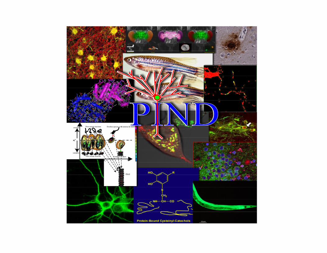
J. Timothy Greenamyre Pittsburgh Institute for Neurodegenerative Diseases
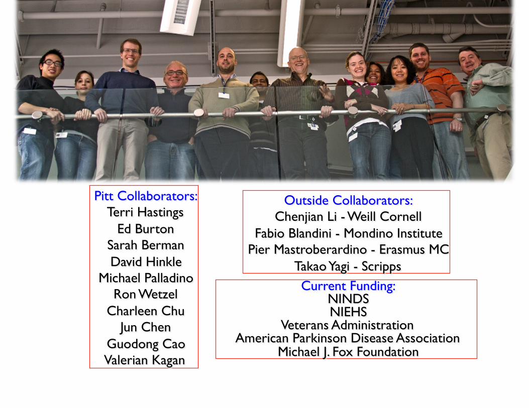
Parkinson’s Disease Prevalence: 1% of people over age 55
(1 million in North America)
Inheritance: Sporadic and Familial
Etiology: Environmental toxins
Complex I defects?
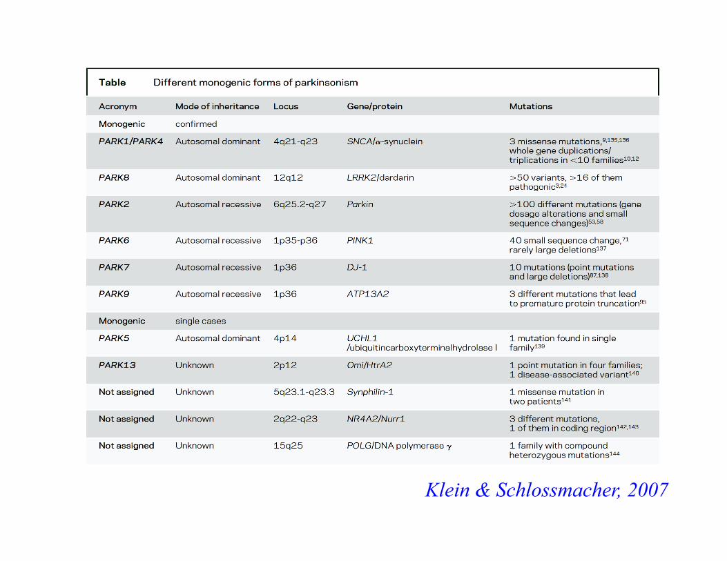
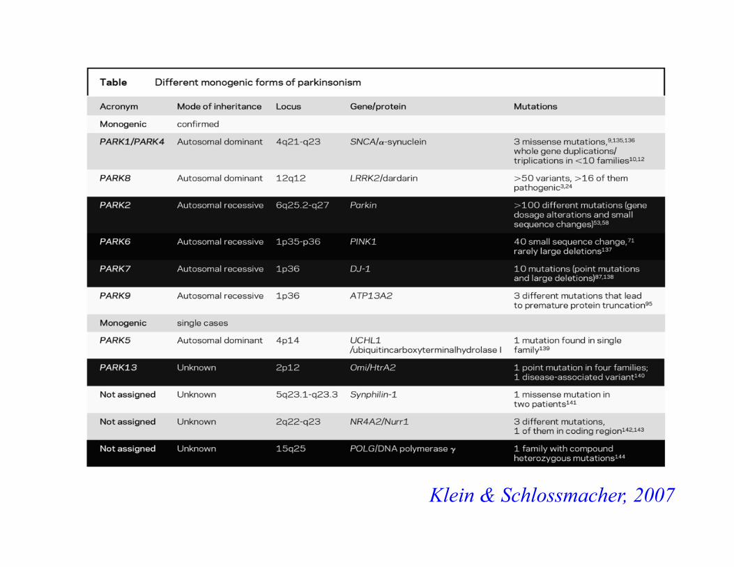
Single gene mutations α-synuclein dupli- & triplications
Cardinal Signs: Tremor, rigidity, bradykinesia, postural instability
Other Signs: Shuffling gait, masked facies, deceased blink rate
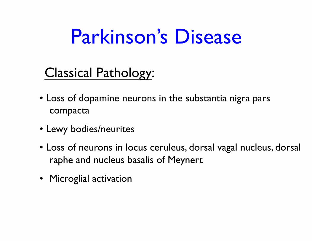
• Loss of dopamine neurons in the substantia nigra pars compacta
• Lewy bodies/neurites
• Loss of neurons in locus ceruleus, dorsal vagal nucleus, dorsal raphe and nucleus basalis of Meynert
• Microglial activation
Classical Pathology:
Parkinson’s Disease
Parkinson’s Disease
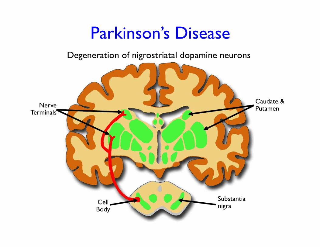
Nerve Terminals
Cell Body
Caudate & Putamen
Degeneration of nigrostriatal dopamine neurons
Substantia nigra
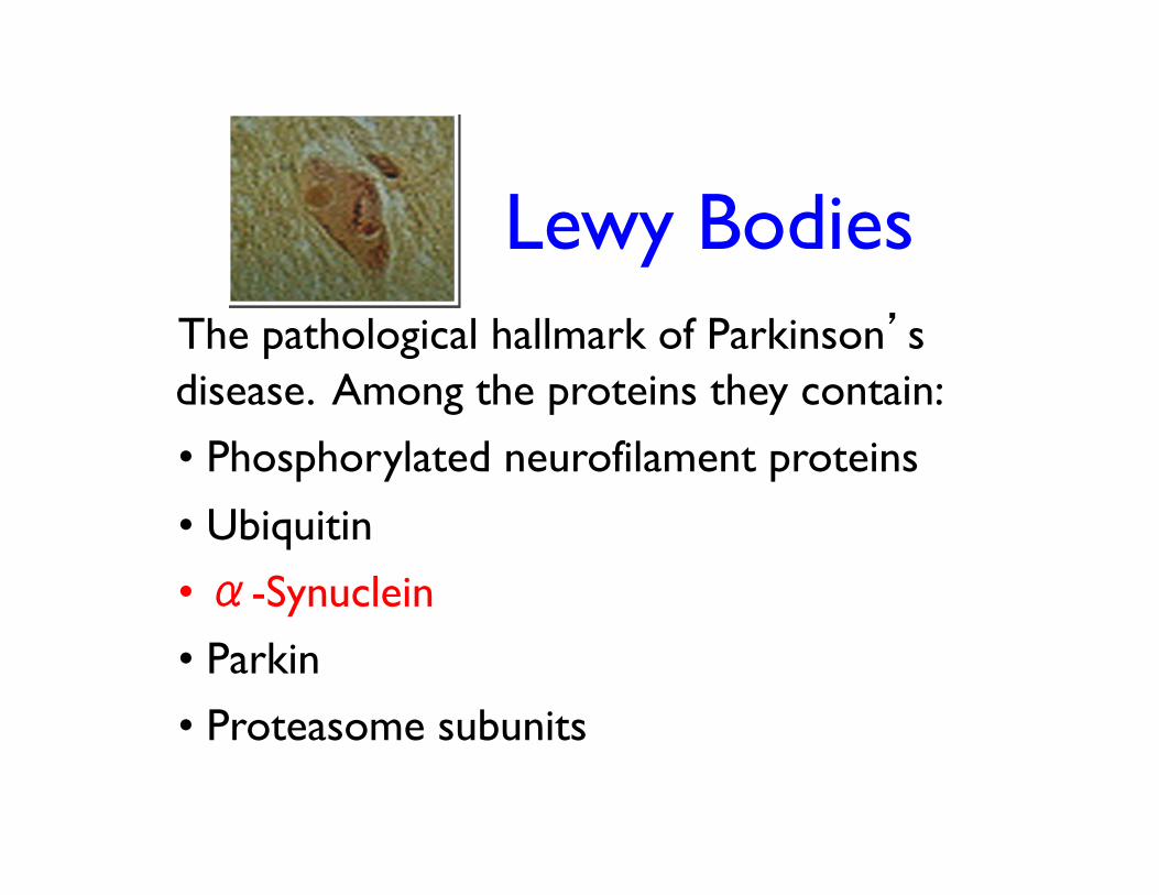
Lewy Bodies The pathological hallmark of Parkinson’s disease. Among the proteins they contain: • Phosphorylated neurofilament proteins
• Ubiquitin
• α-Synuclein
• Parkin
• Proteasome subunits
Parkinson’s Disease
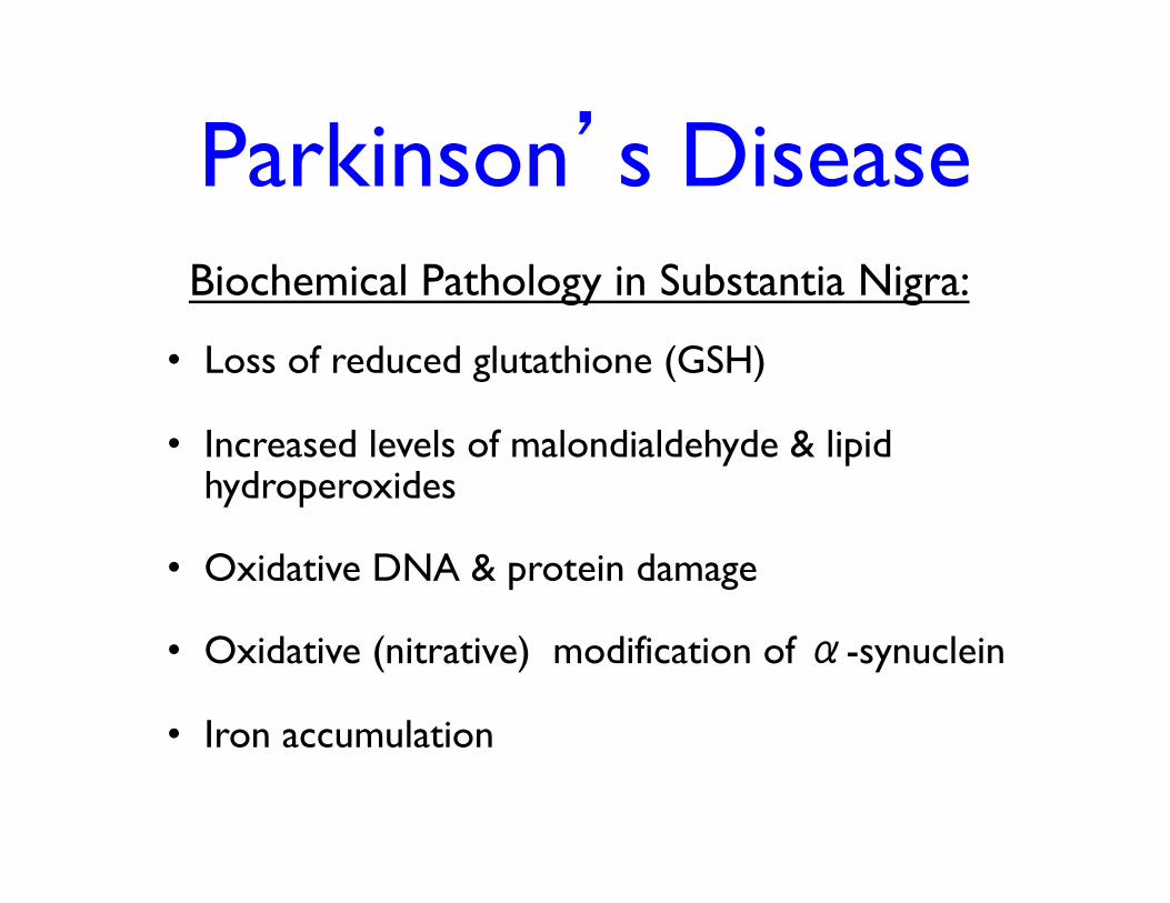
• Loss of reduced glutathione (GSH)
• Increased levels of malondialdehyde & lipid hydroperoxides
• Oxidative DNA & protein damage
• Oxidative (nitrative) modification of α-synuclein
• Iron accumulation
Biochemical Pathology in Substantia Nigra:
MPTP
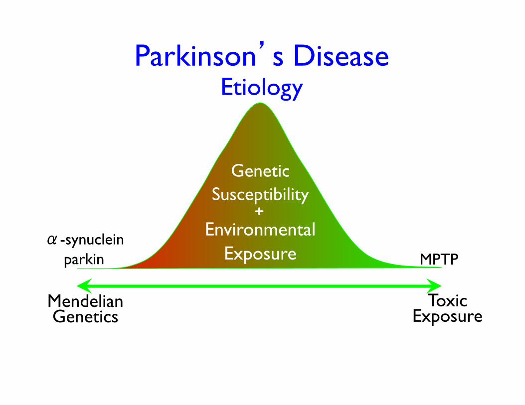
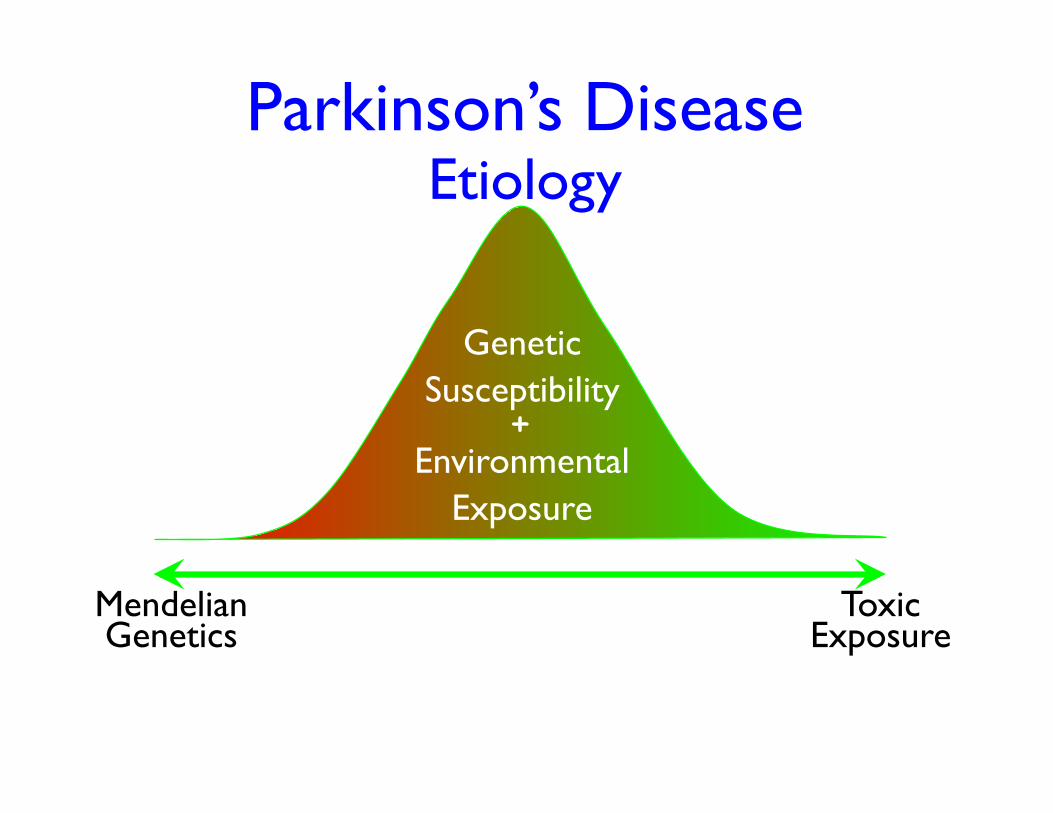
Parkinson’s Disease Etiology
Mendelian Genetics
Toxic Exposure
α-synuclein parkin
Genetic Susceptibility
Environmental Exposure
+
Klein & Schlossmacher, 2007
Klein & Schlossmacher, 2007
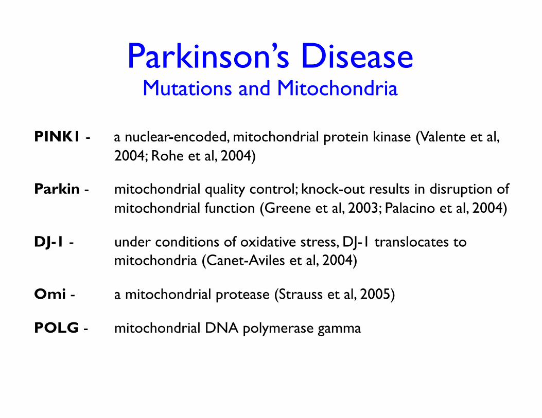
Parkinson’s Disease Mutations and Mitochondria
PINK1 - a nuclear-encoded, mitochondrial protein kinase (Valente et al, 2004; Rohe et al, 2004)
Parkin - mitochondrial quality control; knock-out results in disruption of mitochondrial function (Greene et al, 2003; Palacino et al, 2004)
DJ-1 - under conditions of oxidative stress, DJ-1 translocates to mitochondria (Canet-Aviles et al, 2004)
Omi - a mitochondrial protease (Strauss et al, 2005)
POLG - mitochondrial DNA polymerase gamma
Parkinson’s Disease Etiology
Mendelian Genetics
Toxic Exposure
Genetic Susceptibility
Environmental Exposure
+
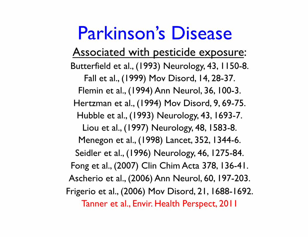
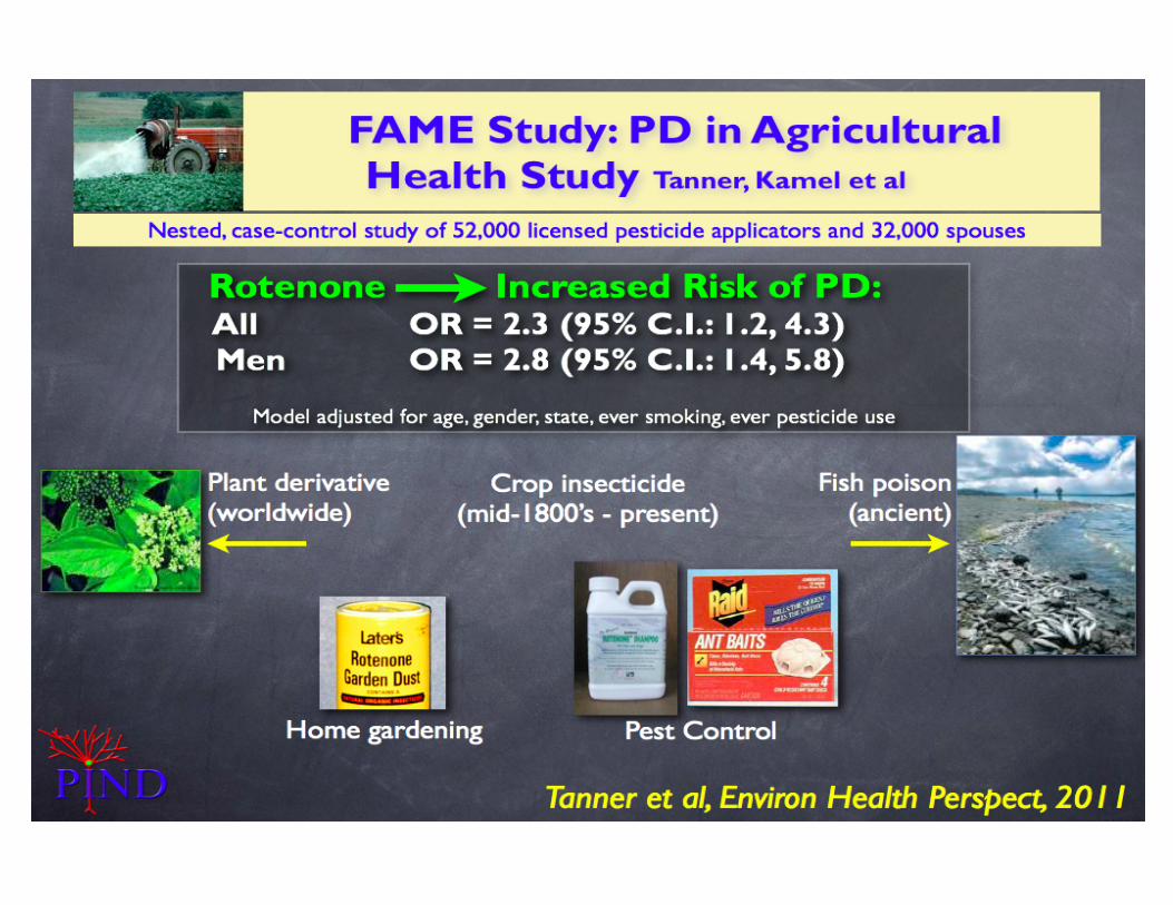
Parkinson’s Disease Associated with pesticide exposure: Butterfield et al., (1993) Neurology, 43, 1150-8.
Fall et al., (1999) Mov Disord, 14, 28-37. Flemin et al., (1994) Ann Neurol, 36, 100-3.
Hertzman et al., (1994) Mov Disord, 9, 69-75. Hubble et al., (1993) Neurology, 43, 1693-7.
Liou et al., (1997) Neurology, 48, 1583-8. Menegon et al., (1998) Lancet, 352, 1344-6.
Seidler et al., (1996) Neurology, 46, 1275-84. Fong et al., (2007) Clin Chim Acta 378, 136-41. Ascherio et al., (2006) Ann Neurol, 60, 197-203.
Frigerio et al., (2006) Mov Disord, 21, 1688-1692. Tanner et al., Envir. Health Perspect, 2011
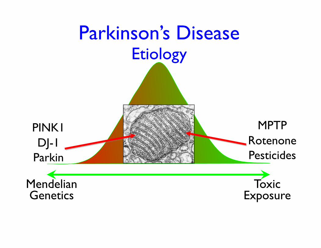
MPTP Rotenone Pesticides
Parkinson’s Disease Etiology
Mendelian Genetics
Toxic Exposure
PINK1 DJ-1
Parkin
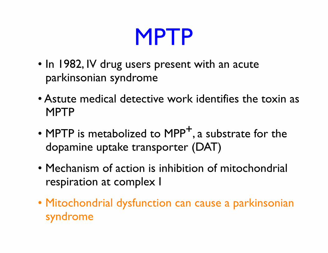
MPTP • In 1982, IV drug users present with an acute
parkinsonian syndrome
• Astute medical detective work identifies the toxin as MPTP
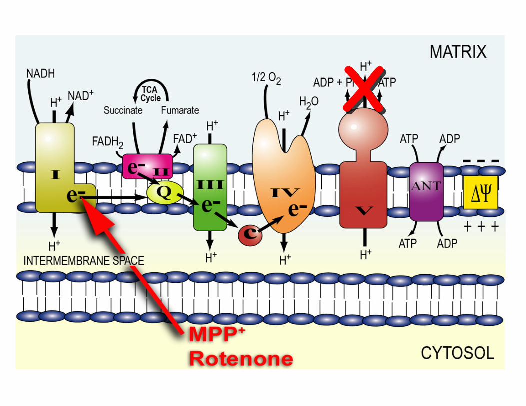
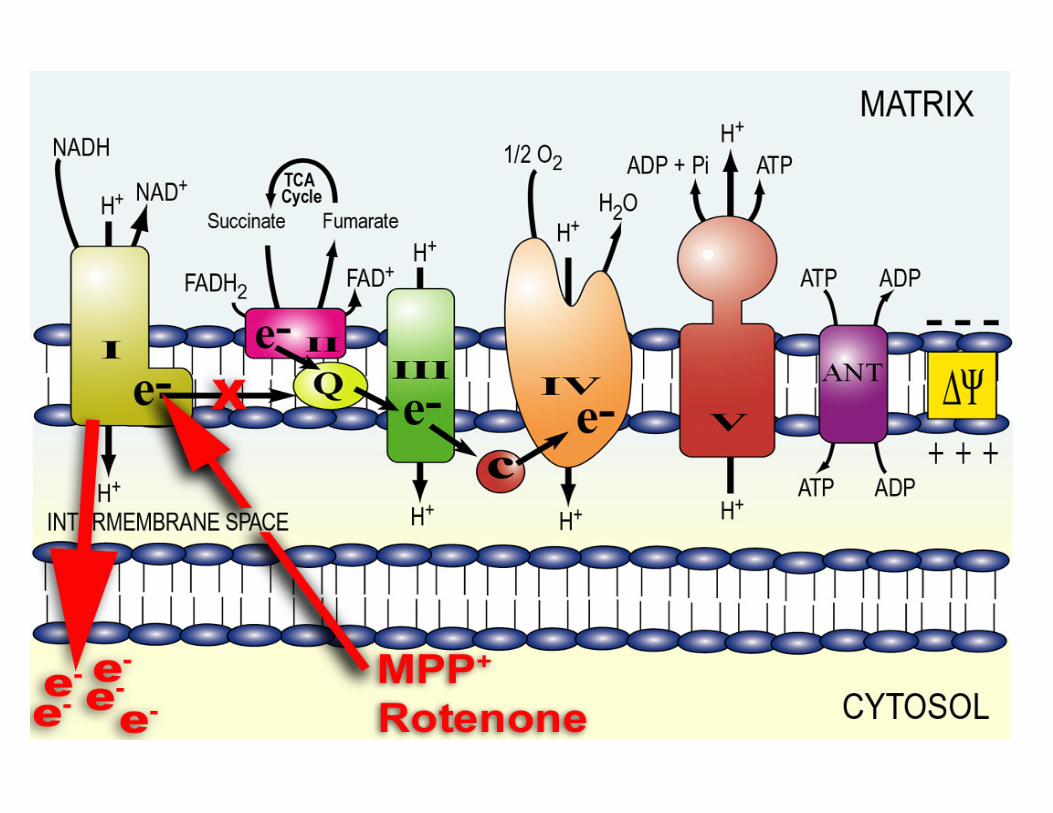
• MPTP is metabolized to MPP+, a substrate for the dopamine uptake transporter (DAT)
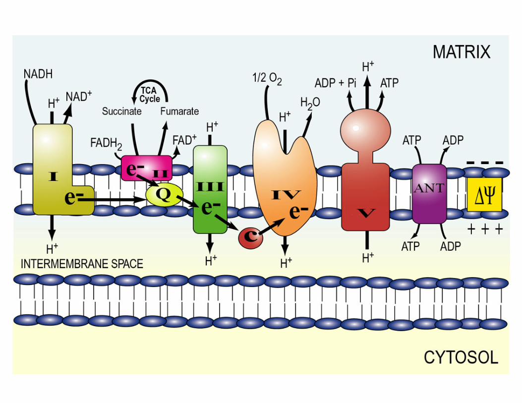
• Mechanism of action is inhibition of mitochondrial respiration at complex I
• Mitochondrial dysfunction can cause a parkinsonian syndrome
Parkinson’s Disease
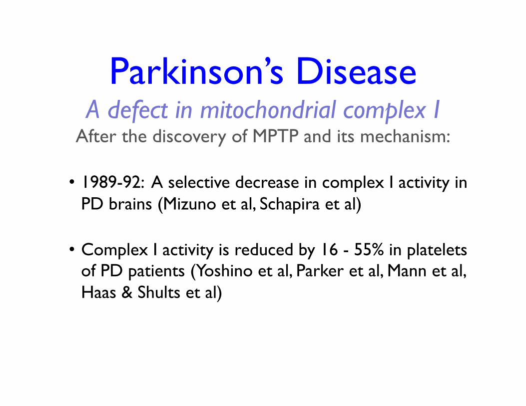
A defect in mitochondrial complex I After the discovery of MPTP and its mechanism:
• 1989-92: A selective decrease in complex I activity in PD brains (Mizuno et al, Schapira et al)
• Complex I activity is reduced by 16 - 55% in platelets of PD patients (Yoshino et al, Parker et al, Mann et al, Haas & Shults et al)
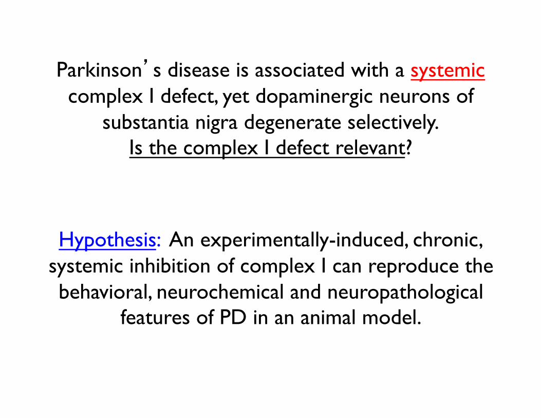
Parkinson’s disease is associated with a systemic complex I defect, yet dopaminergic neurons of
substantia nigra degenerate selectively. Is the complex I defect relevant?
Hypothesis: An experimentally-induced, chronic, systemic inhibition of complex I can reproduce the behavioral, neurochemical and neuropathological
features of PD in an animal model.
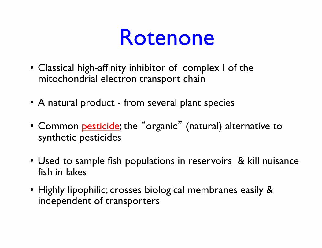
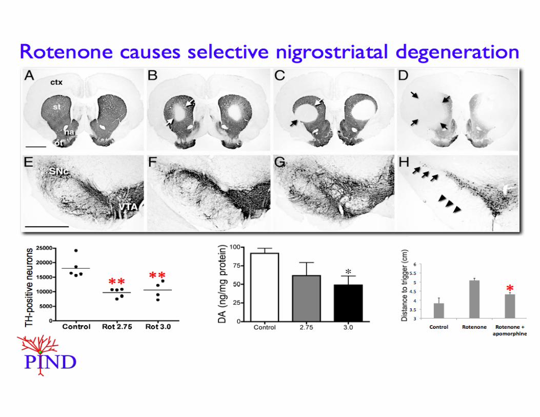
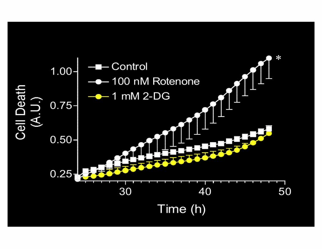
Rotenone
• Classical high-affinity inhibitor of complex I of the mitochondrial electron transport chain
• A natural product - from several plant species
• Common pesticide; the “organic” (natural) alternative to synthetic pesticides
• Used to sample fish populations in reservoirs & kill nuisance fish in lakes
• Highly lipophilic; crosses biological membranes easily & independent of transporters
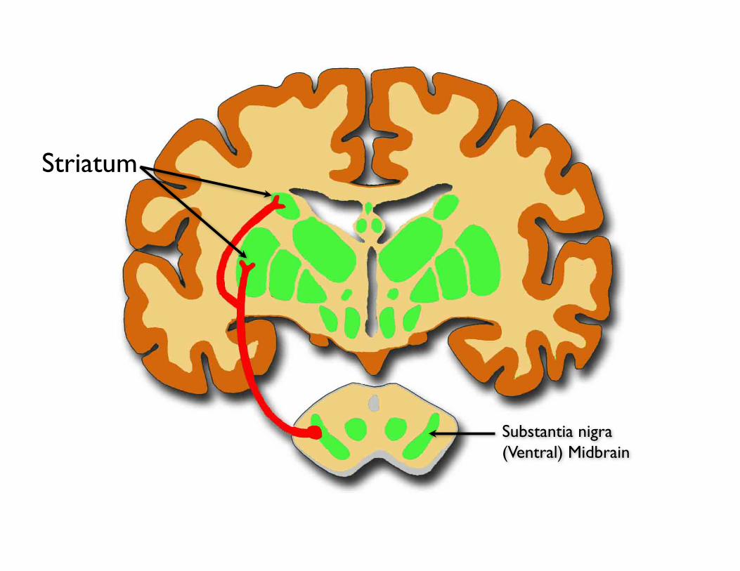
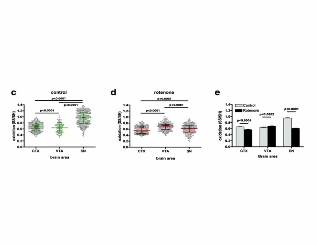
Substantia nigra (Ventral) Midbrain
Striatum
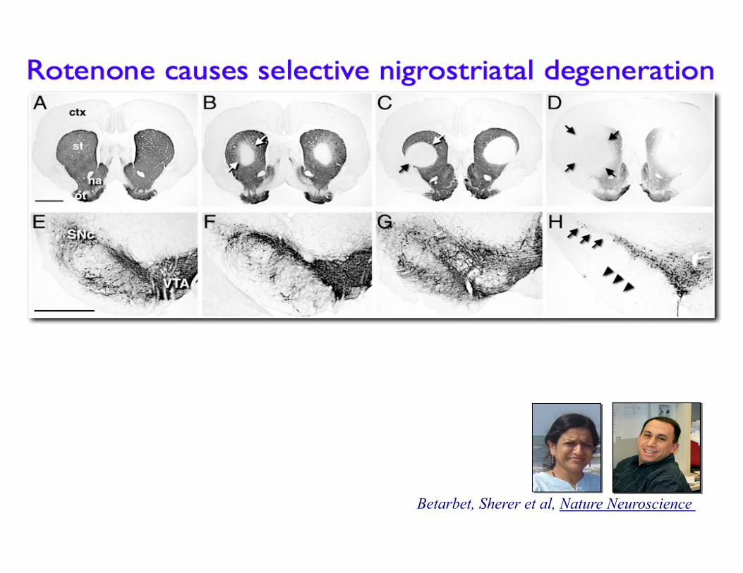
Betarbet, Sherer et al, Nature Neuroscience
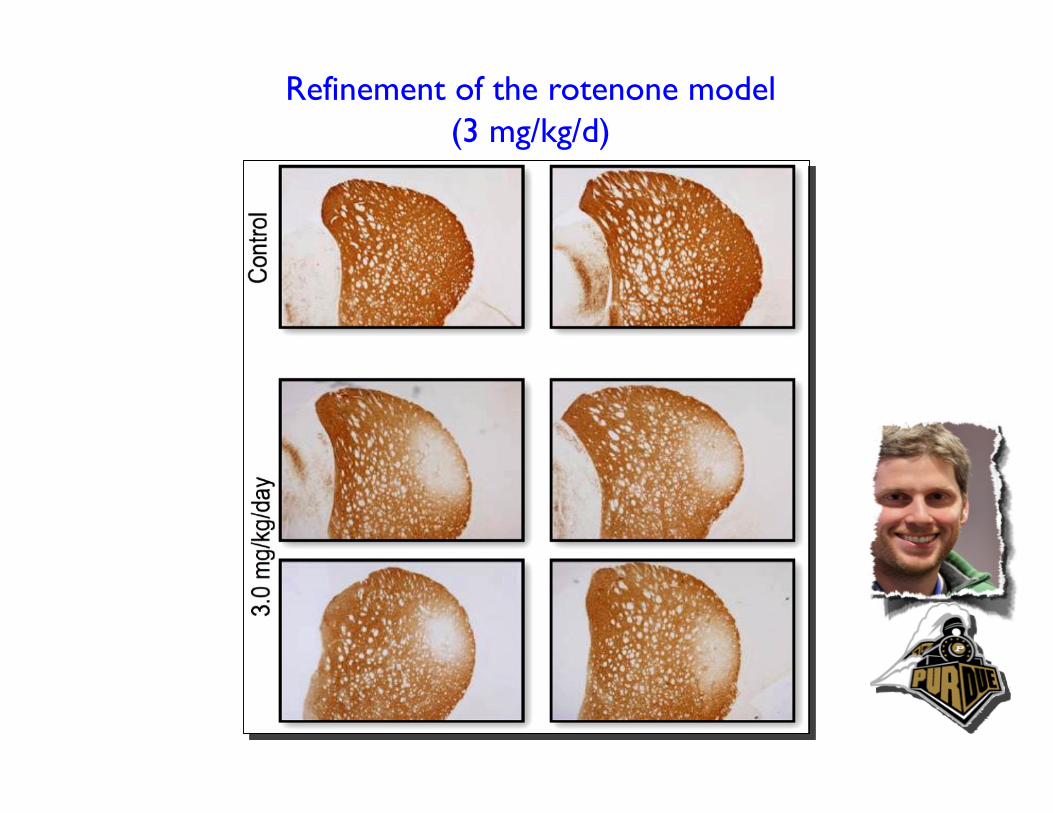
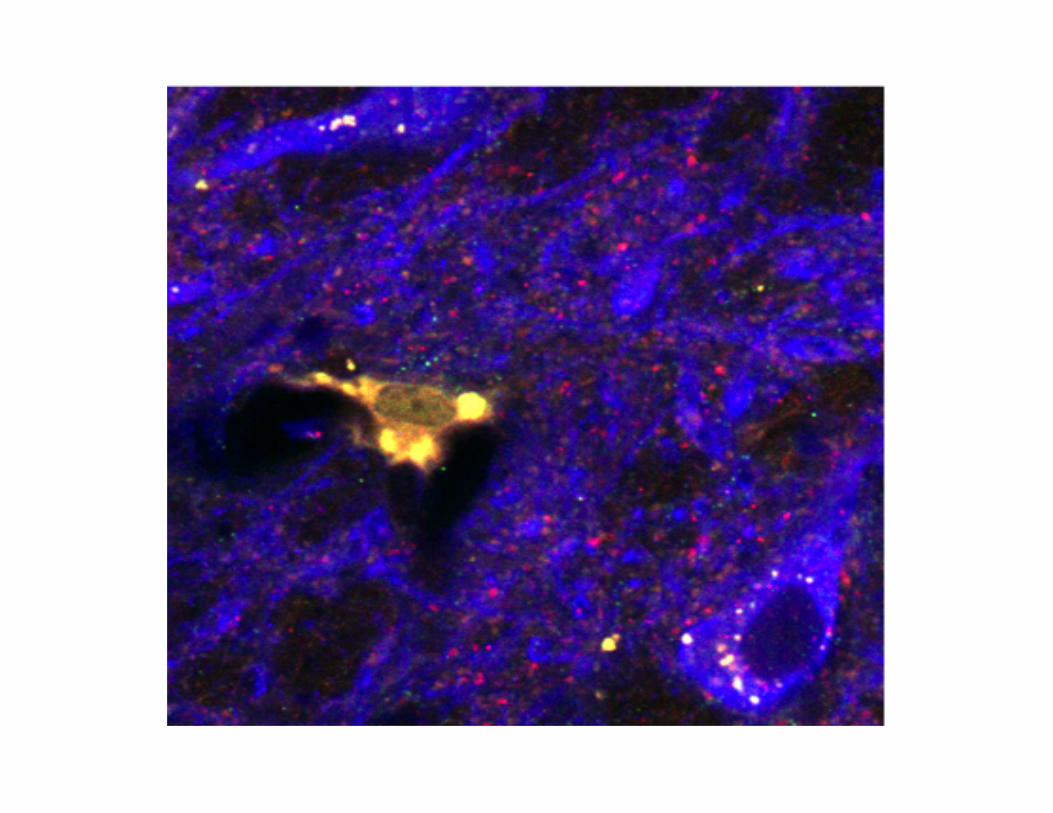
Refinement of the rotenone model (3 mg/kg/d)
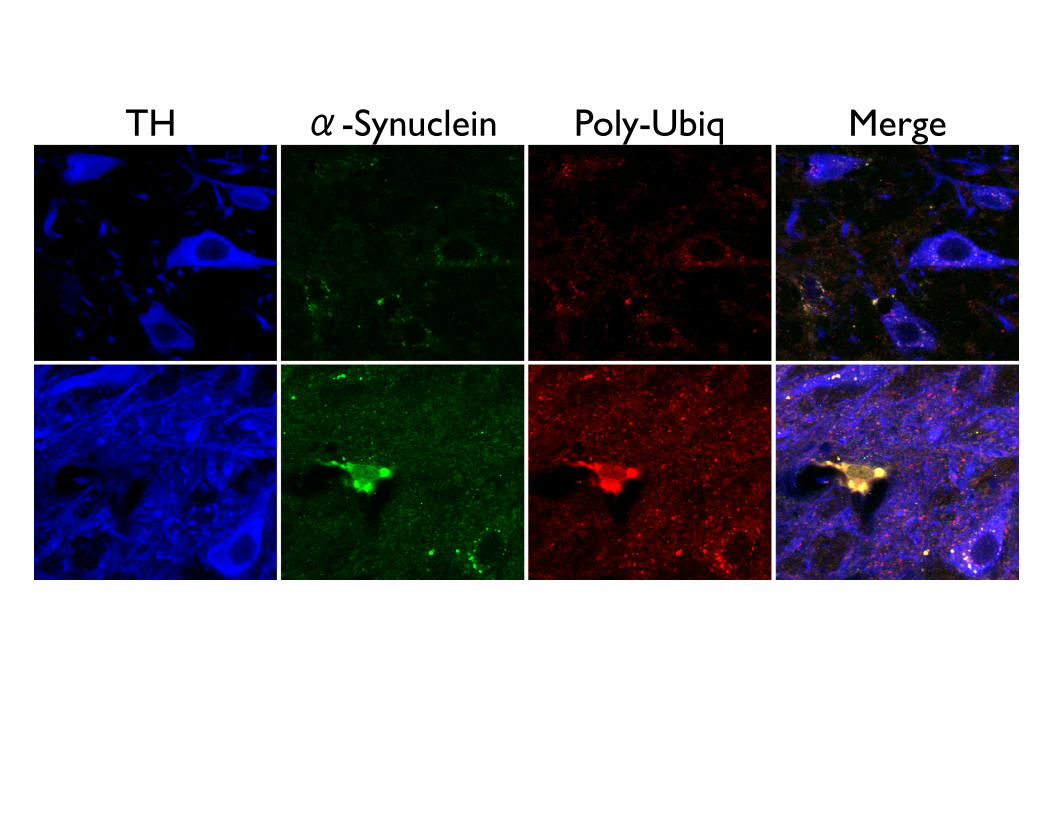
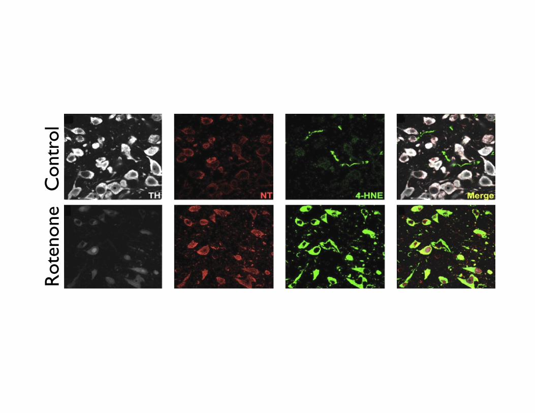
TH α-Synuclein Poly-Ubiq Merge
X
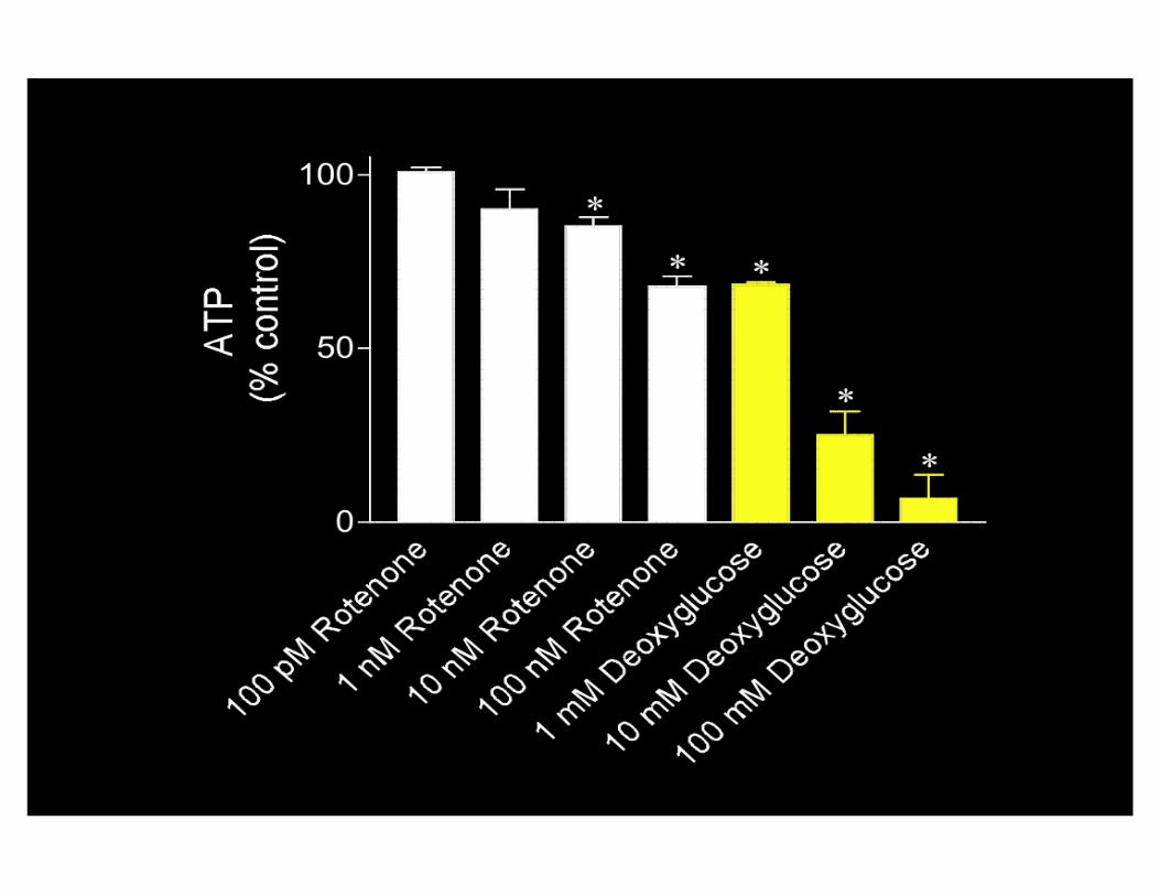
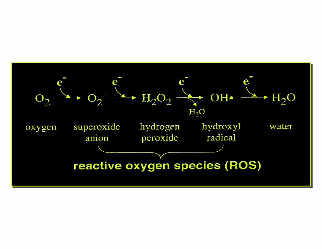
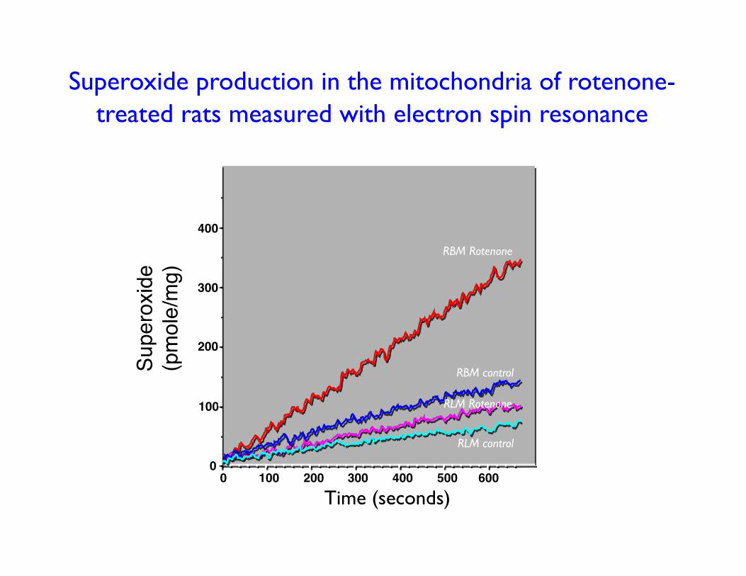
Superoxide production in the mitochondria of rotenone-treated rats measured with electron spin resonance
0 100 200 300 400 500 600
100
200
300
400
0
Superoxid
e (pmole/mg)
RBM control
RLM control
RBM Rotenone
RLM Rotenone
Time (seconds)
Con
trol
R
oten
one
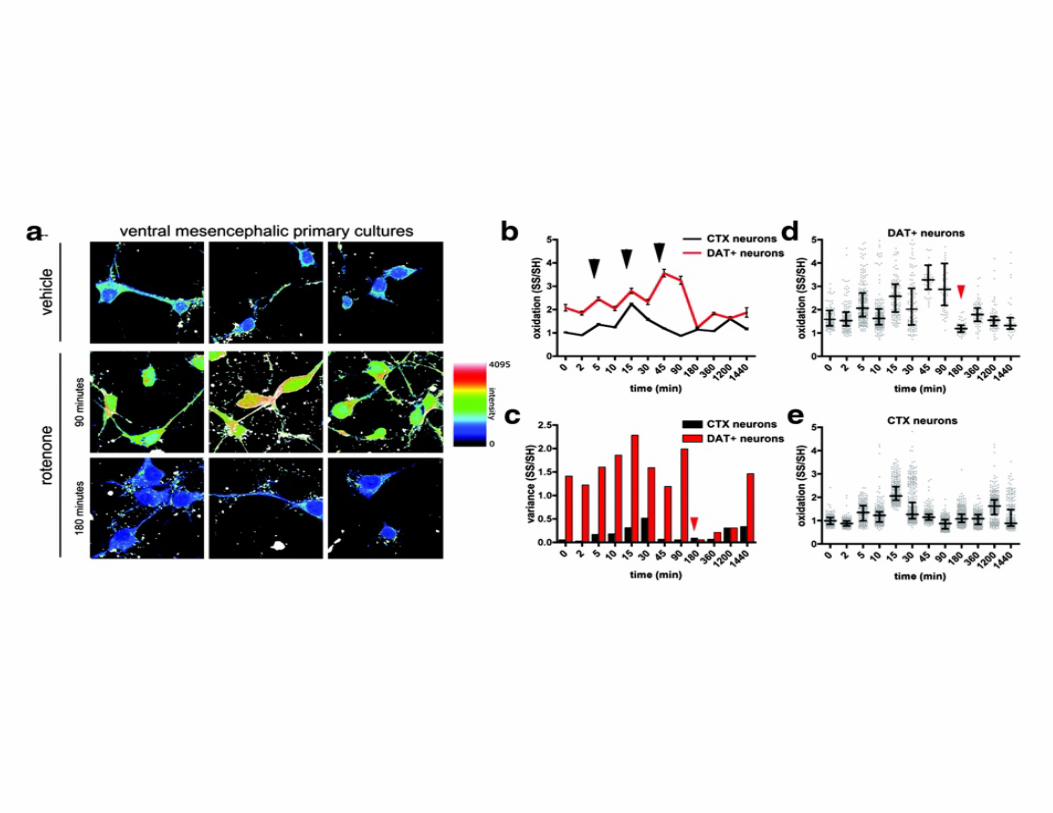
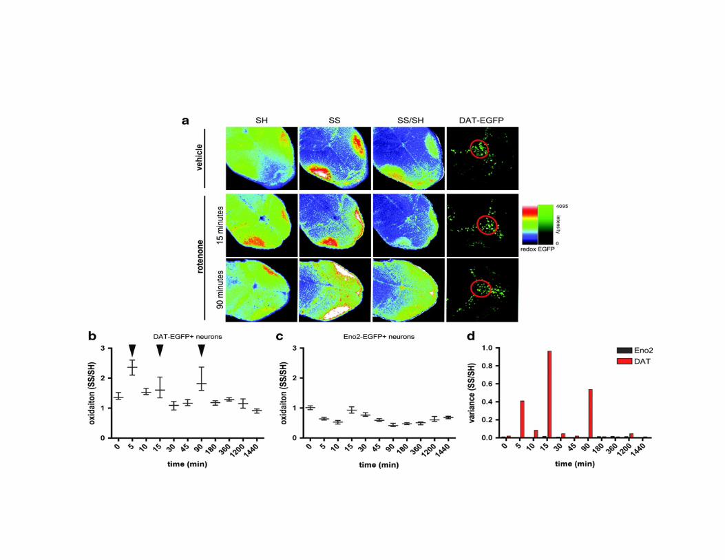
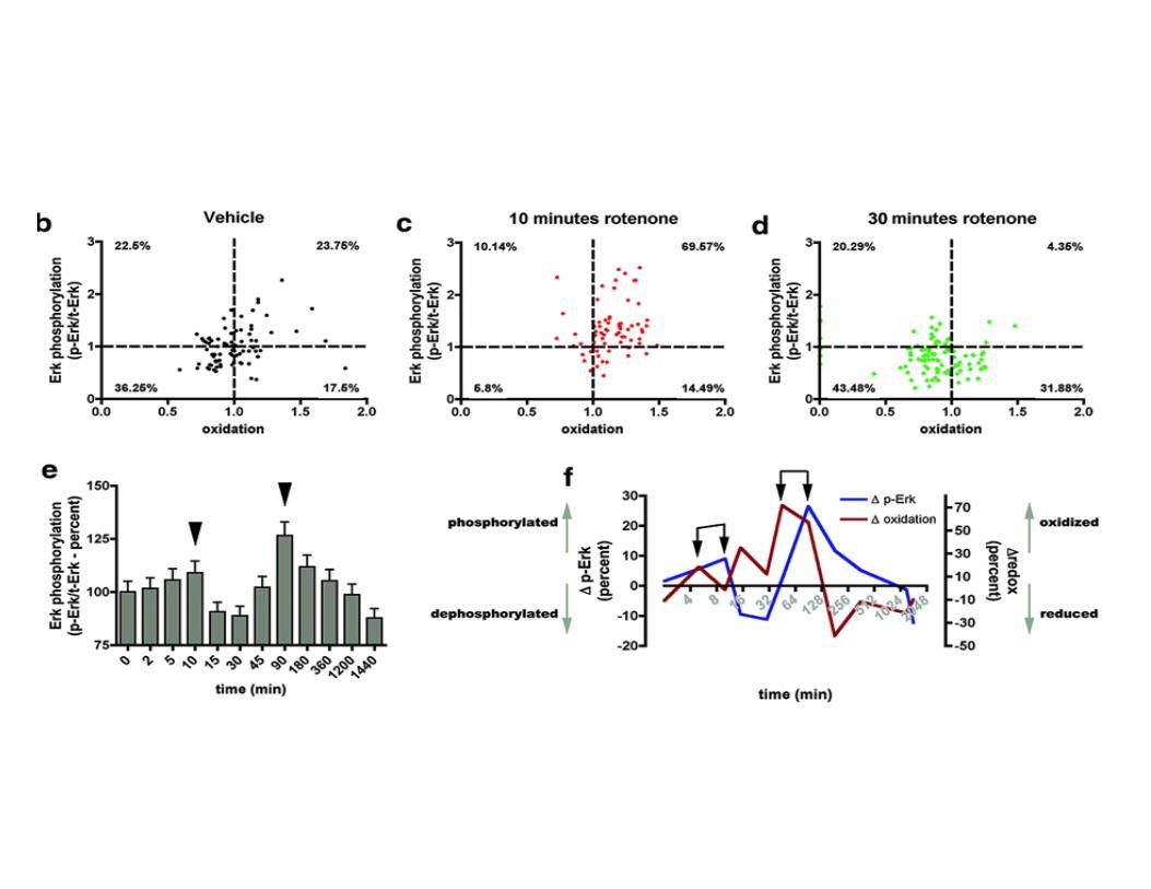
Why are dopamine neurons selectively vulnerable?
Why does degeneration begin in nerve terminals?
Is it dopamine itself?
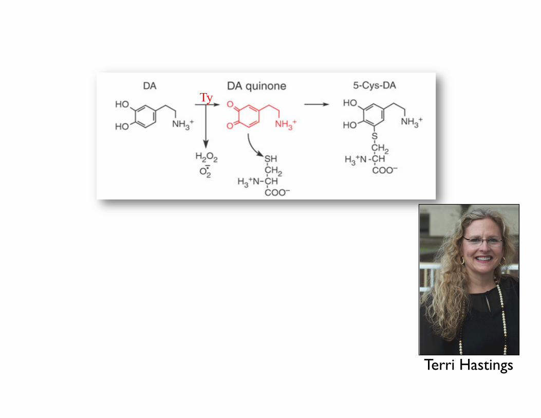
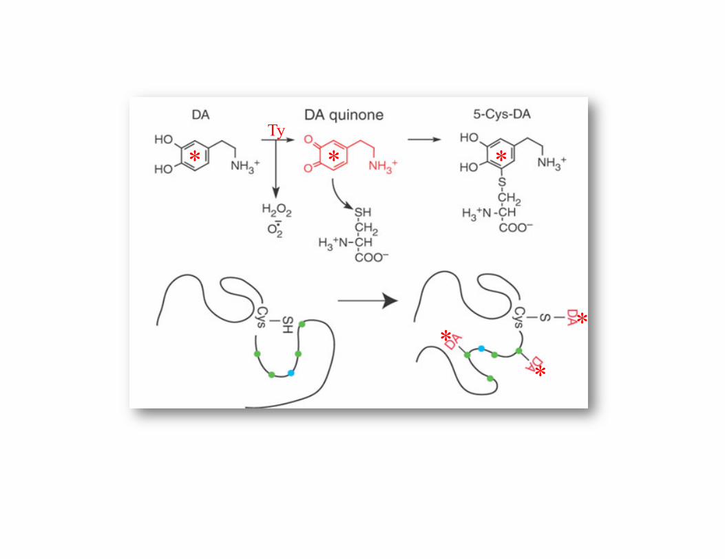
Ty
Terri Hastings
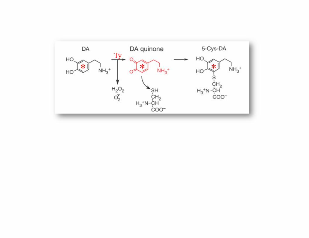
* * * Ty
* * *
*
* *
Ty
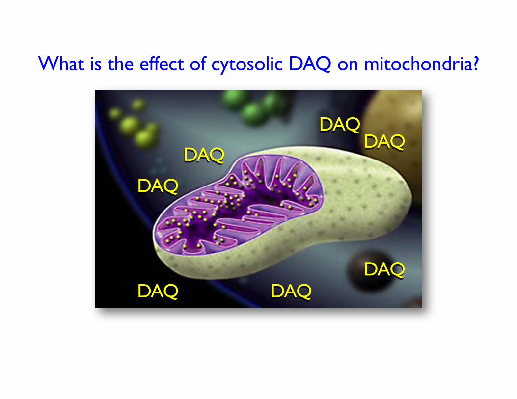
What is the effect of cytosolic DAQ on mitochondria?
DAQ
DAQ
DAQ
DAQ DAQ
DAQ DAQ
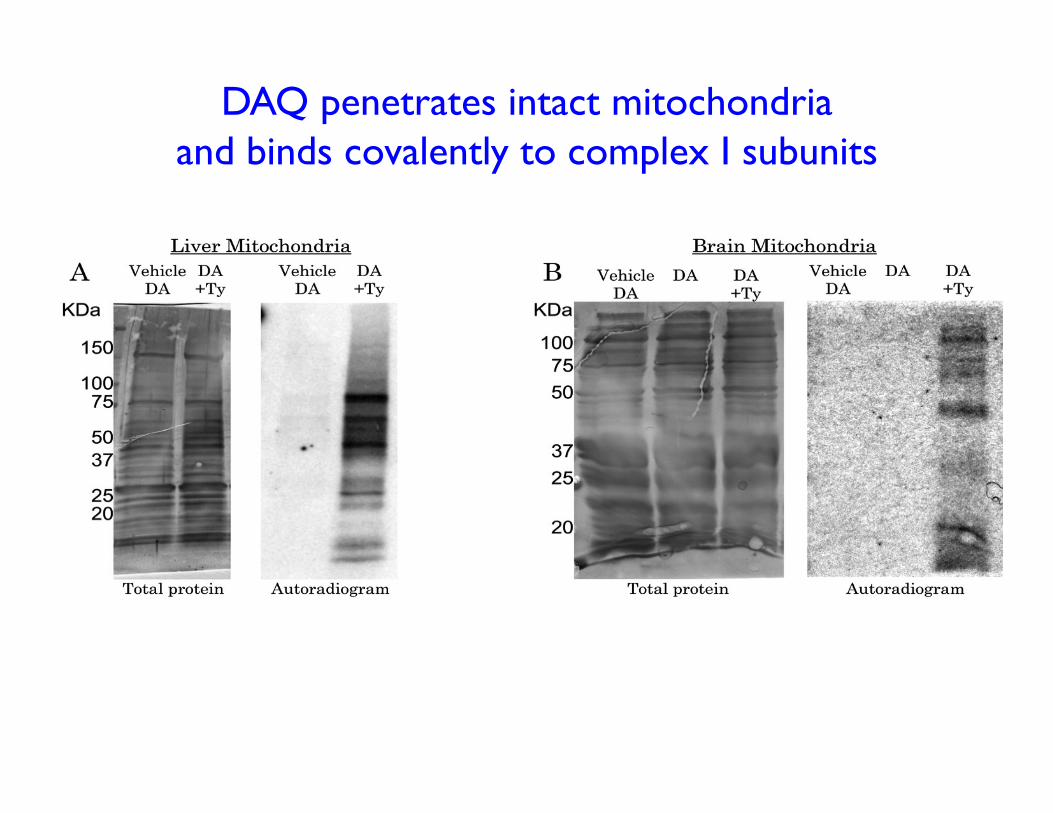
DAQ penetrates intact mitochondria and binds covalently to complex I subunits
-7 -6 -5 -4
0.00
0.25
0.50
0.75
1.00
1.25
+ Tyrosinase– Tyrosinase
IC50 = 1.2 !M
*
*
* * **
Log[dopamine]
-7 -6 -5 -4
0.00
0.25
0.50
0.75
IC50 = 1.8 !M
Log[dopamine]
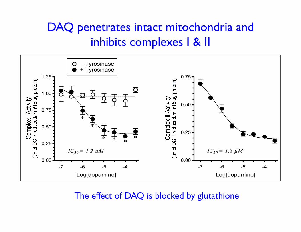
DAQ penetrates intact mitochondria and inhibits complexes I & II
The effect of DAQ is blocked by glutathione
Are these results relevant?
Can DA inhibit mitochondrial function in vivo?
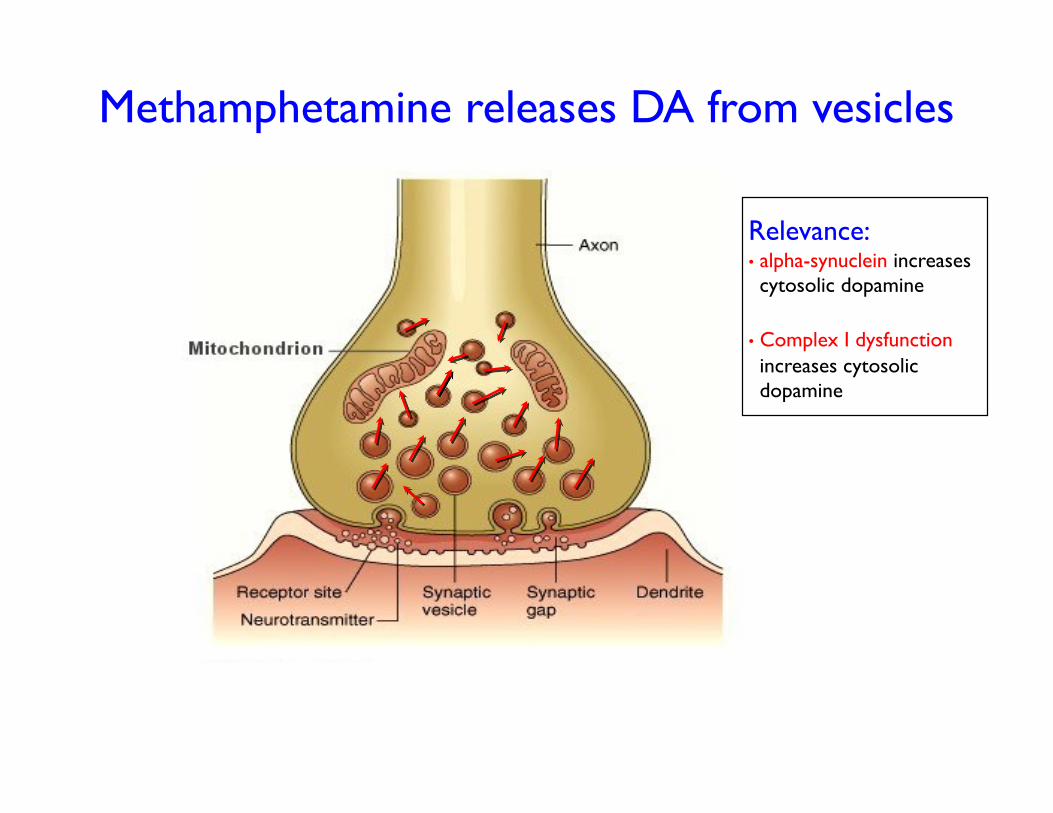
Methamphetamine releases DA from vesicles
Relevance: • alpha-synuclein increases cytosolic dopamine
• Complex I dysfunction increases cytosolic dopamine
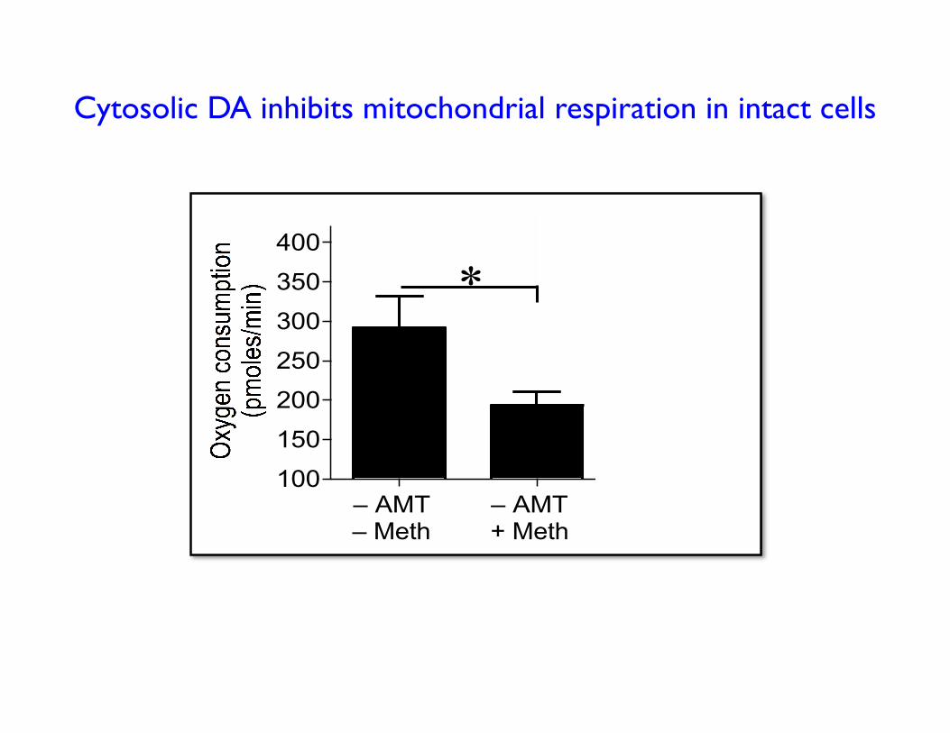
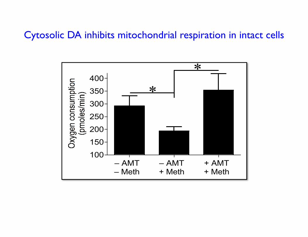
Cytosolic DA inhibits mitochondrial respiration in intact cells
100
150
200
250
300
350
400
– AMT
– Meth
– AMT
+ Meth
+ AMT
+ Meth
*
#
*
*
100
150
200
250
300
350
400
– AMT
– Meth
– AMT
+ Meth
+ AMT
+ Meth
*
#
*
*
Cytosolic DA inhibits mitochondrial respiration in intact cells
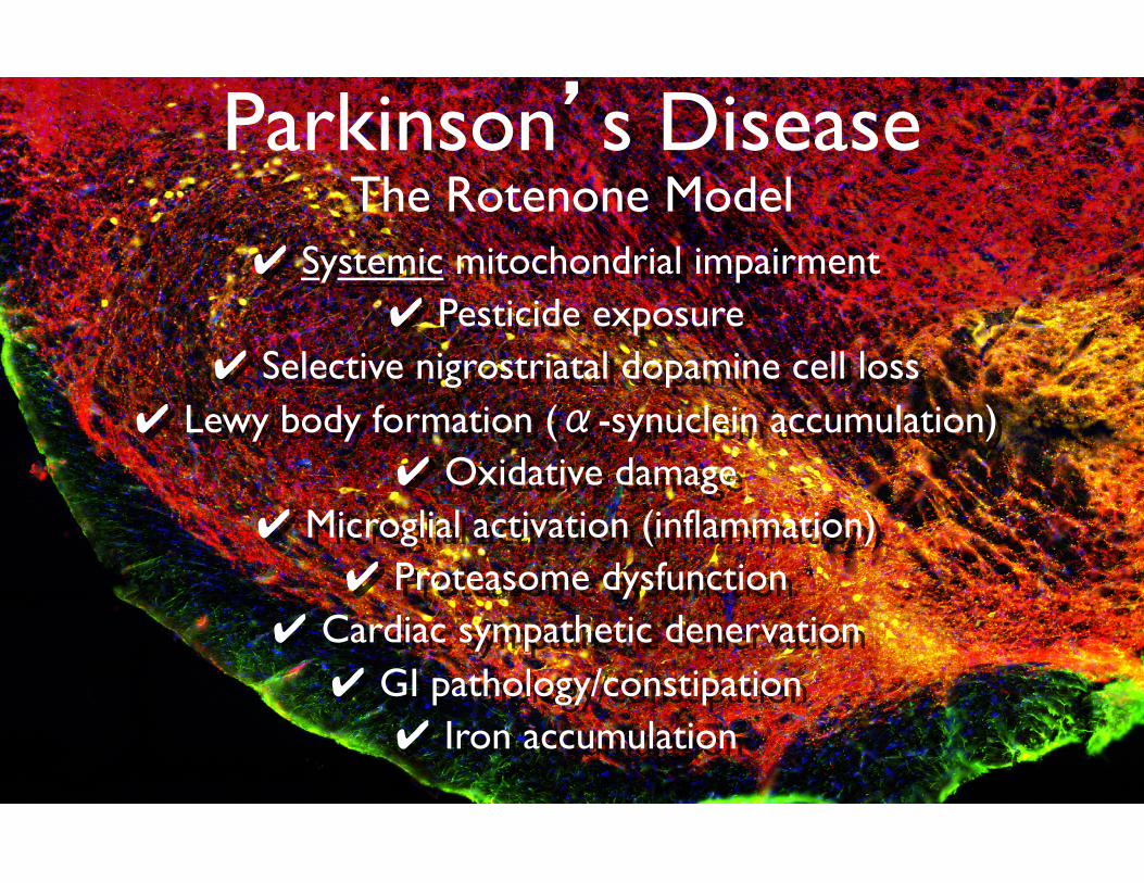
Parkinson’s Disease The Rotenone Model
✔ Systemic mitochondrial impairment ✔ Pesticide exposure
✔ Selective nigrostriatal dopamine cell loss ✔ Lewy body formation (α-synuclein accumulation)
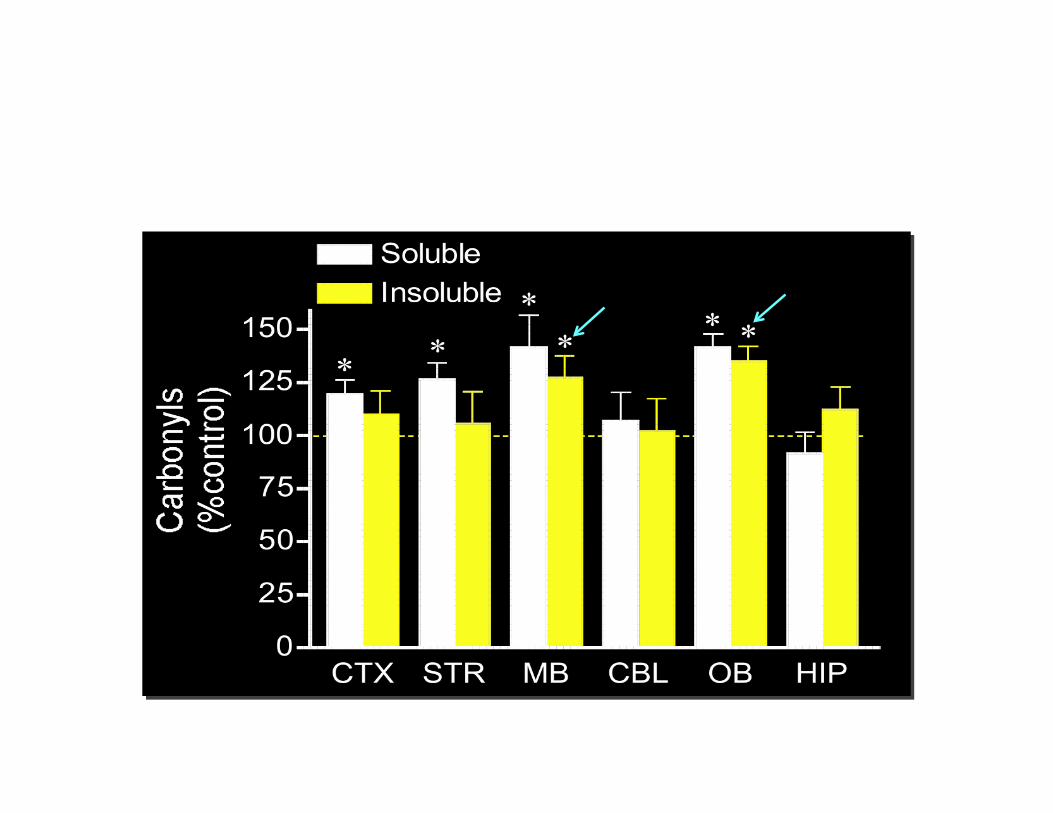
✔ Oxidative damage ✔ Microglial activation (inflammation)
✔ Proteasome dysfunction ✔ Cardiac sympathetic denervation ✔ GI pathology/constipation ✔ Iron accumulation

Top Related