Oxidative Models of Parkinson's Disease J. Timothy Greenamyre
58
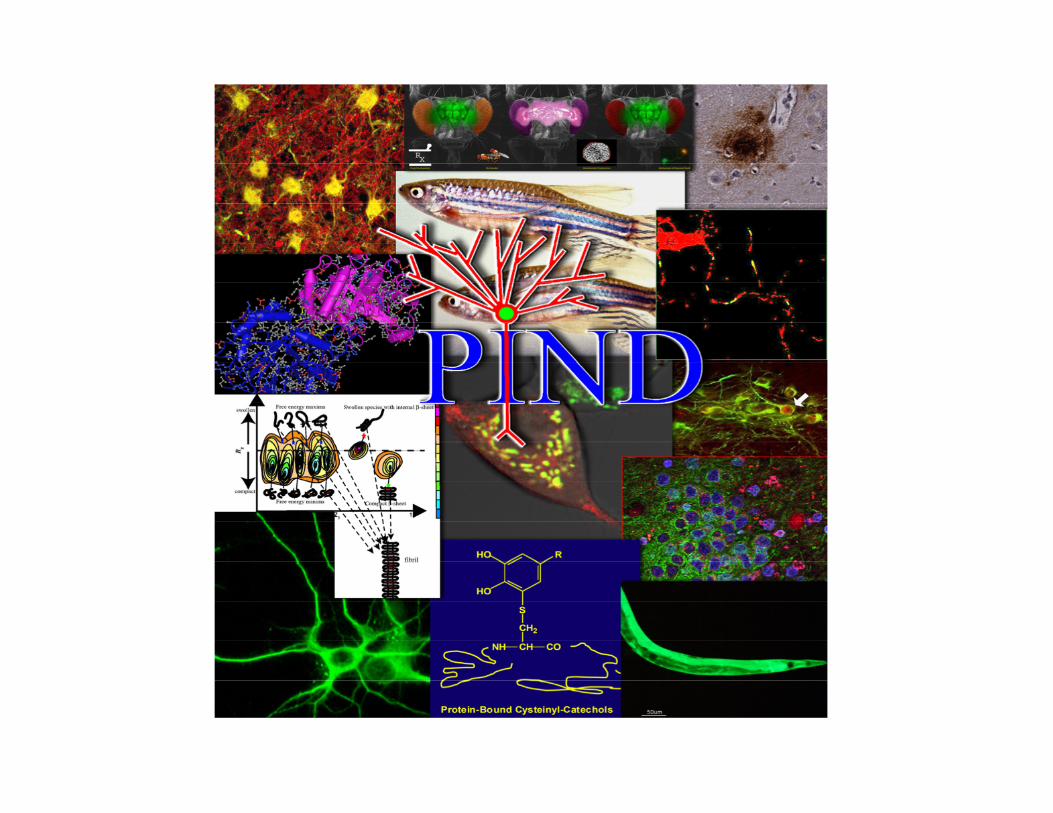
Oxidative Models of Parkinson's Disease J. Timothy Greenamyre Pittsburgh Institute for Neurodegenerative Diseases
Transcript of Oxidative Models of Parkinson's Disease J. Timothy Greenamyre
Oxidative Models of Parkinson's Disease
J. Timothy Greenamyre Pittsburgh Institute for Neurodegenerative Diseases
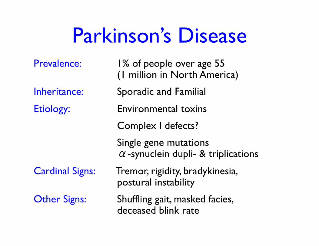
Parkinson’s Disease Prevalence: 1% of people over age 55
(1 million in North America)
Inheritance: Sporadic and Familial
Etiology: Environmental toxins
Complex I defects?
Single gene mutations α-synuclein dupli- & triplications
Cardinal Signs: Tremor, rigidity, bradykinesia, postural instability
Other Signs: Shuffling gait, masked facies, deceased blink rate
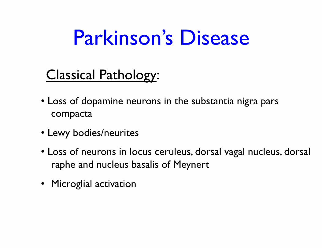
• Loss of dopamine neurons in the substantia nigra pars compacta
• Lewy bodies/neurites
• Loss of neurons in locus ceruleus, dorsal vagal nucleus, dorsal raphe and nucleus basalis of Meynert
• Microglial activation
Classical Pathology:
Parkinson’s Disease
Parkinson’s Disease
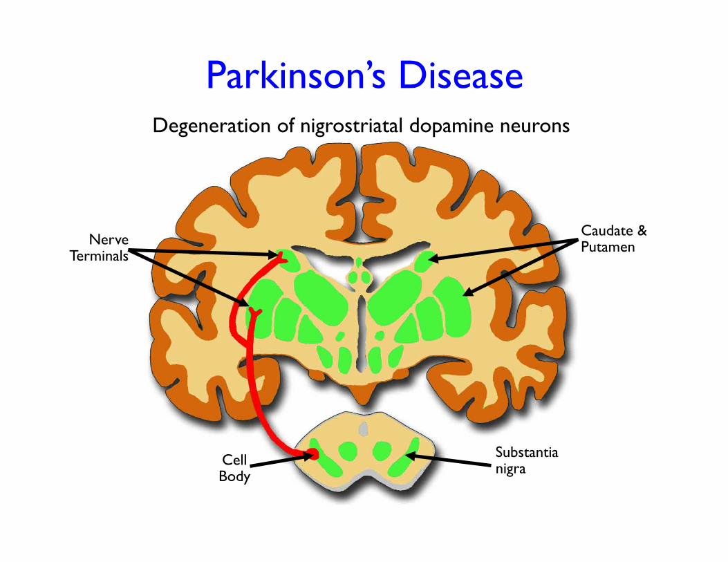
Nerve Terminals
Cell Body
Caudate & Putamen
Degeneration of nigrostriatal dopamine neurons
Substantia nigra
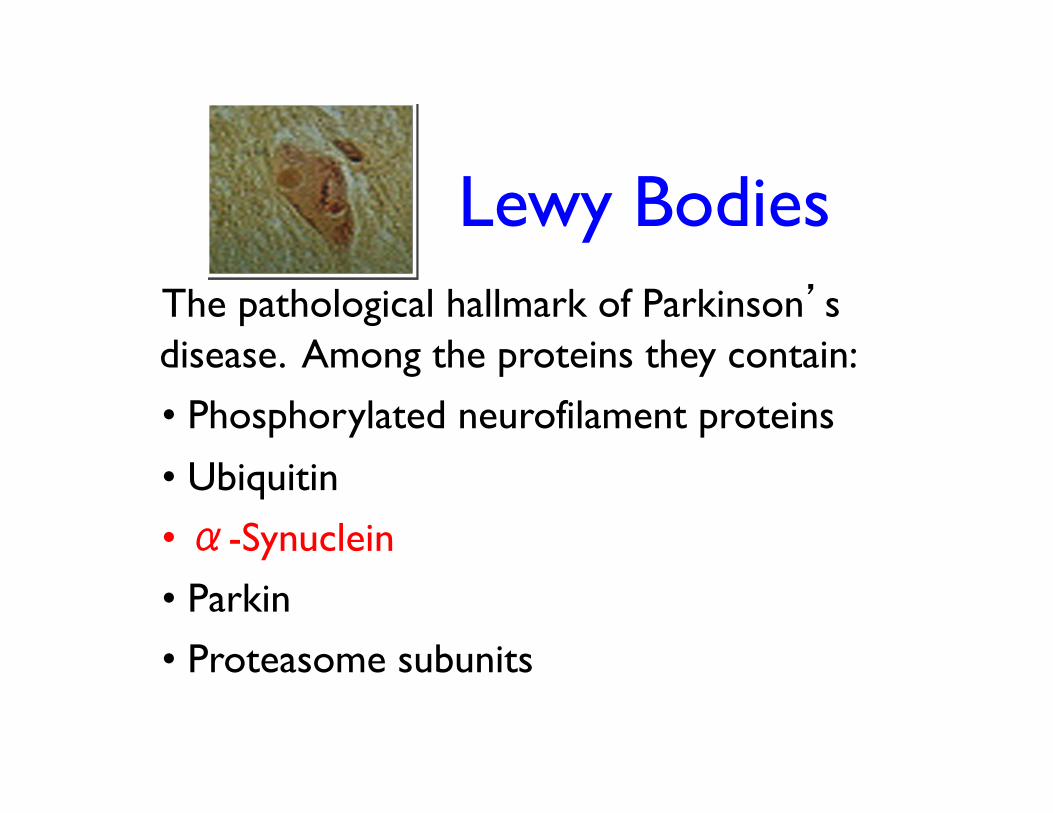
Lewy Bodies The pathological hallmark of Parkinson’s disease. Among the proteins they contain: • Phosphorylated neurofilament proteins
• Ubiquitin
• α-Synuclein
• Parkin
• Proteasome subunits
Parkinson’s Disease
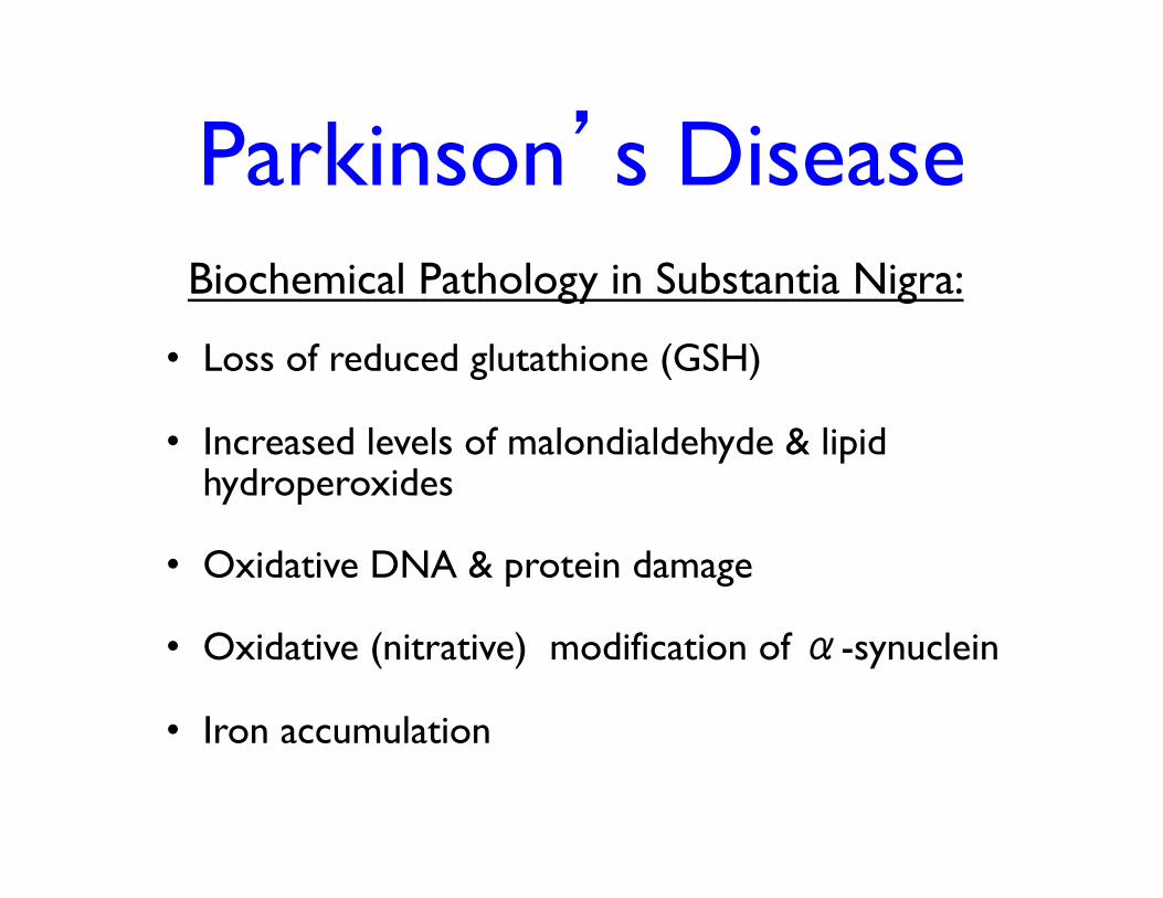
• Loss of reduced glutathione (GSH)
• Increased levels of malondialdehyde & lipid hydroperoxides
• Oxidative DNA & protein damage
• Oxidative (nitrative) modification of α-synuclein
• Iron accumulation
Biochemical Pathology in Substantia Nigra:
MPTP
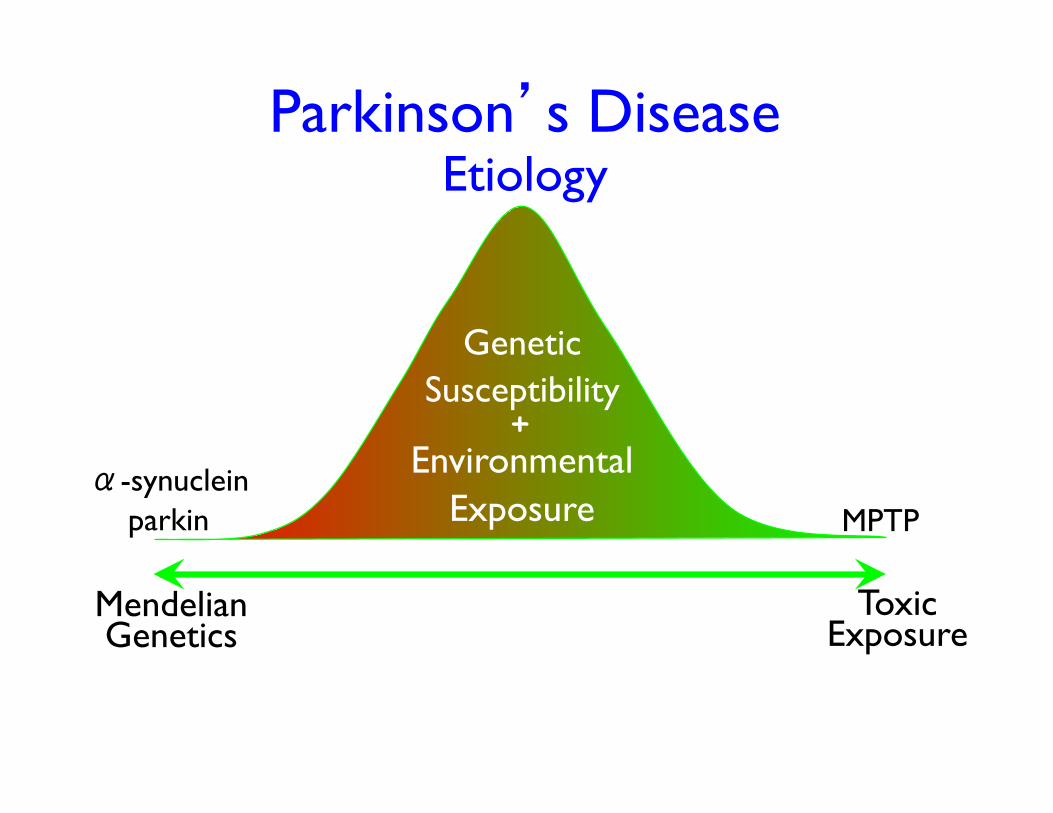
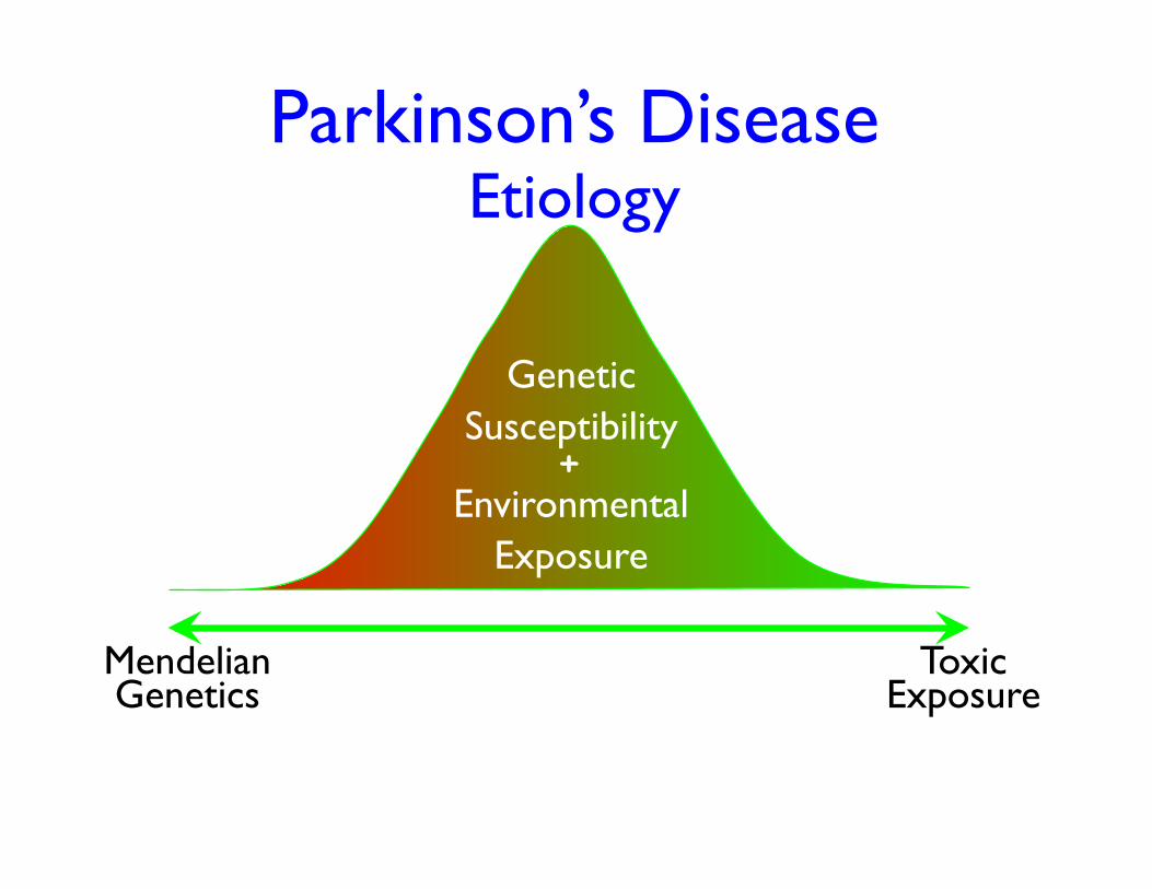
Parkinson’s Disease Etiology
Mendelian Genetics
Toxic Exposure
α-synuclein parkin
Genetic Susceptibility
Environmental Exposure
+
Klein & Schlossmacher, 2007
Klein & Schlossmacher, 2007
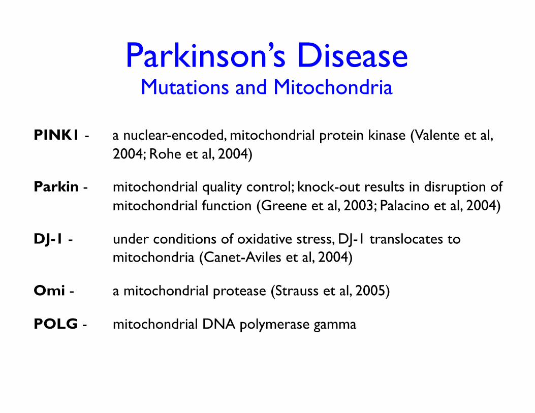
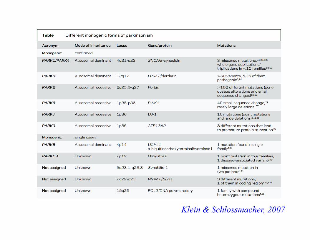
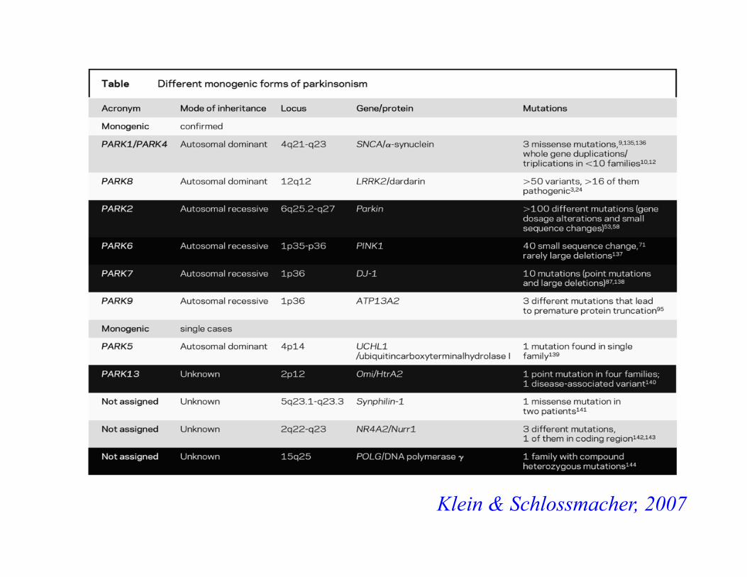
Parkinson’s Disease Mutations and Mitochondria
PINK1 - a nuclear-encoded, mitochondrial protein kinase (Valente et al, 2004; Rohe et al, 2004)
Parkin - mitochondrial quality control; knock-out results in disruption of mitochondrial function (Greene et al, 2003; Palacino et al, 2004)
DJ-1 - under conditions of oxidative stress, DJ-1 translocates to mitochondria (Canet-Aviles et al, 2004)
Omi - a mitochondrial protease (Strauss et al, 2005)
POLG - mitochondrial DNA polymerase gamma
Parkinson’s Disease Etiology
Mendelian Genetics
Toxic Exposure
Genetic Susceptibility
Environmental Exposure
+
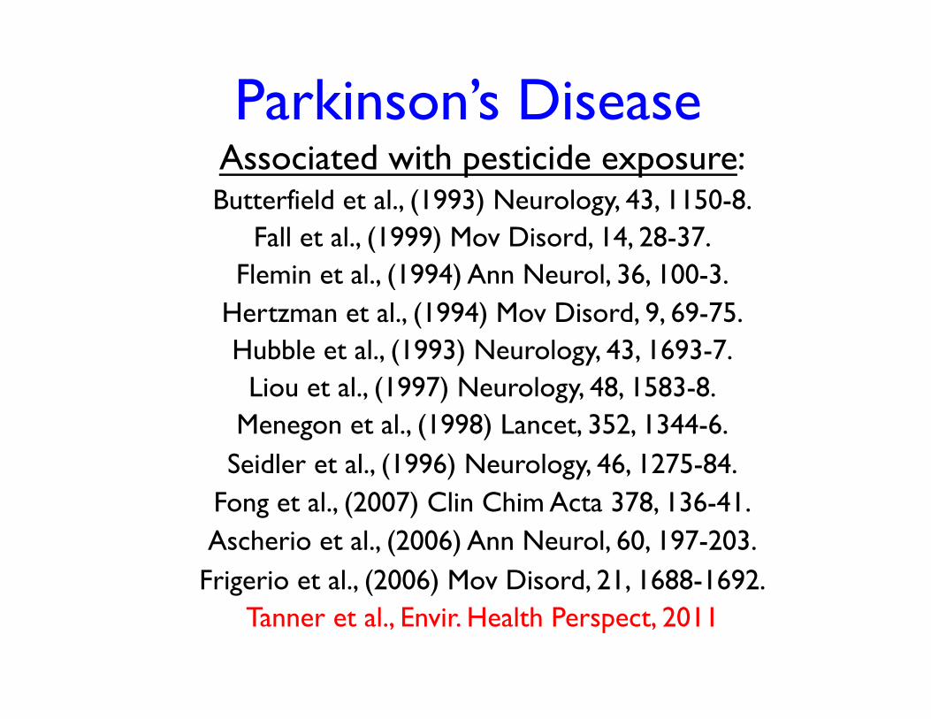
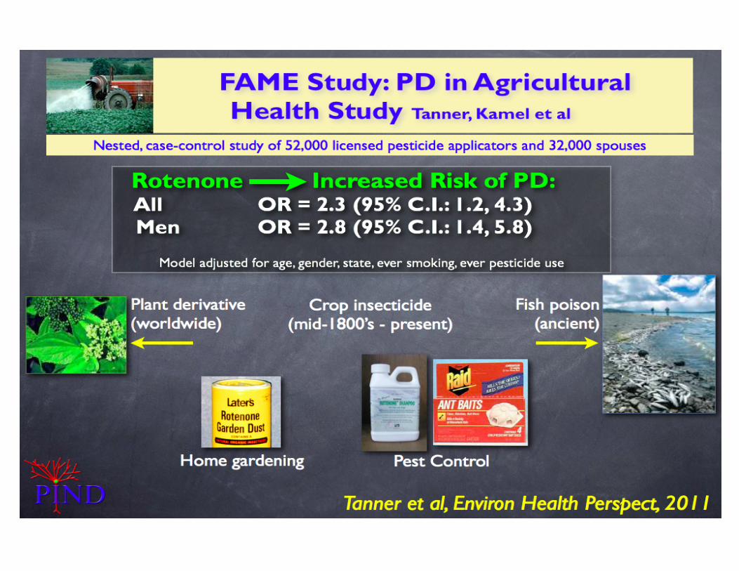
Parkinson’s Disease Associated with pesticide exposure: Butterfield et al., (1993) Neurology, 43, 1150-8.
Fall et al., (1999) Mov Disord, 14, 28-37. Flemin et al., (1994) Ann Neurol, 36, 100-3.
Hertzman et al., (1994) Mov Disord, 9, 69-75. Hubble et al., (1993) Neurology, 43, 1693-7.
Liou et al., (1997) Neurology, 48, 1583-8. Menegon et al., (1998) Lancet, 352, 1344-6.
Seidler et al., (1996) Neurology, 46, 1275-84. Fong et al., (2007) Clin Chim Acta 378, 136-41. Ascherio et al., (2006) Ann Neurol, 60, 197-203.
Frigerio et al., (2006) Mov Disord, 21, 1688-1692. Tanner et al., Envir. Health Perspect, 2011
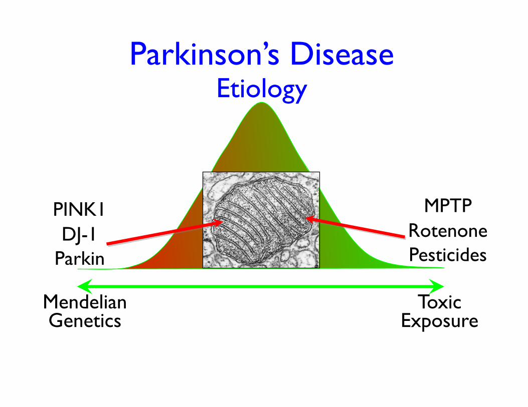
MPTP Rotenone Pesticides
Parkinson’s Disease Etiology
Mendelian Genetics
Toxic Exposure
PINK1 DJ-1
Parkin
MPTP • In 1982, IV drug users present with an acute
parkinsonian syndrome
• Astute medical detective work identifies the toxin as MPTP
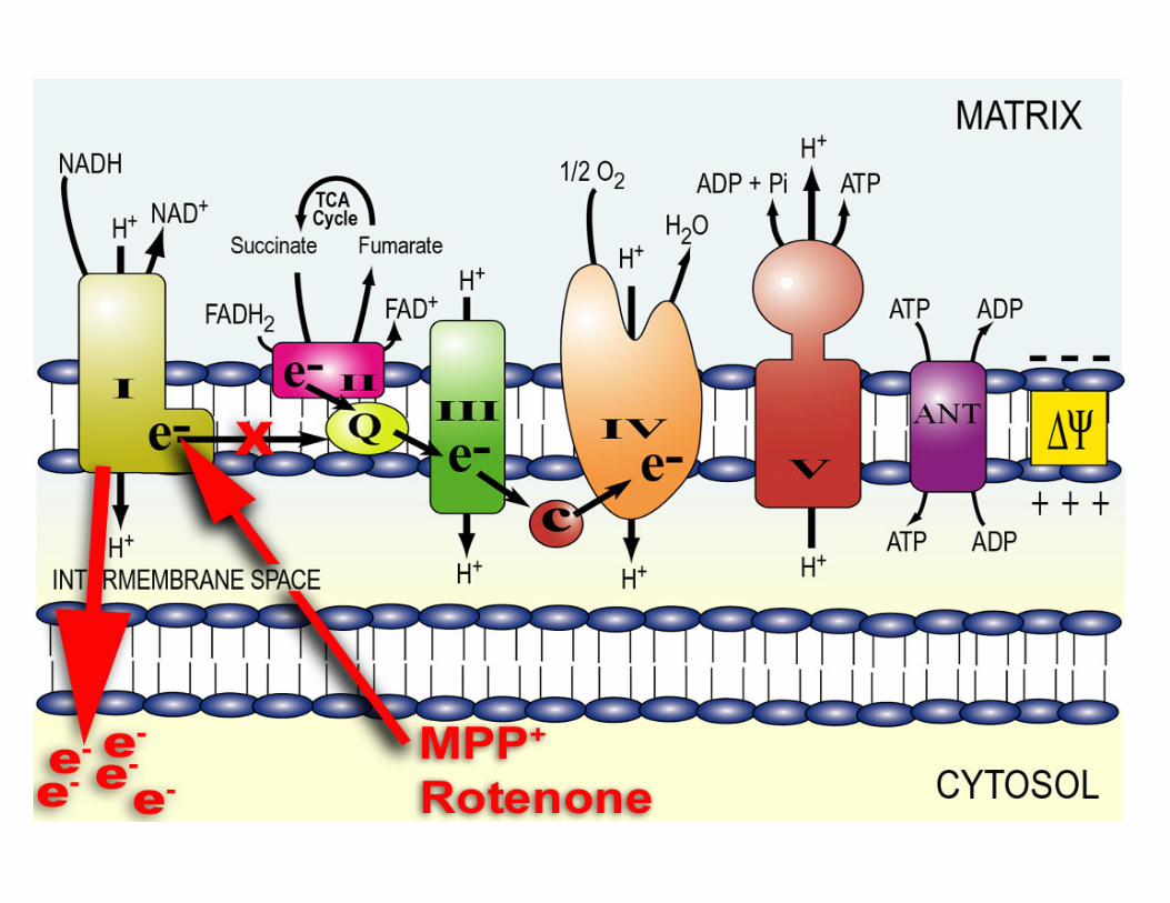
• MPTP is metabolized to MPP+, a substrate for the dopamine uptake transporter (DAT)
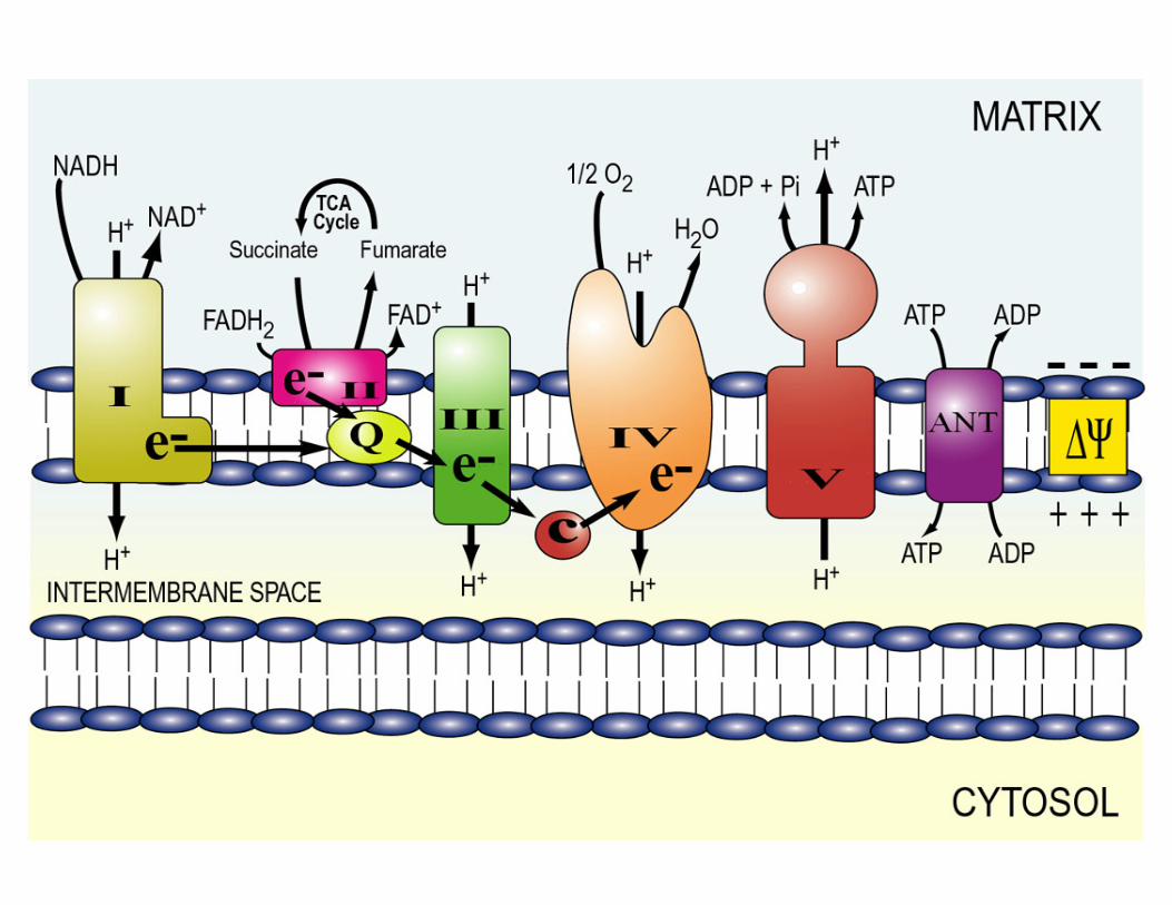
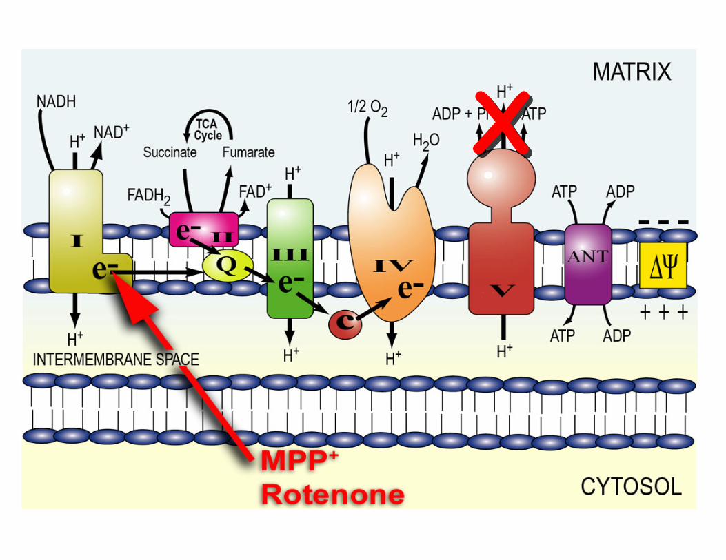
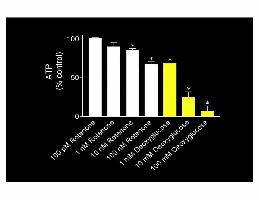
• Mechanism of action is inhibition of mitochondrial respiration at complex I
• Mitochondrial dysfunction can cause a parkinsonian syndrome
Parkinson’s Disease
A defect in mitochondrial complex I After the discovery of MPTP and its mechanism:
• 1989-92: A selective decrease in complex I activity in PD brains (Mizuno et al, Schapira et al)
• Complex I activity is reduced by 16 - 55% in platelets of PD patients (Yoshino et al, Parker et al, Mann et al, Haas & Shults et al)
Parkinson’s disease is associated with a systemic complex I defect, yet dopaminergic neurons of
substantia nigra degenerate selectively. Is the complex I defect relevant?
Hypothesis: An experimentally-induced, chronic, systemic inhibition of complex I can reproduce the behavioral, neurochemical and neuropathological
features of PD in an animal model.
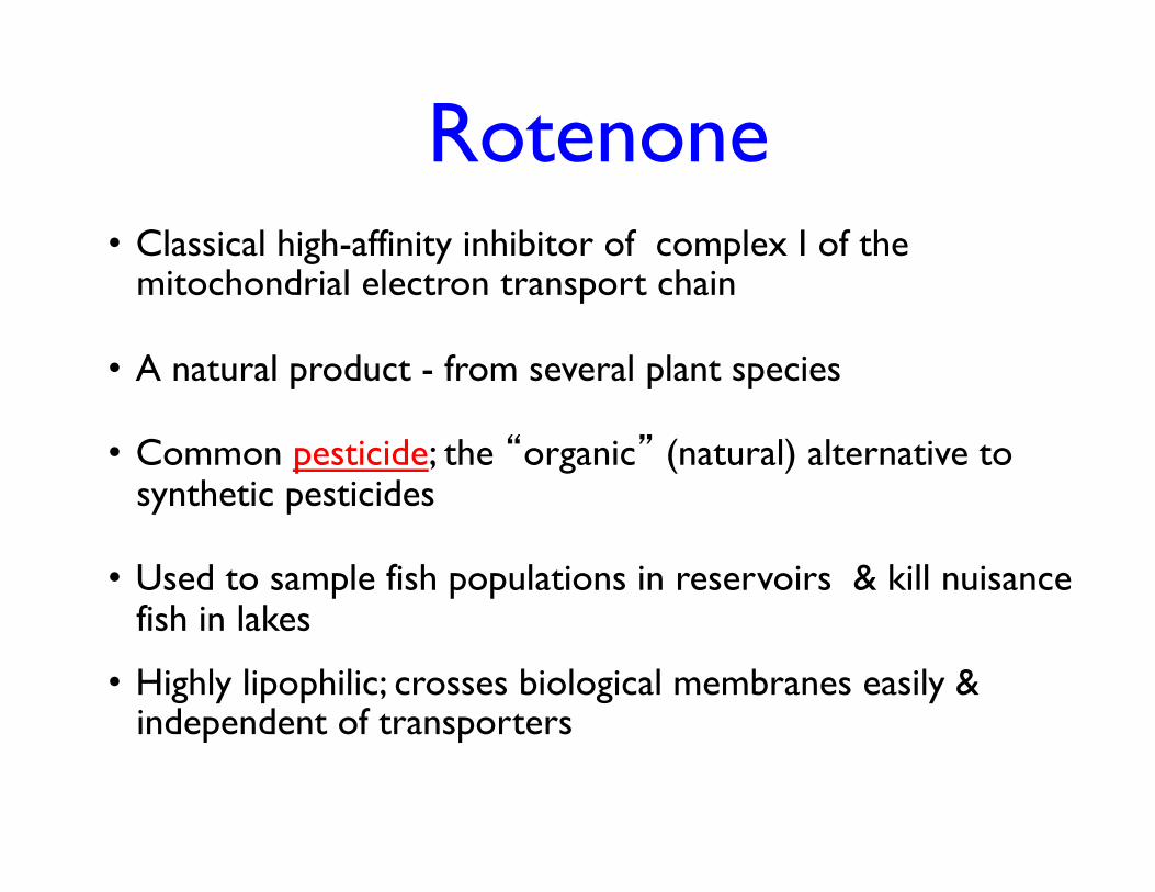
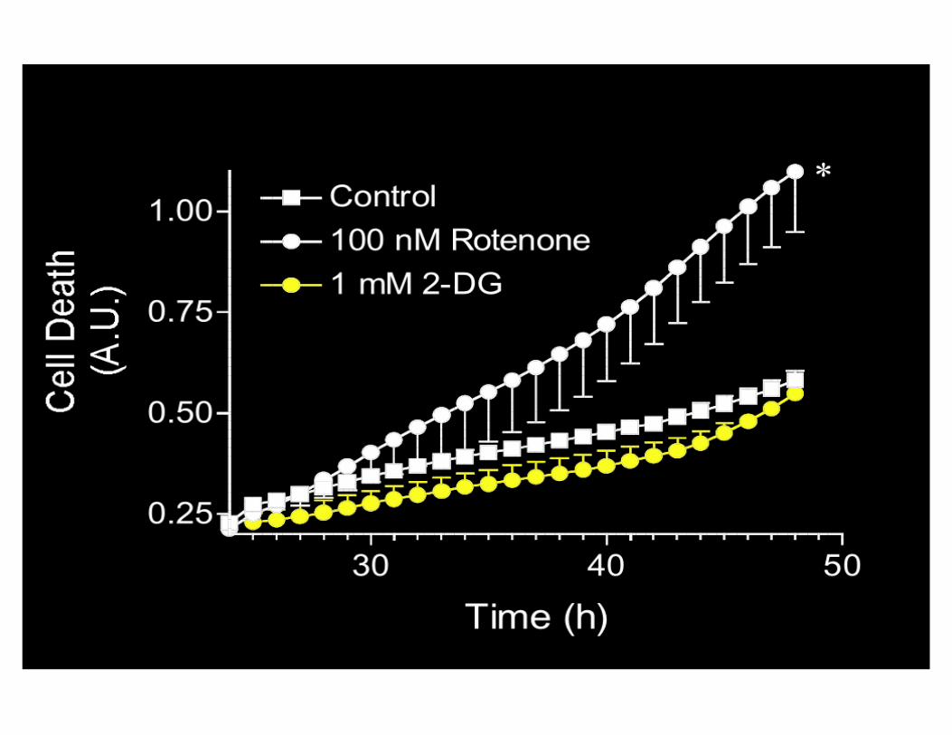
Rotenone
• Classical high-affinity inhibitor of complex I of the mitochondrial electron transport chain
• A natural product - from several plant species
• Common pesticide; the “organic” (natural) alternative to synthetic pesticides
• Used to sample fish populations in reservoirs & kill nuisance fish in lakes
• Highly lipophilic; crosses biological membranes easily & independent of transporters
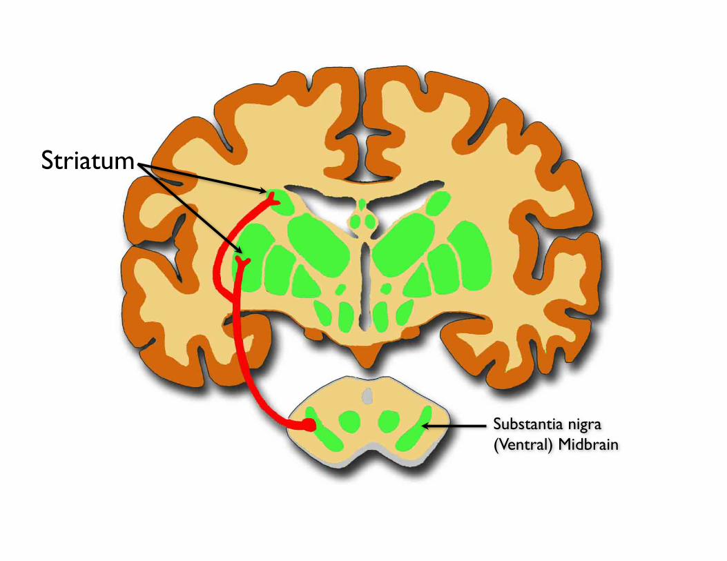
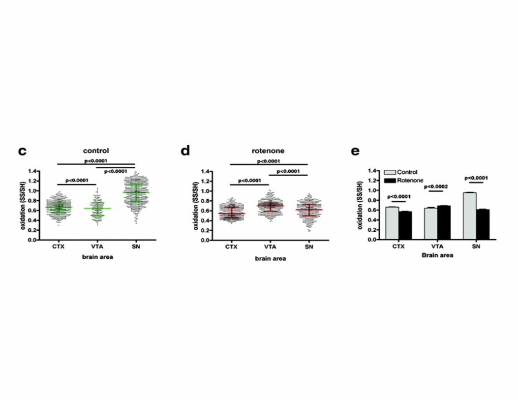
Substantia nigra (Ventral) Midbrain
Striatum
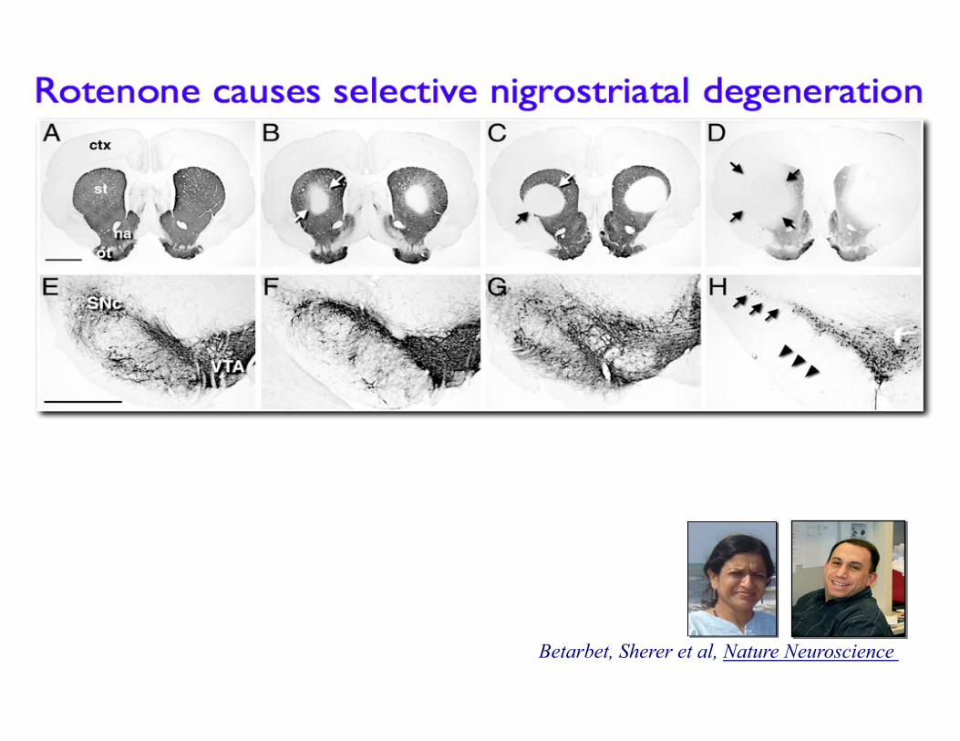
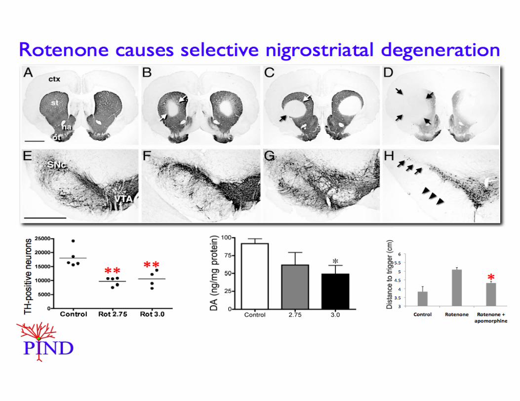
Betarbet, Sherer et al, Nature Neuroscience
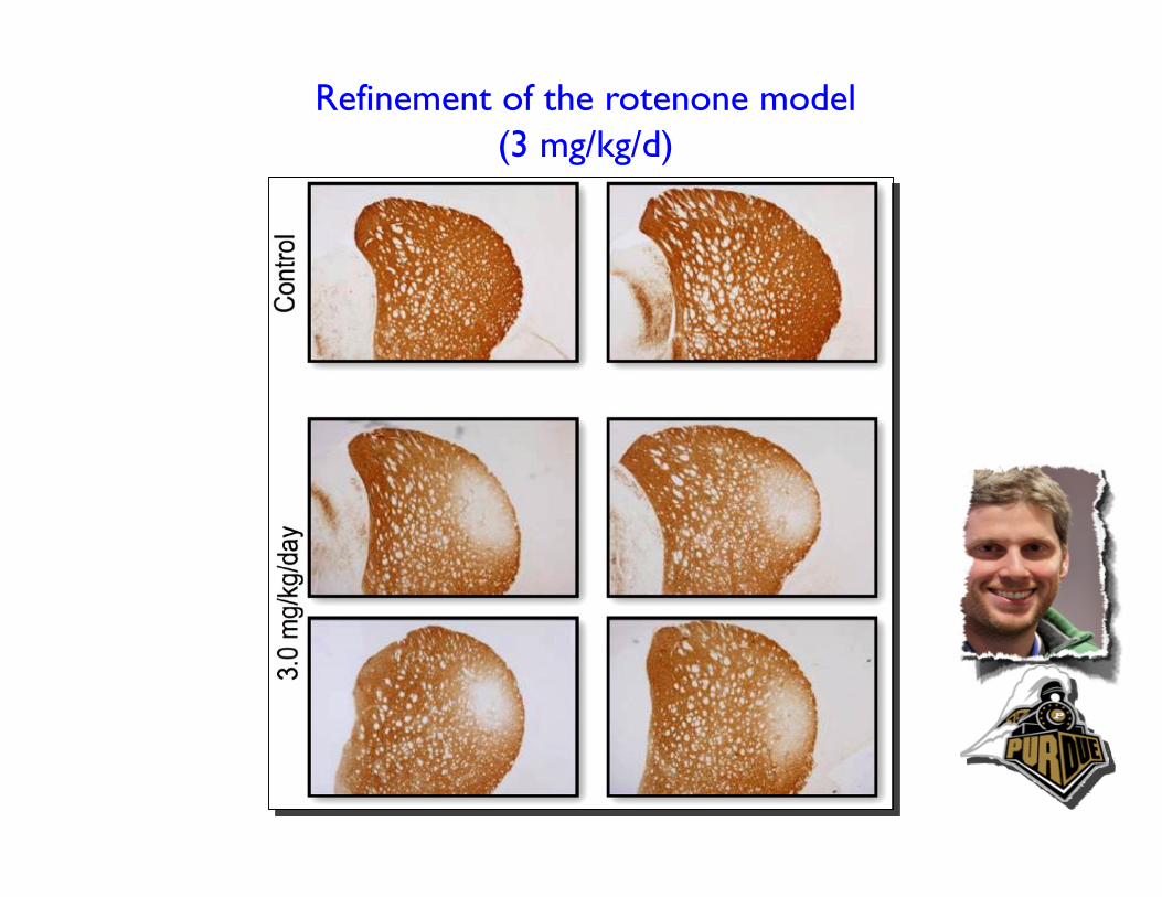
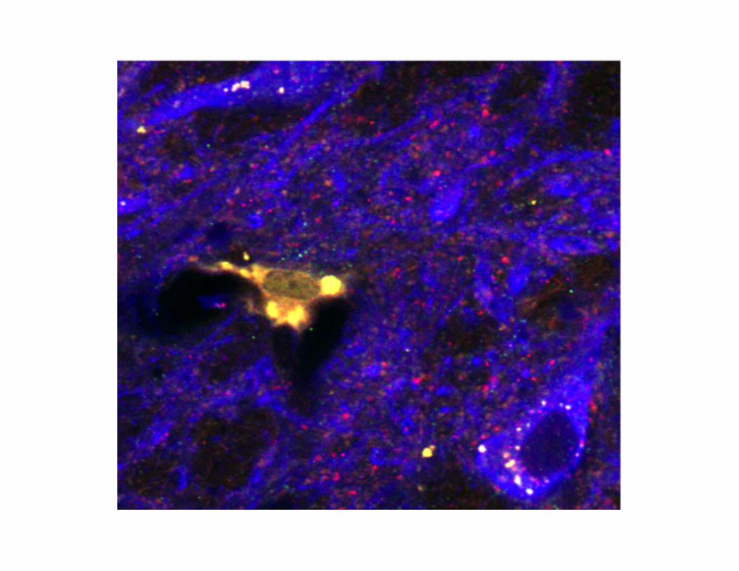
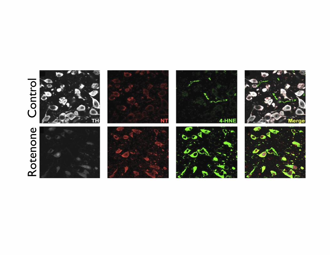
Refinement of the rotenone model (3 mg/kg/d)
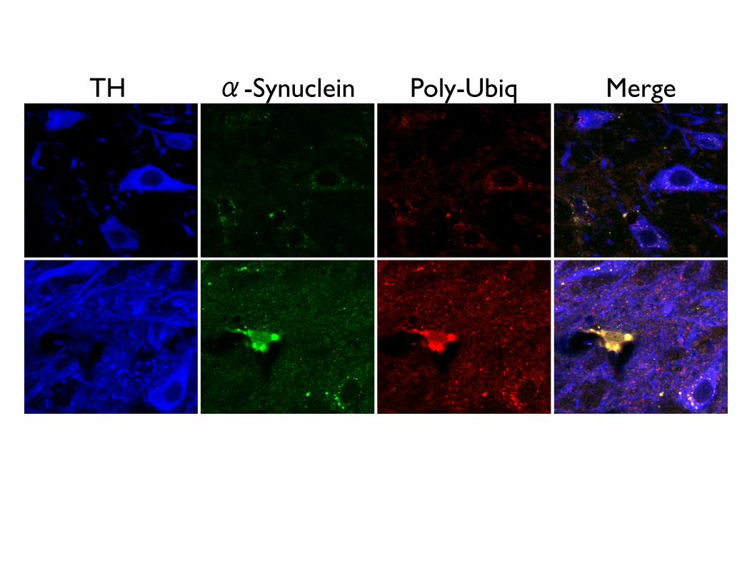
TH α-Synuclein Poly-Ubiq Merge
X
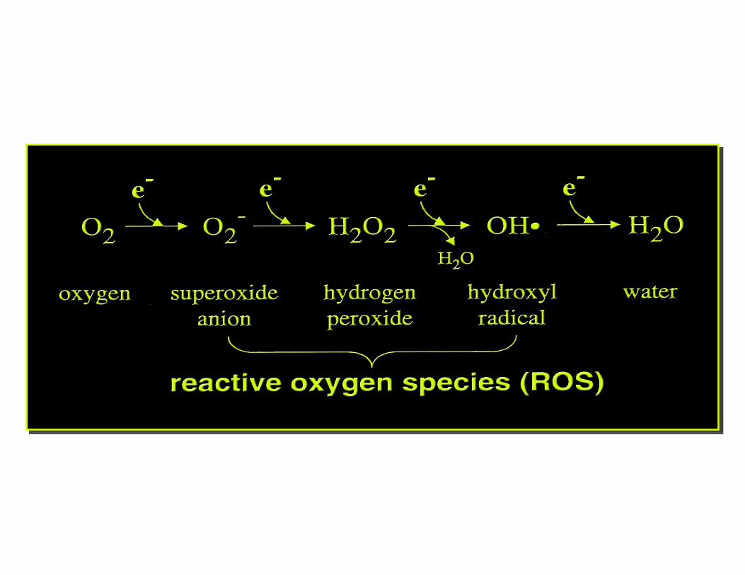
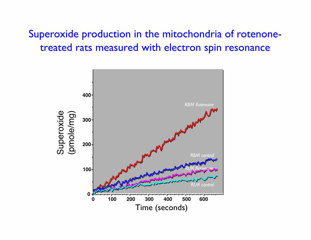
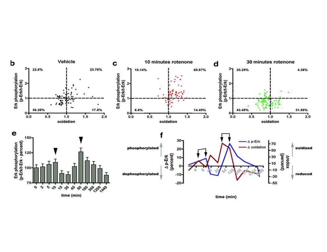
Superoxide production in the mitochondria of rotenone-treated rats measured with electron spin resonance
0 100 200 300 400 500 600
100
200
300
400
0
Superoxid
e (pmole/mg)
RBM control
RLM control
RBM Rotenone
RLM Rotenone
Time (seconds)
Con
trol
R
oten
one
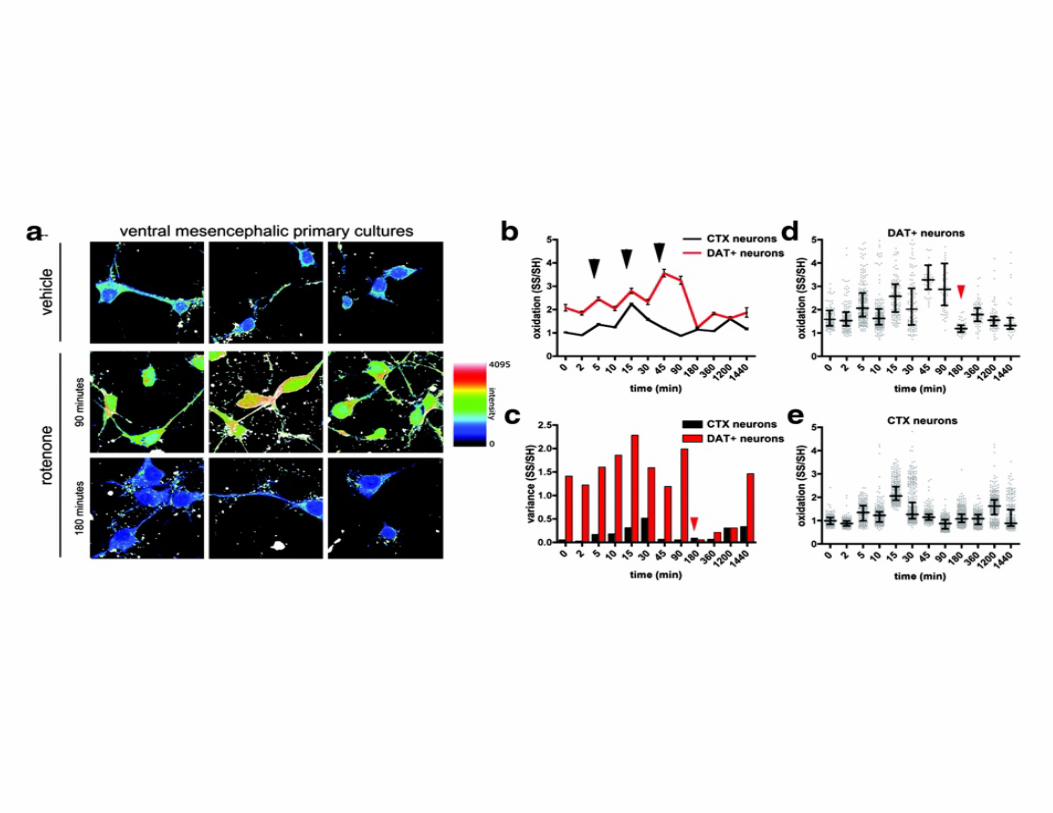
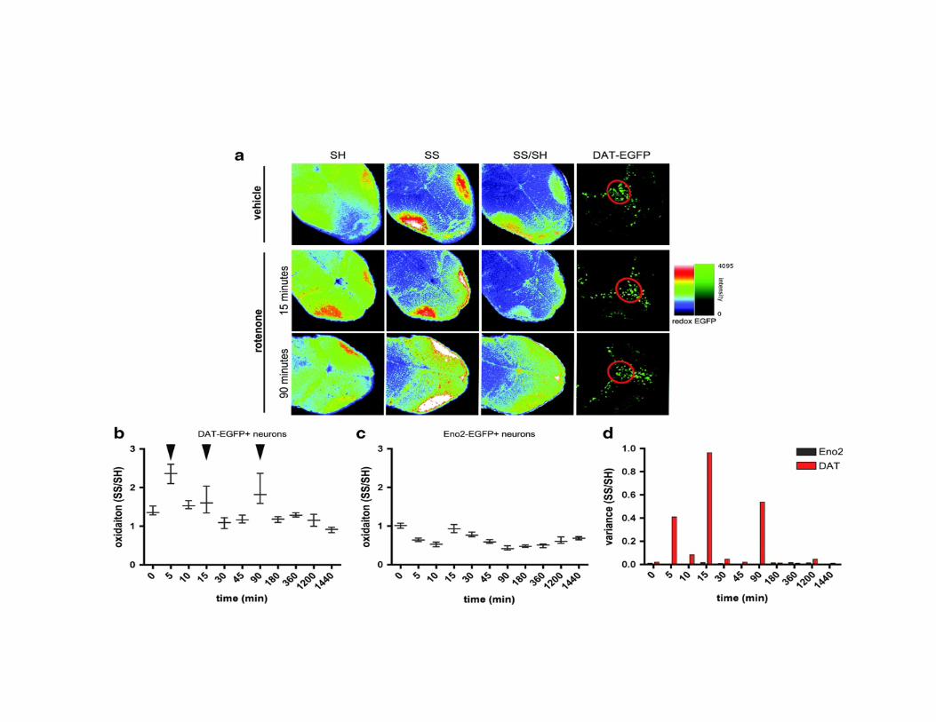
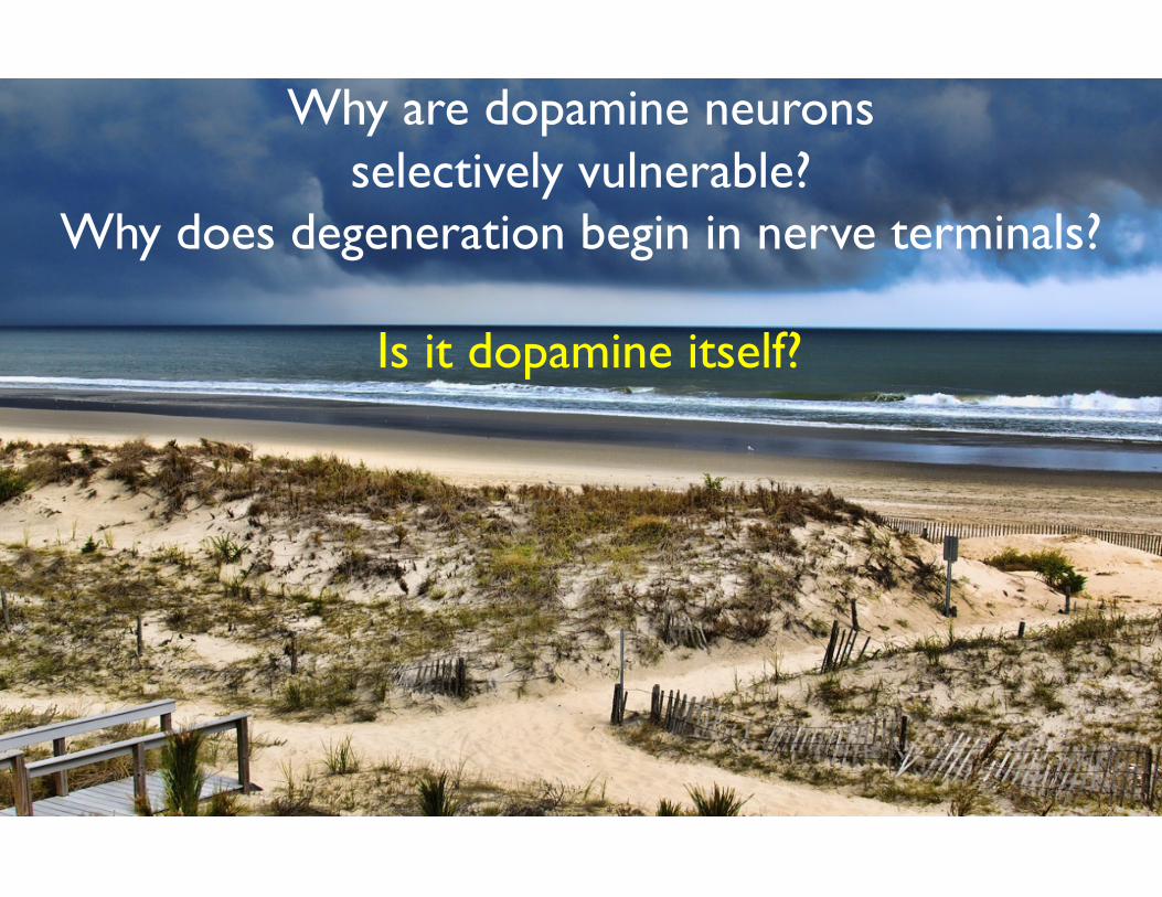
Why are dopamine neurons selectively vulnerable?
Why does degeneration begin in nerve terminals?
Is it dopamine itself?
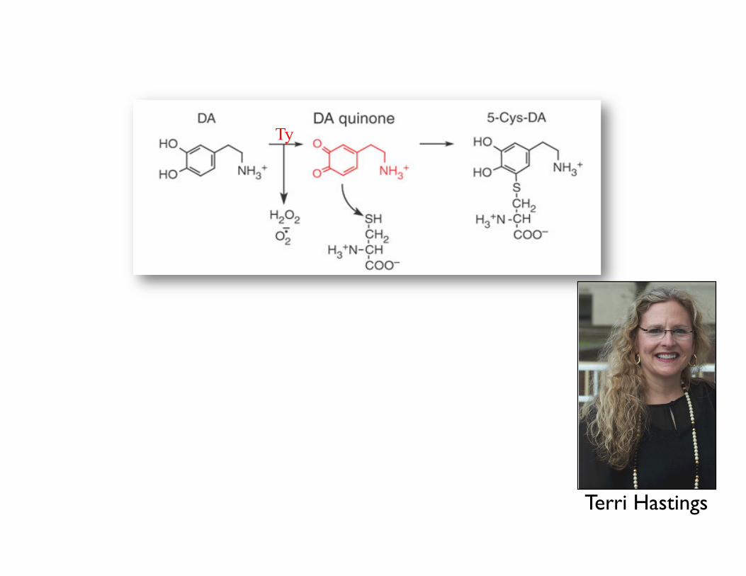
Ty
Terri Hastings
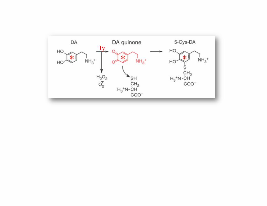
* * * Ty
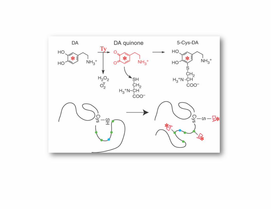
* * *
*
* *
Ty
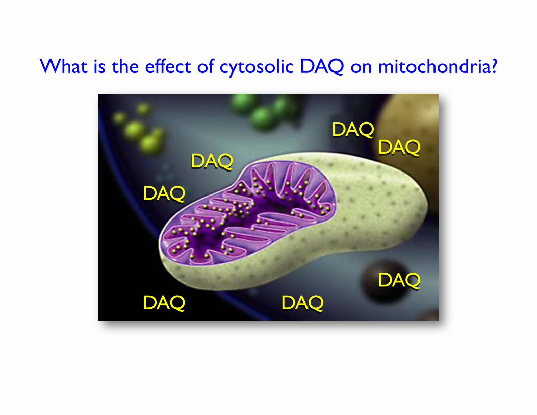
What is the effect of cytosolic DAQ on mitochondria?
DAQ
DAQ
DAQ
DAQ DAQ
DAQ DAQ
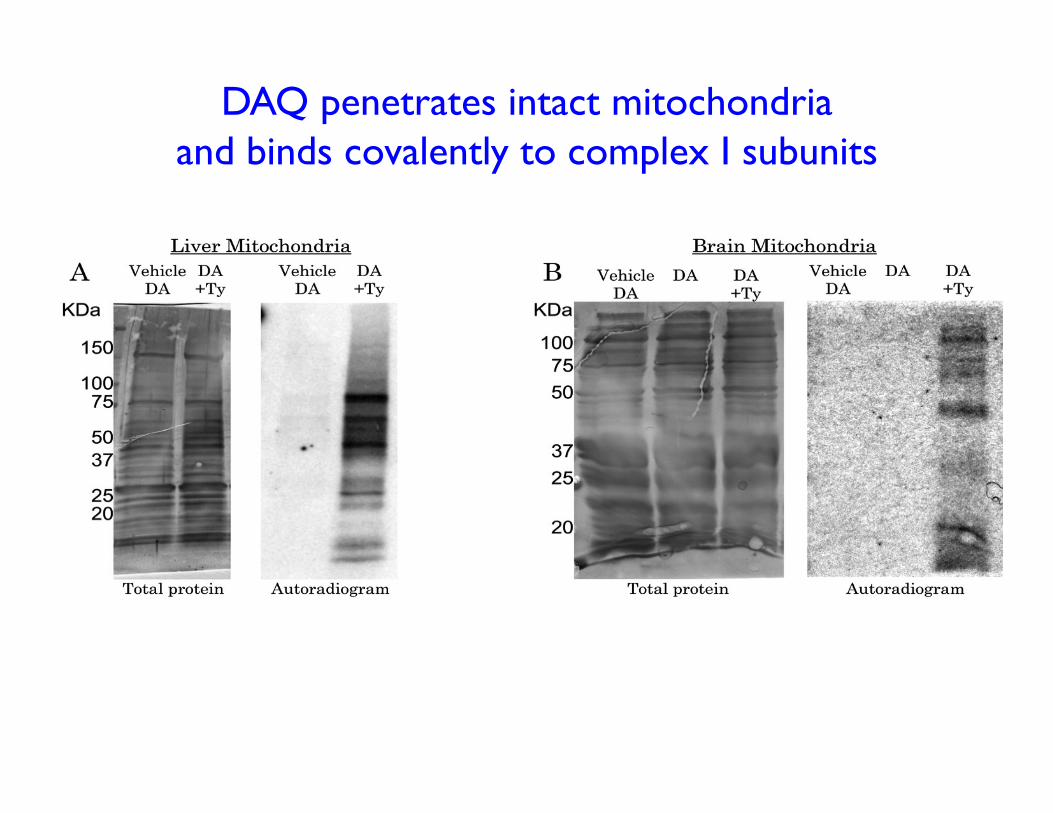
DAQ penetrates intact mitochondria and binds covalently to complex I subunits
-7 -6 -5 -4
0.00
0.25
0.50
0.75
1.00
1.25
+ Tyrosinase– Tyrosinase
IC50 = 1.2 !M
*
*
* * **
Log[dopamine]
-7 -6 -5 -4
0.00
0.25
0.50
0.75
IC50 = 1.8 !M
Log[dopamine]
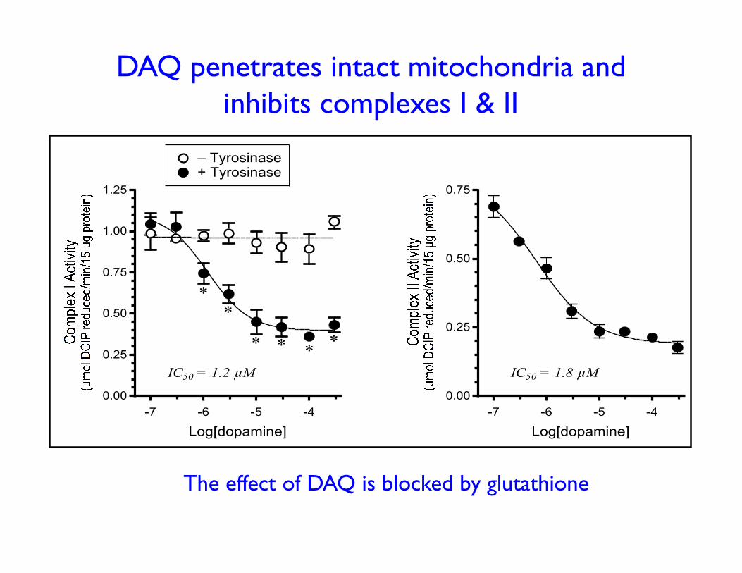
DAQ penetrates intact mitochondria and inhibits complexes I & II
The effect of DAQ is blocked by glutathione
Are these results relevant?
Can DA inhibit mitochondrial function in vivo?
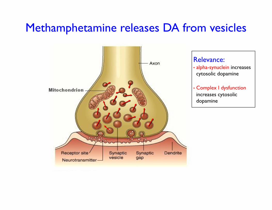
Methamphetamine releases DA from vesicles
Relevance: • alpha-synuclein increases cytosolic dopamine
• Complex I dysfunction increases cytosolic dopamine
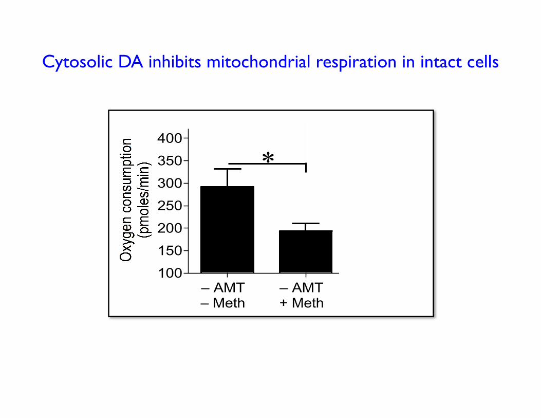
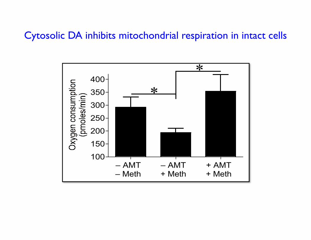
Cytosolic DA inhibits mitochondrial respiration in intact cells
100
150
200
250
300
350
400
– AMT
– Meth
– AMT
+ Meth
+ AMT
+ Meth
*
#
*
*
100
150
200
250
300
350
400
– AMT
– Meth
– AMT
+ Meth
+ AMT
+ Meth
*
#
*
*
Cytosolic DA inhibits mitochondrial respiration in intact cells
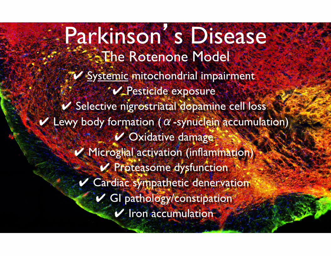
Parkinson’s Disease The Rotenone Model
✔ Systemic mitochondrial impairment ✔ Pesticide exposure
✔ Selective nigrostriatal dopamine cell loss ✔ Lewy body formation (α-synuclein accumulation)
✔ Oxidative damage ✔ Microglial activation (inflammation)
✔ Proteasome dysfunction ✔ Cardiac sympathetic denervation ✔ GI pathology/constipation ✔ Iron accumulation