Wnt/β-catenin signaling requires interaction of the Dishevelled … · VS L FR IT MKH D - GE K VS...
9
Wnt/β-catenin signaling requires interaction of the Dishevelled DEP domain and C terminus with a discontinuous motif in Frizzled Daniele V. F. Tauriello a,1,2 , Ingrid Jordens a,1 , Katharina Kirchner b,3 , Jerry W. Slootstra c,3 , Tom Kruitwagen a , Britta A. M. Bouwman a , Maria Noutsou a , Stefan G. D. Rüdiger d , Klaus Schwamborn c , Alexandra Schambony b , and Madelon M. Maurice a,4 a Department of Cell Biology, University Medical Center Utrecht, 3584 CX Utrecht, The Netherlands; b Developmental Biology Unit, Department of Biology and Developmental Biology, Friedrich-Alexander University Erlangen-Nuremberg, 91058 Erlangen, Germany; c Pepscan Therapeutics BV, 8243 RC Lelystad, The Netherlands; and d Cellular Protein Chemistry, Bijvoet Center for Biomolecular Research, Utrecht University, Utrecht, 3584 CH Utrecht, The Netherlands Edited by Roeland Nusse, Stanford University School of Medicine, Stanford, CA, and approved February 17, 2012 (received for review September 10, 2011) Wnt binding to members of the seven-span transmembrane Frizzled (Fz) receptor family controls essential cell fate decisions and tissue polarity during development and in adulthood. The Fz- mediated membrane recruitment of the cytoplasmic effector Dishevelled (Dvl) is a critical step in Wnt/β-catenin signaling initi- ation, but how Fz and Dvl act together to drive downstream sig- naling events remains largely undefined. Here, we use an Fz peptide-based microarray to uncover a mechanistically important role of the bipartite Dvl DEP domain and C terminal region (DEP-C) in binding a three-segmented discontinuous motif in Fz. We show that cooperative use of two conserved motifs in the third intracel- lular loop and the classic C-terminal motif of Fz is required for DEP- C binding and Wnt-induced β-catenin activation in cultured cells and Xenopus embryos. Within the complex, the Dvl DEP domain mainly binds the Fz C-terminal tail, whereas a short region at the Dvl C-terminal end is required to bind the Fz third loop and stabi- lize the Fz-Dvl interaction. We conclude that Dvl DEP-C binding to Fz is a key event in Wnt-mediated signaling relay to β-catenin. The discontinuous nature of the Fz-Dvl interface may allow for precise regulation of the interaction in the control of Wnt-dependent cellular responses. peptide microarray | protein-protein interaction | Wingless signaling T he Wnt family of secreted glycolipoproteins controls key cellular processes in embryonic development and stem cell maintenance in multicellular animals. Deregulation of Wnt pathway components is associated with various human diseases, most notably cancer (1–3). Depending on the cellular context, Wnts can induce distinct intracellular signaling cascades of which the Wnt/β-catenin and planar cell polarity (PCP) pathways are best described (4–6). Both pathways are initiated by Wnt-medi- ated activation of members of the Frizzled (Fz) receptor family, followed by the recruitment of the cytoplasmic effector protein Dishevelled (Dvl) to the plasma membrane (PM) (7–9). Down- stream signaling cascades are remarkably divergent, however, leading to the activation of β-catenin–mediated gene transcrip- tion in the control of cell fate decisions or to small GTPase- mediated cytoskeletal remodelling in the planar polarization of epithelia (5, 10). How the common Fz-Dvl signaling unit dis- tinguishes among different Wnt pathways remains obscure. In- sight into the molecular basis by which Fz–Dvl complexes are formed at the PM is essential to understand how cell fate choices and morphogenesis are orchestrated. The Fz family of Wnt receptors comprises G protein-coupled receptor (GPCR)–like transmembrane proteins (7, 11–13). Fz carries an extracellular N-terminal cysteine-rich domain, which mediates Wnt binding, followed by a seven-span transmembrane signaling moiety (Fig. 1A). The cytoplasmic C-terminal tail of Fz contains a conserved motif (KTxxxW) that is essential for sig- naling and for membrane relocalization of Dvl (9, 14, 15). Mi- croinjection of peptides comprising this motif interferes with Wnt/β-catenin signaling in Xenopus embryos, further supporting the functional importance of the Fz C-terminal tail. Mutagenesis studies have revealed, however, that other Fz regions, including the intracellular loops, contribute to Wnt signaling by unknown mechanisms (14, 16–18). Indeed, overexpressed Drosophila Fz2 lacking its C-terminal tail still synergizes with Wnt in Dvl-de- pendent signaling (19). Dvl is a cytoplasmic scaffold protein of which three homologs exist in vertebrates (Dvl1, Dvl2, and Dvl3). Dvl carries three structurally conserved domains that are functional in Wnt-induced signaling responses. The N-terminal DIX domain mediates Dvl self-association, leading to the formation of multimerized receptor complexes at the membrane, which are proposed to drive signaling positively by providing a high local concentration of binding sites for Wnt signaling proteins (20–22). The PDZ domain, located in the central region of Dvl, was shown to bind an Fz7-derived peptide containing the conserved KTxxxW motif in vitro (23). The relative low affinity of this in- teraction suggested the need for additional molecular inter- actions in the formation of a stable Fz–Dvl complex in vivo in cells. Moreover, the binding cleft of Dvl PDZ appears highly flexible and accommodates binding of a diverse set of peptide ligands in vitro as well as numerous Dvl PDZ binding partners in cells (24–26). How these interactors compete for Dvl PDZ do- main binding and how specificity is achieved during Wnt sig- naling remain unresolved. The conserved DEP domain, located in the C-terminal half of Dvl, is critical for membrane recruitment of Dvl during Wnt- mediated signaling (8, 27–30). Direct binding of a polybasic stretch in the DEP domain to negatively charged lipids was re- cently shown to contribute to formation of the Fz–Dvl complex at the PM (30). In addition, the Dvl2 DEP domain, aided by a YHEL motif in the Dvl C terminus, interacts with μ2-adaptin, a subunit of the AP-2 clathrin adaptor complex, to facilitate clathrin-mediated endocytosis of Fz4 and Dvl2 on Wnt activation Author contributions: D.V.F.T., I.J., K.K., J.W.S., S.G.D.R., K.S., A.S., and M.M.M. designed research; D.V.F.T., I.J., K.K., J.W.S., T.K., B.A.M.B., and M.N. performed research; D.V.F.T., I.J., K.K., J.W.S., S.G.D.R., K.S., A.S., and M.M.M. analyzed data; and D.V.F.T., I.J., and M.M.M. wrote the paper. The authors declare no conflict of interest. This article is a PNAS Direct Submission. 1 D.V.F.T. and I.J. contributed equally to this work. 2 Present address: Institute for Research in Biomedicine Barcelona, 08028 Barcelona, Spain. 3 K.K. and J.W.S. contributed equally to this work. 4 To whom correspondence should be addressed. E-mail: [email protected]. See Author Summary on page 5154 (volume 109, number 14). This article contains supporting information online at www.pnas.org/lookup/suppl/doi:10. 1073/pnas.1114802109/-/DCSupplemental. E812–E820 | PNAS | Published online March 12, 2012 www.pnas.org/cgi/doi/10.1073/pnas.1114802109 Downloaded by guest on May 19, 2020
Transcript of Wnt/β-catenin signaling requires interaction of the Dishevelled … · VS L FR IT MKH D - GE K VS...
Wnt/β-catenin signaling requires interaction of theDishevelled DEP domain and C terminus witha discontinuous motif in FrizzledDaniele V. F. Taurielloa,1,2, Ingrid Jordensa,1, Katharina Kirchnerb,3, Jerry W. Slootstrac,3, Tom Kruitwagena,Britta A. M. Bouwmana, Maria Noutsoua, Stefan G. D. Rüdigerd, Klaus Schwambornc, Alexandra Schambonyb,and Madelon M. Mauricea,4
aDepartment of Cell Biology, University Medical Center Utrecht, 3584 CX Utrecht, The Netherlands; bDevelopmental Biology Unit, Department of Biology andDevelopmental Biology, Friedrich-Alexander University Erlangen-Nuremberg, 91058 Erlangen, Germany; cPepscan Therapeutics BV, 8243 RC Lelystad, TheNetherlands; and dCellular Protein Chemistry, Bijvoet Center for Biomolecular Research, Utrecht University, Utrecht, 3584 CH Utrecht, The Netherlands
Edited by Roeland Nusse, Stanford University School of Medicine, Stanford, CA, and approved February 17, 2012 (received for review September 10, 2011)
Wnt binding to members of the seven-span transmembraneFrizzled (Fz) receptor family controls essential cell fate decisionsand tissue polarity during development and in adulthood. The Fz-mediated membrane recruitment of the cytoplasmic effectorDishevelled (Dvl) is a critical step in Wnt/β-catenin signaling initi-ation, but how Fz and Dvl act together to drive downstream sig-naling events remains largely undefined. Here, we use an Fzpeptide-based microarray to uncover a mechanistically importantrole of the bipartite Dvl DEP domain and C terminal region (DEP-C)in binding a three-segmented discontinuous motif in Fz. We showthat cooperative use of two conserved motifs in the third intracel-lular loop and the classic C-terminal motif of Fz is required for DEP-C binding and Wnt-induced β-catenin activation in cultured cellsand Xenopus embryos. Within the complex, the Dvl DEP domainmainly binds the Fz C-terminal tail, whereas a short region at theDvl C-terminal end is required to bind the Fz third loop and stabi-lize the Fz-Dvl interaction. We conclude that Dvl DEP-C binding toFz is a key event in Wnt-mediated signaling relay to β-catenin. Thediscontinuous nature of the Fz-Dvl interface may allow for preciseregulation of the interaction in the control of Wnt-dependentcellular responses.
peptide microarray | protein-protein interaction | Wingless signaling
The Wnt family of secreted glycolipoproteins controls keycellular processes in embryonic development and stem cell
maintenance in multicellular animals. Deregulation of Wntpathway components is associated with various human diseases,most notably cancer (1–3). Depending on the cellular context,Wnts can induce distinct intracellular signaling cascades of whichthe Wnt/β-catenin and planar cell polarity (PCP) pathways arebest described (4–6). Both pathways are initiated by Wnt-medi-ated activation of members of the Frizzled (Fz) receptor family,followed by the recruitment of the cytoplasmic effector proteinDishevelled (Dvl) to the plasma membrane (PM) (7–9). Down-stream signaling cascades are remarkably divergent, however,leading to the activation of β-catenin–mediated gene transcrip-tion in the control of cell fate decisions or to small GTPase-mediated cytoskeletal remodelling in the planar polarization ofepithelia (5, 10). How the common Fz-Dvl signaling unit dis-tinguishes among different Wnt pathways remains obscure. In-sight into the molecular basis by which Fz–Dvl complexes areformed at the PM is essential to understand how cell fate choicesand morphogenesis are orchestrated.The Fz family of Wnt receptors comprises G protein-coupled
receptor (GPCR)–like transmembrane proteins (7, 11–13). Fzcarries an extracellular N-terminal cysteine-rich domain, whichmediates Wnt binding, followed by a seven-span transmembranesignaling moiety (Fig. 1A). The cytoplasmic C-terminal tail of Fzcontains a conserved motif (KTxxxW) that is essential for sig-naling and for membrane relocalization of Dvl (9, 14, 15). Mi-
croinjection of peptides comprising this motif interferes withWnt/β-catenin signaling in Xenopus embryos, further supportingthe functional importance of the Fz C-terminal tail. Mutagenesisstudies have revealed, however, that other Fz regions, includingthe intracellular loops, contribute to Wnt signaling by unknownmechanisms (14, 16–18). Indeed, overexpressed Drosophila Fz2lacking its C-terminal tail still synergizes with Wnt in Dvl-de-pendent signaling (19).Dvl is a cytoplasmic scaffold protein of which three homologs
exist in vertebrates (Dvl1, Dvl2, and Dvl3). Dvl carries threestructurally conserved domains that are functional in Wnt-inducedsignaling responses. The N-terminal DIX domain mediates Dvlself-association, leading to the formation of multimerized receptorcomplexes at the membrane, which are proposed to drive signalingpositively by providing a high local concentration of binding sitesfor Wnt signaling proteins (20–22).The PDZ domain, located in the central region of Dvl, was
shown to bind an Fz7-derived peptide containing the conservedKTxxxW motif in vitro (23). The relative low affinity of this in-teraction suggested the need for additional molecular inter-actions in the formation of a stable Fz–Dvl complex in vivo incells. Moreover, the binding cleft of Dvl PDZ appears highlyflexible and accommodates binding of a diverse set of peptideligands in vitro as well as numerous Dvl PDZ binding partners incells (24–26). How these interactors compete for Dvl PDZ do-main binding and how specificity is achieved during Wnt sig-naling remain unresolved.The conserved DEP domain, located in the C-terminal half of
Dvl, is critical for membrane recruitment of Dvl during Wnt-mediated signaling (8, 27–30). Direct binding of a polybasicstretch in the DEP domain to negatively charged lipids was re-cently shown to contribute to formation of the Fz–Dvl complexat the PM (30). In addition, the Dvl2 DEP domain, aided bya YHEL motif in the Dvl C terminus, interacts with μ2-adaptin,a subunit of the AP-2 clathrin adaptor complex, to facilitateclathrin-mediated endocytosis of Fz4 and Dvl2 on Wnt activation
Author contributions: D.V.F.T., I.J., K.K., J.W.S., S.G.D.R., K.S., A.S., and M.M.M. designedresearch; D.V.F.T., I.J., K.K., J.W.S., T.K., B.A.M.B., and M.N. performed research; D.V.F.T., I.J.,K.K., J.W.S., S.G.D.R., K.S., A.S., and M.M.M. analyzed data; and D.V.F.T., I.J., and M.M.M.wrote the paper.
The authors declare no conflict of interest.
This article is a PNAS Direct Submission.1D.V.F.T. and I.J. contributed equally to this work.2Present address: Institute for Research in Biomedicine Barcelona, 08028 Barcelona, Spain.3K.K. and J.W.S. contributed equally to this work.4To whom correspondence should be addressed. E-mail: [email protected].
See Author Summary on page 5154 (volume 109, number 14).
This article contains supporting information online at www.pnas.org/lookup/suppl/doi:10.1073/pnas.1114802109/-/DCSupplemental.
E812–E820 | PNAS | Published online March 12, 2012 www.pnas.org/cgi/doi/10.1073/pnas.1114802109
Dow
nloa
ded
by g
uest
on
May
19,
202
0
(31, 32). Both of these activities of the DEP domain are mainlylinked to PCP signaling, however, leaving the role of the DEPdomain in β-catenin–dependent signaling unexplained (29, 33–36). Moreover, a number of point mutants in the DEP domainwere uncovered that, based on their location in the structure, donot participate in lipid or AP-2 binding yet prevent the trans-location of Dvl to the PM (27–29).Progress in the understanding of how Fz and Dvl function
together during Wnt signaling is hampered by the general lack ofbiochemical evidence for their interaction, partly attributable tothe hydrophobic nature of the Fz protein. Here, we use a peptidescanning method to address the molecular basis of complexformation between Fz and Dvl. We show that the Dvl DEPdomain and C-terminal region (DEP-C) mediates direct bindingto a three-segmented discontinuous motif in Fz, composed of thethird intracellular loop (iLoop3) and C-terminal tail. The in-dividual Dvl binding sequences in the cytosolic interface of Fzcooperate to form stable Fz–Dvl complexes at the PM and ini-tiate Wnt/β-catenin signaling. With these findings, we assigna previously unknown molecular activity to the Dvl DEP domain,identify a functional role of the Dvl C-terminal region, and placethe DEP-C domain central to Wnt/β-catenin signaling initiation.
ResultsIdentification of a Discontinuous Dvl Binding Surface in the FzReceptor. To gain insight into the binding mode betweenmouse Fz5 and Dvl1, we combined high-throughput PEPSCANmicroarray technology (Pepscan Therapeutics BV) with spatiallydefined peptide conformations [chemically linked peptides onscaffolds (CLIPS) technology (37)] to mimic the cytoplasmicinterface of Fz5 and facilitate the discovery of complex molec-ular recognition modes between Fz and Dvl. We generateda combinatorial peptide library comprising 9-mer peptides of theFz5 iLoop3 (V424–F453) in cis-combination with 9-mer peptidesfrom the proximal region of the C-terminal tail of Fz5 (W520–G550) (Fig. 1A). Peptides were flanked by either glycines orcysteines. Glycine-flanked peptides were left in a linear confor-mation, whereas cysteine-flanked peptides were imposed ona trivalent chemical scaffold to mimic a looped conformation(37). To allow for specificity of cysteine-based chemical mod-ifications, the three native cysteines in the distal part of the Fz5tail were changed to alanines in the 9-mer peptides. The library,containing 968 combinatorial iLoop3-tail peptides in total, wasimmobilized on 455-well plates (Fig. 1A).
G
D
A
9-mers
Intracellular loop 3 C-terminal tail (30 AA)
22 x 22
22x
G G G
22x..... ..........
C C
CS
S S
Peptide library
HIS-mDvl1 ∆DIX
484 Linear 484 Scaffolded
VSLFRIRSVIKQGGTKTDKLEKLMIRIGIF WIWSGKTLESWRRFTSRAAASSGHKSGG
VSLFRIRSVSLFRIRSVI
LFRIRSVIK
9-mers
WIWSGKTLEIWSGKTLESWSGKTLESW
Tail
Classical
KTxxxW
motif
Motif I
I II III IV V VI VII
Motif II
Motif III
FR
GGG
H
I
GG T
RSV
Q
L
EI
M
R
K
K
K
K
K
SW
ES
L
W
FT
RR
RRRCCCSSS
D
L
T
I
ITK
iLoop 3
G
E
mFz5CRD
S
L
LL YT
GF AV
FS I I
SG TV
W
Rank # Peptide # Peptide Sequence BI: AU Normalized1 158 GSLFRIRSVIGLESWRRFTSG 1179 2.962 156 GKLMIRIGIFGTLESWRRFTG 1174 2.953 157 GVSLFRIRSVGLESWRRFTSG 1147 2.884 136 GSLFRIRSVIGTLESWRRFTG 1118 2.815 154 GLEKLMIRIGGTLESWRRFTG 1092 2.746 178 GKLMIRIGIFGLESWRRFTSG 1080 2.717 153 GKLEKLMIRIGTLESWRRFTG 1066 2.688 155 GEKLMIRIGIGTLESWRRFTG 1066 2.689 177 GEKLMIRIGIGLESWRRFTSG 1013 2.54
10... ... ... ... ...
135 GVSLFRIRSVGTLESWRRFTG 993 2.49
VSLFRIRSV
SLFRIRSVI
LFRIR
SVIK
FRIRSVIKQ
RIRSVIKQG
IRSVIKQGG
RSVIKQGGT
SVIKQGGTK
VIKQGGTKT
IKQGGTKTD
KQGGTKTDK
QGGTKTDKL
GGTKTDKLE
GTKTDKLEK
TKTDKLEKL
KTDKLEKLM
TDKLEKLM
I
DKLEKLM
IR
KLEKLM
IRI
LEKLMIRIG
EKLMIRIG
I
KLMIRIG
IF
Freq
uenc
y in
the
top
5%
0
2
4
6
8Motif III Other
∆DIXB
Motif I Motif II
DFz
Fz1Fz7Fz2
Fz5Fz8
Fz4Fz9Fz3Fz6
I S L F R I R T V M K T D - - G K R T D K L E R L M L R I G F F
V S L F R I R T I M K H D - - G T K T E K L E KK
L M V R I G V F
V S L F R I R T I M K H D - - G T K T E K L E R L M V R I G V F
V S L F R I R S V I K Q - - G G T K T D K L E K L M I R I G I FV S L F R I R S V I K Q Q G G P T K T H K L E K L M I R L G L F
V A L F K I R S N L Q K D - - G T K T D K L E R L M V K I G V FV A L F H I R K I M K T G - - G T N T E K L E K L M V K I G V FI S L N R V R I E I P L E - - K E N Q D K L V K F M I R I G V FI S L N H V R Q V I Q H D - - G R N Q E K L K K F M I R I G V F
Fz10 V A L F H I R R V M K T G - - G E N T D K L E K L M V R I G V F
V S L F R I R T I M K H D - - G T K T E K L E K L M V R I G V F
BI
Ranked peptides
C
Fig. 1. Identification of a discontinuous Dvl1 binding site in Fz5. (A) (Left) Schematic depiction of the topology of Fz proteins. iLoop3 and the C-terminal tailare boxed. (Right) Design of the Fz5 iLoop3-tail peptide library, based on 30 amino acids from mouse Fz5 iLoop3 (white) and 30 membrane-proximal aminoacids from the Fz5 tail (black). Linear 9-mer peptides (22 each) were combined in cis, flanked and separated by Gly or Cys residues. CLIPS technology was usedon Cys-based combinations, yielding bicyclical peptides; Gly-linked peptides were left linear. The combinatory peptides were immobilized in 455-well plates.Binding of recombinant His-tagged Dvl1-ΔDIX (G86–M695) was determined by an ELISA-based assay. (B) Binding signals were ranked by binding intensity (BI),and the 10 best binding peptides are depicted with their absolute score and normalized BI (relative to the screen average). AU, Arbitrary Units. (C) Frequencyanalysis of the top 5% (48 best binding peptides) reveals a strong preference for binding two motifs in iLoop3 (motifs I and II; pink and green) and one in theC terminus (motif III; blue). Motif III overlaps with the classic PDZ binding motif. Depicted are the frequencies of iLoop3 peptides in the top 5%, whethercombined to motif III (blue) or not (white; other). (D) Diagram of the three Dvl1 binding motifs in the mFz5 iLoop3 and C-terminal tail. (E) Alignment ofregions encompassing motifs I and II of mouse Fz family members and Drosophila Fz (DFz). Motifs I and II are highly conserved among most Fzs.
Tauriello et al. PNAS | Published online March 12, 2012 | E813
DEV
ELOPM
ENTA
LBIOLO
GY
PNASPL
US
Dow
nloa
ded
by g
uest
on
May
19,
202
0
We purified His-tagged, recombinant mouse Dvl1-ΔDIX (G86–M695) protein and investigated its binding to the Fz5 peptide li-brary through detection with peroxidase-conjugated anti–His-tagantibodies. The resulting values were ranked according to bindingsignal, and the top 5% of best binding peptides were analyzed forthe presence of prevalent Dvl binding motifs (Fig. 1 B and C).Clearly, Dvl1-ΔDIX displays a strong preference for binding threelinear motifs in Fz5 (Fig. 1D). In the Fz5 C-terminal tail, Dvl1-ΔDIX binds a sequence that overlaps with the known PDZ bindingmotif (Fig. 1D, motif III). In Fz5 iLoop3, Dvl1-ΔDIX binds twosequences that flank predicted TM regions 5 and 6 (Fig. 1D, motifsI and II). Both Dvl binding motifs in Fz5 iLoop3 are highly con-served among Fz family members, although loss of conservation ofmotif I is evident in Fz3 and Fz6 (Fig. 1E). Strikingly, Dvl1-ΔDIXstrongly preferred binding to linear Fz5 peptides that combinedeither motif I or II with motif III (Fig. 1C). Scaffolded, bicyclicalFz5 peptides in looped conformation displayed an identical bindingpattern, albeit with overall lower binding, suggesting that flexibilityof the individual motifs accommodates optimal Dvl1 interaction.These results indicate that Dvl binds to both iLoop3 and the C-terminal tail of Fz in a cooperative manner.
Dvl Binding Motifs I and II in Fz5 iLoop3 Are Essential for Wnt/β-Catenin Signaling and Recruitment of Dvl in Human Cells. To ad-dress the importance of the Fz5 iLoop3 residues in signaling, we
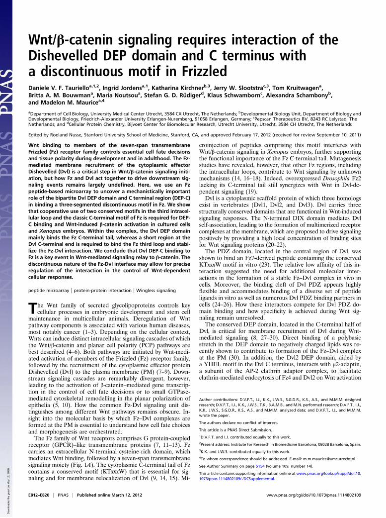
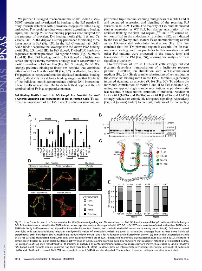
performed triple alanine-scanning mutagenesis of motifs I and IIand compared expression and signaling of the resulting Fz5variants in HEK293T cells. The majority of Fz5 mutants showedsimilar expression as WT Fz5, but alanine substitution of theresidues flanking the sixth TM region (449RIGIF453) caused re-tention of Fz5 in the endoplasmic reticulum (ER), as indicatedby the lack of glycosylated, mature Fz on immunoblotting as wellas an ER-associated subcellular localization (Fig. 2B). Weconclude that this TM proximal region is essential for Fz mat-uration or sorting, and thus precludes further investigation. Allother Fz5 mutants were processed to the mature form andtransported to the PM (Fig. 2B), allowing for analysis of theirsignaling propensity.Overexpression of Fz5 in HEK293T cells strongly induced
β-catenin-dependent transactivation of a luciferase reporterplasmid (TOPflash) on stimulation with Wnt3a-conditionedmedium (Fig. 2A). Single alanine substitutions of key residues inthe classic Dvl binding motif in the Fz5 C terminus significantlyimpaired signaling, as expected (9, 16) (Fig. 2C). To address theindividual contribution of motifs I and II to Fz5-mediated sig-naling, we applied single alanine substitutions to pin down crit-ical residues in these motifs. Mutation of individual residues inFz5 motif I (I429A and R430A) or motif II (L443A and L446A)strongly reduced or completely abrogated signaling, respectively[Fig. 2 A (arrows) and C]. In contrast, mutation of the connecting
A
C
B
0 - 25
% activity
25 - 50
75 - 100
50 - 75
0
0.4
0.8
1.2
1.6
10075
50
Rel
ativ
e Lu
cife
rase
Act
ivity
RIR
>AA
A
SV
I>A
AA
KQ
G>A
AA
GTK
>AA
ATD
K>A
AA
LEK
>AA
AL4
43A
E44
4AK
445A
LMI>
AA
AL4
46A
M44
7AI4
48A
R42
8A
R43
0AI4
29A
Moc
k
V5-Fz5
dimers
actin
WT
LF>A
A
W530A
K525AT526A
L446A
I448A
L443A
R430AI429A
D
E
Complete recruitment
Weak recruitment No recruitment
Partial recruitmentDvl aloneDvl + FrizzledPuncta
IIfitoMIfitoM WT
WT
LFR>A
AALF
>AA
RIR>A
AAR42
8AI42
9AR43
0ASVI>A
AALE
K>AAA
L443
AE44
4ALM
I>AAA
L446
AM44
7AI44
8ARI>A
ARIG
>AAA
matureimmature
PM
Rec
ruitm
ent D
vl
Tail
GGG
HK
SW
ES
L
G
FT
RR
RRCCCSSS
SR
FRI
G G T
RSV
Q
L
I
M
R
K
K
K
KD
LE
T
I
I
iLoop 3
G
TK
W
flag-Dvl1flag-Dvl1V5-Fz5
Complete
Partial
Weak
None
Dvl Recruitment:
Motif IIMotif I
IL
LL YT
GF AV
SG TVF WS I
0
0.2
0.4
0.6
0.8
1.0n=100n=122 n=64 n=58 n=32 n=67
Fz5 WT I429A R430A L443A L446A I448A
Fig. 2. iLoop3 motifs I and II in Fz are essential for Wnt/β-catenin signaling and PM recruitment of Dvl. (A) Alanine scan of iLoop3 residues within full-lengthFz5. Fz5 mutants were tested in the TOPFlash luciferase reporter assay and compared with WT Fz5. HEK293T cells were transfected with either TOPFlash orFOPFlash firefly luciferase reporter, thymidine kinase-Renilla control plasmid, and the indicated mFz5 constructs or empty vector (Mock). Cells were treatedovernight with Wnt3a-conditioned medium. Firefly/Renilla values of TOPFlash/FOPFlash are given as normalized averages from at least three individualexperiments; error bars depict SDs. Critical single residues within motifs I and II for Fz function are indicated with arrows. (B) Immunoblot expression analysisof V5-Fz5 variants, transfected in HEK293T cells; actin loading controls are shown. Immature (ER) and fully glycosylated mature Fz as well as SDS-resistant Fzdimers are indicated. (C) Color-coded luciferase activity map of iLoop3 alanine-scanning data. Fz5 mutations that caused ER retention are indicated in gray.(D) Categories of Flag-Dvl1 recruitment to Fz5 mutants as analyzed by confocal immunofluorescence microscopy are shown. (Scale bars: 10 μm.) (E) InactiveFz5 iLoop3 point mutants display impaired Flag-Dvl1 recruitment. Motif I mutants show an intermediate recruitment phenotype, and motif II mutationsL443A and L446A fail to recruit Dvl1. WT and a control mutant (I448A) are also depicted. The number of counted cells per condition is indicated.
E814 | www.pnas.org/cgi/doi/10.1073/pnas.1114802109 Tauriello et al.
Dow
nloa
ded
by g
uest
on
May
19,
202
0
sequence between motifs I and II did not interfere or just slightlyinterfered with signaling (Fig. 2A). Because mutation of singleresidues in motif I, motif II, or motif III abrogates Fz function inWnt/β-catenin signaling, we propose that the three Dvl bindingmotifs comprise a discontinuous Dvl binding surface (Fig. 2C).To investigate whether the Fz5 iLoop3 motifs I and II con-
tribute to Dvl binding in cells, we studied PM recruitment ofDvl1 in HEK293T cells coexpressing WT or mutant Fz5. Incontrast to WT Fz5, motif II mutants (L443A and L446A) failedto recruit coexpressed Flag-Dvl1; instead, Dvl localized to itscharacteristic cytoplasmic puncta (21). Fz5 motif I mutants(I429A and R430A) displayed an intermediate phenotype. Thecontrol Fz5 variant I448A, however, which signaled equally wellas WT Fz5, displayed robust Dvl recruitment (Fig. 2 A and E).We conclude that, in addition to the classic C-terminal Fz motif,iLoop3 motif II is required for Dvl binding and signaling in thecellular context and that motif I significantly contributes to thisbinding mode.
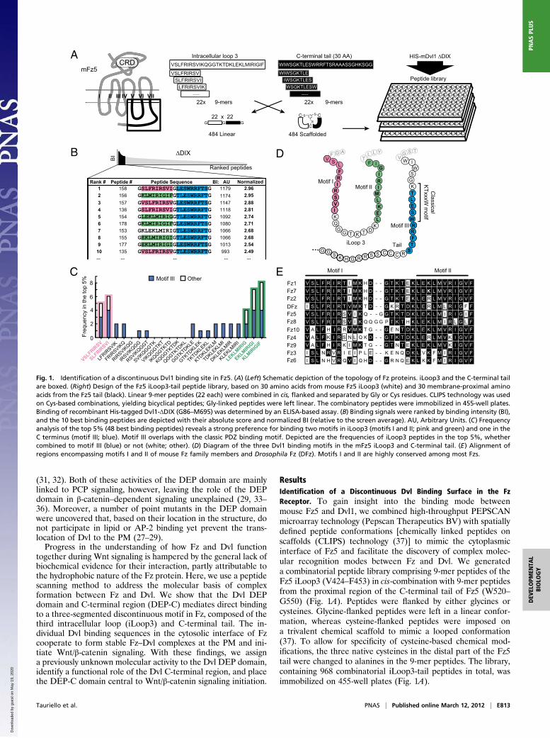
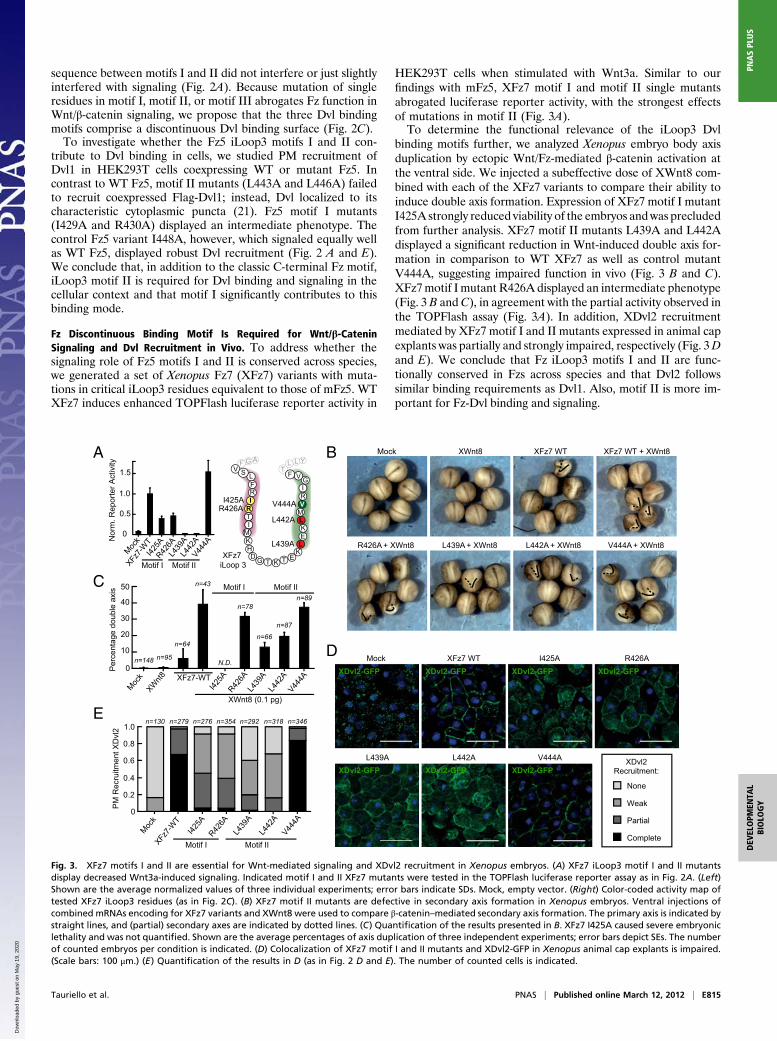
Fz Discontinuous Binding Motif Is Required for Wnt/β-CateninSignaling and Dvl Recruitment in Vivo. To address whether thesignaling role of Fz5 motifs I and II is conserved across species,we generated a set of Xenopus Fz7 (XFz7) variants with muta-tions in critical iLoop3 residues equivalent to those of mFz5. WTXFz7 induces enhanced TOPFlash luciferase reporter activity in
HEK293T cells when stimulated with Wnt3a. Similar to ourfindings with mFz5, XFz7 motif I and motif II single mutantsabrogated luciferase reporter activity, with the strongest effectsof mutations in motif II (Fig. 3A).To determine the functional relevance of the iLoop3 Dvl
binding motifs further, we analyzed Xenopus embryo body axisduplication by ectopic Wnt/Fz-mediated β-catenin activation atthe ventral side. We injected a subeffective dose of XWnt8 com-bined with each of the XFz7 variants to compare their ability toinduce double axis formation. Expression of XFz7 motif I mutantI425A strongly reduced viability of the embryos andwas precludedfrom further analysis. XFz7 motif II mutants L439A and L442Adisplayed a significant reduction in Wnt-induced double axis for-mation in comparison to WT XFz7 as well as control mutantV444A, suggesting impaired function in vivo (Fig. 3 B and C).XFz7motif I mutant R426A displayed an intermediate phenotype(Fig. 3 B and C), in agreement with the partial activity observed inthe TOPFlash assay (Fig. 3A). In addition, XDvl2 recruitmentmediated by XFz7 motif I and II mutants expressed in animal capexplants was partially and strongly impaired, respectively (Fig. 3Dand E). We conclude that Fz iLoop3 motifs I and II are func-tionally conserved in Fzs across species and that Dvl2 followssimilar binding requirements as Dvl1. Also, motif II is more im-portant for Fz-Dvl binding and signaling.
A B
C
D
E
Per
cent
age
doub
le a
xis
0
10
20
30
40
50
XWnt8 (0.1 pg)
XFz7-WT
N.D.
Motif IXFz7
iLoop 3
Mock XWnt8 XFz7 WT XFz7 WT + XWnt8
R426A + XWnt8 L439A + XWnt8 V444A + XWnt8L442A + XWnt8
I425AR426A
L439A
L442A
V444A
Motif II
Motif I
n=148 n=95
n=43
n=64
n=78
n=66
n=87
n=89Motif II
0
0.5
1.0
1.5
Nor
m. R
epor
ter A
ctiv
ity
FL
RI
D G T
RTI
H
L
M
M
R
K
K
KK
E
LE
T
I
V
G
LL YT
GF AV
FS V
L439
A
L442
AV44
4A
Mock
Mock
Mock
XWnt8
XFz7-
WT
I425A
R426A
L439
AL4
42A
V444A
I425A
R426A
L439
AL4
42A
V444A
XFz7-
WT
I425A
R426A
Motif IIMotif I
PM
Rec
ruitm
ent X
Dvl
2
0
0.2
0.4
0.6
0.8
1.0n=279 n=276 n=354 643=n813=n292=n031=n
Mock XFz7 WT R426AI425A
L439A V444AL442A
XDvl2-GFP XDvl2-GFP XDvl2-GFP XDvl2-GFP
XDvl2-GFPXDvl2-GFPXDvl2-GFP
Complete
Partial
Weak
None
XDvl2Recruitment:
Fig. 3. XFz7 motifs I and II are essential for Wnt-mediated signaling and XDvl2 recruitment in Xenopus embryos. (A) XFz7 iLoop3 motif I and II mutantsdisplay decreased Wnt3a-induced signaling. Indicated motif I and II XFz7 mutants were tested in the TOPFlash luciferase reporter assay as in Fig. 2A. (Left)Shown are the average normalized values of three individual experiments; error bars indicate SDs. Mock, empty vector. (Right) Color-coded activity map oftested XFz7 iLoop3 residues (as in Fig. 2C). (B) XFz7 motif II mutants are defective in secondary axis formation in Xenopus embryos. Ventral injections ofcombined mRNAs encoding for XFz7 variants and XWnt8 were used to compare β-catenin–mediated secondary axis formation. The primary axis is indicated bystraight lines, and (partial) secondary axes are indicated by dotted lines. (C) Quantification of the results presented in B. XFz7 I425A caused severe embryoniclethality and was not quantified. Shown are the average percentages of axis duplication of three independent experiments; error bars depict SEs. The numberof counted embryos per condition is indicated. (D) Colocalization of XFz7 motif I and II mutants and XDvl2-GFP in Xenopus animal cap explants is impaired.(Scale bars: 100 μm.) (E) Quantification of the results in D (as in Fig. 2 D and E). The number of counted cells is indicated.
Tauriello et al. PNAS | Published online March 12, 2012 | E815
DEV
ELOPM
ENTA
LBIOLO
GY
PNASPL
US
Dow
nloa
ded
by g
uest
on
May
19,
202
0
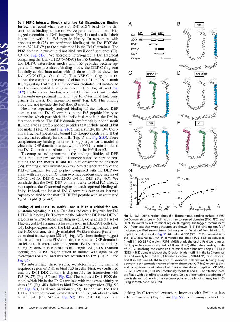
Dvl1 DEP-C Interacts Directly with the Fz5 Discontinuous BindingSurface. To reveal what region of Dvl1-ΔDIX binds to the dis-continuous binding surface on Fz, we generated additional His-tagged recombinant Dvl1 fragments (Fig. 4A) and studied theirinteraction with the Fz5 peptide library. In agreement withprevious work (23), we confirmed binding of the Dvl PDZ do-main (S201–P375) to the classic motif in the Fz5 C terminus. ThePDZ domain, however, did not bind any iLoop3 sequence (Fig.4B and Fig. S1A). We therefore interrogated a Dvl fragmentcomprising the DEP-C (R376–M695) for Fz5 binding. Strikingly,two DEP-C interaction modes with Fz5 peptides became ap-parent. In one prominent binding mode, the DEP-C fragmentfaithfully copied interaction with all three motifs as shown forDvl1-ΔDIX (Figs. 1D and 4C). This DEP-C binding mode re-quired the combined presence of either motif I or II with motifIII, suggesting that the DEP-C domain mediates Dvl binding tothe three-segmented binding surface on Fz5 (Fig. 4C and Fig.S1B). In the second binding mode, DEP-C interacts with a shif-ted membrane-proximal motif in the Fz C-terminal tail, com-prising the classic Dvl interaction motif (Fig. 4D). This bindingmode did not include the Fz5 iLoop3 motifs.Next, we separately analyzed binding of the isolated DEP
domain and the Dvl C terminus to the Fz5 peptide library todetermine which part binds the individual motifs in the Fz5 in-teraction surface. The DEP domain preferentially bound motifIII with a weak preference for peptides that include motif II butnot motif I (Fig. 4E and Fig. S1C). Interestingly, the Dvl C-ter-minal fragment specifically bound Fz5 iLoop3 motifs I and II butentirely lacked affinity for motif III (Fig. 4F and Fig. S1D). Thesecomplementary binding patterns strongly argue for a model inwhich the DEP domain interacts with the Fz5 C-terminal tail andthe Dvl C terminus mediates binding to the Fz5 iLoop3.To compare and approximate the binding affinities of DEP
and DEP-C for Fz5, we used a fluorescein-labeled peptide con-taining the Fz5 motifs II and III in fluorescence polarization(38). Binding curves indicate a 2- to 2.5-fold higher affinity of theDEP-C fragment for Fz5 peptide compared with the DEP do-main, with an apparent Kd from two independent experiments of6.5–12 μM for DEP-C vs. 22–30 μM for DEP (Fig. 4G). Weconclude that the Dvl1 DEP domain is able to bind Fz5 directlybut requires the C-terminal region to attain optimal binding af-finity. Indeed, the isolated Dvl C terminus carries an intrinsiccapacity to bind to the motif II-III Fz5 peptide with an estimatedKd of 13 μM (Fig. 4H).
Binding of Dvl DEP-C to Motifs I and II in Fz Is Critical for Wnt/β-Catenin Signaling in Cells. Our data indicate a key role for DvlDEP-C in binding Fz. To examine the role of theDEP andDEP-Cregions in Wnt/β-catenin signaling in cells, we generated a set ofFlag-taggedDvl1 fragments for expression inHEK293T cells (Fig.5A). Ectopic expression of theDEPandDEP-C fragments, but notthe PDZ domain, strongly inhibited Wnt3a-induced β-catenin–dependent transcription (28, 29) (Fig. 5B). These findings suggestthat in contrast to the PDZ domain, the isolated DEP domain issufficient to interfere with endogenous Fz-Dvl binding and sig-naling. Moreover, in contrast to full-length Dvl1, a Dvl1 variantlacking the DEP-C region failed to induce Wnt signaling onoverexpression (39) and was not recruited to Fz5 (Fig. 5C andFig. S2).To substantiate these results, we determined the minimal
required region of Dvl1 to bind Fz5 in cells. First, we confirmedthat the Dvl1 DIX domain is dispensable for interaction withFz5 (9, 27) (Fig. 5C and Fig. S2). The isolated Dvl1 PDZ do-main, which binds the Fz C terminus with moderate affinity invitro (23) (Fig. 4B), failed to bind Fz5 on coexpression (Fig. 5Cand Fig. S2), as shown previously (29). In contrast, the Dvl1DEP-C fragment robustly colocalized with Fz5, identical to full-length Dvl1 (Fig. 5C and Fig. S2). The Dvl1 DEP domain,
lacking its C-terminal extension, interacts with Fz5 in a lessefficient manner (Fig. 5C and Fig. S2), confirming a role of the
GH
A
C
GG
K
FT
RRCCCSSS
SR
E
L
E
M
R
K
SW
EL
G
RR
I
I
TK
∆DIX
Dvl1PDZ DEP
His
His
His
His
His
DEP-C
C-region
PDZ
DEP
DIX
FRI
GG T
R
SV
Q
IK
K
KD
L
T
WS
L
GFA
V SG
I
LLY
TF I
SGT
VW
DEP
L
E
M
R
K ES
L
FT
R
S
L
I
I
G
G
GG
HK RRCCCSSS R
TK
WR
FRI
GG T
RSV
Q
IK
K
KDT
SW
G
I
LLY
TFL
GFA
V S I
SGT
VW
DEP-C [1]
F
W
B
DG
GG
HK
I
FT
RR
RRCCCSSS
SR
F
L
RI
GG T
R
SV
Q
L
I
M
R
I
K
K
K
KD
LE
T
I
I
G
S
ES
L
G
W
TK
LLY
TGF
A
VSG
T
VF WS
G
GG
HK
FT
RRCCCSSS
SR
L
E
M
R
K
SW
EL
G
RR
I
I
TK
FRI
GG T
RSV
Q
IK
K
KD
L
T
WS
L
GFA
V SG
I
LLY
TF I
SGT
VW
C
G
GG
HK RRSSS
F
L
RI
GG T
R
SV
Q
L
I
M
R
I
K
K
K
KD
LE
T
I
I
G
LLY
TGF
A
VFS
ES
L
FT
R
SCC R
TK
WR
SW
G
I
SGT
VW
DEP-C [2]
PDZ
G
Fluo
resc
ence
pol
ariz
atio
n (m
P)
H
[Dvl fragment] (µM)00
40
80
120
160 C-tail
10 20 30 40
DEP-C DEP
[Dvl fragment] (µM)0
40
80
120
160
20 40 60 80 100
Fig. 4. Dvl1 DEP-C region binds the discontinuous binding surface in Fz5.(A) Domain structure of Dvl1 with three conserved domains (DIX, PDZ, andDEP), followed by a C-terminal region (C-region). His-tagged recombinantDvl1 fragments that were generated are shown. (B–E) Fz5 binding motifs ofindicated purified recombinant Dvl fragments. Details of best binding Fzpeptides are described in Fig. S1. (B) Isolated PDZ (S201–P375) domain bindsthe Fz C-terminal tail, which comprises the classic PDZ binding sequence(motif III). (C) DEP-C region (R376–M695) binds the entire Fz discontinuousbinding surface comprising motifs I, II, and III. (D) Alternative binding modeof DEP-C, involving the classic Fz C-terminal motif but not iLoop3. (E) DEP(S393–N503) domain without the C-region binds motif III in the Fz C-terminaltail and weakly to motif II. (F) Isolated C-region (L500–M695) binds motifs Iand II in Fz5 iLoop3. (G) In vitro fluorescence polarization binding assaybetween a concentration range of recombinant DEP-C (filled) or DEP (open)and a cysteine-maleimide–linked fluorescein-labeled peptide (CKLMIR-IGIFGTLESWRRFTG, 100 nM) combining motifs II and III. The titration dataare fitted with a binding saturation curve. One representative experiment oftwo is shown. (H) In vitro fluorescence polarization binding assay (as in G)using recombinant Dvl C-tail.
E816 | www.pnas.org/cgi/doi/10.1073/pnas.1114802109 Tauriello et al.
Dow
nloa
ded
by g
uest
on
May
19,
202
0
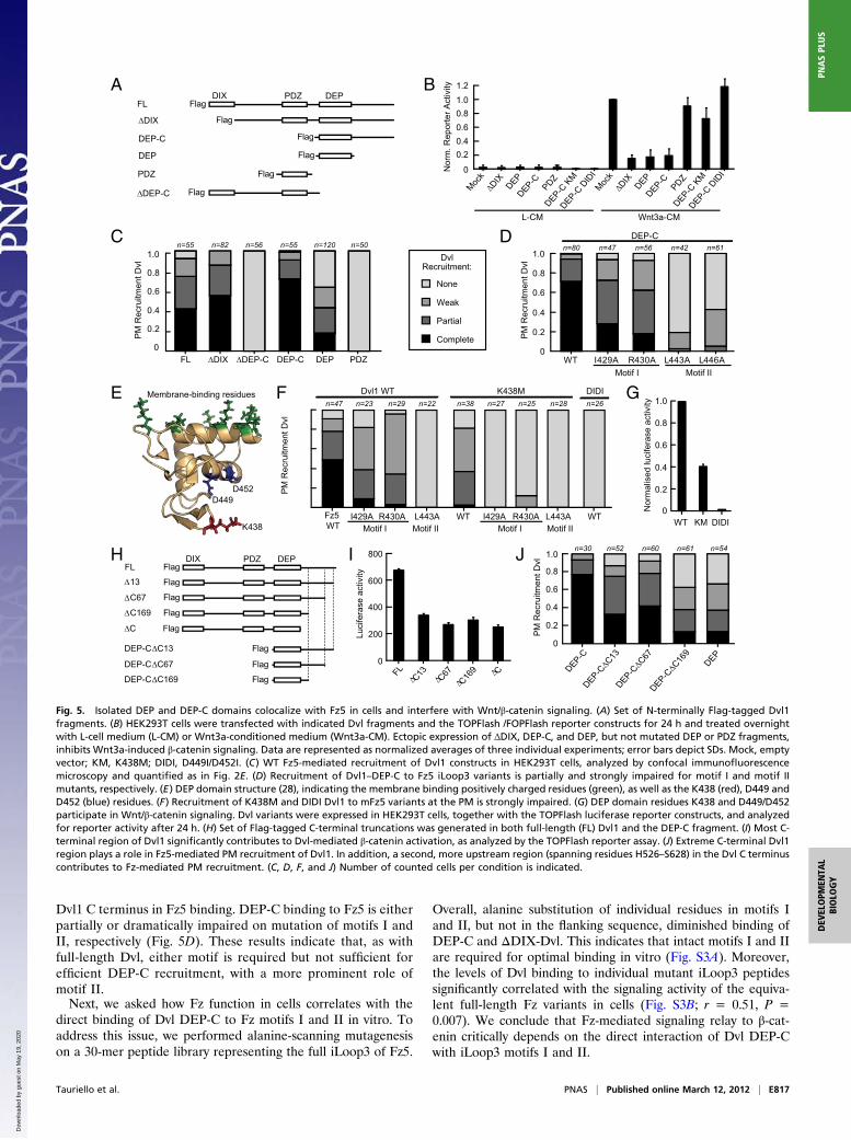
Dvl1 C terminus in Fz5 binding. DEP-C binding to Fz5 is eitherpartially or dramatically impaired on mutation of motifs I andII, respectively (Fig. 5D). These results indicate that, as withfull-length Dvl, either motif is required but not sufficient forefficient DEP-C recruitment, with a more prominent role ofmotif II.Next, we asked how Fz function in cells correlates with the
direct binding of Dvl DEP-C to Fz motifs I and II in vitro. Toaddress this issue, we performed alanine-scanning mutagenesison a 30-mer peptide library representing the full iLoop3 of Fz5.
Overall, alanine substitution of individual residues in motifs Iand II, but not in the flanking sequence, diminished binding ofDEP-C and ΔDIX-Dvl. This indicates that intact motifs I and IIare required for optimal binding in vitro (Fig. S3A). Moreover,the levels of Dvl binding to individual mutant iLoop3 peptidessignificantly correlated with the signaling activity of the equiva-lent full-length Fz variants in cells (Fig. S3B; r = 0.51, P =0.007). We conclude that Fz-mediated signaling relay to β-cat-enin critically depends on the direct interaction of Dvl DEP-Cwith iLoop3 motifs I and II.
A
DC
BFL
∆DIX Flag
Flag
DEP-C
DEP
Flag
PDZ Flag
Flag
Mock
∆DIX
DEPDEP-C
DEP-C K
MDEP-C
DID
I
PDZ
Mock
∆DIX
DEPDEP-C
DEP-C K
MDEP-C
DID
I
PDZ
MC-a3tnWMC-L
0
0.20.40.60.81.01.2
Nor
m. R
epor
ter A
ctiv
ity
Flag∆DEP-C
FL ∆DIX DEP-C
DEP-C
DEP PDZ∆DEP-CMotif IIMotif I
Complete
Partial
Weak
None
DvlRecruitment:
PM
Rec
ruitm
ent D
vl
PM
Rec
ruitm
ent D
vl
PDZ DEPDIX
H JIFL
∆13
∆C67
∆C169
Flag
Flag
Flag
Flag
∆C Flag
Flag
Flag
Flag
DEP-C∆C13
DEP-C∆C67
DEP-C∆C169
PDZ DEPDIX
0
200
400
600
ytivitcaes areficu L
amro
Nsarefic ul
desilytivitca
e
800
FL
∆C13
∆C67
∆C16
9 ∆C DEP-CDEP-C
∆C13
DEP-C∆C
67DEP-C
∆C16
9
DEP
0
0.2
0.4
0.6
0.8
1.0
PM
Rec
ruitm
ent D
vl
F
I429A R430A L443A Fz5 WT
TWTW I429A R430A L443A
IDIDM834KTW1lvD
PM
Rec
ruitm
ent D
vl
G
K438
D449D452
Membrane-binding residues
0
0.4
0.6
0.2
0.8
1.0
WT KM DIDI
E
Motif II IIfitoMIfitoM Motif I
n=30 n=52 n=60 n=61 n=54
0
0.2
0.4
0.6
0.8
1.0n=55 n=82 n=55 n=50n=56 n=120
0
0.2
0.4
0.6
0.8
1.0n=47n=80 n=56 n=42 n=61
WT I429A R430A L443A L446A
0.2
0.4
0.6
0.8
n=47 n=23 n=29 n=22 n=38 n=27 n=25 n=28 n=26
Fig. 5. Isolated DEP and DEP-C domains colocalize with Fz5 in cells and interfere with Wnt/β-catenin signaling. (A) Set of N-terminally Flag-tagged Dvl1fragments. (B) HEK293T cells were transfected with indicated Dvl fragments and the TOPFlash /FOPFlash reporter constructs for 24 h and treated overnightwith L-cell medium (L-CM) or Wnt3a-conditioned medium (Wnt3a-CM). Ectopic expression of ΔDIX, DEP-C, and DEP, but not mutated DEP or PDZ fragments,inhibits Wnt3a-induced β-catenin signaling. Data are represented as normalized averages of three individual experiments; error bars depict SDs. Mock, emptyvector; KM, K438M; DIDI, D449I/D452I. (C) WT Fz5-mediated recruitment of Dvl1 constructs in HEK293T cells, analyzed by confocal immunofluorescencemicroscopy and quantified as in Fig. 2E. (D) Recruitment of Dvl1–DEP-C to Fz5 iLoop3 variants is partially and strongly impaired for motif I and motif IImutants, respectively. (E) DEP domain structure (28), indicating the membrane binding positively charged residues (green), as well as the K438 (red), D449 andD452 (blue) residues. (F) Recruitment of K438M and DIDI Dvl1 to mFz5 variants at the PM is strongly impaired. (G) DEP domain residues K438 and D449/D452participate in Wnt/β-catenin signaling. Dvl variants were expressed in HEK293T cells, together with the TOPFlash luciferase reporter constructs, and analyzedfor reporter activity after 24 h. (H) Set of Flag-tagged C-terminal truncations was generated in both full-length (FL) Dvl1 and the DEP-C fragment. (I) Most C-terminal region of Dvl1 significantly contributes to Dvl-mediated β-catenin activation, as analyzed by the TOPFlash reporter assay. (J) Extreme C-terminal Dvl1region plays a role in Fz5-mediated PM recruitment of Dvl1. In addition, a second, more upstream region (spanning residues H526–S628) in the Dvl C terminuscontributes to Fz-mediated PM recruitment. (C, D, F, and J) Number of counted cells per condition is indicated.
Tauriello et al. PNAS | Published online March 12, 2012 | E817
DEV
ELOPM
ENTA
LBIOLO
GY
PNASPL
US
Dow
nloa
ded
by g
uest
on
May
19,
202
0
We noted that within the 30-mer iLoop3 sequence, the I448Amutant behaved as a single outlier in this analysis. The I448Amutation diminished Dvl binding in vitro but, in the context ofboth the full-length Fz5 and XFz7 receptors, displayed robustDvl recruitment and signaling in vivo (Figs. 2 A and E and 3 Cand E, and Fig. S3A). We speculate that the reduction of Dvlbinding by I448A may be compensated for by interactionsthrough other regions or residues in full-length Fz5.
Identification of Critical Regions in the DEP-C Fragment for Fz Bindingand Wnt/β-Catenin Signaling. Which residues at the surface of theDEP domain participate in Fz binding? Based on interactions ofthe DEP domain with lipid and protein partners at the PM (30–32), we reasoned that unoccupied surface-exposed DEP domainresidues may mediate binding to Fz. A number of DEP domainvariants, including the K438M mutation and the combinedD449I and D452I mutations, were previously proposed to impairFz-dependent PM recruitment (23, 29, 32) (Fig. 5E). Binding ofDvl1 K438M to WT Fz5 was strongly decreased, and the in-teraction was entirely lost in combination with Fz5 motif I/IImutants (Fig. 5F). Concurrently, the K438M Dvl1 mutant dis-plays significantly decreased signaling activity (Fig. 5G). Muta-tion of D449/D452 (DIDI) fully abrogates both Fz5-mediatedPM recruitment and signaling, preventing further analysis of thecontribution of Fz motifs I and II (Fig. 5 F and G). Isolated Dvl1DEP-C fragments with either of these mutations fail to inhibitWnt3a-induced signal relay, confirming their inability to interactand interfere with the activity of endogenous Wnt signalingcomponents (Fig. 5B). We conclude that Fz–DEP-C bindingrequires the cooperative use of Dvl DEP K438 and Fz motifs Iand II at the binding interface to mediate PM recruitment of Dvland Wnt/β-catenin signaling in cells.To determine which regions in the Dvl1 C terminus are im-
portant for Fz association and Dvl signaling, we generateda number of progressive C-terminal truncations in both full-length Dvl1 and the Dvl1 DEP-C fragment (Fig. 5H). Loss of theextreme 13 amino acids (Dvl1ΔC13), which locate to a highlyconserved region in all three Dvl isoforms, reduced the signalingcapability of Dvl1 to the level of Dvl1ΔC. This indicates that theconserved C-terminal region contains important residues for Dvlsignaling (Fig. 5I). Indeed, truncation of the terminal 13 aminoacids from the DEP-C fragment resulted in reduced binding toFz5 (Fig. 5J). Further truncation of the C-terminal region di-minished the interaction with Fz5 even more, pointing to a sec-ond Fz binding region in the Dvl1 C terminus (Fig. 5J).
DiscussionFz and Dvl act together in the control of Wnt-induced β-catenin–dependent and –independent pathways, but the underlying mo-lecular mechanisms remain poorly understood (5, 40). A thor-ough understanding of the mechanism of Fz–Dvl complexformation is crucial to clarify how downstream signaling eventsare controlled. Moreover, Fz-Dvl interactions provide an at-tractive target for the design and development of drugs that in-terfere with Wnt signaling activation (24, 41–43). Although theDvl PDZ domain was shown to bind an Fz C-terminal tail-derived peptide in vitro (9, 23), evidence for in vivo binding ofthe Dvl PDZ domain to Fz remains scarce. In addition, the af-finity of this interaction is insufficient to explain the formation ofa stable Fz–Dvl complex in cells, leading to the suggestion thatadditional intermolecular interactions are required (30, 32).Here, we use a combinatorial peptide library that mimics the
cytoplasmic surface of the Fz5 iLoop3 and C terminus to eluci-date molecular recognition modes between Fz5 and Dvl1. Weuncover a conserved binding mode in which Dvl interacts witha discontinuous binding site in Fz. Our results argue for co-operative binding of Dvl to the Fz C-terminal motif III andiLoop3 motifs I and II based on the following observations. First,
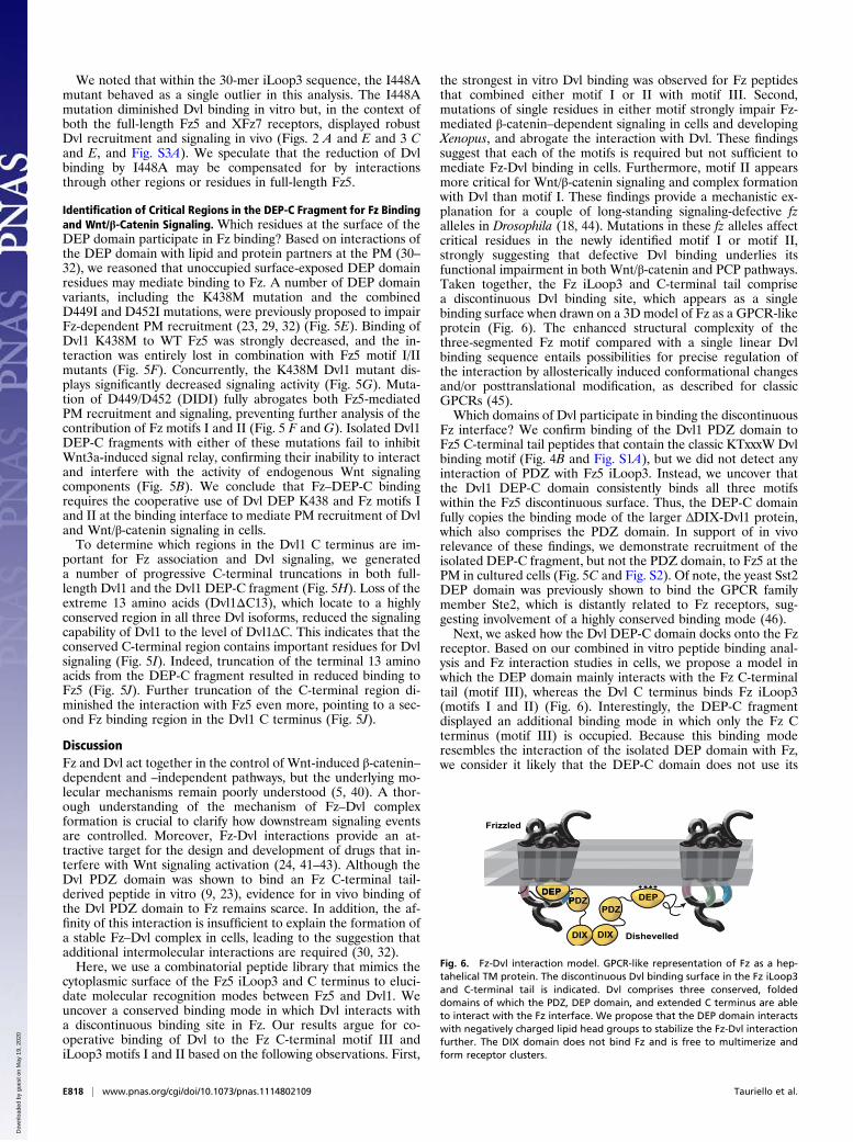
the strongest in vitro Dvl binding was observed for Fz peptidesthat combined either motif I or II with motif III. Second,mutations of single residues in either motif strongly impair Fz-mediated β-catenin–dependent signaling in cells and developingXenopus, and abrogate the interaction with Dvl. These findingssuggest that each of the motifs is required but not sufficient tomediate Fz-Dvl binding in cells. Furthermore, motif II appearsmore critical for Wnt/β-catenin signaling and complex formationwith Dvl than motif I. These findings provide a mechanistic ex-planation for a couple of long-standing signaling-defective fzalleles in Drosophila (18, 44). Mutations in these fz alleles affectcritical residues in the newly identified motif I or motif II,strongly suggesting that defective Dvl binding underlies itsfunctional impairment in both Wnt/β-catenin and PCP pathways.Taken together, the Fz iLoop3 and C-terminal tail comprisea discontinuous Dvl binding site, which appears as a singlebinding surface when drawn on a 3D model of Fz as a GPCR-likeprotein (Fig. 6). The enhanced structural complexity of thethree-segmented Fz motif compared with a single linear Dvlbinding sequence entails possibilities for precise regulation ofthe interaction by allosterically induced conformational changesand/or posttranslational modification, as described for classicGPCRs (45).Which domains of Dvl participate in binding the discontinuous
Fz interface? We confirm binding of the Dvl1 PDZ domain toFz5 C-terminal tail peptides that contain the classic KTxxxW Dvlbinding motif (Fig. 4B and Fig. S1A), but we did not detect anyinteraction of PDZ with Fz5 iLoop3. Instead, we uncover thatthe Dvl1 DEP-C domain consistently binds all three motifswithin the Fz5 discontinuous surface. Thus, the DEP-C domainfully copies the binding mode of the larger ΔDIX-Dvl1 protein,which also comprises the PDZ domain. In support of in vivorelevance of these findings, we demonstrate recruitment of theisolated DEP-C fragment, but not the PDZ domain, to Fz5 at thePM in cultured cells (Fig. 5C and Fig. S2). Of note, the yeast Sst2DEP domain was previously shown to bind the GPCR familymember Ste2, which is distantly related to Fz receptors, sug-gesting involvement of a highly conserved binding mode (46).Next, we asked how the Dvl DEP-C domain docks onto the Fz
receptor. Based on our combined in vitro peptide binding anal-ysis and Fz interaction studies in cells, we propose a model inwhich the DEP domain mainly interacts with the Fz C-terminaltail (motif III), whereas the Dvl C terminus binds Fz iLoop3(motifs I and II) (Fig. 6). Interestingly, the DEP-C fragmentdisplayed an additional binding mode in which only the Fz Cterminus (motif III) is occupied. Because this binding moderesembles the interaction of the isolated DEP domain with Fz,we consider it likely that the DEP-C domain does not use its
PDZPDZ
DEP+ + + +
DIX Dishevelled
P+ + + +DEP
Frizzled
DIX
Fig. 6. Fz-Dvl interaction model. GPCR-like representation of Fz as a hep-tahelical TM protein. The discontinuous Dvl binding surface in the Fz iLoop3and C-terminal tail is indicated. Dvl comprises three conserved, foldeddomains of which the PDZ, DEP domain, and extended C terminus are ableto interact with the Fz interface. We propose that the DEP domain interactswith negatively charged lipid head groups to stabilize the Fz-Dvl interactionfurther. The DIX domain does not bind Fz and is free to multimerize andform receptor clusters.
E818 | www.pnas.org/cgi/doi/10.1073/pnas.1114802109 Tauriello et al.
Dow
nloa
ded
by g
uest
on
May
19,
202
0
C-terminal extension in this alternative orientation. Of note,a number of Fz family members, including Fz3 and Fz6, displayloss of sequence conservation within the region of motif I. Wespeculate that weakening or alteration in Dvl binding to motif Iin these Fzs may affect the orientation of Dvl in the bound state.Both Fz3 and Fz6 were previously linked to Wnt-inducedβ-catenin–independent PCP signaling (47). Thus, alternative Dvlbinding modes may steer selective downstream signaling events.Further probing of the Dvl interaction surface revealed that
a number of surface-exposed residues (K438 and D449/D452) onthe DEP domain as well as the last 13 residues of the C terminusare critical for functional interaction with Fz. Of note, based onsecondary structure prediction, the entire Dvl C region is pre-dicted to be intrinsically disordered, except for the last highlyconserved 15–20 amino acids, which may fold and act as a func-tional terminal unit at the highly flexible Dvl C terminus.How does the DEP domain accommodate different molecular
interactions implicated in membrane recruitment? Recently,a positively charged stretch in the DEP domain was shown tointeract directly with negatively charged lipids at the PM, sug-gesting a prominent role of lipid binding by the DEP domain inFz-mediated signaling (30). Because membrane recruitment ofDEP and DEP-C fragments codepends on the presence of Fzreceptors (Fig. S2), we propose that the Dvl DEP-C domain maysimultaneously bind lipids as well as the three motifs in the Fzreceptor to stabilize the interaction (Fig. 6). Moreover, Fz in-tracellular loops 1 and 2 may contribute to Dvl binding as well.Taken together, we suspect that the affinity of DEP-C for Fzbinding in vivo may be significantly increased through avidityeffects of the local concentration of Dvl binding sites.We conclude that the critical role of the Dvl1 DEP-C region in
Wnt/β-catenin signaling is mediated through its direct interactionwith a discontinuous binding surface on the Fz5 receptor. Ourwork suggests that Dvl employs multiple contact points with Fzand that each interaction is required for efficient downstreamsignaling to β-catenin. Whether binding events that involve thePDZ or DEP-C domain act cooperatively or sequentially awaitsfurther investigation. We speculate that the DEP-C domain maybe dominant over PDZ binding based on its expected higheraffinity for Fz in vivo. Alternatively, simultaneous binding of bothPDZ and DEP-C may allow a single Dvl molecule to forma complex with two Fzs.We propose that the individual interactions may be subject to
regulation by conformational changes and posttranslationalmodifications. Because Fz-Dvl signaling is implicated in at leasttwo important Wnt pathways, detailed studies aimed at un-derstanding how this interaction is induced and regulated byWnt stimulation will help to explain how the Fz-Dvl functionalunit manages to interpret upstream signals to disparate down-stream pathways.
MethodsCell Culture and Transfection. HEK293T cells and mouse L cells were culturedin RPMI or DMEM (Invitrogen), respectively, supplemented with 10% (vol/vol) FBS (Sigma), 100 units/mL penicillin, and 100 μg/mL streptomycin. Lcells stably expressing Wnt3a have been described previously (39). Trans-fections were performed with FuGENE 6 (Roche) according to themanufacturer’s protocol.
Constructs and Antibodies. V5-Fz5, Flag-Dvl1 variants, TOPFlash, and controlFOPFlash luciferase reporter plasmids have been described previously (39).XWnt8 DNA and XFz7-myc were kind gifts of Randy Moon (University ofWashington, Howard Hughes Medical Institute, Seattle, WA) and HerbertSteinbeisser (Heidelberg University Hospital, Institute of Human Genetics,Heidelberg, Germany), respectively. Alanine substitutions were generatedby site-directed mutagenesis. Flag- or His-Dvl1 constructs were PCR-subcl-oned into pcDNA3 or a modified pET-24a(+) vector (Novagen), as describedpreviously (48). For immunoblotting and immunofluorescence, mouse anti-
Flag (M2; Sigma), mouse anti-actin (C4; MP Biomedicals), mouse anti-V5(Invitrogen), or rabbit anti-V5 (Sigma) was used.
TOPFlash Reporter Assays. HEK293T cells were seeded in 24-well platesand transfected with 30 ng of reporter construct TOPFlash or FOPFlash, 5 ngof thymidine kinase (TK)-Renilla, 50 ng of Dvl constructs, 35 ng of Fz con-structs, and mock plasmid to a total of 250 ng per well. The assay was per-formed as described previously (39).
Confocal Microscopy. HEK293T cells were grown on glass coverslips in 24-wellplates (coated with laminin). Cells were transfected andfixed after 24 h in 4%(mass/vol) paraformaldehyde or ice-cold methanol, treated with blockingbuffer [2% (mass/vol) BSA, 0.01% saponin in PBS], and incubated with pri-mary antibodies, followed by secondary antibodies conjugated to Alexa-488,Alexa-568, or Cy5. Cells were mounted in ProLong Gold (Invitrogen) andvisualized using a Zeiss LSM510 confocal microscope.
Protein Purification. His-tagged Dvl fragments were expressed in Rosetta2cells, induced at an OD600 of 0.6 with 0.5 mM isopropyl-β-D-thiogalactopyr-anoside for 6 h at 20 °C. Bacterial cell pellets were resuspended in buffercontaining 50 mM Tris (pH 7.5, 8.0, or 9.0), 300 mM NaCl, 5% (vol/vol)glycerol, 5 mM imidazole, 25 μM PMSF, 1 μg/mL DNase, 1 μg/mL RNase, 2 μg/mL lysozyme, and a 0.1-mL−1 tablet of EDTA-free protease inhibitor mixture(Roche); homogenized by French Press; and purified by ÄKTA purification,using a HisTrap Ni-NTA column (GE Healthcare). Proteins were eluted withan imidazole gradient, buffer-exchanged in 20 mM Tris and 150 mM NaClusing HiTrap and PD-10 desalting columns (GE Healthcare), and concen-trated with vivaspin 6 columns (Sartorius-stedim).
Peptide Library Generation and Screening. The linear and CLIPS peptides weresynthesized using standard Fmoc chemistry and deprotected using trifluoricacid with scavengers. Constrained peptides were synthesized on chemicalscaffolds using CLIPS technology through cysteine residues (37). The Cys sidechains were coupled to CLIPS templates by reacting onto credit-card formatpolypropylene PEPSCAN cards (Pepscan Therapeutics BV; 455 peptide for-mats per card) with a 0.5-mM solution of CLIPS template 1,3,5-Tris (bromo-methyl) benzene in ammonium bicarbonate [20 mM (pH 7.9)]/acetonitrile(1:1 vol/vol)]. The cards were gently shaken for 30–60 min. Finally, the cardswere washed extensively and sonicated in disrupt buffer containing 1% SDS/0.1% β-mercaptoethanol in PBS (pH 7.2) at 70 °C for 30 min, followed bysonication in H2O for another 45 min.
Protein binding was analyzed in a PEPSCAN-based ELISA (Pepscan Ther-apeutics BV). The 455-well cards containing the covalently linked peptideswere incubated with 1 μg/mL protein in blocking solution [4% (vol/vol) horseserum, 5% (mass/vol) ovalbumin, 1% Tween in PBS]. After washing, thepeptides were incubated with a monoclonal mouse anti-His tag antibody(1:1,000; Novagen) and, subsequently, after washing, with a peroxidase-conjugated rabbit–anti-mouse antibody (1:1,000; Southern Biotech). Afterwashing, the peroxidase substrate 2,2′-azino-di-3-ethylbenzthiazoline sul-fonate and 2 μL of 3% H2O2 were added. Color development was measuredand quantified with a CCD and an image processing system (49). Afterbackground correction, these raw data were ranked and the top 5% (48peptides) were analyzed.
Fluorescence Polarization (FP). CKLMIRIGIFGTLESWRRFTG peptide was syn-thesized using standard Fmoc chemistry and deprotected using trifluoric acidwith scavengers. The Cys side chain was labeled with fluorescein-maleimide.Peptide and purified recombinant proteins were diluted in FP buffer [50 mMTris (pH 7.5), 50 mM NaCl, 5 mM β-mercaptoethanol] to 100 nM and 100 μM,respectively. A protein dilution series to subnanomolar range was incubatedwith the peptide for 15–30 min. Fluorescence polarization of the labeledpeptide was analyzed in a Spectramax M5 plate reader (Molecular Devices).Data from the different incubation times were averaged and plotted. UsingSigmaplot (Systat), a binding saturation curve [Y = Bmax × X/(Kd + X)] wasfitted, with Y as the measured FP value (Fk − F∞)/(Fk + F∞), Bmax as themaximum value, and X as the Dvl fragment concentration. From this for-mula, the equilibrium constant, Kd, was derived as the protein concentrationat one-half saturation.
Xenopus Embryo Axis Duplication and Dvl Recruitment Assays. Mature eggswere obtained by injecting females with 500 units of human chorion go-nadotropin (Sigma). One hour after in vitro fertilization, the jelly coat wasremoved by treating the embryos with 2% (mass/vol) cysteine hydrochloridein 0.1× modified Barth’s solution (MBSH) at pH 8.2 [1× MBSH: 10 mM Hepes,
Tauriello et al. PNAS | Published online March 12, 2012 | E819
DEV
ELOPM
ENTA
LBIOLO
GY
PNASPL
US
Dow
nloa
ded
by g
uest
on
May
19,
202
0
88 mM NaCl, 1 mM KCl, 0.33 mM Ca(NO3)2, 0.41 mM CaCl2, 0.82 mM MgSO4,2.4 mM NaHCO3 (pH 7.4)]. Embryos were staged according to the method ofNieuwkoop and Faber (50). Capped mRNAs were transcribed from linearizedDNA templates with a mMessage mMachine kit (Ambion).
For axis duplication assays, embryos were injected at the four-cell stage inthemarginal zone of both ventral blastomereswith 0.1 pg of XWnt8 RNAplus400 pg of XFz7-myc RNAs in a volume of 8 nL and kept in 0.1×MBSH at 16 °Cuntil they had reached stage 21–22. For Dvl recruitment, embryos wereinjected at the two-cell stage in the animal pole of both cells with 200 pg ofXDvl2-GFP RNA and 200 pg of XFz7-myc RNAs and kept in 0.1× MBSH at16 °C until they reached stage 10. Embryos were fixed for 1 h in 4% (mass/mass) formaldehyde/1× MEM [100 mM 3-(N-morpholino)propanesulfonicacid (Mops) (pH 7.4), 2 mM EGTA, 1 mM MgSO4] at room temperature andfor an additional 4 h in DENT’s (80% methanol, 20% DMSO) at −20 °C. After
rehydration, the animal caps were cut and blocked in 20% (vol/vol) horseserum for 1 h. Animal cap explants were stained with anti-GFP (Abcam) andan Alexa-488–conjugated secondary antibody, and nuclei were counter-stained with Hoechst dye. Animal cap explants were mounted in Mowiol(Roth) and analyzed with a Zeiss Axio Imager D1/Z1.
ACKNOWLEDGMENTS. We thank Diana Stork for help with proteinpurification; Zeinab Anvarian, Matthijs Kol, and Maarten Egmond for helpwith FP experiments; and Marc Gentzel for critical reading of the manu-script. This work is supported by Dutch Cancer Society Grant UU 2006-3508(to M.M.M.), European Research Council Grant ERC-StG 242958 (to M.M.M.),a University of Utrecht High Potential Grant High Potential Grant (toM.M.M. and S.G.D.R.), and Deutsche Forschungsgemeinschaft Grant SCHA965/6-1 (to A.S.).
1. Logan CY, Nusse R (2004) The Wnt signaling pathway in development and disease.Annu Rev Cell Dev Biol 20:781–810.
2. Clevers H (2006) Wnt/beta-catenin signaling in development and disease. Cell 127:469–480.
3. Reya T, Clevers H (2005) Wnt signalling in stem cells and cancer. Nature 434:843–850.4. MacDonald BT, Tamai K, He X (2009) Wnt/beta-catenin signaling: Components,
mechanisms, and diseases. Dev Cell 17:9–26.5. Seifert JR, Mlodzik M (2007) Frizzled/PCP signalling: A conserved mechanism
regulating cell polarity and directed motility. Nat Rev Genet 8:126–138.6. van Amerongen R, Mikels A, Nusse R (2008) Alternative wnt signaling is initiated by
distinct receptors. Sci Signal 1:re9.7. Yang-Snyder J, Miller JR, Brown JD, Lai CJ, Moon RT (1996) A frizzled homolog
functions in a vertebrate Wnt signaling pathway. Curr Biol 6:1302–1306.8. Rothbächer U, et al. (2000) Dishevelled phosphorylation, subcellular localization and
multimerization regulate its role in early embryogenesis. EMBO J 19:1010–1022.9. Umbhauer M, et al. (2000) The C-terminal cytoplasmic Lys-thr-X-X-X-Trp motif in
frizzled receptors mediates Wnt/beta-catenin signalling. EMBO J 19:4944–4954.10. Sabharanjak S, Sharma P, Parton RG, Mayor S (2002) GPI-anchored proteins are
delivered to recycling endosomes via a distinct cdc42-regulated, clathrin-independentpinocytic pathway. Dev Cell 2:411–423.
11. Bhanot P, et al. (1996) A new member of the frizzled family from Drosophilafunctions as a Wingless receptor. Nature 382:225–230.
12. Wang HY, Liu T, Malbon CC (2006) Structure-function analysis of Frizzleds. Cell Signal18:934–941.
13. Schulte G, Bryja V (2007) The Frizzled family of unconventional G-protein-coupledreceptors. Trends Pharmacol Sci 28:518–525.
14. Wu J, Klein TJ, Mlodzik M (2004) Subcellular localization of frizzled receptors,mediated by their cytoplasmic tails, regulates signaling pathway specificity. PLoS Biol2:E158.
15. Djiane A, Yogev S, Mlodzik M (2005) The apical determinants aPKC and dPatj regulateFrizzled-dependent planar cell polarity in the Drosophila eye. Cell 121:621–631.
16. Cong F, Schweizer L, Varmus H (2004) Wnt signals across the plasma membrane toactivate the beta-catenin pathway by forming oligomers containing its receptors,Frizzled and LRP. Development 131:5103–5115.
17. Wu J, Jenny A, Mirkovic I, Mlodzik M (2008) Frizzled-Dishevelled signaling specificityoutcome can be modulated by Diego in Drosophila. Mech Dev 125:30–42.
18. Povelones M, Howes R, Fish M, Nusse R (2005) Genetic evidence that Drosophilafrizzled controls planar cell polarity and Armadillo signaling by a commonmechanism. Genetics 171:1643–1654.
19. Schweizer L, Varmus H (2003) Wnt/Wingless signaling through beta-catenin requiresthe function of both LRP/Arrow and frizzled classes of receptors. BMC Cell Biol 4:4.
20. Bilic J, et al. (2007) Wnt induces LRP6 signalosomes and promotes dishevelled-dependent LRP6 phosphorylation. Science 316:1619–1622.
21. Schwarz-Romond T, et al. (2007) The DIX domain of Dishevelled confers Wnt signalingby dynamic polymerization. Nat Struct Mol Biol 14:484–492.
22. Kishida S, et al. (1999) DIX domains of Dvl and axin are necessary for proteininteractions and their ability to regulate beta-catenin stability. Mol Cell Biol 19:4414–4422.
23. Wong H-C, et al. (2003) Direct binding of the PDZ domain of Dishevelled toa conserved internal sequence in the C-terminal region of Frizzled. Mol Cell 12:1251–1260.
24. Zhang Y, et al. (2009) Inhibition of Wnt signaling by Dishevelled PDZ peptides. NatChem Biol 5:217–219.
25. Gao C, Chen Y-G (2010) Dishevelled: The hub of Wnt signaling. Cell Signal 22:717–727.26. Lee HJ, et al. (2010) Identification of transmembrane protein 88 (TMEM88) as
a dishevelled-binding protein. J Biol Chem 285:41549–41556.27. Axelrod JD, Miller JR, Shulman JM, Moon RT, Perrimon N (1998) Differential
recruitment of Dishevelled provides signaling specificity in the planar cell polarity andWingless signaling pathways. Genes Dev 12:2610–2622.
28. Wong HC, et al. (2000) Structural basis of the recognition of the dishevelled DEPdomain in the Wnt signaling pathway. Nat Struct Biol 7:1178–1184.
29. Pan WJ, et al. (2004) Characterization of function of three domains in dishevelled-1:DEP domain is responsible for membrane translocation of dishevelled-1. Cell Res 14:324–330.
30. Simons M, et al. (2009) Electrochemical cues regulate assembly of the Frizzled/Dishevelled complex at the plasma membrane during planar epithelial polarization.Nat Cell Biol 11:286–294.
31. Yu A, et al. (2007) Association of Dishevelled with the clathrin AP-2 adaptor isrequired for Frizzled endocytosis and planar cell polarity signaling. Dev Cell 12:129–141.
32. Yu A, Xing Y, Harrison SC, Kirchhausen T (2010) Structural analysis of the interactionbetween Dishevelled2 and clathrin AP-2 adaptor, a critical step in noncanonical Wntsignaling. Structure 18:1311–1320.
33. Boutros M, Paricio N, Strutt DI, Mlodzik M (1998) Dishevelled activates JNK anddiscriminates between JNK pathways in planar polarity and wingless signaling. Cell94:109–118.
34. Li L, et al. (1999) Dishevelled proteins lead to two signaling pathways. Regulation ofLEF-1 and c-Jun N-terminal kinase in mammalian cells. J Biol Chem 274:129–134.
35. Moriguchi T, et al. (1999) Distinct domains of mouse dishevelled are responsible forthe c-Jun N-terminal kinase/stress-activated protein kinase activation and the axisformation in vertebrates. J Biol Chem 274:30957–30962.
36. Penton A, Wodarz A, Nusse R (2002) A mutational analysis of dishevelled inDrosophila defines novel domains in the dishevelled protein as well as novelsuppressing alleles of axin. Genetics 161:747–762.
37. Timmerman P, Puijk WC, Meloen RH (2007) Functional reconstruction and syntheticmimicry of a conformational epitope using CLIPS technology. J Mol Recognit 20:283–299.
38. Jameson DM, Ross JA (2010) Fluorescence polarization/anisotropy in diagnostics andimaging. Chem Rev 110:2685–2708.
39. Tauriello DVF, et al. (2010) Loss of the tumor suppressor CYLD enhances Wnt/beta-catenin signaling through K63-linked ubiquitination of Dvl. Mol Cell 37:607–619.
40. Veeman MT, Axelrod JD, Moon RT (2003) A second canon. Functions and mechanismsof beta-catenin-independent Wnt signaling. Dev Cell 5:367–377.
41. Shan J, Shi D-L, Wang J, Zheng J (2005) Identification of a specific inhibitor of thedishevelled PDZ domain. Biochemistry 44:15495–15503.
42. Fujii N, et al. (2007) An antagonist of dishevelled protein-protein interactionsuppresses beta-catenin-dependent tumor cell growth. Cancer Res 67:573–579.
43. Mahindroo N, Punchihewa C, Bail AM, Fujii N (2008) Indole-2-amide basedbiochemical antagonist of Dishevelled PDZ domain interaction down-regulatesDishevelled-driven Tcf transcriptional activity. Bioorg Med Chem Lett 18:946–949.
44. Jones KH, Liu J, Adler PN (1996) Molecular analysis of EMS-induced frizzled mutationsin Drosophila melanogaster. Genetics 142:205–215.
45. Christopoulos A, Kenakin T (2002) G protein-coupled receptor allosterism andcomplexing. Pharmacol Rev 54:323–374.
46. Ballon DR, et al. (2006) DEP-domain-mediated regulation of GPCR signalingresponses. Cell 126:1079–1093.
47. Wang Y, Guo N, Nathans J (2006) The role of Frizzled3 and Frizzled6 in neural tubeclosure and in the planar polarity of inner-ear sensory hair cells. J Neurosci 26:2147–2156.
48. Noutsou M, et al. (2011) Critical scaffolding regions of the tumor suppressor Axin1 arenatively unfolded. J Mol Biol 405:773–786.
49. Slootstra JW, Puijk WC, Ligtvoet GJ, Langeveld JP, Meloen RH (1996) Structuralaspects of antibody-antigen interaction revealed through small random peptidelibraries. Mol Divers 1:87–96.
50. Nieuwkoop PD, Faber J (1994) Normal Table of Xenopus laevis (Daudin) (Garland,New York).
E820 | www.pnas.org/cgi/doi/10.1073/pnas.1114802109 Tauriello et al.
Dow
nloa
ded
by g
uest
on
May
19,
202
0