Why the IMQ-induced psoriasis model is a more …...Background and Pathology of Psoriasis Psoriasis...
10
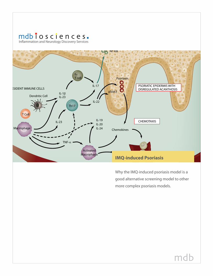
ESIDENT IMMUNE CELLS Dendritic Cell T Cell Macrophage Chemokines Activated Macrophage PSORIATIC EPIDERMIS WITH DISREGULATED ACANTHOSIS CHEMOTAXIS IL-1β IL-23 IL-17 IL-22 Th17 δγ T cell IL-19 pStat3 Psoriasin IL-20 IL-24 NF-kΒ IL-23 TNF-α Inflammation and Neurology Discovery Services IMQ-induced Psoriasis Why the IMQ-induced psoriasis model is a good alternative screening model to other more complex psoriasis models.
Transcript of Why the IMQ-induced psoriasis model is a more …...Background and Pathology of Psoriasis Psoriasis...
RESIDENT IMMUNE CELLS
Dendritic Cell
T Cell
Macrophage Chemokines
VEGFActivated
Macrophage
PSORIATIC EPIDERMIS WITHDISREGULATED ACANTHOSIS
NORMAL EPIDERMAL KERATINOCYTES
CHEMOTAXIS
ANGIOGENESIS
IL-1βIL-23
IL-17
IL-22Th17
δγ T cell
IL-19
pStat3
Psoriasin
IL-20IL-24
NF-kΒ
IL-23
TNF-α
PSORIATIC PLACQUE
Inflammation and Neurology Discovery Services
IMQ-induced Psoriasis
Why the IMQ-induced psoriasis model is a
good alternative screening model to other
more complex psoriasis models.
RESIDENT IMMUNE CELLS
Dendritic Cell
T Cell
Macrophage Chemokines
VEGFActivated
Macrophage
PSORIATIC EPIDERMIS WITHDISREGULATED ACANTHOSIS
NORMAL EPIDERMAL KERATINOCYTES
CHEMOTAXIS
ANGIOGENESIS
IL-1βIL-23
IL-17
IL-22Th17
δγ T cell
IL-19
pStat3
Psoriasin
IL-20IL-24
NF-kΒ
IL-23
TNF-α
PSORIATIC PLACQUE
Psoriasis
2 Whitepaper | IMQ-induced Psoriasis
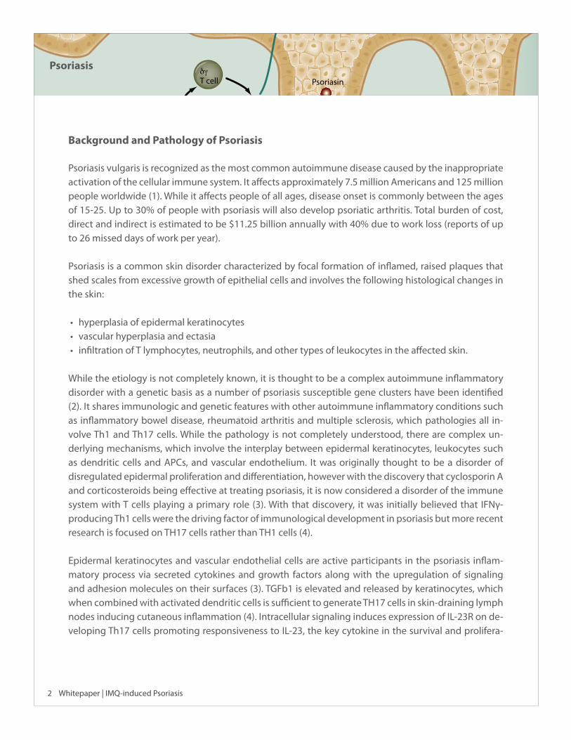
Background and Pathology of Psoriasis
Psoriasis vulgaris is recognized as the most common autoimmune disease caused by the inappropriate activation of the cellular immune system. It affects approximately 7.5 million Americans and 125 million people worldwide (1). While it affects people of all ages, disease onset is commonly between the ages of 15-25. Up to 30% of people with psoriasis will also develop psoriatic arthritis. Total burden of cost, direct and indirect is estimated to be $11.25 billion annually with 40% due to work loss (reports of up to 26 missed days of work per year).
Psoriasis is a common skin disorder characterized by focal formation of inflamed, raised plaques that shed scales from excessive growth of epithelial cells and involves the following histological changes in the skin:
hyperplasia of epidermal keratinocytes•vascular hyperplasia and ectasia•infiltration of T lymphocytes, neutrophils, and other types of leukocytes in the affected skin. •
While the etiology is not completely known, it is thought to be a complex autoimmune inflammatory disorder with a genetic basis as a number of psoriasis susceptible gene clusters have been identified (2). It shares immunologic and genetic features with other autoimmune inflammatory conditions such as inflammatory bowel disease, rheumatoid arthritis and multiple sclerosis, which pathologies all in-volve Th1 and Th17 cells. While the pathology is not completely understood, there are complex un-derlying mechanisms, which involve the interplay between epidermal keratinocytes, leukocytes such as dendritic cells and APCs, and vascular endothelium. It was originally thought to be a disorder of disregulated epidermal proliferation and differentiation, however with the discovery that cyclosporin A and corticosteroids being effective at treating psoriasis, it is now considered a disorder of the immune system with T cells playing a primary role (3). With that discovery, it was initially believed that IFNγ-producing Th1 cells were the driving factor of immunological development in psoriasis but more recent research is focused on TH17 cells rather than TH1 cells (4).
Epidermal keratinocytes and vascular endothelial cells are active participants in the psoriasis inflam-matory process via secreted cytokines and growth factors along with the upregulation of signaling and adhesion molecules on their surfaces (3). TGFb1 is elevated and released by keratinocytes, which when combined with activated dendritic cells is sufficient to generate TH17 cells in skin-draining lymph nodes inducing cutaneous inflammation (4). Intracellular signaling induces expression of IL-23R on de-veloping Th17 cells promoting responsiveness to IL-23, the key cytokine in the survival and prolifera-
RESIDENT IMMUNE CELLS
Dendritic Cell
T Cell
Macrophage Chemokines
VEGFActivated
Macrophage
PSORIATIC EPIDERMIS WITHDISREGULATED ACANTHOSIS
NORMAL EPIDERMAL KERATINOCYTES
CHEMOTAXIS
ANGIOGENESIS
IL-1βIL-23
IL-17
IL-22Th17
δγ T cell
IL-19
pStat3
Psoriasin
IL-20IL-24
NF-kΒ
IL-23
TNF-α
PSORIATIC PLACQUE
Psoriasis
3 Whitepaper | IMQ-induced Psoriasis
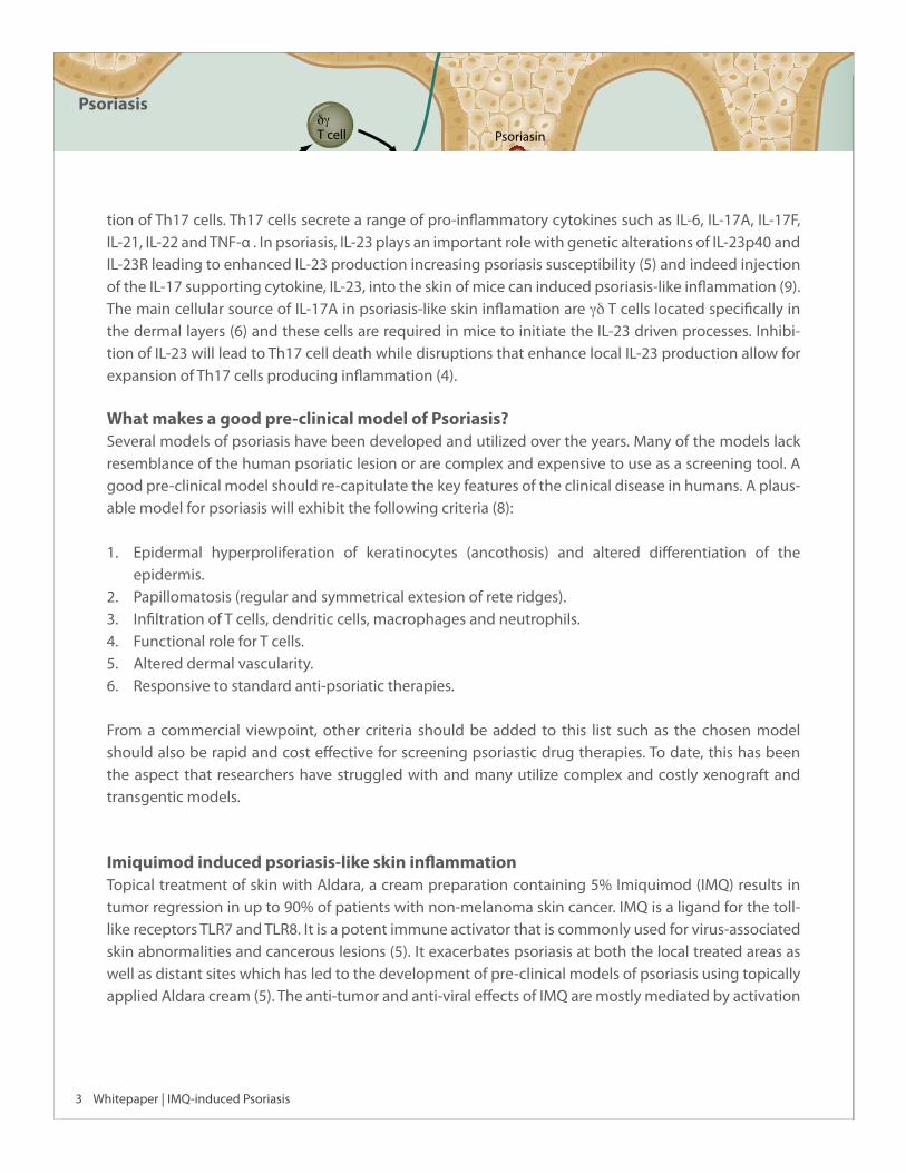
tion of Th17 cells. Th17 cells secrete a range of pro-inflammatory cytokines such as IL-6, IL-17A, IL-17F, IL-21, IL-22 and TNF-α . In psoriasis, IL-23 plays an important role with genetic alterations of IL-23p40 and IL-23R leading to enhanced IL-23 production increasing psoriasis susceptibility (5) and indeed injection of the IL-17 supporting cytokine, IL-23, into the skin of mice can induced psoriasis-like inflammation (9). The main cellular source of IL-17A in psoriasis-like skin inflamation are gd T cells located specifically in the dermal layers (6) and these cells are required in mice to initiate the IL-23 driven processes. Inhibi-tion of IL-23 will lead to Th17 cell death while disruptions that enhance local IL-23 production allow for expansion of Th17 cells producing inflammation (4).
What makes a good pre-clinical model of Psoriasis?Several models of psoriasis have been developed and utilized over the years. Many of the models lack resemblance of the human psoriatic lesion or are complex and expensive to use as a screening tool. A good pre-clinical model should re-capitulate the key features of the clinical disease in humans. A plaus-able model for psoriasis will exhibit the following criteria (8):
Epidermal hyperproliferation of keratinocytes (ancothosis) and altered differentiation of the 1. epidermis.Papillomatosis (regular and symmetrical extesion of rete ridges). 2. Infiltration of T cells, dendritic cells, macrophages and neutrophils.3. Functional role for T cells. 4. Altered dermal vascularity.5. Responsive to standard anti-psoriatic therapies.6.
From a commercial viewpoint, other criteria should be added to this list such as the chosen model should also be rapid and cost effective for screening psoriastic drug therapies. To date, this has been the aspect that researchers have struggled with and many utilize complex and costly xenograft and transgentic models.
Imiquimod induced psoriasis-like skin inflammationTopical treatment of skin with Aldara, a cream preparation containing 5% Imiquimod (IMQ) results in tumor regression in up to 90% of patients with non-melanoma skin cancer. IMQ is a ligand for the toll-like receptors TLR7 and TLR8. It is a potent immune activator that is commonly used for virus-associated skin abnormalities and cancerous lesions (5). It exacerbates psoriasis at both the local treated areas as well as distant sites which has led to the development of pre-clinical models of psoriasis using topically applied Aldara cream (5). The anti-tumor and anti-viral effects of IMQ are mostly mediated by activation
RESIDENT IMMUNE CELLS
Dendritic Cell
T Cell
Macrophage Chemokines
VEGFActivated
Macrophage
PSORIATIC EPIDERMIS WITHDISREGULATED ACANTHOSIS
NORMAL EPIDERMAL KERATINOCYTES
CHEMOTAXIS
ANGIOGENESIS
IL-1βIL-23
IL-17
IL-22Th17
δγ T cell
IL-19
pStat3
Psoriasin
IL-20IL-24
NF-kΒ
IL-23
TNF-α
PSORIATIC PLACQUE
Psoriasis
4 Whitepaper | IMQ-induced Psoriasis
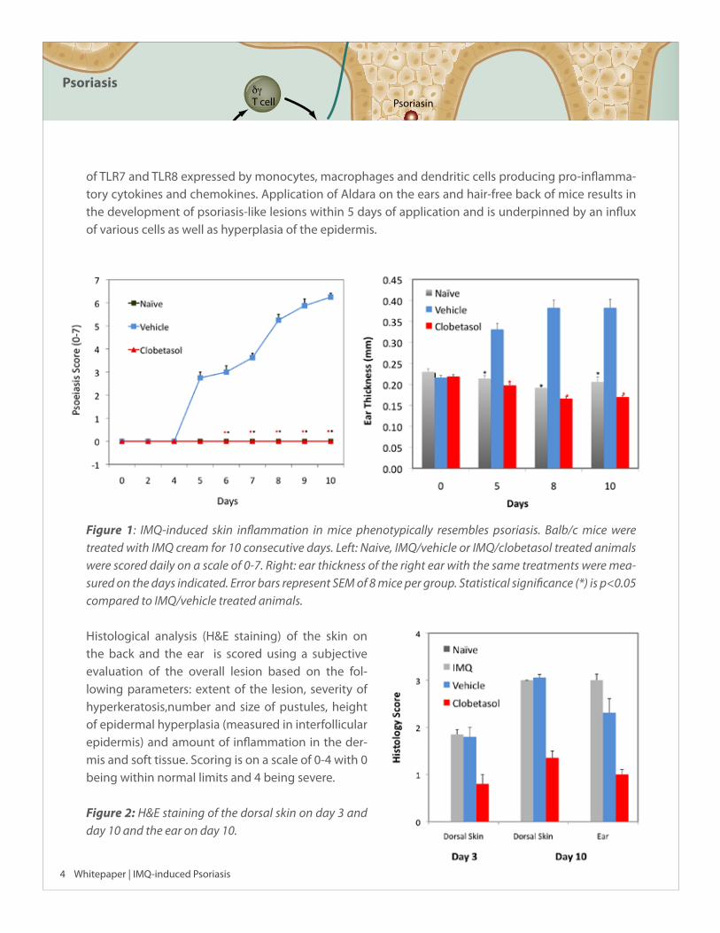
of TLR7 and TLR8 expressed by monocytes, macrophages and dendritic cells producing pro-inflamma-tory cytokines and chemokines. Application of Aldara on the ears and hair-free back of mice results in the development of psoriasis-like lesions within 5 days of application and is underpinned by an influx of various cells as well as hyperplasia of the epidermis.
Figure 1: IMQ-induced skin inflammation in mice phenotypically resembles psoriasis. Balb/c mice were treated with IMQ cream for 10 consecutive days. Left: Naive, IMQ/vehicle or IMQ/clobetasol treated animals were scored daily on a scale of 0-7. Right: ear thickness of the right ear with the same treatments were mea-sured on the days indicated. Error bars represent SEM of 8 mice per group. Statistical significance (*) is p<0.05 compared to IMQ/vehicle treated animals.
Histological analysis (H&E staining) of the skin on the back and the ear is scored using a subjective evaluation of the overall lesion based on the fol-lowing parameters: extent of the lesion, severity of hyperkeratosis,number and size of pustules, height of epidermal hyperplasia (measured in interfollicular epidermis) and amount of inflammation in the der-mis and soft tissue. Scoring is on a scale of 0-4 with 0 being within normal limits and 4 being severe.
Figure 2: H&E staining of the dorsal skin on day 3 and day 10 and the ear on day 10.
RESIDENT IMMUNE CELLS
Dendritic Cell
T Cell
Macrophage Chemokines
VEGFActivated
Macrophage
PSORIATIC EPIDERMIS WITHDISREGULATED ACANTHOSIS
NORMAL EPIDERMAL KERATINOCYTES
CHEMOTAXIS
ANGIOGENESIS
IL-1βIL-23
IL-17
IL-22Th17
δγ T cell
IL-19
pStat3
Psoriasin
IL-20IL-24
NF-kΒ
IL-23
TNF-α
PSORIATIC PLACQUE
Psoriasis
5 Whitepaper | IMQ-induced Psoriasis
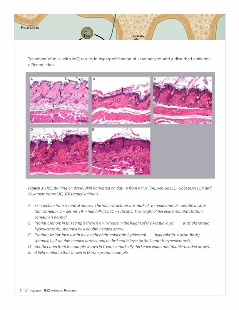
Treatment of mice with IMQ results in hyperproliferation of keratinocytes and a disturbed epidermal differentiation.
Figure 3: H&E staining on dorsal skin harvested on day 10 from naive (3A), vehicle (3E), clobetasol (3B) and dexamethasone (3C, 3D) treated animals.
Skin section from a control mouse. The main structures are marked. E – epidermis, K – keratin of stra-A. tum corneum, D – dermis, HF – hair follicles, SC – subcutis. The height of the epidermis and stratum corneum is normal.Psoriatic lesion: In this sample there is an increase in the height of the keratin layer (orthokeratotic B. hyperkeratosis), spanned by a double-headed arrow. Psoriatic lesion: increase in the height of the epidermis (epidermal hyperplasia = acanthosis), C. spanned by 2 double-headed arrows, and of the keratin layer (orthokeratotic hyperkeratosis). Another area from the sample shown in C with a markedly thickened epidermis (double-headed arrow). D. A field similar to that shown in D from psoriatic sample.E.
RESIDENT IMMUNE CELLS
Dendritic Cell
T Cell
Macrophage Chemokines
VEGFActivated
Macrophage
PSORIATIC EPIDERMIS WITHDISREGULATED ACANTHOSIS
NORMAL EPIDERMAL KERATINOCYTES
CHEMOTAXIS
ANGIOGENESIS
IL-1βIL-23
IL-17
IL-22Th17
δγ T cell
IL-19
pStat3
Psoriasin
IL-20IL-24
NF-kΒ
IL-23
TNF-α
PSORIATIC PLACQUE
Psoriasis
6 Whitepaper | IMQ-induced Psoriasis
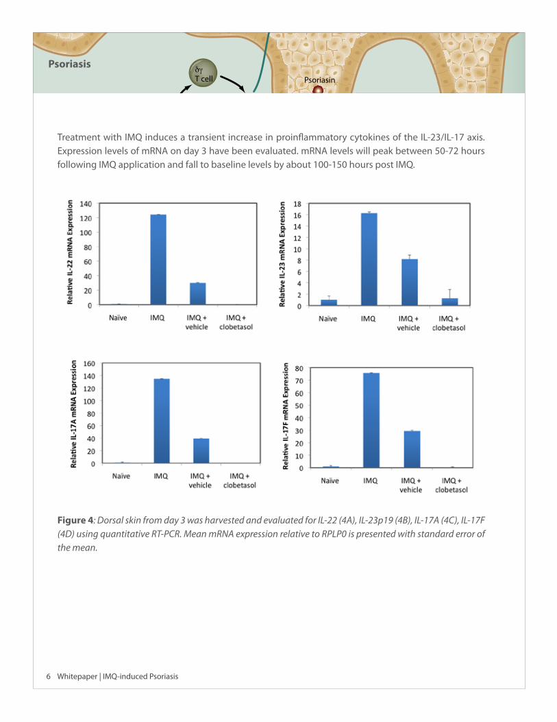
Treatment with IMQ induces a transient increase in proinflammatory cytokines of the IL-23/IL-17 axis. Expression levels of mRNA on day 3 have been evaluated. mRNA levels will peak between 50-72 hours following IMQ application and fall to baseline levels by about 100-150 hours post IMQ.
Figure 4: Dorsal skin from day 3 was harvested and evaluated for IL-22 (4A), IL-23p19 (4B), IL-17A (4C), IL-17F (4D) using quantitative RT-PCR. Mean mRNA expression relative to RPLP0 is presented with standard error of the mean.
RESIDENT IMMUNE CELLS
Dendritic Cell
T Cell
Macrophage Chemokines
VEGFActivated
Macrophage
PSORIATIC EPIDERMIS WITHDISREGULATED ACANTHOSIS
NORMAL EPIDERMAL KERATINOCYTES
CHEMOTAXIS
ANGIOGENESIS
IL-1βIL-23
IL-17
IL-22Th17
δγ T cell
IL-19
pStat3
Psoriasin
IL-20IL-24
NF-kΒ
IL-23
TNF-α
PSORIATIC PLACQUE
Psoriasis
7 Whitepaper | IMQ-induced Psoriasis
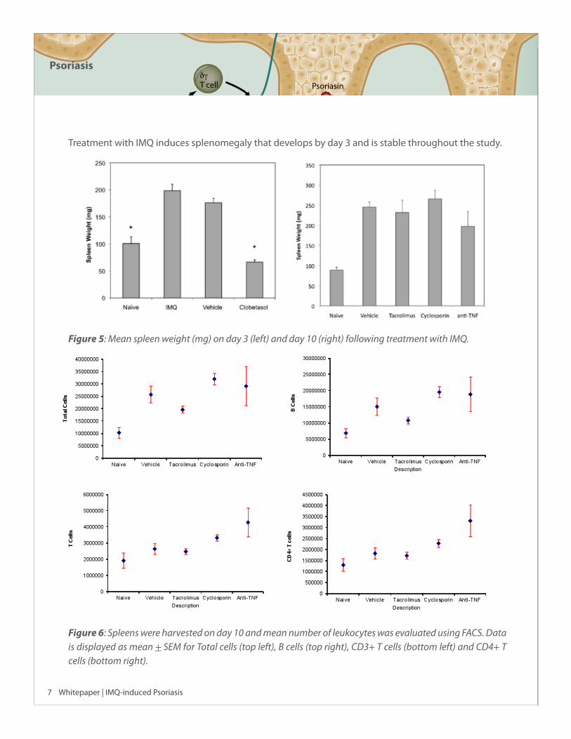
Treatment with IMQ induces splenomegaly that develops by day 3 and is stable throughout the study.
Figure 5: Mean spleen weight (mg) on day 3 (left) and day 10 (right) following treatment with IMQ.
Figure 6: Spleens were harvested on day 10 and mean number of leukocytes was evaluated using FACS. Data is displayed as mean + SEM for Total cells (top left), B cells (top right), CD3+ T cells (bottom left) and CD4+ T cells (bottom right).
! !
! !
RESIDENT IMMUNE CELLS
Dendritic Cell
T Cell
Macrophage Chemokines
VEGFActivated
Macrophage
PSORIATIC EPIDERMIS WITHDISREGULATED ACANTHOSIS
NORMAL EPIDERMAL KERATINOCYTES
CHEMOTAXIS
ANGIOGENESIS
IL-1βIL-23
IL-17
IL-22Th17
δγ T cell
IL-19
pStat3
Psoriasin
IL-20IL-24
NF-kΒ
IL-23
TNF-α
PSORIATIC PLACQUE
Psoriasis
8 Whitepaper | IMQ-induced Psoriasis
How does the IMQ-induced psoriasis-like inflammation model correlate to human psoriasis?
Although it is recognized that mouse skin differs from human skin in several ways such as rodents pos-sess a thinner epidermis and underlying cutaneous muscle layer, a recent study employing functional genomics methods has revealed many similarities between human psoriasis and mouse models across thousands of genes thus supporting the use of mouse pre-clinical models to screen anti-psoriatic com-pounds (8). Over 50,000 transcripts represented on the Affymetrix Human genome were compared to corresponding orthologues in the Affymetrix Mouse genome. This comparative study demonstrated that there was high correlation between psoriatic gene expression in human psoriatic skin samples in com-parison to transgenic mouse keratinocyte models and the mouse Imiquimod (IMQ) induced psoriasis model. In particular, these similarities were related to genes responsible for epidermal development and keratinization.
The mouse IMQ model represents a simple, rapid and cost effective method of inducing psoriasis in mice which avoids the expense of labour intensive breeding programmes which are required for producing the keratinocyte transgenic mouse lines e.g K14-AREG, K5-Stat3C or K5-TGFβ1. Furthermore some of these transgenic mouse lines also suffer from a shortened lifespan due to stunted growth and severe psoriatic skin lesions as a result of the genetic modification (10) thus rendering them less amenable to therapeutic intervention. Additionally, the lesions that develop in the mouse IMQ model resemble human psoriatic lesions phenotypically and histologically and are dependent on IL-23 and IL-17.
ConclusionThe IMQ-induced psoriasis-like inflammation model is based on a single innate antigenic receptor ligand. Since it requires no adjuvants, it is considered a clean model from an immunological viewpoint, resem-bling most of the features of human psoriatic lesions. It presents epidermal changes based on keratino-cyte hyperproliferation and altered differentiation, contains the presence of inflammatory cells including T cells, DC, and neutrophils; has a functional role for T cells and contains altered vascularity. Papillomatosis are sometimes observed. It’s dependence on IL-23, the resemblance of most features of human psoriatic lesions and its short duration makes it a convenient, cost-effective model for screening immunologically targeted therapies.
RESIDENT IMMUNE CELLS
Dendritic Cell
T Cell
Macrophage Chemokines
VEGFActivated
Macrophage
PSORIATIC EPIDERMIS WITHDISREGULATED ACANTHOSIS
NORMAL EPIDERMAL KERATINOCYTES
CHEMOTAXIS
ANGIOGENESIS
IL-1βIL-23
IL-17
IL-22Th17
δγ T cell
IL-19
pStat3
Psoriasin
IL-20IL-24
NF-kΒ
IL-23
TNF-α
PSORIATIC PLACQUE
Psoriasis
9 Whitepaper | IMQ-induced Psoriasis
ReferencesNational Psoriasis Foundation Statistics1. Krueger, J.G. and A. Bowcock (2005) Ann Rheum Dis 64:il30. Psoriasis Pathophysiology: Current Con-2. cepts of Pathogenesis. Danilenko, D.M. (2008) Vet Pathol. 45:563. Review Paper: Preclinical Animal Models of Psoriasis.3. Fitch, A., et al., (2007) Curr. Rheumatol. Rep. 9(6):461. Pathophysiology of Psoriasis: Recent Advances 4. on IL-23 and Th17 Cytokines. Van der fits, L., et al., (2009) The Journal of Immunology 182: 5836. Imiquimod-Induced Psoriasis-5. Like Skin Inflammation in Mice Is Mediated via the IL-23/IL-17 Axis. Cai et al (2011) Immunity. 35(4):596-610. Pivotal role of dermal IL-17-producing γδ T cells in skin 6. inflammation.Walter et al (2013) Nature Communications. Mar 5;4:1560. doi: 10.1038/ncomms2566. Aldara acti-7. vates TLR7-independent immune defence.Swindell et al (2011) PLoS One. Vol 6 (4): e18266. Genome-wide expression profiling of five mouse 8. models identifies similarities and differences with human psoriasis.Chan et al (2006) Journal of Experimental Medicine. Vol 203, No 12. IL-23 stimulates epidermal hy-9. perplasia via TNF and IL20R2-dependent mechanisms with implications for psoriasis pathogenesis.Cook et al (1997) The Journal of Clinical Investigation. Vol 100, No9. Transgenic expression of the hu-10. man amphiregulin gene induces a psoriasis-like phenotype.
RESIDENT IMMUNE CELLS
Dendritic Cell
T Cell
Macrophage Chemokines
VEGFActivated
Macrophage
PSORIATIC EPIDERMIS WITHDISREGULATED ACANTHOSIS
NORMAL EPIDERMAL KERATINOCYTES
CHEMOTAXIS
ANGIOGENESIS
IL-1βIL-23
IL-17
IL-22Th17
δγ T cell
IL-19
pStat3
Psoriasin
IL-20IL-24
NF-kΒ
IL-23
TNF-α
PSORIATIC PLACQUE
Psoriasis
10 Whitepaper | IMQ-induced Psoriasis
About MD BiosciencesMD Biosciences is a Swiss-based global biotechnology company focused in inflammations & neurol-ogy research. The Inflammations & Neurology Discovery Service divisions provide a broad range of disease models and mechanism of action studies for pre-clinical contract research as well as a unique combination of products. Investing time into customizing protocols to meet individual needs, along with learning and understanding clients’ objectives, hurdles and milestone requirements, enables MD Biosciences to contribute suggestions and alternatives to tackle the obstacles challenging the clients business. Cutting edge technologies, the willingness to experiment, in-depth understand-ing of disease processes, flexibility, and a client focused approach gives sponsors an added value over traditional CROs. With specialized laboratories located in Minnesota, Glasgow, and Israel, our panel of internationally recognized experts provide in-depth expertise and technologies to tackle problems and provide total solutions with a shorter project lead time. Whether you need a one time product or service, a combination of products and services, or a fully customized pre-clinical develop-ment program, we are ready to put our expertise and technologies to work for you.
For more information about the IMQ-induced psoriasis model, please visit our website at www.mdbiosciences.com or contact us at one of the offices listed below.
IMPORTANT NOTICE: The information contained in this document is confidential and/or privileged information subject to protection by law or terms of applicable confi-dentiality agreements, and is intended only for the use of the individual or entity sent to. Any unauthorized review, use, disclosure or distribution is prohibited. If you are not the intended recipient, please contact the sender and destroy all copies of the original message. The MD Biosciences logo, ArthritoMab™ and Senerga® are registered trademarks of Morwell Diagnostics and may not be used without permission. April 2013
Inflammation and Neurology Discovery Services
USA/Canada:1-888-USMDBIO
International:+41-44 986 2628