University of Groningen Adipose derived stromal cells in ... stromal cells inhibit TGF-β induced...
19
University of Groningen Adipose derived stromal cells in cardiovascular regenerative medicine Przybyt, Ewa IMPORTANT NOTE: You are advised to consult the publisher's version (publisher's PDF) if you wish to cite from it. Please check the document version below. Document Version Publisher's PDF, also known as Version of record Publication date: 2016 Link to publication in University of Groningen/UMCG research database Citation for published version (APA): Przybyt, E. (2016). Adipose derived stromal cells in cardiovascular regenerative medicine [Groningen]: University of Groningen Copyright Other than for strictly personal use, it is not permitted to download or to forward/distribute the text or part of it without the consent of the author(s) and/or copyright holder(s), unless the work is under an open content license (like Creative Commons). Take-down policy If you believe that this document breaches copyright please contact us providing details, and we will remove access to the work immediately and investigate your claim. Downloaded from the University of Groningen/UMCG research database (Pure): http://www.rug.nl/research/portal. For technical reasons the number of authors shown on this cover page is limited to 10 maximum. Download date: 09-06-2018
Transcript of University of Groningen Adipose derived stromal cells in ... stromal cells inhibit TGF-β induced...
University of Groningen
Adipose derived stromal cells in cardiovascular regenerative medicinePrzybyt, Ewa
IMPORTANT NOTE: You are advised to consult the publisher's version (publisher's PDF) if you wish to cite fromit. Please check the document version below.
Document VersionPublisher's PDF, also known as Version of record
Publication date:2016
Link to publication in University of Groningen/UMCG research database
Citation for published version (APA):Przybyt, E. (2016). Adipose derived stromal cells in cardiovascular regenerative medicine [Groningen]:University of Groningen
CopyrightOther than for strictly personal use, it is not permitted to download or to forward/distribute the text or part of it without the consent of theauthor(s) and/or copyright holder(s), unless the work is under an open content license (like Creative Commons).
Take-down policyIf you believe that this document breaches copyright please contact us providing details, and we will remove access to the work immediatelyand investigate your claim.
Downloaded from the University of Groningen/UMCG research database (Pure): http://www.rug.nl/research/portal. For technical reasons thenumber of authors shown on this cover page is limited to 10 maximum.
Download date: 09-06-2018
adipose-derived stromal cells inhibit tGf-β induced
differentiation of human dermal fibroblasts and keloid fibroblasts
in a paracrine fashion
Plastic and Reconstructive Surgery. 2014 Oct;134(4):699-712
9
maroesjka spiekman, Bs1; ewa przybyt, ms1; Josée a. plantinga, Bs1; susan Gibbs phd2;
Berend van der Lei, md, phd3; martin c. harmsen, phd1
1 department of pathology and medical Biology, university medical centre Groningen, university of Groningen, Groningen, The netherlands.
2 department of dermatology, Vu university medical centre, amsterdam, The netherlands.
3 department of plastic surgery, university medical centre Groningen, university of Groningen, Groningen, The netherlands.
plastic and reconstructive surgery.2014 oct;134(4):699-712
chapter
139
140
CHAPTER 9
138
Abstract Background: Adipose tissue-derived stromal cells (ADSC) augment wound healing and regeneration of the skin. Currently, it is unknown if and how ADSC can also influence dermal scarring. We hypothesized that ADSC inhibit adverse differentiation of dermal fibroblasts induced by the pivotal factor in scarring i.e. Transforming Growth Factor-β (TGF-β). Methods: TGF-β-treated adult human dermal fibroblasts (HDFa) and keloid scar-derived fibroblasts (KLF), were incubated with ADSC conditioned medium (ADSC CM) and assessed for proliferation and differentiation in particular the production of collagen, expression of SM22α and development of hypertrophy and contractility. Results: TGF-β-induced proliferation of HDFa was abolished by ADSC CM. Simultaneously ADSC CM reduced SM22α gene and protein expression of TGF-β-treated HDFa, while their contractility was reduced too. Furthermore, ADSC CM strongly reduced transcription of collagen I and III genes as well as their corresponding proteins. On the other hand the ADSC CM tipped the balance of matrix turnover to degradation through stimulating gene expression of MMP-1, 2 and 14, while MMP-2 activity was upregulated too. Even in end stage myofibroblasts i.e. KLF, ASDC CM suppressed TGF-β-induced myofibroblast contraction, and collagen III gene expression. Conclusion: In this study we show that ADSC inhibit TGF-β-induced adverse differentiation and function of HDFa and TGF- β -induced contraction in KLF, in a paracrine fashion.
Adipose-derived stromal cells inhibit TGF-β induced differentiation of human dermal fibroblasts and keloid fibroblasts in a paracrine fashion
141
9
139
Introduction
Dermal scars are the result of wound healing and remodeling. When dysregulated, they may be painful, cause itching and discomfort, and can become disfiguring as hypertrophic scars or even keloid. Currently, there is no ideal treatment to reverse or reduce such dermal scarring. However, in several clinical reports and clinical trials, it has been reported that lipofilling improves the quality of scars. In addition, it has been demonstrated that lipofilling improves normal aged skin and burn scars histologically1,2, functionally and aesthetically3-6. Among the cellular constituent of fat tissue, we consider adipose tissue-derived stromal cells (ADSC) to be a prime candidate for the observed anti-scarring and skin improving capacity of lipofilling.
Adipose tissue-derived stromal cells (ADSC) are multipotent stem cells, which reside in the perivascular niche as pericytes or peri-adventitial cells7 in white adipose tissue throughout the body8-10. In vitro, ADSC present as fibroblast-like, spindle-shaped cells. They secrete a plethora of trophic factors, which suppress inflammation and apoptosis, yet promote angiogenesis and mitosis of parenchymal cells11. Furthermore, ADSC have the ability to differentiate into several cell types, among which are adipocytes, chondrocytes, osteoblasts, smooth muscle cells and pericytes8-10. In animal models, ADSC speed up wound healing12-15. In vitro, ADSC increase proliferation, migration and extracellular matrix production of dermal fibroblasts16,17. Furthermore, ADSC have been successfully used for organotypic skin culture18 and to tissue engineer skin19. As yet, the influence of ADSC on the remodeling phase of wound healing is unknown, in particular with regard to dermal scarring. In normal wound healing, haemostasis and inflammation are followed by proliferation, remodeling and finally, regeneration or repair. Resident dermal cells, in particular fibroblasts and keratinocytes respond to the inflammatory cues by restoring dermal integrity20. Transforming Growth Factor-β (TGF-β) is the key inflammatory cytokine in wound healing, regeneration and remodeling. During haemostasis and inflammation, TGF-β is secreted by activated platelets and immune cells. In response to TGF-β, fibroblasts differentiate into myofibroblasts21-23, which contract the wound and aid in remodeling of the extracellular matrix (ECM)21,22,24,25. Important ECM molecules secreted by myofibroblasts are collagen types I and III, which are also the main proteinaceous constituents of scars. During the resolution phase of normal wound healing, the morphology of the skin is restored and the myofibroblasts resolve from their transient function21. However, in the presence of chronic pro-inflammatory and/or pro-fibrobrotic stimuli, wound healing may become disturbed and dysregulated. TGF-β is then continuously produced, resulting in an on-going proliferation of myofibroblasts and subsequently excessive deposition of extracellular matrix and contraction of the scar22,26. Based upon clinical observations of the effects of ADSC on disturbed wound healing, we hypothesize that ADSC inhibit TGF-β-induced differentiation of normal dermal fibroblasts and keloid scar-derived fibroblasts and thereby improve dysregulated wound healing. To test this hypothesis in vitro, we investigated matrix production and degradation, mesenchymal markers and contractility of TGF-β-stimulated normal dermal fibroblasts and keloid fibroblasts, after treatment with ADSC conditioned medium (ADSC CM) in order to elucidate the effect of ADSC on myofibroblasts.
Materials and Methods
Adipose tissue-derived stromal cell isolation and culture The research conducted was in agreement with the ethical rules for human experimentation
142
CHAPTER 9
140
as stated in the 1975 Declaration of Helsinki and was approved by the medical ethical committee of University Medical Centre Groningen. Human adipose tissue was obtained with informed consent from healthy donors undergoing elective abdominal liposuction surgery at Bergman Clinics, Heerenveen, the Netherlands. ADSC were isolated from adipose tissue as described previously27. ADSC were maintained in Dulbecco’s Minimal Essentials Medium (DMEM; Lonza) containing 10% FBS, 100 units/mL penicillin, 100 mg/mL streptomycin (DMEM high serum). ADSC were used in experiments up to passage 6. Adult human dermal fibroblasts culture Human dermal fibroblasts, adult type (HDFa) were purchased from ATCC. HDFa were maintained in Eagle's Minimal Essentials Medium (EMEM; Lonza) containing 10% FBS, 100 units/mL penicillin, 100 mg/mL streptomycin. HDFa used in experiments ranged from passage 11-18. Keloid scar-derived fibroblasts culture Keloid scar-derived fibroblasts (KLF) were provided by the Department of Dermatology, VU University Medical Centre, Amsterdam, The Netherlands. KLF were obtained with informed consent, the procedure was approved by the medical ethical committee of VU University Medical Centre. KLF were maintained in DMEM, containing 1% Ultroser-G (Gibco), 100 units/mL penicillin, 100 mg/mL streptomycin. KLF were used in experiments up to passage 3. Collection of adipose-derived stromal cell conditioned medium When ADSC reached ~90% confluency, cells were incubated with culture medium containing 2% FBS. After 24h, conditioned medium was removed and fresh DMEM 2% FBS was added. This procedure was repeated three times, conditioned medium from three different time points was pooled, filtered through a 0.22μm filter and stored at -20°C until experimental need. Conditioned medium for experiments with KLF was generated similarly, but using KLF culture medium. Conditioned medium culture HDFa or KLF were seeded at a density of 10,000 cells/cm2 in appropriate culture media. HDFa were switched to DMEM containing 5% FBS overnight. KLF were maintained on DMEM containing Ultroser-G. At day 1 of the experiment, HDFa or KLF were incubated with ADSC CM, with or without 10ng TGF-β1 (Peprotech) per mL medium. Control groups were incubated with normal culture medium with 2% FBS (HDFa) or 1% Ultroser-G (KLF). Fibroblasts were cultured for 4 days. Immunofluorescent staining HDFa were seeded on Thermanox coverslips (NUNC Brand Products) in 24 wells plates. KLF were seeded in Lab-Tec II 8 well chamber slides (NUNC Brand Products). Cells were fixed in 2% paraformaldehyde. Endogenous biotin was blocked using avidin (Dako) and biotin solutions (Dako). Primary antibodies were rabbit anti-collagen I or rabbit anti-collagen III (Abcam) in PBS with 2% goat serum and 1% BSA. Secondary and tertiary antibodies were goat anti-rabbit biotin-labeled (Dako) and Cy3-labeled Streptavidin (Invitrogen). Nuclei were counterstained using 4′,6-diamidino-2-phenylindole (DAPI; Sigma-Aldrich). Cytoskeleton was visualized using FITC-conjugated phallotoxins (Invitrogen). Slides were examined using a Leica DMRXA microscope and Leica Software (Leica Microsystems). Hypertrophy measurements HDFa were seeded in 24 wells plates. After 4 days, cells were fixed in 2% paraformaldehyde. Cell outlines were visualized with CellMaskTM Deep Red Plasma Membrane Stain (Invitrogen). Nuclei were counterstained using DAPI. Pictures were acquired using a Zeiss
Adipose-derived stromal cells inhibit TGF-β induced differentiation of human dermal fibroblasts and keloid fibroblasts in a paracrine fashion
143
9
141
Axio Observer.Z1 microscope and TissueFAXS Immunofluorescence Cell Analysis system (TissueGnostics USA Inc.). Length and width of HFDa were scored in 10 representative images for control groups and 5 representative images for ADSC CM treated groups (n=3), using ImageJ (Research Services Branch, National Institute of Mental Health). Gene transcript analysis Cells were lysed in Trizol Reagent (Invitrogen). Isolation of total RNA and qRT-PCR analyses were performed as previously described27, using primer sequences as listed below (Table 1). To determine differences in expression of gene of interest, Ct-values were normalized against mean Ct-values of GAPDH and β-actin using dCt-method: dCt= Ctgene of interest – Ct mean (GAPDH
and β-actin). Fold increase was calculated as 2-(ddCt). Table 1. Primer sequences for quantitative Reverse Transcription-Polymerase Chain Reaction Primer Forward Reverse β -actin CCAACCGCGAGAAGATGA CCAGAGGCGTACAGGGATAG Collagen I GGGATTCCCTGGACCTAAAG GGAACACCTCGCTCTCCA Collagen III CTGGACCCCAGGGTCTTC CATCTGATCCAGGGTTTCCA GAPDH AGCCACATCGCTCAGACAC GCCCAATACGACCAAATCC MMP-1 CTGAAAGTGACTGGGAAACC GACAAACTGAGCCACATCAG MMP-2 GTTCCCCTTCTTGTTCAATG CTTGCCATCCTTCTCAAAGT MMP-14 GGGTGAGGAATAACCAAGTG CTTCCTCTCGTAGGCAGTGT SM22a CTGAGGACTATGGGGTCATC TAGTGCCCATCATTCTTGGT TIMP-1 CCAGCGTTATGAGATCAAGA AGTATCCGCAGACACTCTCC TIMP-2 GAAGAGCCTGAACCACAGGT CGGGGAGGAGATGTAGCAC Immunoblot analysis Protein levels of SM22α, collagen I and III were determined by immunoblot analysis. Immunoblot procedures were performed as previously described27. As primary antibodies, rabbit anti-SM22α (Abcam), rabbit anti-collagen I, rabbit anti-collagen III (Abcam) or rabbit anti-β-actin (Cell Signalling) were used. Goat anti-rabbit Irdye680 (Li-Cor Biosciences) was used as secondary antibody. Pictures were acquired with Odyssey infrared imaging system (Li-Cor Biosciences). Densitometric analysis was performed using TotalLab software (Nonlinear Dynamics). Expression of proteins of interest was normalized against β-actin. Gelatin zymography The presence of gelatinase activity in HDFa was determined according to the method of Gogly28. Photographs were acquired using a Nikon D80 camera and were processed with Adobe Photoshop CS5.1. The gelatinase MMP-2 appears at two bands around 70kDa: an upper band of the pro-MMP-2 and a lower band of active MMP-2. The procedure also activates the pro-MMP-2.
Gel contraction assay After 4 days of conditioned medium culture, HDFa were detached and resuspended in DMEM 2% FBS. Gel contraction assay was performed as described by Van Beuge and colleagues29. All cultures were performed in triplicate. After 24h of culture, gels were scanned using a flatbed scanner. Surface area of gels was measured using ImageJ. Statistical analysis All data are represented as mean±SEM. Data were analyzed by one-way analysis of variance (ANOVA) and Bonferroni post-hoc tests, using GraphPad Prism (GraphPad Software Inc.).
144
CHAPTER 9
142
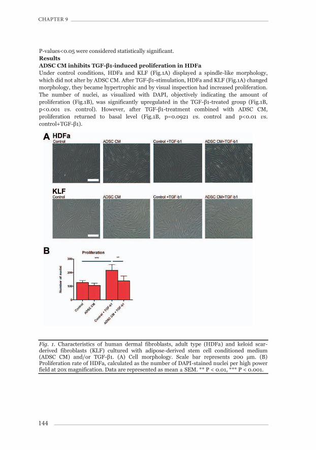
P-values<0.05 were considered statistically significant. Results ADSC CM inhibits TGF-β1-induced proliferation in HDFa Under control conditions, HDFa and KLF (Fig.1A) displayed a spindle-like morphology, which did not alter by ADSC CM. After TGF-β1-stimulation, HDFa and KLF (Fig.1A) changed morphology, they became hypertrophic and by visual inspection had increased proliferation. The number of nuclei, as visualized with DAPI, objectively indicating the amount of proliferation (Fig.1B), was significantly upregulated in the TGF-β1-treated group (Fig.1B, p<0.001 vs. control). However, after TGF-β1-treatment combined with ADSC CM, proliferation returned to basal level (Fig.1B, p=0.0921 vs. control and p<0.01 vs. control+TGF-β1).
Fig. 1. Characteristics of human dermal fibroblasts, adult type (HDFa) and keloid scar-derived fibroblasts (KLF) cultured with adipose-derived stem cell conditioned medium (ADSC CM) and/or TGF-β1. (A) Cell morphology. Scale bar represents 200 μm. (B) Proliferation rate of HDFa, calculated as the number of DAPI-stained nuclei per high power field at 20x magnification. Data are represented as mean ± SEM. ** P < 0.01, *** P < 0.001.
Adipose-derived stromal cells inhibit TGF-β induced differentiation of human dermal fibroblasts and keloid fibroblasts in a paracrine fashion
145
9
143
ADSC CM alters cytoskeletal composition and function in control condition and after TGF-β1-stimulation in HDFa and KLF.
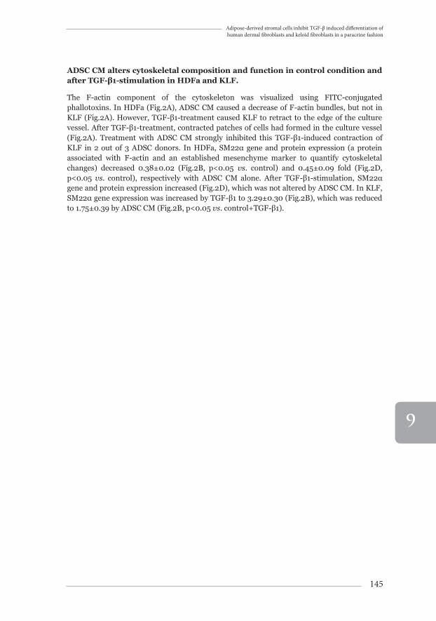
The F-actin component of the cytoskeleton was visualized using FITC-conjugated phallotoxins. In HDFa (Fig.2A), ADSC CM caused a decrease of F-actin bundles, but not in KLF (Fig.2A). However, TGF-β1-treatment caused KLF to retract to the edge of the culture vessel. After TGF-β1-treatment, contracted patches of cells had formed in the culture vessel (Fig.2A). Treatment with ADSC CM strongly inhibited this TGF-β1-induced contraction of KLF in 2 out of 3 ADSC donors. In HDFa, SM22α gene and protein expression (a protein associated with F-actin and an established mesenchyme marker to quantify cytoskeletal changes) decreased 0.38±0.02 (Fig.2B, p<0.05 vs. control) and 0.45±0.09 fold (Fig.2D, p<0.05 vs. control), respectively with ADSC CM alone. After TGF-β1-stimulation, SM22α gene and protein expression increased (Fig.2D), which was not altered by ADSC CM. In KLF, SM22α gene expression was increased by TGF-β1 to 3.29±0.30 (Fig.2B), which was reduced to 1.75±0.39 by ADSC CM (Fig.2B, p<0.05 vs. control+TGF-β1).
146
CHAPTER 9
144
_______________________________________________________________ Fig. 2. Cytoskeletal organization of HDFa and KLF is influenced by ADSC CM in presence an absence of TGF-β1. (A) Staining of HDFa and KLF with FITC-conjugated phallotoxins (green), nuclei counterstained with DAPI (blue). Scale bar represents 300 μm. (B) qRT-PCR for SM22α mRNA expression after ADSC CM treatment and/or TGF-β1 stimulation (C) Representative images of immunoblotting for SM22α protein expression in HDFa (D) Quantification of SM22α protein expression in HDFa. Data are represented as mean ± SEM. * P < 0.05.
Adipose-derived stromal cells inhibit TGF-β induced differentiation of human dermal fibroblasts and keloid fibroblasts in a paracrine fashion
147
9
145
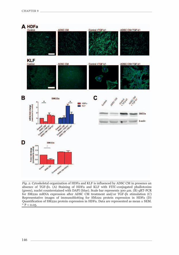
ADSC CM reduces contractility in HDFa To determine the functional implications of the reduction of cytoskeletal components, we performed a collagen gel contraction assay with HDFa (Fig.3A). Under control conditions, HDFa already contracted collagen gels to 54.4%±1.1% of their original gel area (Fig.3B). In ADSC CM pre-treated group, contraction of gels was reduced to 33.0%±3.6% (Fig.3B, p<0.001 vs. control). After TGF-β1 stimulation, contraction of gels increased to 74.7%±1.7%, which was only 64.6%±3.0% (Fig.3B, p<0.05 vs. control+TGF-β1) with ADSC CM.
_______________________________________________________________Fig. 3. HDFa gel contraction assay. (A) Representative images of HDFa (pre-incubated under different conditions for 4 days), 24 hours after seeding in collagen gels. (B) Contraction of collagen gels, represented as percentage of contraction compared to the original volume. Data (n=3) are represented as mean ± SEM. * P < 0.05, *** P < 0.001
ADSC CM prevents hypertrophy in TGF- β stimulated HDFa
In inverted microscopic view (data not shown), HDFa partly lost their typical ‘hill-and-valley’ features after treatment with TGF- β and/or ADSC CM. HDFa shortened and gained width, i.e. they became hypertrophic. The degree of hypertrophy was investigated by measuring length and width of HDFa after staining with CellMaskTM (Fig.4A). Lengths of HDFa decreased 0.80±0.007 fold after ADSC CM treatment only, 0.79±0.008 fold after TGF-β1-stimulation and 0.80±0.007 fold after treatment with ADSC CM and TGF-β1 (Fig.4B, p<0.001 vs. control). Width increased slightly after ADSC CM, to 1.06±0.01 fold (Fig.4C, p<0.001 vs. control). After TGF-β1-stimulation, width increased to 1.33±0.01 fold (Fig.4C, p<0.001 vs. control), which was only 1.04±0.01 fold with concurrent ADSC CM treatment (Fig.4C, p<0.001 vs. control+TGF-β1). Thus, ADSC CM reduced the TGF-β-induced hypertrophy of HDFa cells, in particular through normalization of the width of cells (Fig.4B, C).
148
CHAPTER 9
146
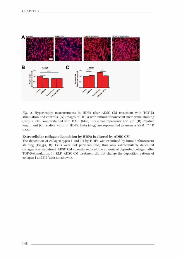
Fig. 4. Hypertrophy measurements in HDFa after ADSC CM treatment with TGF-β1 stimulation and controls. (A) Images of HDFa with immunofluorescent membrane staining (red), nuclei counterstained with DAPI (blue). Scale bar represents 200 μm. (B) Relative length and (C) relative width of HDFa. Data (n=3) are represented as mean ± SEM. *** P 0.001.
Extracellular collagen deposition by HDFa is altered by ADSC CM The deposition of collagen types I and III by HDFa was examined by immunofluorescent staining (Fig.5A, B). Cells were not permeabilized, thus only extracellularly deposited collagen was visualized. ADSC CM strongly reduced the amount of deposited collagen after TGF-β-stimulation. In KLF, ADSC CM treatment did not change the deposition pattern of collagen I and III (data not shown).
Adipose-derived stromal cells inhibit TGF-β induced differentiation of human dermal fibroblasts and keloid fibroblasts in a paracrine fashion
149
9
147
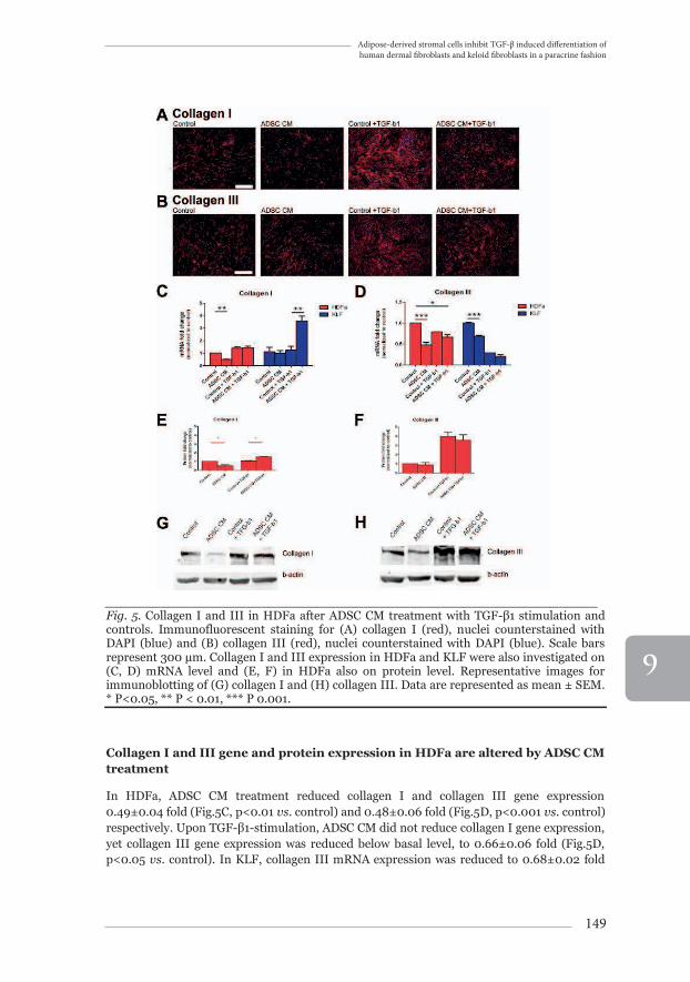
_______________________________________________________________Fig. 5. Collagen I and III in HDFa after ADSC CM treatment with TGF-β1 stimulation and controls. Immunofluorescent staining for (A) collagen I (red), nuclei counterstained with DAPI (blue) and (B) collagen III (red), nuclei counterstained with DAPI (blue). Scale bars represent 300 μm. Collagen I and III expression in HDFa and KLF were also investigated on (C, D) mRNA level and (E, F) in HDFa also on protein level. Representative images for immunoblotting of (G) collagen I and (H) collagen III. Data are represented as mean ± SEM. * P<0.05, ** P < 0.01, *** P 0.001.
Collagen I and III gene and protein expression in HDFa are altered by ADSC CM treatment
In HDFa, ADSC CM treatment reduced collagen I and collagen III gene expression 0.49±0.04 fold (Fig.5C, p<0.01 vs. control) and 0.48±0.06 fold (Fig.5D, p<0.001 vs. control) respectively. Upon TGF-β1-stimulation, ADSC CM did not reduce collagen I gene expression, yet collagen III gene expression was reduced below basal level, to 0.66±0.06 fold (Fig.5D, p<0.05 vs. control). In KLF, collagen III mRNA expression was reduced to 0.68±0.02 fold
150
CHAPTER 9
148
(Fig.5D, p<0.001 vs. control), yet collagen I increased to 3.59±0.38 (Fig.5C, p<0.01 vs. control+TGF-β1).
As for protein production in HDFa, collagen I was reduced 0.51±0.13 fold (Fig.5E, p<0.05 vs. control) after ADSC CM, and increased 1.51±0.05 fold (Fig.5E, p<0.05 vs. control+TGF-β1) after ADSC CM treatment with TGF-β1-stimulation. For collagen III, there was no significant difference (Fig.5F).
Production and activation of matrix metalloproteinases is influenced by ADSC CM treatment
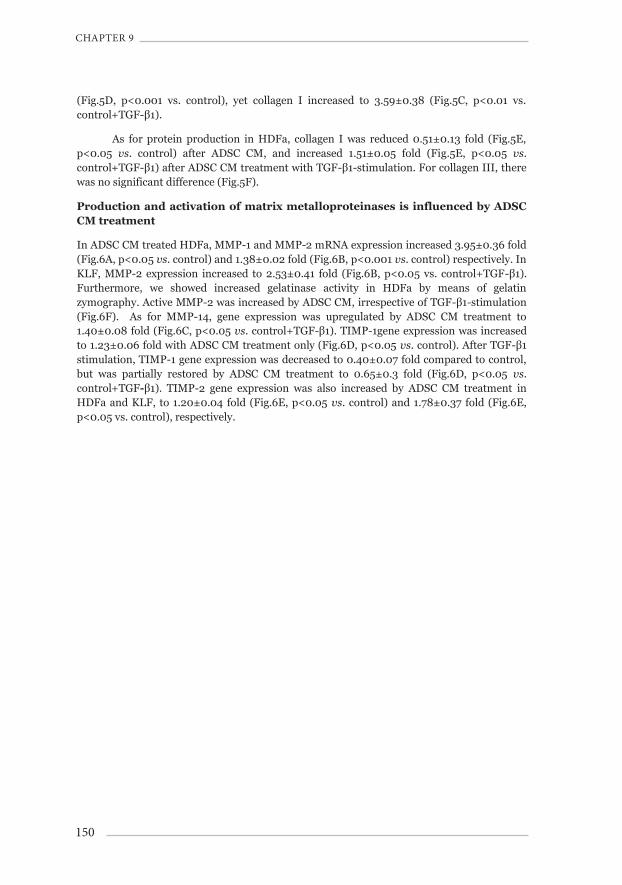
In ADSC CM treated HDFa, MMP-1 and MMP-2 mRNA expression increased 3.95±0.36 fold (Fig.6A, p<0.05 vs. control) and 1.38±0.02 fold (Fig.6B, p<0.001 vs. control) respectively. In KLF, MMP-2 expression increased to 2.53±0.41 fold (Fig.6B, p<0.05 vs. control+TGF-β1). Furthermore, we showed increased gelatinase activity in HDFa by means of gelatin zymography. Active MMP-2 was increased by ADSC CM, irrespective of TGF-β1-stimulation (Fig.6F). As for MMP-14, gene expression was upregulated by ADSC CM treatment to 1.40±0.08 fold (Fig.6C, p<0.05 vs. control+TGF-β1). TIMP-1gene expression was increased to 1.23±0.06 fold with ADSC CM treatment only (Fig.6D, p<0.05 vs. control). After TGF-β1 stimulation, TIMP-1 gene expression was decreased to 0.40±0.07 fold compared to control, but was partially restored by ADSC CM treatment to 0.65±0.3 fold (Fig.6D, p<0.05 vs. control+TGF-β1). TIMP-2 gene expression was also increased by ADSC CM treatment in HDFa and KLF, to 1.20±0.04 fold (Fig.6E, p<0.05 vs. control) and 1.78±0.37 fold (Fig.6E, p<0.05 vs. control), respectively.
Adipose-derived stromal cells inhibit TGF-β induced differentiation of human dermal fibroblasts and keloid fibroblasts in a paracrine fashion
151
9
149
_______________________________________________________________
Fig. 6 Inventory of matrix degradation by HDFa and KLF. Relative gene expression of matrix degrading proteins (A) MMP-1 and (B) MMP-2 and (C) their activator MMP-14. Expression of inhibitors (D) TIMP-1 and (E) TIMP-2. Furthermore, proteolytic activity of (F) MMP-2 protein was analysed. Data (n=5) are represented as mean ± SEM. * P < 0.05, *** P < 0.001.
Discussion
Our set of experiments clearly demonstrates that TFG-β-induced myofibroblast differentiation and function of human dermal fibroblasts is inhibited by ADSC CM and thereby underlines the positive clinical effects observed of ADSC on disturbed wound healing and aged skin. To our knowledge, this is the first report that demonstrates that ADSC can inhibit (dermal) fibroblast differentiation and after TGF-β-stimulation. This implicates that ADSC results in inhibition of fibroblast differentiation which might lead to prevention or even reduction of dermal scars. The effects of ADSC consisted of inhibition of cytoskeletal elements and hypertrophy, ECM secretion, contraction as well as promotion of matrix degrading enzymes. End-stage myofibroblasts, i.e. KLF were less refractory to the inhibitory influence of ADSC CM. Only TGF-β-stimulated KLF contraction was altered by ADSC CM, not the production and degradation of ECM components. In TGF-β-stimulated HDFa, ADSC CM appeared to tip the balance of ECM remodeling towards normalization in contrast to KLF. In HDFa, ADSC CM changed both the pattern of intercellular collagen deposition as
152
CHAPTER 9
150
well as reduced the amount of deposited collagens in a manner that resembles resolution of wound healing. Our in vitro model for skin fibrosis is limited to assess on the one hand only over short times (days) and with strong stimulators (TGF-β). The role of TGF-β in stimulation of collagen incorporation into extracellular matrix, is long known30,31. In our research, collagen I was still present intracellularly, yet extracellular deposition was reduced by ADSC CM after TGF-β stimulation of human dermal fibroblasts. Remarkably, the gene expression of collagen I was also reduced, thus the intracellular presence would merely reflect storage instead of actual synthesis. Therefore, our observation that ADSC CM interfere with the process of extracellular deposition and turnover of collagen I and III is topic of our current research in vivo.The influence of ADSC CM on HDFa was partially replicated in KLF. In the absence of TGF-β, ADSC CM was able to reduce collagen III gene expression in KLF compared to control. In presence of TGF-β, SM22α mRNA expression was decreased too. Collagen production as well as upregulation of the contractile apparatus, are hallmark features of the myofibroblast21. It is striking that in KLF, which have already undergone myofibroblast differentiation, ADSC CM only seems to inhibit one out of these two processes: either matrix production, or contractility. To date, there are no literature data available regarding an effect of lipofilling to reduce keloid scars. Our data suggest that KLF might be differentiated too far to revert the normal fibroblastic phenotypic by means of ADSC. Nevertheless, the ADSC were potent in suppression of various components of myofibroblasts differentiation.
In this study, the molecular mechanism by which ADSC exert their anti-fibrotic influence remains largely unelucidated. In a recent pig study, Yun and co-workers32, showed that ADSC improved healing of full thickness skin wounds, while scarring was minimal. This was partly attributed to the upregulation of TGF-β3. In scarring, TGF-β3 antagonizes the pro-fibrotic action of TGF-β133. Yet, ADSC in our investigations had only very low gene expression levels of any of the three forms of TGF-β (data not shown). Thus other paracrine factors probably were responsible for the suppressive effects of ADSC CM. Interestingly, the immune-modulatory features of ADSC and MSC have been shown to suppress the fibrotic phenotype of scar fibroblasts34 and activated hepatic stellate cells35 through secreted interleukin-10 (IL-10), Tumor Necrosis Factor-α (TNF-α) and Hepatocyte Growth Factor (HGF). Although, IL-10 gene expression was negligible in our ADSC, they expressed strongly anti-inflammatory mediators such as IDO and PGE2 (E. Przybyt, unpublished). In vivo, IDO and PGE2 upregulate IL-10 production by macrophages, which is important in the resolution of wound healing. In contrast, in our experiments the conditioned medium could only directly affect HDFa instead of indirectly through macrophage activation. This notion, and the ease of the use of conditioned media in terms of generation, standardisation, storage to mention, warrants further research in other routes of paracrine communication. In other labs, the role of ADSC has been assessed in dermal wound healing12-15, dermal rejuvenation16,17 and tissue engineering of epidermis19. Previous studies have demonstrated effects of ADSC on proliferation, migration, extracellular matrix production and radical oxygen species formation in fibroblasts12-15. Already, improvement of skin quality1 and scar reduction2-6 have been reported after fat grafting. It has been suggested by Coleman and Mojallal that these regenerative effects can be attributed to ADSC, which are transplanted along with the adipose tissue36,37.
We showed that the paracrine factors secreted by ADSC modulate differentiation of fibroblasts and their subsequent function. As fat reduces dermal scarring, our study is a first in the understanding which cellular component in adipose tissue is involved in scar
Adipose-derived stromal cells inhibit TGF-β induced differentiation of human dermal fibroblasts and keloid fibroblasts in a paracrine fashion
153
9
151
reduction. In addition, development towards application of conditioned medium of ADSC may help prevent adverse wound healing and scarring. Currently, we investigate the molecular mechanisms of the paracrine action of ADSC in wound healing.
References
1. Covarrubias P, Cardenas-Camarena L, Guerrerosantos J, et al. Evaluation of the histologic changes in the fat-grafted facial skin: Clinical trial. Aesthetic Plast Surg. 2013;37(4):778-783.
2. Bruno A, Delli Santi G, Fasciani L, Cempanari M, Palombo M, Palombo P. Burn scar lipofilling: Immunohistochemical and clinical outcomes. J Craniofac Surg. 2013;24(5):1806-1814.
3. Klinger M, Caviggioli F, Klinger FM, et al. Autologous fat graft in scar treatment. J Craniofac Surg. 2013;24(5):1610-1615.
4. Klinger M, Marazzi M, Vigo D, Torre M. Fat injection for cases of severe burn outcomes: A new perspective of scar remodeling and reduction. Aesthetic Plast Surg. 2008;32(3):465-469.
5. Mazzola IC, Cantarella G, Mazzola RF. Management of tracheostomy scar by autologous fat transplantation: A minimally invasive new approach. J Craniofac Surg. 2013;24(4):1361-1364.
6. Ulrich D, Ulrich F, van Doorn L, Hovius S. Lipofilling of perineal and vaginal scars: A new method for improvement of pain after episiotomy and perineal laceration. Plast Reconstr Surg. 2012;129(3):593e-594e.
7. Corselli M, Chen CW, Sun B, Yap S, Rubin JP, Peault B. The tunica adventitia of human arteries and veins as a source of mesenchymal stem cells. Stem Cells Dev. 2012;21(8):1299-1308.
8. Mizuno H, Tobita M, Uysal AC. Concise review: Adipose-derived stem cells as a novel tool for future regenerative medicine. Stem Cells. 2012;30(5):804-810.
9. Philips BJ, Marra KG, Rubin JP. Adipose stem cell-based soft tissue regeneration. Expert Opin Biol Ther. 2012;12(2):155-163.
10. Wilson A, Butler PE, Seifalian AM. Adipose-derived stem cells for clinical applications: A review. Cell Prolif. 2011;44(1):86-98.
11. Hong SJ, Traktuev DO, March KL. Therapeutic potential of adipose-derived stem cells in vascular growth and tissue repair. Curr Opin Organ Transplant. 2010;15(1):86-91.
12. Kim WS, Park BS, Sung JH, et al. Wound healing effect of adipose-derived stem cells: A critical role of secretory factors on human dermal fibroblasts. J Dermatol Sci. 2007;48(1):15-24.
13. Jeon YK, Jang YH, Yoo DR, Kim SN, Lee SK, Nam MJ. Mesenchymal stem cells' interaction with skin: Wound-healing effect on fibroblast cells and skin tissue. Wound Repair Regen. 2010;18(6):655-661.
14. Heo SC, Jeon ES, Lee IH, Kim HS, Kim MB, Kim JH. Tumor necrosis factor-alpha-activated human adipose tissue-derived mesenchymal stem cells accelerate cutaneous wound healing through paracrine mechanisms. J Invest Dermatol. 2011;131(7):1559-1567.
15. Luo G, Cheng W, He W, et al. Promotion of cutaneous wound healing by local application of mesenchymal stem cells derived from human umbilical cord blood. Wound Repair Regen. 2010;18(5):506-513.
16. Kim WS, Park BS, Park SH, Kim HK, Sung JH. Antiwrinkle effect of adipose-derived stem cell: Activation of dermal fibroblast by secretory factors. J Dermatol Sci. 2009;53(2):96-102.
154
CHAPTER 9
152
17. Kim WS, Park BS, Sung JH. Protective role of adipose-derived stem cells and their soluble factors in photoaging. Arch Dermatol Res. 2009;301(5):329-336.
18. Kim DW, Lee JS, Yoon ES, Lee BI, Park SH, Dhong ES. Influence of human adipose-derived stromal cells on wnt signaling in organotypic skin culture. J Craniofac Surg. 2011;22(2):694-698.
19. Lu W, Yu J, Zhang Y, et al. Mixture of fibroblasts and adipose tissue-derived stem cells can improve epidermal morphogenesis of tissue-engineered skin. Cells Tissues Organs. 2012;195(3):197-206.
20. Gurtner GC, Werner S, Barrandon Y, Longaker MT. Wound repair and regeneration. Nature. 2008;453(7193):314-321.
21. Hinz B. Formation and function of the myofibroblast during tissue repair. J Invest Dermatol. 2007;127(3):526-537.
22. Desmouliere A, Chaponnier C, Gabbiani G. Tissue repair, contraction, and the myofibroblast. Wound Repair Regen. 2005;13(1):7-12.
23. Desmouliere A, Geinoz A, Gabbiani F, Gabbiani G. Transforming growth factor-beta 1 induces alpha-smooth muscle actin expression in granulation tissue myofibroblasts and in quiescent and growing cultured fibroblasts. J Cell Biol. 1993;122(1):103-111.
24. Grotendorst GR, Rahmanie H, Duncan MR. Combinatorial signaling pathways determine fibroblast proliferation and myofibroblast differentiation. FASEB J. 2004;18(3):469-479.
25. Widgerow AD. Current concepts in scar evolution and control. Aesthetic Plast Surg. 2011;35(4):628-635.
26. Kis K, Liu X, Hagood JS. Myofibroblast differentiation and survival in fibrotic disease. Expert Rev Mol Med. 2011;13:e27.
27. Przybyt E, Krenning G, Brinker MG, Harmsen MC. Adipose stromal cells primed with hypoxia and inflammation enhance cardiomyocyte proliferation rate in vitro through STAT3 and Erk1/2. J Transl Med. 2013;11:39-5876-11-39.
28. Gogly B, Groult N, Hornebeck W, Godeau G, Pellat B. Collagen zymography as a sensitive and specific technique for the determination of subpicogram levels of interstitial collagenase. Anal Biochem. 1998;255(2):211-216.
29. van Beuge MM, Prakash J, Lacombe M, et al. Reduction of fibrogenesis by selective delivery of a rho kinase inhibitor to hepatic stellate cells in mice. J Pharmacol Exp Ther. 2011;337(3):628-635.
30. Ignotz RA, Massague J. Transforming growth factor-beta stimulates the expression of fibronectin and collagen and their incorporation into the extracellular matrix. J Biol Chem. 1986;261(9):4337-4345.
31. Schiller M, Javelaud D, Mauviel A. TGF-beta-induced SMAD signaling and gene regulation: Consequences for extracellular matrix remodeling and wound healing. J Dermatol Sci. 2004;35(2):83-92.
32. Yun IS, Jeon YR, Lee WJ, et al. Effect of human adipose derived stem cells on scar formation and remodeling in a pig model: A pilot study. Dermatol Surg. 2012;38(10):1678-1688.
33. O'Kane S, Ferguson MW. Transforming growth factor beta s and wound healing. Int J Biochem Cell Biol. 1997;29(1):63-78.
34. Kumai Y, Kobler JB, Park H, Galindo M, Herrera VL, Zeitels SM. Modulation of vocal fold scar fibroblasts by adipose-derived stem/stromal cells. Laryngoscope. 2010;120(2):330-337.

Adipose-derived stromal cells inhibit TGF-β induced differentiation of human dermal fibroblasts and keloid fibroblasts in a paracrine fashion
155
9
153
35. Parekkadan B, van Poll D, Megeed Z, et al. Immunomodulation of activated hepatic stellate cells by mesenchymal stem cells. Biochem Biophys Res Commun. 2007;363(2):247-252.
36. Coleman SR. Structural fat grafting: More than a permanent filler. Plast Reconstr Surg. 2006;118(3 Suppl):108S-120S.
37. Mojallal A, Lequeux C, Shipkov C, et al. Improvement of skin quality after fat grafting: Clinical observation and an animal study. Plast Reconstr Surg. 2009;124(3):765-774.


![Epac2 signaling at the β-cell plasma membrane920771/FULLTEXT01.pdf · small fraction of cells are pancreatic polypeptide-secreting PP-cells [6] and ghrelin-releasing ε-cells [7].](https://static.fdocument.org/doc/165x107/6065b034c80f1b4fbb7d2949/epac2-signaling-at-the-cell-plasma-membrane-920771fulltext01pdf-small-fraction.jpg)