University of Dundee Proteomic identification of the UDP ...
15
University of Dundee Proteomic identification of the UDP-GlcNAc Ji, Zhe; Tinti, Michele; Ferguson, Michael A. J. Published in: PLoS ONE DOI: 10.1371/journal.pone.0244699 Publication date: 2021 Licence: CC BY Document Version Publisher's PDF, also known as Version of record Link to publication in Discovery Research Portal Citation for published version (APA): Ji, Z., Tinti, M., & Ferguson, M. A. J. (2021). Proteomic identification of the UDP-GlcNAc: PI 1-6 GlcNAc- transferase subunits of the glycosylphosphatidylinositol biosynthetic pathway of Trypanosoma brucei. PLoS ONE, 16(3), [e0244699]. https://doi.org/10.1371/journal.pone.0244699 General rights Copyright and moral rights for the publications made accessible in Discovery Research Portal are retained by the authors and/or other copyright owners and it is a condition of accessing publications that users recognise and abide by the legal requirements associated with these rights. • Users may download and print one copy of any publication from Discovery Research Portal for the purpose of private study or research. • You may not further distribute the material or use it for any profit-making activity or commercial gain. • You may freely distribute the URL identifying the publication in the public portal. Take down policy If you believe that this document breaches copyright please contact us providing details, and we will remove access to the work immediately and investigate your claim. Download date: 26. Oct. 2021
Transcript of University of Dundee Proteomic identification of the UDP ...
University of Dundee
Proteomic identification of the UDP-GlcNAc
Ji, Zhe; Tinti, Michele; Ferguson, Michael A. J.
Published in:PLoS ONE
DOI:10.1371/journal.pone.0244699
Publication date:2021
Licence:CC BY
Document VersionPublisher's PDF, also known as Version of record
Link to publication in Discovery Research Portal
Citation for published version (APA):Ji, Z., Tinti, M., & Ferguson, M. A. J. (2021). Proteomic identification of the UDP-GlcNAc: PI 1-6 GlcNAc-transferase subunits of the glycosylphosphatidylinositol biosynthetic pathway of Trypanosoma brucei. PLoSONE, 16(3), [e0244699]. https://doi.org/10.1371/journal.pone.0244699
General rightsCopyright and moral rights for the publications made accessible in Discovery Research Portal are retained by the authors and/or othercopyright owners and it is a condition of accessing publications that users recognise and abide by the legal requirements associated withthese rights.
• Users may download and print one copy of any publication from Discovery Research Portal for the purpose of private study or research. • You may not further distribute the material or use it for any profit-making activity or commercial gain. • You may freely distribute the URL identifying the publication in the public portal.
Take down policyIf you believe that this document breaches copyright please contact us providing details, and we will remove access to the work immediatelyand investigate your claim.
Download date: 26. Oct. 2021
RESEARCH ARTICLE
Proteomic identification of the UDP-GlcNAc: PI
α1–6 GlcNAc-transferase subunits of the
glycosylphosphatidylinositol biosynthetic
pathway of Trypanosoma brucei
Zhe Ji, Michele TintiID, Michael A. J. FergusonID*
The Wellcome Centre for Anti-Infectives Research, School of Life Sciences, University of Dundee, Dundee,
United Kingdom
Abstract
The first step of glycosylphosphatidylinositol (GPI) anchor biosynthesis in all eukaryotes is
the addition of N-acetylglucosamine (GlcNAc) to phosphatidylinositol (PI) which is catalysed
by a UDP-GlcNAc: PI α1–6 GlcNAc-transferase, also known as GPI GnT. This enzyme has
been shown to be a complex of seven subunits in mammalian cells and a similar complex of
six homologous subunits has been postulated in yeast. Homologs of these mammalian and
yeast subunits were identified in the Trypanosoma brucei predicted protein database. The
putative catalytic subunit of the T. brucei complex, TbGPI3, was epitope tagged with three
consecutive c-Myc sequences at its C-terminus. Immunoprecipitation of TbGPI3-3Myc fol-
lowed by native polyacrylamide gel electrophoresis and anti-Myc Western blot showed that
it is present in a ~240 kDa complex. Label-free quantitative proteomics were performed to
compare anti-Myc pull-downs from lysates of TbGPI-3Myc expressing and wild type cell
lines. TbGPI3-3Myc was the most highly enriched protein in the TbGPI3-3Myc lysate pull-
down and the expected partner proteins TbGPI15, TbGPI19, TbGPI2, TbGPI1 and TbERI1
were also identified with significant enrichment. Our proteomics data also suggest that an
Arv1-like protein (TbArv1) is a subunit of the T. brucei complex. Yeast and mammalian Arv1
have been previously implicated in GPI biosynthesis, but here we present the first experi-
mental evidence for physical association of Arv1 with GPI biosynthetic machinery. A puta-
tive E2-ligase has also been tentatively identified as part of the T. brucei UDP-GlcNAc: PI
α1–6 GlcNAc-transferase complex.
Introduction
Trypanosoma brucei is a protozoan pathogen that undergoes a complex life cycle between its
tsetse fly vector and mammalian hosts. The parasite causes human African trypanosomiasis in
humans and nagana in cattle in sub-Saharan Africa.
The bloodstream form (BSF) of T. brucei produces a dense coat of GPI anchored variant
surface protein (VSG) to protect it from the innate immune system and, through antigenic
variation, the acquired immune response [1]. Other T. brucei surface molecules that have been
PLOS ONE
PLOS ONE | https://doi.org/10.1371/journal.pone.0244699 March 18, 2021 1 / 14
a1111111111
a1111111111
a1111111111
a1111111111
a1111111111
OPEN ACCESS
Citation: Ji Z, Tinti M, Ferguson MAJ (2021)
Proteomic identification of the UDP-GlcNAc: PI α1–
6 GlcNAc-transferase subunits of the
glycosylphosphatidylinositol biosynthetic pathway
of Trypanosoma brucei. PLoS ONE 16(3):
e0244699. https://doi.org/10.1371/journal.
pone.0244699
Editor: Ziyin Li, University of Texas Medical School
at Houston, UNITED STATES
Received: December 11, 2020
Accepted: February 21, 2021
Published: March 18, 2021
Peer Review History: PLOS recognizes the
benefits of transparency in the peer review
process; therefore, we enable the publication of
all of the content of peer review and author
responses alongside final, published articles. The
editorial history of this article is available here:
https://doi.org/10.1371/journal.pone.0244699
Copyright: © 2021 Ji et al. This is an open access
article distributed under the terms of the Creative
Commons Attribution License, which permits
unrestricted use, distribution, and reproduction in
any medium, provided the original author and
source are credited.
Data Availability Statement: The data underlying
this study are available at the PRIDE database
through accession number PXD022979.
shown experimentally to possess a GPI membrane anchor are the ESAG6-subunit of the BSF
transferrin receptor (TfR) [2] and the procyclins, the major surface glycoproteins of the tsetse
mid-gut dwelling procyclic form (PCF) of the parasite [3]. In addition, many other surface
molecules with N-terminal signal peptides and C-terminal GPI addition signal peptides are
predicted to be GPI-anchored in T. brucei, including the BSF haptaglobin-haemaglobin recep-
tor [4] and the factor H receptor [5], the epimastigote BARP glycoprotein [6] and the metacyc-
lic trypomastigote invariant surface protein (MISP) [7]. Thus far, GPI anchor structures have
been completely or partially solved for four T. brucei VSGs [8–11], the TfR [2] and the procy-
clins [3]. As for the structure of GPIs, research on T. brucei was the first to yield methodologies
to delineate the steps of GPI biosynthesis that were subsequently applied to mammalian cells
and yeast [12–14]. However, it was the power of mammalian cell and yeast genetics that led to
the identification of the majority of GPI biosynthesis genes, reviewed in [15–17].
We currently have reasonably advanced models for GPI anchor biosynthesis and processing
in trypanosomes, mammalian cells and yeast and the similarities and differences in these path-
ways have been reviewed extensively elsewhere [15–18]. For most organisms, the functions
and interactions of putative GPI pathway gene products have been inferred from experimental
work in mammalian or yeast cells. In a few cases these functions have been experimentally
confirmed in T. brucei, i.e., for the GlcNAc-PI de-N-acetylase (TbGPI12) [19], the third man-
nosyltransferase (TbGPI10) [20] and the catalytic (TbGPI8) [21] and other subunits (TTA1
and 2 [22] and TbGPI16) [23]) of the GPI transamidase complex.
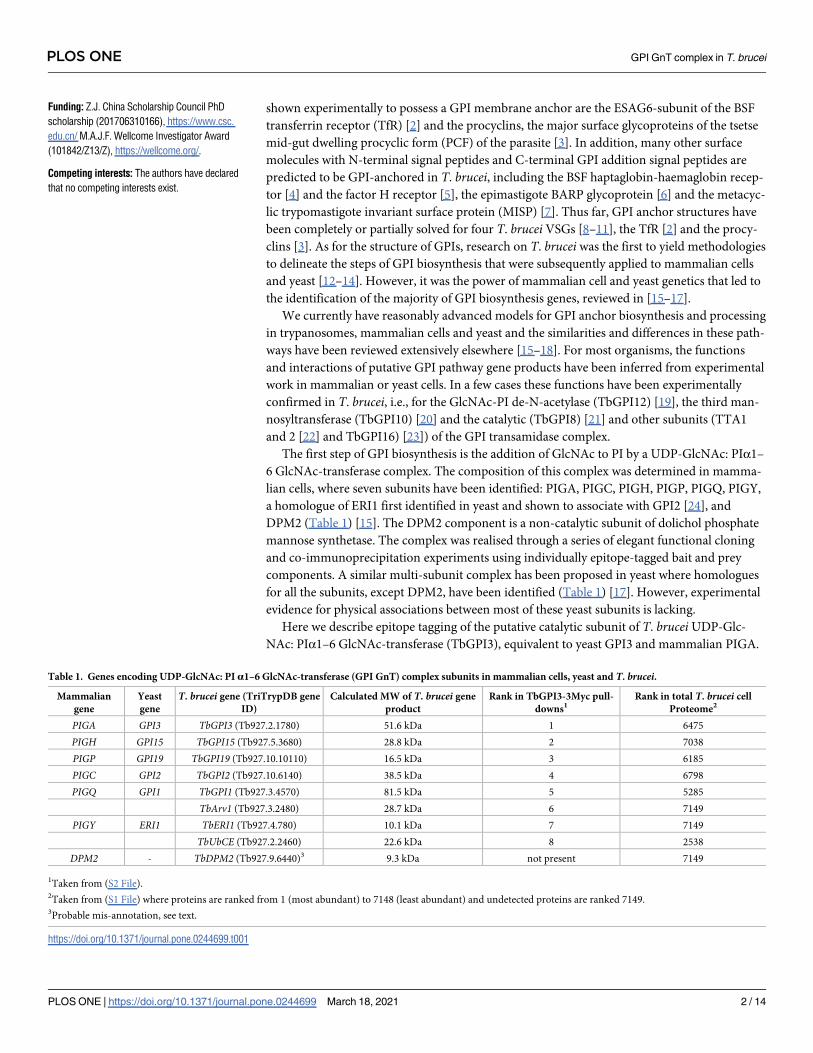
The first step of GPI biosynthesis is the addition of GlcNAc to PI by a UDP-GlcNAc: PIα1–
6 GlcNAc-transferase complex. The composition of this complex was determined in mamma-
lian cells, where seven subunits have been identified: PIGA, PIGC, PIGH, PIGP, PIGQ, PIGY,
a homologue of ERI1 first identified in yeast and shown to associate with GPI2 [24], and
DPM2 (Table 1) [15]. The DPM2 component is a non-catalytic subunit of dolichol phosphate
mannose synthetase. The complex was realised through a series of elegant functional cloning
and co-immunoprecipitation experiments using individually epitope-tagged bait and prey
components. A similar multi-subunit complex has been proposed in yeast where homologues
for all the subunits, except DPM2, have been identified (Table 1) [17]. However, experimental
evidence for physical associations between most of these yeast subunits is lacking.
Here we describe epitope tagging of the putative catalytic subunit of T. bruceiUDP-Glc-
NAc: PIα1–6 GlcNAc-transferase (TbGPI3), equivalent to yeast GPI3 and mammalian PIGA.
Table 1. Genes encoding UDP-GlcNAc: PI α1–6 GlcNAc-transferase (GPI GnT) complex subunits in mammalian cells, yeast and T. brucei.
Mammalian
gene
Yeast
gene
T. brucei gene (TriTrypDB gene
ID)
Calculated MW of T. brucei gene
product
Rank in TbGPI3-3Myc pull-
downs1Rank in total T. brucei cell
Proteome2
PIGA GPI3 TbGPI3 (Tb927.2.1780) 51.6 kDa 1 6475
PIGH GPI15 TbGPI15 (Tb927.5.3680) 28.8 kDa 2 7038
PIGP GPI19 TbGPI19 (Tb927.10.10110) 16.5 kDa 3 6185
PIGC GPI2 TbGPI2 (Tb927.10.6140) 38.5 kDa 4 6798
PIGQ GPI1 TbGPI1 (Tb927.3.4570) 81.5 kDa 5 5285
TbArv1 (Tb927.3.2480) 28.7 kDa 6 7149
PIGY ERI1 TbERI1 (Tb927.4.780) 10.1 kDa 7 7149
TbUbCE (Tb927.2.2460) 22.6 kDa 8 2538
DPM2 - TbDPM2 (Tb927.9.6440)3 9.3 kDa not present 7149
1Taken from (S2 File).2Taken from (S1 File) where proteins are ranked from 1 (most abundant) to 7148 (least abundant) and undetected proteins are ranked 7149.3Probable mis-annotation, see text.
https://doi.org/10.1371/journal.pone.0244699.t001
PLOS ONE GPI GnT complex in T. brucei
PLOS ONE | https://doi.org/10.1371/journal.pone.0244699 March 18, 2021 2 / 14
Funding: Z.J. China Scholarship Council PhD
scholarship (201706310166), https://www.csc.
edu.cn/ M.A.J.F. Wellcome Investigator Award
(101842/Z13/Z), https://wellcome.org/.
Competing interests: The authors have declared
that no competing interests exist.
We demonstrate its presence in a protein complex and identify its partner proteins through
label-free quantitative proteomics.
Materials and methods
Cultivation of trypanosomes
T. brucei brucei Lister strain 427 bloodstream form (BSF) parasites expressing VSG variant 221
and transformed to stably express T7 polymerase and the tetracycline repressor protein under
G418 antibiotic selection [25] was used in this study and will be referred as bloodstream form
wild type (BSF WT). Cells were cultivated in HMI-11T medium containing 2.5 μg/mL of G418
at 37˚C in a 5% CO2 incubator as previously described [25]. HMI-11T is a modification of the
original HMI-9 [26] that uses 56 mM 1-thioglycerol in place of 200 mM 2-mercaptoethanol,
and contains 10% heat inactivated fetal bovine serum (PAA) and lacks of serum plus (Hazleton
Biologics, Lenexa, Kansas).
DNA isolation and manipulation
Plasmid DNA was purified from Escherichia coli (chemically competent DH5α cells) using
Qiagen Miniprep kits and Maxiprep was performed by the University of Dundee DNA
sequencing service. Gel extraction and PCR purification were performed using QIAquick kits
(Qiagen). Custom oligonucleotides were obtained from Eurofins MWG Operon or Thermo
Fisher. T. brucei genomic DNA was isolated from ~5 × 10⁷ BSF cells using lysis buffer contain-
ing 100 mM Tris-HCl (pH 8.0), 100 mM NaCl, 25 mM EDTA, 0.5% SDS, and 0.1 mg/mL pro-
teinase K (Sigma) by standard methods.
Generation of in-situ tagging constructs
The tagging cassette was amplified from the pMOTag43M plasmid [27] using the forward
primer: 5’-TGATTGATATTGCACCAGATTTTCCACTGGAGTTGTACTCTCGTAACCGGGAGAAGCTTCAAGTTGTGGGAAGCCCATCCgaacaaaagctgggtacc-3’ and the reverse
primer: 5’-CAACGCGAAACAATGACagAGAGAGAGAGAGAAGGGCGAAAACAAAAAGGATCGCGGTAGAGAGGACCCCGCCCATACCCctattcctttgccctcggac-3’. The PCR prod-
uct contains 80 bp corresponding to the 3’-end of the TbGPI3 open reading frame (capital let-
ters of forward primer) followed by a sequence encoding the 3Myc epitope tag, an intergenic
region (igr) from the T. brucei α-β tubulin locus, the hygromycin phosphotransferase (HYG)
selectable marker gene and the 3’-UTR of TbGPI3 (capital letters of reverse primer).
Transformation of BSF T. bruceiConstructs for in situ tagging were purified and precipitated, washed with 70% ethanol, and
re-dissolved in sterile water. The released DNA was electroporated into BSF WT cell line. Cell
culture and transformation were carried out as described previously [25, 27]. After five days of
selection with hygromycin, cells were sub-cloned and four independent clones were selected
and cultured.
Western blot of cell lysates
To confirm the C-terminal tagging of TbGPI3 with 3Myc, cells from the four selected clones in
parallel with BSF WT cells were lysed in SDS sample buffer. Aliquots corresponding to 5×10⁶
cells per sample, were subjected to SDS-PAGE on NuPAGE bis-Tris 10% acrylamide gels
(Invitrogen) and transferred to a nitrocellulose membrane (Invitrogen). Ponceau staining con-
firmed equal loading and transfer. The blot was further probed with anti-Myc rat monoclonal
PLOS ONE GPI GnT complex in T. brucei
PLOS ONE | https://doi.org/10.1371/journal.pone.0244699 March 18, 2021 3 / 14
antibody (Chromotek, 9E1) in a 1:1,000 dilution. Detection was carried out using IRDye
800CW conjugated goat anti-rat IgG antibody (1:15,000) and LI-COR Odyssey infrared imag-
ing system (LICOR Biosciences, Lincoln, NE).
Co-immunoprecipitation and Native-PAGE Western blotting
To investigate detergent solubilisation conditions for the immunoprecipitation of TbGPI3-
3Myc complexes, aliquots of 2 × 10⁸ cells were harvested and lysed in 500 μL of 50 mM Tris-
HCl, pH 7.4, 150 mM NaCl containing different detergents; 0.5% digitonin, 1% digitonin, 1%
Triton X-100 (TX-100), 1% n-octyl-beta-glucoside (NOG) or 1% decyl-β-D-maltopyranoside
(DM). After centrifugation at 16,000 g, 4˚C for 20 min, aliquots of the supernatants equivalent
to 2 x 10⁸ cells were incubated with 10 μL anti-Myc agarose beads (Myc-TrapTM, Chromotek)
for 1 h at 4˚C. The beads were washed three times in 50 mM Tris-HCl, pH 7.4, 150 mM NaCl
containing the corresponding detergents and bound proteins were eluted three times with
10 μL 0.5 mg/mL c-Myc peptide (Sigma M2435) in the corresponding detergent containing
buffer. The combining eluates for each detergent condition, equivalent to 2 × 10⁸ cells, were
subjected to NativePAGE (Invitrogen) and transferred to a PVDF membrane (Invitrogen) fol-
lowed by immunoblotting with anti-Myc antibody (Chromotek, 9E1) diluted 1:1,000. The blot
was then developed by ECL using an HRP-conjugated secondary antibody (Sigma, A9037,
1:3,000).
Label free proteomics of TbGPI3-3Myc and BSF WT lysate pull downs
BSF WT and TbGPI3-3Myc expressing cell lines were cultured and 1 × 10⁹ cells of each were
harvested and lysed in 1 mL of lysis buffer containing 0.5% digitonin. After centrifugation
16,000 g, 4˚C for 20 min, the supernatants were mixed with 20 μL of Myc-TrapTM beads and
incubated for 1 h at 4˚C. The beads were washed three times in the same buffer, and bound
proteins were eluted with 1×SDS sample buffer and subjected to SDS-PAGE, running the pro-
teins only 10 cm into the gel. Whole lanes containing TbGPI3 and wild type cell lines samples
were cut identically into 3 slices and the gel pieces were dried in Speed-vac (Thermo Scientific)
for in-gel reduction with 0.01 M dithiothreitol and alkylation with 0.05 M iodoacetamide
(Sigma) for 30 min in the dark. The gel slices were washed in 0.1 M NH4HCO3, and digested
with 12.5 μg/mL modified sequence grade trypsin (Roche) in 0.02 M NH4HCO3 for 16 h at
30˚C. Samples were dried and re-suspended in 50 μL 1% formic acid and then subjected to liq-
uid chromatography on Ultimate 3000 RSLC nano-system (Thermo Scientific) fitted with a 3
Acclaim PepMap 100 (C18, 100 μM × 2 cm) and then separated on an Easy-Spray PepMap
RSLC C18 column (75 μM × 50 cm) (Thermo Scientific). Samples (15μL) were loaded in 0.1%
formic acid (buffer A) and separated using a binary gradient consisting of buffer A and buffer
B (80% acetonitrile, 0.1% formic acid). Peptides were eluted with a linear gradient from 2 to
35% buffer B over 70 min. The HPLC system was coupled to a Q-Exactive Plus Mass Spec-
trometer (Thermo Scientific) equipped with an Easy-Spray source with temperature set at
50˚C and a source voltage of 2.0 kV. The mass spectrometry proteomics data have been depos-
ited to the ProteomeXchange Consortium via the PRIDE partner repository with the dataset
identifier Project PXD022979 [28] https://www.ebi.ac.uk/pride/archive/projects/PXD022979.
Protein identification by MaxQuant
RAW data files were analysed using MaxQuant version 1.6.10.43, with the in-built Andromeda
search engine [29], using the T. brucei brucei 927 annotated protein sequences from Tri-
TrypDB release 46 [30], supplemented with the T. brucei brucei 427 VSG221 (Tb427.
BES40.22) protein sequence. The mass tolerance was set to 4.5 ppm for precursor ions and
PLOS ONE GPI GnT complex in T. brucei
PLOS ONE | https://doi.org/10.1371/journal.pone.0244699 March 18, 2021 4 / 14
MS/MS mass tolerance was set at 20 ppm (MaxQuant default parameters). The enzyme was set
to trypsin, allowing up to 2 missed cleavages. Carbamidomethyl on cysteine was set as a fixed
modification. Acetylation of protein N-termini, and oxidation of methionine were set as vari-
able modifications. Match between runs was enabled, allowing transfer of peptide identifica-
tions of sequenced peptides from one LC-MS run to non-sequenced ions with the same mass
and retention time in another run. A 20-min time window was set for alignment of separate
LC-MS runs. The false-discovery rate for protein and peptide level identifications was set at
1%, using a target-decoy based strategy.
Data analysis
Data analysis was performed using custom Python scripts, using the SciPy ecosystem of open-
source software libraries [31]. The data analysis pipeline is available at GitHub https://github.com/
mtinti/TbGPI3 and Zenodo https://zenodo.org/record/4310034 repositories, DOI:10.5281/
zenodo.3735036. The MaxQuant proteinGroups.txt output file was used to extract the iBAQ
scores for forward trypanosome protein sequences identified with at least two unique peptides
and with an Andromeda score>4. The protein iBAQ scores were normalised for sample loading
by dividing each iBAQ value by the median of all the iBAQ values in each experiment. Missing
values were replaced by the smallest iBAQ value in each sample. Differential abundance analysis
between the bait and control samples was performed with the ProtRank Python package [32].
Briefly, ProtRank performs a rank test between each control and bait sample pair to output as
signed-rank and false discovery rate values. The signed-rank is proportional to the significance of
the differential abundance of the protein groups between the bait and control samples.
The BSF protein intensity (abundance) rank (S1 File) was computed from a recent dataset
published by our laboratory [33] of T. brucei protein half-lives computed from a label-chase
experiment. In those experiments, BSF parasites were labelled to steady-state in medium
SILAC culture medium (M) and then placed into light SILAC culture medium (L). Seven time
points, with three biological replicates, were sampled and each mixed 1:1 with BSF lysate
labelled to steady state in heavy SILAC culture medium (H) to provide an internal standard
for normalisation. Each sample was then separated into 10 sub-fractions for LC-MS/MS (thus,
a total of 210 LC-MS/MS analyses were performed). Here, we exploited the heavy-labelled
internal standard in every sample: The log10 summed eXtracted Ion Currents (XICs) of the
heavy-labelled peptides for each protein were averaged across the BSF replicates and used to
rank a very deep BSF proteome from the most abundant (rank = 1) to the least abundant
(rank = 7148). Undetected proteins were given a rank of 7149.
Results
Identification of putative T. brucei UDP-GlcNAc: PI α1–6 GlcNAc-
transferase complex components
Conventional BLASTp searches with default settings [34] were sufficient to identify T. bruceihomologues of PIGA(GPI3), PIGC(GPI2), PIGP(GPI19), PIGQ(GPI1) and DPM2. However,
the results for PIGH(GPI15) and PIGY(ERI1) were equivocal so a Domain Enhanced Lookup
Time Accelerated BLAST [35] using a PAM250 matrix was applied to find the corresponding
T. brucei homologues (Table 1).
In situ epitope tagging of TbGPI3
To investigate whether a multi-subunit UDP-GlcNAc: PI α1–6 GlcNAc-transferase complex
might exist in T. brucei we selected TbGPI3, which encodes a 455 amino acid protein with two
PLOS ONE GPI GnT complex in T. brucei
PLOS ONE | https://doi.org/10.1371/journal.pone.0244699 March 18, 2021 5 / 14
predicted transmembrane domains, one near its N-terminus and one near its C-terminus [36],
for epitope tagging. We chose the PIGA(GPI3) homologue as the bait protein because PIGA
has been shown to have either direct or indirect interactions with all other subunits in the
mammalian UDP-GlcNAc: PI α1–6 GlcNAc-transferase complex [15]. Alignment of putative
TbGPI3, yeast GPI3 and PIGA protein sequences show that the T. brucei sequence has 43.9%
and 50.8% sequence identity with the yeast and human sequences, respectively (S1 Fig).
In situ tagging of the TbGPI3 gene was achieved by transfecting BSF T. brucei with PCR
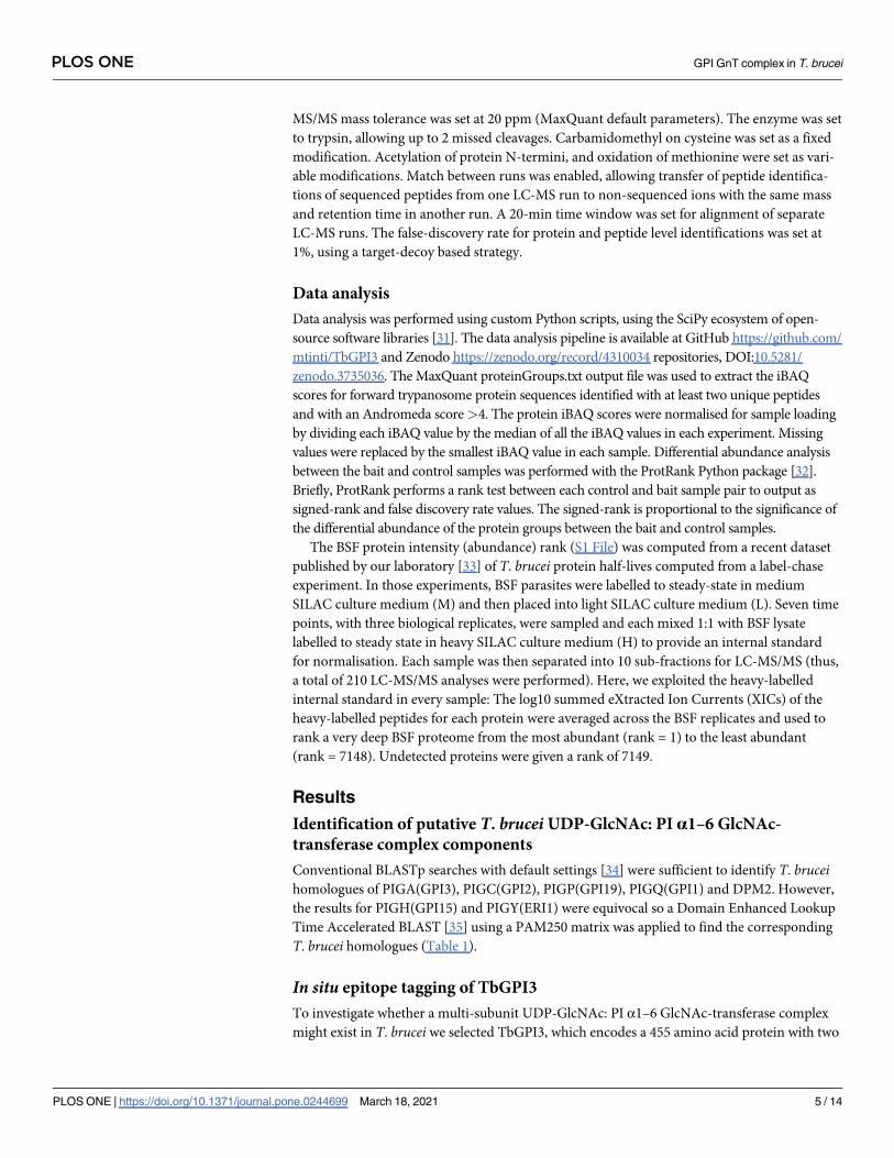
products amplified from the pMOTag43M plasmid [27] (Fig 1A). Transfected cells were
selected using hygromycin and subsequently cloned by limit-dilution. Lysates of four separate
clones were subjected to anti-Myc Western blotting (Fig 1B and 1C). In situ tagged TbGPI3-
3Myc protein was detected in all four clones at an apparent molecular weight of ~47 kDa,
somewhat lower than the predicted molecular weight of 55 kDa (Fig 1C, lanes 1–4). The speci-
ficity of this staining, although weak, is illustrated by the absence of comparable staining for
BSF WT sample (Fig 1C, lane 5). The weakness of the TbGPI3-3Myc staining with anti-Myc is
a function of the extremely low abundance of TbGPI3 in the total cell proteome, where it
ranks 6475th out of 7148 detectable protein groups (Table 1), and the limitations of protein
loading for BSF cell lysates on SDS-PAGE caused by the abundance of VSG and tubulin in
these cells (S1 File) from their surface coat and subpellicular cytoskeleton, respectively. These
(glyco)proteins can be seen running above and below the 50 kDa marker in (Fig 1B).
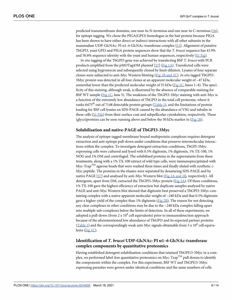
Solubilisation and native-PAGE of TbGPI3-3Myc
The analysis of epitope-tagged membrane bound multiprotein complexes requires detergent
extraction and anti-epitope pull-down under conditions that preserve intermolecular interac-
tions within the complex. To investigate detergent extraction conditions, TbGPI-3Myc
expressing cells were cultured and lysed with 0.5% digitonin, 1% digitonin, 1% TX-100, 1%
NOG and 1% DM and centrifuged. The solubilised proteins in the supernatants from these
treatments, along with a 1% TX-100 extract of wild type cells, were immunoprecipitated with
Myc-TrapTM agarose beads that were washed three times and finally eluted with synthetic c-
Myc peptide. The proteins in the eluates were separated by denaturing SDS-PAGE and by
native PAGE [37] and analysed by anti-Myc Western blot (Fig 2A and 2B, respectively). All
detergents, apart from DM, extracted the TbGPI3-3Myc protein (Fig 2A). Of these conditions,
1% TX-100 gave the highest efficiency of extraction but duplicate samples analysed by native
PAGE and anti-Myc Western blot showed that digitonin best preserved a TbGPI3-3Myc-con-
taining complex with a native apparent molecular weight of ~240 kDa and that 0.5% digitonin
gave a higher-yield of the complex than 1% digitonin (Fig 2B). The reason for not detecting
any clear complexes in other conditions may be due to the ~240 kDa complex falling apart
into multiple sub-complexes below the limits of detection. In all of these experiments, we
adopted a pull-down (from 2 x 10⁸ cell equivalents) prior to immunodetection approach
because of the aforementioned low abundance of TbGPI3 and its expected partner proteins
(Table 1) and the correspondingly weak anti-Myc signals obtainable from 5 x 10⁶ cell equiva-
lents (Fig 1C).
Identification of T. brucei UDP-GlcNAc: PI α1–6 GlcNAc-transferase
complex components by quantitative proteomics
Having established detergent solubilisation conditions that retained TbGPI13-3Myc in a com-
plex, we performed label-free quantitative proteomics on Myc-TrapTM pull downs to identify
the components within the complex. For this experiment, BSF WT and TbGPI13-3Myc
expressing parasites were grown under identical conditions and the same numbers of cells
PLOS ONE GPI GnT complex in T. brucei
PLOS ONE | https://doi.org/10.1371/journal.pone.0244699 March 18, 2021 6 / 14
were harvested and lysed in 0.5% digitonin lysis buffer. Immunoprecipitation was performed
using Myc-TrapTM beads and the proteins eluted from these two samples with SDS sample
buffer were processed to tryptic peptides for LC-MS/MS analysis (Fig 3A). The experiments
Fig 1. In situ C-terminal tagging of TbGPI3 with 3Myc. (A) Map of plasmid pMOTag43M [27] used for the in situ tagging of TbGPI3, and a
scheme of how the PCR product generated with the indicated forward (For) and reverse (Rev) primers inserts into the 3’-end of the TbGPI3ORF
(checked box) and 3’-UTR (striped box) in the parasite genome to effect in-situ tagging. HYG = hygromycin phosphotransferase selectable marker;
igr = α-β tublin intergenic region. (B) Ponceau staining of denaturing SDS-PAGE Western blot shows similar loading and transfer of lysates
(corresponding to 5×10⁶ cells) from four in-situ tagged clones (lanes 1–4) and wild type cells (lane 5). (C) The identical blot was probed with anti-
Myc antibody. TbGPI-3Myc is indicated by the arrow. The positions of molecular weight markers are indicated on the left of (B) and (C).
https://doi.org/10.1371/journal.pone.0244699.g001
PLOS ONE GPI GnT complex in T. brucei
PLOS ONE | https://doi.org/10.1371/journal.pone.0244699 March 18, 2021 7 / 14
were performed in biological triplicates and the data were analysed using MaxQuant software
and a newly developed data analysis method written in Python called ProtRank [32], see Mate-
rials and Methods. The protein groups identified (S2 File) were displayed on a plot of the
minus log10 value of their False Discovery Rate (y-axis) and enrichment rank (x-axis) between
the bait versus control samples (Fig 3B). As expected, the bait protein TbGPI3-3Myc was the
most highly enriched protein and its putative partner proteins TbGPI15, TbGPI19, TbGPI2,
TbGPI1 and TbERI1 were also significantly enriched and present in the top-7 proteins in the
pull-downs (Table 1). Notably, TbDPM2 (dolichol-phosphate-mannose synthetase 2) was not
detected. However, although TbDPM2 is annotated in the TriTrypDB database it should be
noted that, like yeast, T. bruceimakes a single-chain dolichol-phospho-mannose synthetase
(DPM1) [38] rather than a trimeric enzyme made of a soluble catalytic DPM1 subunit associ-
ated with small transmembrane DPM2 and DPM3 subunits, as found in mammalian cells. For
these reasons, we feel that the absence of DPM protein components in the T. brucei complex is
to be expected.
Interestingly, an Arv1-like protein (hereon referred to as TbArv1, Tb927.3.2480) and a
putative ubiquitin-conjugating enzyme E2 (UbCE, Tb927.2.2460) were also co-immunopre-
cipitated with TbGPI3 (Fig 3B). The data were also processed in a different way (see Materials
and Methods) that plots the experimental rank (x-axis) against the rank order of estimated
abundances of the protein groups in the total cell proteome (S1 File), generated from data in
[33], on the y-axis (Fig 3C). In this plot, the very low abundance TbArv1 (undetectable in the
total cell proteome) clusters well with the canonical and similarly low abundance UDP-Glc-
NAc: PI α1–6 GlcNAc-transferase subunits. By contrast, although UbCE is clearly highly-
enriched in the pull-down it is a much more abundant protein, suggesting that only some frac-
tion of it may be associated with the complex.
Fig 2. TbGPI3-3Myc is present in complexes in BSF T. brucei. (A) Aliquots of 2 × 10⁸ cells were harvested and lysed in lysis buffer containing different detergents
to assess TbGPI3-3Myc solubilisation. After immunoprecipitation of the supernatants with anti-Myc agarose beads, proteins were eluted with 0.5 mg/mL c-Myc
peptide and aliquots were subjected to SDS-PAGE followed by anti-Myc Western blotting. (B) Identical samples were also separated by native-PAGE and subjected
to anti-Myc Western blotting. In both cases, lane 1 corresponds to wild type cells lysed with 1% TX-100, as a negative control for anti-Myc blotting, and lanes 2–6
correspond to TbGPI3-3Myc clone1 lysed with 0.5% digitonin, 1% digitonin, 1% TX-100, 1% NOG or 1% DM, respectively.
https://doi.org/10.1371/journal.pone.0244699.g002
PLOS ONE GPI GnT complex in T. brucei
PLOS ONE | https://doi.org/10.1371/journal.pone.0244699 March 18, 2021 8 / 14
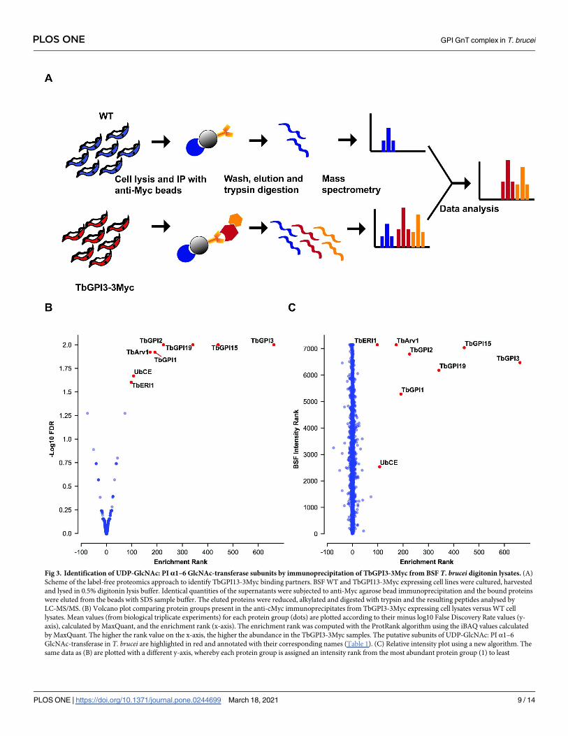
Fig 3. Identification of UDP-GlcNAc: PI α1–6 GlcNAc-transferase subunits by immunoprecipitation of TbGPI3-3Myc from BSF T. brucei digitonin lysates. (A)
Scheme of the label-free proteomics approach to identify TbGPI13-3Myc binding partners. BSF WT and TbGPI13-3Myc expressing cell lines were cultured, harvested
and lysed in 0.5% digitonin lysis buffer. Identical quantities of the supernatants were subjected to anti-Myc agarose bead immunoprecipitation and the bound proteins
were eluted from the beads with SDS sample buffer. The eluted proteins were reduced, alkylated and digested with trypsin and the resulting peptides analysed by
LC-MS/MS. (B) Volcano plot comparing protein groups present in the anti-cMyc immunoprecipitates from TbGPI3-3Myc expressing cell lysates versus WT cell
lysates. Mean values (from biological triplicate experiments) for each protein group (dots) are plotted according to their minus log10 False Discovery Rate values (y-
axis), calculated by MaxQuant, and the enrichment rank (x-axis). The enrichment rank was computed with the ProtRank algorithm using the iBAQ values calculated
by MaxQuant. The higher the rank value on the x-axis, the higher the abundance in the TbGPI3-3Myc samples. The putative subunits of UDP-GlcNAc: PI α1–6
GlcNAc-transferase in T. brucei are highlighted in red and annotated with their corresponding names (Table 1). (C) Relative intensity plot using a new algorithm. The
same data as (B) are plotted with a different y-axis, whereby each protein group is assigned an intensity rank from the most abundant protein group (1) to least
PLOS ONE GPI GnT complex in T. brucei
PLOS ONE | https://doi.org/10.1371/journal.pone.0244699 March 18, 2021 9 / 14
Discussion
The proteomics data herein suggest that: (i) The T. bruceiUDP-GlcNAc: PI α1–6 GlcNAc-
transferase complex (GPI GnT) contains the expected subunits TbGPI3, TbGPI15, TbGPI19,
TbGPI2, TbGPI1 and TbERI1. (ii) Like yeast, but unlike mammalian cells, DPM components
are not subunits of the parasite complex. (iii) An Arv1-like protein (TbArv1) is a part of the
parasite complex. (iv) A putative E2-ligase UbCE may be a part of the parasite complex.
The limitations of this study are that it does not attempt to assess stoichiometry or topology
of the T. bruceiUDP-GlcNAc: PI α1–6 GlcNAc-transferase complex components and that it
lacks an orthogonal confirmation of the presence of TbArv1 and TbUbCE in the complex.
With respect to the latter we rely, instead, on the extremely high and reproducible enrichment
of these two components in the TbGPI3-3Myc pull-downs: From undetectable (>7148th) and
2538th in the total cell proteome to 6th and 7th in the TbGPI3-3Myc pull-downs, respectively.
TbArv1 is predicted to contain four transmembrane domains and an Arv1 domain
(PF04161). Previous studies in yeast have indicated that Arv1p, although non-essential for
growth and therefore GPI-anchoring of proteins at 25˚C [39, 40], is required for the efficient
synthesis of Man1GlcN-acylPI (mannosyl-glucosaminyl-acyl-phosphatidylinositol) [41] It has
been postulated to be a GPI flippase [41, 42] helping deliver GlcN-acylPI, which is made on
the cytoplasmic face of the ER, to the active site of mannosyl-transferase I (MT I) on the lumi-
nal face of the ER. The complementation of yeast Arv1 mutants by the human Arv1 [43] and
recent findings that human Arv1 mutations lead to deficiencies in GPI anchoring [44, 45]
strongly suggest a related role in mammalian cells. However, whether it is a component of the
mammalian and yeast UDP-GlcNAc: PI α1–6 GlcNAc-transferase complexes, or indeed of
possible GlcNAc-PI de-N-acetylase of GPI flippase complexes, is unclear. It is possible that
TbArv1 plays an analogous role to that proposed for Arv1 in yeast and mammalian cells in the
T. brucei GPI pathway. However, since (unlike yeast and mammalian cells) acylation of the PI
moiety occurs strictly after the action of MT I in T. brucei [46], TbArv1 would need to facilitate
the delivery of GlcN-PI rather than GlcN-acylPI to TbMT I in this parasite. Further, it is worth
noting that unlike mammalian cells and yeast, which are thought to only translocate GlcN-
acylPI, T. brucei appears to flip-flop most of its GPI intermediates between the cytoplasmic
and lumenal surfaces of the ER [47]. Thus, it is possible that T. brucei Arv1 protein, given its
location, may play some other, perhaps regulatory, role in the UDP-GlcNAc: PI α1–6 GlcNAc-
transferase reaction. This may also be the case in mammalian and yeast cells.
Finally, a recent study in mammalian cells showed that GPI anchor biosynthesis is upregu-
lated in ERAD (endoplasmic-reticulum-associated protein degradation) deficient and PIGS
mutant cell lines, suggesting that the GPI anchor biosynthetic pathway is somehow linked to
and regulated by the ERAD system [48]. Since ERAD involves E2-dependent ubiquitylation of
misfolded proteins as they exit the ER, it is possible that the UbCE associated, in part, with
UDP-GlcNAc: PI α1–6 GlcNAc-transferase complex might play some role in regulation of the
T. brucei GPI pathway.
Supporting information
S1 Fig. Sequence alignment of GPI3, PIGA and TbGPI3.
(PDF)
abundant protein groups (7148) based on their summed eXtracted Ion Currents (XICs) for the total BSF proteome. (Details of the mass spectrometry and data analysis
are provided in in Materials and Methods).
https://doi.org/10.1371/journal.pone.0244699.g003
PLOS ONE GPI GnT complex in T. brucei
PLOS ONE | https://doi.org/10.1371/journal.pone.0244699 March 18, 2021 10 / 14
S1 Graphical abstract.
(TIF)
S1 File. Total cell proteome of bloodstream form T. brucei ranked by intensity.
(XLSX)
S2 File. Label free quantitative proteomics data.
(XLSX)
S1 Raw images. Raw images of Figs 1B, 1C, 2A and 2B.
(PDF)
Acknowledgments
We thank Drs Alvaro Acosta Serrano, Lucia Guther and Samuel Duncan for helpful advice
and the Fingerprints Proteomics Facility for expert assistance with quantitative proteomics.
Author Contributions
Conceptualization: Zhe Ji, Michele Tinti, Michael A. J. Ferguson.
Data curation: Zhe Ji, Michele Tinti, Michael A. J. Ferguson.
Formal analysis: Zhe Ji, Michele Tinti, Michael A. J. Ferguson.
Funding acquisition: Michael A. J. Ferguson.
Investigation: Zhe Ji, Michele Tinti, Michael A. J. Ferguson.
Methodology: Zhe Ji, Michele Tinti, Michael A. J. Ferguson.
Project administration: Michael A. J. Ferguson.
Resources: Michael A. J. Ferguson.
Supervision: Michael A. J. Ferguson.
Validation: Zhe Ji, Michele Tinti.
Visualization: Zhe Ji, Michele Tinti, Michael A. J. Ferguson.
Writing – original draft: Zhe Ji, Michele Tinti, Michael A. J. Ferguson.
Writing – review & editing: Zhe Ji, Michele Tinti, Michael A. J. Ferguson.
References
1. Horn D. Antigenic variation in African trypanosomes. Mol Biochem Parasitol. 2014; 195(2):123–9.
https://doi.org/10.1016/j.molbiopara.2014.05.001 PMID: 24859277
2. Mehlert A, Ferguson MAJ. Structure of the glycosylphosphatidylinositol anchor of the Trypanosoma
brucei transferrin receptor. Mol Biochem Parasitol. 2007; 151(2):220–3. https://doi.org/10.1016/j.
molbiopara.2006.11.001 PMID: 17140675
3. Treumann A, Zitzmann N, Hulsmeier A, Prescott AR, Almond A, Sheehan J, et al. Structural characteri-
sation of two forms of procyclic acidic repetitive protein expressed by procyclic forms of Trypanosoma
brucei. J Mol Biol. 1997; 269(4):529–47. https://doi.org/10.1006/jmbi.1997.1066 PMID: 9217258
4. Lane-Serff H, MacGregor P, Lowe ED, Carrington M, Higgins MK. Structural basis for ligand and innate
immunity factor uptake by the trypanosome haptoglobin-haemoglobin receptor. Elife. 2014; 3:e05553.
https://doi.org/10.7554/eLife.05553 PMID: 25497229
5. Macleod OJS, Bart JM, MacGregor P, Peacock L, Savill NJ, Hester S, et al. A receptor for the comple-
ment regulator factor H increases transmission of trypanosomes to tsetse flies. Nat Commun. 2020; 11
(1):1–12. https://doi.org/10.1038/s41467-020-15125-y PMID: 32165615
PLOS ONE GPI GnT complex in T. brucei
PLOS ONE | https://doi.org/10.1371/journal.pone.0244699 March 18, 2021 11 / 14
6. Urwyler S, Studer E, Renggli CK, Roditi I. A family of stage-specific alanine-rich proteins on the surface
of epimastigote forms of Trypanosoma brucei. Mol Microbiol. 2007; 63(1):218–28. https://doi.org/10.
1111/j.1365-2958.2006.05492.x PMID: 17229212
7. Casas-Sanchez A, Perally S, Ramaswamy R, Haines LR, Rose C, Yunta C, et al. The crystal structure
and localization of Trypanosoma brucei invariant surface glycoproteins suggest a more permissive
VSG coat in the tsetse-transmitted metacyclic stage. bioRxiv. 2018;
8. Ferguson M, Homans S, Dwek R, Rademacher T. Glycosyl-phosphatidylinositol moiety that anchors
Trypanosoma brucei variant surface glycoprotein to the membrane. Science (80-). 1988 Feb; 239
(4841):753–9. https://doi.org/10.1126/science.3340856 PMID: 3340856
9. Mehlert A, Richardson JM, Ferguson MAJ. Structure of the Glycosylphosphatidylinositol Membrane
Anchor Glycan of a Class-2 Variant Surface Glycoprotein from Trypanosoma brucei. 1998; 3. https://
doi.org/10.1006/jmbi.1997.1600 PMID: 9514751
10. Mehlert A, Sullivan L, Ferguson MAJ. Glycotyping of Trypanosoma brucei variant surface glycoprotein
MITat1.8. Mol Biochem Parasitol. 2010; 174(1):74–7. https://doi.org/10.1016/j.molbiopara.2010.06.007
PMID: 20558211
11. Treumann A, Guther MLS, Schneider P, Ferguson MAJ. Analysis of the Carbohydrate and Lipid Com-
ponents of Glycosylphosphatidylinositol Structures. In: Glycoanalysis Protocols. New Jersey: Humana
Press; 2010. p. 213–36.
12. Masterson WJ, Doering TL, Hart GW, Englund PT. A novel pathway for glycan assembly: Biosynthesis
of the glycosyl-phosphatidylinositol anchor of the trypanosome variant surface glycoprotein. Cell. 1989;
56(5):793–800. https://doi.org/10.1016/0092-8674(89)90684-3 PMID: 2924349
13. Masterson WJ, Raper J, Doering TL, Hart GW, Englund PT. Fatty acid remodeling: A novel reaction
sequence in the biosynthesis of trypanosome glycosyl phosphatidylinositol membrane anchors. Cell.
1990; 62(1):73–80. https://doi.org/10.1016/0092-8674(90)90241-6 PMID: 1694728
14. Menon AK, Schwarz RT, Mayor S, Cross GAM. Cell-free synthesis of glycosyl-phosphatidylinositol pre-
cursors for the glycolipid membrane anchor of Trypanosoma brucei variant surface glycoproteins.
Structural characterization of putative biosynthetic intermediates. J Biol Chem. 1990; 265(16):9033–42.
PMID: 1693147
15. Kinoshita T. Biosynthesis and biology of mammalian GPI-anchored proteins. Open Biol. 2020; 10
(3):190290. https://doi.org/10.1098/rsob.190290 PMID: 32156170
16. Kinoshita T, Fujita M. Biosynthesis of GPI-anchored proteins: Special emphasis on GPI lipid remodel-
ing. J Lipid Res. 2016; 57(1):6–24. https://doi.org/10.1194/jlr.R063313 PMID: 26563290
17. Pittet M, Conzelmann A. Biosynthesis and function of GPI proteins in the yeast Saccharomyces cerevi-
siae. Biochim Biophys Acta—Mol Cell Biol Lipids. 2007; 1771(3):405–20. https://doi.org/10.1016/j.
bbalip.2006.05.015 PMID: 16859984
18. Ferguson MAJ, Kinoshita T HGW. Glycosylphosphatidylinositol Anchors. In: et al. Varki A, Cummings
RD, Esko JD, editor. Essentials of Glycobiology. 2nd ed. Cold Spring Harbor (NY): Cold Spring Harbor
Laboratory Press; 2009. p. 137–50.
19. Chang T, Milne KG, Guther MLS, Smith TK, Ferguson MAJ. Cloning of Trypanosoma brucei and Leish-
mania major genes encoding the GlcNAc-phosphatidylinositol De-N-acetylase of glycosylphosphatidyli-
nositol biosynthesis that is essential to the African sleeping sickness parasite. J Biol Chem. 2002; 277
(51):50176–82. https://doi.org/10.1074/jbc.M208374200 PMID: 12364327
20. Nagamune K, Nozaki T, Maeda Y, Ohishi K, Fukuma T, Hara T, et al. Critical roles of glycosylphosphati-
dylinositol for Trypanosoma brucei. Proc Natl Acad Sci U S A. 2000; 97(19):10336–41. https://doi.org/
10.1073/pnas.180230697 PMID: 10954751
21. Lillico S, Field MC, Blundell P, Coombs GH, Mottram JC. Essential Roles for GPI-anchored Proteins in
African Trypanosomes Revealed Using Mutants Deficient in GPI8. Gilmore R, editor. Mol Biol Cell.
2003 Mar; 14(3):1182–94. https://doi.org/10.1091/mbc.e02-03-0167 PMID: 12631733
22. Nagamune K, Ohishi K, Ashida H, Hong Y, Hino J, Kangawa K, et al. GPI transamidase of Trypano-
soma brucei has two previously uncharacterized (trypanosomatid transamidase 1 and 2) and three
common subunits. Proc Natl Acad Sci U S A. 2003; 100(19):10682–7. https://doi.org/10.1073/pnas.
1833260100 PMID: 12958211
23. Hong Y, Nagamune K, Ohishi K, Morita YS, Ashida H, Maeda Y, et al. TbGPI16 is an essential compo-
nent of GPI transamidase in Trypanosoma brucei. FEBS Lett. 2006; 580(2):603–6. https://doi.org/10.
1016/j.febslet.2005.12.075 PMID: 16405969
24. Sobering AK, Watanabe R, Romeo MJ, Yan BC, Specht CA, Orlean P, et al. Yeast Ras regulates the
complex that catalyzes the first step in GPI-anchor biosynthesis at the ER. Cell. 2004; 117(5):637–48.
https://doi.org/10.1016/j.cell.2004.05.003 PMID: 15163411
PLOS ONE GPI GnT complex in T. brucei
PLOS ONE | https://doi.org/10.1371/journal.pone.0244699 March 18, 2021 12 / 14
25. Wirtz E, Leal S, Ochatt C, Cross GAM. A tightly regulated inducible expression system for conditional
gene knock-outs and dominant-negative genetics in Trypanosoma brucei. Mol Biochem Parasitol.
1999; 99(1):89–101. https://doi.org/10.1016/s0166-6851(99)00002-x PMID: 10215027
26. Hirumi H, Hirumi K. Continuous Cultivation of Trypanosoma brucei Blood Stream Forms in a Medium
Containing a Low Concentration of Serum Protein without Feeder Cell Layers. J Parasitol. 1989 Dec; 75
(6):985. PMID: 2614608
27. Oberholzer M, Morand S, Kunz S, Seebeck T. A vector series for rapid PCR-mediated C-terminal in situ
tagging of Trypanosoma brucei genes. Mol Biochem Parasitol. 2006; 145(1):117–20. https://doi.org/10.
1016/j.molbiopara.2005.09.002 PMID: 16269191
28. Perez-Riverol Y, Csordas A, Bai J, Bernal-Llinares M, Hewapathirana S, Kundu DJ, et al. The PRIDE
database and related tools and resources in 2019: Improving support for quantification data. Nucleic
Acids Res. 2019; 47(D1):D442—D450. https://doi.org/10.1093/nar/gky1106 PMID: 30395289
29. Tyanova S, Temu T, Cox J. The MaxQuant computational platform for mass spectrometry-based shot-
gun proteomics. Nat Protoc. 2016; 11(12):2301–19. https://doi.org/10.1038/nprot.2016.136 PMID:
27809316
30. Aslett M, Aurrecoechea C, Berriman M, Brestelli J, Brunk BP, Carrington M, et al. TriTrypDB: a func-
tional genomic resource for the Trypanosomatidae. Nucleic Acids Res. 2010 Jan; 38(suppl_1):D457—
D462. https://doi.org/10.1093/nar/gkp851 PMID: 19843604
31. Virtanen P, Gommers R, Oliphant TE, Haberland M, Reddy T, Cournapeau D, et al. SciPy 1.0: funda-
mental algorithms for scientific computing in Python. Nat Methods. 2020; 17(3):261–72. https://doi.org/
10.1038/s41592-019-0686-2 PMID: 32015543
32. Medo M, Aebersold DM, Medova M. ProtRank: Bypassing the imputation of missing values in differen-
tial expression analysis of proteomic data. BMC Bioinformatics. 2019; 20(1):1–12. https://doi.org/10.
1186/s12859-018-2565-8 PMID: 30606105
33. Tinti M, Guther MLS, Crozier TWM, Lamond AI, Ferguson MAJ. Proteome turnover in the bloodstream
and procyclic forms of trypanosoma brucei measured by quantitative proteomics [version 1; peer
review: 3 approved]. Wellcome Open Res. 2019; 4:1–26. https://doi.org/10.12688/wellcomeopenres.
14976.2 PMID: 31245630
34. Acland A, Agarwala R, Barrett T, Beck J, Benson DA, Bollin C, et al. Database resources of the National
Center for Biotechnology Information. Nucleic Acids Res. 2012 Nov 26; 41(D1):D8–20. https://doi.org/
10.1093/nar/gks1189 PMID: 23193264
35. Boratyn GM, Schaffer AA, Agarwala R, Altschul SF, Lipman DJ, Madden TL. Domain enhanced lookup
time accelerated BLAST. Biol Direct. 2012; 7:1–14. https://doi.org/10.1186/1745-6150-7-1 PMID:
22221860
36. Kall L, Krogh A, Sonnhammer ELL. A combined transmembrane topology and signal peptide prediction
method. J Mol Biol. 2004; 338(5):1027–36. https://doi.org/10.1016/j.jmb.2004.03.016 PMID: 15111065
37. Schagger H, von Jagow G. Blue native electrophoresis for isolation of membrane protein complexes in
enzymatically active form. Anal Biochem. 1991; 199(2):223–31. https://doi.org/10.1016/0003-2697(91)
90094-a PMID: 1812789
38. Mazhari-Tabrizi R, Eckert V, Blank M, Muller R, Mumberg D, Funk M, et al. Cloning and functional
expression of glycosyltransferases from parasitic protozoans by heterologous complementation in
yeast: The dolichol phosphate mannose synthase from Trypanosoma brucei brucei. Biochem J. 1996;
316(3):853–8. https://doi.org/10.1042/bj3160853 PMID: 8670162
39. McDonough V, Germann M, Liu Y, Sturley SL, Nickels JT. Yeast cells lacking the ARV1 gene harbor
defects in sphingolipid metabolism: Complementation by human ARV1. J Biol Chem. 2002; 277
(39):36152–60. https://doi.org/10.1074/jbc.M206624200 PMID: 12145310
40. Georgiev AG, Johansen J, Ramanathan VD, Sere YY, Beh CT, Menon AK. Arv1 regulates PM and ER
membrane structure and homeostasis but is dispensable for intracellular sterol transport. Traffic. 2013;
14(8):912–21. https://doi.org/10.1111/tra.12082 PMID: 23668914
41. Kajiwara K, Watanabe R, Pichler H, Ihara K, Murakami S, Riezman H, et al. Yeast ARV1 Is Required for
Efficient Delivery of an Early GPI Intermediate to the First Mannosyltransferase during GPI Assembly
and Controls Lipid Flow from the Endoplasmic Reticulum. Munro S, editor. Mol Biol Cell. 2008 May; 19
(5):2069–82. https://doi.org/10.1091/mbc.e07-08-0740 PMID: 18287539
42. Okai H, Ikema R, Nakamura H, Kato M, Araki M, Mizuno A, et al. Cold-sensitive phenotypes of a yeast
null mutant of ARV1 support its role as a GPI flippase. FEBS Lett. 2020; 594(15):2431–9. https://doi.
org/10.1002/1873-3468.13843 PMID: 32449190
43. Ikeda A, Kajiwara K, Iwamoto K, Makino A, Kobayashi T, Mizuta K, et al. Complementation analysis
reveals a potential role of human ARV1 in GPI anchor biosynthesis. Yeast. 2016; 33(2):37–42. https://
doi.org/10.1002/yea.3138 PMID: 26460143
PLOS ONE GPI GnT complex in T. brucei
PLOS ONE | https://doi.org/10.1371/journal.pone.0244699 March 18, 2021 13 / 14
44. Davids M, Menezes M, Guo Y, Mclean SD, Hakonarson H, Collins F, et al. Homozygous splice-variants
in human ARV1 cause GPI-anchor synthesis deficiency. Mol Genet Metab. 2020; 130(1):49–57. https://
doi.org/10.1016/j.ymgme.2020.02.005 PMID: 32165008
45. Segel R, Aran A, Gulsuner S, Nakamura H, Rosen T, Walsh T, et al. A defect in GPI synthesis as a sug-
gested mechanism for the role of ARV1 in intellectual disability and seizures. Neurogenetics. 2020; 21
(4):259–67. https://doi.org/10.1007/s10048-020-00615-4 PMID: 32462292
46. Guther MLS, Ferguson MAJ. The role of inositol acylation and inositol deacylation in GPI biosynthesis
in Trypanosoma brucei. Parasitol Today. 1995; 11(9):319. PMID: 7621823
47. Vidugiriene J, Menon AK. The GPI anchor of cell-surface proteins is synthesized on the cytoplasmic
face of the endoplasmic reticulum. J Cell Biol. 1994; 127(2):333–41. https://doi.org/10.1083/jcb.127.2.
333 PMID: 7929579
48. Wang Y, Maeda Y, Liu YS, Takada Y, Ninomiya A, Hirata T, et al. Cross-talks of glycosylphosphatidyli-
nositol biosynthesis with glycosphingolipid biosynthesis and ER-associated degradation. Nat Commun.
2020; 11(1):1–18. https://doi.org/10.1038/s41467-019-13993-7 PMID: 31911652
PLOS ONE GPI GnT complex in T. brucei
PLOS ONE | https://doi.org/10.1371/journal.pone.0244699 March 18, 2021 14 / 14