
THE β1 INTEGRIN ACTIVATES JNK AND PAXILLIN … · The H. pylori strain G27 was used in this study...
25
THE β 1 INTEGRIN ACTIVATES JNK INDEPENDENT OF CAGA, AND JNK ACTIVATION IS REQUIRED FOR HELICOBACTER PYLORI CAGA+ INDUCED MOTILITY OF GASTRIC CANCER CELLS Jared L. Snider a , Cody Allison b , Bryan H. Bellaire c , Richard L. Ferrero b , and James A Cardelli a a Department of Microbiology and Immunology and the Feist-Weiller Cancer Center, Louisiana State University Health Sciences Center, Shreveport, LA, USA; b Department of Microbiology, Monash University, Clayton 3800, Victoria, Australia; c Department of Veterinary Microbiology and Preventive Medicine, College of Veterinary Medicine, Iowa State University of Science and Technology, Ames, IA, USA. Running Title: β 1 Integrin Activates JNK and paxillin signaling in H. pylori-induced cell motility Address correspondence to: James A. Cardelli, Ph.D, Department of Microbiology and Immunology, Louisiana State University Health Sciences Center, 1501 Kings Highway, Shreveport, LA 71130, USA. E-mail: [email protected] The Helicobacter pylori CagA protein is translocated into gastric epithelial cells through a t ype IV s ecretion s ystem (TFSS), and published studies suggest CagA is critical for H. pylori-associated carcinogenesis. CagA is thought to be necessary and sufficient to induce the motogenic response observed in response to CagA + strains, as CagA interacts with proteins involved in adhesion and motility. We report that H. pylori strain 60190 stimulated AGS cell motility through a CagA- and TFSS-dependent mechanism, since strains 60190∆cagA or 60190∆cagE (TFSS-defective) did not increase motility. The JNK pathway is critical for H. pylori-dependent cell motility, as inhibition using SP600125 (JNK1/2/3 inhibitor) or a JNK2/3-specific inhibitor blocked motility. JNK mediates H. pylori-induced cell motility by activating paxillin, since JNK inhibition blocked paxillin Y118 phosphorylation, and paxillin expression knockdown completely abrogated bacterial-induced motility. Furthermore, JNK and paxillin Y118 were activated by 60190∆cagA but not 60190∆cagE, demonstrating CagA-independent signaling critical for cell motility. A β 1 integrin-blocking antibody significantly inhibited JNK and paxillin Y118 phosphorylation and cell scattering, demonstrating that CagA-independent signaling required for cell motility occurs through β 1. The requirement of both Src and FAK for signaling and motility further suggest the importance of integrin signaling in H. pylori-induced cell motility. Finally, we show that JNK activation occurs independent of known upstream kinases and signaling molecules, including Nod1, Cdc42, Rac1, MKK4, and MKK7, which demonstrates novel signaling leading to JNK activation. We report for the first time that H. pylori mediates CagA- independent signaling that promote cell motility through the β 1 integrin pathway. H. pylori infects one-half of the world’s population, establishing a chronic infection in the gastric mucosa that persists for the lifetime of the host (1,2). Although most infections only manifest as superficial gastritis, many progress to mucosal necrosis, ulceration, and atrophic gastritis, a precursor lesion of gastric adenocarcinoma (3). In animal studies, 37% of gerbils infected with virulent strains of H. pylori developed stomach tumors, demonstrating a direct link between H. pylori and gastric carcinogenesis (4). Additionally, epidemiological studies suggest that H. pylori infection increases the risk of developing gastric cancer six-fold, emphasizing the importance of this bacterium in gastric carcinogenesis (5). H. pylori pathogenesis varies based on the expression of virulence factors used for bacterial colonization and disease progression. The vacA gene is encoded by virtually all H. pylori strains, but the intense vacuolation caused by VacA varies based on genetic mosaicism (6). Peptic ulceration strongly correlates with strains encoding the most active forms of VacA (6). The cag pathogenicity island (cag PAI) contains 32 genes, many of which encode components of a putative type IV secretion system (TFSS). The only known protein transported by the TFSS is CagA, which is also expressed from the cag locus (7). During infection, CagA 1 http://www.jbc.org/cgi/doi/10.1074/jbc.M800289200 The latest version is at JBC Papers in Press. Published on March 20, 2008 as Manuscript M800289200 Copyright 2008 by The American Society for Biochemistry and Molecular Biology, Inc. by guest on June 25, 2018 http://www.jbc.org/ Downloaded from
Transcript of THE β1 INTEGRIN ACTIVATES JNK AND PAXILLIN … · The H. pylori strain G27 was used in this study...
THE β1 INTEGRIN ACTIVATES JNK INDEPENDENT OF CAGA, AND JNK ACTIVATION IS REQUIRED FOR HELICOBACTER PYLORI CAGA+
INDUCED MOTILITY OF GASTRIC CANCER CELLS Jared L. Snidera, Cody Allisonb, Bryan H. Bellairec, Richard L. Ferrerob, and James A Cardellia
aDepartment of Microbiology and Immunology and the Feist-Weiller Cancer Center, Louisiana State University Health Sciences Center, Shreveport, LA, USA;
bDepartment of Microbiology, Monash University, Clayton 3800, Victoria, Australia; cDepartment of Veterinary Microbiology and Preventive Medicine, College of Veterinary Medicine, Iowa
State University of Science and Technology, Ames, IA, USA. Running Title: β1 Integrin Activates JNK and paxillin signaling in H. pylori-induced cell motility Address correspondence to: James A. Cardelli, Ph.D, Department of Microbiology and Immunology, Louisiana State University Health Sciences Center, 1501 Kings Highway, Shreveport, LA 71130, USA. E-mail: [email protected] The Helicobacter pylori CagA protein is translocated into gastric epithelial cells through a type IV secretion system (TFSS), and published studies suggest CagA is critical for H. pylori-associated carcinogenesis. CagA is thought to be necessary and sufficient to induce the motogenic response observed in response to CagA+ strains, as CagA interacts with proteins involved in adhesion and motility. We report that H. pylori strain 60190 stimulated AGS cell motility through a CagA- and TFSS-dependent mechanism, since strains 60190∆cagA or 60190∆cagE (TFSS-defective) did not increase motility. The JNK pathway is critical for H. pylori-dependent cell motility, as inhibition using SP600125 (JNK1/2/3 inhibitor) or a JNK2/3-specific inhibitor blocked motility. JNK mediates H. pylori-induced cell motility by activating paxillin, since JNK inhibition blocked paxillinY118 phosphorylation, and paxillin expression knockdown completely abrogated bacterial-induced motility. Furthermore, JNK and paxillinY118 were activated by 60190∆cagA but not 60190∆cagE, demonstrating CagA-independent signaling critical for cell motility. A β1 integrin-blocking antibody significantly inhibited JNK and paxillinY118 phosphorylation and cell scattering, demonstrating that CagA-independent signaling required for cell motility occurs through β1. The requirement of both Src and FAK for signaling and motility further suggest the importance of integrin signaling in H. pylori-induced cell motility. Finally, we show that JNK activation occurs independent of known upstream kinases and signaling
molecules, including Nod1, Cdc42, Rac1, MKK4, and MKK7, which demonstrates novel signaling leading to JNK activation. We report for the first time that H. pylori mediates CagA-independent signaling that promote cell motility through the β1 integrin pathway.
H. pylori infects one-half of the world’s population, establishing a chronic infection in the gastric mucosa that persists for the lifetime of the host (1,2). Although most infections only manifest as superficial gastritis, many progress to mucosal necrosis, ulceration, and atrophic gastritis, a precursor lesion of gastric adenocarcinoma (3). In animal studies, 37% of gerbils infected with virulent strains of H. pylori developed stomach tumors, demonstrating a direct link between H. pylori and gastric carcinogenesis (4). Additionally, epidemiological studies suggest that H. pylori infection increases the risk of developing gastric cancer six-fold, emphasizing the importance of this bacterium in gastric carcinogenesis (5).
H. pylori pathogenesis varies based on the expression of virulence factors used for bacterial colonization and disease progression. The vacA gene is encoded by virtually all H. pylori strains, but the intense vacuolation caused by VacA varies based on genetic mosaicism (6). Peptic ulceration strongly correlates with strains encoding the most active forms of VacA (6).
The cag pathogenicity island (cag PAI) contains 32 genes, many of which encode components of a putative type IV secretion system (TFSS). The only known protein transported by the TFSS is CagA, which is also expressed from the cag locus (7). During infection, CagA
1
http://www.jbc.org/cgi/doi/10.1074/jbc.M800289200The latest version is at JBC Papers in Press. Published on March 20, 2008 as Manuscript M800289200
Copyright 2008 by The American Society for Biochemistry and Molecular Biology, Inc.
by guest on June 25, 2018http://w
ww
.jbc.org/D
ownloaded from
translocates into gastric epithelial cells via the TFSS and is phosphorylated at multiple sites by Src family kinases and Abl (8-10). CagA then influences signal transduction pathways by docking with host signaling proteins (11-14). Patients infected with CagA-positive H. pylori strains exhibit higher grades of gastric inflammation, atrophic gastritis, and an increased risk of the development of gastric adenocarcinoma (15-17).
In vitro experiments show that epithelial cells cultured with CagA+ bacteria transition from the unstimulated “cobblestone” morphology to the “hummingbird” phenotype indicative of motile cells (18-21). Additionally, H. pylori stimulates gastric cancer cell invasion through in vitro basement membranes, suggesting a role for H. pylori in cancer progression to metastasis (22-24). The mechanism of H. pylori-induced cell motility and invasion are unclear, although recent studies show that CagA associates with key regulators of cell adhesion, motility and invasion, including c-Met, Grb2, SHP-2 phosphatase and ZO-1 (11,12,14,22,25). The critical role of CagA in cancer cell motility was emphasized by Higashi, et al. who showed that CagA transfection of AGS cells was sufficient to induce the motile phenotype (11). These data suggest that CagA stimulates all signaling necessary to induce cell motility, although this hypothesis is not universally accepted (22).
In this study, we identified JNK as a key mediator of H. pylori-stimulated cell motility, and found that JNK was activated through a CagA-independent, but still TFSS-dependent mechanism. We then evaluated the molecular mechanism of CagA-independent JNK activation, and determined that CagA-independent activation of JNK occurs through β1 integrin and Src signaling. Furthermore, we identified paxillin as a downstream target of H. pylori-dependent JNK activation, and that paxillin activity is required for H. pylori-stimulated gastric cancer cell motility. JNK mediates H. pylori-induced cell motility through activation of paxillin. These data show for the first time a mechanism of CagA-independent signaling that promotes cell motility, and we conclude that a combination of CagA-dependent and CagA-independent cell signaling are required for H. pylori-induced gastric cancer cell motility.
EXPERIMENTAL PROCEDURES Cell culture and reagents AGS gastric adenocarcinoma cells (ATCC, Manassas, VA) were cultured in Ham’s F12 media (Mediatech, Herndon, VA) supplemented with 10% fetal calf serum (Gemini, West Sacramento, CA), 100 µg/mL streptomycin, 100 IU/mL penicillin (Mediatech, Herndon, VA), and 0.0375% (w/v) sodium bicarbonate (Mediatech, Herndon, VA) at 37°C in 5% CO2. Cells were cultured to 75% confluency and split either 1:5 or 1:10 using 0.025% EDTA to gently detach cells from plastic. To prevent spontaneous cell scattering associated with increased passage number, fresh stocks were thawed out monthly.
Pharmacological inhibitors LY294002, SP600125, JNK2/3 inhibitor (IX), and Bay11-7082 were obtained from EMD Biosciences (San Diego, CA). Cycloheximide and PP2 were obtained from Sigma (St. Louis, MO). The β1 blocking antibody AIIB2 was obtained from Developmental Studies Hybridoma Bank (Iowa City, IA). Prior to experiments, sub-toxic concentrations of each inhibitor were selected and Western blots to detect phosphorylated proteins were performed to confirm the functionality of the inhibitors at the concentrations used. For experiments, cells were pretreated with inhibitors for 30 minutes (LY294002, 50 µM; SP600125, 40 µM; cycloheximide, 10 µg/mL), 2 hours (Inhibitor IX, 625 nM), 2 hours (PP2, 25 µM) or 3 hours (Bay11-7082, 0.5 µg/mL) prior to the addition of bacteria Bacterial strains and culture conditions H. pylori strains 60190 (ATCC 49503, cag PAI+, vacA s1/m1) and Tx30a (ATCC 51932, cag PAI-, vacA s2/m2) were obtained from ATCC (Manassas, VA) and grown on trypticase soy agar plates supplemented with 5% adult defibrinated bovine blood (Gemini, West Sacramento, CA) at 37°C in 5% CO2 overnight prior to use in experiments. H. pylori mutant strains with disrupted cagA (60190∆cagA), cagE (60190∆cagE), and vacA (60190∆vacA) genes were a kind gift from Dr. Richard Peek (Vanderbilt University, Nashville, TN). These strains were grown on the same plates as wild-type bacteria, but under kanamycin selection (50 µg/mL). Bacteria were passaged daily, and fresh bacteria were thawed on a monthly basis.
2
by guest on June 25, 2018http://w
ww
.jbc.org/D
ownloaded from
The H. pylori strain G27 was used in this study and isogenic mutants, cagA and cagM, were constructed by natural transformation of a kanamycin cassette flanked by regions homologous to the disrupted genes (26,27). H. pylori G27 and mutant strains were routinely cultured on horse blood agar (HBA) (blood base agar No. 2, 8% (v/v) horse blood (Bio-Lab, VIC, Australia)) supplemented with antibiotics (27). Bacteria were grown at 37°C for 1-2 days under microaerobic conditions in an anaerobic jar containing a Campygen gas mix of 5% O2, 10% CO2, and 85% N2 (Oxoid, Hampshire, UK). Liquid broth cultures were incubated in 25 cm3 tissue culture flasks (Iwaki, Japan) in a final volume of 10 ml brain heart infusion (BHI) broth containing 10% (v/v) fetal bovine serum (FBS) (Thermo Electro, Melbourne, Australia) shaking at 125 rpm and 37°C overnight, prior to use in experiments. H. pylori isogenic mutants with disrupted cagA, (G27∆cagA), and cagM (G27∆cagM) were grown on the same plates as wild-type bacteria, but under kanamycin selection (20 µg/mL). Infection of AGS cells At least one day prior to infection, AGS cells were seeded at a density of 2.5-6x10⁴ cells/mL, respectively, in antibiotic-free media. For each experiment, one day-old bacteria were suspended in warmed, CO2-charged antibiotic-free media and bacterial density measured by spectrophotometer at 600 nm. Bacteria were then added to cells at a multiplicity of infection (MOI) of 100. Bacterial contact with cells was synchronized by centrifugation at 600 x g for 4 minutes, after which cells were maintained at 37°C and 5% CO2 throughout each experiment. Control cells were prepared under identical conditions. Scatter assays Cells were cultured alone or with bacteria +/- inhibitors for 18-20 hours, and monolayers were fixed with 4% paraformaldehyde. After three washes with PBS, F-actin was fluorescently labeled with Oregon Green 488 phalloidin (Molecular Probes, Eugene, OR) suspended in BSP (BSA, saponin, PBS). Fluorescent and phase images were acquired by wide-field fluorescent microscopy. Western blot analysis Lysates from bacteria alone or cells co-cultured with bacteria were collected in boiling protein loading buffer (25% 0.5 M Tris HCl, pH 6.8, 20% SDS, 4.5% bromophenol blue, 1 M sucrose). Proteins were
separated by 7.5-12% acrylamide gel electrophoresis and transferred to nitrocellulose or PVDF membranes (Pall, Pensacola, FL). Membranes were blocked with 5% milk or 0.1% gelatin in TBST (20 mM Tris, 137 mM NaCl, 0.1% Tween-20, pH 7.5) and incubated overnight with antibodies specific for tubulin (NeoMarkers, Fremont, CA), CagA (Austral Biologicals, San Ramon, CA), β-actin (Sigma, Steinheim, Germany), Rac1 (BDBiosciences, San Jose, CA), phospho-JNK, phospho-AKT, JNK, Cdc42, phospho-MKK4, MKK4, phospho-MKK7, MKK7, phospho-paxillinY118, and paxillin (Cell Signaling Technology, Beverly, MA) and phospho-paxillinS178 (EMD Biosciences, San Diego, CA) in 5% BSA or 0.1% gelatin in TBST. Blots were followed with HRP-conjugated secondary antibodies (Amersham Biosciences, Piscataway, NJ), and proteins were detected by ECL (Amersham Biosciences, Piscataway, NJ). Colloidal gold motility assay Assay derived from colloidal gold phagokinetic assay as described previously (28). Briefly, 12 mm coverslips were immersed in a gelatin solution (Sigma, 300 bloom; 500 mg in 300 mL water) and heated at 90°C for 10 minutes. The gelatin was then removed and the coverslips were dried at 70°C for 45 minutes. After the coverslips cooled, they were aseptically transferred to 24-well plates. The colloidal gold suspension was prepared by mixing 11 mL water, 6 mL of a 36.5 mM Na2CO3 solution, and 2 mL of a 14.5 mM AuHCl4 solution (Fisher Scientific, Hampton, NH). The mixture was gently swirled high over a Bunsen burner until the first sign of boiling, after which the flask was immediately removed from heat and 2 mL of a 0.1% formaldehyde solution was quickly added.
The hot solution was allowed to slightly cool on the bench top as the flask was swirled, during which time the solution turned moderately brown in color. After the gold solution changed color, 2 mL were added atop each coverslip in the 24-well plate, and the plates were incubated at 37°C and 5% CO2 for 3 hours to allow the gold particles to settle onto the coverslips. Coverslips were then gently washed and stored in PBS at 4°C until use. For motility assays, 1x10⁴ cells were seeded onto prepared coverslips and spun at 600 x g to maximize cell attachment to the substratum (personal observation). After 6-12 hours of recovery time, bacteria were added as described
3
by guest on June 25, 2018http://w
ww
.jbc.org/D
ownloaded from
above. In inhibition assays, the inhibitors were added 30 minutes prior to the addition of bacteria, except for SU11274, which was added overnight following a 6 hour recovery period.
After 18-22 hours, cells were fixed with 4% paraformaldehyde and permeabilized with BSP for 1 hour at room temperature without agitation. Coverslips were rinsed three times with PBS and mounted onto slides with Slowfade Gold antifade reagent with Dapi (Molecular Probes, Eugene, Oregon). Phase and fluorescent images were taken of each field, and the area cleared by single or small colonies of cells measured using ImageJ software (NIH). The area was then divided by the number of nuclei in the corresponding fluorescent image to give the average area cleared per cell. Fifteen to thirty fields were visualized in this manner for an average of 100 cells per coverslip, and between one and three coverslips were used per experimental condition. Data for two to three separate experiments were compared and presented as the fold change over control or area cleared per cell. Standard deviation and error were included, and p-values were calculated by either the paired two-sample t-test for means (Microsoft Excel) or ANOVA with Tukey’s HSD test (GraphPad Prism). Short Interfering RNA (siRNA) Transfection Transfection protocol was modified from Chan et al. (29). Briefly, 1.25X10⁴ AGS cells were seeded into a 24-well plate, and the following day were transfected with Lipofectamine 2000 (Invitrogen, San Diego, CA) according to product protocol using 1.5 uL of stock Cdc42 or luciferase siRNA (20 uM; a kind gift from Marc Symons, Feinstein Institute for Medical Research, Manhasset, NY). After two days fresh medium was added, and cells were either infected with H. pylori for scatter assays or collected for Western blot analysis. Lentiviral Delivery of Short Hairpin RNA (shRNA) Stable shRNA cell lines were generated using MISSION shRNA lentiviruses (Sigma, St. Louis, MO) according to manufacturer’s protocol. Briefly, 5X10⁴ AGS cells were seeded in 6-well dishes and each well was left untreated or infected with one of the five lentiviral clones provided in each target transcript kit (MOI 0.5) plus polybrene (8 µg/mL). The following day, virus was aspirated and replaced with fresh media, and the next day the cells were washed and media containing puromycin (0.6 µg/mL) was added for selection of
stable transfectants. After three days of daily washes, stable clones were screened for maximal target protein expression knock down. The clones selected were: Rac1 (MISSION shRNA TRCN0000004873), MKK4 (MISSION shRNA TRCN0000039916), MKK7 (MISSION shRNA TRCN0000000587), paxillin (MISSION shRNA TRCN0000123138), and non-target control (MISSION shRNA SHC002V). Stable GFP shRNA cell lines were generated by infecting AGS cells with the lentiviral shRNA vector GFP-FSIPPW (a kind gift from Dr. Andrew Kung, Dana-Farber Cancer Institute, Boston, MA). Stable cells were cultured in puromycin-containing media. Adenovirus Infection Transient expression of GFP-tagged FAK-related non-kinase (Ad-GFP-FRNK) or GFP (Ad-GFP) was facilitated through adenoviral delivery. Ad-GFP-FRNK and Ad-GFP viruses were a kind gift of Joan Taylor (UNC-Chapel Hill, NC). AGS cells were incubated overnight with virus at an MOI of 10. The following day, cells were then infected with H. pylori strains for either 1 hour (Western blot analysis) or overnight. After overnight incubation, cells were fixed and stained for F-actin using Alexa Fluor 546 phalloidin (Molecular Probes). GFP and Texas Red images were captured, and merge images were generated using ImageJ (NIH). Nod1KD Cell Analysis AGS cells stably expressing siRNA’s to either the caspase-activation recruitment domain of Nod1 or an irrelevant gene (enhanced green fluorescent protein, EGFP) were generated by Dr. J. Viala (Institut Pasteur, Paris) (27). Briefly, AGS cells were transfected with plasmid constructs in which short hairpin RNA to the genes of interest had been cloned into psiRNA-hHIneo (InvivoGen, San Diego, CA). To select for clones that had stably incorporated the respective plasmids into their genomic DNA, the cells were grown in RPMI medium containing 10% FCS and 400 µg/mL G418 (Invitrogen Life Technologies, Cergy-Pontoise, France). G418-resistant cells were isolated and expanded so as to generate stable knockdown clones for Nod1 or EGFP genes. The characterization of these clones will be described in detail elsewhere (RLF, unpublished data). Successful knock down of Nod1 expression in these cells was previously published (30).
4
by guest on June 25, 2018http://w
ww
.jbc.org/D
ownloaded from
Chemokine Analysis IL-8 production by EGFP and Nod1KD cells induced by H. pylori co-culture was determined by collecting 24-hour supernatants and using the BD OptEIA kit (BD Biosciences, San Diego, CA).
RESULTS H. pylori-dependent AGS cell scattering and motility require CagA and the TFSS
Epithelial cell lines co-cultured with CagPAI+
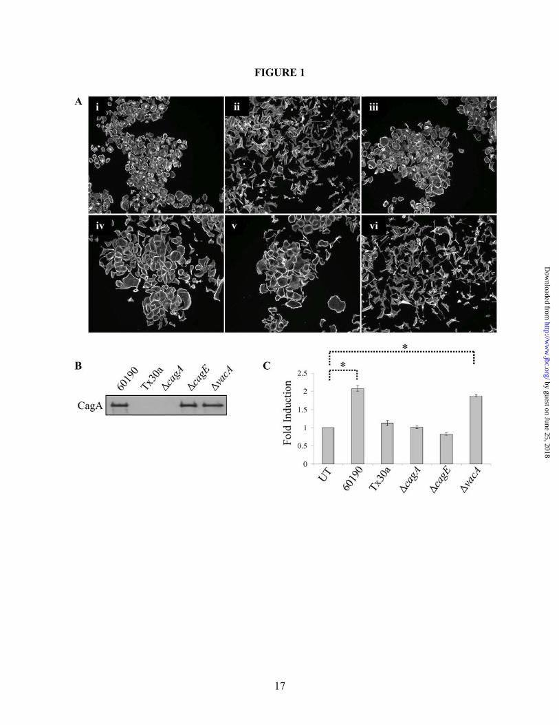
H. pylori strains exhibit the “hummingbird” phenotype associated with EMT (14,18). To demonstrate that a similar phenotype occurred in response to exposure to H. pylori in our experimental system, AGS gastric cancer cells were co-cultured with H. pylori 60190 (Cag PAI+, vacuolating) or Tx30a (Cag PAI-, non-vacuolating) for 18 hours, and cells were fixed and fluorescently labeled for F-actin. As shown in Figure 1A(ii), AGS cells cultured with H. pylori 60190 exhibited the “hummingbird” phenotype indicative of motile cells, while cells cultured with H. pylori Tx30a (Figure 1A(iii)) showed no morphological changes compared to untreated cells (Figure 1A(i)).
To determine the bacterial factors required for AGS cell scattering, isogenic mutants of H. pylori 60190 with gene disruptions in cagA (60190∆cagA), cagE, encoding a gene product required for full TFSS functionality (60190∆cagE), or vacA (60190∆vacA) were used in co-culture assays. Western blot analysis was first performed on whole bacteria lysates to confirm that the mutant strains 60190∆cagE and 60190∆vacA, but not 60190∆cagA, expressed CagA (Figure 1B). AGS cells were then co-cultured with each of these strains, and the extent of cell scattering was determined by immunofluorescence microscopy. As shown in Figures 1A(iv) and 1A(v), 60190∆cagA and 60190∆cagE, respectively, did not induce wild-type cell scattering, demonstrating that the delivery of CagA into host cells is necessary to induce the hummingbird phenotype. The mutant strain 60190∆vacA caused a scattering phenotype similar to wild-type 60190 (Figures 1A(vi) and 1A(ii), respectively), demonstrating that the vacuolating cytotoxin of H. pylori plays no role in the induction of this morphological response.
Previous studies have measured H. pylori-induced cell motility as the percent of cells per
field that exhibit the “hummingbird” phenotype (21,31-33). To quantitate the level of participation of bacterial and host proteins in cell motility, a modified colloidal gold phagokinetic assay was utilized to investigate the role of CagA, the TFSS itself (using the CagE mutant), and the vacuolating cytotoxin in cell motility. Briefly, cells and bacteria were seeded onto a colloidal gold substrate and cell motility was measured as a function of the area that cells cleared as they moved during the assay. As shown in Figure 1C, H. pylori 60190 stimulated a two-fold increase in cell motility over untreated cells. Additionally, only the VacA mutant caused a comparable increase in motility over untreated cells, while no significant increase in motility was observed by Tx30a and the CagA or TFSS mutants. These data correlate with our scatter data from Figure 1A, and demonstrate that cell motility is a CagA- and TFSS-dependent, but VacA-independent event.
H. pylori-induced cell scattering and motility require JNK signaling
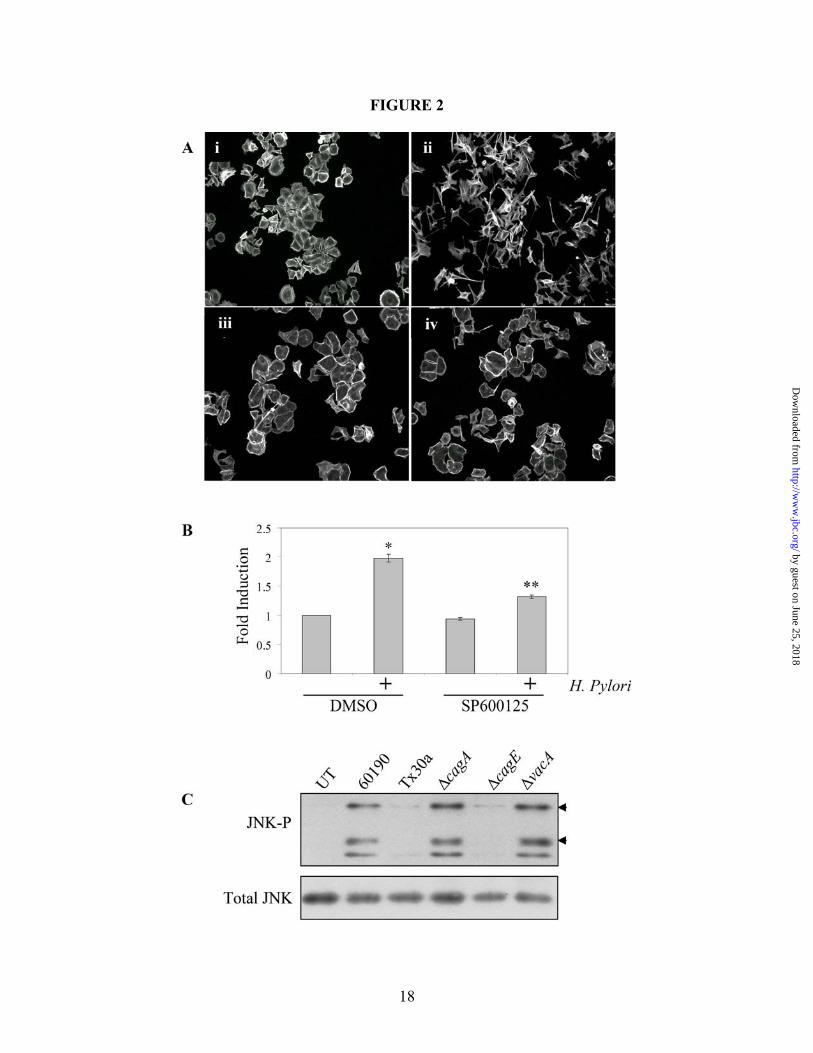
Recent evidence shows that the JNK pathway is a key mediator of cytoskeletal extensions and cell motility in a number of experimental systems (34,35). To determine if JNK plays a role in H. pylori-induced cell scattering and motility, AGS cells were pretreated with the pan-JNK inhibitor, SP600125 (Figure 2A (iii and iv)), or DMSO (Figure 2A (i and ii)) for 30 minutes prior to the addition of H. pylori strains for 18 hours. Cells pretreated with SP600125 did not scatter in response to H. pylori compared to DMSO control cells (Figures 2A iv and ii, respectively), demonstrating that JNK is required for cell scattering. Motility assays were then employed, and Figure 2B shows that SP600125 significantly decreased AGS cell motility, demonstrating that JNK signaling is required for H. pylori-induced cell scattering and motility. H. pylori activates JNK in a CagA- independent, TFSS-dependent manner
The report by Higashi, et al. suggests that CagA is sufficient to stimulate all pathways required for cell motility (11). If this hypothesis is true, then all signaling pathways required for H. pylori-induced cell motility would require CagA delivery for activation. To test this hypothesis, Western blot analysis was performed on cells co-
5
by guest on June 25, 2018http://w
ww
.jbc.org/D
ownloaded from
cultured with the parental and mutant H. pylori strains, and JNK phosphorylation in response to different bacterial stimuli was analyzed (Figure 2C). Surprisingly, JNK was phosphorylated in response to co-culture with 60190, 60190∆cagA or 60190∆vacA, while strains lacking a functional TFSS (Tx30a and 60190∆cagE) showed no induction. These data show that JNK signaling, which is required for H. pylori-stimulated cell motility, is activated in a CagA-independent, but still TFSS-dependent manner.
H. pylori stimulates JNK through a Nod1-independent mechanism
Although activation of JNK occurs in a CagA-independent, but still TFSS-dependent manner, the mechanism of TFSS-dependent JNK signaling is unknown. A recent report by Viala et al. demonstrated that peptidoglycan, a component of the bacterial cell wall, is transported into the cytoplasm and recognized by the pathogen recognition molecule Nod1, causing NF-κB activation and IL-8 secretion (27). Nod1 is also reported to regulate JNK and p38 activity, and therefore peptidoglycan-mediated Nod1 induction may be the mechanism of CagA-independent JNK activation leading to cell motility (36,37). To test this hypothesis, cells stably expressing siRNAs targeting Nod1 (Nod1KD) or an irrelevant gene (EGFP) were analyzed for JNK phosphorylation after co-culture with H. pylori strain G27 and isogenic mutant strains G27∆cagA or G27 ∆cagM (TFSS-defective) (26). Nod1 expression was significantly abolished in the Nod1KD cells, as published previously (30). To confirm a knockdown of Nod1 expression, H. pylori-stimulated IL-8 production was analyzed by ELISA, and Supplemental Figure 1A confirms a significant decrease in IL-8 production by Nod1KD cells in response to H. pylori G27. In Supplemental Figure 1B, G27 stimulated JNK phosphorylation in a CagA-independent manner both EGFP and Nod1KD cell lines, which demonstrates two important points. First, CagA-independent JNK activation is not specific to H. pylori strain 60190. Second, the bacterium stimulates JNK phosphorylation independent of Nod1 signaling. Further, Supplemental Figure 1C shows that Nod1KD cells exhibit the scattered phenotype in response to H. pylori similar to
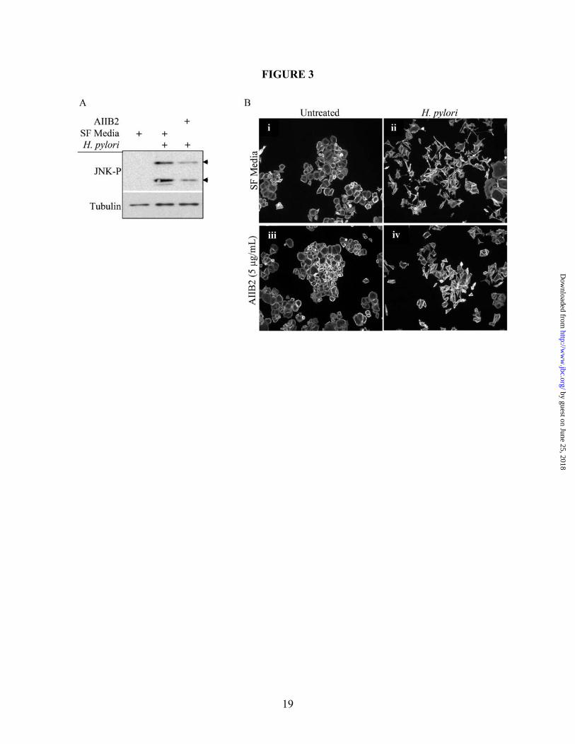
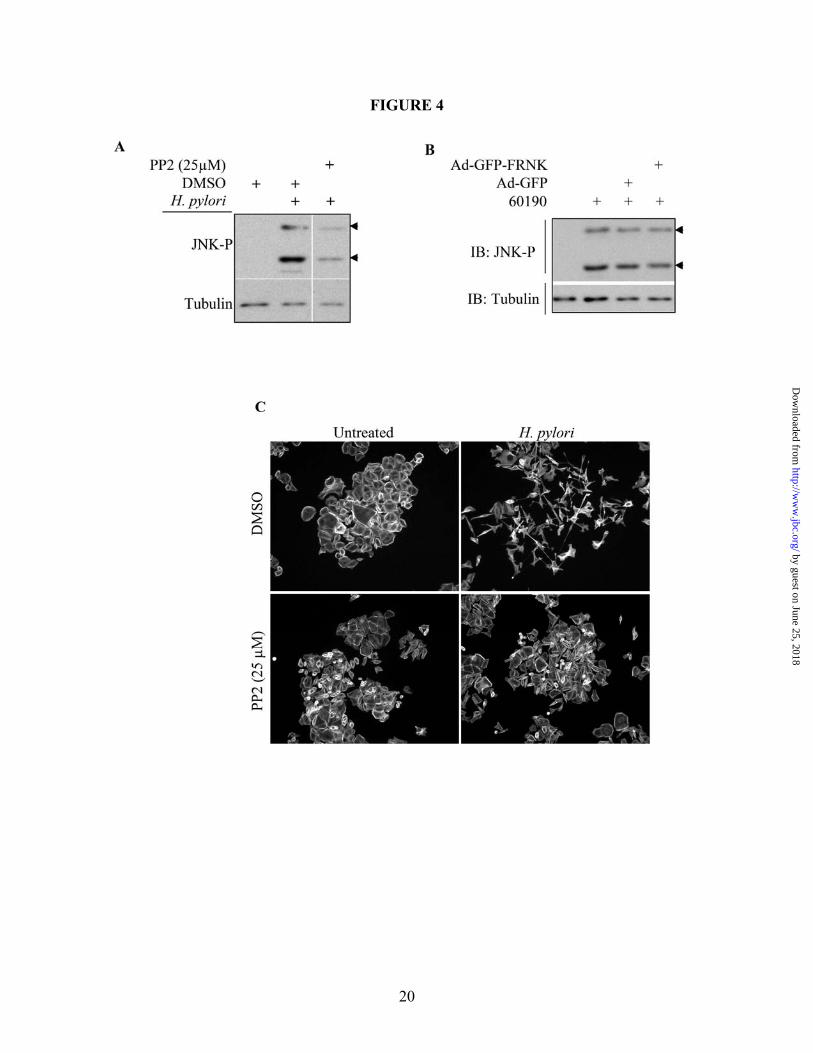
EGFP control cells. These observations demonstrate that H. pylori activates JNK-dependent cell scattering through a Nod1-independent mechanism. H. pylori-induced JNK phosphorylation and cell scattering occur through integrin signaling A recent publication by Kwok, et al., showed that the TFSS requires the interaction between the TFSS-associated CagL protein and the α5β1 integrin complex to initiate translocation of CagA into the cell. This interaction also activates FAK and Src, both of which play a key regulatory role in integrin signaling (38). This group pretreated AGS cells with a β1-blocking antibody (AIIB2), which prevented CagA translocation and phosphorylation (38). Evidence also shows that integrin signaling can stimulate JNK activity (39). To determine if TFSS-dependent β1 signaling causes activation of JNK leading to H. pylori-induced cell motility, AGS cells were pretreated with AIIB2 for one hour prior to co-culture with H. pylori 60190. Western blot analysis was then performed, and Figure 3A shows that incubation with the β1 blocking antibody significantly, though not completely, blocked JNK phosphorylation. Scatter assays were also performed, and Figure 3B shows that pretreatment with AIIB2 significantly blocked the robust scattering phenotype induced by H. pylori alone, although the cell colonies loosen up, indicative of incomplete inhibition of motility. These data demonstrate, though, that β1 integrin signaling is required for CagA-independent JNK phosphorylation and cell scattering. The previously-cited authors also show that H. pylori-mediated β1 integrin stimulation results in activation of both Src and FAK (38). These two kinases form a signaling complex that mediates downstream integrin signaling (40). Therefore, we tested if Src and FAK influenced JNK phosphorylation and cell scattering. In Figure 4A, cells pretreated with the Src inhibitor, PP2, showed a significant decrease in H. pylori-stimulated JNK phosphorylation compared to DMSO alone. Additionally, PP2 blocked H. pylori-induced cell scattering, as shown in Figure 4C. These data demonstrate that Src is required for H. pylori-induced JNK activation and cell scattering.
6
by guest on June 25, 2018http://w
ww
.jbc.org/D
ownloaded from
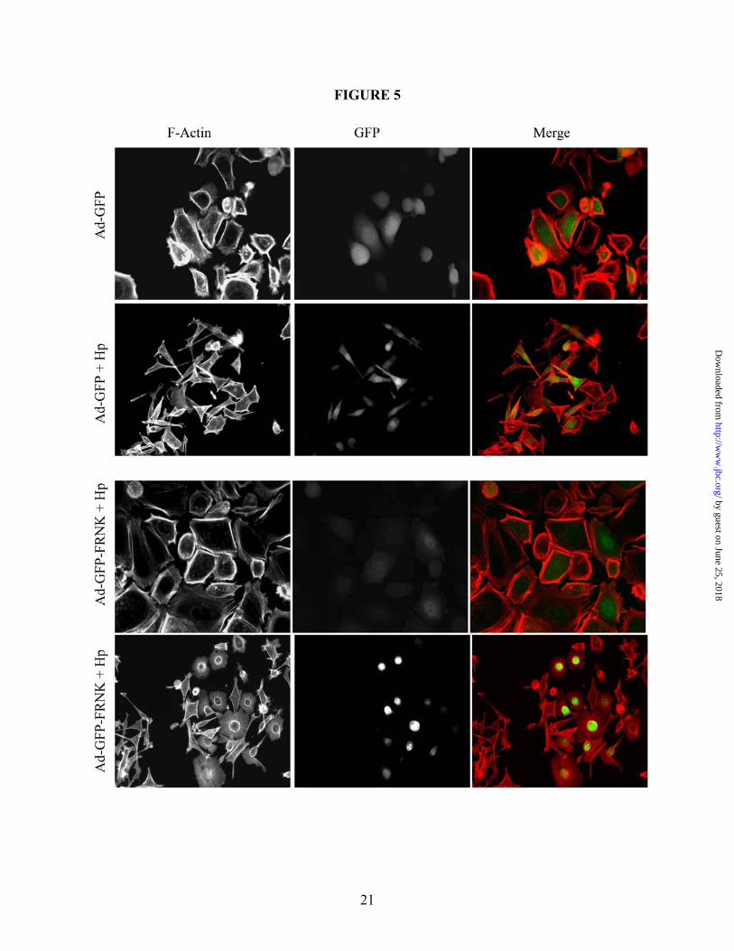
To address the role of FAK in JNK activation and cell scattering, we employed a protein consisting of the carboxy-terminal noncatalytic domain of FAK, termed FAK-related non-kinase (FRNK). FRNK is a separate protein endogenously expressed which, when over expressed, inhibits FAK activity (41). Therefore, to inhibit FAK in our studies, GFP-labeled FRNK or GFP alone was expressed in AGS cells using a replication-defective adenovirus construct (Ad-GFP-FRNK and Ad-GFP, respectively) prior to co-culture with H. pylori 60190 for 1 hour. As shown in Figure 4B, FRNK expression did not inhibit H. pylori-induced JNK phosphorylation compared to the GFP control construct, demonstrating that FAK is not required for JNK activity. In Figure 5, though, cells that expressed GFP-FRNK showed a striking inhibition of H. pylori-induced cell scattering. These cells showed high nuclear FRNK localization and loss of cortical actin and stress fibers, compared to GFP control cells, which still scattered in response to H. pylori. These data demonstrate that while JNK requires Src activity, JNK phosphorylation occurs independent of FAK. But both Src and FAK inhibition blocks the H. pylori-induced morphogenic response. H. pylori stimulates JNK through a PI3K, Cdc42- and Rac1-independent mechanism
A key pathway that regulates cancer cell survival is the PI3K pathway, which is known to play a role in integrin signaling and JNK activation (42,43). To determine if PI3K regulates JNK activity, AGS cells were pretreated with LY294002 prior to co-culture with H. pylori 60190 for 1 hour. Western blot analysis was performed on these lysates, and Supplemental Figure 2A shows that H. pylori-induced JNK activity was not affected by LY294002, which demonstrates that JNK is not regulated by PI3K activity.
The Rho GTPases Cdc42 and Rac1 are well known regulators of actin cytoskeletal changes; Cdc42 mediates filopodial protrusions at the leading edge of motile cells and Rac1 controls the lamellipodial sheets that extend the cell forward (44). Yamauchi, et al., used dominant negative constructs to show that Cdc42 and Rac1 also regulate neuronal extension by stimulating JNK activity (45). Additionally, H. pylori is known to
activate both Cdc42 and Rac1 (46). To determine if these Rho GTPases play a role in H. pylori-dependent JNK activation and cell motility, siRNA technology was applied to knock down expression of Cdc42 or Rac1 for Western blot analyses and scatter assays. AGS cells were transiently transfected with Cdc42 siRNAs or control siRNAs (luciferase), and these cells were co-cultured with H. pylori 60190 for 1 hour and lysates were collected for Western blot analysis. Supplemental Figure 2B shows that Cdc42 siRNA (siCdc42)-treated cells show considerable loss of Cdc42 compared to control cells (siLuc), but no difference in H. pylori-stimulated JNK phosphorylation was observed between the two transfection conditions. Furthermore, Supplemental Figure 2C shows that there was also no difference in H. pylori-stimulated cell scattering between siCdc42 (ii) and siLuc cells (i), demonstrating that Cdc42 is not required for JNK phosphorylation or cell scattering. To determine if JNK activity is regulated by H. pylori-dependent Rac1 activation, AGS cells stably expressing Rac1 shRNAs were generated by lentiviral delivery (see Methods). Supplemental Figure 2D shows that Rac1 shRNA-expressing cells (shRac1) exhibit significant Rac1 protein knockdown compared to GFP shRNA-expressing control cells (shGFP). Additionally, in Supplemental Figures 2E and 2F, respectively, Rac1 knockdown did not affect H. pylori-induced JNK phosphorylation or cell scattering. These data demonstrate that H. pylori stimulates JNK through a Cdc42- and Rac1-independent mechanism. H. pylori stimulates MKK4 phosphorylation but activates JNK independent of MKK4 The Map Kinase Kinase 4 (MKK4) is one of two Map kinase kinases identified as direct upstream JNK kinases (47,48). To determine if H. pylori activates MKK4 to stimulate JNK activation, AGS cells were co-cultured with the indicated H. pylori strains and collected for Western blot analysis. As shown in Supplemental Figure 3A, MKK4 was phosphorylated in response to H. pylori 60190, H. pylori 60190∆cagA and H. pylori 60190∆vacA, but not H. pylori 60190∆cagE, demonstrating that MKK4 is activated in a CagA-independent, TFSS-dependent manner similar to JNK. To further determine if MKK4 was required for JNK phosphorylation,
7
by guest on June 25, 2018http://w
ww
.jbc.org/D
ownloaded from
AGS cells stably expressing shRNAs against MKK4 (shMKK4) were generated. Supplemental Figure 3B shows successful and efficient MKK4 expression knock down compared to non-target lentivirus-infected cells (shCtrl). Also, although H. pylori-stimulated phosphorylation of JNK isoforms 2 and 3 (JNK2/3) was unaffected in the shMKK4 cells (upper arrow head), JNK isoform 1 (JNK1) phosphorylation was significantly reduced in the knock down cells compared to shCtrl cells (lower arrow head). This suggests that H. pylori-stimulated MKK4 demonstrates specificity for the JNK1 isoform. Cells were then co-cultured with H. pylori 60190 for scatter assays, and Supplemental Figure 3C shows that shMKK4 cells still scattered in response to H. pylori, suggesting that neither MKK4 nor JNK1 phosphorylation are required for the H. pylori-induced morphological response. H. pylori stimulates JNK phosphorylation and cell scattering through an MKK7-independent mechanism Besides MKK4, only Map Kinase Kinase 7 (MKK7) is known to directly regulate JNK activity (48). Therefore, one would predict that if JNK is required for cell scattering and shMKK4 cells show a loss of JNK1 phosphorylation but the cells still scatter, then MKK7 would regulate JNK2/3 activity and be required for H. pylori-dependent cell scattering. Indeed, when JNK2/3 activity was pharmacologically inhibited using an inhibitor specific to isoforms 2 and 3 but not 1, H. pylori-stimulated cell scattering was significantly blocked although the normal phenotype was not completely restored; this demonstrates a requirement for JNK2/3 activity in H. pylori-stimulated cell scattering (Supplemental Figure 4A). Stable AGS cells expressing shRNAs against MKK7 were then generated (shMKK7) to address the role of MKK7 in H. pylori-stimulated signaling and cell scattering. Surprisingly, Supplemental Figure 4B shows no difference between shCtrl and shMKK7 cells in the phosphorylation of any JNK isoforms in the presence of H. pylori 60190, although shMKK7 cells showed significant loss of MKK7 expression. Additionally, shMKK7 cells scattered to a similar extent to control cells in response to H. pylori 60190 (Supplemental Figure 4C), demonstrating
that H. pylori induces JNK phosphorylation and cell scattering independent of MKK7. JNK phosphorylation and cell scattering occur independent of both MKK4 and MKK7 Although the JNK2/3 inhibitor blocked H. pylori-dependent cell scattering, MKK7 expression knock down failed to prevent JNK2/3 phosphorylation. To test the hypothesis that loss of one of these kinases is complemented by the presence of the other, AGS cells stably expressing both MKK4 and MKK7 shRNAs (shMKK4/7) cells were generated by lentiviral delivery and used in co-culture experiments. Supplemental Figure 5A confirms that MKK4 and MKK7 expression is almost completely abrogated in shMKK4/7 cells, and Supplemental Figure 5B indicates that H. pylori-induced JNK 2/3 phosphorylation was unaffected in these both control and shMKK4/7 cells. Additionally, shCtrl cells or shMKK4/7 cells were co-cultured with H. pylori 60190 for 18 hours and fixed and stained for F-actin. Supplemental Figure 5C shows that H. pylori-stimulated cell scattering was not blocked, demonstrating that H. pylori-stimulated JNK2/3 phosphorylation and cell scattering occur independent of both MKK4 and MKK7. JNK mediates H. pylori-dependent cell motility through paxillin A major function of JNK is to regulate activity of the AP-1 transcription factor, which in turn alters gene expression (48-50). Additionally, recent evidence shows that JNK can influence cell motility by activating downstream effectors that stabilize microtubules and regulate actin reorganization and cell adhesion (35,51,52). Cells pretreated with the protein synthesis inhibitor, cycloheximide, or the NF-κB inhibitor, Bay11-7082, were unable to block H. pylori-stimulated cell scattering in separate experiments, demonstrating that JNK mediates H. pylori-induced cell scattering through a gene expression-independent mechanism (Supplemental Figure 6). Paxillin is a component of focal adhesions, which facilitate attachment of the actin cytoskeleton to the extracellular matrix (51). Paxillin is phosphorylated at multiple serine and tyrosine residues by different upstream activators to regulate focal adhesion turnover and cell migration (53). Phosphorylation at tyrosine residue
8
by guest on June 25, 2018http://w
ww
.jbc.org/D
ownloaded from
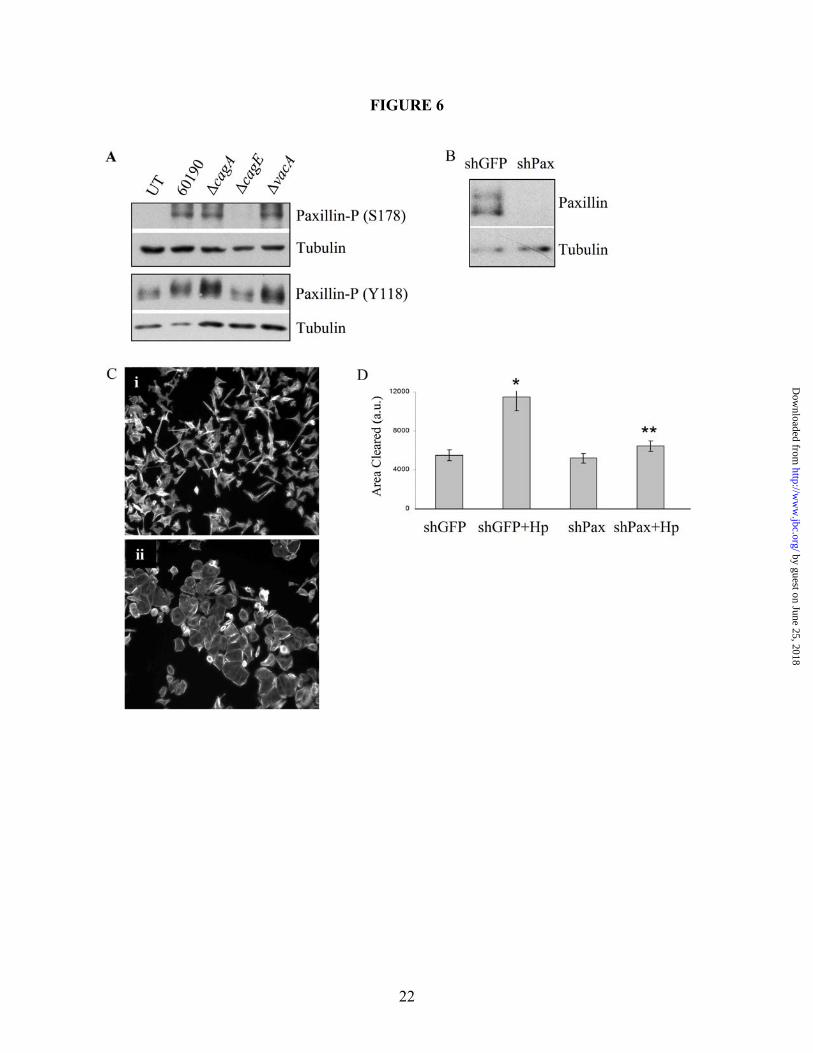
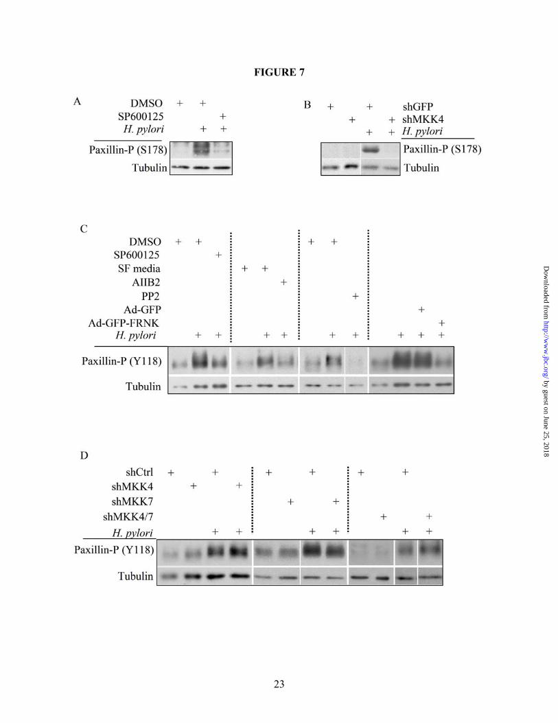
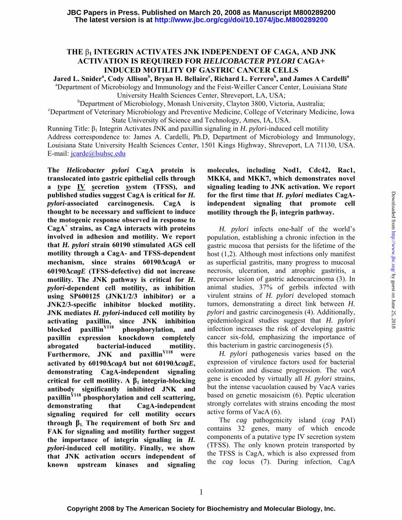
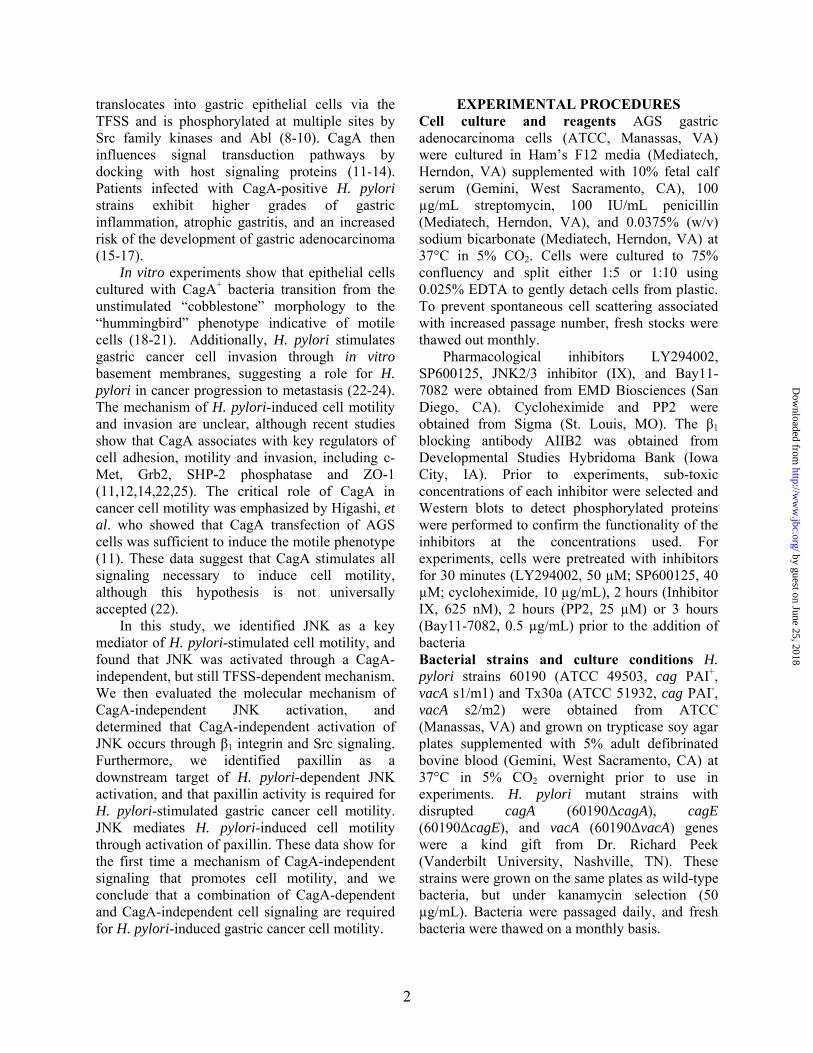
118 (paxillinY118) occurs through Src and FAK in response to growth factors, and serine 178 (paxillinS178) is known to be phosphorylated by JNK (35,51-53). Due to this link between JNK and paxillin, Western blot analyses were performed on AGS co-culture lysates to determine paxillin activation in response to H. pylori 60190. As shown in Figure 6A, paxillin was phosphorylated at both Y118 and S178 in a CagA-independent but TFSS-dependent manner. AGS cells stably expressing paxillin shRNAs were generated (shPax) to address the role of paxillin in H. pylori-induced cell signaling and motility, and Figure 6B shows loss of detectable paxillin in shPax cells compared to shGFP control cells. Cell scattering and motility assays show a striking decrease in shPax cell scattering and motility compared to shGFP cells in response to H. pylori co-culture (Figures 6C and 6D, respectively), which demonstrates that paxillin is required for H. pylori-induced cell scattering and motility. To determine if JNK regulates paxillin activity, AGS cells were pretreated with the pan-JNK inhibitor SP600125 (targets isoforms 1-3) prior to co-culture with H. pylori 60190 for 1 hour. Western blot analysis was then performed on the lysates and Figure 7A shows that phosphorylation at paxillinS178 was significantly decreased in response to the JNK inhibitor. Next, shGFP or MKK4 cells were co-cultured with H. pylori 60190 for 1 hour and collected for Western blot analysis to determine if paxillinS178 phosphorylation is induced by JNK1. As shown in Figure 7B, shMKK4 cells showed no paxillinS178 phosphorylation compared to strong induction in shGFP cells. This suggests that paxillinS178 may be a substrate of JNK1, but phosphorylation at S178 is not required for H. pylori-stimulated cell scattering. Figure 7C shows that paxillinY118 phosphorylation was also blocked in the presence of SP600125, demonstrating that paxillinY118 is regulated by JNK activity. Figure 7C also shows that paxillinY118 phosphorylation is reduced in response to pretreatment with AIIB (β1 blocking antibody), PP2 (Src inhibitor), or FRNK (endogenous FAK inhibitor) compared to control conditions, demonstrating that H. pylori stimulates paxillinY118 through the integrin signaling pathway. Western blot analysis also showed no change in paxillinY118 phosphorylation in response
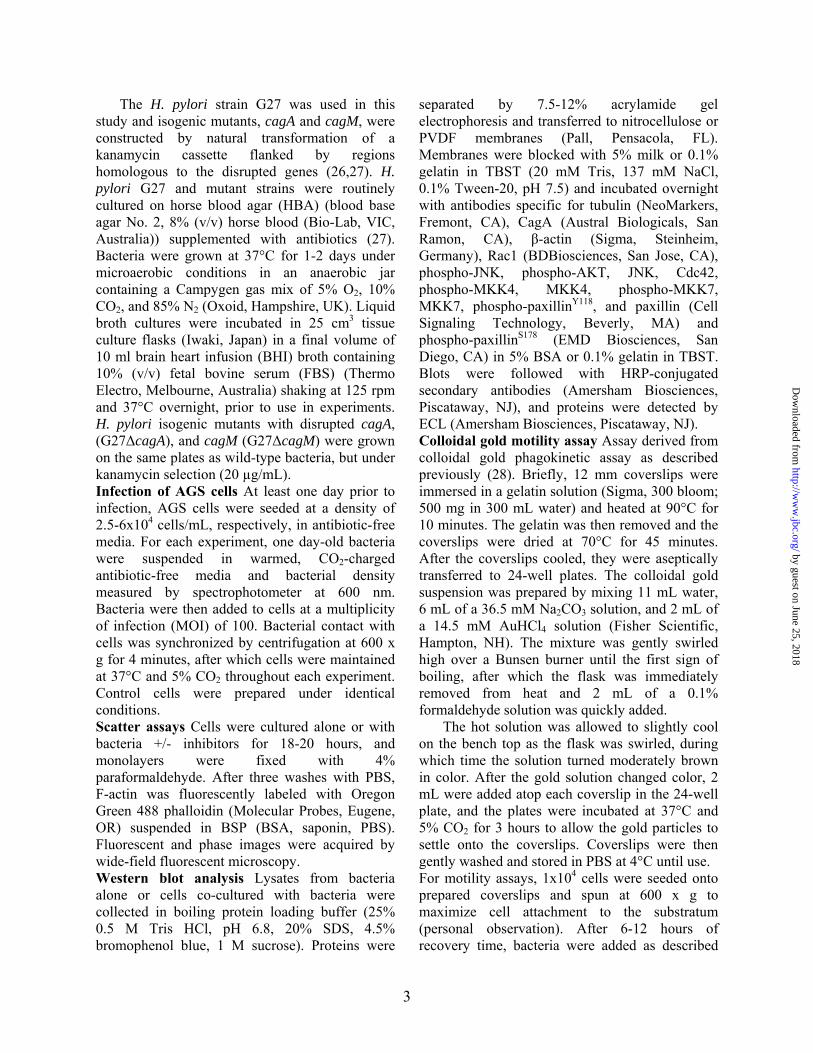
to H. pylori in shMKK4, shMKK7, and shMKK4/7 cells compared to shCtrl cells, which shows that paxillinY118 is phosphorylated independent of known upstream kinases (Figure 7D). These data also suggest that this paxillinY118 may be important in H. pylori-dependent motility.
DISCUSSION In recent years, CagA has been shown to be
sufficient to induce cell scattering (“hummingbird” morphology) when transiently expressed in cells, and CagA is thought to be the major stimulus of cell morphological changes (11,12,21). These previous data conflict with our findings, in that the JNK pathway, which is required for H. pylori-stimulated cell motility, is induced independent of CagA. When comparing experimental systems, we propose that live bacterial co-culture is more physiologically relevant than overexpression of CagA, since proper expression levels and intracellular location are maintained, and any protein modifications occurring inside the bacteria or during translocation that may influence CagA activity are more likely to occur (54,55). The report by Al-Ghoul, et al. demonstrating that Cag PAI gene products independent of CagA were required for cell scattering further suggests that CagA alone is insufficient to stimulate cell scattering (32).
Our data are consistent with reports of TFSS-dependent, but CagA-independent host responses, such as cyclin D1 expression, NF-κB activation and the production of cytokines (27,56,57). Recent studies also showed that H. pylori induced cell invasion through a combination of CagA-dependent and CagA-independent (but TFSS-dependent) signaling (22,32).
Mechanisms of TFSS-dependent signaling independent of CagA have only recently been reported. The cytoplasmic pattern recognition receptor, Nod1, was shown to be activated in response to peptidoglycan that was transported through the H. pylori TFSS to stimulate NF-κB activity (27). Evidence also showed that Nod1 can regulate both JNK and p38 activity, although Supplemental Figure 1 shows that AGS cells lacking Nod1 expression still exhibited H. pylori-induced JNK activity and cell scattering, suggesting that the TFSS does not activate JNK signaling through Nod1 (36,37). More recently, Kwok et al. showed that the TFSS activates
9
by guest on June 25, 2018http://w
ww
.jbc.org/D
ownloaded from
integrin signaling through interaction of the bacterial CagL protein with the α5β1 integrin heterodimer (38). Further, this interaction resulted in the activation of Src and FAK, both known to participate in integrin-mediated signaling leading to JNK activation (58). Figures 3 and 7C strongly suggest that the H. pylori CagL-β1 integrin interaction stimulates CagA-independent JNK-mediated cell motility through paxillin. One may argue that the β1 blocking antibody inhibits focal adhesions, but since the cells still attach to the substratum we believe that focal adhesions remain intact.
Upon translocation via the TFSS, CagA is phosphorylated at multiple EPIYA residues by Src family kinases and c-Abl (8-10,59). CagA then causes dephosphorylation of the activation domain of Src, leading to deregulation of multiple cytoskeletal regulatory pathways and cell motility (60,61). The inactivation of Src by CagA does not conflict with our data showing Src-mediated JNK phosphorylation, since we detect JNK activation within 30 minutes of stimulation by H. pylori and CagA-mediated Src inactivation occurs after 3-4 hours.
We also show that FAK is not required for H. pylori-induced JNK phosphorylation, but is required for paxillinY118 phosphorylation and cell scattering, which suggests that FAK bypasses JNK to directly target paxillin. This was not surprising, since paxillinY118 is a known target of FAK (62). Integrin-mediated stimulation occurs through an undefined mechanism at tyrosine 397, which causes a conformational change that creates a high-affinity Src-homology 2 (SH2) domain for Src. Src then mediates further tyrosine phosphorylation of FAK, leading to downstream signaling (40). Therefore, as shown in Figure 8, we propose that Src-mediated FAK activity leads to paxillinY118 phosphorylation, which is also stimulated by Src-mediated JNK activation.
Since the integrin-mediated signaling pathway leading to JNK activation is reported to occur independent of Rac1 and PI3K, our data showing that Cdc42, Rac1 and PI3K are not important for H. pylori-dependent JNK phosphorylation strengthens our model of JNK activation (39).
H. pylori-dependent JNK2/3 is required for AGS cell scattering (Supplemental Figure 4A), but JNK2/3 activation and cell scattering occur independent of MKK4 and MKK7, as shown in
Supplemental Figures 3-5. We therefore propose that JNK2/3 phosphorylation and H. pylori-dependent cell scattering occur through activation of a third unidentified Map kinase kinase that targets JNK. Yamauchi, et al. reported activation of JNK independent of either MKK4 or MKK7 using dominant-negative constructs and termed this unknown kinase MKK-X (58). This hypothesis is conceivable, as JNK isoforms interact with scaffolding proteins that form signaling complexes to promote JNK activation and downstream signaling (48,63). Since many Map kinase kinases and Map kinase kinase kinases, such as MKK1, MKK3, and mixed-lineage kinases, are known to be recruited to JNK scaffold proteins, JNK2/3 may be activated by one of these other kinases (48,63). One of these signaling complexes was reported by Takino, et al., who showed that the α5β1 ligand, fibronectin, stimulated JNK activity through complex formation of FAK and the JSAP1 scaffold protein. This interaction was enhanced by Src, and led to cell migration (63). These data strengthen the hypothesis of integrin-mediated activation of JNK and the role of scaffold proteins in cell motility.
JNK signaling is emerging as a key mediator in cell migration and invasion. Besides regulating gene expression through the AP-1 transcription factor complex, JNK can activate proteins that regulate microtubule stabilization and focal adhesion turnover (35,48-50,64). Supplemental Figure 6 demonstrates that H. pylori-dependent cell scattering does not require de novo protein synthesis, which agrees with prior reports of JNK mediating cell migration through a gene expression-independent mechanism (34). We show that H. pylori-induced motility requires paxillin expression, and paxillinS178 phosphorylation is dependent upon MKK4 and JNK activity but is not required for cell motility (Figures 6 and 7, Supplemental Figure 3). PaxillinY118 phosphorylation is dependent upon JNK activity, as shown using the pan-JNK inhibitor, SP600125 (Figure 7C). Since the JNK2/3 inhibitor blocked H. pylori-induced cell scattering, the fact that paxillinY118 phosphorylation requires JNK activation and occurs independent of MKK4/7 ablation suggests that JNK2/3 mediates cell motility through Y118 phosphorylation.
10
by guest on June 25, 2018http://w
ww
.jbc.org/D
ownloaded from
Figure 8 summarizes our model of H. pylori-induced cell motility. The process is initiated by the interaction of CagL with the β1 integrin, which promotes translocation of CagA via the TFSS into tumor cells. Beta-1 signaling causes Src-mediated CagA phosphorylation (along with c-Abl) to facilitate CagA-dependent signaling that promotes cell motility. Src also stimulates FAK activation, which leads to activation of paxillin. Src also mediates JNK2/3 activation through a mechanism independent of known upstream JNK kinases, and JNK2/3 also stimulates paxillin phosphorylation, which promotes focal adhesion turnover necessary for cell motility.
While these CagA-independent events are necessary, they are insufficient to induce cell
scattering and motility without CagA-dependent signaling. Thus, we propose that the combination of CagA-dependent and CagA–independent (but still TFSS-dependent) events are required to stimulate an EMT-like response in gastric cancer cells in a gene expression-independent manner.
These data demonstrate that the TFSS plays a greater role in host cell physiology than just to deliver CagA from the bacterium into the host cell cytoplasm, and further studies are needed to determine the greater scope of CagA-independent signaling. These studies will also identify additional host cell players that participate in cell motility that may contribute further insight into the mechanisms of gastric cancer progression.
References Cited
1. Montecucco, C., and Rappuoli, R. (2001) Nat Rev Mol Cell Biol 2(6), 457-466 2. Blaser, M. J., and Atherton, J. C. (2004) J Clin Invest 113(3), 321-333 3. Peek, R. M., Jr., and Blaser, M. J. (2002) Nature Rev Cancer 2(1), 28-37 4. Watanabe, T., Tada, M., Nagai, H., Sasaki, S., and Nakao, M. (1998) Gastroenterology 115(3),
642-648 5. (1993) Lancet 341(8857), 1359-1362 6. Atherton, J. C., Cao, P., Peek, R. M., Jr., Tummuru, M. K., Blaser, M. J., and Cover, T. L. (1995)
J Biol Chem 270(30), 17771-17777 7. Odenbreit, S., Puls, J., Sedlmaier, B., Gerland, E., Fischer, W., and Haas, R. (2000) Science
287(5457), 1497-1500 8. Selbach, M., Moese, S., Hauck, C. R., Meyer, T. F., and Backert, S. (2002) J. Biol. Chem. 277(9),
6775-6778 9. Stein, M., Bagnoli, F., Halenbeck, R., Rappuoli, R., Fantl, W. J., and Covacci, A. (2002) Mol
Microbiol 43(4), 971-980 10. Tammer, I., Brandt, S., Hartig, R., Konig, W., and Backert, S. (2007) Gastroenterology 132(4),
1309-1319 11. Higashi, H., Tsutsumi, R., Muto, S., Sugiyama, T., Azuma, T., Asaka, M., and Hatakeyama, M.
(2002) Science 295(5555), 683-686 12. Mimuro, H., Suzuki, T., Tanaka, J., Asahi, M., Haas, R., and Sasakawa, C. (2002) Mol Cell 10(4),
745-755 13. Suzuki, M., Mimuro, H., Suzuki, T., Park, M., Yamamoto, T., and Sasakawa, C. (2005) J Exp
Med 202(9), 1235-1247 14. Churin, Y., Al-Ghoul, L., Kepp, O., Meyer, T. F., Birchmeier, W., and Naumann, M. (2003) The
Journal of cell biology 161(2), 249-255 15. Hatakeyama, M., and Higashi, H. (2005) Cancer Sci 96(12), 835-843 16. Blaser, M. J., Perez-Perez, G. I., Kleanthous, H., Cover, T. L., Peek, R. M., Chyou, P. H.,
Stemmermann, G. N., and Nomura, A. (1995) Cancer Res 55(10), 2111-2115 17. Parsonnet, J., Friedman, G. D., Orentreich, N., and Vogelman, H. (1997) Gut 40(3), 297-301 18. Segal, E. D., Cha, J., Lo, J., Falkow, S., and Tompkins, L. S. (1999) Proc Natl Acad Sci U S A
96(25), 14559-14564
11
by guest on June 25, 2018http://w
ww
.jbc.org/D
ownloaded from
19. Backert, S., Moese, S., Selbach, M., Brinkmann, V., and Meyer, T. F. (2001) Mol Microbiol 42(3), 631-644
20. Moese, S., Selbach, M., Meyer, T. F., and Backert, S. (2002) Infect. Immun. 70(8), 4687-4691 21. Higashi, H., Nakaya, A., Tsutsumi, R., Yokoyama, K., Fujii, Y., Ishikawa, S., Higuchi, M.,
Takahashi, A., Kurashima, Y., Teishikata, Y., Tanaka, S., Azuma, T., and Hatakeyama, M. (2004) J Biol Chem 279(17), 17205-17216
22. Oliveira, M. J., Costa, A. C., Costa, A. M., Henriques, L., Suriano, G., Atherton, J. C., Machado, J. C., Carneiro, F., Seruca, R., Mareel, M., Leroy, A., and Figueiredo, C. (2006) J Biol Chem
23. Chang, Y. J., Wu, M. S., Lin, J. T., and Chen, C. C. (2005) J Immunol 175(12), 8242-8252 24. Wroblewski, L. E., Noble, P.-J. M., Pagliocca, A., Pritchard, D. M., Hart, C. A., Campbell, F.,
Dodson, A. R., Dockray, G. J., and Varro, A. (2003) Journal of cell science 116(14), 3017-3026 25. Amieva, M. R., Vogelmann, R., Covacci, A., Tompkins, L. S., Nelson, W. J., and Falkow, S.
(2003) Science 300(5624), 1430-1434 26. Covacci, A., Censini, S., Bugnoli, M., Petracca, R., Burroni, D., Macchia, G., Massone, A.,
Papini, E., Xiang, Z., Figura, N., and et al. (1993) Proc Natl Acad Sci U S A 90(12), 5791-5795 27. Viala, J., Chaput, C., Boneca, I. G., Cardona, A., Girardin, S. E., Moran, A. P., Athman, R.,
Memet, S., Huerre, M. R., Coyle, A. J., DiStefano, P. S., Sansonetti, P. J., Labigne, A., Bertin, J., Philpott, D. J., and Ferrero, R. L. (2004) Nat Immunol 5(11), 1166-1174
28. Scott, W. N., McCool, K., and Nelson, J. (2000) Anal Biochem 287(2), 343-344 29. Chan, A. Y., Coniglio, S. J., Chuang, Y. Y., Michaelson, D., Knaus, U. G., Philips, M. R., and
Symons, M. (2005) Oncogene 24(53), 7821-7829 30. Kufer, T. A., Kremmer, E., Adam, A. C., Philpott, D. J., and Sansonetti, P. J. (2007) Cell
Microbiol 31. Moese, S., Selbach, M., Kwok, T., Brinkmann, V., Konig, W., Meyer, T. F., and Backert, S.
(2004) Infect Immun 72(6), 3646-3649 32. Al-Ghoul, L., Wessler, S., Hundertmark, T., Kruger, S., Fischer, W., Wunder, C., Haas, R.,
Roessner, A., and Naumann, M. (2004) Biochem Biophys Res Commun 322(3), 860-866 33. Tsutsumi, R., Takahashi, A., Azuma, T., Higashi, H., and Hatakeyama, M. (2006) Molecular and
cellular biology 26(1), 261-276 34. Altan, Z. M., and Fenteany, G. (2004) Biochem Biophys Res Commun 322(1), 56-67 35. Huang, C., Rajfur, Z., Borchers, C., Schaller, M. D., and Jacobson, K. (2003) Nature 424(6945),
219-223 36. Girardin, S. E., Tournebize, R., Mavris, M., Page, A. L., Li, X., Stark, G. R., Bertin, J.,
DiStefano, P. S., Yaniv, M., Sansonetti, P. J., and Philpott, D. J. (2001) EMBO Rep 2(8), 736-742 37. Opitz, B., Puschel, A., Beermann, W., Hocke, A. C., Forster, S., Schmeck, B., van Laak, V.,
Chakraborty, T., Suttorp, N., and Hippenstiel, S. (2006) J Immunol 176(1), 484-490 38. Kwok, T., Zabler, D., Urman, S., Rohde, M., Hartig, R., Wessler, S., Misselwitz, R., Berger, J.,
Sewald, N., Konig, W., and Backert, S. (2007) Nature 449(7164), 862-866 39. Oktay, M., Wary, K. K., Dans, M., Birge, R. B., and Giancotti, F. G. (1999) The Journal of cell
biology 145(7), 1461-1469 40. Mitra, S. K., and Schlaepfer, D. D. (2006) Current opinion in cell biology 18(5), 516-523 41. Taylor, J. M., Mack, C. P., Nolan, K., Regan, C. P., Owens, G. K., and Parsons, J. T. (2001)
Molecular and cellular biology 21(5), 1565-1572 42. Shaw, L. M., Rabinovitz, I., Wang, H. H., Toker, A., and Mercurio, A. M. (1997) Cell 91(7), 949-
960 43. Shintani, Y., Wheelock, M. J., and Johnson, K. R. (2006) Molecular biology of the cell 17(7),
2963-2975 44. Friedl, P., and Wolf, K. (2003) Nat Rev Cancer 3(5), 362-374 45. Yamauchi, J., Miyamoto, Y., Sanbe, A., and Tanoue, A. (2006) Exp Cell Res 312(15), 2954-2961 46. Churin, Y., Kardalinou, E., Meyer, T. F., and Naumann, M. (2001) Mol Microbiol 40(4), 815-823
12
by guest on June 25, 2018http://w
ww
.jbc.org/D
ownloaded from
47. Yang, D., Tournier, C., Wysk, M., Lu, H. T., Xu, J., Davis, R. J., and Flavell, R. A. (1997) Proc Natl Acad Sci U S A 94(7), 3004-3009
48. Weston, C. R., and Davis, R. J. (2002) Curr Opin Genet Dev 12(1), 14-21 49. Amagasaki, K., Kaneto, H., Heldin, C. H., and Lennartsson, J. (2006) J Biol Chem 281(31),
22173-22179 50. Davis, R. J. (2000) Cell 103(2), 239-252 51. Huang, C., Jacobson, K., and Schaller, M. D. (2004) Journal of cell science 117(Pt 20), 4619-
4628 52. Huang, C., Jacobson, K., and Schaller, M. D. (2004) Cell Cycle 3(1), 4-6 53. Brown, M. C., and Turner, C. E. (2004) Physiol Rev 84(4), 1315-1339 54. Backert, S., Muller, E. C., Jungblut, P. R., and Meyer, T. F. (2001) Proteomics 1(4), 608-617 55. Selbach, M., Moese, S., Meyer, T. F., and Backert, S. (2002) Infect. Immun. 70(2), 665-671 56. Maeda, S., Akanuma, M., Mitsuno, Y., Hirata, Y., Ogura, K., Yoshida, H., Shiratori, Y., and
Omata, M. (2001) J Biol Chem 276(48), 44856-44864 57. Hirata, Y., Maeda, S., Mitsuno, Y., Akanuma, M., Yamaji, Y., Ogura, K., Yoshida, H., Shiratori,
Y., and Omata, M. (2001) Infect Immun 69(6), 3965-3971 58. Yamauchi, J., Kawano, T., Nagao, M., Kaziro, Y., and Itoh, H. (2000) J Biol Chem 275(11),
7633-7640 59. Poppe, M., Feller, S. M., Romer, G., and Wessler, S. (2007) Oncogene 26(24), 3462-3472 60. Selbach, M., Moese, S., Hurwitz, R., Hauck, C. R., Meyer, T. F., and Backert, S. (2003) Embo J
22(3), 515-528 61. Tsutsumi, R., Higashi, H., Higuchi, M., Okada, M., and Hatakeyama, M. (2003) J Biol Chem
278(6), 3664-3670 62. Bellis, S. L., Miller, J. T., and Turner, C. E. (1995) J Biol Chem 270(29), 17437-17441 63. Takino, T., Nakada, M., Miyamori, H., Watanabe, Y., Sato, T., Gantulga, D., Yoshioka, K.,
Yamada, K. M., and Sato, H. (2005) J Biol Chem 280(45), 37772-37781 64. Almeida, E. A., Ilic, D., Han, Q., Hauck, C. R., Jin, F., Kawakatsu, H., Schlaepfer, D. D., and
Damsky, C. H. (2000) The Journal of cell biology 149(3), 741-754
FOOTNOTES
The research in this manuscript was supported in part by an NIH grant # CA104242.The authors thank Kathleen Llorens for her technical help on microscopy, M. Shane Smith and Gretchen Bentz for instruction on colloidal gold assay, and Shile Huang for help with mTOR data. The work in RLF’s laboratory was financed by the National Health and medical Research Council of Australia (project grant # 334127). CA is supported by a postgraduate scholarship from the Faculty of Medicine, Nursing and Health Sciences and the Department of Microbiology.
FIGURE LEGENDS
Figure 1. H. pylori-stimulated AGS cell scattering and motility are dependent upon CagA and the TFSS. (A) AGS cells were cultured alone (i) or with 60190 (ii), Tx30a (iii), 60190∆cagA (iv), 60190∆cagE (v), or 60190∆vacA (vi) for 18 hours, after which cells were fixed and stained for F-actin. (B) Western blot analysis was performed on bacterial lysates using a CagA-specific antibody. (C) Graph represents the effect H. pylori strains have on cell motility. Data presented as a fold change compared to control cells in three separate experiments and includes standard error. UT—Untreated. *UT vs. 60190 or VacA (p<0.01, using paired two-sample t-test for means). All micrographs, blots and motility data are representative of multiple experiments.
13
by guest on June 25, 2018http://w
ww
.jbc.org/D
ownloaded from
Figure 2. H. pylori-induced cell motility requires JNK signaling. (A) Cells were co-cultured alone (i and iii) or with H. pylori strain 60190 (ii and iv) for 18 hours following pretreatment for 30 minutes with JNK inhibitor SP600125 (40 µM; iii and iv) or with DMSO as a carrier control (i and ii). Cells were fixed and stained for F-actin. (B) Graph represents the effects of JNK inhibition on H. pylori-induced cell motility. Data presented as fold change compared to control cells in three separate experiments and includes standard error. (C) AGS cells were co-cultured with the indicated H. pylori strains for two hours and lysates were collected. Western blot analysis was performed to determine the JNK activation profile using phospho-specific antibodies. Total protein was also probed as a load control. Arrow heads indicate the 46-kD JNK2/3 isoforms and the 54-kD JNK1 isoform. UT—Untreated. *UT vs. H. pylori (p<0.01); **H. pylori vs. H. pylori + inhibitor (p<0.01, using paired two-sample t-test for means). All micrographs, blots, and motility data are representative of multiple experiments. Figure 3. The β1 integrin is required for H. pylori-dependent JNK phosphorylation and cell scattering. (A) AGS cells were pretreated for 1 hour with the β1 blocking antibody AIIB2 (5 µg/mL) or an equal aliquot of serum-free media (SF Media) prior to co-culture with H. pylori 60190, and cell lysates were analyzed for phospho-JNK by Western blot analysis using phospho-specific antibodies. Tubulin was also probed as a load control. (B) AGS cells were pretreated for 1 hour as indicated in A and then cultured alone or with H. pylori 60190 for 18 hours. Cells were then fixed and stained for F-actin. All blots and micrographs are representative of multiple experiments. Figure 4. Src, but not FAK, is required for H. pylori-induced JNK phosphorylation. (A) AGS cells were pretreated with DMSO or PP2 for 2 hours before culture alone or with H. pylori 60190 for 1 hour. Cells were collected and analyzed for phospho-JNK activity by Western blot analysis using phospho-specific antibodies. (B) AGS cells were incubated overnight with adenovirus encoding GFP-tagged FAK-related non-kinase (Ad-GFP-FRNK) or GFP alone (Ad-GFP) as a control (MOI 10). Cells were then co-cultured for 1 hour with H. pylori 60190. Lysates were analyzed for JNK activity. Tubulin was also probed as a load control. Arrow heads indicate Arrow heads indicate the 46-kD JNK2/3 isoforms and the 54-kD JNK1 isoform. (C) Cells pretreated with either DMSO or PP2 were incubated overnight alone or with H. pylori 60190, and cells were fixed and stained for F-actin. Figure 5. H. pylori-induced cell scattering requires FAK activity. AGS cells were incubated overnight with Ad-GFP-FRNK or Ad-GFP. Cells were then cultured overnight alone or with H. pylori 60190. Cells were then fixed and stained for F-actin. Images of GFP-labeled and F-actin-labeled cells were then captured. Figure 6. H. pylori requires paxillin to induce cell scattering and motility. (A) AGS cells were co-cultured with indicated H. pylori strains for 1 hour and lysates were collected for Western blot analysis. Paxillin phosphorylation at S178 or Y118 was detected using phospho-S178- or –Y118-specific antibodies. (B) AGS cells stably expressing shRNAs targeting paxillin (shPax) or GFP (shGFP) were collected for Western blot analysis, and total paxillin was detected using anti-paxillin antibodies. Tubulin was also probed as a load control. (C) shGFP (i) or shPax (ii) cells were co-cultured with H. pylori 60190 for 18 hours, and fixed and stained for F-actin. (D). Graph represents the effects of paxillin expression knock down on H. pylori-induced cell motility compared to control cells in three separate experiments and includes standard error. Data presented as average area cleared per cell from multiple experiments and includes standard error. *shGFP+H. pylori vs. shGFP alone (p<0.001); **shPax+H. pylori vs. shPax alone (p>0.05; determined by ANOVA and Tukey’s HSD test). UT—untreated cells. Figure 7. H. pylori-stimulates β1-mediated paxillinY118 phosphorylation independent of MKK4 or MKK7. All Western blot analyses were derived from lysates of cells co-cultured with H. pylori 60190 for 1 hour unless indicated otherwise. (A) AGS wild-type cells were pretreated with the JNK1/2/3 inhibitor
14
by guest on June 25, 2018http://w
ww
.jbc.org/D
ownloaded from
SP600125 (40 µM) or DMSO, and (B) shGFP or shMKK4 cells were cultured alone or with H. pylori, and paxillinS178 phosphorylation at was detected using phospho-PaxillinS178-specific antibodies. (C) AGS wild-type cells were pretreated with the DMSO, SP600125 (40 µM), AIIB2 antibody (5 µg/mL), an equal volume of serum-free media (SF media), PP2 (25 µM), Ad-GFP-FRNK (MOI 10), or Ad-GFP (MOI 10) and (D) untreated shCtrl, shMKK4, shMKK7, or shMKK4/7 cells were co-culture with H. pylori, and paxillinY118 phosphorylation at was detected using phospho-paxillinY118-specific antibodies. Tubulin was also probed as a load control. All blots are representative of multiple experiments. Figure 8. H. pylori stimulates cancer cell motility through the combined CagA-dependent and –independent signaling. CagA is injected into the cell via the α5β1 integrin and associates with known regulators of cell adhesion and motility. CagA-independent activation of the β1 integrin signaling pathway leads to Src-induced JNK stimulation, which in combination with FAK causes paxillinY118 phosphorylation. Although both CagA-dependent and CagA-independent signaling pathways are necessary, neither pathway is sufficient by itself for H. pylori-induced cell motility. Supplemental Figure 1. Nod1 is not required for H. pylori-dependent JNK phosphorylation. (A) EGFP control or Nod1 knockdown (Nod1KD) cells were co-cultured with H. pylori strain G27 for 24 hours, and culture supernatant was analyzed for IL-8 secretion. (B) EGFP or Nod1KD cells were co-cultured with indicated H. pylori strains for 1.5 hours and lysates were analyzed for JNK phosphorylation by Western blot using phospho-JNK-specific antibodies. F-actin was also probed as a load control. Arrow heads indicate the 46-kD JNK2/3 isoforms and the 54-kD JNK1 isoform. (C) EGFP or Nod1KD cells were cultured alone or with H. pylori G27 for 18 hours and fixed and stained for F-actin. ELISA data, blots, and micrographs are representative of multiple experiments. Supplemental Figure 2. H. pylori does not require PI3K, Cdc42 or Rac1 to induce JNK phosphorylation and cell scattering. (A) AGS cells were pretreated with LY294002 or DMSO, and phosphorylated Akt or JNK were detected using phospho-Akt- or –JNK-specific antibodies. (B) AGS cells transiently expressing siRNAs targeting Cdc42 (siCdc42) or luciferase (siLuc) were co-cultured with H. pylori 60190. After 1 hour, lysates were collected for Western blot analysis and total Cdc42 or phospho-JNK was detected using anti-Cdc42 or phospho-JNK-specific antibodies. (C) After 18 hours co-culture with H. pylori 60190, siLuc (i) and siCdc42 (ii) cells were fixed and stained for F-actin. (D) Western blot analysis was performed on lysates of AGS cells stably expressing shRNAs targeting Rac1 (shRac1) or GFP (shGFP) using anti-Rac1 antibodies. (E) Western blot analysis was performed on shRac1 and shGFP cells co-cultured with H. pylori 60190 for 1 hour using phospho-JNK-specific antibodies. Tubulin was also probed as a load control. UT-Untreated cells. Arrow indicates Akt phosphorylation. Arrow heads indicate the 46-kD JNK2/3 isoforms and the 54-kD JNK1 isoform. (F) After 18 hours, cells co-cultured with H. pylori 60190 cells were fixed and stained for F-actin. Blots and micrographs are representative of multiple experiments. Supplemental Figure 3. H. pylori induces phosphorylation of MKK4, which is required for JNK1 phosphorylation but not required for cell scattering. (A) Western blot analysis was performed on AGS cell lysates after co-cultured with indicated H. pylori strains for 1 hour. Phosphorylated MKK4 was detected using phospho-MKK4-specific antibodies. (B) AGS cells stably expressing shRNAs targeting MKK4 (shMKK4) or non-target shRNAs (shCtrl) were co-cultured with H. pylori 60190 for 1 hour and collected for Western blot analysis. Total MKK4 or phosphorylated JNK isoforms were detected using anti-MKK4 and phospho-JNK-specific antibodies. Tubulin was also probed as a load control. (C) Control cells (shGFP) or shMKK4 cells were also co-cultured with H. pylori 60190 for 18 hours and fixed and stained for F-actin. Arrow heads indicate the 46-kD JNK2/3 isoforms and the 54-kD JNK1 isoform. UT—untreated cells.
15
by guest on June 25, 2018http://w
ww
.jbc.org/D
ownloaded from
Supplemental Figure 4. H. pylori requires JNK2/3 to induce cell scattering, which occurs independent of MKK7. (A) AGS cells were pretreated with DMSO or the JNK2/3-specific inhibitor IX (625 nM) for 2 hour prior to co-culture with H. pylori 60190 for 18 hours. Cells were then fixed and stained for F-actin. (B) AGS cells stably expressing shRNAs targeting MKK7 or non-target shRNAs (shCtrl) were co-cultured with H. pylori 60190 for 1 hour and collected for Western blot analysis. Total MKK7 and phosphorylated JNK were detected using anti-MKK7 or phospho-JNK-specific antibodies. Tubulin was also probed as a load control. (C) shCtrl and shMKK7 cells were co-cultured with H. pylori 60190 for 18 hours and fixed and stained for F-actin. Arrow heads indicate the 46-kD JNK2/3 isoforms and the 54-kD JNK1 isoform. UT—untreated cells. Supplemental Figure 5. JNK2/3 phosphorylation and cell scattering occur independent of both MKK4 and MKK7. (A) AGS cells stably expressing shRNAs against both MKK4 and MKK7 (shMKK4/7) and shCtrl cells were collected and analyzed for total MKK4 and MKK7 protein by Western blot analysis. (B) shCtrl or shMKK4/7 cells were co-cultured with H. pylori 60190 for 1 hour and lysates were analyzed for phospho-JNK activity by Western blot analysis using phospho-specific antibodies. Tubulin was also probed as a load. Arrow heads indicate the 46-kD JNK2/3 isoforms and the 54-kD JNK1 isoform. (C) shCtrl or shMKK4/7 cells were cultured alone or with H. pylori 60190 for 18 hours and then fixed and stained for F-actin. All blots and micrographs are representative of multiple experiments. Supplemental Figure 6. NF-κB activity and protein synthesis are not required for cell scattering induced by H. pylori. (A) Cells were pretreated for 3 hours with DMSO or the NF-κB inhibitor Bay11-7082 (5 µg/mL), followed by co-culture with H. pylori strain 60190 for 18 hours. Cells were fixed and stained for F-actin. (B) AGS cells were pretreated with DMSO (i and ii) or cycloheximide (10 µg/mL; iii) prior to co-culture with H. pylori 60190 for 18 hours (ii and iii). Cells were fixed and stained for F-actin. Micrographs are representative of multiple experiments.
16
by guest on June 25, 2018http://w
ww
.jbc.org/D
ownloaded from
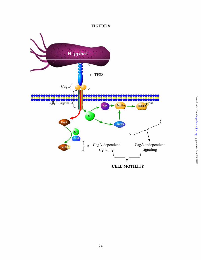
FIGURE 8
α5β1 Integrin
CagL
TFSS
H. pylori
CagA-independent signaling
CagA-dependent signaling
CELL MOTILITY
α5β1 Integrin
CagL
TFSS
H. pylori
CagA-independent signaling
CagA-dependent signaling
CELL MOTILITY
24
by guest on June 25, 2018http://w
ww
.jbc.org/D
ownloaded from

Jared L. Snider, Cody Allison, Bryan H. Bellaire, Richard L. Ferrero and James A. Cardellihelicobacter pylori caga+ induced motility of gastric cancer cells
1 integrin activates jnk independent of caga, and jnk activation is required forβThe
published online March 20, 2008J. Biol. Chem.
10.1074/jbc.M800289200Access the most updated version of this article at doi:
Alerts:
When a correction for this article is posted•
When this article is cited•
to choose from all of JBC's e-mail alertsClick here
Supplemental material:
http://www.jbc.org/content/suppl/2008/04/08/M800289200.DC1
by guest on June 25, 2018http://w
ww
.jbc.org/D
ownloaded from