Helicobacter Pylori
18
Helicobacter pylori Bahan tutorial B3 Sri wahyuni 070100076
-
Upload
sri-wahyuni -
Category
Documents
-
view
80 -
download
0
Transcript of Helicobacter Pylori
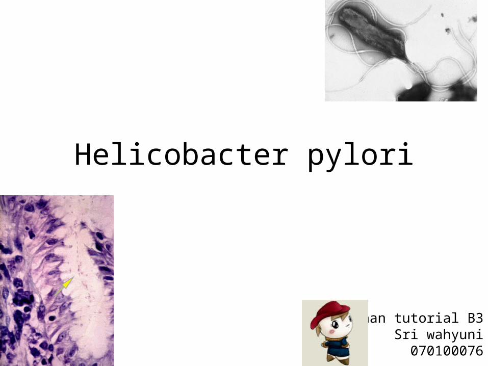
Helicobacter pylori
Bahan tutorial B3 Sri wahyuni070100076
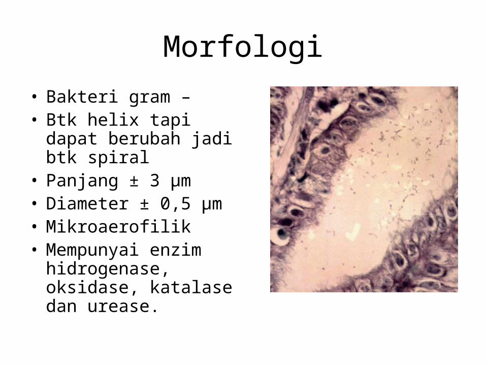
Morfologi
• Bakteri gram –• Btk helix tapi dapat
berubah jadi btk spiral• Panjang ± 3 μm• Diameter ± 0,5 μm• Mikroaerofilik• Mempunyai enzim
hidrogenase, oksidase, katalase dan urease.
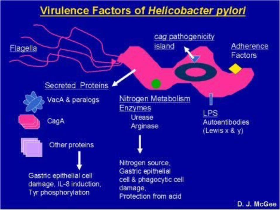
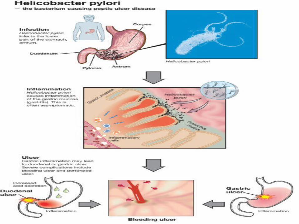
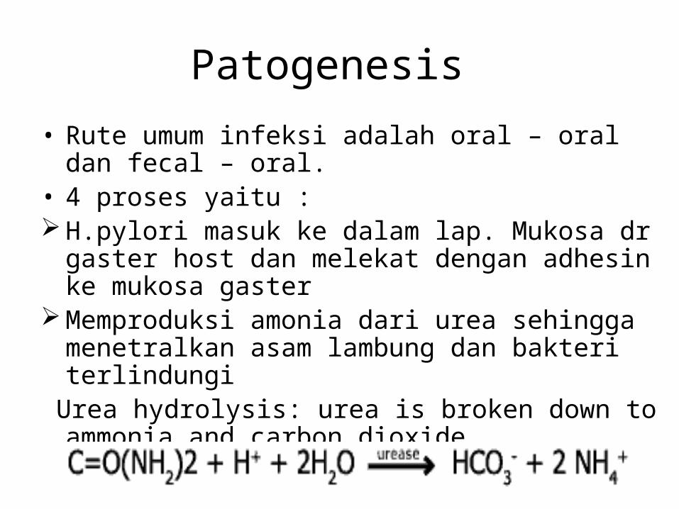
Patogenesis
• Rute umum infeksi adalah oral – oral dan fecal – oral.
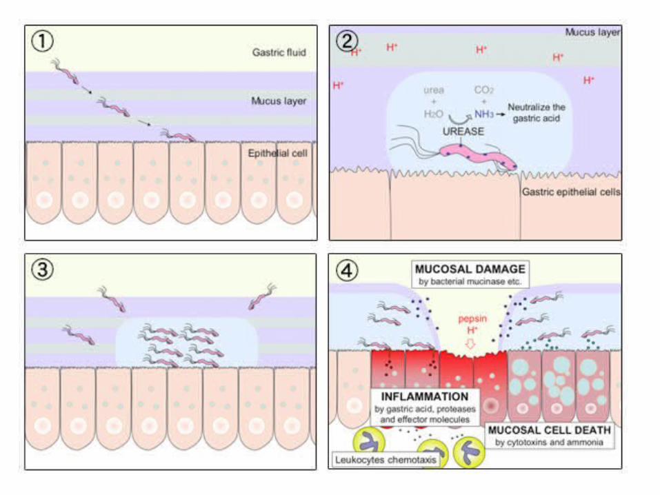
• 4 proses yaitu : H.pylori masuk ke dalam lap. Mukosa dr gaster
host dan melekat dengan adhesin ke mukosa gaster
Memproduksi amonia dari urea sehingga menetralkan asam lambung dan bakteri terlindungi
Urea hydrolysis: urea is broken down to ammonia and carbon dioxide

Berpoliferasi, migrasi, dan akhirnya membentuk fokus infeksi
Ulkus gaster akan dihasilkan dengan adanya destruksi oleh mukosa, inflamasi dan kematian sel mukosa.
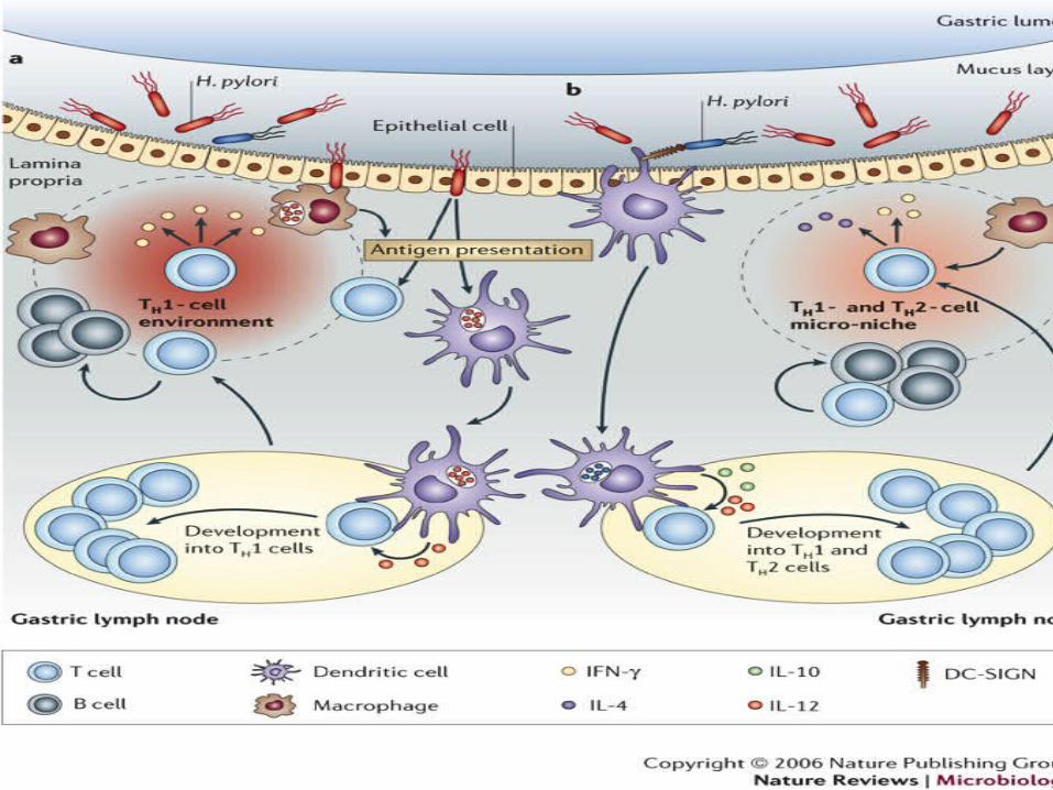
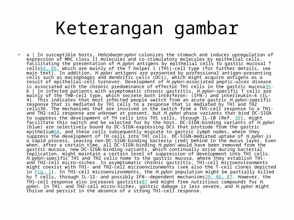
Keterangan gambar• a | In susceptible hosts, Helicobacter pylori colonizes the stomach and induces upregulation of
expression of MHC class II molecules and co-stimulatory molecules by epithelial cells, facilitating the presentation of H. pylori antigens by epithelial cells to gastric mucosal T cells84, 85, which are mainly of the T helper 1 (TH1)-cell type (for further details, see main text). In addition, H. pylori antigens are presented by professional antigen-presenting cells such as macrophages and dendritic cells (DCs), which might acquire antigens as a result of epithelial-cell turnover. Development of H. pylori-associated peptic-ulcer disease is associated with the chronic predominance of effector TH1 cells in the gastric mucosa35. b | In infected patients with asymptomatic chronic gastritis, H. pylori-specific T cells are mainly of the TH0-cell type, which secrete both interferon- (IFN-) and interleukin-4 (IL-4). This indicates that most infected people switch from an acute gastric H. pylori-specific response that is mediated by TH1 cells to a response that is mediated by TH1 and TH2 cells36. The mechanisms that are involved in the switch from a TH1-cell response to a TH1- and TH2-cell response are unknown at present, but H. pylori phase variants that bind DC-SIGN to suppress the development of TH cells into TH1 cells, through IL-10 (Ref. 39), might facilitate this switch and be selected for by the host. DC-SIGN-binding variants of H. pylori (blue) are selectively bound by DC-SIGN-expressing DCs that protrude from the gastric epithelium54, and these cells subsequently migrate to gastric lymph nodes, where they suppress the development of TH cells into TH1 cells. DC-SIGN-mediated uptake of H. pylori is a rapid process, leaving non-DC-SIGN-binding bacteria (red) behind in the mucus layer. Even when, after a certain time, all DC-SIGN-binding H. pylori would have been removed from the gastric mucosa, new DC-SIGN-binding variants, which continually arise during bacterial replication, might maintain a certain level of suppression of development into TH1 cells. H. pylori-specific TH1 and TH2 cells home to the gastric mucosa, where they establish TH1- and TH2-cell micro-niches. In asymptomatic chronic gastritis, TH1-cell microenvironments might coexist with TH1- and TH2-cell microenvironments (see also the T-cell clones depicted in Fig. 1). In TH1-cell microenvironments, the H. pylori population might be partially killed by T cells, through IL-12- and possibly IFN--dependent mechanisms29, 86, 87. However, the TH1-cell response also increases gastritis87 and might free nutritious compounds for H. pylori. In TH1- and TH2-cell micro-niches, gastric damage is less severe, and H. pylori might thrive and persist in the absence of a strong TH1-cell response.
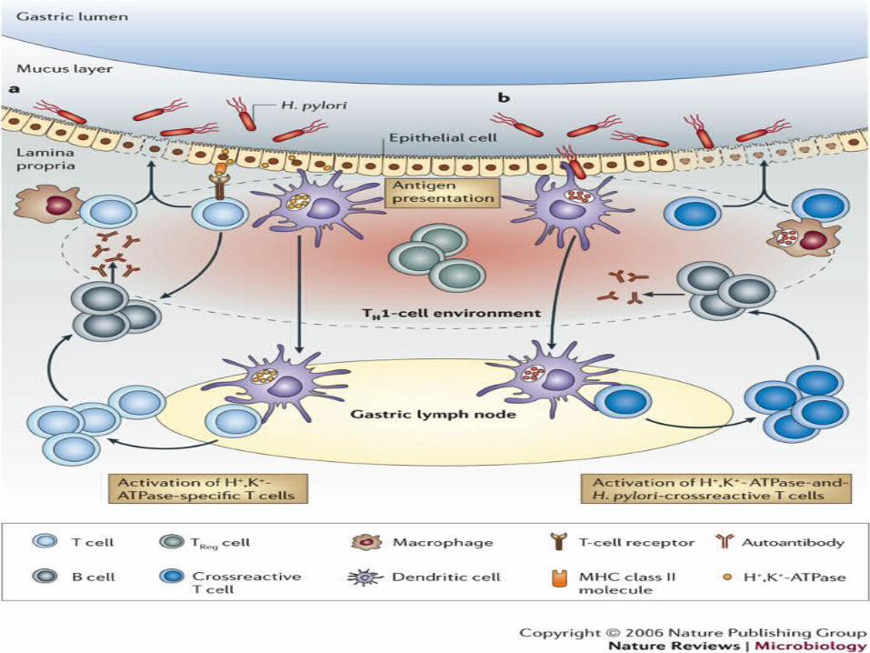
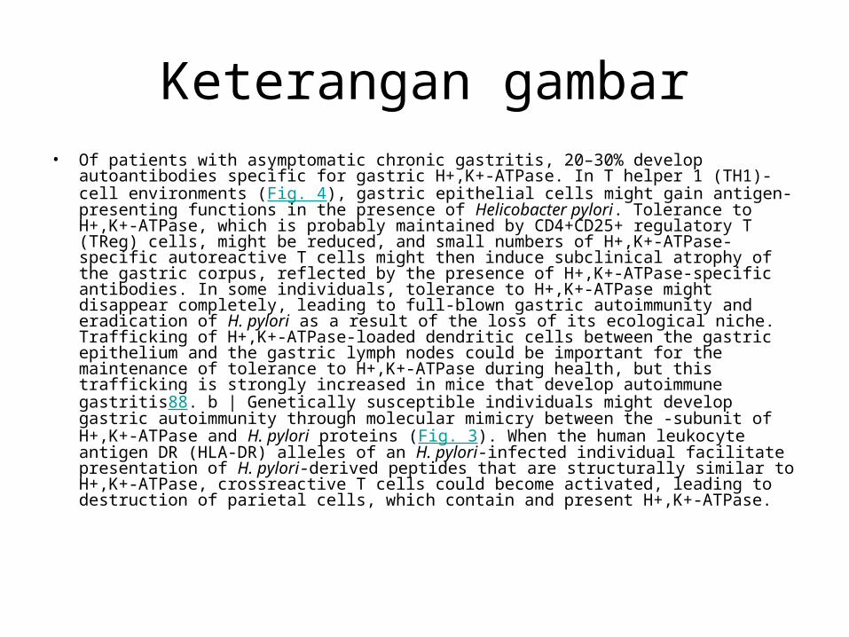
Keterangan gambar• Of patients with asymptomatic chronic gastritis, 20–30% develop autoantibodies
specific for gastric H+,K+-ATPase. In T helper 1 (TH1)-cell environments (Fig. 4), gastric epithelial cells might gain antigen-presenting functions in the presence of Helicobacter pylori. Tolerance to H+,K+-ATPase, which is probably maintained by CD4+CD25+ regulatory T (TReg) cells, might be reduced, and small numbers of H+,K+-ATPase-specific autoreactive T cells might then induce subclinical atrophy of the gastric corpus, reflected by the presence of H+,K+-ATPase-specific antibodies. In some individuals, tolerance to H+,K+-ATPase might disappear completely, leading to full-blown gastric autoimmunity and eradication of H. pylori as a result of the loss of its ecological niche. Trafficking of H+,K+-ATPase-loaded dendritic cells between the gastric epithelium and the gastric lymph nodes could be important for the maintenance of tolerance to H+,K+-ATPase during health, but this trafficking is strongly increased in mice that develop autoimmune gastritis88. b | Genetically susceptible individuals might develop gastric autoimmunity through molecular mimicry between the -subunit of H+,K+-ATPase and H. pylori proteins (Fig. 3). When the human leukocyte antigen DR (HLA-DR) alleles of an H. pylori-infected individual facilitate presentation of H. pylori-derived peptides that are structurally similar to H+,K+-ATPase, crossreactive T cells could become activated, leading to destruction of parietal cells, which contain and present H+,K+-ATPase.
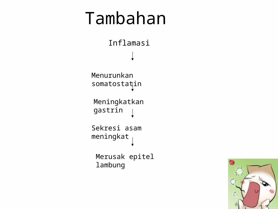
Tambahan Inflamasi
Menurunkan somatostatin
Meningkatkan gastrin
Sekresi asam meningkat
Merusak epitel lambung
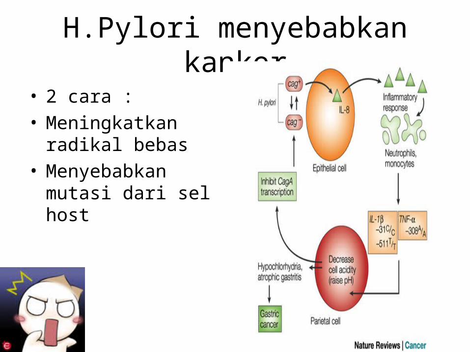
H.Pylori menyebabkan kanker
• 2 cara :• Meningkatkan radikal
bebas• Menyebabkan mutasi
dari sel host
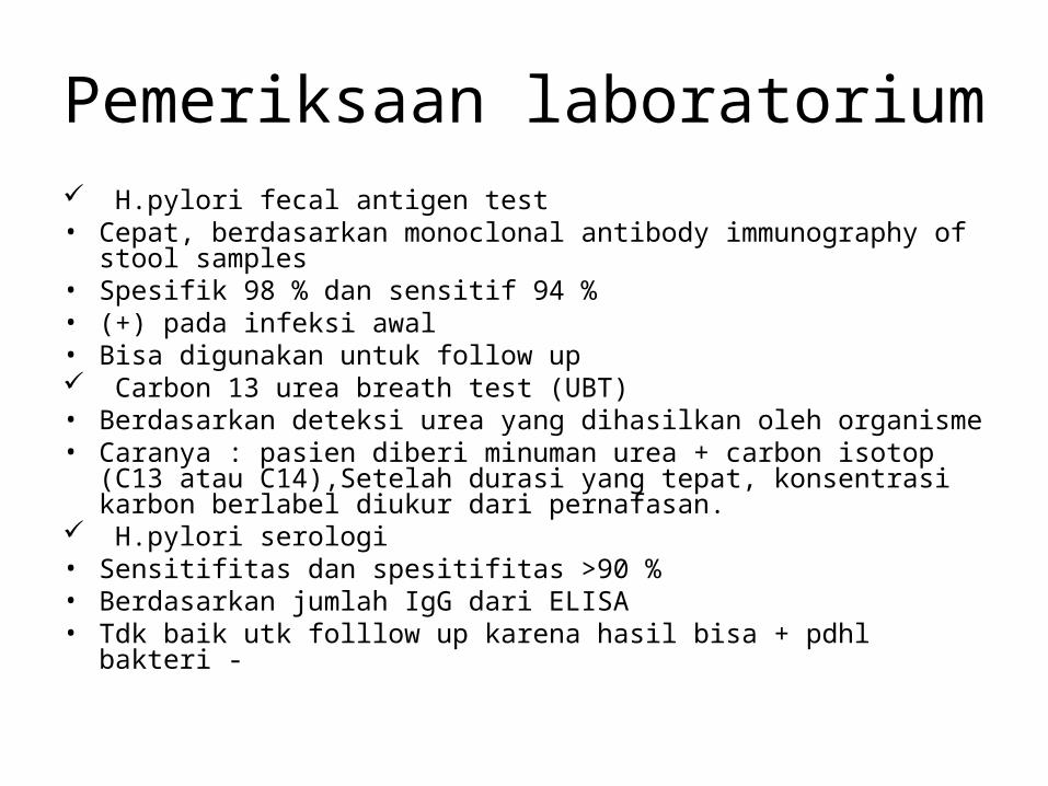
Pemeriksaan laboratorium H.pylori fecal antigen test• Cepat, berdasarkan monoclonal antibody immunography of stool
samples• Spesifik 98 % dan sensitif 94 %• (+) pada infeksi awal• Bisa digunakan untuk follow up Carbon 13 urea breath test (UBT)• Berdasarkan deteksi urea yang dihasilkan oleh organisme• Caranya : pasien diberi minuman urea + carbon isotop (C13 atau
C14),Setelah durasi yang tepat, konsentrasi karbon berlabel diukur dari pernafasan.
H.pylori serologi• Sensitifitas dan spesitifitas >90 %• Berdasarkan jumlah IgG dari ELISA• Tdk baik utk folllow up karena hasil bisa + pdhl bakteri -
Imaging study, tdk membantu diagnosisEGD ( esophagogastroduodenoscopy),
penting pada pasien peptic ulcer dan untuk biopsi.
Yang paling baru adalah dengan menggunakan pewarnaan HE yang dimodifikasi
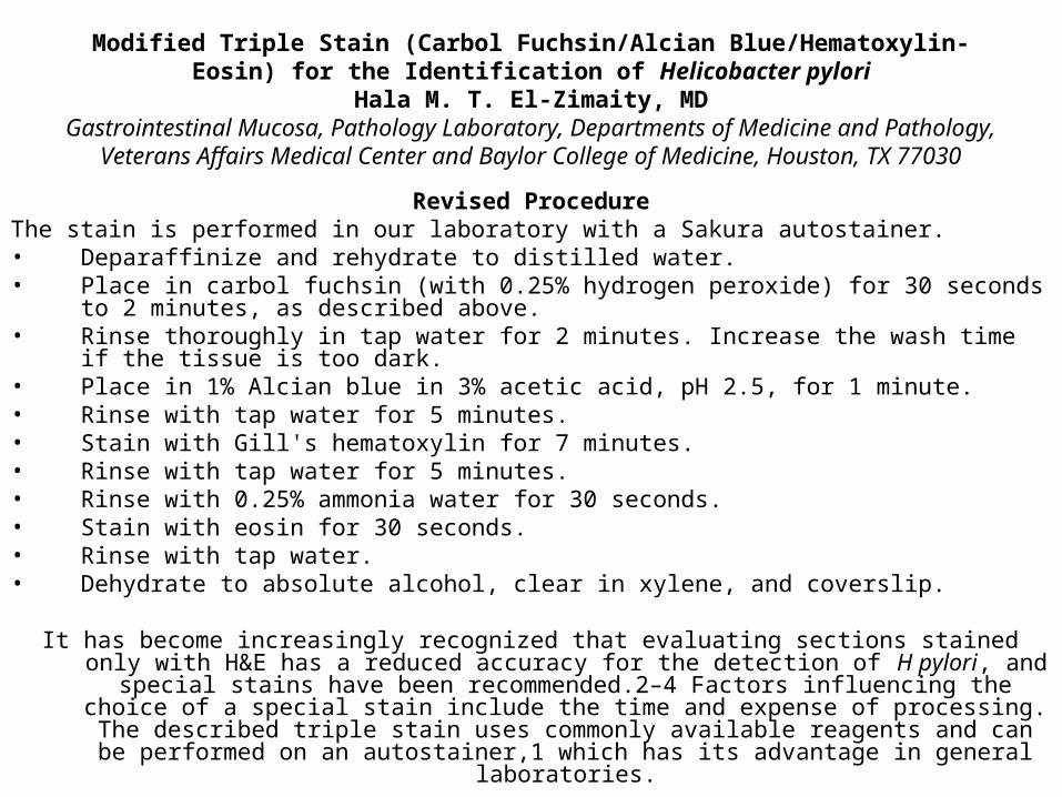
Modified Triple Stain (Carbol Fuchsin/Alcian Blue/Hematoxylin-Eosin) for the Identification of Helicobacter pylori
Hala M. T. El-Zimaity, MDGastrointestinal Mucosa, Pathology Laboratory, Departments of Medicine and Pathology,
Veterans Affairs Medical Center and Baylor College of Medicine, Houston, TX 77030
Revised ProcedureThe stain is performed in our laboratory with a Sakura autostainer. • Deparaffinize and rehydrate to distilled water.• Place in carbol fuchsin (with 0.25% hydrogen peroxide) for 30 seconds to 2 minutes, as
described above.• Rinse thoroughly in tap water for 2 minutes. Increase the wash time if the tissue is too dark.• Place in 1% Alcian blue in 3% acetic acid, pH 2.5, for 1 minute.• Rinse with tap water for 5 minutes.• Stain with Gill's hematoxylin for 7 minutes.• Rinse with tap water for 5 minutes.• Rinse with 0.25% ammonia water for 30 seconds.• Stain with eosin for 30 seconds.• Rinse with tap water.• Dehydrate to absolute alcohol, clear in xylene, and coverslip.
It has become increasingly recognized that evaluating sections stained only with H&E has a reduced accuracy for the detection of H pylori, and special stains have been
recommended.2–4 Factors influencing the choice of a special stain include the time and expense of processing. The described triple stain uses commonly available reagents and
can be performed on an autostainer,1 which has its advantage in general laboratories.
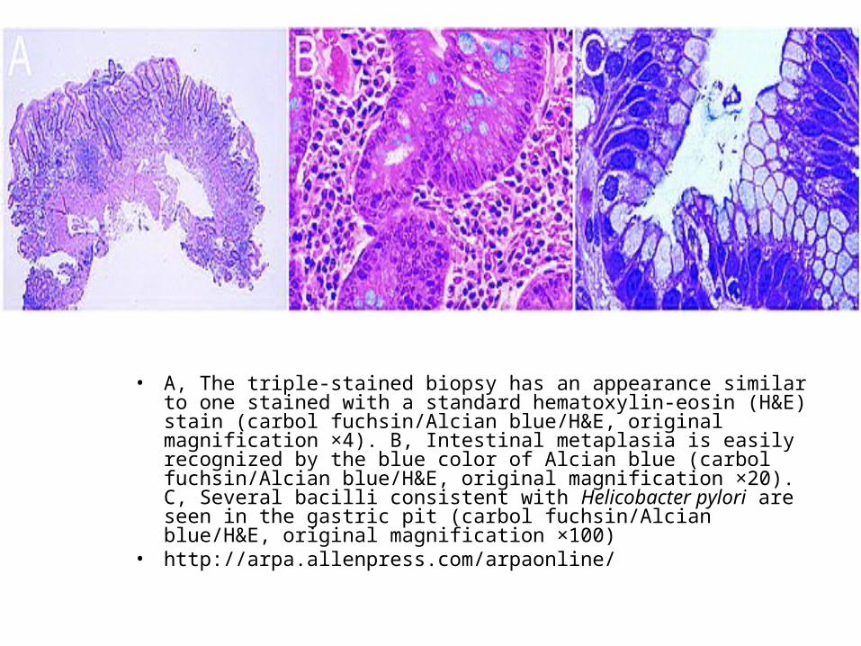
• A, The triple-stained biopsy has an appearance similar to one stained with a standard hematoxylin-eosin (H&E) stain (carbol fuchsin/Alcian blue/H&E, original magnification ×4). B, Intestinal metaplasia is easily recognized by the blue color of Alcian blue (carbol fuchsin/Alcian blue/H&E, original magnification ×20). C, Several bacilli consistent with Helicobacter pylori are seen in the gastric pit (carbol fuchsin/Alcian blue/H&E, original magnification ×100)
• http://arpa.allenpress.com/arpaonline/

Referensi www.nature.comhttp://arpa.allenpress.com/arpaonline/www.emedicine.comwww.wikipedia.comhttp://southmed.usouthal.edu/.../faculty/mcgee.html Color atlas microbiologyDll...