
Tali Apoptosis Kit – Annexin V Alexa Fluor 488 and ... · PDF fileTali ® Apoptosis...
5
MAN0004128 | MP 10788 Revision: 2.0 For Research Use Only. Not for use in diagnostic procedures. Tali ® Apoptosis Kit – Annexin V Alexa Fluor ® 488 and Propidium Iodide *for use with Tali ® Assay: Apoptosis* Catalog no. A10788 Table 1 Contents and storage Material Amount Composition Storage Stability Annexin V Alexa Fluor ® 488 (Component A) 500 μL Solution in 25 mM HEPES, 140 mM NaCl, 1 mM EDTA, pH 7.4, 0.1% bovine serum albumin (BSA) • 2–8°C • Protect from light • Do not freeze When stored as directed, the product is stable for at least 6 months. Tali ® Propidium Iodide (PI) (Component B) 100 μL 100 μg/mL in water • 2–30°C 5X Annexin Binding Buffer (ABB) (Component C) 25 mL 50 mM HEPES, 700 mM NaCl, 12.5 mM CaCl 2 , pH 7.4 • 2–8°C • Protect from light Number of assays: Sufficient material is supplied for 100 assays of 100 μL each. Approximate fluorescence excitation/emission maxima: Annexin V Alexa Fluor ® 488: 488/499 in nm; Propidium iodide: 535/617 in nm, bound to DNA. Introduction Apoptosis is a carefully regulated process of cell death that occurs as a normal part of development. Inappropriately regulated apoptosis is implicated in disease states, such as Alzheimer’s disease and cancer. Apoptosis is distinguished from necrosis by characteristic morphological and biochemical changes, including compaction and fragmentation of the nuclear chromatin, shrinkage of the cytoplasm, and loss of membrane asymmetry. 1–5 In normal live cells, phosphatidyl serine (PS) is located on the cytoplasmic surface of the cell membrane. However, in apoptotic cells, PS is translocated from the inner to the outer leaflet of the plasma membrane, thus exposing PS to the external cellular environment. 6 In leukocyte apoptosis, PS on the outer surface of the cell marks the cell for recognition and phagocytosis by macrophages. 7,8 The human anticoagulant, annexin V, is a 35–36 kDa Ca 2+ -dependent phospholipid- binding protein that has a high affinity for PS. 9 Annexin V labeled with a fluorophore or biotin can identify apoptotic cells by binding to PS exposed on the outer leaflet. 10 Propridium Iodide (PI) is a cell-impermeant and fluorogenic DNA-binding dye used foridentifying necrotic cells. PI is impermeant to live cells, but easily enters dead cells where it binds to nucleic acids and becomes fluorescent.
Transcript of Tali Apoptosis Kit – Annexin V Alexa Fluor 488 and ... · PDF fileTali ® Apoptosis...
MAN0004128 | MP 10788 Revision: 2.0For Research Use Only. Not for use in diagnostic procedures.
Tali® Apoptosis Kit – Annexin V Alexa Fluor® 488 and Propidium Iodide*for use with Tali® Assay: Apoptosis*
Catalog no. A10788
Table 1 Contents and storage
Material Amount Composition Storage Stability
Annexin V Alexa Fluor® 488 (Component A) 500 μL
Solution in 25 mM HEPES, 140 mM NaCl, 1 mM EDTA, pH 7.4, 0.1% bovine serum albumin (BSA)
• 2–8°C• Protect from light• Do not freeze When stored as
directed, the product is stable for at least 6 months.
Tali® Propidium Iodide (PI) (Component B) 100 μL 100 μg/mL in water • 2–30°C
5X Annexin Binding Buffer (ABB) (Component C) 25 mL 50 mM HEPES, 700 mM NaCl, 12.5
mM CaCl2, pH 7.4• 2–8°C• Protect from light
Number of assays: Sufficient material is supplied for 100 assays of 100 μL each.
Approximate fluorescence excitation/emission maxima: Annexin V Alexa Fluor® 488: 488/499 in nm; Propidium iodide: 535/617 in nm, bound to DNA.
Introduction
Apoptosis is a carefully regulated process of cell death that occurs as a normal part of development. Inappropriately regulated apoptosis is implicated in disease states, such as Alzheimer’s disease and cancer. Apoptosis is distinguished from necrosis by characteristic morphological and biochemical changes, including compaction and fragmentation of the nuclear chromatin, shrinkage of the cytoplasm, and loss of membrane asymmetry.1–5 In normal live cells, phosphatidyl serine (PS) is located on the cytoplasmic surface of the cell membrane. However, in apoptotic cells, PS is translocated from the inner to the outer leaflet of the plasma membrane, thus exposing PS to the external cellular environment.6 In leukocyte apoptosis, PS on the outer surface of the cell marks the cell for recognition and phagocytosis by macrophages.7,8
The human anticoagulant, annexin V, is a 35–36 kDa Ca2+-dependent phospholipid-binding protein that has a high affinity for PS.9 Annexin V labeled with a fluorophore or biotin can identify apoptotic cells by binding to PS exposed on the outer leaflet.10
Propridium Iodide (PI) is a cell-impermeant and fluorogenic DNA-binding dye used foridentifying necrotic cells. PI is impermeant to live cells, but easily enters dead cells where it binds to nucleic acids and becomes fluorescent.
Tali® Apoptosis Kit – Annexin V Alexa Fluor® 488 and Propidium Iodide | 2
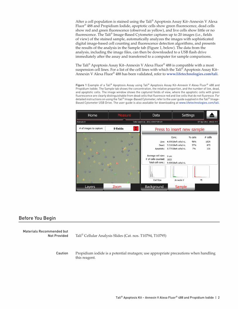
Figure 1 Example of a Tali® Apoptosis Assay using Tali® Apoptosis Assay Kit–Annexin V Alexa Fluor® 488 and Propidium Iodide. The Sample tab shows the concentration, the relative proportion, and the number of live, dead, and apoptotic cells. The image window shows the captured fields of view, where the apoptotic cells with green fluorescence are clearly distinguishable from dead cells that fluoresce red and live cells that do not fluoresce. For detailed instructions on using the Tali® Image-Based Cytometer, refer to the user guide supplied in the Tali® Image-Based Cytometer USB Drive. The user guide is also available for downloading at www.lifetechnologies.com/tali.
After a cell population is stained using the Tali® Apoptosis Assay Kit–Annexin V Alexa Fluor® 488 and Propidium Iodide, apoptotic cells show green fluorescence, dead cells show red and green fluorescence (observed as yellow), and live cells show little or no fluorescence. The Tali® Image-Based Cytometer captures up to 20 images (i.e., fields of view) of the stained sample, automatically analyzes the images with sophisticated digital image-based cell counting and fluorescence detection algorithms, and presents the results of the analysis in the Sample tab (Figure 1, below). The data from the analysis, including the image files, can then be downloaded to a USB flash drive immediately after the assay and transferred to a computer for sample comparisons.
The Tali® Apoptosis Assay Kit–Annexin V Alexa Fluor® 488 is compatible with a most suspension cell lines. For a list of the cell lines with which the Tali® Apoptosis Assay Kit–Annexin V Alexa Fluor® 488 has been validated, refer to www.lifetechnologies.com/tali.
Before You Begin
Materials Recommended but Not Provided Tali® Cellular Analysis Slides (Cat. nos. T10794, T10795)
Caution Propidium iodide is a potential mutagen; use appropriate precautions when handling this reagent.
Tali® Apoptosis Kit – Annexin V Alexa Fluor® 488 and Propidium Iodide | 3
Experimental Protocol
Follow the instructions below to perform the Tali® Apoptosis Assay using the Tali® Apoptosis Assay Kit–Annexin V Alexa Fluor® 488 and Propidium Iodide. For detailed instructions on using the Tali® Image-Based Cytometer, refer to the user guide supplied in the Tali® Image-Based Cytometer USB Drive. The user guide is also available for downloading at www.lifetechnologies.com/tali. The recommended sample concentration range for the Tali® Image-Based Cytometer is 1 × 10⁵ to 1 × 10⁷ cells/mL; however, the sample concentration does not need to be exact to perform an assay.
1. If required, induce apoptosis in cells using the desired method. Prepare a negative control by incubating the cells in the absence of the apoptosis inducing agent. If your cells exhibit significant autofluorescence, you may additionally prepare a negative control of unstained cells.
2. Prepare 1X Annexin binding buffer by diluting the 5X Annexin binding buffer (ABB, Component C) in deionized water. For example, for 10 assays, add 200 μL of 5X Annexin binding buffer to 800 μL of deionized water.
3. Harvest the cells after the incubation period, centrifuge, and discard the supernatent.
4. Resuspend the cells in 1X Annexin binding buffer, so that there is at least 100 μL of cells per individual assay at a concentration of approximately 5 × 10⁵ to 5 × 10⁶ cells/mL. The concentration of the cells does not need to be exact.
5. To each 100 μL of sample, add 5 μL of Annexin V Alexa Fluor® 488 (Component A). Mix well.
6. Incubate the cell-Annexin V Alexa Fluor® 488 mixture at room temperature in the dark for 20 minutes.
7. Centrifuge the cells and resuspend them in 100 μL of ABB.
8. Add 1 μL of Tali® Propidium Iodide (PI, component B) to each 100 μL sample. Mix well.
9. Incubate the samples at room temperature in the dark for 1–5 minutes.
10. Load 25 μL of the stained cells into a Tali® Cellular Analysis Slide by pipetting the sample at an angle of approximately 80° into the half moon-shaped sample loading area. The sample is loaded into the chamber through capillary action. Take care to avoid forming bubbles in the sample or to cause back splatter.
11. Insert the slide into the slide port of the Tali® Image-Based Cytometer until it stops. Do not forcefully push the slide any further.
12. Touch Cell Health on the Home screen of the Tali® Image-Based Cytometer and then touch Apoptosis to select the Tali® Apoptosis Assay.
13. Name the sample series, if desired.
14. Touch Press to insert new sample; the slide will automatically be pulled into the instrument.
15. When prompted, focus your cells using the image adjustment (focus) knob on the right side of the instrument.
Tali® Apoptosis Kit – Annexin V Alexa Fluor® 488 and Propidium Iodide | 4
16. Specify the number of fields of view to capture using the # of images to capture drop-down menu, and then touch Press to run sample. The Tali® Image-Based Cytometer will automatically capture and analyze the images of your sample, and present the results of the analysis in the analysis window.
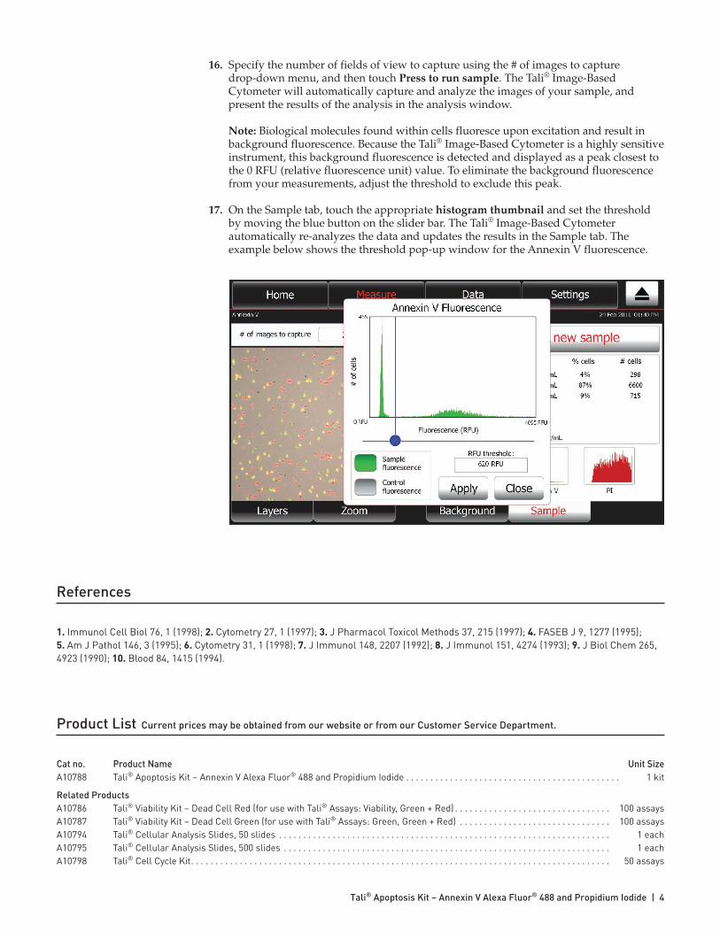
Note: Biological molecules found within cells fluoresce upon excitation and result in background fluorescence. Because the Tali® Image-Based Cytometer is a highly sensitive instrument, this background fluorescence is detected and displayed as a peak closest to the 0 RFU (relative fluorescence unit) value. To eliminate the background fluorescence from your measurements, adjust the threshold to exclude this peak.
17. On the Sample tab, touch the appropriate histogram thumbnail and set the threshold by moving the blue button on the slider bar. The Tali® Image-Based Cytometer automatically re-analyzes the data and updates the results in the Sample tab. The example below shows the threshold pop-up window for the Annexin V fluorescence.
References
1. Immunol Cell Biol 76, 1 (1998); 2. Cytometry 27, 1 (1997); 3. J Pharmacol Toxicol Methods 37, 215 (1997); 4. FASEB J 9, 1277 (1995); 5. Am J Pathol 146, 3 (1995); 6. Cytometry 31, 1 (1998); 7. J Immunol 148, 2207 (1992); 8. J Immunol 151, 4274 (1993); 9. J Biol Chem 265, 4923 (1990); 10. Blood 84, 1415 (1994).
Product List Current prices may be obtained from our website or from our Customer Service Department.
Cat no. Product Name Unit SizeA10788 Tali® Apoptosis Kit – Annexin V Alexa Fluor® 488 and Propidium Iodide . . . . . . . . . . . . . . . . . . . . . . . . . . . . . . . . . . . . . . . . . . . . 1 kit
Related ProductsA10786 Tali® Viability Kit – Dead Cell Red (for use with Tali® Assays: Viability, Green + Red) . . . . . . . . . . . . . . . . . . . . . . . . . . . . . . . . 100 assaysA10787 Tali® Viability Kit – Dead Cell Green (for use with Tali® Assays: Green, Green + Red) . . . . . . . . . . . . . . . . . . . . . . . . . . . . . . . 100 assaysA10794 Tali® Cellular Analysis Slides, 50 slides . . . . . . . . . . . . . . . . . . . . . . . . . . . . . . . . . . . . . . . . . . . . . . . . . . . . . . . . . . . . . . . . . . . . 1 eachA10795 Tali® Cellular Analysis Slides, 500 slides . . . . . . . . . . . . . . . . . . . . . . . . . . . . . . . . . . . . . . . . . . . . . . . . . . . . . . . . . . . . . . . . . . . 1 eachA10798 Tali® Cell Cycle Kit. . . . . . . . . . . . . . . . . . . . . . . . . . . . . . . . . . . . . . . . . . . . . . . . . . . . . . . . . . . . . . . . . . . . . . . . . . . . . . . . . . . . . . 50 assays
Purchaser Notification
Corporate Headquarters 5791 Van Allen Way Carlsbad, CA 92008 USA Phone: +1 760 603 7200 Fax: +1 760 602 6500 Email: [email protected]
European Headquarters Inchinnan Business Park 3 Fountain Drive Paisley PA4 9RF UK Phone: +44 141 814 6100 Toll-Free Phone: 0800 269 210 Toll-Free Tech: 0800 838 380 Fax: +44 141 814 6260 Tech Fax: +44 141 814 6117 Email: [email protected] Email Tech: [email protected]
Japanese Headquarters LOOP-X Bldg. 6F 3-9-15, Kaigan Minato-ku, Tokyo 108-0022 Japan Phone: +81 3 5730 6509 Fax: +81 3 5730 6519 Email: [email protected]
Additional international offices are listed at www.lifetechnologies.com
These high-quality reagents and materials must be used by, or directl y under the super vision of, a tech nically qualified individual experienced in handling potentially hazardous chemicals. Read the Safety Data Sheet provided for each product; other regulatory considerations may apply.
Obtaining Support For the latest services and support information for all locations, go to www.lifetechnologies.com.
At the website, you can:• Access worldwide telephone and fax numbers to contact Technical Support and Sales facilities• Search through frequently asked questions (FAQs)• Submit a question directly to Technical Support ([email protected])• Search for user documents, SDSs, vector maps and sequences, application notes, formulations, handbooks, certificates of
analysis, citations, and other product support documents• Obtain information about customer training• Download software updates and patches
SDS Safety Data Sheets (SDSs) are available at www.lifetechnologies.com/sds.
Certificate of Analysis The Certificate of Analysis provides detailed quality control and product qualification information for each product. Certificates of Analysis are available on our website. Go to www.lifetechnologies.com/support and search for the Certificate of Analysis by product lot number, which is printed on the product packaging (tube, pouch, or box).
Limited Product Warranty Life Technologies Corporation and/or its affiliate(s) warrant their products as set forth in the Life Technologies’ General Terms and Conditions of Sale found on Life Technologies’ website at www.lifetechnologies.com/termsandconditions. If you have any questions, please contact Life Technologies at www.lifetechnologies.com/support.
Disclaimer LIFE TECHNOLOGIES CORPORATION AND/OR ITS AFFILIATE(S) DISCLAIM ALL WARRANTIES WITH RESPECT TO THIS DOCUMENT, EXPRESSED OR IMPLIED, INCLUDING BUT NOT LIMITED TO THOSE OF MERCHANTABILITY, FITNESS FOR A PARTICULAR PURPOSE, OR NON-INFRINGEMENT. TO THE EXTENT ALLOWED BY LAW, IN NO EVENT SHALL LIFE TECHNOLOGIES AND/OR ITS AFFILIATE(S) BE LIABLE, WHETHER IN CONTRACT, TORT, WARRANTY, OR UNDER ANY STATUTE OR ON ANY OTHER BASIS FOR SPECIAL, INCIDENTAL, INDIRECT, PUNITIVE, MULTIPLE OR CONSEQUENTIAL DAMAGES IN CONNECTION WITH OR ARISING FROM THIS DOCUMENT, INCLUDING BUT NOT LIMITED TO THE USE THEREOF.
Limited Use Label License: Research Use Only The purchase of this product conveys to the purchaser the limited, non-transferable right to use the purchased amount of the product only to perform internal research for the sole benefit of the purchaser. No right to resell this product or any of its components is conveyed expressly, by implication, or by estoppel. This product is for internal research purposes only and is not for use in commercial services of any kind, including, without limitation, reporting the results of purchaser’s activities for a fee or other form of consideration. For information on obtaining additional rights, please contact [email protected] or Out Licensing, Life Technologies Corporation, 5791 Van Allen Way, Carlsbad, California 92008.
The trademarks mentioned herein are the property of Life Technologies Corporation and/or its affiliate(s) or their respective owners.
©2013 Life Technologies Corporation. All rights reserved.
18 July 2013


![La tecnologia alexa[1]](https://static.fdocument.org/doc/165x107/55d11f4bbb61ebe2398b47f2/la-tecnologia-alexa1.jpg)