Bacterial RNA Polymerase New Insights on a Fundamental Molecular Machine
1
Table S1. Bacterial strains and plasmids used in this study � �
� � Strain or plasmid Relevant characteristics� Source/Reference�
E. coli strain
TOP10 F- mcrAΔ(mrr-hsdRMS-mcrBC) φ80lacZΔM15ΔlacX74 recA1
araD139 Δ(ara-leu)7697 galU galK rpsL(StrR) endA1 nupG λ-
Life Technologies
BL21 F- ompT hsdSB(rB-, mB
-) gal dcm GE Healthcare
XL1-Blue recA1 endA1 gyrA96 thi-1 hsdR17 supE44 relA1 lac [F'proAB
lacIqZ△M15 Tn10 (Tetr)]
(1)
E. coli 18H278 Heat-stable enterotoxin (ST+, LT-) producing E. coli O128:H12 This study
Clostridium perfringens strain
OS1 becAB positive isolate from diarrheal patient in Osaka case This study
TS1 becAB positive isolate from diarrheal patient in Tochigi case This study
Iz1 cpe negative isolate from human feces This study
Ta1 cpe negative isolate from human feces This study
NCTC8239 cpe positive isolate from diarrheal patient (2)
NCTC8798 cpe positive isolate from diarrheal patient (2)
OS1ΔbecB OS1 becB Ω targetron This study
Y1 cpe positive isolate from diarrheal patient This study
Y2 cpe positive isolate from diarrheal patient This study
Y3 cpe positive isolate from diarrheal patient This study
Y4 cpe positive isolate from diarrheal patient This study
Y5 cpe positive isolate from diarrheal patient This study
Y6 cpe positive isolate from diarrheal patient This study
Y7 cpe positive isolate from diarrheal patient This study
Y8 cpe positive isolate from diarrheal patient This study
Y9 cpe negative isolate from diarrheal patient This study
Y10 cpe negative isolate from diarrheal patient This study
Y11 cpe negative isolate from diarrheal patient This study
Y12 cpe negative isolate from diarrheal patient This study
Y13 cpe negative isolate from diarrheal patient This study
Y14 cpe negative isolate from diarrheal patient This study
2
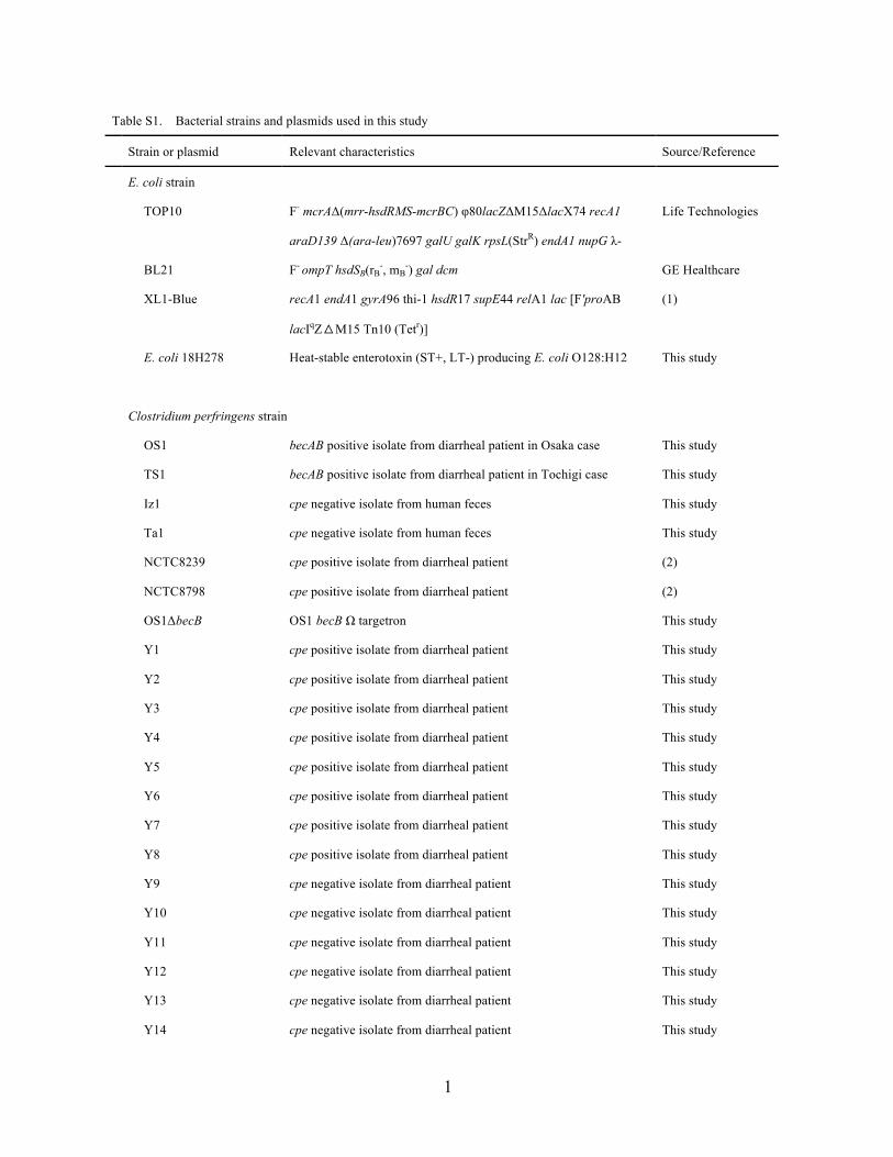
Y15 cpe negative isolate from diarrheal patient This study
Y16 cpe negative isolate from diarrheal patient This study
Y17 cpe negative isolate from diarrheal patient This study
Y18 cpe positive isolate from healthy individual This study
Y19 cpe negative isolate from healthy individual This study
Y20 cpe negative isolate from healthy individual This study
Y21 cpe negative isolate from healthy individual This study
Y22 cpe negative isolate from healthy individual This study
Y23 cpe negative isolate from healthy individual This study
Y24 cpe negative isolate from healthy individual This study
Y25 cpe negative isolate from healthy individual This study
Y26 cpe negative isolate from healthy individual This study
Y27 cpe negative isolate from healthy individual This study
Y28 cpe negative isolate from healthy individual This study
Y29 cpe negative isolate from healthy individual This study
Y30 cpe negative isolate from healthy individual This study
Y31 cpe negative isolate from healthy individual This study
Y32 cpe negative isolate from healthy individual This study
Y33 cpe negative isolate from healthy individual This study
Y34 cpe negative isolate from healthy individual This study
Plasmid
pCR™2.1-TOPO® TA Cloning vector Life Technologies
pGEX-4T-2 Expression vector GE Healthcare
pJIR750 C. perfringens-E. coli shuttle vector, Cmr (3)
pJIR750ai pJIR750Ωalpha-toxin targetron Sigma
� � � � pJIR750becB pJIR750ΩBECb targetron This study
References
1. Sambrook J, Russell DW. 2001. Molecular cloning: a laboratory manual. Cold Spring
Harbor (New York): Cold Spring Harbor Laboratory Press.

3
2. Li J, Sayeed S, McClane BA. 2007. Prevalence of enterotoxigenic Clostridium perfringens
isolates in Pittsburgh (Pennsylvania) area soils and home kitchens. Appl Environ Microbiol.
73:7218-7224.
3. Bannam TL, Rood JI. 1993. Clostridium perfringens-Escherichia coli shuttle vectors that
carry single antibiotic resistance determinants. Plasmid. 29:233-235.
4
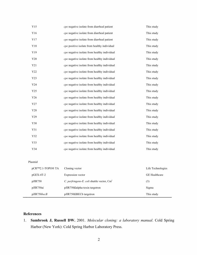
Table S2. Primers used in this study � �
Primer Sequence(5'-3') use Reference
Sequencing of bec
bec_S1 tttagaaagagattttctagcaagttcata sequencing of bec this study
bec_S2 attatcaaacatttcatgggaagtt sequencing of bec this study
bec_S3 ctgggtttgagctattaggtgc sequencing of bec this study
bec_S4 cgaattcccctgtacaaataga sequencing of bec this study
bec_S5 gaaaaacaaatagcaggtttgga sequencing of bec this study
bec_S6 aaggagaatccacaacagaagg sequencing of bec this study
bec_S7 atcctgtagttggagttaaaatggaa sequencing of bec this study
bec_S8 ctttccacttagctgcaacctt sequencing of bec this study
bec_S9 aaggttgcagctaagtggaaag sequencing of bec this study
bec_S10 tatagaagaatgggcccaaatg sequencing of bec this study
bec_S11 agcccaaagtgtagctgaattc sequencing of bec this study
bec_S12 caggctccttctgtaagctcat sequencing of bec this study
bec_S13 tttctcattgtattcattgcatacc sequencing of bec this study
bec_S14 gacgataaccgacctatggatt sequencing of bec this study
bec_S15 catttgggcccattcttctat sequencing of bec this study
Cloning of becAB gene
rbecA F atacccgggatgttagacgataaccgacct cloning of becA this study
� � rbecA R atactcgagttatattaaagtagcatcaat cloning of becA this study
rbecB F atacccgggatgataaataatactttttttatgggctac cloning of becB this study
rbecB R atactcgagttaaaaagggtattcaagcacaatagtatc cloning of becB this study
Targentron
becB 1285a-IBS aaaaaagcttataattatccttaaccatcaatactgtgcgcccagatagggtg retargeting for becB, sequencing this study
becB 1285a-EBS1d cagattgtacaaatgtggtgataacagataagtcaatactattaacttacctttctttgt retargeting for becB, sequencing this study
becB 1285a-EBS2 tgaacgcaagtttctaatttcgattatggttcgatagaggaaagtgtct retargeting for becB this study
EBS Universal cgaaattagaaacttgcgttcagtaaac retargeting for becB Sigma
bec_S3 ctgggtttgagctattaggtgc insert check, sequencing this study
becB F tgcaaatgacccttacactga insert check, sequencing this study
5
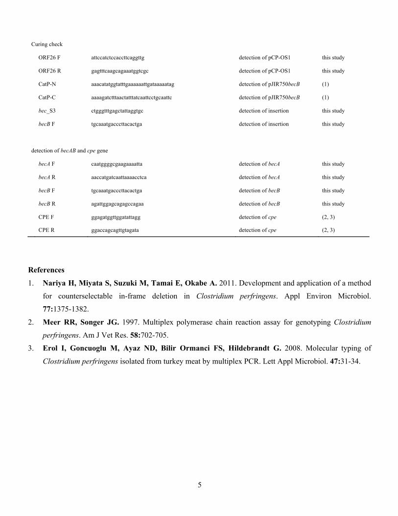
Curing check
ORF26 F attccatctccaccttcaggttg detection of pCP-OS1 this study
ORF26 R gagtttcaagcagaaatggtcgc detection of pCP-OS1 this study
CatP-N aaacatatggtatttgaaaaaattgataaaaatag detection of pJIR750becB (1)
CatP-C aaaagatctttaactatttatcaattcctgcaattc detection of pJIR750becB (1)
bec_S3 ctgggtttgagctattaggtgc detection of insertion this study
becB F tgcaaatgacccttacactga detection of insertion this study
detection of becAB and cpe gene
becA F caatggggcgaagaaaatta detection of becA this study
becA R aaccatgatcaattaaaacctca detection of becA this study
becB F tgcaaatgacccttacactga detection of becB this study
becB R agattggagcagagccagaa detection of becB this study
CPE F ggagatggttggatattagg detection of cpe (2, 3)
� CPE R ggaccagcagttgtagata detection of cpe (2, 3)
References
1. Nariya H, Miyata S, Suzuki M, Tamai E, Okabe A. 2011. Development and application of a method
for counterselectable in-frame deletion in Clostridium perfringens. Appl Environ Microbiol.
77:1375-1382.
2. Meer RR, Songer JG. 1997. Multiplex polymerase chain reaction assay for genotyping Clostridium
perfringens. Am J Vet Res. 58:702-705.
3. Erol I, Goncuoglu M, Ayaz ND, Bilir Ormanci FS, Hildebrandt G. 2008. Molecular typing of
Clostridium perfringens isolated from turkey meat by multiplex PCR. Lett Appl Microbiol. 47:31-34.
6
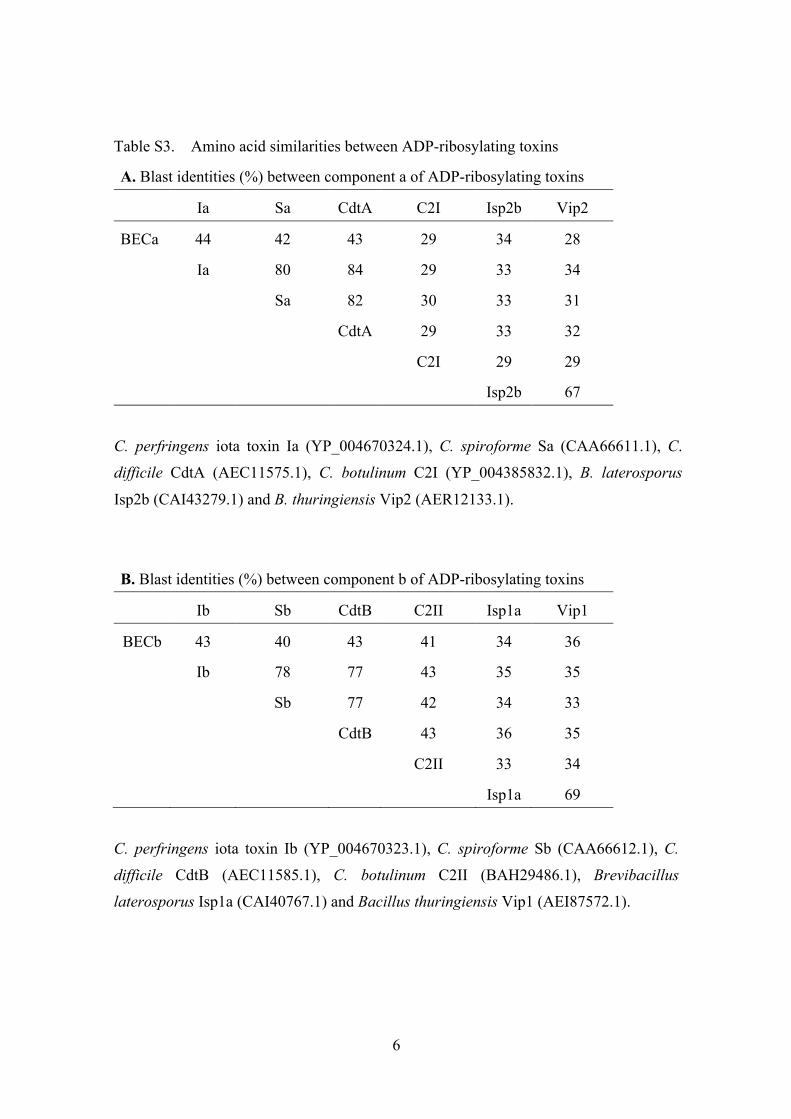
Table S3. Amino acid similarities between ADP-ribosylating toxins
A. Blast identities (%) between component a of ADP-ribosylating toxins
� Ia Sa CdtA C2I Isp2b Vip2
BECa 44 42 43 29 34 28
Ia 80 84 29 33 34
Sa 82 30 33 31
CdtA 29 33 32
C2I 29 29
� Isp2b 67
C. perfringens iota toxin Ia (YP_004670324.1), C. spiroforme Sa (CAA66611.1), C.
difficile CdtA (AEC11575.1), C. botulinum C2I (YP_004385832.1), B. laterosporus
Isp2b (CAI43279.1) and B. thuringiensis Vip2 (AER12133.1).
B. Blast identities (%) between component b of ADP-ribosylating toxins
� Ib Sb CdtB C2II Isp1a Vip1
BECb 43 40 43 41 34 36
Ib 78 77 43 35 35
Sb 77 42 34 33
CdtB 43 36 35
C2II 33 34
� Isp1a 69
C. perfringens iota toxin Ib (YP_004670323.1), C. spiroforme Sb (CAA66612.1), C.
difficile CdtB (AEC11585.1), C. botulinum C2II (BAH29486.1), Brevibacillus
laterosporus Isp1a (CAI40767.1) and Bacillus thuringiensis Vip1 (AEI87572.1).
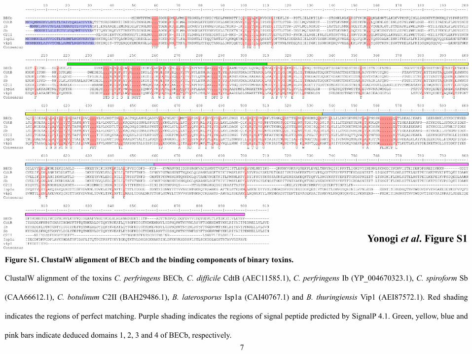
Yonogi et al. Figure S1�
Figure S1. ClustalW alignment of BECb and the binding components of binary toxins. �
ClustalW alignment of the toxins C. perfringens BECb, C. difficile CdtB (AEC11585.1), C. perfringens Ib (YP_004670323.1), C. spiroform Sb
(CAA66612.1), C. botulinum C2II (BAH29486.1), B. laterosporus Isp1a (CAI40767.1) and B. thuringiensis Vip1 (AEI87572.1). Red shading
indicates the regions of perfect matching. Purple shading indicates the regions of signal peptide predicted by SignalP 4.1. Green, yellow, blue and
pink bars indicate deduced domains 1, 2, 3 and 4 of BECb, respectively.�7
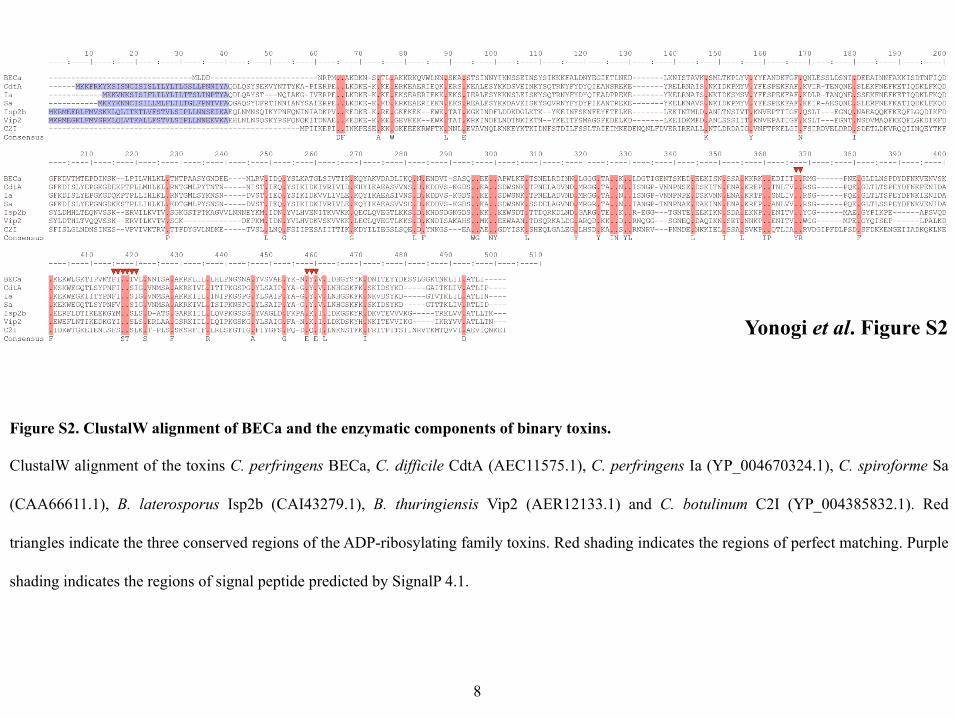
Yonogi et al. Figure S2�
Figure S2. ClustalW alignment of BECa and the enzymatic components of binary toxins. �
ClustalW alignment of the toxins C. perfringens BECa, C. difficile CdtA (AEC11575.1), C. perfringens Ia (YP_004670324.1), C. spiroforme Sa
(CAA66611.1), B. laterosporus Isp2b (CAI43279.1), B. thuringiensis Vip2 (AER12133.1) and C. botulinum C2I (YP_004385832.1). Red
triangles indicate the three conserved regions of the ADP-ribosylating family toxins. Red shading indicates the regions of perfect matching. Purple
shading indicates the regions of signal peptide predicted by SignalP 4.1.�
8
BECb��
BECa��Actin��
rBECa rBECb NAD+
Biotin-NAD+
Actin
+ - - + - - + - - + - - - + - - + - - + - - + - + + + - - - - - - - - - - - - + + + - - - + + + + + + + + + + + + - - - probe: streptavidin��
Yonogi et al. Figure S3�
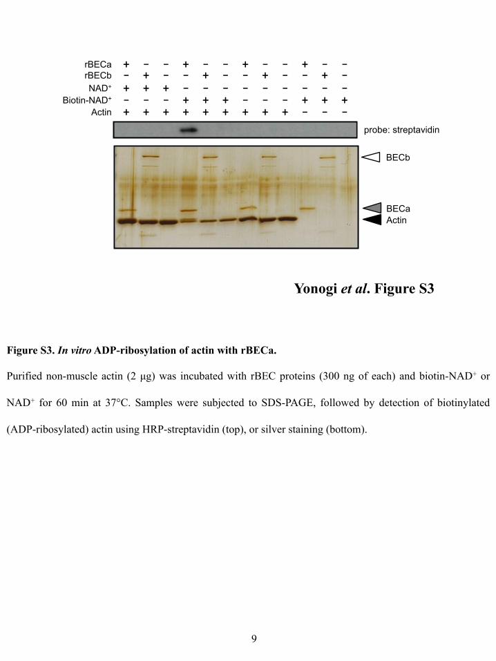
Figure S3. In vitro ADP-ribosylation of actin with rBECa.�
Purified non-muscle actin (2 µg) was incubated with rBEC proteins (300 ng of each) and biotin-NAD+ or
NAD+ for 60 min at 37°C. Samples were subjected to SDS-PAGE, followed by detection of biotinylated
(ADP-ribosylated) actin using HRP-streptavidin (top), or silver staining (bottom). �
9�
Yonogi et al. Figure S4�
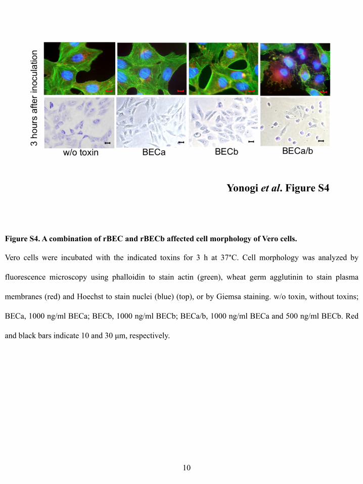
Figure S4. A combination of rBEC and rBECb affected cell morphology of Vero cells.�
Vero cells were incubated with the indicated toxins for 3 h at 37°C. Cell morphology was analyzed by
fluorescence microscopy using phalloidin to stain actin (green), wheat germ agglutinin to stain plasma
membranes (red) and Hoechst to stain nuclei (blue) (top), or by Giemsa staining. w/o toxin, without toxins;
BECa, 1000 ng/ml BECa; BECb, 1000 ng/ml BECb; BECa/b, 1000 ng/ml BECa and 500 ng/ml BECb. Red
and black bars indicate 10 and 30 µm, respectively.�
10�