Formation of Electrically Conductive Bacterial Nanowires .... Alshehri, et al.pdf · Formation of...
15
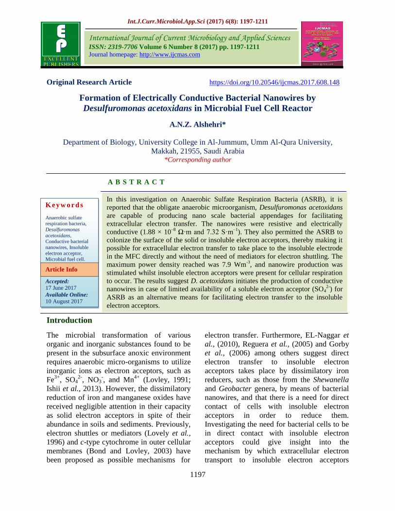
Int.J.Curr.Microbiol.App.Sci (2017) 6(8): 1197-1211 1197 Original Research Article https://doi.org/10.20546/ijcmas.2017.608.148 Formation of Electrically Conductive Bacterial Nanowires by Desulfuromonas acetoxidans in Microbial Fuel Cell Reactor A.N.Z. Alshehri* Department of Biology, University College in Al-Jummum, Umm Al-Qura University, Makkah, 21955, Saudi Arabia *Corresponding author ABSTRACT Introduction The microbial transformation of various organic and inorganic substances found to be present in the subsurface anoxic environment requires anaerobic micro-organisms to utilize inorganic ions as electron acceptors, such as Fe 3+ , SO 4 2- , NO 3 - , and Mn 4+ (Lovley, 1991; Ishii et al., 2013). However, the dissimilatory reduction of iron and manganese oxides have received negligible attention in their capacity as solid electron acceptors in spite of their abundance in soils and sediments. Previously, electron shuttles or mediators (Lovely et al., 1996) and c-type cytochrome in outer cellular membranes (Bond and Lovley, 2003) have been proposed as possible mechanisms for electron transfer. Furthermore, EL-Naggar et al., (2010), Reguera et al., (2005) and Gorby et al., (2006) among others suggest direct electron transfer to insoluble electron acceptors takes place by dissimilatory iron reducers, such as those from the Shewanella and Geobacter genera, by means of bacterial nanowires, and that there is a need for direct contact of cells with insoluble electron acceptors in order to reduce them. Investigating the need for bacterial cells to be in direct contact with insoluble electron acceptors could give insight into the mechanism by which extracellular electron transport to insoluble electron acceptors International Journal of Current Microbiology and Applied Sciences ISSN: 2319-7706 Volume 6 Number 8 (2017) pp. 1197-1211 Journal homepage: http://www.ijcmas.com In this investigation on Anaerobic Sulfate Respiration Bacteria (ASRB), it is reported that the obligate anaerobic microorganism, Desulfuromonas acetoxidans are capable of producing nano scale bacterial appendages for facilitating extracellular electron transfer. The nanowires were resistive and electrically conductive (1.88 × 10 –8 Ω·m and 7.32 S·m –1 ). They also permitted the ASRB to colonize the surface of the solid or insoluble electron acceptors, thereby making it possible for extracellular electron transfer to take place to the insoluble electrode in the MFC directly and without the need of mediators for electron shuttling. The maximum power density reached was 7.9 Wm -3 , and nanowire production was stimulated whilst insoluble electron acceptors were present for cellular respiration to occur. The results suggest D. acetoxidans initiates the production of conductive nanowires in case of limited availability of a soluble electron acceptor (SO 4 2- ) for ASRB as an alternative means for facilitating electron transfer to the insoluble electron acceptors. Keywords Anaerobic sulfate respiration bacteria, Desulfuromonas acetoxidans, Conductive bacterial nanowires, Insoluble electron acceptor, Microbial fuel cell. Accepted: 17 June 2017 Available Online: 10 August 2017 Article Info
Transcript of Formation of Electrically Conductive Bacterial Nanowires .... Alshehri, et al.pdf · Formation of...
Int.J.Curr.Microbiol.App.Sci (2017) 6(8): 1197-1211
1197
Original Research Article https://doi.org/10.20546/ijcmas.2017.608.148
Formation of Electrically Conductive Bacterial Nanowires by
Desulfuromonas acetoxidans in Microbial Fuel Cell Reactor
A.N.Z. Alshehri*
Department of Biology, University College in Al-Jummum, Umm Al-Qura University,
Makkah, 21955, Saudi Arabia *Corresponding author
A B S T R A C T
Introduction
The microbial transformation of various
organic and inorganic substances found to be
present in the subsurface anoxic environment
requires anaerobic micro-organisms to utilize
inorganic ions as electron acceptors, such as
Fe3+
, SO42-
, NO3-, and Mn
4+ (Lovley, 1991;
Ishii et al., 2013). However, the dissimilatory
reduction of iron and manganese oxides have
received negligible attention in their capacity
as solid electron acceptors in spite of their
abundance in soils and sediments. Previously,
electron shuttles or mediators (Lovely et al.,
1996) and c-type cytochrome in outer cellular
membranes (Bond and Lovley, 2003) have
been proposed as possible mechanisms for
electron transfer. Furthermore, EL-Naggar et
al., (2010), Reguera et al., (2005) and Gorby
et al., (2006) among others suggest direct
electron transfer to insoluble electron
acceptors takes place by dissimilatory iron
reducers, such as those from the Shewanella
and Geobacter genera, by means of bacterial
nanowires, and that there is a need for direct
contact of cells with insoluble electron
acceptors in order to reduce them.
Investigating the need for bacterial cells to be
in direct contact with insoluble electron
acceptors could give insight into the
mechanism by which extracellular electron
transport to insoluble electron acceptors
International Journal of Current Microbiology and Applied Sciences ISSN: 2319-7706 Volume 6 Number 8 (2017) pp. 1197-1211 Journal homepage: http://www.ijcmas.com
In this investigation on Anaerobic Sulfate Respiration Bacteria (ASRB), it is
reported that the obligate anaerobic microorganism, Desulfuromonas acetoxidans
are capable of producing nano scale bacterial appendages for facilitating
extracellular electron transfer. The nanowires were resistive and electrically
conductive (1.88 × 10⁻⁸
Ω·m and 7.32 S·m–1
). They also permitted the ASRB to
colonize the surface of the solid or insoluble electron acceptors, thereby making it
possible for extracellular electron transfer to take place to the insoluble electrode
in the MFC directly and without the need of mediators for electron shuttling. The
maximum power density reached was 7.9 Wm-3
, and nanowire production was
stimulated whilst insoluble electron acceptors were present for cellular respiration
to occur. The results suggest D. acetoxidans initiates the production of conductive
nanowires in case of limited availability of a soluble electron acceptor (SO42-
) for
ASRB as an alternative means for facilitating electron transfer to the insoluble
electron acceptors.
K e y w o r d s
Anaerobic sulfate respiration bacteria,
Desulfuromonas
acetoxidans, Conductive bacterial
nanowires, Insoluble
electron acceptor, Microbial fuel cell.
Accepted:
17 June 2017
Available Online: 10 August 2017
Article Info
Int.J.Curr.Microbiol.App.Sci (2017) 6(8): 1197-1211
1198
occurs in nature. Similarly, nano scale
bacterial appendages reduce iron during direct
electron transfer to Fe (III) oxides or other
solid electron receptors. The microbial fuel
cell (MFC) device utilizes electrically
catalytic microorganisms for generating
electricity from organic and inorganic
substances (Logan et al., 2006). This makes it
useful in evaluating extracellular electron
transfer reactions that occur close to electrode
surfaces, thereby acting as a naturally
insoluble or solid-phase electron acceptor in
the environment (Kiely et al., 2011). Indirect
electron transfer by mediators from cells to
electrodes is one strategy formed for
explaining extracellular electron transfer in
MFC (Newman and Kolter, 2000). Mediators
capable of functioning as electron shuttles
include iron oxides, sulfate, phenazines and
quinones. Another strategy is direct electron
transfer from cell to electrode by means of the
c-type cytochrome related with outer
membranes (Chaudhuri and Lovley, 2003),
and an alternative of interest to this study is
direct electron transfer by means of
filamentous structures such as bacterial
nanowires (Gorby et al., 2006). In the case of
MFCs that are mediator-less, micro-
organisms enable direct electron transfer to
the electrodes without the need for mediators,
and the electrodes function as sole electron
acceptors (Chaudhuri and Lovley, 2003;
Gregory et al., 2004).
Although many micro-organisms exist with
the capability of electron donation to the MFC
anode, and thereby of electricity production,
only some iron reducing species have been
discovered to produce nanowires as mediators
during direct electron transfer to the electrode,
such as Geobacter sulfurreducens and
Shewanella oneidensis MR-I (Gorby et al.,
2006; El-Naggar et al., 2010). ASRB
(Anaerobic Sulfate Respiration Bacteria)
present in highly reducing environments
normally use soluble sulfate as a terminal
electron acceptor during the process of
respiration to aid in their growth (Heidelberg
et al., 2004). It is by means of this process
that ASRB contribute to the carbon and sulfur
cycles considerably, as well as bioremediation
of contaminated subsurface systems (Martins
et al., 2009). ASRB such as Desulfobulbus
propionicus are capable of using other
electron acceptors, such as Fe3+
(Lovley et al.,
1993), NO3-
(Marietou et al., 2009), Mn4+
(Myers and Nealson, 1988), and fumarate
(Tomei et al., 1995), i.e. other than sulfate.
Notably, ASRB are also capable of being
used as microbes for generating electricity in
MFC (Cordas et al., 2008; Zhao et al., 2008).
A number of strategies have been devised to
facilitate electron transfer to date, from ASRB
to solid electrodes in an MFC system. For
instance, indirect electron transfer to
electrodes has been demonstrated for ASRB
by means of inorganic electrons as mediators
functioning as shuttles to thereby generate
electricity, such as sulfate or sulfide (Zhao et
al., 2008). In a mediatorless MFC, it has also
been shown that when functioning as a
microbial catalyst, ASRB facilitate electron
transfer to the electrode by means of contact
between the microbes and electrodes through
a c-type cytochrome present in an outer cell
membrane (Cordas et al., 2008). However,
the transfer of electrons by ASRB through
microbial appendages directly has not been
adequately addressed previously as it has for
iron reducers (Reguera et al., 2005; Gorby et
al., 2006; El-Naggar et al., 2010).
This study assesses the utility of nanoscale
bacterial appendages produced by ASRB as
filaments for facilitating the transfer of
electrons to insoluble electron acceptors
directly. It hypothesized that the bacterium D.
acetoxidans, selected as representative of
ASRB based on it having a well-characterized
genome and its prevalence in diverse anoxic
environments (Devereux and Mundfrom,
1994), produces nanowires only when there
Int.J.Curr.Microbiol.App.Sci (2017) 6(8): 1197-1211
1199
are insoluble electron acceptors present to
facilitate extracellular electron transfer, as
opposed to SO42-
as the soluble electron
acceptor. Furthermore, distinguishing
characteristics of the nanowires are identified
with respect to their morphological features
and functions by using an MFC inoculated
with D. acetoxidans as the chief ASRB.
Materials and Methods
Microorganism, media and cultivation
The ASRB D. acetoxidans was obtained from
a Germanic collection of micro-organisms in
Braunschweig, Germany. The cells underwent
preharvestation within a growth medium in
strict anaerobic conditions at 37oC. This
medium contained (per liter): 0.5 g of
K2HPO4, 1.0 g of NH4Cl, 1.0 g of Na2SO4,
0.1 g of CaCl2.H2O, 2.0 g of MgSO4.7H2O,
2.0 g of sodium lactate, 1.0 g of yeast extract,
1.0 mg of resazurin, 0.5 g of FeSO4.7H2O, 0.1
g of sodium thioglycolate, 0.1 g of ascorbic
acid, and 1 mL of a trace element solution.
Prior to sterilization at 121oC by autoclaving
for 15 minutes, the pH 7 of the medium was
adjusted with 0.1 M NaOH. The cells were
harvested by centrifugation for 20 min at
10,000g and 4oC, then washed twice in a
buffer solution of 50 mM of phosphate at pH
7, and seeded in the anodic MFC chamber.
A mixed culture of ASRB was then isolated
from an anaerobic sewage digestion of sludge,
which was collected from a treatment plant of
domestic wastewater by subculturing the
bacteria in a selective medium of ASRB. The
medium was composed of 2 g/L of sodium
lactate, 0.3 g/L of sodium citrate, 0.1 g/L of
yeast extract, 4.5 g/L of Na2SO4, 0.06 g/L of
CaCl2.2H2O, 1.0 g/L of NH4Cl, 0.5 g/L of
KH2PO4, 2.0 g/L of MgSO4.4H2O, 0.5 g/L of
FeSO4.7H2O, 0.3 g/L of disodium ethylene
diamine tetra acetate, and 0.2 g/L of K2CrO4.
The mixed culture of ASBR was retained for
a week under strict anaerobic conditions
before being subcultured. The microbial
inocula were washed with the phosphate
buffer solution (50 mM at pH7), and a mixed
ASRB culture was seeded in the MFC anodic
chamber.
Bacterial nanowire formation
The D. acetoxidans developed anaerobically
in the growth medium at 37oC. The cells were
given 50 mM of sulfate as soluble and 50 mM
of Fe (III) oxide (αFe2O3) as insoluble
electron acceptors. The cells were grown in
the growth medium without an electron
acceptor as a control experiment.
MFC set up and operation
Experiments were conducted with the
electrodes as solid electron acceptors in a
dual-chamber MFC, similar to the
arrangement of Alshehri et al., (2016) with
some modifications. A Nafion 117 proton
exchange membrane (PEM; Dupont, USA)
separated the two chambers. The working
volume was 100 mL, and the total volume
was 200 mL. The cathode chamber was then
filled with a catholyte solution involving 30
mMTris buffer solution (pH 7) and
continuously purged using water-saturated air.
The pure culture of D. acetoxidans was
seeded in the anode chamber, and a mixed
ASRB culture was added in a separate
experiment. Electron donor was given
through a supply of organic substrate as fuel,
and with the exception of the electrode, no
electron acceptor or electron-shuttling
mediator was used. The anode chamber
medium was purged with a N2 and CO2
mixture in a ratio of 9:3 (v/v) in order to
maintain the anaerobic conditions. Graphite
felt with a surface area of 30 cm2 was used as
the cathodic and anodic electrodes (GF series,
Electrosynthesis, USA). The electrodes were
then connected to a platinum wire with a 150
Ω external resistance.
Int.J.Curr.Microbiol.App.Sci (2017) 6(8): 1197-1211
1200
Monitoring and calculation
When the MFC reached a steady state,
automatic measurements of the voltage were
taken using a digital multimeter (Sanwa
CD800a, Japan) connected to a personal
computer and Picolog software (Pico
Technology Limited) at one second intervals.
The corresponding current was based on
equation I=E/Rext, where: I is current (mA), E
is voltage (mV) and Rext is external resistance.
The power (P) was obtained by P=IE. The
current density and the power density have
been normalized based on the projected
surface area of the anode via equations IAn=
I/AAn, where IAn is current density and AAn is
the surface area of anode, PAn=E2/AAnRext,
where PAn is power density. The polarization
curve was obtained at different external
resistance (50 - 1000Ω). Internal resistance
was derived from the polarization curve as the
slope. Coulombic efficiency (CE) was derived
from the equations Cp=It, Cmax=FfSCODVAn,
and CE=Cp/Cmax, where Cp is the coulombs
of energy produced, t is the time of stable
voltage output, Cmax is the theoretical
maximum coulombs, F is Faraday's constant
(96.485 C/mol of electrons), f is a factor of
1mol electrons/8g COD, SCOD is substrate
concentration g COD/L, and VAn is a net
volume of anolyte (mL). All the experiments
were replicated twice at 37 oC under the same
conditions.
Phylogenetic analyses
Purity of the culture was done by DNA
extraction and PCR amplification of the 16S
rRNA gene with the aid of a forward primer
(5'- ACC GTT AGA TGG CTC TAC TTG
GGC AGA TTC GCT -3') and a reverse
primer (5'-TGC CGC TGA AGC AGG TTC
ACC TCC TAC GGC A-3'), and subsequent
sequencing of the purified PCR products.
Phylogenetic analysis was then done using
BLAST (NCBI) and the standard DNA
sequencing program, and PCR amplification
was done similarly for the mixed culture of
ASRB using the forward and reverse primers
as listed in table 1. The neighbor-joining
method (Daly et al., 2000) was then used
during the phylogenetic analysis based on six
ASRB predominant subgroups.
Scanning electron microscopy
The surfaces of the graphite felt electrodes
(both bare and treated) and the biofilm
formation present on these surfaces during
runs of the MFC were examined with
scanning electron microscope (SEM, JSM-
5410LV, JEOL Ltd., Japan). Distilled water
was used to rinse the electrodes, fixed firstly
with 2% of glutaraldehyde for 2 hours at 4oC.
It was then rinsed 3 times using 0.05 M of
sodium cacodylate buffer (pH 7.2) for 10
minutes at 4oC, and fixed further with 1% of
osmium tetroxide in 0.05 M sodium
cacodylate buffer for 2 hours at 4oC. Mill-Q
water was then used to rinse the fixed
electrode twice at room temperature before
dehydrating with increased ethanol
concentrations (30%, 50%, 70%, 80%, 90%,
and 100%) for 10 minutes each at room
temperature. After gently washing the
electrode with 100% hexamethyldisilazane
for 15 minutes at room temperature and
making it air-dried overnight, the electrode
was then coated before the SEM analysis with
platinum.
Atomic force microscopy (AFM)
After centrifugation of the microbial samples
collected from MFC for 5 minutes at 3000 g
and removal of the supernatant, distilled water
was used to wash the pellet, and an aliquot
sample (20µL) was applied onto a highly
oriented pyrolytic graphite (HOPG) plate as a
substrate (10×10×1.5 mm, grade ZYA, Ted
Pella, USA). This sample was then air-dried
and washed with distilled water 3 times. The
Int.J.Curr.Microbiol.App.Sci (2017) 6(8): 1197-1211
1201
HOPG plate loaded with the cells was
subjected to examination using Atomic Force
Microscope (AFM, XE-100 series, Park
System Scanning Probe Microscope, Park
System, Santa Clara, USA) in either the
contact or non-contact modes. In the case of
the contact mode (conducting probe AFM), a
nominal spring constant of 0.2 N/m was
applied to a conductive cantilever (CDT-
CONTR 3M-T, Park System) with a
platinum-coated probe (Park system). The
current response profile was obtained by
applying a +5.0V bias voltage, and the current
responses to sweep bias voltages in the range
-1 V to +1 V were measured to form an I-V
(current-voltage) curve. The formula p = R ×
A / L was used to calculate the nanowire
resistivity where p = resistivity (Ωm), R =
resistance (Ω), L = length of the nanowires,
and A = cross-sectional area of the nanowires
(m2 was calculated using AFM height
measurement), and the formula σ = 1/p was
used to calculate the conductivity was
calculated as the inverse of resistivity, where
σ = electrical conductivity (S·m–1
) and p =
resistivity (Ω m).
Analytical techniques
A Petroff-Hauser counting chamber was used
to determine the cell density, and
quantification of the cellular proteins was
done as previously described (Chaudhuri and
Lovley, 2003). The Bradford method was
used with bovine serum albumin as standard
to extract and measure the protein attached to
the electrode (Quick Start Bradford Protein
Assay, Bio-Rad, USA).
Determination of the substrate concentration
was made by using visible spectroscope, then
ion chromatographe (Cordas et al., 2008). The
sulfide concentration was determined using a
silver/sulfide ion selective electrode.
Quantification of the Fe2+
was done by a
ferrozine method (lovley and Phillips, 1986).
Purity testing of the cell cultures was done
through PCR amplification of the 16S rRNA
gene.
Results and Discussion
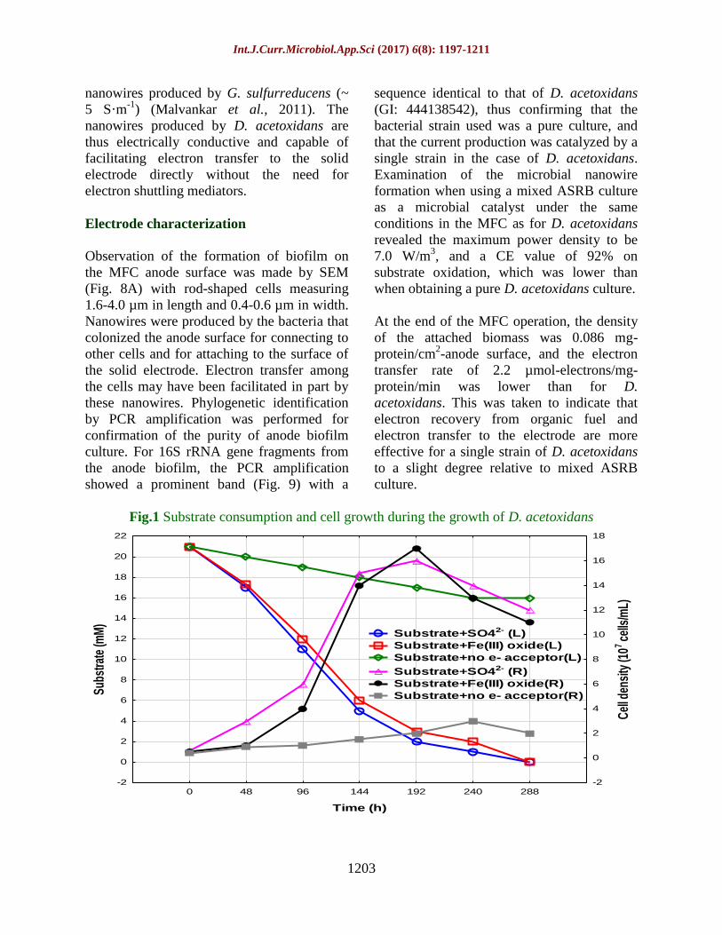
Bacterial nanowire formation
Examination of the bacterial nanowire
formation was done by growing a pure culture
of D. acetoxidans under strict anaerobic
conditions in the presence or absence of
electron acceptors. Sulfate or Fe (III) oxide
was utilized by the D. acetoxidans for the
growth as the sole electron acceptor. This led
to an increase in its biomass, whereas almost
no growth was evident in the absence of an
inorganic electron acceptor (Fig. 1).
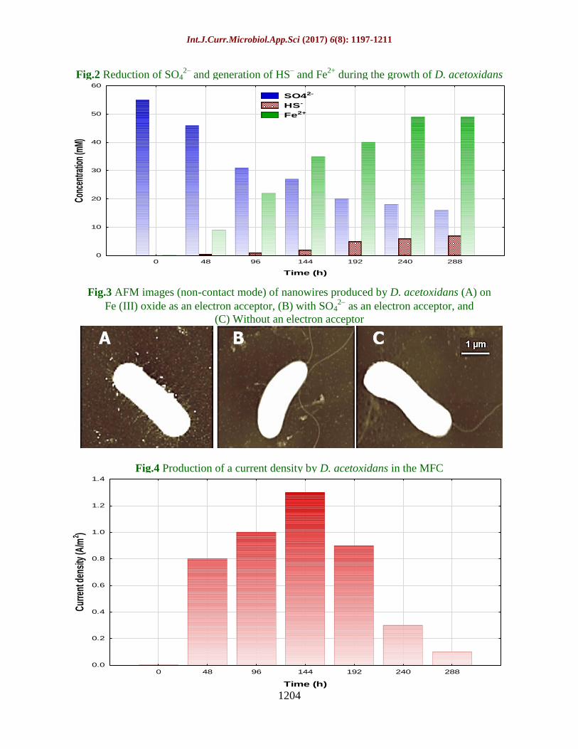
Dissimilatory reduction occurred of sulfate to
sulfide, and of Fe (III) oxide to Fe2+
(Fig. 2),
and the production of nanowires during this
period was confirmed by AFM. In the
presence of Fe (III) oxide, nanowire
formation around the cell was induced
markedly in numbers, as the Fe (III) oxide
served as an electron (Fig. 3A). This finding
confirms earlier demonstrations of bacterial
nanowire production by iron reducing G.
sulfurreducens and S. oneidensis when they
were grown on poorly crystalline Fe (III)
oxides (Reguera et al., 2005; Groby et al.,
2006). With D. acetoxidans, only a single
filament was produced when grown on the
soluble electron acceptor (SO42-
).
This filament appeared as a polar flagellum
for cell motility (Fig. 3B), and the detection
of a single filament was in the absence of an
inorganic electron acceptor (Fig. 3C).
Microbial nanowire formation in numerous
quantities can therefore be stimulated (as it
clear from these results) for allowing contact
with insoluble electron acceptors, as for metal
oxides present in surface systems, and for
transferring electrons for dissimilatory
reduction directly.
Int.J.Curr.Microbiol.App.Sci (2017) 6(8): 1197-1211
1202
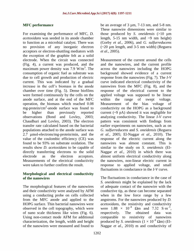
MFC performance
For examining the performance of MFC, D.
acetoxidans was seeded in its anode chamber
to function as a microbial catalyst. There was
no provision of any inorganic electron
acceptors or electron-shuttling mediators with
the exception of the graphite felt as a solid
electrode. When the circuit was connected
(Fig. 4), a current was produced, and the
maximum power density was 7.9 W/m3. The
consumption of organic fuel as substrate was
due to cell growth and production of electric
current. This was indicated by a gradual
increase in the cell’s biomass in the anode
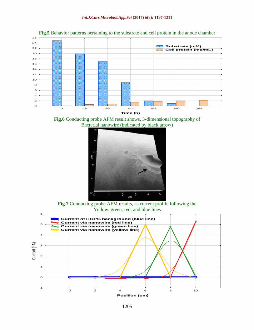
chamber over time (Fig. 5). Dense biofilms
were formed continuously by the cells on the
anode surface, and at the end of the MFC
operation, the biomass which reached 0.08
mg-protein/cm2-anode surface was found to
be higher than previously reported
observations (Bond and Lovley, 2003;
Chaudhuri and Lovley, 2003). The electron
transfer rate calculated based on the bacterial
populations attached to the anode surface was
2.7 µmol-electrons/mg-protein/min, and the
value of the coulombic efficiency (CE) was
found to be 93% on substrate oxidation. The
results show D. acetoxidans to be capable of
directly transferring electrons to the solid
electrode as the electron acceptors.
Measurements of the electrical conductivity
were taken to further confirm the result.
Morphological and electrical conductivity
of the nanowires
The morphological features of the nanowires
and their conductivity were analysed by AFM
using a conducting probe on cells collected
from the MFC anode and applied to the
HOPG surface. Thin bacterial nanowires were
observed in the cell topography, which were
of nano scale thickness like wires (Fig. 6).
Using non-contact mode AFM for additional
characterization, the length, width and height
of the nanowires were measured and found to
be an average of 3 µm, 7-13 nm, and 5-8 nm.
These nanowire dimensions were similar to
those produced by S. oneidensis (>10 µm
length, 5-15 nm width, and ~9 nm height)
(Gorby et al., 2006), and G. sulfurreducens
(~20 µm length, and 3-5 nm width) (Reguera
et al., 2005).
Measurement of the current around the cells
and the nanowires, and the current profile
across the nanowires including the HOPG
background showed evidence of a current
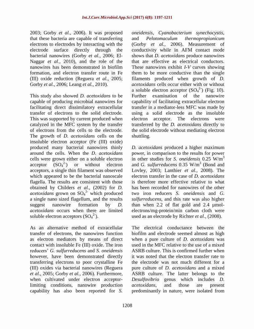
response from the nanowires (Fig. 7). The I-V
curve indicated electrical conductivity of the
nanowires from the MFC (Fig. 8), and the
response of the electrical current to the
applied voltage was supposed to be linear
within the range of swept voltage.
Measurement of the bias voltage of
conductivity on the HOPG as a background
current (~9 pA) showed it was negligible for
analyzing conductivity. The linear I-V curve
pattern was consistent with findings from
earlier experiments on nanowires, such as on
G. sulfurreducens and S. oneidensis (Reguera
et al., 2005; El-Naggar et al., 2010). The
response of the electric current along the
nanowires was almost constant. This is
similar to the study on S. oneidensis (El-
Naggar et al., 2010) in which there was
almost uniform electrical conductivity along
the nanowires, non-linear electric current in
response to applied voltage, and small
fluctuations in conductance in the I-V curve.
The fluctuations in conductance in the case of
S. oneidensis might be explained by the lack
of adequate contact of the nanowire with the
conductive tip, as these can become separated
when in the low force range by a few
angstroms. For the nanowires produced by D.
acetoxidans, the resistivity and conductivity
were 1.88 × 10⁻⁸
Ωm and 7.32 S·m–1
,
respectively. The obtained data was
comparable to resistivity of nanowires
produced by S. oneidensis (1 × 10⁻⁸
Ωm) (El-
Naggar et al., 2010) m and conductivity of
Int.J.Curr.Microbiol.App.Sci (2017) 6(8): 1197-1211
1203
nanowires produced by G. sulfurreducens (~
5 S·m-1
) (Malvankar et al., 2011). The
nanowires produced by D. acetoxidans are
thus electrically conductive and capable of
facilitating electron transfer to the solid
electrode directly without the need for
electron shuttling mediators.
Electrode characterization
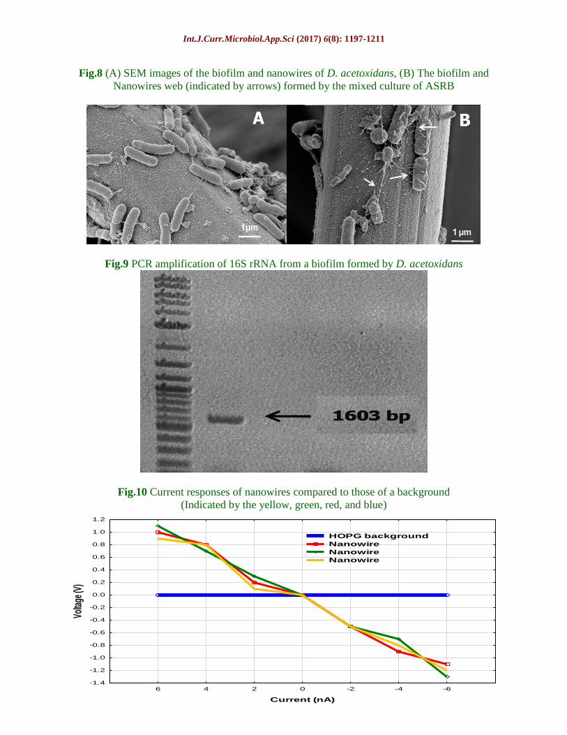
Observation of the formation of biofilm on
the MFC anode surface was made by SEM
(Fig. 8A) with rod-shaped cells measuring
1.6-4.0 µm in length and 0.4-0.6 µm in width.
Nanowires were produced by the bacteria that
colonized the anode surface for connecting to
other cells and for attaching to the surface of
the solid electrode. Electron transfer among
the cells may have been facilitated in part by
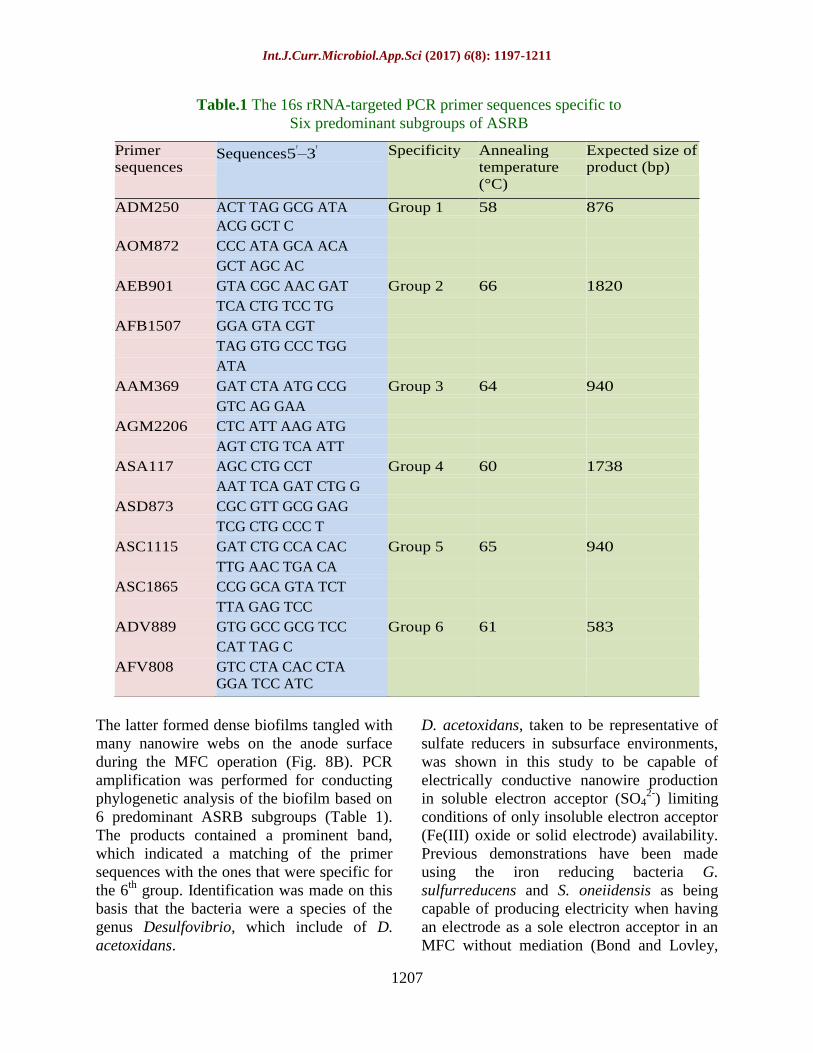
these nanowires. Phylogenetic identification
by PCR amplification was performed for
confirmation of the purity of anode biofilm
culture. For 16S rRNA gene fragments from
the anode biofilm, the PCR amplification
showed a prominent band (Fig. 9) with a
sequence identical to that of D. acetoxidans
(GI: 444138542), thus confirming that the
bacterial strain used was a pure culture, and
that the current production was catalyzed by a
single strain in the case of D. acetoxidans.
Examination of the microbial nanowire
formation when using a mixed ASRB culture
as a microbial catalyst under the same
conditions in the MFC as for D. acetoxidans
revealed the maximum power density to be
7.0 W/m3, and a CE value of 92% on
substrate oxidation, which was lower than
when obtaining a pure D. acetoxidans culture.
At the end of the MFC operation, the density
of the attached biomass was 0.086 mg-
protein/cm2-anode surface, and the electron
transfer rate of 2.2 µmol-electrons/mg-
protein/min was lower than for D.
acetoxidans. This was taken to indicate that
electron recovery from organic fuel and
electron transfer to the electrode are more
effective for a single strain of D. acetoxidans
to a slight degree relative to mixed ASRB
culture.
Fig.1 Substrate consumption and cell growth during the growth of D. acetoxidans
0 48 96 144 192 240 288
Time (h)
-2
0
2
4
6
8
10
12
14
16
18
20
22
Sub
stra
te (m
M)
-2
0
2
4
6
8
10
12
14
16
18
Cel
l den
sity
(107 c
ells
/mL)
Substrate+SO42- (L)
Substrate+Fe(III) oxide(L)
Substrate+no e- acceptor(L)
Substrate+SO42- (R)
Substrate+Fe(III) oxide(R)
Substrate+no e- acceptor(R)
Int.J.Curr.Microbiol.App.Sci (2017) 6(8): 1197-1211
1204
Fig.2 Reduction of SO42–
and generation of HS– and Fe
2+ during the growth of D. acetoxidans
Fig.3 AFM images (non-contact mode) of nanowires produced by D. acetoxidans (A) on
Fe (III) oxide as an electron acceptor, (B) with SO42
as an electron acceptor, and
(C) Without an electron acceptor
Fig.4 Production of a current density by D. acetoxidans in the MFC
0 48 96 144 192 240 288
Time (h)
0
10
20
30
40
50
60Co
ncen
tratio
n (m
M)
SO42-
HS-
Fe2+
0 48 96 144 192 240 288
Time (h)
0.0
0.2
0.4
0.6
0.8
1.0
1.2
1.4
Cur
rent
den
sity
(A/m
2 )
Int.J.Curr.Microbiol.App.Sci (2017) 6(8): 1197-1211
1205
Fig.5 Behavior patterns pertaining to the substrate and cell protein in the anode chamber
Fig.6 Conducting probe AFM result shows, 3-dimensional topography of
Bacterial nanowire (indicated by black arrow)
Fig.7 Conducting probe AFM results, as current profile following the
Yellow, green, red, and blue lines
0 48 96 144 192 240 288
Time (h)
0
2
4
6
8
10
12
14
16
18
20
22
24
26
Substrate (mM)
Cell protein (mg/mL)
0 2 4 6 8 10
Position (um)
-1
0
1
2
3
4
5
6
Curre
nt (n
A)
Current of HOPG background (blue line)
Current via nanowire (red line)
Current via nanowire (green line)
Current via nanowire (yellow line)
Int.J.Curr.Microbiol.App.Sci (2017) 6(8): 1197-1211
1206
Fig.8 (A) SEM images of the biofilm and nanowires of D. acetoxidans, (B) The biofilm and
Nanowires web (indicated by arrows) formed by the mixed culture of ASRB
Fig.9 PCR amplification of 16S rRNA from a biofilm formed by D. acetoxidans
Fig.10 Current responses of nanowires compared to those of a background
(Indicated by the yellow, green, red, and blue)
6 4 2 0 -2 -4 -6
Current (nA)
-1.4
-1.2
-1.0
-0.8
-0.6
-0.4
-0.2
0.0
0.2
0.4
0.6
0.8
1.0
1.2
Volta
ge (V
)
HOPG background
Nanowire
Nanowire
Nanowire
Int.J.Curr.Microbiol.App.Sci (2017) 6(8): 1197-1211
1207
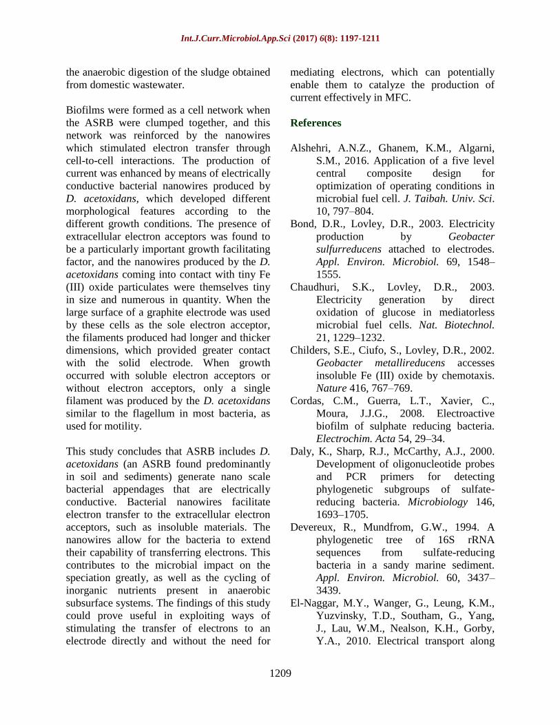
Table.1 The 16s rRNA-targeted PCR primer sequences specific to
Six predominant subgroups of ASRB
The latter formed dense biofilms tangled with
many nanowire webs on the anode surface
during the MFC operation (Fig. 8B). PCR
amplification was performed for conducting
phylogenetic analysis of the biofilm based on
6 predominant ASRB subgroups (Table 1).
The products contained a prominent band,
which indicated a matching of the primer
sequences with the ones that were specific for
the 6th
group. Identification was made on this
basis that the bacteria were a species of the
genus Desulfovibrio, which include of D.
acetoxidans.
D. acetoxidans, taken to be representative of
sulfate reducers in subsurface environments,
was shown in this study to be capable of
electrically conductive nanowire production
in soluble electron acceptor (SO42-
) limiting
conditions of only insoluble electron acceptor
(Fe(III) oxide or solid electrode) availability.
Previous demonstrations have been made
using the iron reducing bacteria G.
sulfurreducens and S. oneiidensis as being
capable of producing electricity when having
an electrode as a sole electron acceptor in an
MFC without mediation (Bond and Lovley,
Primer
sequences Sequences5ʹ–3ʹ Specificity Annealing
temperature
(°C)
Expected size of
product (bp)
ADM250 ACT TAG GCG ATA Group 1 58 876
ACG GCT C
AOM872 CCC ATA GCA ACA
GCT AGC AC
AEB901 GTA CGC AAC GAT Group 2 66 1820
TCA CTG TCC TG
AFB1507 GGA GTA CGT
TAG GTG CCC TGG
ATA
AAM369 GAT CTA ATG CCG Group 3 64 940
GTC AG GAA
AGM2206 CTC ATT AAG ATG
AGT CTG TCA ATT
ASA117 AGC CTG CCT Group 4 60 1738
AAT TCA GAT CTG G
ASD873 CGC GTT GCG GAG
TCG CTG CCC T
ASC1115 GAT CTG CCA CAC Group 5 65 940
TTG AAC TGA CA
ASC1865 CCG GCA GTA TCT
TTA GAG TCC
ADV889 GTG GCC GCG TCC Group 6 61 583
CAT TAG C
AFV808 GTC CTA CAC CTA
GGA TCC ATC
Int.J.Curr.Microbiol.App.Sci (2017) 6(8): 1197-1211
1208
2003; Gorby et al., 2006). It was proposed
that these bacteria are capable of transferring
electrons to electrodes by interacting with the
electrode surface directly through the
bacterial nanowires (Gorby et al., 2006; El-
Naggar et al., 2010), and the role of the
nanowires has been demonstrated in biofilm
formation, and electron transfer route in Fe
(III) oxide reduction (Reguera et al., 2005;
Gorby et al., 2006; Leang et al., 2010).
This study also showed D. acetoxidans to be
capable of producing microbial nanowires for
facilitating direct dissimilatory extracellular
transfer of electrons to the solid electrode.
This was supported by current produced when
catalyzed in the MFC system by the transfer
of electrons from the cells to the electrode.
The growth of D. acetoxidans cells on the
insoluble electron acceptor (Fe (III) oxide)
produced many bacterial nanowires thinly
around the cells. When the D. acetoxidans
cells were grown either on a soluble electron
acceptor (SO42-
) or without electron
acceptors, a single thin filament was observed
which appeared to be the bacterial nanoscale
flagella. The results are consistent with those
obtained by Childers et al., (2002) for D.
acetoxidans grown on SO42-
which produced
a single nano sized flagellum, and the results
suggest nanowire formation by D.
acetoxidans occurs when there are limited
soluble electron acceptors (SO42-
).
As an alternative method of extracellular
transfer of electrons, the nanowires function
as electron mediators by means of direct
contact with insoluble Fe (III) oxide. The iron
reducers’ G. sulfurreducens and S. oneidensis
however, have been demonstrated directly
transferring electrons to poor crystalline Fe
(III) oxides via bacterial nanowires (Reguera
et al., 2005; Gorby et al., 2006). Furthermore,
when cultivated under electron acceptor-
limiting conditions, nanowire production
capability has also been reported for S.
oneidensis, Cyanobacterium synechocystis,
and Pelotomaculum thermopropionicum
(Gorby et al., 2006). Measurement of
conductivity while in AFM contact mode
shows that D. acetoxidans produce nanowires
that are effective as electrical conductors.
These nanowires exhibit I-V curves showing
them to be more conductive than the single
filaments produced when growth of D.
acetoxidans cells occur either with or without
a soluble electron acceptor (SO42-
) (Fig. 10).
Further examination of the nanowire
capability of facilitating extracellular electron
transfer in a mediator-less MFC was made by
using a solid electrode as the insoluble
electron acceptor. The electrons were
transferred by the D. acetoxidans directly to
the solid electrode without mediating electron
shuttling.
D. acetoxidans produced a higher maximum
power, in comparison to the results for power
in other studies for S. oneidensis 0.25 W/m3
and G. sulfurreducens 0.35 W/m3 (Bond and
Lovley, 2003; Lanthier et al., 2008). The
electron transfer in the case of D. acetoxidans
is therefore more effective relative to what
has been recorded for nanowires of the other
two iron reducers S. oneidensis and G.
sulfurreducens, and this rate was also higher
than when 2.2 of flat gold and 2.4 µmol-
electrons/mg-protein/min carbon cloth were
used as an electrode by Richter et al., (2008).
The electrical conductance between the
biofilm and electrode seemed almost as high
when a pure culture of D. acetoxidans was
used in the MFC relative to the use of a mixed
ASRB culture. This is confirmed further when
it was noted that the electron transfer rate to
the electrode was not much different for a
pure culture of D. acetoxidans and a mixed
ASRB culture. The latter belongs to the
Desulfovibrio genus which includes D.
acetoxidans, and those are present
predominantly in nature, were isolated from
Int.J.Curr.Microbiol.App.Sci (2017) 6(8): 1197-1211
1209
the anaerobic digestion of the sludge obtained
from domestic wastewater.
Biofilms were formed as a cell network when
the ASRB were clumped together, and this
network was reinforced by the nanowires
which stimulated electron transfer through
cell-to-cell interactions. The production of
current was enhanced by means of electrically
conductive bacterial nanowires produced by
D. acetoxidans, which developed different
morphological features according to the
different growth conditions. The presence of
extracellular electron acceptors was found to
be a particularly important growth facilitating
factor, and the nanowires produced by the D.
acetoxidans coming into contact with tiny Fe
(III) oxide particulates were themselves tiny
in size and numerous in quantity. When the
large surface of a graphite electrode was used
by these cells as the sole electron acceptor,
the filaments produced had longer and thicker
dimensions, which provided greater contact
with the solid electrode. When growth
occurred with soluble electron acceptors or
without electron acceptors, only a single
filament was produced by the D. acetoxidans
similar to the flagellum in most bacteria, as
used for motility.
This study concludes that ASRB includes D.
acetoxidans (an ASRB found predominantly
in soil and sediments) generate nano scale
bacterial appendages that are electrically
conductive. Bacterial nanowires facilitate
electron transfer to the extracellular electron
acceptors, such as insoluble materials. The
nanowires allow for the bacteria to extend
their capability of transferring electrons. This
contributes to the microbial impact on the
speciation greatly, as well as the cycling of
inorganic nutrients present in anaerobic
subsurface systems. The findings of this study
could prove useful in exploiting ways of
stimulating the transfer of electrons to an
electrode directly and without the need for
mediating electrons, which can potentially
enable them to catalyze the production of
current effectively in MFC.
References
Alshehri, A.N.Z., Ghanem, K.M., Algarni,
S.M., 2016. Application of a five level
central composite design for
optimization of operating conditions in
microbial fuel cell. J. Taibah. Univ. Sci.
10, 797–804.
Bond, D.R., Lovley, D.R., 2003. Electricity
production by Geobacter
sulfurreducens attached to electrodes.
Appl. Environ. Microbiol. 69, 1548–
1555.
Chaudhuri, S.K., Lovley, D.R., 2003.
Electricity generation by direct
oxidation of glucose in mediatorless
microbial fuel cells. Nat. Biotechnol.
21, 1229–1232.
Childers, S.E., Ciufo, S., Lovley, D.R., 2002.
Geobacter metallireducens accesses
insoluble Fe (III) oxide by chemotaxis.
Nature 416, 767–769.
Cordas, C.M., Guerra, L.T., Xavier, C.,
Moura, J.J.G., 2008. Electroactive
biofilm of sulphate reducing bacteria.
Electrochim. Acta 54, 29–34.
Daly, K., Sharp, R.J., McCarthy, A.J., 2000.
Development of oligonucleotide probes
and PCR primers for detecting
phylogenetic subgroups of sulfate-
reducing bacteria. Microbiology 146,
1693–1705.
Devereux, R., Mundfrom, G.W., 1994. A
phylogenetic tree of 16S rRNA
sequences from sulfate-reducing
bacteria in a sandy marine sediment.
Appl. Environ. Microbiol. 60, 3437–
3439.
El-Naggar, M.Y., Wanger, G., Leung, K.M.,
Yuzvinsky, T.D., Southam, G., Yang,
J., Lau, W.M., Nealson, K.H., Gorby,
Y.A., 2010. Electrical transport along
Int.J.Curr.Microbiol.App.Sci (2017) 6(8): 1197-1211
1210
bacterial nanowires from Shewanella
oneidensis MR-1. Proc. Natl. Acad. Sci.
U.S.A. 107, 18127–18131.
Gorby, Y.A., Yanina, S., McLean, J.S.,
Rosso, K.M., Moyles, D., Dohnalkova,
A., Beveridge, T.J., Chang, I.S., Kim,
B.H., Kim, K.S., Culley, D.E., Reed,
S.B., Romine, M. F., Saffarini, D.A.,
Hill, E.A., Shi, L., Elias, D.A.,
Kennedy, D.W., Pinchuk, G.,
Watanabe, K., Ishii, S., Logan, B.,
Nealson, K.H., Fredrickson, J.K., 2006.
Electrically conductive bacterial
nanowires produced by Shewanella
oneidensis strain MR-1 and other
microorganisms. Proc. Natl. Acad. Sci.
U.S.A. 103, 1358–11363.
Gregory, K.B., Bond, D.R., Lovley, D.R.,
2004. Graphite electrodes as electron
donors for anaerobic respiration.
Environ. Microbiol. 6, 596–604.
Heidelberg, J.F., Seshadri, R., Haveman,
S.A., Hemme, C.L., Paulsen, I.T.,
Kolonay, J.F., Eisen, J.A., Ward, N.,
Methe, B., Brinkac, L.M., Daugherty,
S.C., Deboy, R.T., Dodson, R.J.,
Durkin, A.S., Madupu, R., Nelson,
W.C., Sullivan, S.A., Fouts, D., Haft,
D.H., Selengut, J., Peterson, J.D.,
Davidsen, T.M., Zafar, N., Zhou, L.,
Radune, D., Dimitrov, G., Hance, M.,
Tran, K., Khouri, H., Gill, J., Utterback,
T.R., Feldblyum, T. V., Wall, J.D.,
Voordouw, G., Fraser, C.M., 2004. The
genome sequence of the anaerobic,
sulfate-reducing bacterium
Desulfovibrio vulgaris Hildenborough.
Nat. Biotechnol. 22, 554–559.
Ishii, S., Suzuki, S., Norden-Krichmar, T.M.,
Tenney, A., Chain, P.S.G., Scholz,
M.B., Nealson, K.H., Bretschger, O.,
2013. A novel metatranscriptomic
approach to identify gene expression
dynamics during extracellular electron
transfer. Nat. Commun. 4, 1601.
http://dx.doi.org/10.1038/ncomms2615.
Kiely, P.D., Regan, J.M., Logan, B.E., 2011.
The electric picnic: synergistic
requirements for exoelectrogenic
microbial communities. Curr. Opin.
Biotechnol. 22, 378–385.
Lanthier, M., Gregory, K.B., Lovley, D.R.,
2008. Growth with high planktonic
biomass in Shewanella oneidensis fuel
cells. FEMS Microbiol. Lett. 278, 29–
35.
Leang, C., Qian, X., Mester, T., Lovley, D.R.,
2010. Alignment of the c-type
cytochrome OmcS along pili of
Geobacter sulfurreducens. Appl.
Environ. Microbiol. 76, 4080–4084.
Logan, B.E., Hamelers, B., Rozendal, R.,
Shröder, U., Keller, J., Freguia, S.,
Aelterman, P., Verstraete, W., Rabaey,
K., 2006. Microbial fuel cells:
methodology and technology. Environ.
Sci. Technol. 40, 5181–5192.
Lovley, D.R., 1991. Dissimilatory Fe (III) and
Mn (IV) reduction. Microbiol. Rev. 55,
259–287.
Lovley, D.R., Coates, J.D., Blunt-Harris, E.L.,
Phillips, E.J.P., Woodward, J.C., 1996.
Humic substances as electron acceptors
for microbial respiration. Nature 382,
445–448.
Lovley, D.R., Phillips, E.J.P., 1986. Organic
matter mineralization with reduction of
ferric iron in anaerobic sediments. Appl.
Environ. Microbiol. 51, 683–689.
Lovley, D.R., Roden, E.E., Phillips, E.J.P.,
Woodward, J.C., 1993. Enzymatic iron
and uranium reduction by sulfate-
reducing bacteria. Mar. Geol. 113, 41–
53.
Malvankar, N.S., Vargas, M., Nevin, K.P.,
Franks, A.E., Leang, C., Kim, B.-C.,
Inoue, K., Mester, T., Covalla, S.F.,
Johnson, J.P., Rotello, V.M., Tuominen,
M.T., Lovley, D.R., 2011. Tunable
metallic-like conductivity in microbial
nanowire networks. Nat. Nanotechnol.
6, 573–579.
Int.J.Curr.Microbiol.App.Sci (2017) 6(8): 1197-1211
1211
Marietou, A., Griffiths, L., Cole, J., 2009.
Preferential reduction of the
thermodynamically less favorable
electron acceptor, sulfate, by a
nitratereducing strain of the sulfate-
reducing bacterium Desulfovibrio
acetoxidans 27774. J. Bacteriol. 191,
882–889.
Martins, M., Faleio, M.L., Barros, R.J.,
Verissimo, A.R., Barreiros, M.A.,
Costa, M.C., 2009. Characterization and
activity studies of highly heavy metal
resistant sulfate-reducing bacteria to be
in acid mine drainage decontamination.
J. Hazard. Mater. 166, 706–713.
Myers, C.R., Nealson, K.H., 1988. Bacterial
manganese reduction and growth with
manganese oxide as the sole electron
acceptor. Science 240, 1319–1321.
Newman, D.K., Kolter, R., 2000. A role for
excreted quinones in extracellular
electron transfer. Nature 405, 94–97.
Reguera, G., McCarthy, K.D., Mehta, T.,
Nicoll, J.S., Tuominen, M.T., Lovley,
D.R., 2005. Extracellular electron
transfer via microbial nanowires.
Nature 435, 1098–1101.
Richter, H., McCarthy, K., Nevin, K.P.,
Johnson, J.P., Rotello, V.M., Lovley,
D.R., 2008. Electricity generation by
Geobactersulfurreducens attached to
gold electrodes. Langmuir 24, 4376–
4379.
Tomei, F.A., Barton, L.L., Lemanski, C.L.,
Zocco, T.G., Fink, N.H., Sillerud, L.O.,
1995. Transformation of selenate and
selenite to elemental selenium by
Desulfovibrio acetoxidans. J. Ind.
Microbiol. 14, 329–336.
Zhao, F., Rahunen, N., Varcoe, J.R., Chandra,
A., Avignone-Rossa, C., Thumser, A.E.,
Slade, R.C.T., 2008. Activated carbon
cloth as anode for sulfate removal in a
microbial fuel cell. Environ. Sci.
Technol. 42, 4971–4976.
How to cite this article:
Alshehri, A.N.Z. 2017. Formation of Electrically Conductive Bacterial Nanowires by
Desulfuromonas Acetoxidans in Microbial Fuel Cell Reactor. Int.J.Curr.Microbiol.App.Sci.
6(8): 1197-1211. doi: https://doi.org/10.20546/ijcmas.2017.608.148