Table S1. Bacterial strains and plasmids Strain or plasmid ...
20
Table S1. Bacterial strains and plasmids Strain or plasmid Description Reference S. pyogenes MGAS5005 Wild-type, M1-serotype strain (Sumby et al., 2005) 5005ΔspyA spyA deletion mutant (Hoff et al., 2011) 5005ΔspyB spyB deletion mutant This study 5005ΔspyB spyB + spyB deletion mutant complemented with spyB in cis This study E. coli DH5 Cloning host Invitrogen Rosetta (DE3) Protein expression host Novagen Plasmids pBBL740 S. pyogenes integrational plasmid (Zhu et al., 2009) pBBL740spyB pBBL740 containing a 1.4 kbp DNA fragment carrying the spyB locus and regions flanking either side This study pBBL740ΔspyB pBBL740spyB derived plasmid containing a spyB in frame deletion This study pspyBs pBBL740spyB derived plasmid containing a synonymous mutation in Arg 9 of spyB. This study pET-22b(+) E. coli protein expression vector harboring T7 promoter Novagen pmalE pET-22b(+) derived plasmid containing malE followed by a multiple cloning site and the sequence encoding a His-tag This study pmalEspyB pmalE derived plasmid containing spyB fused at the N- terminus with malE and at the C-terminus with a TEV protease recognition site followed by a His-tag sequence. This study pKV1111 pET-22b(+) derived plasmid containing malE (Dunstan et al., 2013)
Transcript of Table S1. Bacterial strains and plasmids Strain or plasmid ...
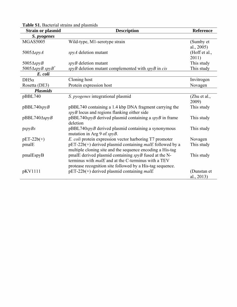
Table S1. Bacterial strains and plasmids Strain or plasmid Description Reference
S. pyogenes MGAS5005 Wild-type, M1-serotype strain (Sumby et
al., 2005) 5005ΔspyA spyA deletion mutant (Hoff et al.,
2011) 5005ΔspyB spyB deletion mutant This study 5005ΔspyB spyB+ spyB deletion mutant complemented with spyB in cis This study
E. coli DH5 Cloning host Invitrogen Rosetta (DE3) Protein expression host Novagen
Plasmids pBBL740 S. pyogenes integrational plasmid (Zhu et al.,
2009) pBBL740spyB pBBL740 containing a 1.4 kbp DNA fragment carrying the
spyB locus and regions flanking either side This study
pBBL740ΔspyB pBBL740spyB derived plasmid containing a spyB in frame deletion
This study
pspyBs pBBL740spyB derived plasmid containing a synonymous mutation in Arg 9 of spyB.
This study
pET-22b(+) E. coli protein expression vector harboring T7 promoter Novagen pmalE pET-22b(+) derived plasmid containing malE followed by a
multiple cloning site and the sequence encoding a His-tag This study
pmalEspyB pmalE derived plasmid containing spyB fused at the N-terminus with malE and at the C-terminus with a TEV protease recognition site followed by a His-tag sequence.
This study
pKV1111 pET-22b(+) derived plasmid containing malE (Dunstan et al., 2013)
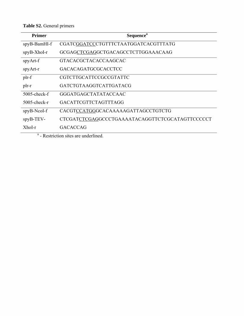
Table S2. General primers
Primer Sequencea
spyB-BamHI-f CGATCGGATCCCTGTTTCTAATGGATCACGTTTATG
spyB-XhoI-r GCGAGCTCGAGGCTGACAGCCTCTTGGAAACAAG
spyArt-f GTACACGCTACACCAAGCAC
spyArt-r GACACAGATGCGCACCTCC
plr-f CGTCTTGCATTCCGCCGTATTC
plr-r GATCTGTAAGGTCATTGATACG
5005-check-f GGGATGAGCTATATACCAAC
5005-check-r GACATTCGTTCTAGTTTAGG
spyB-NcoI-f CACGTCCATGGGCACAAAAAGATTAGCCTGTCTG
spyB-TEV-
XhoI-r
CTCGATCTCGAGGCCCTGAAAATACAGGTTCTCGCATAGTTCCCCCT
GACACCAG a - Restriction sites are underlined.
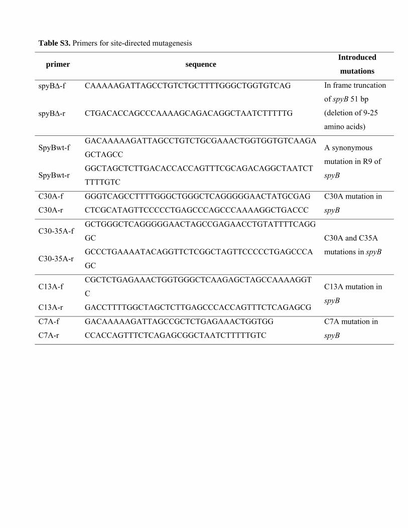
Table S3. Primers for site-directed mutagenesis
primer sequence Introduced
mutations
spyB-f CAAAAAGATTAGCCTGTCTGCTTTTGGGCTGGTGTCAG In frame truncation
of spyB 51 bp
(deletion of 9-25
amino acids)
spyB-r CTGACACCAGCCCAAAAGCAGACAGGCTAATCTTTTTG
SpyBwt-f GACAAAAAGATTAGCCTGTCTGCGAAACTGGTGGTGTCAAGA
GCTAGCC A synonymous
mutation in R9 of
spyB SpyBwt-r GGCTAGCTCTTGACACCACCAGTTTCGCAGACAGGCTAATCT
TTTTGTC
C30A-f GGGTCAGCCTTTTGGGCTGGGCTCAGGGGGAACTATGCGAG C30A mutation in
spyB C30A-r CTCGCATAGTTCCCCCTGAGCCCAGCCCAAAAGGCTGACCC
C30-35A-f GCTGGGCTCAGGGGGAACTAGCCGAGAACCTGTATTTTCAGG
GC C30A and C35A
mutations in spyB C30-35A-r
GCCCTGAAAATACAGGTTCTCGGCTAGTTCCCCCTGAGCCCA
GC
C13A-f CGCTCTGAGAAACTGGTGGGCTCAAGAGCTAGCCAAAAGGT
C C13A mutation in
spyB C13A-r GACCTTTTGGCTAGCTCTTGAGCCCACCAGTTTCTCAGAGCG
C7A-f GACAAAAAGATTAGCCGCTCTGAGAAACTGGTGG C7A mutation in
spyB C7A-r CCACCAGTTTCTCAGAGCGGCTAATCTTTTTGTC
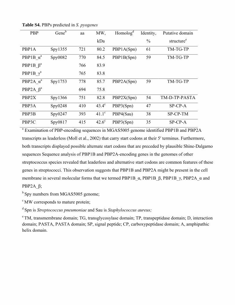
Table S4. PBPs predicted in S. pyogenes
PBP Geneb aa MW,
kDa
Homologd Identity,
%
Putative domain
structuree
PBP1A Spy1355 721 80.2 PBP1A(Spn) 61 TM-TG-TP
PBP1B_αa Spy0082
770 84.5 PBP1B(Spn) 59 TM-TG-TP
PBP1B_βa 766 83.9
PBP1B_γa 765 83.8
PBP2A_αa Spy1753
778 85.7 PBP2A(Spn) 59 TM-TG-TP
PBP2A_βa 694 75.8
PBP2X Spy1366 751 82.8 PBP2X(Spn) 54 TM-D-TP-PASTA
PBP3A Spy0248 410 43.4c PBP3(Spn) 47 SP-CP-A
PBP3B Spy0247 393 41.1c PBP4(Sau) 38 SP-CP-TM
PBP3C Spy0817 415 42.6c PBP3(Spn) 35 SP-CP-A a Examination of PBP-encoding sequences in MGAS5005 genome identified PBP1B and PBP2A
transcripts as leaderless (Moll et al., 2002) that carry start codons at their 5′ terminus. Furthermore,
both transcripts displayed possible alternate start codons that are preceded by plausible Shine-Dalgarno
sequences Sequence analysis of PBP1B and PBP2A-encoding genes in the genomes of other
streptococcus species revealed that leaderless and alternative start codons are common features of these
genes in streptococci. This observation suggests that PBP1B and PBP2A might be present in the cell
membrane in several molecular forms that we termed PBP1B_α, PBP1B_β, PBP1B_γ, PBP2A_α and
PBP2A_β; b Spy numbers from MGAS5005 genome; c MW corresponds to mature protein; d Spn is Streptococcus pneumoniae and Sau is Staphylococcus aureus; e TM, transmembrane domain; TG, transglycosylase domain; TP, transpeptidase domain; D, interaction domain; PASTA, PASTA domain; SP, signal peptide; CP, carboxypeptidase domain; A, amphipathic helix domain.

Table S5. Concentration of total carbohydrates in bacteria
Strains nM CHO/µg of protein
MGAS5005 0.1059± 0.0024
5005ΔspyB 0.1094± 0.0045
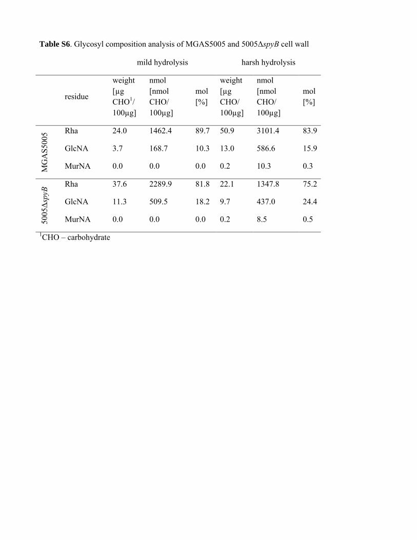
Table S6. Glycosyl composition analysis of MGAS5005 and 5005ΔspyB cell wall
mild hydrolysis harsh hydrolysis
residue
weight [µg CHO1/ 100µg]
nmol [nmol CHO/ 100µg]
mol [%]
weight [µg CHO/ 100µg]
nmol [nmol CHO/ 100µg]
mol [%]
MG
AS
5005
Rha 24.0 1462.4 89.7 50.9 3101.4 83.9
GlcNA 3.7 168.7 10.3 13.0 586.6 15.9
MurNA 0.0 0.0 0.0 0.2 10.3 0.3
5005Δ
spyB
Rha 37.6 2289.9 81.8 22.1 1347.8 75.2
GlcNA 11.3 509.5 18.2 9.7 437.0 24.4
MurNA 0.0 0.0 0.0 0.2 8.5 0.5
1CHO – carbohydrate
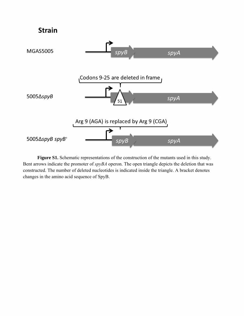
Figure S1. Schematic representations of the construction of the mutants used in this study. Bent arrows indicate the promoter of spyBA operon. The open triangle depicts the deletion that was constructed. The number of deleted nucleotides is indicated inside the triangle. A bracket denotes changes in the amino acid sequence of SpyB.
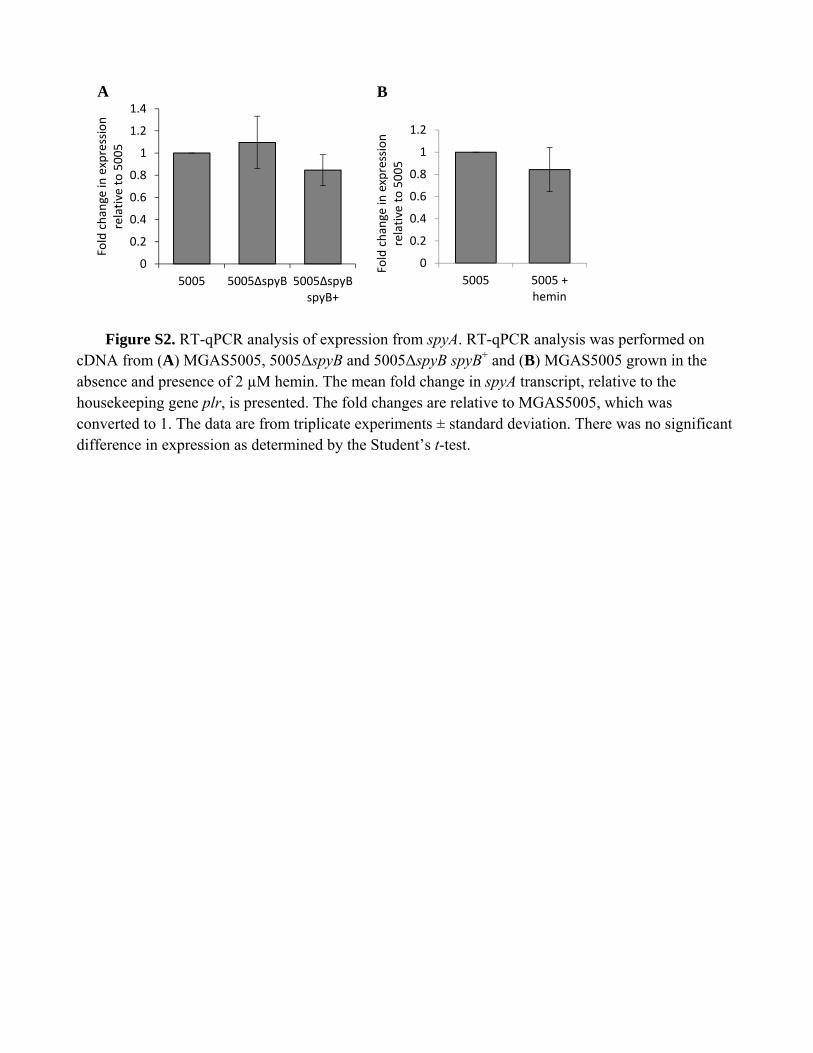
Figure S2. RT-qPCR analysis of expression from spyA. RT-qPCR analysis was performed on cDNA from (A) MGAS5005, 5005ΔspyB and 5005ΔspyB spyB+ and (B) MGAS5005 grown in the absence and presence of 2 µM hemin. The mean fold change in spyA transcript, relative to the housekeeping gene plr, is presented. The fold changes are relative to MGAS5005, which was converted to 1. The data are from triplicate experiments ± standard deviation. There was no significant difference in expression as determined by the Student’s t-test.
0
0.2
0.4
0.6
0.8
1
1.2
1.4
5005 5005ΔspyB 5005ΔspyB spyB+
Fold chan
ge in
expression
relative to 5005
0
0.2
0.4
0.6
0.8
1
1.2
5005 5005 +hemin
Fold change in
expression
relative to 5005
A B


Figure S3. Phenotype MicroArray data for MGAS5005 vs 5005ΔspyB and 5005ΔspyA. Growth phenotypes of MGAS5005 vs 5005ΔspyA (A) and MGAS5005 vs 5005ΔspyB (B) were assessed using the Biolog plates at Biolog's PM Services facility. A total of 20 96-well PM plates constituting eight metabolic panels (PM1 to PM8); and 12 sensitivity panels (PM9 to PM20) were used. Two replicates were conducted for each strain and the consensus plots are presented. (C) Phenotypes gained and lost for 5005ΔspyA and 5005ΔspyB
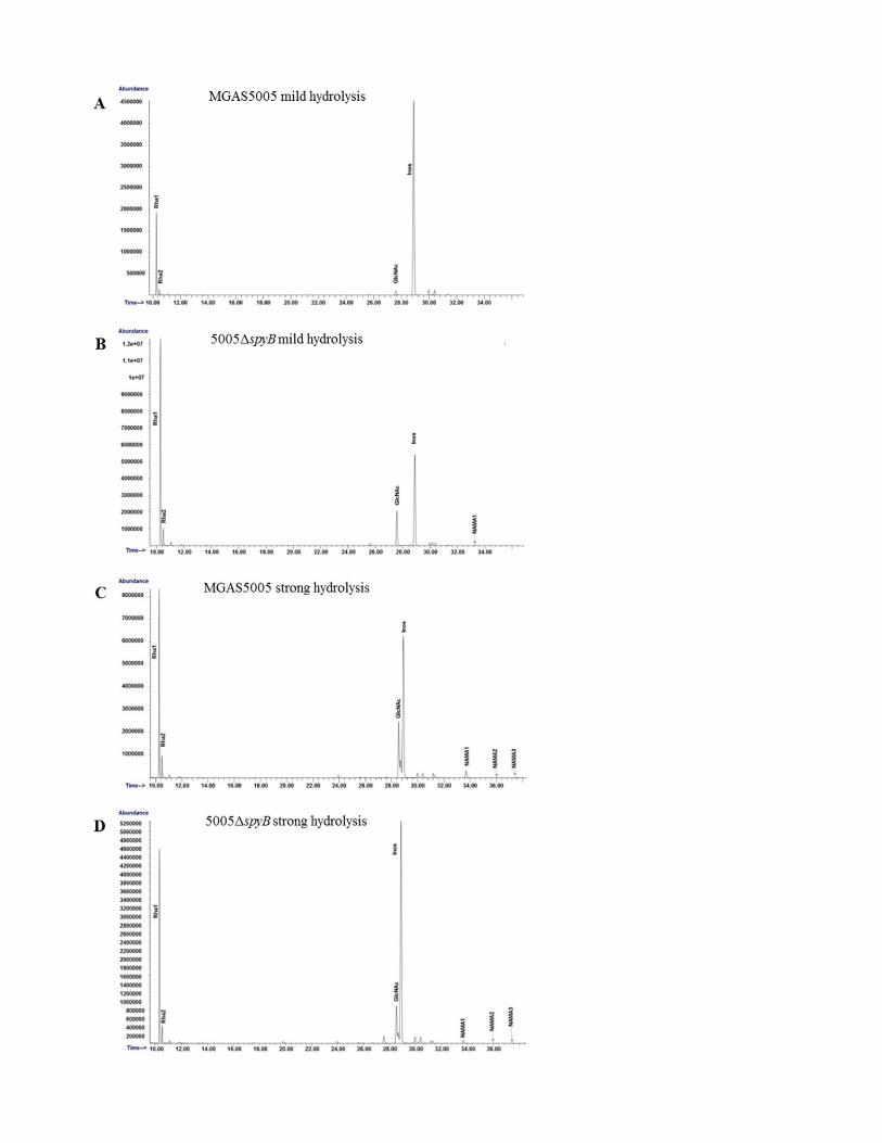

Figure S4. GC-MS chromatograms for glycosyl composition analysis of MGAS5005 and 5005
ΔspyB. (A) and (E) MGAS5005 mild hydrolysis, (B) and (F) 5005ΔspyB mild hydrolysis, (C) and (G)
MGAS5005 strong hydrolysis, (D) and (H) 5005ΔspyB strong hydrolysis. (A-D) and (E-H) are data
from two separate analyses performed on different cell wall preparations.
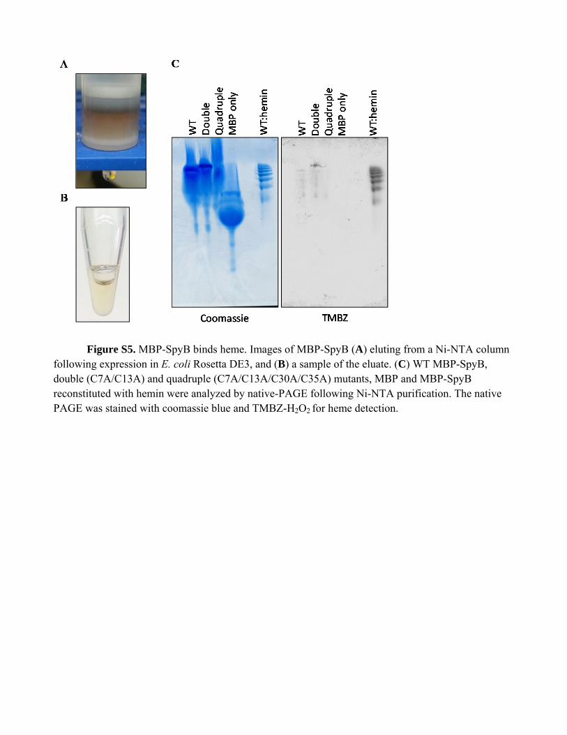
Figure S5. MBP-SpyB binds heme. Images of MBP-SpyB (A) eluting from a Ni-NTA column following expression in E. coli Rosetta DE3, and (B) a sample of the eluate. (C) WT MBP-SpyB, double (C7A/C13A) and quadruple (C7A/C13A/C30A/C35A) mutants, MBP and MBP-SpyB reconstituted with hemin were analyzed by native-PAGE following Ni-NTA purification. The native PAGE was stained with coomassie blue and TMBZ-H2O2 for heme detection.
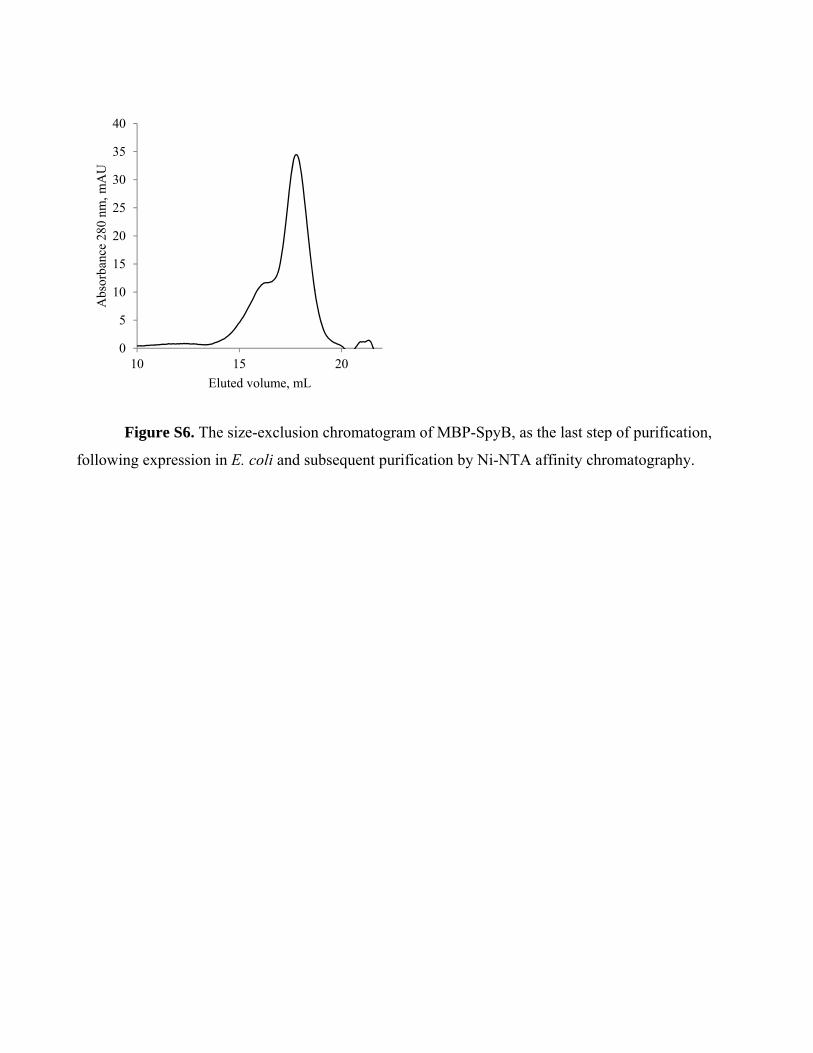
Figure S6. The size-exclusion chromatogram of MBP-SpyB, as the last step of purification,
following expression in E. coli and subsequent purification by Ni-NTA affinity chromatography.
0
5
10
15
20
25
30
35
40
10 15 20
Abs
orba
nce
280
nm, m
AU
Eluted volume, mL
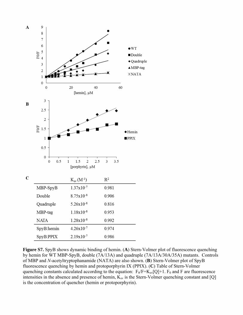
Figure S7. SpyB shows dynamic binding of hemin. (A) Stern-Volmer plot of fluorescence quenching by hemin for WT MBP-SpyB, double (7A/13A) and quadruple (7A/13A/30A/35A) mutants. Controls of MBP and N-acetyltryptophanamide (NATA) are also shown. (B) Stern-Volmer plot of SpyB fluorescence quenching by hemin and protoporphyrin IX (PPIX). (C) Table of Stern-Volmer quenching constants calculated according to the equation: F0/F=Ksv[Q]+1. F0 and F are fluorescence intensities in the absence and presence of hemin, Ksv is the Stern-Volmer quenching constant and [Q] is the concentration of quencher (hemin or protoporphyrin).
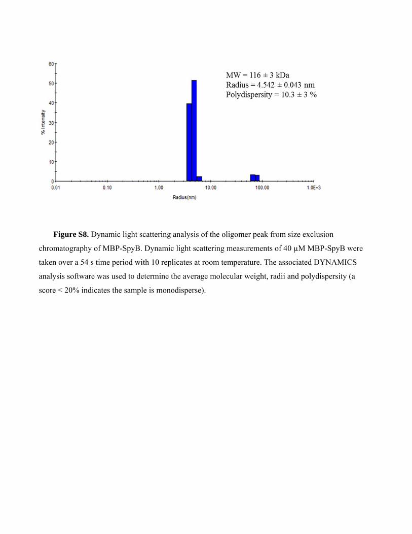
Figure S8. Dynamic light scattering analysis of the oligomer peak from size exclusion
chromatography of MBP-SpyB. Dynamic light scattering measurements of 40 µM MBP-SpyB were
taken over a 54 s time period with 10 replicates at room temperature. The associated DYNAMICS
analysis software was used to determine the average molecular weight, radii and polydispersity (a
score < 20% indicates the sample is monodisperse).
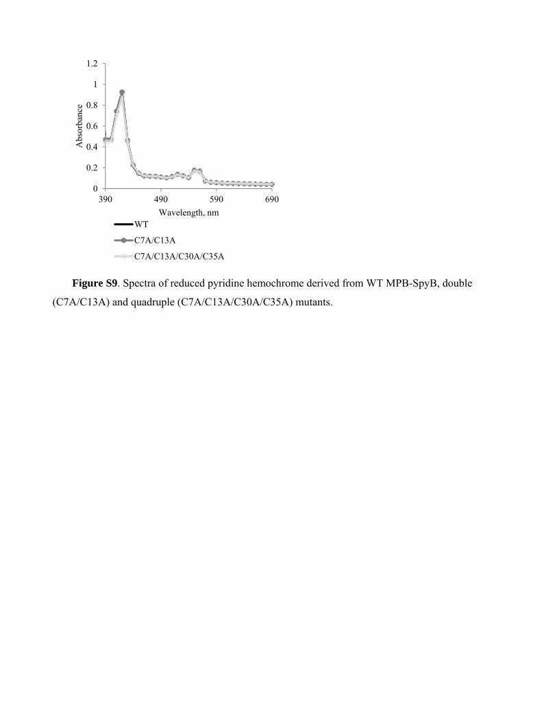
Figure S9. Spectra of reduced pyridine hemochrome derived from WT MPB-SpyB, double
(C7A/C13A) and quadruple (C7A/C13A/C30A/C35A) mutants.
0
0.2
0.4
0.6
0.8
1
1.2
390 490 590 690
Abs
orba
nce
Wavelength, nmWT
C7A/C13A
C7A/C13A/C30A/C35A
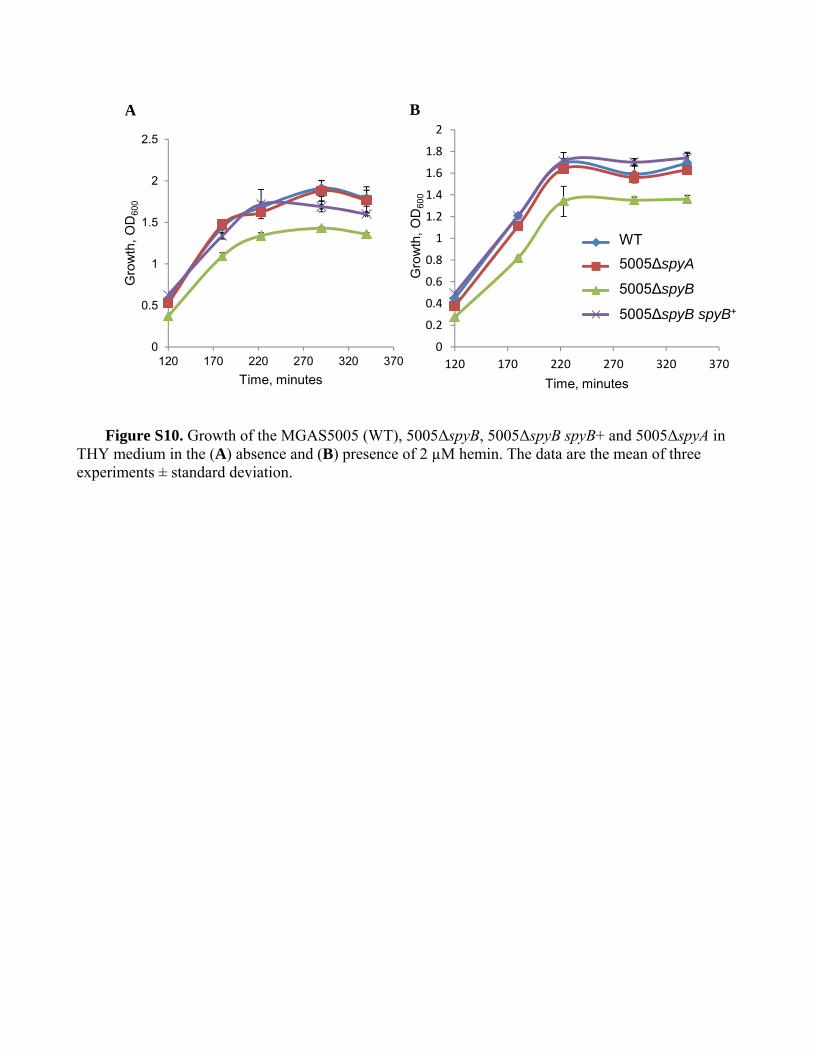
Figure S10. Growth of the MGAS5005 (WT), 5005ΔspyB, 5005ΔspyB spyB+ and 5005ΔspyA in
THY medium in the (A) absence and (B) presence of 2 µM hemin. The data are the mean of three experiments ± standard deviation.
0
0.2
0.4
0.6
0.8
1
1.2
1.4
1.6
1.8
2
120 170 220 270 320 370
Gro
wth
, O
D60
0
Time, minutes
wt+heme
spyA+heme
spyB+heme
compl+heme
WT
5005∆spyB
5005∆spyB spyB+
5005∆spyA
0
0.5
1
1.5
2
2.5
120 170 220 270 320 370
Gro
wth
, O
D60
0
Time, minutes
A B
References
Dunstan, R.A., Heinz, E., Wijeyewickrema, L.C., Pike, R.N., Purcell, A.W., Evans, T.J., Praszkier, J., Robins‐Browne, R.M., Strugnell, R.A., Korotkov, K.V., and Lithgow, T. (2013). Assembly of the type II secretion system such as found in Vibrio cholerae depends on the novel Pilotin AspS. PLoS Pathog 9, e1003117, doi:10.1371/journal.ppat.1003117.
Hoff, J.S., Dewald, M., Moseley, S.L., Collins, C.M., and Voyich, J.M. (2011). SpyA, a C3‐like ADP‐ribosyltransferase, contributes to virulence in a mouse subcutaneous model of Streptococcus pyogenes infection. Infection and immunity 79, 2404‐2411, doi:10.1128/IAI.01191‐10.
Moll, I., Grill, S., Gualerzi, C.O., and Blasi, U. (2002). Leaderless mRNAs in bacteria: surprises in ribosomal recruitment and translational control. Mol Microbiol 43, 239‐246.
Sumby, P., Porcella, S.F., Madrigal, A.G., Barbian, K.D., Virtaneva, K., Ricklefs, S.M., Sturdevant, D.E., Graham, M.R., Vuopio‐Varkila, J., Hoe, N.P., and Musser, J.M. (2005). Evolutionary origin and emergence of a highly successful clone of serotype M1 group a Streptococcus involved multiple horizontal gene transfer events. J Infect Dis 192, 771‐782.
Zhu, H., Liu, M., Sumby, P., and Lei, B. (2009). The secreted esterase of group a streptococcus is important for invasive skin infection and dissemination in mice. Infection and immunity 77, 5225‐5232, doi:10.1128/IAI.00636‐09.