Structure−Function Relationships in Aminoquinolines: Effect of Amino and Chloro Groups on...
9
Structure-Function Relationships in Aminoquinolines: Effect of Amino and Chloro Groups on Quinoline-Hematin Complex Formation, Inhibition of -Hematin Formation, and Antiplasmodial Activity Timothy J. Egan,* ,² Roger Hunter, ² Catherine H. Kaschula, ² Helder M. Marques, ‡ Ashley Misplon, ² and Jason Walden § Departments of Chemistry and Pharmacology, University of Cape Town, and Department of Chemistry, University of the Witwatersrand, Johannesburg, South Africa Received August 25, 1999 Comparison of 19 aminoquinolines supports the hypothesis that chloroquine and related antimalarials act by complexing ferriprotoporphyrin IX (Fe(III)PPIX), inhibiting its conversion to -hematin (hemozoin) and hence its detoxification. The study suggests that a basic amino side chain is also essential for antiplasmodial activity. 2- And 4-aminoquinolines are unique in their strong affinity for Fe(III)PPIX, and attachment of side chains to the amino group has relatively little influence on the strength of complex formation. Association with Fe(III)PPIX is necessary, but not sufficient, for inhibiting -hematin formation. Presence of a 7-chloro group in the 4-aminoquinoline ring is a requirement for -hematin inhibitory activity, and this is also unaffected by side chains attached to the amino group. In turn, -hematin inhibitory activity is necessary, but not sufficient, for antiplasmodial activity as the presence of an aminoalkyl group attached to the 4-amino-7-chloroquinoline template is essential for strong activity. We thus propose that the 4-aminoquinoline nucleus of chloroquine and related antimalarials is responsible for complexing Fe(III)PPIX, the 7-chloro group is required for inhibition of -hematin formation, and the basic amino side chain is required for drug accumulation in the food vacuole of the parasite. Introduction Considerable evidence has been presented in recent years that antimalarial drugs such as chloroquine and amodiaquine (type-1 blood schizontocides) act by form- ing complexes with hematin, the hydroxo or aqua complex of ferriprotoporphyrin IX (Fe(III)PPIX), derived from parasite proteolysis of host hemoglobin. 1 Associa- tion constants for various antimalarials with hematin both in aqueous solution, 2 where hematin is expected to be aggregated, 3 and in 40% aqueous DMSO, 4 where it is monomeric, 5 have been reported recently. Ward and co-workers 6,7 have provided evidence that such interac- tion occurs within the food vacuole of the malaria parasite, the probable site of action of these drugs. Most compellingly, they have shown 6,7 that the activities of these drugs are Fe(III)PPIX-dependent, since inhibitors of plasmepsin (the enzyme responsible for host hemo- globin degradation in the food vacuole) are antagonists of drug activity. They have also shown this dependence in the case of mefloquine, a type-2 blood schizontocide, suggesting that drugs such as quinine, halofantrine, and mefloquine act by a similar mechanism. It is generally believed 8 that detoxification of Fe(III)- PPIX in the parasite food vacuole is brought about by polymerization to form malaria pigment, or hemozoin. Although this has recently been questioned by Ginsburg and co-workers 9 who have suggested that the major portion of Fe(III)PPIX is transported across the vacuolar membrane and degraded by glutathione-dependent re- dox processes involving reduced oxygen species, 10 no evidence has yet been presented for the large quantities of iron which would then have to be sequestered in the parasite. Indeed, to date hemozoin is the only known store of large quantities of iron in the parasite. Based on current evidence, it would then seem likely that type-1 and possibly also type-2 drugs inhibit hemozoin formation resulting in the buildup of significant quanti- ties of toxic Fe(III)PPIX-drug complex which is prob- ably then responsible for killing the parasite. It is now known that hemozoin is chemically identical to -hematin, 11 which is generally believed to be a polymer of hematin. In 1994 we showed 12 for the first time that several type-1 and type-2 blood schizontocides can specifically inhibit chemical formation of -hematin by direct interaction between the drugs and Fe(III)PPIX. There is now substantial evidence that antimalarial drugs can inhibit both synthetic -hematin formation and hemozoin formation in both chemical and biological systems by direct interaction between the drugs and Fe(III)PPIX. 2,12-20 The precise mechanism by which this occurs in the parasite food vacuole is still uncertain, but Goldberg and co-workers have shown that such hema- tin-drug complexes can bind to and inhibit further incorporation of Fe(III)PPIX into hemozoin. 19,20 They have thus suggested that the drugs cap the growing polymer and inhibit its further growth. Recently 9 direct evidence for decreased hemozoin formation in Plasmo- * To whom correspondence should be addressed at: Department of Chemistry, University of Cape Town, Private Bag, Rondebosch 7701, South Africa. Tel: +27-21-650-2528. Fax: +27-21-689-7499. E-mail: [email protected]. ² Department of Chemistry, University of Cape Town. ‡ Department of Chemistry, University of the Witwatersrand. § Department of Pharmacology, University of Cape Town. 283 J. Med. Chem. 2000, 43, 283-291 10.1021/jm990437l CCC: $19.00 © 2000 American Chemical Society Published on Web 12/31/1999
Transcript of Structure−Function Relationships in Aminoquinolines: Effect of Amino and Chloro Groups on...
Structure-Function Relationships in Aminoquinolines: Effect of Amino andChloro Groups on Quinoline-Hematin Complex Formation, Inhibition ofâ-Hematin Formation, and Antiplasmodial Activity
Timothy J. Egan,*,† Roger Hunter,† Catherine H. Kaschula,† Helder M. Marques,‡ Ashley Misplon,† andJason Walden§
Departments of Chemistry and Pharmacology, University of Cape Town, and Department of Chemistry, University of theWitwatersrand, Johannesburg, South Africa
Received August 25, 1999
Comparison of 19 aminoquinolines supports the hypothesis that chloroquine and relatedantimalarials act by complexing ferriprotoporphyrin IX (Fe(III)PPIX), inhibiting its conversionto â-hematin (hemozoin) and hence its detoxification. The study suggests that a basic aminoside chain is also essential for antiplasmodial activity. 2- And 4-aminoquinolines are uniquein their strong affinity for Fe(III)PPIX, and attachment of side chains to the amino group hasrelatively little influence on the strength of complex formation. Association with Fe(III)PPIXis necessary, but not sufficient, for inhibiting â-hematin formation. Presence of a 7-chloro groupin the 4-aminoquinoline ring is a requirement for â-hematin inhibitory activity, and this isalso unaffected by side chains attached to the amino group. In turn, â-hematin inhibitory activityis necessary, but not sufficient, for antiplasmodial activity as the presence of an aminoalkylgroup attached to the 4-amino-7-chloroquinoline template is essential for strong activity. Wethus propose that the 4-aminoquinoline nucleus of chloroquine and related antimalarials isresponsible for complexing Fe(III)PPIX, the 7-chloro group is required for inhibition of â-hematinformation, and the basic amino side chain is required for drug accumulation in the food vacuoleof the parasite.
Introduction
Considerable evidence has been presented in recentyears that antimalarial drugs such as chloroquine andamodiaquine (type-1 blood schizontocides) act by form-ing complexes with hematin, the hydroxo or aquacomplex of ferriprotoporphyrin IX (Fe(III)PPIX), derivedfrom parasite proteolysis of host hemoglobin.1 Associa-tion constants for various antimalarials with hematinboth in aqueous solution,2 where hematin is expectedto be aggregated,3 and in 40% aqueous DMSO,4 whereit is monomeric,5 have been reported recently. Ward andco-workers6,7 have provided evidence that such interac-tion occurs within the food vacuole of the malariaparasite, the probable site of action of these drugs. Mostcompellingly, they have shown6,7 that the activities ofthese drugs are Fe(III)PPIX-dependent, since inhibitorsof plasmepsin (the enzyme responsible for host hemo-globin degradation in the food vacuole) are antagonistsof drug activity. They have also shown this dependencein the case of mefloquine, a type-2 blood schizontocide,suggesting that drugs such as quinine, halofantrine, andmefloquine act by a similar mechanism.
It is generally believed8 that detoxification of Fe(III)-PPIX in the parasite food vacuole is brought about bypolymerization to form malaria pigment, or hemozoin.Although this has recently been questioned by Ginsburg
and co-workers9 who have suggested that the majorportion of Fe(III)PPIX is transported across the vacuolarmembrane and degraded by glutathione-dependent re-dox processes involving reduced oxygen species,10 noevidence has yet been presented for the large quantitiesof iron which would then have to be sequestered in theparasite. Indeed, to date hemozoin is the only knownstore of large quantities of iron in the parasite. Basedon current evidence, it would then seem likely thattype-1 and possibly also type-2 drugs inhibit hemozoinformation resulting in the buildup of significant quanti-ties of toxic Fe(III)PPIX-drug complex which is prob-ably then responsible for killing the parasite.
It is now known that hemozoin is chemically identicalto â-hematin,11 which is generally believed to be apolymer of hematin. In 1994 we showed12 for the firsttime that several type-1 and type-2 blood schizontocidescan specifically inhibit chemical formation of â-hematinby direct interaction between the drugs and Fe(III)PPIX.There is now substantial evidence that antimalarialdrugs can inhibit both synthetic â-hematin formationand hemozoin formation in both chemical and biologicalsystems by direct interaction between the drugs andFe(III)PPIX.2,12-20 The precise mechanism by which thisoccurs in the parasite food vacuole is still uncertain, butGoldberg and co-workers have shown that such hema-tin-drug complexes can bind to and inhibit furtherincorporation of Fe(III)PPIX into hemozoin.19,20 Theyhave thus suggested that the drugs cap the growingpolymer and inhibit its further growth. Recently9 directevidence for decreased hemozoin formation in Plasmo-
* To whom correspondence should be addressed at: Department ofChemistry, University of Cape Town, Private Bag, Rondebosch 7701,South Africa. Tel: +27-21-650-2528. Fax: +27-21-689-7499. E-mail:[email protected].
† Department of Chemistry, University of Cape Town.‡ Department of Chemistry, University of the Witwatersrand.§ Department of Pharmacology, University of Cape Town.
283J. Med. Chem. 2000, 43, 283-291
10.1021/jm990437l CCC: $19.00 © 2000 American Chemical SocietyPublished on Web 12/31/1999
dium falciparum in the presence of chloroquine hasbeen provided.
In light of the above, a mechanism of action of type-1antimalarials can be proposed in which the drug:1,21,22
i. enters the food vacuole, possibly via diffusion of thefree base across intervening membranes; ii. accumulatesin the food vacuole, at least in part due to pH trappingof the protonated drug at the low pH of the vacuole23
(this process involves diffusion of only the free baseacross the food vacuole membrane; increased protona-tion of the drug at the low pH of the vacuole results ininflow of more drug until free base concentrations areequal on both sides of the membrane); iii. forms acomplex (which is predominantly a π-π complex24-26)with Fe(III)PPIX, which may further enhance drugaccumulation;6,7 iv. inhibits formation of hemozoin viathe formation of this complex; and v. exerts a toxic effecton the parasite in the form of the Fe(III)PPIX-drugcomplex.
The development of this detailed model of actionprovides an opportunity to test the activity of variousquinolines in unprecedented detail. In this article wereport the results of Fe(III)PPIX complexation, â-he-matin inhibition, and antiplasmodial activity of 6 com-mercially available quinolines and 15 quinolines specif-ically synthesized for this study. The compounds werechosen to probe the effect of amino group position andof chlorination at the 7-position on the activities of thesecompounds with the aim of establishing a detailedstructure-activity profile of chloroquine.
Chemistry
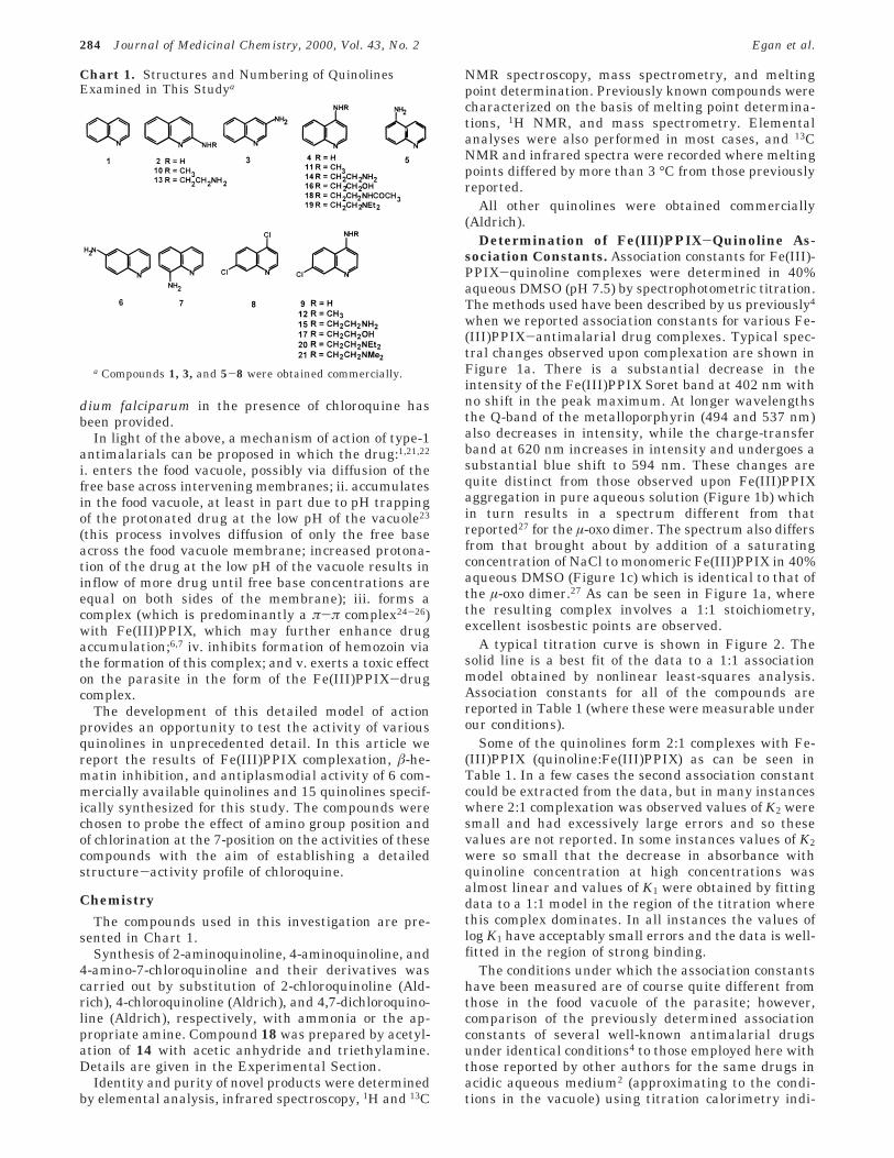
The compounds used in this investigation are pre-sented in Chart 1.
Synthesis of 2-aminoquinoline, 4-aminoquinoline, and4-amino-7-chloroquinoline and their derivatives wascarried out by substitution of 2-chloroquinoline (Ald-rich), 4-chloroquinoline (Aldrich), and 4,7-dichloroquino-line (Aldrich), respectively, with ammonia or the ap-propriate amine. Compound 18 was prepared by acetyl-ation of 14 with acetic anhydride and triethylamine.Details are given in the Experimental Section.
Identity and purity of novel products were determinedby elemental analysis, infrared spectroscopy, 1H and 13C
NMR spectroscopy, mass spectrometry, and meltingpoint determination. Previously known compounds werecharacterized on the basis of melting point determina-tions, 1H NMR, and mass spectrometry. Elementalanalyses were also performed in most cases, and 13CNMR and infrared spectra were recorded where meltingpoints differed by more than 3 °C from those previouslyreported.
All other quinolines were obtained commercially(Aldrich).
Determination of Fe(III)PPIX-Quinoline As-sociation Constants. Association constants for Fe(III)-PPIX-quinoline complexes were determined in 40%aqueous DMSO (pH 7.5) by spectrophotometric titration.The methods used have been described by us previously4
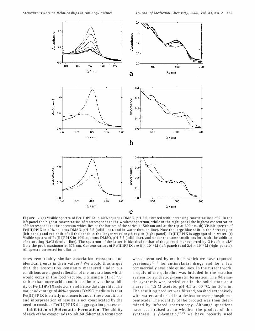
when we reported association constants for various Fe-(III)PPIX-antimalarial drug complexes. Typical spec-tral changes observed upon complexation are shown inFigure 1a. There is a substantial decrease in theintensity of the Fe(III)PPIX Soret band at 402 nm withno shift in the peak maximum. At longer wavelengthsthe Q-band of the metalloporphyrin (494 and 537 nm)also decreases in intensity, while the charge-transferband at 620 nm increases in intensity and undergoes asubstantial blue shift to 594 nm. These changes arequite distinct from those observed upon Fe(III)PPIXaggregation in pure aqueous solution (Figure 1b) whichin turn results in a spectrum different from thatreported27 for the µ-oxo dimer. The spectrum also differsfrom that brought about by addition of a saturatingconcentration of NaCl to monomeric Fe(III)PPIX in 40%aqueous DMSO (Figure 1c) which is identical to that ofthe µ-oxo dimer.27 As can be seen in Figure 1a, wherethe resulting complex involves a 1:1 stoichiometry,excellent isosbestic points are observed.
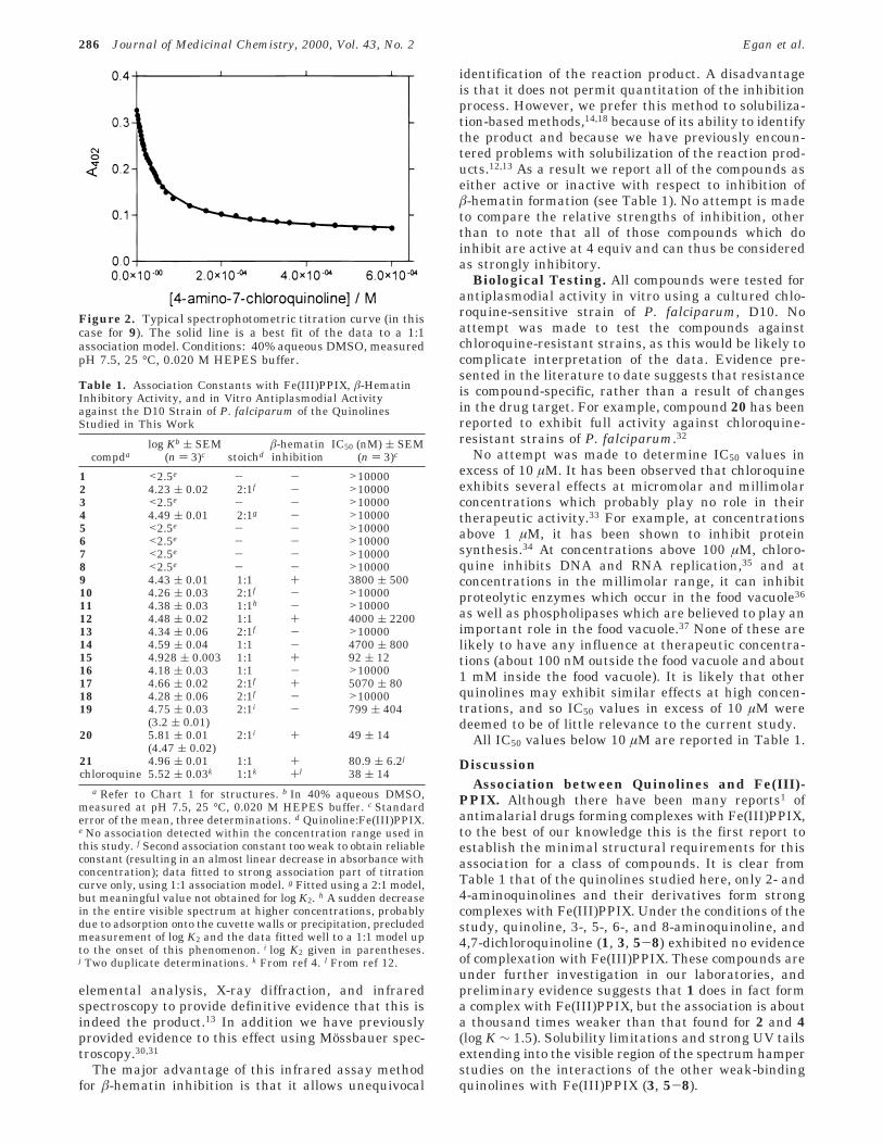
A typical titration curve is shown in Figure 2. Thesolid line is a best fit of the data to a 1:1 associationmodel obtained by nonlinear least-squares analysis.Association constants for all of the compounds arereported in Table 1 (where these were measurable underour conditions).
Some of the quinolines form 2:1 complexes with Fe-(III)PPIX (quinoline:Fe(III)PPIX) as can be seen inTable 1. In a few cases the second association constantcould be extracted from the data, but in many instanceswhere 2:1 complexation was observed values of K2 weresmall and had excessively large errors and so thesevalues are not reported. In some instances values of K2were so small that the decrease in absorbance withquinoline concentration at high concentrations wasalmost linear and values of K1 were obtained by fittingdata to a 1:1 model in the region of the titration wherethis complex dominates. In all instances the values oflog K1 have acceptably small errors and the data is well-fitted in the region of strong binding.
The conditions under which the association constantshave been measured are of course quite different fromthose in the food vacuole of the parasite; however,comparison of the previously determined associationconstants of several well-known antimalarial drugsunder identical conditions4 to those employed here withthose reported by other authors for the same drugs inacidic aqueous medium2 (approximating to the condi-tions in the vacuole) using titration calorimetry indi-
Chart 1. Structures and Numbering of QuinolinesExamined in This Studya
a Compounds 1, 3, and 5-8 were obtained commercially.
284 Journal of Medicinal Chemistry, 2000, Vol. 43, No. 2 Egan et al.
cates remarkably similar association constants andidentical trends in their values.1 We would thus arguethat the association constants measured under ourconditions are a good reflection of the interactions whichwould occur in the food vacuole. Utilizing a pH of 7.5,rather than more acidic conditions, improves the stabil-ity of Fe(III)PPIX solutions and hence data quality. Themajor advantage of 40% aqueous DMSO medium is thatFe(III)PPIX is strictly monomeric under these conditionsand interpretation of results is not complicated by theneed to consider Fe(III)PPIX disaggregation processes.
Inhibition of â-Hematin Formation. The abilityof each of the compounds to inhibit â-hematin formation
was determined by methods which we have reportedpreviously12,13 for antimalarial drugs and for a fewcommercially available quinolines. In the current work,4 equiv of the quinoline was included in the reactionsystem for synthetic â-hematin formation. The â-hema-tin synthesis was carried out in the solid state as aslurry in 4.5 M acetate, pH 4.5 at 60 °C, for 30 min.The resulting product was filtered, washed extensivelywith water, and dried in a desiccator over phosphoruspentoxide. The identity of the product was then deter-mined by infrared spectroscopy. Although questionshave been raised as to whether the product of thissynthesis is â-hematin,28,29 we have recently used
Figure 1. (a) Visible spectra of Fe(III)PPIX in 40% aqueous DMSO, pH 7.5, titrated with increasing concentrations of 9. In theleft panel the highest concentration of 9 corresponds to the weakest spectrum, while in the right panel the highest concentrationof 9 corresponds to the spectrum which lies at the bottom of the series at 500 nm and at the top at 600 nm. (b) Visible spectra ofFe(III)PPIX in 40% aqueous DMSO, pH 7.5 (solid line), and in water (broken line). Note the large blue shift in the Soret region(left panel) and red shift of all the bands in the longer wavelength region (right panel). Fe(III)PPIX is aggregated in water. (c)Visible spectra of Fe(III)PPIX in 40% aqueous DMSO, pH 7.5 (solid line), and under the same conditions but with the additionof saturating NaCl (broken line). The spectrum of the latter is identical to that of the µ-oxo dimer reported by O’Keefe et al.27
Note the peak maximum at 575 nm. Concentrations of Fe(III)PPIX are 8 × 10⁻⁶ M (left panels) and 2.4 × 10⁻⁵ M (right panels).All spectra corrected for dilution.
Structure-Function Relationships in Aminoquinolines Journal of Medicinal Chemistry, 2000, Vol. 43, No. 2 285
elemental analysis, X-ray diffraction, and infraredspectroscopy to provide definitive evidence that this isindeed the product.13 In addition we have previouslyprovided evidence to this effect using Mossbauer spec-troscopy.30,31
The major advantage of this infrared assay methodfor â-hematin inhibition is that it allows unequivocal
identification of the reaction product. A disadvantageis that it does not permit quantitation of the inhibitionprocess. However, we prefer this method to solubiliza-tion-based methods,14,18 because of its ability to identifythe product and because we have previously encoun-tered problems with solubilization of the reaction prod-ucts.12,13 As a result we report all of the compounds aseither active or inactive with respect to inhibition ofâ-hematin formation (see Table 1). No attempt is madeto compare the relative strengths of inhibition, otherthan to note that all of those compounds which doinhibit are active at 4 equiv and can thus be consideredas strongly inhibitory.
Biological Testing. All compounds were tested forantiplasmodial activity in vitro using a cultured chlo-roquine-sensitive strain of P. falciparum, D10. Noattempt was made to test the compounds againstchloroquine-resistant strains, as this would be likely tocomplicate interpretation of the data. Evidence pre-sented in the literature to date suggests that resistanceis compound-specific, rather than a result of changesin the drug target. For example, compound 20 has beenreported to exhibit full activity against chloroquine-resistant strains of P. falciparum.32
No attempt was made to determine IC50 values inexcess of 10 µM. It has been observed that chloroquineexhibits several effects at micromolar and millimolarconcentrations which probably play no role in theirtherapeutic activity.33 For example, at concentrationsabove 1 µM, it has been shown to inhibit proteinsynthesis.34 At concentrations above 100 µM, chloro-quine inhibits DNA and RNA replication,35 and atconcentrations in the millimolar range, it can inhibitproteolytic enzymes which occur in the food vacuole36
as well as phospholipases which are believed to play animportant role in the food vacuole.37 None of these arelikely to have any influence at therapeutic concentra-tions (about 100 nM outside the food vacuole and about1 mM inside the food vacuole). It is likely that otherquinolines may exhibit similar effects at high concen-trations, and so IC50 values in excess of 10 µM weredeemed to be of little relevance to the current study.
All IC50 values below 10 µM are reported in Table 1.
DiscussionAssociation between Quinolines and Fe(III)-
PPIX. Although there have been many reports1 ofantimalarial drugs forming complexes with Fe(III)PPIX,to the best of our knowledge this is the first report toestablish the minimal structural requirements for thisassociation for a class of compounds. It is clear fromTable 1 that of the quinolines studied here, only 2- and4-aminoquinolines and their derivatives form strongcomplexes with Fe(III)PPIX. Under the conditions of thestudy, quinoline, 3-, 5-, 6-, and 8-aminoquinoline, and4,7-dichloroquinoline (1, 3, 5-8) exhibited no evidenceof complexation with Fe(III)PPIX. These compounds areunder further investigation in our laboratories, andpreliminary evidence suggests that 1 does in fact forma complex with Fe(III)PPIX, but the association is abouta thousand times weaker than that found for 2 and 4(log K ∼ 1.5). Solubility limitations and strong UV tailsextending into the visible region of the spectrum hamperstudies on the interactions of the other weak-bindingquinolines with Fe(III)PPIX (3, 5-8).
Figure 2. Typical spectrophotometric titration curve (in thiscase for 9). The solid line is a best fit of the data to a 1:1association model. Conditions: 40% aqueous DMSO, measuredpH 7.5, 25 °C, 0.020 M HEPES buffer.
Table 1. Association Constants with Fe(III)PPIX, â-HematinInhibitory Activity, and in Vitro Antiplasmodial Activityagainst the D10 Strain of P. falciparum of the QuinolinesStudied in This Work
compdalog Kb ( SEM
(n ) 3)c stoichdâ-hematininhibition
IC50 (nM) ( SEM(n ) 3)c
1 <2.5e - - >100002 4.23 ( 0.02 2:1f - >100003 <2.5e - - >100004 4.49 ( 0.01 2:1g - >100005 <2.5e - - >100006 <2.5e - - >100007 <2.5e - - >100008 <2.5e - - >100009 4.43 ( 0.01 1:1 + 3800 ( 50010 4.26 ( 0.03 2:1f - >1000011 4.38 ( 0.03 1:1h - >1000012 4.48 ( 0.02 1:1 + 4000 ( 220013 4.34 ( 0.06 2:1f - >1000014 4.59 ( 0.04 1:1 - 4700 ( 80015 4.928 ( 0.003 1:1 + 92 ( 1216 4.18 ( 0.03 1:1 - >1000017 4.66 ( 0.02 2:1f + 5070 ( 8018 4.28 ( 0.06 2:1f - >1000019 4.75 ( 0.03
(3.2 ( 0.01)2:1i - 799 ( 404
20 5.81 ( 0.01(4.47 ( 0.02)
2:1i + 49 ( 14
21 4.96 ( 0.01 1:1 + 80.9 ( 6.2j
chloroquine 5.52 ( 0.03k 1:1k +l 38 ( 14a Refer to Chart 1 for structures. b In 40% aqueous DMSO,
measured at pH 7.5, 25 °C, 0.020 M HEPES buffer. c Standarderror of the mean, three determinations. d Quinoline:Fe(III)PPIX.e No association detected within the concentration range used inthis study. f Second association constant too weak to obtain reliableconstant (resulting in an almost linear decrease in absorbance withconcentration); data fitted to strong association part of titrationcurve only, using 1:1 association model. g Fitted using a 2:1 model,but meaningful value not obtained for log K2. h A sudden decreasein the entire visible spectrum at higher concentrations, probablydue to adsorption onto the cuvette walls or precipitation, precludedmeasurement of log K2 and the data fitted well to a 1:1 model upto the onset of this phenomenon. i log K2 given in parentheses.j Two duplicate determinations. k From ref 4. l From ref 12.
286 Journal of Medicinal Chemistry, 2000, Vol. 43, No. 2 Egan et al.
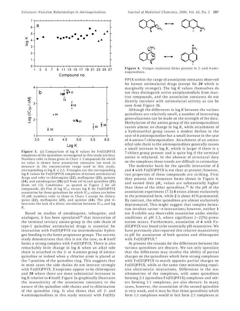
Based on studies of amodiaquine, tebuquine, andanalogues, it has been speculated38 that interaction ofthe terminal tertiary amino group in the side chain oftype-1 quinoline antimalarial drugs is essential forinteraction with Fe(III)PPIX via intermolecular hydro-gen bonding to the heme propionate groups. The currentstudy demonstrates that this is not the case, as 4 itselfforms a strong complex with Fe(III)PPIX. There is alsoremarkably little change in log K when an alkyl sidechain is attached to the 2- or 4-amino group of amino-quinoline or indeed when a chlorine atom is placed atthe 7-position of the quinoline ring. This suggests thatin most cases the side chains do not interact stronglywith Fe(III)PPIX. Exceptions appear to be chloroquineand 20 where there are more substantial increases inlog K relative to 4 itself. Figure 3a graphically illustratesthe insensitivity of the association constants to thenature of the quinoline side chains and to chlorinationof the quinoline ring. It also shows that all 2- and4-aminoquinolines in this study interact with Fe(III)-
PPIX within the range of association constants observedfor known antimalarial drugs (except for 20 which ismarginally stronger). The log K values themselves donot thus distinguish active antiplasmodials from inac-tive compounds, and the association constants do notdirectly correlate with antimalarial activity as can beseen from Figure 3b.
Although the differences in log K between the variousquinolines are relatively small, a number of interestinggeneralizations can be made on the strength of the data.Methylation of the amino group of the aminoquinolinescauses almost no change in log K, while attachment ofa hydroxyethyl group causes a modest decline in thecase of 4-aminoquinoline but a small increase in the caseof 4-amino-7-chloroquinoline. Attachment of an amino-ethyl side chain to the aminoquinolines generally causesa small increase in log K, which is larger if there is a7-chloro group present and is quite big if the terminalamine is ethylated. In the absence of structural dataon the complexes these trends are difficult to rationalize.
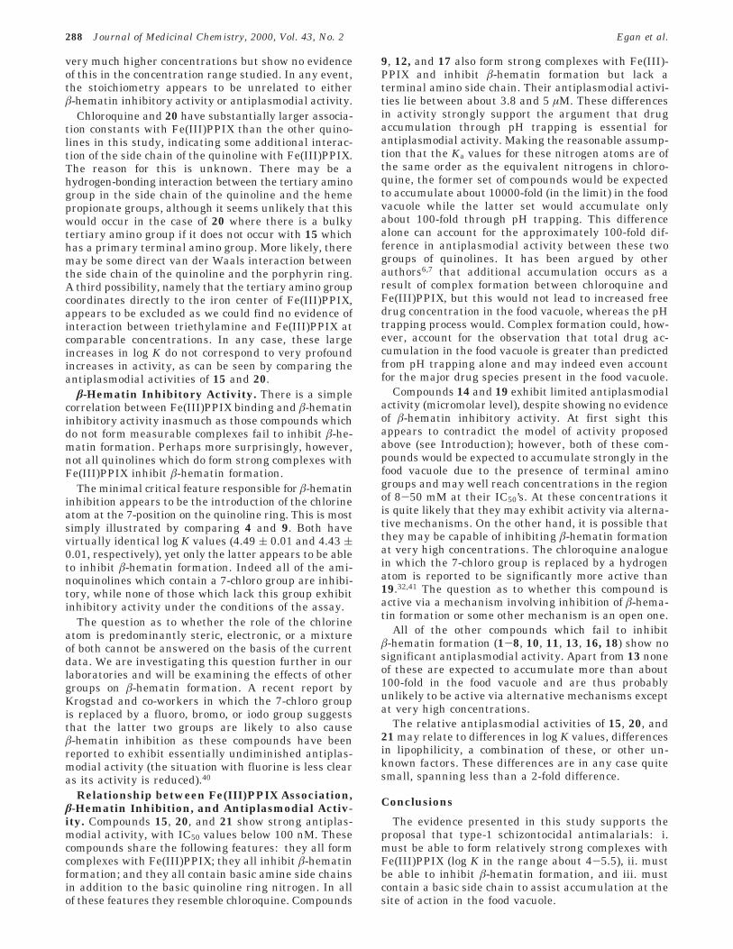
The molecular basis for the specific interaction of 2and 4 with Fe(III)PPIX is not clear at present; however,two properties of these compounds are striking. Firstthey possess the resonance forms shown in Figure 4,and second their pKa values are substantially higherthan those of the other quinolines.39 At the pH of theassociation experiment (7.5) 4 exists almost exclusivelyin the protonated form, while 2 is about 41% protonated.By contrast, the other quinolines are almost exclusivelydeprotonated. This might suggest that complex forma-tion involves cation-π interactions; however, neither 1nor 3 exhibit any observable association under similarconditions at pH 5.5, where significant (∼22%) proto-nation occurs. Furthermore, association of 4 with Fe-(III)PPIX was found to be essentially pH-insensitive. Wehave previously also reported this relative insensitivityto pH for association of both quinine and chloroquinewith Fe(III)PPIX.4
At present the reasons for the differences between thevarious quinolines are obscure. We can only speculatethat the differences may involve the ability of partialcharges on the quinolines which form strong complexeswith Fe(III)PPIX to match opposite partial charges onFe(III)PPIX, while at the same time minimizing repul-sive electrostatic interactions. Differences in the sto-ichiometries of the complexes, with some quinolinesforming 2:1 (quinoline:Fe(III)PPIX) complexes and oth-ers forming 1:1 complexes, are also obscure. In manycases, however, the association of the second quinolineis very weak, and it may be that those quinolines whichform 1:1 complexes would in fact form 2:1 complexes at
Figure 3. (a) Comparison of log K values for Fe(III)PPIXcomplexes of the quinolines investigated in this study (circles).Numbers refer to those given in Chart 1. Compounds for whichno value is shown have association constants too weak tomeasure in the concentration range used in this study,corresponding to log K j 2.5. Triangles are the correspondinglog K values for Fe(III)PPIX complexes of known antimalarialdrugs and refer to chloroquine (22), mefloquine (23), quinine(24), and amodiaquine (26) (all from ref 4) and quinidine (25)(from ref 13). Conditions: as quoted in Figure 2 for allcompounds. (b) Plot of log IC50 versus log K for Fe(III)PPIXassociation for those quinolines for which IC50 values are below10 µM; numbers refer to those in Chart 1 except for chloro-quine (22), mefloquine (23), and quinine (24). The plot il-lustrates the lack of a direct correlation between IC50 and logK.
Figure 4. Unique resonance forms present in 2- and 4-ami-noquinolines.
Structure-Function Relationships in Aminoquinolines Journal of Medicinal Chemistry, 2000, Vol. 43, No. 2 287
very much higher concentrations but show no evidenceof this in the concentration range studied. In any event,the stoichiometry appears to be unrelated to eitherâ-hematin inhibitory activity or antiplasmodial activity.
Chloroquine and 20 have substantially larger associa-tion constants with Fe(III)PPIX than the other quino-lines in this study, indicating some additional interac-tion of the side chain of the quinoline with Fe(III)PPIX.The reason for this is unknown. There may be ahydrogen-bonding interaction between the tertiary aminogroup in the side chain of the quinoline and the hemepropionate groups, although it seems unlikely that thiswould occur in the case of 20 where there is a bulkytertiary amino group if it does not occur with 15 whichhas a primary terminal amino group. More likely, theremay be some direct van der Waals interaction betweenthe side chain of the quinoline and the porphyrin ring.A third possibility, namely that the tertiary amino groupcoordinates directly to the iron center of Fe(III)PPIX,appears to be excluded as we could find no evidence ofinteraction between triethylamine and Fe(III)PPIX atcomparable concentrations. In any case, these largeincreases in log K do not correspond to very profoundincreases in activity, as can be seen by comparing theantiplasmodial activities of 15 and 20.
â-Hematin Inhibitory Activity. There is a simplecorrelation between Fe(III)PPIX binding and â-hematininhibitory activity inasmuch as those compounds whichdo not form measurable complexes fail to inhibit â-he-matin formation. Perhaps more surprisingly, however,not all quinolines which do form strong complexes withFe(III)PPIX inhibit â-hematin formation.
The minimal critical feature responsible for â-hematininhibition appears to be the introduction of the chlorineatom at the 7-position on the quinoline ring. This is mostsimply illustrated by comparing 4 and 9. Both havevirtually identical log K values (4.49 ( 0.01 and 4.43 (0.01, respectively), yet only the latter appears to be ableto inhibit â-hematin formation. Indeed all of the ami-noquinolines which contain a 7-chloro group are inhibi-tory, while none of those which lack this group exhibitinhibitory activity under the conditions of the assay.
The question as to whether the role of the chlorineatom is predominantly steric, electronic, or a mixtureof both cannot be answered on the basis of the currentdata. We are investigating this question further in ourlaboratories and will be examining the effects of othergroups on â-hematin formation. A recent report byKrogstad and co-workers in which the 7-chloro groupis replaced by a fluoro, bromo, or iodo group suggeststhat the latter two groups are likely to also causeâ-hematin inhibition as these compounds have beenreported to exhibit essentially undiminished antiplas-modial activity (the situation with fluorine is less clearas its activity is reduced).40
Relationship between Fe(III)PPIX Association,â-Hematin Inhibition, and Antiplasmodial Activ-ity. Compounds 15, 20, and 21 show strong antiplas-modial activity, with IC50 values below 100 nM. Thesecompounds share the following features: they all formcomplexes with Fe(III)PPIX; they all inhibit â-hematinformation; and they all contain basic amine side chainsin addition to the basic quinoline ring nitrogen. In allof these features they resemble chloroquine. Compounds
9, 12, and 17 also form strong complexes with Fe(III)-PPIX and inhibit â-hematin formation but lack aterminal amino side chain. Their antiplasmodial activi-ties lie between about 3.8 and 5 µM. These differencesin activity strongly support the argument that drugaccumulation through pH trapping is essential forantiplasmodial activity. Making the reasonable assump-tion that the Ka values for these nitrogen atoms are ofthe same order as the equivalent nitrogens in chloro-quine, the former set of compounds would be expectedto accumulate about 10000-fold (in the limit) in the foodvacuole while the latter set would accumulate onlyabout 100-fold through pH trapping. This differencealone can account for the approximately 100-fold dif-ference in antiplasmodial activity between these twogroups of quinolines. It has been argued by otherauthors6,7 that additional accumulation occurs as aresult of complex formation between chloroquine andFe(III)PPIX, but this would not lead to increased freedrug concentration in the food vacuole, whereas the pHtrapping process would. Complex formation could, how-ever, account for the observation that total drug ac-cumulation in the food vacuole is greater than predictedfrom pH trapping alone and may indeed even accountfor the major drug species present in the food vacuole.
Compounds 14 and 19 exhibit limited antiplasmodialactivity (micromolar level), despite showing no evidenceof â-hematin inhibitory activity. At first sight thisappears to contradict the model of activity proposedabove (see Introduction); however, both of these com-pounds would be expected to accumulate strongly in thefood vacuole due to the presence of terminal aminogroups and may well reach concentrations in the regionof 8-50 mM at their IC50’s. At these concentrations itis quite likely that they may exhibit activity via alterna-tive mechanisms. On the other hand, it is possible thatthey may be capable of inhibiting â-hematin formationat very high concentrations. The chloroquine analoguein which the 7-chloro group is replaced by a hydrogenatom is reported to be significantly more active than19.32,41 The question as to whether this compound isactive via a mechanism involving inhibition of â-hema-tin formation or some other mechanism is an open one.
All of the other compounds which fail to inhibitâ-hematin formation (1-8, 10, 11, 13, 16, 18) show nosignificant antiplasmodial activity. Apart from 13 noneof these are expected to accumulate more than about100-fold in the food vacuole and are thus probablyunlikely to be active via alternative mechanisms exceptat very high concentrations.
The relative antiplasmodial activities of 15, 20, and21 may relate to differences in log K values, differencesin lipophilicity, a combination of these, or other un-known factors. These differences are in any case quitesmall, spanning less than a 2-fold difference.
Conclusions
The evidence presented in this study supports theproposal that type-1 schizontocidal antimalarials: i.must be able to form relatively strong complexes withFe(III)PPIX (log K in the range about 4-5.5), ii. mustbe able to inhibit â-hematin formation, and iii. mustcontain a basic side chain to assist accumulation at thesite of action in the food vacuole.
288 Journal of Medicinal Chemistry, 2000, Vol. 43, No. 2 Egan et al.
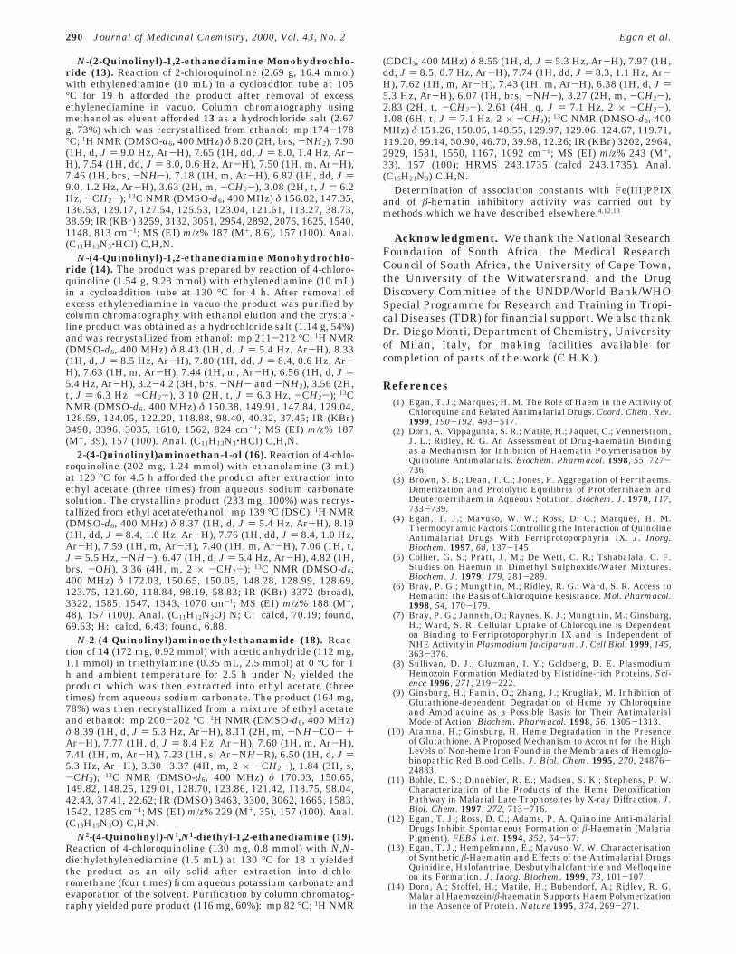
We are now able to propose a detailed model of thestructure-function relationships in chloroquine as fol-lows (see Figure 5): i. the 4-aminoquinoline nucleusalone provides an Fe(III)PPIX complexing template butis not sufficient for inhibition of hemozoin formation;ii. introduction of the 7-chloro group is responsible forinhibition of hemozoin formation but probably has littleinfluence on the strength of association with Fe(III)-PPIX; and iii. the aminoalkyl side chain is a require-ment for strong antiplasmodial activity. It probablyassists in drug accumulation in the food vacuole. It alsoappears to enhance the strength of association with Fe-(III)PPIX in some cases, but this effect does not appearto be essential for its activity.
Based on these findings, it should be possible toelaborate other molecules which are found to form π-πcomplexes with Fe(III)PPIX and which inhibit â-hema-tin formation so as to arrive at novel antimalarials bysemirational design. These findings are also of consider-able interest when combined with the recent structure-function investigations of Krogstad and co-workers32,40
which have shown that changes in the length of theaminoalkyl side chain have little influence on activityagainst chloroquine-sensitive strains of P. falciparum(in agreement with our proposed structure-functionmodel) but a profound influence on activity againstchloroquine-resistant strains of the parasite. It wouldappear that sufficiently large changes in the side chainalone overcome chloroquine resistance without havingto make changes to the 4-amino-7-haloquinoline tem-plate responsible for Fe(III)PPIX complexation andinhibition of â-hematin formation.
Experimental Section
Antiplasmodial Testing. All antiplasmodial experimentswere carried out on the chloroquine-sensitive P. falciparum
clone D10. The parasites were cultured continuously accordingto the method of Trager and Jensen,42 with slight modifica-tions. Cultures were maintained at a hematocrit of between3% and 5% and at parasitemias of between 2% and 8%.Parasitized O+ human red blood cells were suspended inculture flasks containing RPMI culture medium supplementedwith 10% A+ human serum. The flasks were gassed with amixture of 3% O2, 4% CO2, and 93% N2.
Chloroquine diphosphate was dissolved in water and allother compounds were dissolved in methanol and then dilutedwith medium to achieve the required concentrations (finalconcentrations all contained less than 1% methanol, which wasfound to be nontoxic to the parasites). Antiplasmodial activitywas assessed by measuring the activity of the parasite lactatedehydrogenase enzyme, according to a modified version of themethod of Makler.43 All antiplasmodial testing was performedat 1% hematocrit and 2% parasitemia. Compounds were addedat the trophozoite stage and applied for a period of 48 h beforeLDH measurement.
Dose-response curves were then constructed using nonlin-ear least-squares fitting with Graphpad Prism software44 andthe 50% inhibitory concentrations (IC50) were calculated. EachIC50 value is the result of at least three separate experimentsperformed in duplicate.
Chemistry. Quinoline, 2-chloroquinoline, 4-chloroquinoline,4,7-dichloroquinoline, 3-aminoquinoline, 5-aminoquinoline,6-aminoquinoline, 8-aminoquinoline, ethylenediamine, N,N-diethylethylenediamine, N,N-dimethylethylenediamine, andethanolamine were obtained from Sigma-Aldrich, Vorna Val-ley, South Africa. Methylamine (33% in methanol) was ob-tained from Merck, South Africa, and aqueous ammonia, aceticanhydride, triethylamine, and zinc chloride as well as commonsalts and solvents were obtained from Sarchem, Krugersdorp,South Africa. Precoated silica gel plates were obtained fromMerck, South Africa.
Column chromatography was carried out on Merck silicagel 60. Infrared (IR) spectroscopy was carried out on a Perkin-Elmer 983 infrared spectrometer in the range 4000-600 cm-1.Proton and carbon NMR spectra were recorded on a Varian400 MHz spectrometer or a Varian VXR200 instrument at 200MHz. Solvents are as indicated. Mass spectra were obtainedon a VG Micromass 16F spectrometer operating at 70 eV withan accelerating voltage of 4 kV and a variable temperaturesource. Melting points were determined on a Reichert-JungThermovar hot stage microscope except compound 16 whichwas determined by differential scanning calorimetry (DSC).Microanalyses were performed by the University of Cape TownMicroanalysis Service.
In the case of compounds which were synthesized, but arenot novel, melting points are shown in Table 2 and arecompared with those previously reported. These compoundswere all further characterized by proton NMR, mass spec-trometry, and in most cases elemental analysis. Where adiscrepancy of 3 °C or more was observed in the melting point,the compound was further characterized by 13C NMR. In allcases the identity of these products was confirmed and theywere also found to be of acceptable purity for further study.In the case of novel compounds, full synthetic details andcharacterization are supplied below.
Table 2. Melting Points for Previously Known Aminoquinolines Synthesized in This Work
compd mp (°C)
2-aminoquinoline (2) 129 (lit.45 128-129)4-aminoquinoline (4) 151-153 (lit.45 153-154)4-amino-7-chloroquinoline (9) 134 (lit.46 105-110, lit.47 146-147, lit.48 152-154.4)49
2-methylaminoquinoline (10) 70-72 (lit.50 70.5-71.5)4-methylaminoquinoline (11) 233a (lit.51 227-227.5, lit.52 238-240)7-chloro-4-methylaminoquinoline (12) 245 sublimation (lit.53 245-246)N-(7-chloro-4-quinolinyl)-1,2-ethanediamine (15) 137-139 (lit.54 137-139)2-(7-chloro-4-quinolinyl)aminoethan-1-ol (17) 217-218a (lit.55 214)N2-(7-chloro-4-quinolinyl)-N1,N1-diethyl-1,2-ethanediamine (20) 106-108a (lit.56 94-97)N2-(7-chloro-4-quinolinyl)-N1,N1-dimethyl-1,2-ethanediamine (21) 122-124 (lit.57 121-122.8)
a Purity confirmed by elemental analysis.
Figure 5. Proposed structure-function relationships in chlo-roquine based on findings in the current work.
Structure-Function Relationships in Aminoquinolines Journal of Medicinal Chemistry, 2000, Vol. 43, No. 2 289
N-(2-Quinolinyl)-1,2-ethanediamine Monohydrochlo-ride (13). Reaction of 2-chloroquinoline (2.69 g, 16.4 mmol)with ethylenediamine (10 mL) in a cycloaddion tube at 105°C for 19 h afforded the product after removal of excessethylenediamine in vacuo. Column chromatography usingmethanol as eluent afforded 13 as a hydrochloride salt (2.67g, 73%) which was recrystallized from ethanol: mp 174-178°C; 1H NMR (DMSO-d6, 400 MHz) δ 8.20 (2H, brs, -NH2), 7.90(1H, d, J ) 9.0 Hz, Ar-H), 7.65 (1H, dd, J ) 8.0, 1.4 Hz, Ar-H), 7.54 (1H, dd, J ) 8.0, 0.6 Hz, Ar-H), 7.50 (1H, m, Ar-H),7.46 (1H, brs, -NH-), 7.18 (1H, m, Ar-H), 6.82 (1H, dd, J )9.0, 1.2 Hz, Ar-H), 3.63 (2H, m, -CH2-), 3.08 (2H, t, J ) 6.2Hz, -CH2-); 13C NMR (DMSO-d6, 400 MHz) δ 156.82, 147.35,136.53, 129.17, 127.54, 125.53, 123.04, 121.61, 113.27, 38.73,38.59; IR (KBr) 3259, 3132, 3051, 2954, 2892, 2076, 1625, 1540,1148, 813 cm-1; MS (EI) m/z% 187 (M+, 8.6), 157 (100). Anal.(C11H13N3‚HCl) C,H,N.
N-(4-Quinolinyl)-1,2-ethanediamine Monohydrochlo-ride (14). The product was prepared by reaction of 4-chloro-quinoline (1.54 g, 9.23 mmol) with ethylenediamine (10 mL)in a cycloaddition tube at 130 °C for 4 h. After removal ofexcess ethylenediamine in vacuo the product was purified bycolumn chromatography with ethanol elution and the crystal-line product was obtained as a hydrochloride salt (1.14 g, 54%)and was recrystallized from ethanol: mp 211-212 °C; 1H NMR(DMSO-d6, 400 MHz) δ 8.43 (1H, d, J ) 5.4 Hz, Ar-H), 8.33(1H, d, J ) 8.5 Hz, Ar-H), 7.80 (1H, dd, J ) 8.4, 0.6 Hz, Ar-H), 7.63 (1H, m, Ar-H), 7.44 (1H, m, Ar-H), 6.56 (1H, d, J )5.4 Hz, Ar-H), 3.2-4.2 (3H, brs, -NH- and -NH2), 3.56 (2H,t, J ) 6.3 Hz, -CH2-), 3.10 (2H, t, J ) 6.3 Hz, -CH2-); 13CNMR (DMSO-d6, 400 MHz) δ 150.38, 149.91, 147.84, 129.04,128.59, 124.05, 122.20, 118.88, 98.40, 40.32, 37.45; IR (KBr)3498, 3396, 3035, 1610, 1562, 824 cm-1; MS (EI) m/z% 187(M+, 39), 157 (100). Anal. (C11H13N3‚HCl) C,H,N.
2-(4-Quinolinyl)aminoethan-1-ol (16). Reaction of 4-chlo-roquinoline (202 mg, 1.24 mmol) with ethanolamine (3 mL)at 120 °C for 4.5 h afforded the product after extraction intoethyl acetate (three times) from aqueous sodium carbonatesolution. The crystalline product (233 mg, 100%) was recrys-tallized from ethyl acetate/ethanol: mp 139 °C (DSC); 1H NMR(DMSO-d6, 400 MHz) δ 8.37 (1H, d, J ) 5.4 Hz, Ar-H), 8.19(1H, dd, J ) 8.4, 1.0 Hz, Ar-H), 7.76 (1H, dd, J ) 8.4, 1.0 Hz,Ar-H), 7.59 (1H, m, Ar-H), 7.40 (1H, m, Ar-H), 7.06 (1H, t,J ) 5.5 Hz, -NH-), 6.47 (1H, d, J ) 5.4 Hz, Ar-H), 4.82 (1H,brs, -OH), 3.36 (4H, m, 2 × -CH2-); 13C NMR (DMSO-d6,400 MHz) δ 172.03, 150.65, 150.05, 148.28, 128.99, 128.69,123.75, 121.60, 118.84, 98.19, 58.83; IR (KBr) 3372 (broad),3322, 1585, 1547, 1343, 1070 cm-1; MS (EI) m/z% 188 (M+,48), 157 (100). Anal. (C11H12N2O) N; C: calcd, 70.19; found,69.63; H: calcd, 6.43; found, 6.88.
N-2-(4-Quinolinyl)aminoethylethanamide (18). Reac-tion of 14 (172 mg, 0.92 mmol) with acetic anhydride (112 mg,1.1 mmol) in triethylamine (0.35 mL, 2.5 mmol) at 0 °C for 1h and ambient temperature for 2.5 h under N2 yielded theproduct which was then extracted into ethyl acetate (threetimes) from aqueous sodium carbonate. The product (164 mg,78%) was then recrystallized from a mixture of ethyl acetateand ethanol: mp 200-202 °C; 1H NMR (DMSO-d6, 400 MHz)δ 8.39 (1H, d, J ) 5.3 Hz, Ar-H), 8.11 (2H, m, -NH-CO- +Ar-H), 7.77 (1H, d, J ) 8.4 Hz, Ar-H), 7.60 (1H, m, Ar-H),7.41 (1H, m, Ar-H), 7.23 (1H, s, Ar-NH-R), 6.50 (1H, d, J )5.3 Hz, Ar-H), 3.30-3.37 (4H, m, 2 × -CH2-), 1.84 (3H, s,-CH3); 13C NMR (DMSO-d6, 400 MHz) δ 170.03, 150.65,149.82, 148.25, 129.01, 128.70, 123.86, 121.42, 118.75, 98.04,42.43, 37.41, 22.62; IR (DMSO) 3463, 3300, 3062, 1665, 1583,1542, 1285 cm-1; MS (EI) m/z% 229 (M+, 35), 157 (100). Anal.(C13H15N3O) C,H,N.
N2-(4-Quinolinyl)-N1,N1-diethyl-1,2-ethanediamine (19).Reaction of 4-chloroquinoline (130 mg, 0.8 mmol) with N,N-diethylethylenediamine (1.5 mL) at 130 °C for 18 h yieldedthe product as an oily solid after extraction into dichlo-romethane (four times) from aqueous potassium carbonate andevaporation of the solvent. Purification by column chromatog-raphy yielded pure product (116 mg, 60%): mp 82 °C; 1H NMR
(CDCl3, 400 MHz) δ 8.55 (1H, d, J ) 5.3 Hz, Ar-H), 7.97 (1H,dd, J ) 8.5, 0.7 Hz, Ar-H), 7.74 (1H, dd, J ) 8.3, 1.1 Hz, Ar-H), 7.62 (1H, m, Ar-H), 7.43 (1H, m, Ar-H), 6.38 (1H, d, J )5.3 Hz, Ar-H), 6.07 (1H, brs, -NH-), 3.27 (2H, m, -CH2-),2.83 (2H, t, -CH2-), 2.61 (4H, q, J ) 7.1 Hz, 2 × -CH2-),1.08 (6H, t, J ) 7.1 Hz, 2 × -CH3); 13C NMR (DMSO-d6, 400MHz) δ 151.26, 150.05, 148.55, 129.97, 129.06, 124.67, 119.71,119.20, 99.14, 50.90, 46.70, 39.98, 12.26; IR (KBr) 3202, 2964,2929, 1581, 1550, 1167, 1092 cm-1; MS (EI) m/z% 243 (M+,33), 157 (100); HRMS 243.1735 (calcd 243.1735). Anal.(C15H21N3) C,H,N.
Determination of association constants with Fe(III)PPIXand of â-hematin inhibitory activity was carried out bymethods which we have described elsewhere.4,12,13
Acknowledgment. We thank the National ResearchFoundation of South Africa, the Medical ResearchCouncil of South Africa, the University of Cape Town,the University of the Witwatersrand, and the DrugDiscovery Committee of the UNDP/World Bank/WHOSpecial Programme for Research and Training in Tropi-cal Diseases (TDR) for financial support. We also thankDr. Diego Monti, Department of Chemistry, Universityof Milan, Italy, for making facilities available forcompletion of parts of the work (C.H.K.).
References(1) Egan, T. J.; Marques, H. M. The Role of Haem in the Activity of
Chloroquine and Related Antimalarial Drugs. Coord. Chem. Rev.1999, 190-192, 493-517.
(2) Dorn, A.; Vippagunta, S. R.; Matile, H.; Jaquet, C.; Vennerstrom,J. L.; Ridley, R. G. An Assessment of Drug-haematin Bindingas a Mechanism for Inhibition of Haematin Polymerisation byQuinoline Antimalarials. Biochem. Pharmacol. 1998, 55, 727-736.
(3) Brown, S. B.; Dean, T. C.; Jones, P. Aggregation of Ferrihaems.Dimerization and Protolytic Equilibria of Protoferrihaem andDeuteroferrihaem in Aqueous Solution. Biochem. J. 1970, 117,733-739.
(4) Egan, T. J.; Mavuso, W. W.; Ross, D. C.; Marques, H. M.Thermodynamic Factors Controlling the Interaction of QuinolineAntimalarial Drugs With Ferriprotoporphyrin IX. J. Inorg.Biochem. 1997, 68, 137-145.
(5) Collier, G. S.; Pratt, J. M.; De Wett, C. R.; Tshabalala, C. F.Studies on Haemin in Dimethyl Sulphoxide/Water Mixtures.Biochem. J. 1979, 179, 281-289.
(6) Bray, P. G.; Mungthin, M.; Ridley, R. G.; Ward, S. R. Access toHematin: the Basis of Chloroquine Resistance. Mol. Pharmacol.1998, 54, 170-179.
(7) Bray, P. G.; Janneh, O.; Raynes, K. J.; Mungthin, M.; Ginsburg,H.; Ward, S. R. Cellular Uptake of Chloroquine is Dependenton Binding to Ferriprotoporphyrin IX and is Independent ofNHE Activity in Plasmodium falciparum. J. Cell Biol. 1999, 145,363-376.
(8) Sullivan, D. J.; Gluzman, I. Y.; Goldberg, D. E. PlasmodiumHemozoin Formation Mediated by Histidine-rich Proteins. Sci-ence 1996, 271, 219-222.
(9) Ginsburg, H.; Famin, O.; Zhang, J.; Krugliak, M. Inhibition ofGlutathione-dependent Degradation of Heme by Chloroquineand Amodiaquine as a Possible Basis for Their AntimalarialMode of Action. Biochem. Pharmacol. 1998, 56, 1305-1313.
(10) Atamna, H.; Ginsburg, H. Heme Degradation in the Presenceof Glutathione. A Proposed Mechanism to Account for the HighLevels of Non-heme Iron Found in the Membranes of Hemoglo-binopathic Red Blood Cells. J. Biol. Chem. 1995, 270, 24876-24883.
(11) Bohle, D. S.; Dinnebier, R. E.; Madsen, S. K.; Stephens, P. W.Characterization of the Products of the Heme DetoxificationPathway in Malarial Late Trophozoites by X-ray Diffraction. J.Biol. Chem. 1997, 272, 713-716.
(12) Egan, T. J.; Ross, D. C.; Adams, P. A. Quinoline Anti-malarialDrugs Inhibit Spontaneous Formation of â-Haematin (MalariaPigment). FEBS Lett. 1994, 352, 54-57.
(13) Egan, T. J.; Hempelmann, E.; Mavuso, W. W. Characterisationof Synthetic â-Haematin and Effects of the Antimalarial DrugsQuinidine, Halofantrine, Desbutylhalofantrine and Mefloquineon its Formation. J. Inorg. Biochem. 1999, 73, 101-107.
(14) Dorn, A.; Stoffel, H.; Matile, H.; Bubendorf, A.; Ridley, R. G.Malarial Haemozoin/â-haematin Supports Haem Polymerizationin the Absence of Protein. Nature 1995, 374, 269-271.
290 Journal of Medicinal Chemistry, 2000, Vol. 43, No. 2 Egan et al.
(15) Dorn, A.; Vippagunta, S. R.; Matile, H.; Bubendorf, A.; Venner-strom, J. L.; Ridley, R. G. A Comparison and Analysis of SeveralWays to Promote Haematin (Haem) Polymerisation and anAssessment of its Initiation in Vitro. Biochem. Pharmacol. 1998,55, 737-747.
(16) Hawley, S. R.; Bray, P. G.; Mungthin, M.; Atkinson, J. D.;O’Neill, P. M.; Ward, S. A. Relationship Between AntimalarialDrug Activity, Accumulation, and Inhibition of Heme Polymer-ization in Plasmodium falciparum in Vitro. Antimicrob. AgentsChemother. 1998, 42, 682-686.
(17) Basilico, N.; Monti, D.; Olliaro, P.; Taramelli, D. Non-ironPorphyrins Inhibit â-Haematin (Malaria Pigment) Polymerisa-tion. FEBS Lett. 1997, 409, 297-299.
(18) Basilico, N.; Pagani, E.; Monti, D.; Olliaro, P.; Taramelli, D. AMicrotitre-based Method for Measuring the Haem Polymeriza-tion Inhibitory Activity (HPIA) of Antimalarial Drugs. J. Anti-microb. Chemother. 1998, 42, 55-60.
(19) Sullivan, D. J.; Gluzman, I. Y.; Russell, D. G.; Goldberg, D. E.On the Molecular Mechanism of Chloroquine’s AntimalarialAction. Proc. Natl. Acad. Sci. U.S.A. 1996, 93, 11865-11870.
(20) Sullivan, D. J.; Matile, H.; Ridley, R. G.; Goldberg, D. E. ACommon Mechanism for Blockade of Heme Polymerization byAntimalarial Quinolines. J. Biol. Chem. 1998, 273, 31103-31107.
(21) Egan, T. J.; Ross, D. C.; Adams, P. A. The Mechanism of Actionof Quinolines and Related Antimalarial Drugs. S. Afr. J. Sci.1996, 92, 11-14.
(22) Egan, T. J.; Hunter, R.; Kaschula, C. H.; Marques, H. M.;Mavuso, W. W. Towards the Rational Design of AntimalarialDrugs. S. Afr. J. Sci. 1998, 94, 277-278.
(23) Homewood, C. A.; Warhurst, D. C.; Peters, W.; Baggaley, V. C.Lysosomes, pH and the Anti-malarial Action of Chloroquine.Nature 1972, 235, 50-52.
(24) Shellnutt, J. A. Metal Effects on Metalloporphyrins and on Theirπ-π Charge-transfer Complexes with Aromatic Acceptors: Uro-hemin Complexes. Inorg. Chem. 1983, 22, 2535-2544.
(25) Moreau, S.; Perly, B.; Chachaty, C.; Deleuze, C. A NuclearMagnetic Resonance Study of the Interactions of AntimalarialDrugs with Porphyrins. Biochim. Biophys. Acta 1985, 840, 107-116.
(26) Constantinidis, I.; Satterlee, J. D. UV-Visible and Carbon NMRStudies of Chloroquine Binding to Urohemin I Chloride andUroporphyrin I in Aqueous Solution. J. Am. Chem. Soc. 1988,110, 4391-4395.
(27) O’Keefe, D. H.; Barlow, C. H.; Smythe, G. A.; Fuchsman, W. H.;Moss, T. H.; Lilienthal, H. R.; Caughey, W. S. Magnetic andSpectroscopic Probes for FeOFe Linkages in Hemin Systems.Bioinorg. Chem. 1975, 5, 125-147.
(28) Pandey, V. A.; Tekwani, B. L. Formation of Haemozoin/â-haematin Under Physiological Conditions is Not Spontaneous.FEBS Lett. 1996, 393, 189-192.
(29) Ignatushchenko, M. V.; Winter, R. W.; Bachinger, H. P.; Hin-richs, D. J.; Riscoe, M. K. Xanthones as Antimalarial Agents;Studies of a Possible Mode of Action. FEBS Lett. 1997, 409, 67-73.
(30) Adams, P. A.; Berman, P. A. M.; Egan, T. J.; Marsh, P. J.; Silver,J. The Iron Environment in Heme and Heme-antimalarialComplexes of Pharmacological Interest. J. Inorg. Biochem. 1996,63, 69-77.
(31) Adams, P. A.; Egan, T. J.; Ross, D. C.; Silver, J.; Marsh, P. J.The Chemical Mechanism of â-Haematin Formation Studied byMossbauer Spectroscopy. Biochem. J. 1996, 318, 25-27.
(32) De, D.; Krogstad, F. M.; Cogswell, F. B.; Krogstad, D. J.Aminoquinolines That Circumvent Resistance in Plasmodiumfalciparum in Vitro. Am. J. Trop. Med. Hyg. 1996, 55, 579-583.
(33) Slater, A. F. G. Chloroquine: Mechanism of Drug Action andResistance in Plasmodium falciparum. Pharmacol. Ther. 1993,57, 203-235.
(34) Surolia, N.; Padmanaban, G. Chloroquine Inhibits Heme-de-pendent Protein Synthesis in Plasmodium falciparum. Proc.Natl. Acad. Sci. U.S.A. 1991, 88, 4786-4790.
(35) Cohen, S. N.; Yielding, K. L. Inhibition of DNA and RNAPolymerase Reactions by Chloroquine. Proc. Natl. Acad. Sci.U.S.A. 1965, 54, 521-527.
(36) Gyang, F. N.; Poole, B.; Trager, W. Peptidases from Plasmodiumfalciparum Cultured in Vitro. Mol. Biochem. Parasitol. 1982, 5,263-273.
(37) Ginsburg, H.; Krugliak, M. Quinoline-containing Antimalarials- Mode of Action, Drug Resistance and its Reversal. Biochem.Pharmacol. 1992, 43, 63-70.
(38) O’Neill, P. M.; Willock, D. J.; Hawley, S. R.; Bray, P. G.; Storr,R. C.; Ward, S. R.; Park, B. K. Synthesis, Antimalarial Activityand Molecular Modeling of Tebuquine Analogues. J. Med. Chem.1997, 40, 437-448.
(39) Barton, D. H. R.; Ollis, W. D. In Comprehensive OrganicChemistry: the Synthesis and Reactions of Organic Compounds,Vol. 4: Heterocyclic Compounds; Sammes, P. G., Ed.; PergamonPress: Oxford, 1979; pp 191-193.
(40) De, D.; Krogstad, F. M.; Byers, L. D.; Krogstad, D. J. Structure-activity Relationships for Antiplasmodial Activity Among 7-Sub-stituted 4-Aminoquinolines. J. Med. Chem. 1998, 41, 4918-4926.
(41) Veigne, E.; Moreau, S. The Mode of Action of Chloroquine.Nonweak Base Properties of 4-Aminoquinolines and Antima-larial Effects on Strains of Plasmodium. Ann. Trop. Med.Parasitol. 1991, 85, 229-237.
(42) Trager, W.; Jensen, J. B. Human Malaria Parasites in Continu-ous Culture. Science 1976, 193, 674-675.
(43) Makler, M. T.; Ries, J. M.; Williams, J. A.; Bancroft, J. E.; Piper,R. C.; Gibbins, B. L.; Hinrichs, D. J. Parasite Lactate Dehydro-genase as an Assay for Plasmodium falciparum Drug Sensitivity.Am. J. Trop. Med. Hyg. 1993, 48, 739-741.
(44) GraphPad Prism, version 2.0, GraphPad Software Inc., 10855Sorrento Valley Rd. #203, San Diego, CA 92121.
(45) Brown, V. E.; Plasz, A. C. Spectrophotometric Determination ofthe Second Dissociation Constants of the Aminoquinolines. J.Heterocycl. Chem. 1970, 7, 335-338.
(46) Price, C. C.; Leonard, N. J.; Peel, E. W.; Reitsema, R. H. Some4-Amino-7-chloroquinoline Derivatives. J. Am. Chem. Soc. 1946,68, 1807-1808.
(47) Lin, A. J.; Loo, T. L. Synthesis and Antitumor Activity of HalogenSubstituted 4-(3,3-dimethyl-1-triazeno)quinolines. J. Med. Chem.1978, 21, 268-272.
(48) Roseman, K. A.; Gould, M. M.; Linfield, W. M.; Edwards, B. E.Antimalarials. 8-Chloro-4-(2′-N,N-dibutylamino-1′-hydroxyeth-yl)benzo[h]-1,6-naphthyridine. J. Med. Chem. 1970, 13, 230-233.
(49) The melting point of 9 is reported46 to be strongly dependent onthe degree of hydration of the compound. When hydrated themelting point is 105-110 °C, but this rises substantially afterextensive drying.
(50) Ito, H. Formation of cis-2-Nitrocinnamonitrile by Oxidation of2-Methylaminoquinoline with Hydrogen Peroxide. Chem. Pharm.Bull. 1964, 12, 350-352.
(51) Luthy, N. G.; Bergstrom, F. W.; Mosher, H. S. Introduction ofAlkylamino and Dialkylamino Groups into the Quinoline Nucleus.J. Am. Chem. Soc. 1949, 71, 1109-1110.
(52) Pentimalli, L.; Milani, G. Peracid Oxidation of (Methylamino)-quinolines. Gazz. Chim. Ital. 1983, 113, 803-806.
(53) Craig, J. C.; Pearson, D. E. NMR Proof of the Structure of4-Aminoquinolines and Pyridines. J. Heterocycl. Chem. 1968, 5,631-637.
(54) Peck, R. M.; Preston, R. K.; Creech, H. J. Nitrogen MustardAnalogues of Antimalarial Drugs. J. Am. Chem. Soc. 1959, 81,3984-3989.
(55) Elderfield, R. C.; Gensler, W. J.; Birstein, O.; Kreysa, F. J.;Maynard, J. T.; Galbreath, J. Synthesis of Certain Simple4-Aminoquinoline Derivatives. J. Am. Chem. Soc. 1946, 68,1250-1251.
(56) Surrey, A. R.; Hammer, H. F. Some 7-Substituted 4-Amino-quinoline Derivatives. J. Am. Chem. Soc. 1946, 68, 113-114.
(57) Surrey, A. R.; Lesher, G. Y.; Mayer, J. R.; Webb, W. G.Hypotensive Agents. II. The Preparation of Quaternary Saltsof Some 4-Dialkylaminoalkylaminoquinolines. J. Am. Chem. Soc.1959, 81, 2894-2897.
JM990437L
Structure-Function Relationships in Aminoquinolines Journal of Medicinal Chemistry, 2000, Vol. 43, No. 2 291