Structural and Functional Elucidation of Yeast Lanosterol ... · (CYP51) in the ergosterol...
18
RESEARCH ARTICLE Structural and Functional Elucidation of Yeast Lanosterol 14α-Demethylase in Complex with Agrochemical Antifungals Joel D. A. Tyndall 1☯ *, Manya Sabherwal 2☯ , Alia A. Sagatova 2 , Mikhail V. Keniya 2,3 , Jacopo Negroni 4 , Rajni K. Wilson 2 , Matthew A. Woods 2 , Klaus Tietjen 5 , Brian C. Monk 2,3 * 1 New Zealand’s National School of Pharmacy, University of Otago, Dunedin, New Zealand, 2 Sir John Walsh Research Institute, New Zealand’s National Centre for Dentistry, University of Otago, Dunedin, New Zealand, 3 Department of Oral Sciences, New Zealand’s National Centre for Dentistry, University of Otago, Dunedin, New Zealand, 4 Bayer SAS, Division Crop Science, Disease Control Research, Lyon, France, 5 Bayer AG, Division Crop Science, Disease Control Research, Monheim, Germany ☯ These authors contributed equally to this work. * [email protected] (JDAT); [email protected] (BCM) Abstract Azole antifungals, known as demethylase inhibitors (DMIs), target sterol 14α-demethylase (CYP51) in the ergosterol biosynthetic pathway of fungal pathogens of both plants and humans. DMIs remain the treatment of choice in crop protection against a wide range of fun- gal phytopathogens that have the potential to reduce crop yields and threaten food security. We used a yeast membrane protein expression system to overexpress recombinant hexa- histidine-tagged S. cerevisiae lanosterol 14α-demethylase and the Y140F or Y140H mutants of this enzyme as surrogates in order characterize interactions with DMIs. The whole-cell antifungal activity (MIC 50 values) of both the R- and S-enantiomers of tebucona- zole, prothioconazole (PTZ), prothioconazole-desthio, and oxo-prothioconazole (oxo-PTZ) as well as for fluquinconazole, prochloraz and a racemic mixture of difenoconazole were determined. In vitro binding studies with the affinity purified enzyme were used to show tight type II binding to the yeast enzyme for all compounds tested except PTZ and oxo-PTZ. High resolution X-ray crystal structures of ScErg11p6×His in complex with seven DMIs, including four enantiomers, reveal triazole-mediated coordination of all compounds and the specific orientation of compounds within the relatively hydrophobic binding site. Comparison with CYP51 structures from fungal pathogens including Candida albicans, Candida glabrata and Aspergillus fumigatus provides strong evidence for a highly conserved CYP51 structure including the drug binding site. The structures obtained using S. cerevisiae lanosterol 14α- demethylase in complex with these agrochemicals provide the basis for understanding the impact of mutations on azole susceptibility and a platform for the structure-directed design of the next-generation of DMIs. PLOS ONE | DOI:10.1371/journal.pone.0167485 December 1, 2016 1 / 18 a11111 OPEN ACCESS Citation: Tyndall JDA, Sabherwal M, Sagatova AA, Keniya MV, Negroni J, Wilson RK, et al. (2016) Structural and Functional Elucidation of Yeast Lanosterol 14α-Demethylase in Complex with Agrochemical Antifungals. PLoS ONE 11(12): e0167485. doi:10.1371/journal.pone.0167485 Editor: Joseph T. Nickels, Venenum Biodesign, UNITED STATES Received: September 16, 2016 Accepted: November 15, 2016 Published: December 1, 2016 Copyright: © 2016 Tyndall et al. This is an open access article distributed under the terms of the Creative Commons Attribution License, which permits unrestricted use, distribution, and reproduction in any medium, provided the original author and source are credited. Data Availability Statement: All PDB files are available from the Protein Data Bank (accession number(s) 5EAB, 5EAC, 5EAD, 5EAE, 5EAF, 5EAG, 5EAH). Funding: This programme was funded by the Health Research Council and Bayer Crop Science, https://www.cropscience.bayer.com. The funding was provided to BM and used for this study. The commercial funders provided financial support in the form of authors’ salaries and/or research materials. JN and KT had a minor role in data analysis, and preparation of the manuscript. The
Transcript of Structural and Functional Elucidation of Yeast Lanosterol ... · (CYP51) in the ergosterol...
RESEARCH ARTICLE
Structural and Functional Elucidation of Yeast
Lanosterol 14α-Demethylase in Complex with
Agrochemical Antifungals
Joel D. A. Tyndall1☯*, Manya Sabherwal2☯, Alia A. Sagatova2, Mikhail V. Keniya2,3,
Jacopo Negroni4, Rajni K. Wilson2, Matthew A. Woods2, Klaus Tietjen5, Brian C. Monk2,3*
1 New Zealand’s National School of Pharmacy, University of Otago, Dunedin, New Zealand, 2 Sir John
Walsh Research Institute, New Zealand’s National Centre for Dentistry, University of Otago, Dunedin, New
Zealand, 3 Department of Oral Sciences, New Zealand’s National Centre for Dentistry, University of Otago,
Dunedin, New Zealand, 4 Bayer SAS, Division Crop Science, Disease Control Research, Lyon, France,
5 Bayer AG, Division Crop Science, Disease Control Research, Monheim, Germany
☯ These authors contributed equally to this work.
* [email protected] (JDAT); [email protected] (BCM)
Abstract
Azole antifungals, known as demethylase inhibitors (DMIs), target sterol 14α-demethylase
(CYP51) in the ergosterol biosynthetic pathway of fungal pathogens of both plants and
humans. DMIs remain the treatment of choice in crop protection against a wide range of fun-
gal phytopathogens that have the potential to reduce crop yields and threaten food security.
We used a yeast membrane protein expression system to overexpress recombinant hexa-
histidine-tagged S. cerevisiae lanosterol 14α-demethylase and the Y140F or Y140H
mutants of this enzyme as surrogates in order characterize interactions with DMIs. The
whole-cell antifungal activity (MIC50 values) of both the R- and S-enantiomers of tebucona-
zole, prothioconazole (PTZ), prothioconazole-desthio, and oxo-prothioconazole (oxo-PTZ)
as well as for fluquinconazole, prochloraz and a racemic mixture of difenoconazole were
determined. In vitro binding studies with the affinity purified enzyme were used to show tight
type II binding to the yeast enzyme for all compounds tested except PTZ and oxo-PTZ. High
resolution X-ray crystal structures of ScErg11p6×His in complex with seven DMIs, including
four enantiomers, reveal triazole-mediated coordination of all compounds and the specific
orientation of compounds within the relatively hydrophobic binding site. Comparison with
CYP51 structures from fungal pathogens including Candida albicans, Candida glabrata and
Aspergillus fumigatus provides strong evidence for a highly conserved CYP51 structure
including the drug binding site. The structures obtained using S. cerevisiae lanosterol 14α-
demethylase in complex with these agrochemicals provide the basis for understanding the
impact of mutations on azole susceptibility and a platform for the structure-directed design
of the next-generation of DMIs.
PLOS ONE | DOI:10.1371/journal.pone.0167485 December 1, 2016 1 / 18
a11111
OPENACCESS
Citation: Tyndall JDA, Sabherwal M, Sagatova AA,
Keniya MV, Negroni J, Wilson RK, et al. (2016)
Structural and Functional Elucidation of Yeast
Lanosterol 14α-Demethylase in Complex with
Agrochemical Antifungals. PLoS ONE 11(12):
e0167485. doi:10.1371/journal.pone.0167485
Editor: Joseph T. Nickels, Venenum Biodesign,
UNITED STATES
Received: September 16, 2016
Accepted: November 15, 2016
Published: December 1, 2016
Copyright: © 2016 Tyndall et al. This is an open
access article distributed under the terms of the
Creative Commons Attribution License, which
permits unrestricted use, distribution, and
reproduction in any medium, provided the original
author and source are credited.
Data Availability Statement: All PDB files are
available from the Protein Data Bank (accession
number(s) 5EAB, 5EAC, 5EAD, 5EAE, 5EAF, 5EAG,
5EAH).
Funding: This programme was funded by the
Health Research Council and Bayer Crop Science,
https://www.cropscience.bayer.com. The funding
was provided to BM and used for this study. The
commercial funders provided financial support in
the form of authors’ salaries and/or research
materials. JN and KT had a minor role in data
analysis, and preparation of the manuscript. The
Introduction
Since the introduction in 1973 of the imidazole antifungal agent enilconazole (imazalil, chlora-
mazole) and the triazole triadimefon, multiple generations of the azole antifungals [1–4] have
underpinned global food security by preventing or treating a wide range of diseases in plants
caused by fungal pathogens (for review see Parker et al.) [5]. Azoles are routinely used to treat
diseases caused by phytopathogenic fungi including eyespot disease and powdery mildew in
wheat and barley, black rust, headblight and septoria leaf blotch in wheat, leaf scald in cereals,
leaf spot in sugar beet, powdery mildew on grapes, black sigatoka in bananas, apple scab, and
mold on citrus fruit as well as the production of toxins. Without agrochemical intervention
such diseases can substantially reduce crop yields, wipe out important crops, or result in crop
spoilage due to rots or mycotoxins such as aflotoxins in peanuts. While numerous classes of
antimycotics are employed in agriculture, such as the benzimidazoles, phenylamides, dicar-
boximides, anilinopyrimidines, quinone outside inhibitors and carboxylic amides, the triazole
antifungals alone account for ~20% of the global market share for systemic fungicides. In the
United Kingdom prothioconazole (PTZ), tebuconazole (TBZ) and epoxiconazole are the three
most commonly used fungicides [6]. In addition, the azoles are well suited for use in agricul-
ture, since in some cases they have the advantage of enhancing crop growth independent of
their effects on phytopathogens [7]. The continuing success of azole antifungals has been
attributed to their efficacy in the field and the slow evolution of azole resistance that has usually
been countered by the development of more active triazole fungicides such as PTZ. This tria-
zolinethione was introduced in 2002. Some problems have been associated with the agricul-
tural use of azole antifungals, particularly in relation to human health. These include the
development of antifungal resistance (reduced susceptibility) not only in the targeted phyto-
pathogens but also in the ubiquitous and medically important pathogen Aspergillus fumigatus[8–13]. Endocrine disruption has also become a consideration. The imidazoles appear more
potent than triazoles in eliciting possible side-effects that have been detected in animal tissue
culture, such as inhibition of the conversion of progesterone to testosterone as well as anti-
androgenic effects due to aromatase inhibition [14–16]. In a large study profiling the toxicities
of environmental chemicals, it was found that the reproductive toxicity caused by individual
azoles varies widely [17].
Lanosterol 14α-demethylase (CYP51 or Erg11p in yeast and some fungal pathogens; sterol
14α-demethylase in fungal phytopathogens) is the primary target of the imidazole and triazole
(azole) antifungal drugs [18], often referred to as demethylase inhibitors (DMIs). The CYP51
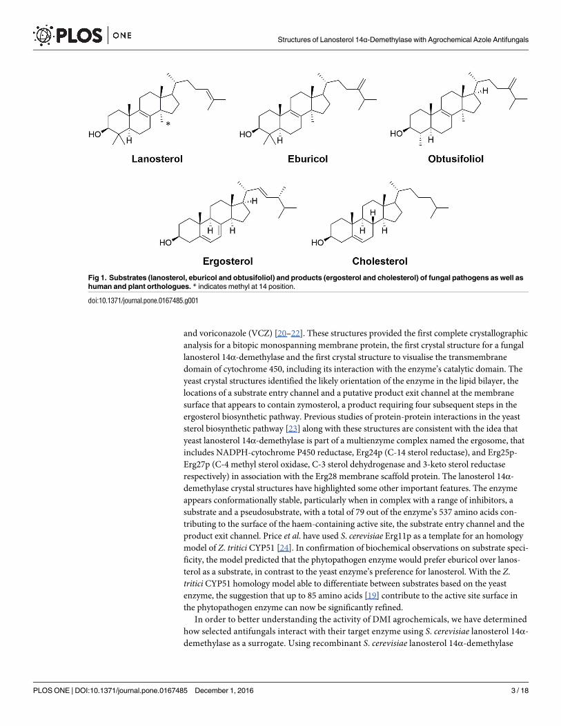
enzymes of fungal pathogens and yeast use eburicol or lanosterol as substrates to produce
ergosterol while their orthologues in plants and humans use obtusifoliol and lanosterol,
respectively, to produce plant sterols and cholesterol (Fig 1). In the absence of any X-ray struc-
tures of a fungal sterol 14α-demethylase, Mullins et al. (2011) proposed a structural rationale
for the emergence of azole resistance associated with CYP51 mutations in the wheat blotch
pathogen Zymoseptoria tritici (formerly Mycosphaerella graminicola) [19]. Homology models
of the wild type protein and 13 variant proteins were generated by selectively combining the
structures forMycobacterium tuberculosis CYP51 plus a range of other CYP51 proteins as tem-
plates [19]. Modelling of the wild type and mutant binding pockets with the ligands TBZ, pro-
chloraz (PRZ) and epoxiconazole implicated 85 amino acid residues in determining active site
volume and azole binding, thereby suggesting a basis for the genetic selection of single and
multiple mutations that led to reduced affinity for these antifungals.
We have determined the X-ray crystal structures of hexahistidine-tagged Saccharomyces cer-evisiae lanosterol 14α-demethylase in complex with its substrate lanosterol, the pseudosub-
strate estriol and the triazole drugs itraconazole (ITC), posaconazole (PCZ), fluconazole (FLC)
Structures of Lanosterol 14α-Demethylase with Agrochemical Azole Antifungals
PLOS ONE | DOI:10.1371/journal.pone.0167485 December 1, 2016 2 / 18
funders had no role in study design, data collection
and analysis, decision to publish, or preparation of
the manuscript.
Competing Interests: I have read the journal’s
policy and the authors of this manuscript have the
following competing interests: the research was
funded by Bayer Crop Science. This does not alter
our adherence to PLOS ONE policies on sharing
data and materials.
and voriconazole (VCZ) [20–22]. These structures provided the first complete crystallographic
analysis for a bitopic monospanning membrane protein, the first crystal structure for a fungal
lanosterol 14α-demethylase and the first crystal structure to visualise the transmembrane
domain of cytochrome 450, including its interaction with the enzyme’s catalytic domain. The
yeast crystal structures identified the likely orientation of the enzyme in the lipid bilayer, the
locations of a substrate entry channel and a putative product exit channel at the membrane
surface that appears to contain zymosterol, a product requiring four subsequent steps in the
ergosterol biosynthetic pathway. Previous studies of protein-protein interactions in the yeast
sterol biosynthetic pathway [23] along with these structures are consistent with the idea that
yeast lanosterol 14α-demethylase is part of a multienzyme complex named the ergosome, that
includes NADPH-cytochrome P450 reductase, Erg24p (C-14 sterol reductase), and Erg25p-
Erg27p (C-4 methyl sterol oxidase, C-3 sterol dehydrogenase and 3-keto sterol reductase
respectively) in association with the Erg28 membrane scaffold protein. The lanosterol 14α-
demethylase crystal structures have highlighted some other important features. The enzyme
appears conformationally stable, particularly when in complex with a range of inhibitors, a
substrate and a pseudosubstrate, with a total of 79 out of the enzyme’s 537 amino acids con-
tributing to the surface of the haem-containing active site, the substrate entry channel and the
product exit channel. Price et al. have used S. cerevisiae Erg11p as a template for an homology
model of Z. tritici CYP51 [24]. In confirmation of biochemical observations on substrate speci-
ficity, the model predicted that the phytopathogen enzyme would prefer eburicol over lanos-
terol as a substrate, in contrast to the yeast enzyme’s preference for lanosterol. With the Z.
tritici CYP51 homology model able to differentiate between substrates based on the yeast
enzyme, the suggestion that up to 85 amino acids [19] contribute to the active site surface in
the phytopathogen enzyme can now be significantly refined.
In order to better understanding the activity of DMI agrochemicals, we have determined
how selected antifungals interact with their target enzyme using S. cerevisiae lanosterol 14α-
demethylase as a surrogate. Using recombinant S. cerevisiae lanosterol 14α-demethylase
Fig 1. Substrates (lanosterol, eburicol and obtusifoliol) and products (ergosterol and cholesterol) of fungal pathogens as well as
human and plant orthologues. * indicates methyl at 14 position.
doi:10.1371/journal.pone.0167485.g001
Structures of Lanosterol 14α-Demethylase with Agrochemical Azole Antifungals
PLOS ONE | DOI:10.1371/journal.pone.0167485 December 1, 2016 3 / 18
(ScErg11p6×His; wildtype) or the Y140F or Y140H mutants of this enzyme [25] we have
determined the whole cell antifungal activity (MIC50 values) of the R- and S- enantiomers
of TBZ, PTZ, prothioconazole-desthio (DPZ), oxo-prothioconazole (oxo-PTZ) as well as
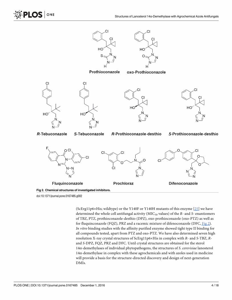
for fluquinconazole (FQZ), PRZ and a racemic mixture of difenoconazole (DFC, Fig 2).
In vitro binding studies with the affinity purified enzyme showed tight type II binding for
all compounds tested, apart from PTZ and oxo-PTZ. We have also determined seven high
resolution X-ray crystal structures of ScErg11p6×His in complex with R- and S-TBZ, R-
and S-DPZ, FQZ, PRZ and DFC. Until crystal structures are obtained for the sterol
14α-demethylases of individual phytopathogens, the structures of S. cerevisiae lanosterol
14α-demethylase in complex with these agrochemicals and with azoles used in medicine
will provide a basis for the structure-directed discovery and design of next-generation
DMIs.
Fig 2. Chemical structures of investigated inhibitors.
doi:10.1371/journal.pone.0167485.g002
Structures of Lanosterol 14α-Demethylase with Agrochemical Azole Antifungals
PLOS ONE | DOI:10.1371/journal.pone.0167485 December 1, 2016 4 / 18
Results
X-ray crystallographic analysis of phytopathogen CYP51 inhibitors in
complex with ScErg11p6×His
Crystal structures of the full length S. cerevisiae lanosterol 14α-demethylase (ScErg11p6×His)
in complex with S-TBZ (PDB ID:5EAB), R-TBZ (PDB ID:5EAC), S-DPZ (PDB ID:5EAD), R-
DPZ PDB ID:5EAE), FQZ (PDB ID:5EAF), PRZ (PDB ID:5EAG), and all four stereoisomers
of DFC (PDB ID:5EAH) were determined at resolutions from 2.65 to 2.00 Å. Data collection
and refinement statistics are given in S1 Table. All complexes were crystallised in the space
group P 1 211, with a single ScErg11p6×His-ligand complex in the asymmetric unit, with the
exception of the ScErg11p6×His -FQZ complex, which was crystallised in space group P1 with
two protomers found in the asymmetric unit. Comparison of the ScErg11p6×His-FQZ com-
plex with the others shows an almost identical protein structure, including the transmembrane
and amphipathic helices. All ligand-protein complexes were isomorphous with the original
ScErg11p6×His lanosterol complex (PDB ID:4LXJ) used for molecular replacement [20].
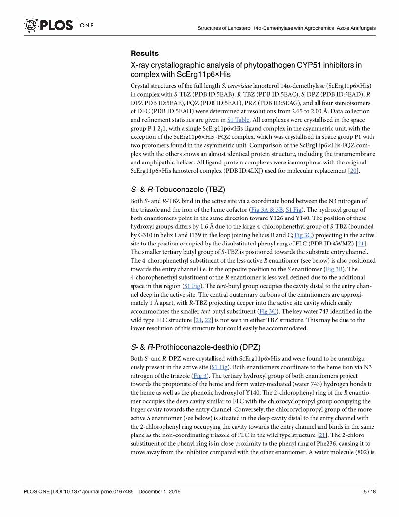
S- & R-Tebuconazole (TBZ)
Both S- and R-TBZ bind in the active site via a coordinate bond between the N3 nitrogen of
the triazole and the iron of the heme cofactor (Fig 3A & 3B, S1 Fig). The hydroxyl group of
both enantiomers point in the same direction toward Y126 and Y140. The position of these
hydroxyl groups differs by 1.6 Å due to the large 4-chlorophenethyl group of S-TBZ (bounded
by G310 in helix I and I139 in the loop joining helices B and C; Fig 3C) projecting in the active
site to the position occupied by the disubstituted phenyl ring of FLC (PDB ID:4WMZ) [21].
The smaller tertiary butyl group of S-TBZ is positioned towards the substrate entry channel.
The 4-chorophenethyl substituent of the less active R enantiomer (see below) is also positioned
towards the entry channel i.e. in the opposite position to the S enantiomer (Fig 3B). The
4-chorophenethyl substituent of the R enantiomer is less well defined due to the additional
space in this region (S1 Fig). The tert-butyl group occupies the cavity distal to the entry chan-
nel deep in the active site. The central quaternary carbons of the enantiomers are approxi-
mately 1 Å apart, with R-TBZ projecting deeper into the active site cavity which easily
accommodates the smaller tert-butyl substituent (Fig 3C). The key water 743 identified in the
wild type FLC structure [21, 22] is not seen in either TBZ structure. This may be due to the
lower resolution of this structure but could easily be accommodated.
S- & R-Prothioconazole-desthio (DPZ)
Both S- and R-DPZ were crystallised with ScErg11p6×His and were found to be unambigu-
ously present in the active site (S1 Fig). Both enantiomers coordinate to the heme iron via N3
nitrogen of the triazole (Fig 3). The tertiary hydroxyl group of both enantiomers project
towards the propionate of the heme and form water-mediated (water 743) hydrogen bonds to
the heme as well as the phenolic hydroxyl of Y140. The 2-chlorophenyl ring of the R enantio-
mer occupies the deep cavity similar to FLC with the chlorocyclopropyl group occupying the
larger cavity towards the entry channel. Conversely, the chlorocyclopropyl group of the more
active S enantiomer (see below) is situated in the deep cavity distal to the entry channel with
the 2-chlorophenyl ring occupying the cavity towards the entry channel and binds in the same
plane as the non-coordinating triazole of FLC in the wild type structure [21]. The 2-chloro
substituent of the phenyl ring is in close proximity to the phenyl ring of Phe236, causing it to
move away from the inhibitor compared with the other enantiomer. A water molecule (802) is
Structures of Lanosterol 14α-Demethylase with Agrochemical Azole Antifungals
PLOS ONE | DOI:10.1371/journal.pone.0167485 December 1, 2016 5 / 18
present between F384 and the chlorocyclopropyl group of the R enantiomer. This is displaced
by the phenyl ring in the S enantiomer complex. Consequently, F384 and Y126 are oriented
closer to the hydrophobic ring of the ligand. Attempts were made to crystallise PTZ and oxo-
PTZ but crystals could not be obtained.
Fluquinconazole (FQZ)
FQZ binds unambiguously in the active site via a coordinate bond between the N3 of the tria-
zole ring and the iron of the heme cofactor (Fig 4A; data in S1 Fig). The dichlorophenyl ring
binds deep in the active site adjacent to G310 in a similar manner to FLC (Fig 4B). The rigid
quinazolin-4(3H)-one ring projects towards the entry channel in an orientation with the aro-
matic ring perpendicular to the non-coordinating azole of FLC. No water (743/843) is seen
adjacent to Y140. Y126 is shifted away compared to the FLC structure to accommodate the
rigid and bulky ring system with a close contact between the fluoro substituent of the fungicide
and the phenolic ring. The N1 nitrogen of the quinazolin-4(3H)-one ring is in close proximity
to Cδ1 of L380, the carbonyl oxygen is close to Cγ2 of T130 and the coordinating triazole abuts
G310 as seen previously with S-TBZ.
Fig 3. Phytopathogen inhibitors complexed in the active site of ScERG11p6×His. The structures reveal the active site binding
orientation of the enantiomers (A) S-TBZ (green carbons, PDB ID:5EAB), (B) R-TBZ (cyan carbons, PDB ID:5EAC), and (C) the
superimposition of S-TBZ and R-TBZ, the active site orientation of the enantiomers (D) S-DPZ (magenta carbons, PDB ID:5EAD), (E) R-
DPZ (salmon carbons, PDB ID:5EAE), and (F) the superimposition of S-DPZ and R-DPZ. The heme cofactor and selected residues (Y126
and Y140) are shown as sticks. Water-mediated (w743, red sphere) hydrogen bonds are shown as yellow dashed lines. Helix I is shown as a
yellow ribbon at the bottom right of each panel. Nitrogen atoms are blue, oxygen red and chlorine green.
doi:10.1371/journal.pone.0167485.g003
Structures of Lanosterol 14α-Demethylase with Agrochemical Azole Antifungals
PLOS ONE | DOI:10.1371/journal.pone.0167485 December 1, 2016 6 / 18
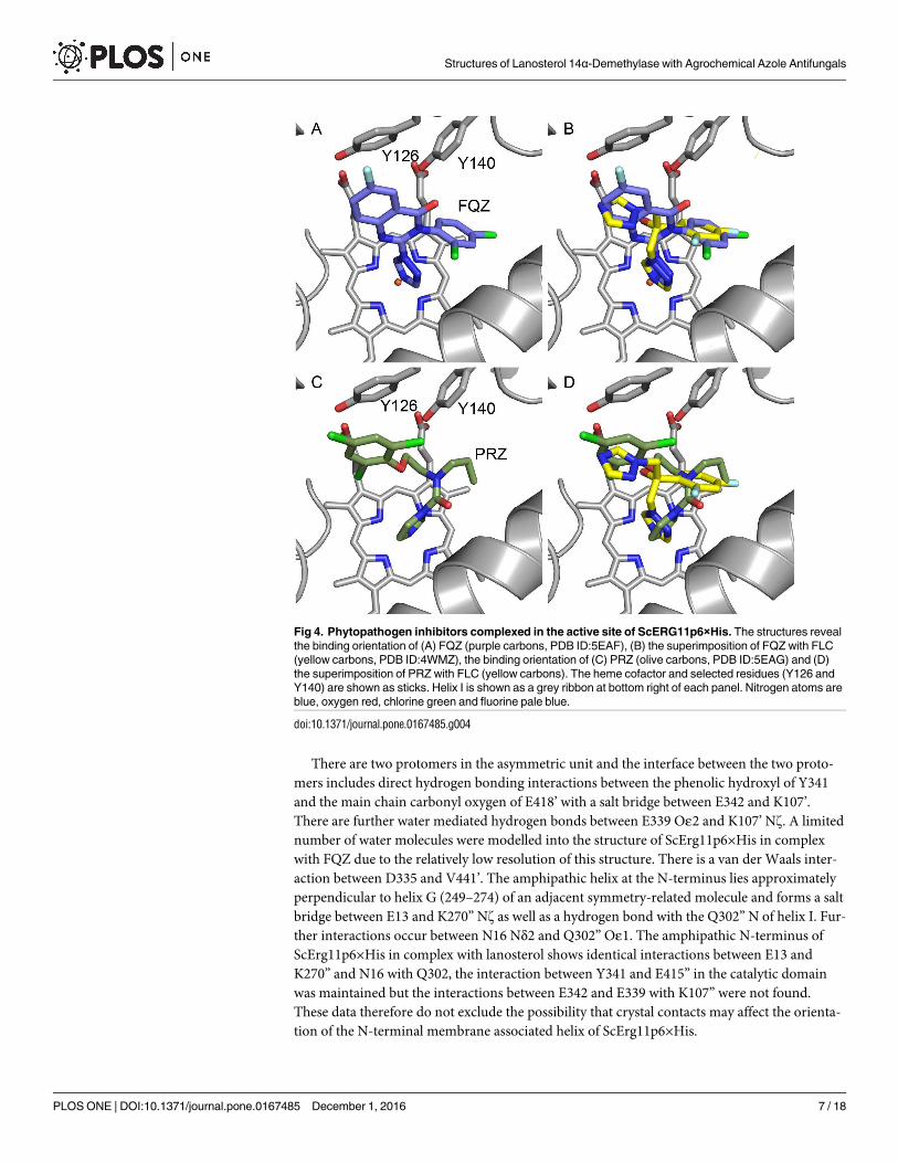
There are two protomers in the asymmetric unit and the interface between the two proto-
mers includes direct hydrogen bonding interactions between the phenolic hydroxyl of Y341
and the main chain carbonyl oxygen of E418’ with a salt bridge between E342 and K107’.
There are further water mediated hydrogen bonds between E339 Oε2 and K107’ Nz. A limited
number of water molecules were modelled into the structure of ScErg11p6×His in complex
with FQZ due to the relatively low resolution of this structure. There is a van der Waals inter-
action between D335 and V441’. The amphipathic helix at the N-terminus lies approximately
perpendicular to helix G (249–274) of an adjacent symmetry-related molecule and forms a salt
bridge between E13 and K270” Nz as well as a hydrogen bond with the Q302” N of helix I. Fur-
ther interactions occur between N16 Nδ2 and Q302” Oε1. The amphipathic N-terminus of
ScErg11p6×His in complex with lanosterol shows identical interactions between E13 and
K270” and N16 with Q302, the interaction between Y341 and E415” in the catalytic domain
was maintained but the interactions between E342 and E339 with K107” were not found.
These data therefore do not exclude the possibility that crystal contacts may affect the orienta-
tion of the N-terminal membrane associated helix of ScErg11p6×His.
Fig 4. Phytopathogen inhibitors complexed in the active site of ScERG11p6×His. The structures reveal
the binding orientation of (A) FQZ (purple carbons, PDB ID:5EAF), (B) the superimposition of FQZ with FLC
(yellow carbons, PDB ID:4WMZ), the binding orientation of (C) PRZ (olive carbons, PDB ID:5EAG) and (D)
the superimposition of PRZ with FLC (yellow carbons). The heme cofactor and selected residues (Y126 and
Y140) are shown as sticks. Helix I is shown as a grey ribbon at bottom right of each panel. Nitrogen atoms are
blue, oxygen red, chlorine green and fluorine pale blue.
doi:10.1371/journal.pone.0167485.g004
Structures of Lanosterol 14α-Demethylase with Agrochemical Azole Antifungals
PLOS ONE | DOI:10.1371/journal.pone.0167485 December 1, 2016 7 / 18
Prochloraz (PRZ)
PRZ differs chemically from other inhibitors tested as it coordinates to the heme iron via the
N3 nitrogen of an imidazole ring compared with a triazole ring. The plane of the amide group
is approximately 60˚ to that of the imidazole ring and the carbonyl oxygen is in close proximity
to G314 of helix I (Fig 4C). The n-propyl group occupies part of the deep hydrophobic pocket
lined by I139 in the loop between helices B and C, with the acyclic nitrogen in a similar posi-
tion to that of the ipso carbon of the dichlorophenyl ring of FLC (Fig 4D). This restricted con-
formation pushes the ethyl linker into the space near water 743 (PDB ID:4WMZ) with the
second carbon being 3.3 Å from the phenolic hydroxyl of Y140, thereby excluding any water
mediated hydrogen bonding. The 2-chloro substituent is in close proximity to both Cδ1 of
L380 and Cγ2 of L383, while the phenyl ring pushes Y126 away from the active site into a posi-
tion approaching a pi stacking interaction.
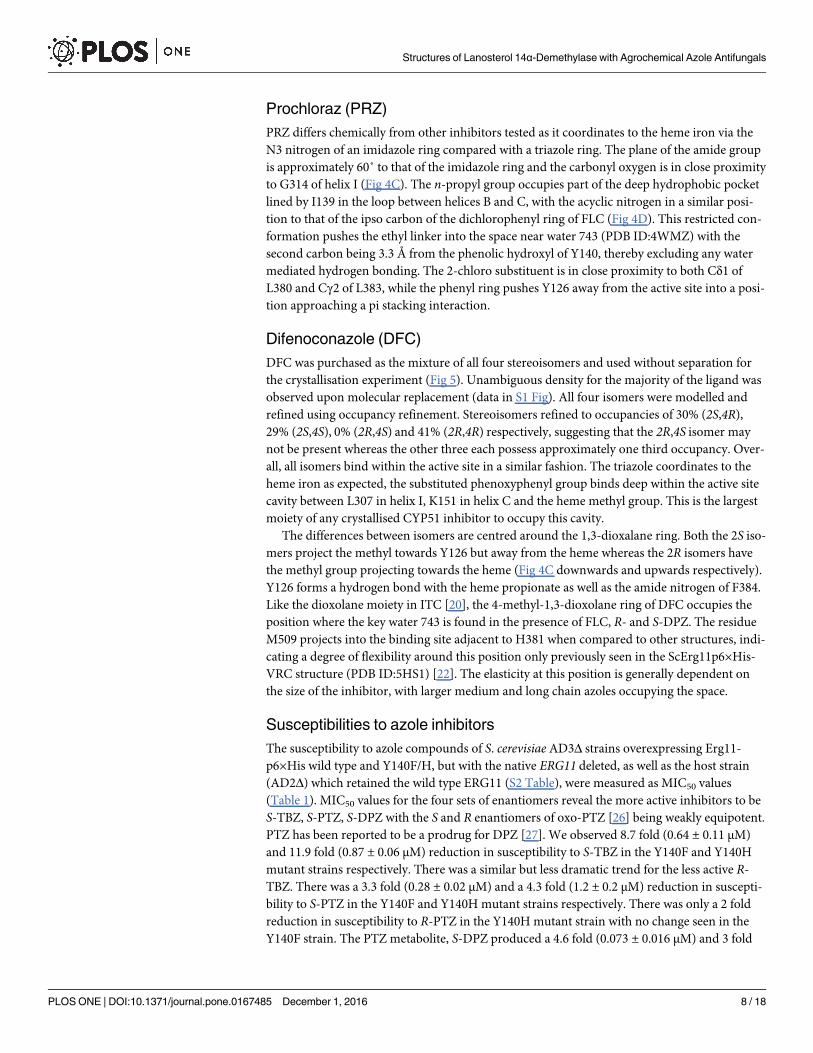
Difenoconazole (DFC)
DFC was purchased as the mixture of all four stereoisomers and used without separation for
the crystallisation experiment (Fig 5). Unambiguous density for the majority of the ligand was
observed upon molecular replacement (data in S1 Fig). All four isomers were modelled and
refined using occupancy refinement. Stereoisomers refined to occupancies of 30% (2S,4R),
29% (2S,4S), 0% (2R,4S) and 41% (2R,4R) respectively, suggesting that the 2R,4S isomer may
not be present whereas the other three each possess approximately one third occupancy. Over-
all, all isomers bind within the active site in a similar fashion. The triazole coordinates to the
heme iron as expected, the substituted phenoxyphenyl group binds deep within the active site
cavity between L307 in helix I, K151 in helix C and the heme methyl group. This is the largest
moiety of any crystallised CYP51 inhibitor to occupy this cavity.
The differences between isomers are centred around the 1,3-dioxalane ring. Both the 2S iso-
mers project the methyl towards Y126 but away from the heme whereas the 2R isomers have
the methyl group projecting towards the heme (Fig 4C downwards and upwards respectively).
Y126 forms a hydrogen bond with the heme propionate as well as the amide nitrogen of F384.
Like the dioxolane moiety in ITC [20], the 4-methyl-1,3-dioxolane ring of DFC occupies the
position where the key water 743 is found in the presence of FLC, R- and S-DPZ. The residue
M509 projects into the binding site adjacent to H381 when compared to other structures, indi-
cating a degree of flexibility around this position only previously seen in the ScErg11p6×His-
VRC structure (PDB ID:5HS1) [22]. The elasticity at this position is generally dependent on
the size of the inhibitor, with larger medium and long chain azoles occupying the space.
Susceptibilities to azole inhibitors
The susceptibility to azole compounds of S. cerevisiae AD3Δ strains overexpressing Erg11-
p6×His wild type and Y140F/H, but with the native ERG11 deleted, as well as the host strain
(AD2Δ) which retained the wild type ERG11 (S2 Table), were measured as MIC50 values
(Table 1). MIC50 values for the four sets of enantiomers reveal the more active inhibitors to be
S-TBZ, S-PTZ, S-DPZ with the S and R enantiomers of oxo-PTZ [26] being weakly equipotent.
PTZ has been reported to be a prodrug for DPZ [27]. We observed 8.7 fold (0.64 ± 0.11 μM)
and 11.9 fold (0.87 ± 0.06 μM) reduction in susceptibility to S-TBZ in the Y140F and Y140H
mutant strains respectively. There was a similar but less dramatic trend for the less active R-
TBZ. There was a 3.3 fold (0.28 ± 0.02 μM) and a 4.3 fold (1.2 ± 0.2 μM) reduction in suscepti-
bility to S-PTZ in the Y140F and Y140H mutant strains respectively. There was only a 2 fold
reduction in susceptibility to R-PTZ in the Y140H mutant strain with no change seen in the
Y140F strain. The PTZ metabolite, S-DPZ produced a 4.6 fold (0.073 ± 0.016 μM) and 3 fold
Structures of Lanosterol 14α-Demethylase with Agrochemical Azole Antifungals
PLOS ONE | DOI:10.1371/journal.pone.0167485 December 1, 2016 8 / 18
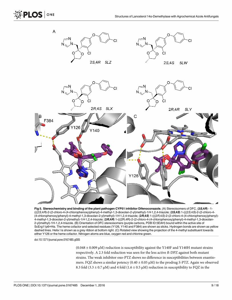
(0.048 ± 0.009 μM) reduction is susceptibility against the Y140F and Y140H mutant strains
respectively. A 2.3 fold reduction was seen for the less active R-DPZ against both mutant
strains. The weak inhibitor oxo-PTZ shows no difference in susceptibilities between enantio-
mers. FQZ shows a similar potency (0.40 ± 0.05 μM) to the prodrug S-PTZ. Again we observed
8.3 fold (3.3 ± 0.7 μM) and 4 fold (1.6 ± 0.5 μM) reduction in susceptibility to FQZ in the
Fig 5. Stereochemistry and binding of the plant pathogen CYP51 inhibitor Difenoconazole. (A) Stereoisomers of DFC, (2S,4R) - 1-
(((2S,4R)-2-(2-chloro-4-(4-chlorophenoxy)phenyl)-4-methyl-1,3-dioxolan-2-yl)methyl)-1H-1,2,4-triazole; (2S,4S) 1-(((2S,4S)-2-(2-chloro-4-
(4-chlorophenoxy)phenyl)-4-methyl-1,3-dioxolan-2-yl)methyl)-1H-1,2,4-triazole; (2R,4S) 1-(((2R,4S)-2-(2-chloro-4-(4-chlorophenoxy)phenyl)-
4-methyl-1,3-dioxolan-2-yl)methyl)-1H-1,2,4-triazole; (2R,4R) 1-(((2R,4R)-2-(2-chloro-4-(4-chlorophenoxy)phenyl)-4-methyl-1,3-dioxolan-
2-yl)methyl)-1H-1,2,4-triazole. (B) Orientation of DFC stereoisomers (purple carbons, PDB ID:5EAH) bound within the active site of
ScErg11p6×His. The heme cofactor and selected residues (Y126, Y140 and F384) are shown as sticks. Hydrogen bonds are shown as yellow
dashed lines. Helix I is shown as a grey ribbon at bottom right. (C) Rotated view showing the projection of the 4-methyl substituent towards
either Y126 or the heme cofactor. Nitrogen atoms are blue, oxygen red and chlorine green.
doi:10.1371/journal.pone.0167485.g005
Structures of Lanosterol 14α-Demethylase with Agrochemical Azole Antifungals
PLOS ONE | DOI:10.1371/journal.pone.0167485 December 1, 2016 9 / 18
Y140F and Y140H mutant strains respectively. PRZ is less active (1.9 ± 0.5 μM) with reduced
susceptibility (2.3 fold and 3.5 fold) to Y140F and Y140H mutant strains respectively. DFC is
the second most potent compound in our assay (behind S-DPZ) but also shows 5.2 fold
(0.243 ± 0.003 μM) and 5 fold (0.237 ± 0.003 μM) reduced susceptibility against the Y140F and
Y140H mutant strains respectively. All compounds showed significantly lower MIC50 values
when tested with the AD2Δ strain expressing the native Erg11p, which led to higher fold differ-
ences in susceptibility and showed that the overexpressed enzyme was functional.
Type II binding to affinity purified ScErg11p6×His
The absolute absorbance spectra for Ni-NTA affinity and SEC purified ScErg11p6×His showed
a Soret peak at 417 nm [21]. Type II azole binding is characterised by a shift of the Soret peak
from 417 nm to 421–424 nm in ScErg11p6×His and other cytochrome P450s. The type II dif-
ference spectra were used to detect and quantitate binding of the inhibitors S-TBZ, R-TBZ, S-
PTZ, R-PTZ, S-DPZ, R-DPZ, S-oxo-PTZ and R-oxo-PTZ, FQZ, and PRZ using Ni-NTA affin-
ity purified wild type ScErg11p6×His preparations at 1 μM. Spectral shifts, [Azole]0.5 values,
Hill coefficients as well as Type II difference spectra binding curves are shown in S3 Table
(selected spectra shown in S2 Fig) and Fig 6, respectively. Equivalent spectral shifts from 417
nm to ~421 nm were obtained for enantiomers of TBZ and DPZ as well as for FQZ and PRZ.
The absence of a spectral shift for enantiomers of PTZ or oxo-PTZ indicated there is no azole
coordination at the heme iron. This was confirmed in the type II binding experiments. All
other compounds showed type II binding with [Azole]0.5 values at ~ 0.4 μM in the presence of
1 μM functional ScErg11p6×His and Kd values in the low nanomolar range indicating tight
binding. These results are comparable to a previous study which determined Kd values of DPZ
and PTZ against Candida albicans CYP51 which showed tight type II binding (Kd 36.4 ± 10.7
nM) for the former, with the latter producing only a weak type I difference spectra (Kd 6.3 ±1.5 μM) [27]. TBZ, DPZ, FQZ and PRZ were all shown to be non-competitive inhibitors of
Table 1. MIC50 data for compounds against Saccharomyces cerevisiae strains.
Compound MIC50 (μM)
AD2Δ AD3Δ Y140F Y140H
S-Tebuconazole 0.0197 ± 0.0003 0.073 ± 0.015 0.64 ± 0.11 0.87 ± 0.06
R-Tebuconazole 0.12 ± 0.0 1.1 ± 0.3 2.0 ± 0.2 5.2 ± 1.3
S-Prothioconazole 0.077 ± 0.003 0.28 ± 0.02 0.93 ± 0.0 1.2 ± 0.2
R-Prothioconazole 5.2 ± 1.3 16 ± 3 16 ± 4 31 ± 4
S-Prothioconazole-desthio 0.006 ± 0.001 0.0157 ± 0.0003 0.073 ± 0.016 0.048 ± 0.009
R-Prothioconazole-desthio 0.47 ± 0.05 3.2 ± 0.2 7.5 ± 0.0 7.5 ± 0.0
S-Oxo-prothioconazole 75.0 ± 0.0 84 ± 3 87 ± 7 97 ± 3
R-Oxo-prothioconazole 75.0 ± 3.0 80 ± 2 87 ± 7 95 ± 5
Fluquinconazole 0.17 ± 0.02 0.40 ± 0.05 3.3 ± 0.7 1.6 ± 0.5
Prochloraz 0.25 ± 0.02 1.9 ± 0.5 4.3 ± 0.8 6.7 ± 0.7
Difenoconazole 0.0147 ± 0.0003 0.047 ± 0.009 0.243 ± 0.003 0.237 ± 0.003
Values are the mean for 3 separate clones of each strain using data obtained in triplicate measurements from at least 3 different experiments (Standard
error is shown).
AD2Δ host with native ERG11 retained.
AD3Δ - ScErg11p6×His overexpressed from PDR5 locus, native ERG11 deleted.
Y140F - ScErg11p6×His Y140F overexpressed from PDR5 locus, native ERG11 deleted.
Y140H –ScErg11p6×His Y140H overexpressed from PDR5 locus, native ERG11 deleted.
doi:10.1371/journal.pone.0167485.t001
Structures of Lanosterol 14α-Demethylase with Agrochemical Azole Antifungals
PLOS ONE | DOI:10.1371/journal.pone.0167485 December 1, 2016 10 / 18
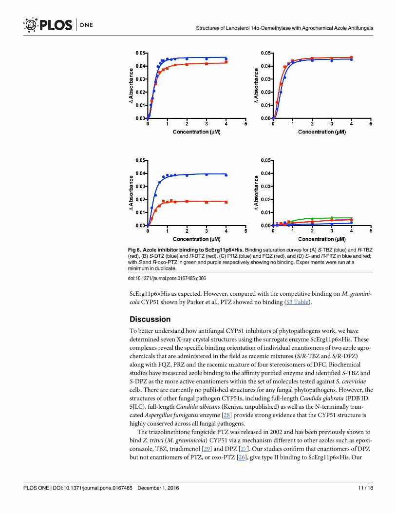
ScErg11p6×His as expected. However, compared with the competitive binding on M. gramini-cola CYP51 shown by Parker et al., PTZ showed no binding (S3 Table).
Discussion
To better understand how antifungal CYP51 inhibitors of phytopathogens work, we have
determined seven X-ray crystal structures using the surrogate enzyme ScErg11p6×His. These
complexes reveal the specific binding orientation of individual enantiomers of two azole agro-
chemicals that are administered in the field as racemic mixtures (S/R-TBZ and S/R-DPZ)
along with FQZ, PRZ and the racemic mixture of four stereoisomers of DFC. Biochemical
studies have measured azole binding to the affinity purified enzyme and identified S-TBZ and
S-DPZ as the more active enantiomers within the set of molecules tested against S. cerevisiaecells. There are currently no published structures for any fungal phytopathogens. However, the
structures of other fungal pathogen CYP51s, including full-length Candida glabrata (PDB ID:
5JLC), full-length Candida albicans (Keniya, unpublished) as well as the N-terminally trun-
cated Aspergillus fumigatus enzyme [28] provide strong evidence that the CYP51 structure is
highly conserved across all fungal pathogens.
The triazolinethione fungicide PTZ was released in 2002 and has been previously shown to
bind Z. tritici (M. graminicola) CYP51 via a mechanism different to other azoles such as epoxi-
conazole, TBZ, triadimenol [29] and DPZ [27]. Our studies confirm that enantiomers of DPZ
but not enantiomers of PTZ, or oxo-PTZ [26], give type II binding to ScErg11p6×His. Our
Fig 6. Azole inhibitor binding to ScErg11p6×His. Binding saturation curves for (A) S-TBZ (blue) and R-TBZ
(red), (B) S-DTZ (blue) and R-DTZ (red), (C) PRZ (blue) and FQZ (red), and (D) S- and R-PTZ in blue and red;
with S and R-oxo-PTZ in green and purple respectively showing no binding. Experiments were run at a
minimum in duplicate.
doi:10.1371/journal.pone.0167485.g006
Structures of Lanosterol 14α-Demethylase with Agrochemical Azole Antifungals
PLOS ONE | DOI:10.1371/journal.pone.0167485 December 1, 2016 11 / 18
whole cell MIC50 data for both S-PTZ and R-PTZ reveal that both compounds are active
against yeast. The S-enantiomers of both PTZ and DPZ were found to be the more active enan-
tiomers [57 fold and 200 fold more active over the R enantiomers respectively (MIC50 values
for ADΔ3 strains)]. Differences in activity between S-PTZ and S-DPZ could be attributed to
incomplete conversion of S-DPZ. S-TBZ was found to be the more active enantiomer with
15-fold lower MIC50 than R-TBZ. Both enantiomers of TBZ and DTZ bind in the active site
via azole coordination to the heme iron, with the tertiary hydroxyl on the central quaternary
carbon oriented in the direction towards Y140. Like FLC, water 743 was present in both DPZ
structures but was not detected in the TBZ structures, possibly due to the lower resolution of
the crystal structure obtained. S-TBZ binds with its phenylethyl substituent anchored deep in
the hydrophobic pocket. The R enantiomer presents the flexible phenylethyl substituent into
the more open cavity and is subsequently disordered (data in S1 Fig). Conversely the shorter
and more rigid chlorobenzyl substituent of S-DPZ appears to be an optimal fit for this cavity,
with a close contact between the 2-chloro substituent and F236. The same 2-chloro substituent
on the R enantiomer is also close to F236 but in a different orientation which pushes the chlor-
ocyclopropyl group close to Y126. Overall this makes S-DPZ the more optimal fit.
The triazole fungicide DFC was first introduced in 1989 as a mixture of four stereoisomers.
It was found to be the second most potent compound behind S-DPZ tested in this study
against yeast. The crystal structure reveals all four stereoisomers bind in the same orientation,
with three stereoisomers showing equivalent occupancy. The potency of DFC when compared
with S-TBZ and S-DPZ can be attributed to deep binding of the phenoxyphenyl group in com-
bination with the 1,3-dioxalane ring. A study of the stereoselective bioactivity of DFC against 4
different fungal pathogens (Alternaria sonali, Fulvia fulva, Botrytis cinerea and Rhizoctoniasolani) identified the 2S,4R stereoisomer (5LZ; our nomenclature) as the most active com-
pound [30]. The methyl group of this isomer projects towards Y126 (away from the heme) and
would therefore be the most optimal fit of the four compounds.
Numerous amino acid mutations and deletions have been associated with reduced azole
susceptibility in CYP51 in the phytopathogen Z. tritici (M. graminicola) [31] as well as the
human pathogens Candida albicans and Aspergillus fumigatus [5, 19, 32]. Mutation of tyrosine
140 (Y140, S. cerevisiae numbering) is commonly found in many fungal species. For example,
Y137F in Z. tritici confers resistance to triadimenol, Y132F/H confer resistance to FLC and
VCZ in C. albicans, Y132H in combination with I471T confers enhanced azole resistance to
FLC and VCZ in the C. albicans Darlington strain [33], and TR46/Y121F/T289A in A. fumiga-tus CYP51A, which is thought to have arisen due to agricultural use of azoles [5, 6], confers
resistance to VCZ. Other mutations have been associated with reduced susceptibility to azole
agrochemicals. For example, Z. tritici I381V confers resistance to TBZ [34] and Z. triticiV136A confers resistance to PRZ [35]. The structures presented here help to clarify the posi-
tions of such mutations relative to the bound ligands and justify susceptibility shifts seen
though specific mutations. Residues V136, Y137 and I381 in Z. tritici Cyp51 (I139, Y140 and
L380 in ScErg11p6×His) are all within the active site and project into the ligand binding cavity.
Mutations associated with reduced sensitivity such as V136A (TBZ) and I381V (PRZ) induce
an increase in the size of the binding pocket and lead to a less optimal fit for the ligand. Previ-
ous work has identified a key water mediated hydrogen bonding network between ScErg11-
p6×His Y140 and hydroxyl-containing azole inhibitors such as FLC and other inhibitors with
structural similarities such as triadimenol and TBZ [22]. When mutations occur (either to phe-
nylalanine or histidine) this hydrogen bonding network is disrupted and binding affinity
decreases leading to reduced susceptibility. The cell based data presented here also shows
reduced susceptibility to azole compounds in Y140F and Y140H mutant strains particularly
when a hydroxyl group is present. As none of the compounds tested in the present study have
Structures of Lanosterol 14α-Demethylase with Agrochemical Azole Antifungals
PLOS ONE | DOI:10.1371/journal.pone.0167485 December 1, 2016 12 / 18
a tail comparable to ITC or PCZ that penetrates deep into the substrate entry channel, the
effects on affinity imposed by mutations at Y140F/H are not compensated for. The deletions
ΔY459/G460 in Z. tritici CYP51 (corresponding to Y447/G448 in ScErg11p6×His), in conjunc-
tion with other mutations, confers resistance to TBZ and epoxiconazole [35, 36]. The homolo-
gous residues in ScErg11p6×His are situated at a turn at the end of the fungal specific loop
region which is positioned on the opposite side of the heme cofactor well away from the ligand
binding site. How this deletion impacts resistance from its distant position remains unclear.
Evidence has emerged [12, 13] around the development of cross-resistance in Candida glab-rata [37] and Aspergillus fumigatus [38] to medically used triazoles as a result of agricultural
compounds. This signals an urgent need to develop new fungicides to overcome resistance.
The results presented allow for a clear understanding of the how the agrochemical fungicides
act at their molecular target and reveal how mutations can affect drug binding. Until we able
to achieve molecular resolution of CYP51s from plant pathogens, the yeast model provides a
highly informative platform to investigate ligand binding, the impact of these mutations and a
template for molecular models that will aid the discovery of next-generation antifungals for
medical and agrochemical intervention.
Materials and Methods
The plant pathogen inhibitors R-prothioconazole, S-prothioconazole, R-prothioconazole-
desthio, S-prothioconazole-desthio, R-oxo-prothioconazole, S-oxo-prothioconazole, R-tebu-
conazole, S-tebuconazole, fluquinconazole and prochloraz were supplied by Bayer AG. Dife-
noconazole was purchased from Sigma-Aldrich as a mixture of four stereoisomers.
Yeast strains overexpressing wild type and mutant ScErg11p6×His
The yeast strains used in this study have been reported previously [20–22, 25] and are
described in S2 table.
Phytopathogen CYP51 inhibitor susceptibility of strains overexpressing
ScErg11p
The susceptibilities of strains overexpressing wild type ScErg11p6×His and ScErg11p6×His
Y140F/H to CYP51 inhibitors were measured as MIC50 values using broth microdilution
assays as described previously.[21] The MIC50s were defined as the concentration of drug
required for 50% growth inhibition compared to no drug controls.
Protein expression and purification
ScErg11p6×His expression and purification was carried out according to the methods
described previously [20, 21]. In brief, crude membranes were prepared from liquid cultures
grown overnight in YPD medium (1% (wt/vol) yeast extract (BD Difco™), 2% (wt/vol) peptone
(BD Difco™), 2% (wt/vol) dextrose) at 30˚C with shaking at 200 rpm. Cells were harvested and
broken using bead beating and crude membranes were obtained by differential centrifugation.
These crude membranes (5 mg/ml) were solubilized with 10× critical micelle concentration
(CMC) n-decyl-β-D-maltoside (DM, Affymetrix Inc., Santa Clara, US). ScErg11p6×His was
purified from the solubilized crude membrane fraction by affinity chromatography using 2 ml
of packed Ni-NTA-agarose matrix (Qiagen) per 1 g of protein. Affinity purification buffer con-
taining 10% (wt/vol) glycerol, 250 mM NaCl, 20 mM Tris pH 7.5, 0.5 mM PMSF, 16 mM
(10×CMC) DM, 20 mM imidazole and 1 EDTA-free protease inhibitor pill per 200 mL. The
affinity purified ScErg11p6×His was concentrated by centrifugal filtration using a 50 kDa
Structures of Lanosterol 14α-Demethylase with Agrochemical Azole Antifungals
PLOS ONE | DOI:10.1371/journal.pone.0167485 December 1, 2016 13 / 18
molecular-weight cut-off Amicon Ultra-4 centrifugal filter (Millipore) and further purified by
size exclusion chromatography (SEC) using a Superdex™ 200 10/300 GL column (GE Health-
care Life Sciences, UK). The column was equilibrated with SEC buffer containing 10% (wt/
vol) glycerol, 250 mM NaCl, 20 mM Tris, pH 7.5, 0.5 mM phenylmethanesulfonyl fluoride, 6.4
mM DM (4×CMC) and 1 Roche EDTA-free protease inhibitor pill per 200 mL. CYP51 inhibi-
tors dissolved in dimethyl sulfoxide (DMSO) were added to the pooled fractions obtained by
size-exclusion chromatography with final concentrations of 40 μM to 1% of the final volume.
The samples were then concentrated using a 50 kDa molecular-weight cut-off Amicon Ultra-4
centrifugal filter prior to crystallisation.
Crystallization and X-ray data collection
Ni-NTA-agarose affinity and SEC purified ScErg11p6×His was concentrated and co-crystal-
lized with ligands using the hanging-drop vapour-diffusion method (30). Crystals formed
within one week at 18˚C. Reservoir solutions contained 45% polyethylene glycol-400 in 100
mM glycine at a pH range of 9.3–9.5. The drops were 4 μl in a 1:1 ratio of reservoir solution
and ~20 mg/ml of the protein in SEC buffer. Crystals were flash-cooled in liquid nitrogen
prior to storage and data collection. Single datasets were collected on the MX2 beamline at the
Australian Synchrotron using an ADSC Quantum ADSC Quantum 315 detector. Data were
indexed and integrated using iMosflm [39] and scaled with SCALA [40]. Molecular replace-
ment was carried out using Phaser-MR [41] from Phenix [42] using ScErg11p6×His co-crystal-
lized with lanosterol (PDB ID: 4LXJ) as template [20]. Refinement and modelling was
performed using phenix.refine [42] and Coot [43] respectively. Geometric restraints for inhibi-
tors were generated via the Grade Web Server (http://grade.globalphasing.org/cgi-bin/grade/
server.cgi) from mol2 files created within SYBYL-X2.1.1. The inhibitors were modelled into
the appropriate density in the active site and waters were added if at least one hydrogen bond
was detected (2.5–3.3 Å).
Type II binding CYP51 inhibitors to ScErg11p6×His
Samples of Ni-NTA-agarose affinity purified ScErg11p6×His were eluted with 40 mM histi-
dine instead of imidazole as described by Warrilow et al. [44]. Samples were washed free of the
histidine and concentrated by centrifugal filtration using a 50 kDa molecular-weight cut-off
Amicon Ultra-4 centrifugal filter (Millipore) to give protein with a Soret peak at 417 nm. Car-
bon monoxide binding was used to determine the concentration of functional cytochrome
P450 for drug binding studies as described previously [21] based on the protocol described by
Guengerich et al. [45] Saturation curves for the binding of type II ligands were determined by
obtaining difference spectra for functional enzyme at 1 μM as described previously [21]. The
dissociation constant Kd for type II binding of CYP51 inhibitors was calculated by applying
the Hill equation using the formula ΔA = ΔAmax [Azole]n / ([Azole]n + Kdn), with ΔAmax being
the maximum change in absorbance, [Azole] the azole concentration and n the Hill coefficient.
All calculations were carried out using GraphPad Prism 6 Software (GraphPad Prism, San
Diego, CA). [Azole]0.5 values were defined as the concentration of the azole drug that gave half
ΔAmax.
Supporting Information
S1 Fig. OMIT maps following initial refinement for (A) S-TBZ (green carbons), (B) R-TBC
(cyan carbons), (C) S-DPZ (magenta carbons), (D) R-DPZ (salmon carbons), (E) FQZ
(blue carbons), (F) PRZ (olive carbons), and (G) all four stereoisomers of DFC (purple car-
bons). Ligands are the final refined conformation. Fo-Fc map [green mesh] contoured at 3σ;
Structures of Lanosterol 14α-Demethylase with Agrochemical Azole Antifungals
PLOS ONE | DOI:10.1371/journal.pone.0167485 December 1, 2016 14 / 18
2Fo-Fc map [blue mesh] contoured at 1σ. Maps were calculated using Fcalc refined from coor-
dinates with no ligand at the active site. N atoms are coloured blue, Oxygen red, Chlorine
green and Fluorine pale blue. The heme cofactor is shown as sticks with the iron atom (where
visible) an orange sphere.
(PNG)
S2 Fig. Difference spectra illustrating type II binding of (A) S-tebuconazole, (B) R-tebuco-
nazole (C) S-prothioconazole-desthio (D) R-prothioconazole-desthio (E) Prochloraz (F)
Fluquinconazole and the spectra for (G) S-oxo-prothioconazole and (H) R-oxo-prothioco-
nazole indicating neither type I nor type II binding. The curves shown were obtained by
incremental additions of the azole up to 2 μM, in the presence of 1 μM ScErg11p6×His. Repre-
sentative examples of at least two experiments are shown.
(PNG)
S1 Table. Data collection and refinement statistics.
(DOCX)
S2 Table. Yeast strains used in this study.
(DOCX)
S3 Table. Binding of azoles to affinity-purified ScErg11p6×His.
(DOCX)
Acknowledgments
Paul Adams and Nigel W. Moriarty, Lawrence Berkeley National Laboratory, for assistance
with Phenix.
Author Contributions
Conceptualization: JT BM KT.
Formal analysis: JT MS BM JN KT.
Funding acquisition: BM.
Investigation: MS AS MK RW MW JT JN.
Project administration: BM.
Resources: JT.
Supervision: JT MK BM.
Validation: MS AS MK MW.
Visualization: JT MS.
Writing – original draft: JT BM.
Writing – review & editing: JT BM KT MS.
References1. Russell PE. A century of fungicide evolution. J Agri Sci 143(1):11–25.
2. Bowyer P, Denning DW. Environmental fungicides and triazole resistance in Aspergillus. Pest Manag
Sci. 2014; 70(2):173–8. doi: 10.1002/ps.3567 PMID: 23616354
Structures of Lanosterol 14α-Demethylase with Agrochemical Azole Antifungals
PLOS ONE | DOI:10.1371/journal.pone.0167485 December 1, 2016 15 / 18
3. Bhanderi BB, Yadav MM, Roy A. Antifungal Drug Resistance—Concerns for Veterinarians. Vet World.
2009; 2(5):204–7.
4. Kuck K-H, Stenzel K, Vors J-P. Sterol Biosynthesis Inhibitors. In: Kraemer W, editor. Modern Crop Pro-
tection Compounds 1–3. 2nd ed. Weinheim, Germany: Wiley-VCH Verlag GmbH & Co. KGaA; 2012.
5. Parker JE, Warrilow AG, Price CL, Mullins JG, Kelly DE, Kelly SL. Resistance to antifungals that target
CYP51. J Chem Biol. 2014; 7(4):143–61. doi: 10.1007/s12154-014-0121-1 PMID: 25320648
6. Price CL, Parker JE, Warrilow AG, Kelly DE, Kelly SL. Azole fungicides—understanding resistance
mechanisms in agricultural fungal pathogens. Pest Manag Sci. 2015; 71(8):1054–8. doi: 10.1002/ps.
4029 PMID: 25914201
7. Tietjen K. Contribution of plant responses to efficacy of fungicides—A perspective. In: Deising HB, B. F,
A. M, E.C. O, H. S, G. S, editors. Modern Fungicides and Antifungal Compounds. VIII. (Submitted)
2016.
8. Snelders E, Karawajczyk A, Verhoeven RJ, Venselaar H, Schaftenaar G, Verweij PE, et al. The struc-
ture-function relationship of the Aspergillus fumigatus cyp51A L98H conversion by site-directed muta-
genesis: the mechanism of L98H azole resistance. Fungal Genet Biol. 2011; 48(11):1062–70. doi: 10.
1016/j.fgb.2011.08.002 PMID: 21907818
9. Chowdhary A, Kathuria S, Xu J, Meis JF. Emergence of azole-resistant Aspergillus fumigatus strains
due to agricultural azole use creates an increasing threat to human health. PLoS pathogens. 2013; 9
(10):e1003633. doi: 10.1371/journal.ppat.1003633 PMID: 24204249
10. van der Linden JW, Camps SM, Kampinga GA, Arends JP, Debets-Ossenkopp YJ, Haas PJ, et al.
Aspergillosis due to voriconazole highly resistant Aspergillus fumigatus and recovery of genetically
related resistant isolates from domiciles. Clin Infect Dis. 2013; 57(4):513–20. doi: 10.1093/cid/cit320
PMID: 23667263
11. Camps SM, Rijs AJ, Klaassen CH, Meis JF, O’Gorman CM, Dyer PS, et al. Molecular epidemiology of
Aspergillus fumigatus isolates harboring the TR34/L98H azole resistance mechanism. J Clin Microbiol.
2012; 50(8):2674–80. doi: 10.1128/JCM.00335-12 PMID: 22675126
12. Verweij PE, Chowdhary A, Melchers WJ, Meis JF. Azole resistance in Aspergillus fumigatus: can we
retain the clinical use of mold-active antifungal azoles? Clin Infect Dis. 2016; 62(3):362–8. doi: 10.1093/
cid/civ885 PMID: 26486705
13. Franceschini S, Chitarra W, Pugliese M, Gisi U, Garibaldi A, Gullino ML. Quantification of Aspergillus
fumigatus and enteric bacteria in European compost and biochar. Compost Sci Util. 2016; 24(1):20–9.
14. Kjaerstad MB, Taxvig C, Nellemann C, Vinggaard AM, Andersen HR. Endocrine disrupting effects in
vitro of conazole antifungals used as pesticides and pharmaceuticals. Reprod Toxicol. 2010; 30
(4):573–82. doi: 10.1016/j.reprotox.2010.07.009 PMID: 20708073
15. Trosken E-R. Toxicological Evaluation of Azole Fungicides in Agriculture and Food Chemistry: Univer-
sity Wurzburg, Germany; 2005.
16. Chen ZF, Ying GG. Occurrence, fate and ecological risk of five typical azole fungicides as therapeutic
and personal care products in the environment: A review. Environ Int. 2015; 84:142–53. doi: 10.1016/j.
envint.2015.07.022 PMID: 26277639
17. Reif DM, Martin MT, Tan SW, Houck KA, Judson RS, Richard AM, et al. Endocrine profiling and prioriti-
zation of environmental chemicals using ToxCast data. Environ Health Perspect. 2010; 118(12):1714–
20. doi: 10.1289/ehp.1002180 PMID: 20826373
18. Becher R, Wirsel SG. Fungal cytochrome P450 sterol 14alpha-demethylase (CYP51) and azole resis-
tance in plant and human pathogens. Appl Microbiol Biotechnol. 2012; 95(4):825–40. doi: 10.1007/
s00253-012-4195-9 PMID: 22684327
19. Mullins JG, Parker JE, Cools HJ, Togawa RC, Lucas JA, Fraaije BA, et al. Molecular modelling of the
emergence of azole resistance in Mycosphaerella graminicola. PLoS One. 2011; 6(6):e20973. doi: 10.
1371/journal.pone.0020973 PMID: 21738598
20. Monk BC, Tomasiak TM, Keniya MV, Huschmann FU, Tyndall JD, O’Connell JD 3rd, et al. Architecture
of a single membrane spanning cytochrome P450 suggests constraints that orient the catalytic domain
relative to a bilayer. Proc Natl Acad Sci U S A. 2014; 111(10):3865–70. doi: 10.1073/pnas.1324245111
PMID: 24613931
21. Sagatova AA, Keniya MV, Wilson RK, Monk BC, Tyndall JD. Structural Insights into Binding of the Anti-
fungal Drug Fluconazole to Saccharomyces cerevisiae Lanosterol 14alpha-Demethylase. Antimicrob
Agents Chemother. 2015; 59(8):4982–9. doi: 10.1128/AAC.00925-15 PMID: 26055382
22. Sagatova AA, Keniya MV, Wilson RK, Sabherwal M, Tyndall JD, Monk BC. Triazole resistance medi-
ated by mutations of a conserved active site tyrosine in fungal lanosterol 14alpha-demethylase. Scien-
tific reports. 2016; 6:26213. doi: 10.1038/srep26213 PMID: 27188873
Structures of Lanosterol 14α-Demethylase with Agrochemical Azole Antifungals
PLOS ONE | DOI:10.1371/journal.pone.0167485 December 1, 2016 16 / 18
23. Mo C, Bard M. A systematic study of yeast sterol biosynthetic protein-protein interactions using the
split-ubiquitin system. Biochim Biophys Acta. 2005; 1737(2–3):152–60. doi: 10.1016/j.bbalip.2005.11.
002 PMID: 16300994
24. Price CL, Warrilow AG, Parker JE, Mullins JG, Nes WD, Kelly DE, et al. Novel Substrate Specificity and
Temperature-Sensitive Activity of Mycosphaerella graminicola CYP51 Supported by the Native NADPH
Cytochrome P450 Reductase. Applied and environmental microbiology. 2015; 81(10):3379–86. doi: 10.
1128/AEM.03965-14 PMID: 25746994
25. Lamping E, Monk BC, Niimi K, Holmes AR, Tsao S, Tanabe K, et al. Characterization of three classes
of membrane proteins involved in fungal azole resistance by functional hyperexpression in Saccharo-
myces cerevisiae. Eukaryot Cell. 2007; 6(7):1150–65. doi: 10.1128/EC.00091-07 PMID: 17513564
26. Haas M, Justus K. Metabolism of Prothioconazole (JAU6476) in animals and plants. Pflanzenschutz-
Nachr Bayer. 2004; 57(2):207–24.
27. Parker JE, Warrilow AG, Cools HJ, Fraaije BA, Lucas JA, Rigdova K, et al. Prothioconazole and prothio-
conazole-desthio activities against Candida albicans sterol 14-alpha-demethylase. Applied and environ-
mental microbiology. 2013; 79(5):1639–45. doi: 10.1128/AEM.03246-12 PMID: 23275516
28. Hargrove TY, Wawrzak Z, Lamb DC, Guengerich FP, Lepesheva GI. Structure-Functional Characteri-
zation of Cytochrome P450 Sterol 14alpha-Demethylase (CYP51B) from Aspergillus fumigatus and
Molecular Basis for the Development of Antifungal Drugs. J Biol Chem. 2015; 290(39):23916–34. doi:
10.1074/jbc.M115.677310 PMID: 26269599
29. Parker JE, Warrilow AG, Cools HJ, Martel CM, Nes WD, Fraaije BA, et al. Mechanism of binding of
prothioconazole to Mycosphaerella graminicola CYP51 differs from that of other azole antifungals.
Applied and environmental microbiology. 2011; 77(4):1460–5. doi: 10.1128/AEM.01332-10 PMID:
21169436
30. Dong F, Li J, Chankvetadze B, Cheng Y, Xu J, Liu X, et al. Chiral triazole fungicide difenoconazole:
absolute stereochemistry, stereoselective bioactivity, aquatic toxicity, and environmental behavior in
vegetables and soil. Environ Sci Technol. 2013; 47(7):3386–94. doi: 10.1021/es304982m PMID:
23451708
31. Cools HJ, Fraaije BA. Update on mechanisms of azole resistance in Mycosphaerella graminicola and
implications for future control. Pest Manag Sci. 2013; 69(2):150–5. doi: 10.1002/ps.3348 PMID:
22730104
32. Sanglard D, Ischer F, Koymans L, Bille J. Amino acid substitutions in the cytochrome P-450 lanosterol
14alpha-demethylase (CYP51A1) from azole-resistant Candida albicans clinical isolates contribute to
resistance to azole antifungal agents. Antimicrob Agents Chemother. 1998; 42(2):241–53. PMID:
9527767
33. Kakeya H, Miyazaki Y, Miyazaki H, Nyswaner K, Grimberg B, Bennett JE. Genetic analysis of azole
resistance in the Darlington strain of Candida albicans. Antimicrob Agents Chemother. 2000; 44
(11):2985–90. PMID: 11036010
34. Fraaije BA, Cools HJ, Kim SH, Motteram J, Clark WS, Lucas JA. A novel substitution I381V in the sterol
14alpha-demethylase (CYP51) of Mycosphaerella graminicola is differentially selected by azole fungi-
cides. Mol Plant Pathol. 2007; 8(3):245–54. doi: 10.1111/j.1364-3703.2007.00388.x PMID: 20507496
35. Leroux P, Albertini C, Gautier A, Gredt M, Walker AS. Mutations in the CYP51 gene correlated with
changes in sensitivity to sterol 14 alpha-demethylation inhibitors in field isolates of Mycosphaerella gra-
minicola. Pest Manag Sci. 2007; 63(7):688–98. doi: 10.1002/ps.1390 PMID: 17511023
36. Stammler G, Carstensen M, Koch AE, Semar M, Strobel D, Schlehuber. Frequency of different CYP51-
haplotypes of Mycosphaerella graminicola and their impact on epoxiconazole-sensitivity and -field effi-
cacy. Crop Protection. 2008; 27(11):1448–56.
37. Faria-Ramos I, Tavares PR, Farinha S, Neves-Maia J, Miranda IM, Silva RM, et al. Environmental
azole fungicide, prochloraz, can induce cross-resistance to medical triazoles in Candida glabrata.
FEMS yeast research. 2014; 14(7):1119–23. doi: 10.1111/1567-1364.12193 PMID: 25132632
38. Snelders E, Camps SM, Karawajczyk A, Schaftenaar G, Kema GH, van der Lee HA, et al. Triazole fun-
gicides can induce cross-resistance to medical triazoles in Aspergillus fumigatus. PLoS One. 2012; 7
(3):e31801. doi: 10.1371/journal.pone.0031801 PMID: 22396740
39. Battye TG, Kontogiannis L, Johnson O, Powell HR, Leslie AG. iMOSFLM: a new graphical interface for
diffraction-image processing with MOSFLM. Acta Crystallogr D Biol Crystallogr. 2011; 67(Pt 4):271–81.
doi: 10.1107/S0907444910048675 PMID: 21460445
40. Evans P. Scaling and assessment of data quality. Acta Crystallogr D Biol Crystallogr. 2006; 62(Pt
1):72–82. Epub 2005/12/22. doi: 10.1107/S0907444905036693 PMID: 16369096
41. McCoy AJ, Grosse-Kunstleve RW, Adams PD, Winn MD, Storoni LC, Read RJ. Phaser crystallographic
software. Journal of applied crystallography. 2007; 40(Pt 4):658–74. Epub 2007/08/01. doi: 10.1107/
S0021889807021206 PMID: 19461840
Structures of Lanosterol 14α-Demethylase with Agrochemical Azole Antifungals
PLOS ONE | DOI:10.1371/journal.pone.0167485 December 1, 2016 17 / 18
42. Adams PD, Afonine PV, Bunkoczi G, Chen VB, Davis IW, Echols N, et al. PHENIX: a comprehensive
Python-based system for macromolecular structure solution. Acta Crystallogr D Biol Crystallogr. 2010;
66(Pt 2):213–21. doi: 10.1107/S0907444909052925 PMID: 20124702
43. Emsley P, Lohkamp B, Scott WG, Cowtan K. Features and development of Coot. Acta Crystallogr D
Biol Crystallogr. 2010; 66(Pt 4):486–501. Epub 2010/04/13. doi: 10.1107/S0907444910007493 PMID:
20383002
44. Warrilow AG, Melo N, Martel CM, Parker JE, Nes WD, Kelly SL, et al. Expression, purification, and char-
acterization of Aspergillus fumigatus sterol 14-alpha demethylase (CYP51) isoenzymes A and B. Anti-
microb Agents Chemother. 2010; 54(10):4225–34. doi: 10.1128/AAC.00316-10 PMID: 20660663
45. Guengerich FP, Martin MV, Sohl CD, Cheng Q. Measurement of cytochrome P450 and NADPH-cyto-
chrome P450 reductase. Nat Protoc. 2009; 4(9):1245–51. doi: 10.1038/nprot.2009.121 PMID:
19661994
Structures of Lanosterol 14α-Demethylase with Agrochemical Azole Antifungals
PLOS ONE | DOI:10.1371/journal.pone.0167485 December 1, 2016 18 / 18


![Structure Elucidation of Benzhexol-β-Cyclodextrin Complex ... · of inclusion complex, but also provides information useful for detailed structure elucidation of the complex [13].](https://static.fdocument.org/doc/165x107/5e7e1d38e07ed352d60daf63/structure-elucidation-of-benzhexol-cyclodextrin-complex-of-inclusion-complex.jpg)