The Fumagillin Biosynthetic Gene Cluster in Aspergillus ...
32
Supporting Information The Fumagillin Biosynthetic Gene Cluster in Aspergillus fumigatus Encodes a Cryptic Terpene Cyclase Involved in the Formation of β-trans-Bergamotene Hsiao-Ching Lin † , Yit-Heng Chooi † , Sourabh Dhingra ‡ , Wei Xu † , Ana M. Calvo ‡ and Yi Tang *,†,§ Corresponding Author Yi Tang Department of Chemical and Biomolecular Engineering Department of Chemistry and Biochemistry University of California, Los Angeles, CA 90095 [email protected] S1
Transcript of The Fumagillin Biosynthetic Gene Cluster in Aspergillus ...

Supporting Information
The Fumagillin Biosynthetic Gene Cluster in Aspergillus fumigatus Encodes a Cryptic Terpene Cyclase Involved in the
Formation of β-trans-Bergamotene Hsiao-Ching Lin†, Yit-Heng Chooi†, Sourabh Dhingra‡, Wei Xu†, Ana M. Calvo‡ and Yi
Tang*,†,§
Corresponding Author Yi Tang
Department of Chemical and Biomolecular Engineering Department of Chemistry and Biochemistry
University of California, Los Angeles, CA 90095 [email protected]
S1
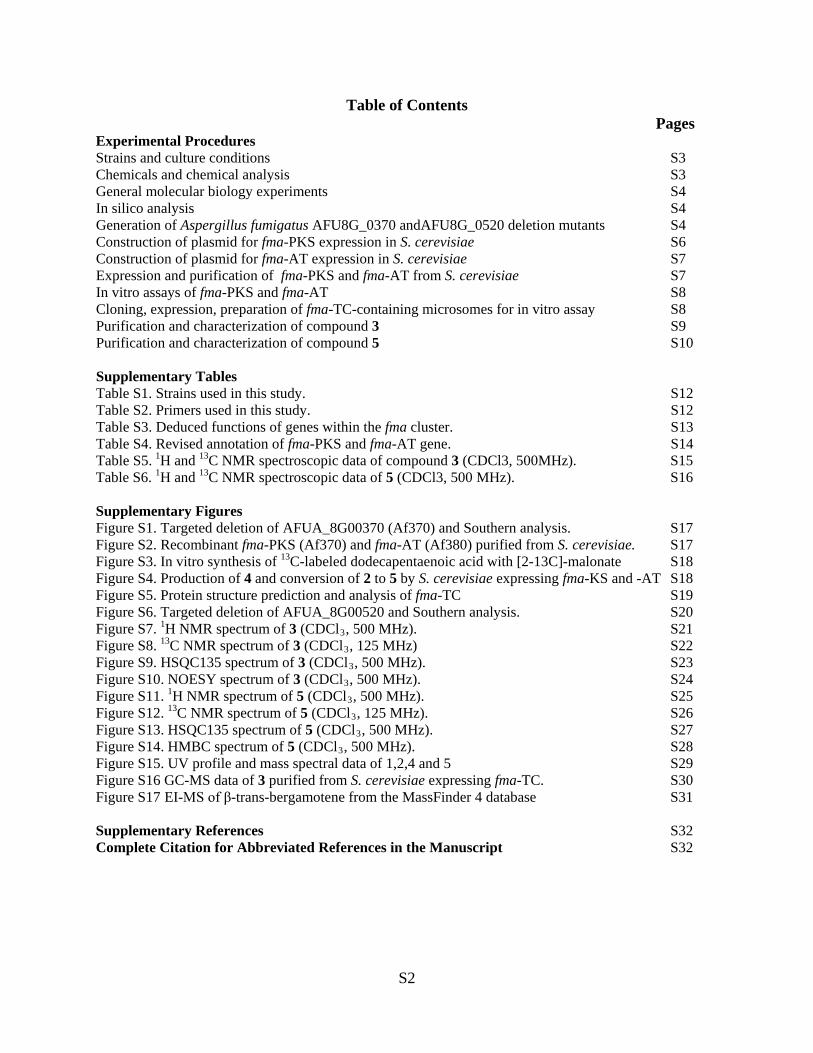
Table of Contents Pages
Experimental Procedures Strains and culture conditions S3 Chemicals and chemical analysis S3 General molecular biology experiments S4 In silico analysis S4 Generation of Aspergillus fumigatus AFU8G_0370 andAFU8G_0520 deletion mutants S4 Construction of plasmid for fma-PKS expression in S. cerevisiae S6 Construction of plasmid for fma-AT expression in S. cerevisiae S7 Expression and purification of fma-PKS and fma-AT from S. cerevisiae S7 In vitro assays of fma-PKS and fma-AT S8 Cloning, expression, preparation of fma-TC-containing microsomes for in vitro assay S8 Purification and characterization of compound 3 S9 Purification and characterization of compound 5 S10 Supplementary Tables Table S1. Strains used in this study. S12 Table S2. Primers used in this study. S12 Table S3. Deduced functions of genes within the fma cluster. S13 Table S4. Revised annotation of fma-PKS and fma-AT gene. S14 Table S5. 1H and 13C NMR spectroscopic data of compound 3 (CDCl3, 500MHz). S15 Table S6. 1H and 13C NMR spectroscopic data of 5 (CDCl3, 500 MHz). S16 Supplementary Figures Figure S1. Targeted deletion of AFUA_8G00370 (Af370) and Southern analysis. S17 Figure S2. Recombinant fma-PKS (Af370) and fma-AT (Af380) purified from S. cerevisiae. S17 Figure S3. In vitro synthesis of 13C-labeled dodecapentaenoic acid with [2-13C]-malonate S18 Figure S4. Production of 4 and conversion of 2 to 5 by S. cerevisiae expressing fma-KS and -AT S18 Figure S5. Protein structure prediction and analysis of fma-TC S19 Figure S6. Targeted deletion of AFUA_8G00520 and Southern analysis. S20 Figure S7. 1H NMR spectrum of 3 (CDCl3, 500 MHz). S21 Figure S8. 13C NMR spectrum of 3 (CDCl3, 125 MHz) S22 Figure S9. HSQC135 spectrum of 3 (CDCl3, 500 MHz). S23 Figure S10. NOESY spectrum of 3 (CDCl3, 500 MHz). S24 Figure S11. 1H NMR spectrum of 5 (CDCl3, 500 MHz). S25 Figure S12. 13C NMR spectrum of 5 (CDCl3, 125 MHz). S26 Figure S13. HSQC135 spectrum of 5 (CDCl3, 500 MHz). S27 Figure S14. HMBC spectrum of 5 (CDCl3, 500 MHz). S28 Figure S15. UV profile and mass spectral data of 1,2,4 and 5 S29 Figure S16 GC-MS data of 3 purified from S. cerevisiae expressing fma-TC. S30 Figure S17 EI-MS of β-trans-bergamotene from the MassFinder 4 database S31 Supplementary References S32 Complete Citation for Abbreviated References in the Manuscript S32
S2
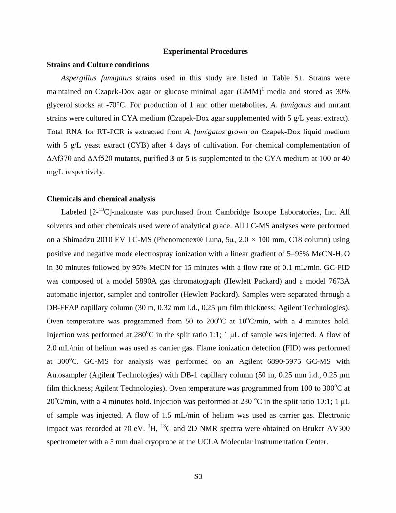
Experimental Procedures
Strains and Culture conditions
Aspergillus fumigatus strains used in this study are listed in Table S1. Strains were
maintained on Czapek-Dox agar or glucose minimal agar (GMM)1 media and stored as 30%
glycerol stocks at -70°C. For production of 1 and other metabolites, A. fumigatus and mutant
strains were cultured in CYA medium (Czapek-Dox agar supplemented with 5 g/L yeast extract).
Total RNA for RT-PCR is extracted from A. fumigatus grown on Czapek-Dox liquid medium
with 5 g/L yeast extract (CYB) after 4 days of cultivation. For chemical complementation of
ΔAf370 and ΔAf520 mutants, purified 3 or 5 is supplemented to the CYA medium at 100 or 40
mg/L respectively.
Chemicals and chemical analysis
Labeled [2-13C]-malonate was purchased from Cambridge Isotope Laboratories, Inc. All
solvents and other chemicals used were of analytical grade. All LC-MS analyses were performed
on a Shimadzu 2010 EV LC-MS (Phenomenex® Luna, 5µ, 2.0 × 100 mm, C18 column) using
positive and negative mode electrospray ionization with a linear gradient of 5−95% MeCN-H2O
in 30 minutes followed by 95% MeCN for 15 minutes with a flow rate of 0.1 mL/min. GC-FID
was composed of a model 5890A gas chromatograph (Hewlett Packard) and a model 7673A
automatic injector, sampler and controller (Hewlett Packard). Samples were separated through a
DB-FFAP capillary column (30 m, 0.32 mm i.d., 0.25 µm film thickness; Agilent Technologies).
Oven temperature was programmed from 50 to 200oC at 10oC/min, with a 4 minutes hold.
Injection was performed at 280oC in the split ratio 1:1; 1 μL of sample was injected. A flow of
2.0 mL/min of helium was used as carrier gas. Flame ionization detection (FID) was performed
at 300oC. GC-MS for analysis was performed on an Agilent 6890-5975 GC-MS with
Autosampler (Agilent Technologies) with DB-1 capillary column (50 m, 0.25 mm i.d., 0.25 µm
film thickness; Agilent Technologies). Oven temperature was programmed from 100 to 300oC at
20oC/min, with a 4 minutes hold. Injection was performed at 280 oC in the split ratio 10:1; 1 μL
of sample was injected. A flow of 1.5 mL/min of helium was used as carrier gas. Electronic
impact was recorded at 70 eV. 1H, 13C and 2D NMR spectra were obtained on Bruker AV500
spectrometer with a 5 mm dual cryoprobe at the UCLA Molecular Instrumentation Center.
S3
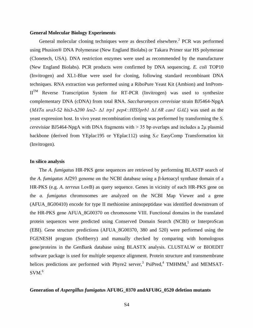
General Molecular Biology Experiments
General molecular cloning techniques were as described elsewhere.2 PCR was performed
using Phusion® DNA Polymerase (New England Biolabs) or Takara Primer star HS polymerase
(Clonetech, USA). DNA restriction enzymes were used as recommended by the manufacturer
(New England Biolabs). PCR products were confirmed by DNA sequencing. E. coli TOP10
(Invitrogen) and XL1-Blue were used for cloning, following standard recombinant DNA
techniques. RNA extraction was performed using a RiboPure Yeast Kit (Ambion) and ImProm-
IITM Reverse Transcription System for RT-PCR (Invitrogen) was used to synthesize
complementary DNA (cDNA) from total RNA. Saccharomyces cerevisiae strain BJ5464-NpgA
(MATα ura3-52 his3-∆200 leu2- ∆1 trp1 pep4::HIS3prb1 ∆1.6R can1 GAL) was used as the
yeast expression host. In vivo yeast recombination cloning was performed by transforming the S.
cerevisiae BJ5464-NpgA with DNA fragments with > 35 bp overlaps and includes a 2μ plasmid
backbone (derived from YEplac195 or YEplac112) using S.c EasyComp Transformation kit
(Invitrogen).
In silico analysis
The A. fumigatus HR-PKS gene sequences are retrieved by performing BLASTP search of
the A. fumigatus Af293 genome on the NCBI database using a β-ketoacyl synthase domain of a
HR-PKS (e.g. A. terreus LovB) as query sequence. Genes in vicinity of each HR-PKS gene on
the a. fumigatus chromosomes are analyzed on the NCBI Map Viewer and a gene
(AFUA_8G00410) encode for type II methionine aminopeptidase was identified downstream of
the HR-PKS gene AFUA_8G00370 on chromosome VIII. Functional domains in the translated
protein sequences were predicted using Conserved Domain Search (NCBI) or InterproScan
(EBI). Gene structure predictions (AFUA_8G00370, 380 and 520) were performed using the
FGENESH program (Softberry) and manually checked by comparing with homologous
gene/proteins in the GenBank database using BLASTX analysis. CLUSTALW or BIOEDIT
software package is used for multiple sequence alignment. Protein structure and transmembrane
helices predictions are performed with Phyre2 server,3 PsiPred,4 TMHMM,5 and MEMSAT-
SVM.6
Generation of Aspergillus fumigatus AFU8G_0370 andAFU8G_0520 deletion mutants
S4
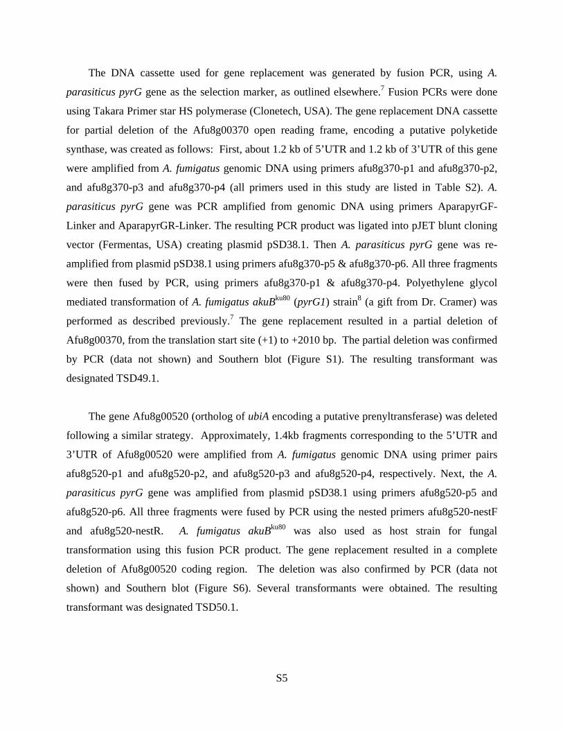
The DNA cassette used for gene replacement was generated by fusion PCR, using A.
parasiticus pyrG gene as the selection marker, as outlined elsewhere.7 Fusion PCRs were done
using Takara Primer star HS polymerase (Clonetech, USA). The gene replacement DNA cassette
for partial deletion of the Afu8g00370 open reading frame, encoding a putative polyketide
synthase, was created as follows: First, about 1.2 kb of 5’UTR and 1.2 kb of 3’UTR of this gene
were amplified from A. fumigatus genomic DNA using primers afu8g370-p1 and afu8g370-p2,
and afu8g370-p3 and afu8g370-p4 (all primers used in this study are listed in Table S2). A.
parasiticus pyrG gene was PCR amplified from genomic DNA using primers AparapyrGF-
Linker and AparapyrGR-Linker. The resulting PCR product was ligated into pJET blunt cloning
vector (Fermentas, USA) creating plasmid pSD38.1. Then A. parasiticus pyrG gene was re-
amplified from plasmid pSD38.1 using primers afu8g370-p5 & afu8g370-p6. All three fragments
were then fused by PCR, using primers afu8g370-p1 & afu8g370-p4. Polyethylene glycol
mediated transformation of A. fumigatus akuBku80 (pyrG1) strain8 (a gift from Dr. Cramer) was
performed as described previously.7 The gene replacement resulted in a partial deletion of
Afu8g00370, from the translation start site (+1) to +2010 bp. The partial deletion was confirmed
by PCR (data not shown) and Southern blot (Figure S1). The resulting transformant was
designated TSD49.1.
The gene Afu8g00520 (ortholog of ubiA encoding a putative prenyltransferase) was deleted
following a similar strategy. Approximately, 1.4kb fragments corresponding to the 5’UTR and
3’UTR of Afu8g00520 were amplified from A. fumigatus genomic DNA using primer pairs
afu8g520-p1 and afu8g520-p2, and afu8g520-p3 and afu8g520-p4, respectively. Next, the A.
parasiticus pyrG gene was amplified from plasmid pSD38.1 using primers afu8g520-p5 and
afu8g520-p6. All three fragments were fused by PCR using the nested primers afu8g520-nestF
and afu8g520-nestR. A. fumigatus akuBku80 was also used as host strain for fungal
transformation using this fusion PCR product. The gene replacement resulted in a complete
deletion of Afu8g00520 coding region. The deletion was also confirmed by PCR (data not
shown) and Southern blot (Figure S6). Several transformants were obtained. The resulting
transformant was designated TSD50.1.
S5
An isogenic control strain was also obtained by transforming A. fumigatus akuBku8 with a
wild-type copy of the A. fumigatus pyrG gene. The DNA fragment containing pyrG used for this
transformation was PCR amplified from p1439 (Stinnett et al., 2007) with primers
CoLinkerF_641 and ColinkerR_642. The resulting transformant was designated TSD51.1.
Construction of plasmid for fma-PKS expression in S. cerevisiae
The fma-PKS expression plasmid, pHCfmaPKS, was constructed using in vivo yeast
recombination cloning. The complete fma-PKS cDNA was constructed by three overlapping
fragments (Table S4). The middle largest exon region (4479 bp) that was predicted to not
contain any intron was amplified from A. fumigatus gDNA using primer pair FG370-P2-F with
FG370-P2-R (fragment designated as FG370-P2). The 3′ and 5′ end regions were predicted to
contain introns and were thus amplified from cDNA. RNA was extracted from a four-day old
culture in CYB medium using the RiboPure Yeast Kit (Ambion) following the manufacturer’s
instructions and residual gDNA in the total RNA was digested with DNase (2 U/μL) (Invitrogen)
at 37 oC for four hours. The 3′ and 5′ fma-PKS cDNA was synthesized using ImProm-IITM
Reverse Transcription System with the reverse primers FG370-P1-R and FG370-P3-R,
respectively (Table S4). The cDNA was used as template for PCR. The 3′ and 5′ intron-free
cDNA fragments were amplified using primer pairs FG370-P1-F/FG370-P1-R-TADH2 and PADH2-
FG370-P3-F/FG370-P3-R3 (designated as PADH2-FG370-P3 and FG370-P1-TADH2 respectively).
For in vivo yeast recombination, the three overlapping fragments, PADH2-FG370-P3, FG370-P2
and FG370-P1-TADH2, were combined with a PmlI/NdeI-linearized YEplac195-derived 2μ
expression plasmid (containing URA3 marker) flanking with PADH2 promoter and TADH2
terminator (also encode a 6×His-tag and stop codon);9 co-transfomation of S. cerevisiae BJ5464-
NpgA using an S. c EasyComp™ Transformation kit (Invitrogen) yield the expression plasmid
pHCfmaPKS by in vivo recombination. The resulting pHCfmaPKS plasmid was recovered by
Zymoprep™ Yeast Plasmid Miniprep (Zymo Research) for propagation in E. coli XL1 and
verified by sequencing.
Construction of plasmid for fma-AT expression in S. cerevisiae
An intron-free fma-AT was constructed from cDNA which was synthesized with the reverse
primer pHis8-FG380-R using ImProm-IITM Reverse Transcription System. The cDNA was used
S6
as template for PCR and amplified by using primer pair pHis8-FG380-F and pHis8-FG380-R,
which contained EcoRI and NotI restriction sites, respectively. To introduce 8×His-tag to the N-
terminal of fma-AT, the PCR product was digested with EcoRI and NotI and ligated into the
EcoRI/NotI-digested pHis8 expression vector,10 to yield plasmid pHCHis8fmaAT. This plasmid
was used as template for PCR using primer pair ADH2p-380-F with ADH2t-380-R. The
resulting PCR product and an NdeI/HindIII-linearized YEplac112-derived 2 μ expression
plasmid (containing TRP1 marker) were combined; co-transformation of S. cerevisiae BJ5464-
NpgA using an S. c EasyComp™ Transformation kit (Invitrogen) yield the expression plasmid
pHCfmaAT by in vivo recombination. The resulting pHCfmaAT plasmid was recovered by
Zymoprep™ Yeast Plasmid Miniprep (Zymo Research) for propagation in E. coli XL1 and
verified by sequencing.
Expression and purification of fma-PKS and fma-AT from S. cerevisiae
For 1 L culture, the cells were grown at YPD (10 g/L yeast extract, 20 g/L peptone)
supplemented with 1% dextrose and incubated at 28 oC with shaking for 72 hours. The cells were
harvested by centrifugation (3,750 rpm at 4oC for 10 mins) and the cell pellet was resuspended in
20 mL lysis buffer (50 mM NaH2PO4, 150 mM NaCl, 10 mM imidazole, pH 8.0) and lysed by
sonication on ice in one minute intervals until homogenous. To remove cellular debris, the
homogenous mixture was centrifuged at 17,000 rpm for 1 hour at 4oC. Ni-NTA agarose resin
was added to the supernatant (2 mL) and the solution was stirred at 4oC overnight. Soluble fma-
PKS was purified by gravity-flow column chromatography with increasing concentrations of
imidazole in Buffer A (50 mM Tris-HCl, 500 mM NaCl, 20 mM–250 mM imidazole, pH 7.9).
Purified protein was concentrated and buffer was exchanged into Buffer B (50 mM Tris-HCl, 2
mM EDTA, 100 mM NaCl, pH 8.0) using an Amicon Ultra-15 Centrifugal Filter Unit and stored
in 10% glycerol. The purified fma-PKS and fma-AT were analyzed by SDS-PAGE (Figure S2A)
and their concentration were calculated to be 3.5 mg/L and 18.0 mg/L, respectively, using the
Bradford assay with BSA as a standard.
In vitro assays of fma-PKS and fma-AT
For in vitro synthesis of dodecapentaenoic acid 4, 10 µM fma-PKS and 10 µM fma-AT were
incubated with 2 mM malonyl-CoA, 1 mM NADPH in 100 mM PBS, pH 7.4 in a total 100 µL
S7

reaction. The reaction was incubated at room temperature overnight and extracted with 100 µL
ethyl acetate with 1% acetic acid twice. The organic phase was dried and dissolved in 20 µL
MeOH for analysis on LC-MS. For in vitro synthesis of 5-dodecapentaenoyl fumagillol 5, 1 mM
fumagillol 2 was supplemented to the same reaction condition above in 100 µL volume
following the same protocols. For in vitro synthesis of 13C-labeled dodecapentaenoic acid, 10
µM fma-PKS and 10 µM fma-AT were incubated with 100 mM [2-13C]-malonate (unlabeled
malonate used for control reaction) and a malonyl-CoA regeneration system (10 mM Mg2Cl2, 25
mM ATP, 10 mM CoA, 25 μM MatB, 1 mM NADPH) in 100 mM PBS buffer, pH 7.4 in total
100 µL volume. The reaction was incubated at room temperature for overnight, extracted and
analyzed by LCMS as above (Figure S3).
Cloning, expression, and preparation of fma-TC-containing microsomes for in vitro assay
The complete fma-PKS cDNA was constructed by two overlapping fragments using yeast in
vivo recombination cloning (Table S4). The 3′ and 5′ end regions were predicted to contain
introns and were thus amplified from cDNA. cDNA was synthesized using ImProm-IITM Reverse
Transcription System with the anchored oligo-dT reverse primer. The cDNA was used as
template for PCR. The 3’ and 5’ intron-free cDNA fragments were amplified using primer pairs
PADH2-FG520-P1-F/FG520-P1-R (PADH2-FG520-P1 fragment) and FG520-P2-F/FG520-P2-R-5-
TADH2 (FG520-P2-TADH2 fragment). For in vivo recombination, S. cerevisiae BJ5464-NpgA
were co-transformed with three overlapping fragments, PADH2-FG520-P1, FG520-P2-TADH2 and
a SwaI/NdeI-linearized YEplac195-derived 2μ expression plasmid (containing URA3 marker)
flanking with PADH2 promoter and TADH2 terminator (no His tag) to yield the expression plasmid
pHCfmaTC.
For expression of fma-TC, the cells were grown in YPD medium supplemented with 1%
dextrose at 28oC with shaking for 48 hours. The microsomes were prepared according to the
protocol described previously.11 Briefly, the cells were harvested by centrifugation (3,750 rpm at
4oC for 10 mins) and the cell pellet was washed with 100 mL of TES buffer (50 mM Tris–HCl,
pH, 7.5, 1 mM EDTA, 0.6 M sorbitol). The cells were centrifuged as above, resuspended in 100
mL of TES-M (TES supplemented with 10 mM 2-mercaptoethanol), and allowed to incubate at
room temperature for 10 min. The yeast cells were centrifuged again at 3,750 rpm for 10 min,
and the pellet was resuspended in 2.5 mL of extraction buffer (1% bovine serum albumin,
S8
fraction V, 2 mM 2-mercaptoethanol, 1 mM phenylmethylsulfonyl fluoride, all dissolved in
TES). Zirconia/silica beads (0.5 mm in diameter, Biospec Products) were added until skimming
the surface of the cell suspension. Cell walls were disrupted manually by hand shaking in a cold
room for 10 min at 30-s intervals separated by 30-s intervals on ice. Cell extracts were
transferred to a 50-mL centrifuge tube, the Zirconia/silica beads were washed three times with 5
mL of extraction buffer, and the washes were pooled with the original cell extracts. Finally,
microsomes were obtained by differential centrifugation at 10,000g for 10 min at 4°C to remove
cellular debris followed by centrifugation at 100,000g for 70 min at 4°C. The microsomal pellets
were weighed prior to resuspension in 1.5 mL of TEG-M buffer (50 mM Tris–HCl, pH 7.5, 1
mM EDTA, 20% glycerol, and 1.5 mM 2-mercaptoethanol) and stored frozen at -80 °C.
For in vitro microsomes assay, 1 or 10 mg/mL (wet weight) microsomal fractions containing
fma-TC and 1 mM FPP were incubated with or without 5 mM MgCl2 in a 100 µL reaction. The
reaction was incubated at room temperature for overnight and extracted with 100 µL hexanes
twice. The organic phase was dried and re-dissolved in 100 µL hexanes for analysis by GC-FID
or GC-MS. For the measurement of the conversion rate of FPP into 3, 10 mg/mL microsome
fractions containing fma-TC, 1 mM FPP and 5 mM MgCl2 were incubated at room temperature.
The reactions were stopped at 15 min, 30 min, 1 hours and 2 hours by adding 100 μL hexanes
and extracted twice, respectively. The organic phase was dried and re-dissolved in 5 µL DMF for
analysis by GC-FID. The concentration of β-trans-bergamotene produced was measured by the
peak area compared to the standard curve. The conversion rate of 3 was measured to be 4.4
μMmin-1 in the presence 10 mg/mL microsomes. The amount of protein in 10 mg/mL
microsomes was calculated to be 250 μg/mL based on a modified Bradford assay against a BSA
standard curve (protein samples were pre-denatured in 0.1 M NaOH).
Purification and characterization of compound 3
S. cerevisiae strain BJ5464-NpgA harboring pHCfmaTC plasmid was inoculated to 4 mL
Yeast Synthetic Drop-Out medium without uracil. The cells were grown for 72 hours with
constant shaking at 28oC. A 1 mL aliquot of the seed culture was inoculated to 1 L YPD
medium supplemented with 1% dextrose. The culture was shaken at 28 oC for 96 hours. The cells
were harvested by centrifugation (3750 rpm, 10 minuntes, 4oC). The cell pellet was extracted by
actetone (100 mL) at room temperature and concentrated in vacuo. The acetone extract was
S9

suspended in MeCN (15 mL) and partitioned with hexanes (15 mL × 3). The hexanes soluble
fraction (55.1 mg out of 65.0 mg) was separated on a Silica gel column (230―400 mesh,
hexanes) to give 3 (39.0 mg).
Compound 3 had a molecular formula C15H24, as deduced from EI-MS, and showed [α] 22D
−5.6 (c 2.5, CHCl3). The 1H NMR data showed the characteristic signals of one set of isoprenyl
protons (δ 1.94, br.q, J = 7.7 Hz, H-9, δ 5.15, tqq, J = 7.0, 1.3, 1.3 Hz, H-10; δ 1.68, br.s, H-12; δ
1.61, br.s, H-13;), two olefinic methine protons (δ 4.62, br.s, H-15a and δ 4.54, br.s, H-15b) and
one methyl proton (δ 0.70, s, H-14). The 13C NMR data exhibited signals of three methyls (δ
25.7, 18.6 and 17.6), five methylenes (δ 23.5, 23.5, 23.8, 27.1 and 38.2), two methines (δ 38.7
and 50.2), one tert-methyl (δ 43.8), and two olefinic group (δ 125.1 and 130.1; δ 152.1 and 106.0)
(Table S5). These 1H, 13C and EI-MS spectroscopic features were identical to those of β-trans-
bergamotene.12-14 The NOESY spectrum (Figure S10) showed the correlations of δ 1.68 (H-12)
to δ 2.51 (H-1), δ 2.05 (H-5) and δ 2.28 (H-6β) and the correlations of 0.70 (H-14) to δ 2.24 (H-
3β) and δ 1.82 (H-4) confirming the trans-configuration of structure. Accordingly, compound 3
was established as (−)-β-trans-bergamotene.
Purification and characterization of compound 5
S. cerevisiae strain BJ5464-NpgA harboring both pHCfmaPKS and pHCfmaAT plasmids
was inoculated to 4 mL Yeast Synthetic Drop-Out medium without uracil and tryptophan. The
cells were grown for 72 hours with constant shaking at 28oC. A 1 mL aliquot of the seed culture
was inoculated 50 mL YPD (10 g yeast extract, 20 g peptone and 950 mL Milli-Q water)
supplemented with 1% dextrose. Fumagillol (2, 2.5 mg in 100 μL MeOH) was added to the
culture after 48 hours at 28oC with shaking and the cells were cultivated for another 48 hours.
The cells were harvested by centrifugation (3750 rpm, 10 minuntes, 4oC). The cell pellet was
extracted by acetone (100 mL) at room temperature and concentrated in vacuo. The acetone
extract was further separated on a semi-preparative RP-18 HPLC column (Luna®, ODS-3, 5 μm,
semi-preparative: 250 × 10 mm) elutaed by 85% MeCNaq with flow rate of 2.5 mL/min to give 5
(1.1 mg, tR: 12.1 min).
Compound 5 had the molecular formula C28H38O5, as deduced from ESI-MS showing the
quasi-molecular ion [M+Na]+ at m/z 477. The UV spectrum showed a typical polyene pattern
with maximum absorption at 359 nm. The 1H and 13C NMR data of 5 were similar to those in
S10
fumagillin15 except the signals of the polyene moiety (Table S6). Both 1H and 13C spectra
showed the characteristic signals of one dodecapentaenoyl moiety (1H data, δ 5.85−7.30 and δ
1.79, br.d, J = 6.8 Hz; 13C, Table S6). The stereochemistry of H-2' to H-11' showed all trans
based on the coupling constant (J = 14.5−15.5 Hz). The 1H and 13C NMR assignments of 5 were
made by 2D NMR analyses and are listed in Table S6. Thus, compound 5 was established as 5-
dodecapentaenoyl fumagillol.
S11
Supplementary Tables
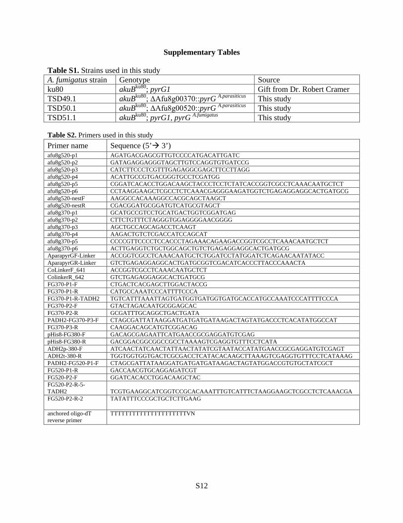
Table S1. Strains used in this study A. fumigatus strain Genotype Source ku80 akuBku80; pyrG1 Gift from Dr. Robert Cramer TSD49.1 akuBku80; ΔAfu8g00370::pyrG A.parasiticus This study TSD50.1 akuBku80; ΔAfu8g00520::pyrG A.parasiticus This study TSD51.1 akuBku80; pyrG1, pyrG A.fumigatus This study Table S2. Primers used in this study Primer name Sequence (5’ 3’) afu8g520-p1 AGATGACGAGCGTTGTCCCCATGACATTGATC afu8g520-p2 GATAGAGGAGGGTAGCTTGTCCAGGTGTGATCCG afu8g520-p3 CATCTTCCCTCGTTTGAGAGGCGAGCTTCCTTAGG afu8g520-p4 ACATTGCCGTGACGGGTGCCTCGATGG afu8g520-p5 CGGATCACACCTGGACAAGCTACCCTCCTCTATCACCGGTCGCCTCAAACAATGCTCT afu8g520-p6 CCTAAGGAAGCTCGCCTCTCAAACGAGGGAAGATGGTCTGAGAGGAGGCACTGATGCG afu8g520-nestF AAGGCCACAAAGGCCACGCAGCTAAGCT afu8g520-nestR CGACGGATGCGGATGTCATGCGTAGCT afu8g370-p1 GCATGCCGTCCTGCATGACTGGTCGGATGAG afu8g370-p2 CTTCTGTTTCTAGGGTGGAGGGGAACGGGG afu8g370-p3 AGCTGCCAGCAGACCTCAAGT afu8g370-p4 AAGACTGTCTCGACCATCCAGCAT afu8g370-p5 CCCCGTTCCCCTCCACCCTAGAAACAGAAGACCGGTCGCCTCAAACAATGCTCT afu8g370-p6 ACTTGAGGTCTGCTGGCAGCTGTCTGAGAGGAGGCACTGATGCG AparapyrGF-Linker ACCGGTCGCCTCAAACAATGCTCTGGATCCTATGGATCTCAGAACAATATACC AparapyrGR-Linker GTCTGAGAGGAGGCACTGATGCGGTCGACATCACCCTTACCCAAACTA CoLinkerF_641 ACCGGTCGCCTCAAACAATGCTCT ColinkerR_642 GTCTGAGAGGAGGCACTGATGCG FG370-P1-F CTGACTCACGAGCTTGGACTACCG FG370-P1-R CATGCCAAATCCCATTTTCCCA FG370-P1-R-TADH2 TGTCATTTAAATTAGTGATGGTGATGGTGATGCACCATGCCAAATCCCATTTTCCCA FG370-P2-F GTACTAGACAATGCGGAGCAC FG370-P2-R GCGATTTGCAGGCTGACTGATA PADH2-FG370-P3-F CTAGCGATTATAAGGATGATGATGATAAGACTAGTATGACCCTCACATATGGCCAT FG370-P3-R CAAGGACAGCATGTCGGACAG pHis8-FG380-F GACAGCGAGAATTCATGAACCGCGAGGATGTCGAG pHis8-FG380-R GACGGACGGCGGCCGCCTAAAAGTCGAGGTGTTTCCTCATA ADH2p-380-F ATCAACTATCAACTATTAACTATATCGTAATACCATATGAACCGCGAGGATGTCGAGT ADH2t-380-R TGGTGGTGGTGACTCGCGACCTCATACACAAGCTTAAAGTCGAGGTGTTTCCTCATAAAG PADH2-FG520-P1-F CTAGCGATTATAAGGATGATGATGATAAGACTAGTATGGACCGTGTGCTATCGCT FG520-P1-R GACCAACGTGCAGGAGATCGT FG520-P2-F GGATCACACCTGGACAAGCTAC FG520-P2-R-5-TADH2 TCGTGAAGGCATCGGTCCGCACAAATTTGTCATTTCTAAGGAAGCTCGCCTCTCAAACGA FG520-P2-R-2 TATATTTCCCGCTGCTCTTGAAG
anchored oligo-dT reverse primer
TTTTTTTTTTTTTTTTTTTTTVN
S12
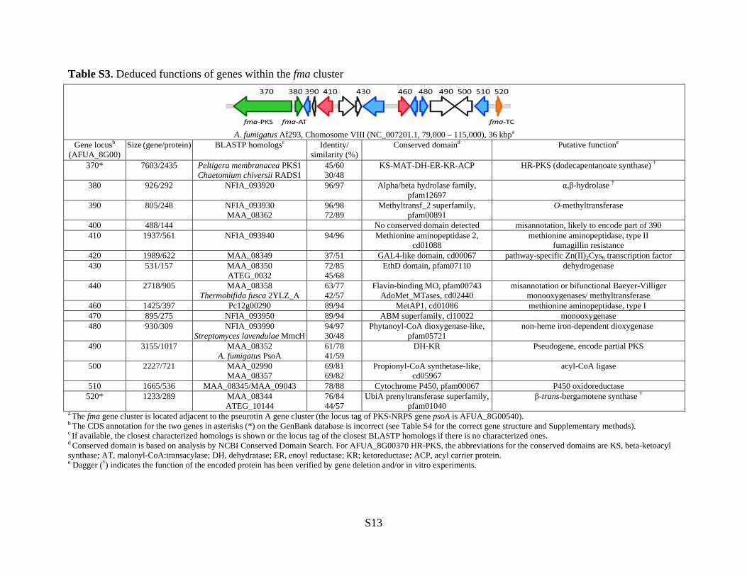
Table S3. Deduced functions of genes within the fma cluster
a The fma gene cluster is located adjacent to the pseurotin A gene cluster (the locus tag of PKS-NRPS gene psoA is AFUA_8G00540). b The CDS annotation for the two genes in asterisks (*) on the GenBank database is incorrect (see Table S4 for the correct gene structure and Supplementary methods). c If available, the closest characterized homologs is shown or the locus tag of the closest BLASTP homologs if there is no characterized ones. d Conserved domain is based on analysis by NCBI Conserved Domain Search. For AFUA_8G00370 HR-PKS, the abbreviations for the conserved domains are KS, beta-ketoacyl synthase; AT, malonyl-CoA:transacylase; DH, dehydratase; ER, enoyl reductase; KR; ketoreductase; ACP, acyl carrier protein. e Dagger (†) indicates the function of the encoded protein has been verified by gene deletion and/or in vitro experiments.
A. fumigatus Af293, Chomosome VIII (NC_007201.1, 79,000 – 115,000), 36 kbpa
Gene locusb (AFUA_8G00)
Size (gene/protein) BLASTP homologsc Identity/ similarity (%)
Conserved domaind Putative functione
370* 7603/2435 Peltigera membranacea PKS1 Chaetomium chiversii RADS1
45/60 30/48
KS-MAT-DH-ER-KR-ACP HR-PKS (dodecapentanoate synthase) †
380 926/292 NFIA_093920 96/97 Alpha/beta hydrolase family, pfam12697
α,β-hydrolase †
390 805/248 NFIA_093930 MAA_08362
96/98 72/89
Methyltransf_2 superfamily, pfam00891
O-methyltransferase
400 488/144 No conserved domain detected misannotation, likely to encode part of 390 410 1937/561 NFIA_093940 94/96 Methionine aminopeptidase 2,
cd01088 methionine aminopeptidase, type II
fumagillin resistance 420 1989/622 MAA_08349 37/51 GAL4-like domain, cd00067 pathway-specific Zn(II)2Cys6 transcription factor 430 531/157 MAA_08350
ATEG_0032 72/85 45/68
EthD domain, pfam07110 dehydrogenase
440 2718/905 MAA_08358 Thermobifida fusca 2YLZ_A
63/77 42/57
Flavin-binding MO, pfam00743 AdoMet_MTases, cd02440
misannotation or bifunctional Baeyer-Villiger monooxygenases/ methyltransferase
460 1425/397 Pc12g00290 89/94 MetAP1, cd01086 methionine aminopeptidase, type I 470 895/275 NFIA_093950 89/94 ABM superfamily, cl10022 monooxygenase 480 930/309 NFIA_093990
Streptomyces lavendulae MmcH 94/97 30/48
Phytanoyl-CoA dioxygenase-like, pfam05721
non-heme iron-dependent dioxygenase
490 3155/1017 MAA_08352 A. fumigatus PsoA
61/78 41/59
DH-KR Pseudogene, encode partial PKS
500 2227/721 MAA_02990 MAA_08357
69/81 69/82
Propionyl-CoA synthetase-like, cd05967
acyl-CoA ligase
510 1665/536 MAA_08345/MAA_09043 78/88 Cytochrome P450, pfam00067 P450 oxidoreductase 520* 1233/289 MAA_08344
ATEG_10144 76/84 44/57
UbiA prenyltransferase superfamily, pfam01040
β-trans-bergamotene synthase †
S13

Table S4. Revised annotation of fma-PKS and fma-TC gene.
Locus tag/Gene AFUA_8G00370 (fma-PKS gene)a AFUA_8G00520 (fma-TC gene)b
GenBank ID (mRNA)
XM_742074.1 XM_742060.1
CDS annotation in NC_007201.1
complement (<79285..80133,80197..80511,80560..80749, 80794..86223,86283..>86887)
join(<113744..113909,113972..114042,114101..114358, 114412..>114438)
Revised (actual) CDS annotation
complement (join(79285..80133,80197..80511,80560..80733, 80794..86223,86283..86400,86464 >86887))
join(113249..>113347,113411..113503, 113559..113570, 113636..113909, 113972.. 114042,114101..114358,114419..>114481)
New GenBank ID KC407775 KC407776 a The complete coding sequence of fma-PKS gene is constructed by yeast recombination from two cDNA fragments amplified from total RNA by RT-PCR using 370-P1-F/P1-R and 370-P3-F/P3-R primers, and an intron-less fragment amplified from genomic DNA of A. fumigatus using 370-P2-F/P2-R. Sequencing of the cloned 370-P1-F/P1-R and 370-P3-F/P3-R cDNA fragments revealed the correct exon regions (*misannotated introns). b The complete coding sequence of fma-TC gene is constructed by yeast recombination from two cDNA fragments using FG520-P1-F/P1-R and FG520-P2-F/P2-R-5 primers amplified from total RNA by RT-PCR. The exon regions of 5′ fragment is determined by sequencing of the cloned FG520-P1-F/P1-R cDNA fragment, while sequencing of the 3′ cDNA fragment amplified with FG520-P2-F and FG520-P2-R-2 (located at 3′ untranslated region) revealed the correct exon regions and the stop codon. NOTE For more details for cloning of the two genes for expression in S. cerevisiae, see Supplementary Methods.
400 bp
370-P1-R P3-RP2-R
P3-F
520-P1-F
P1-R
P2-F
P2-R-2P2-R-5
AFUA_8G00520
100 bpP1-F P2-F
AFUA_8G00370
GenBank annotation
FGENESH prediction
Actual gene structure
* *
S14
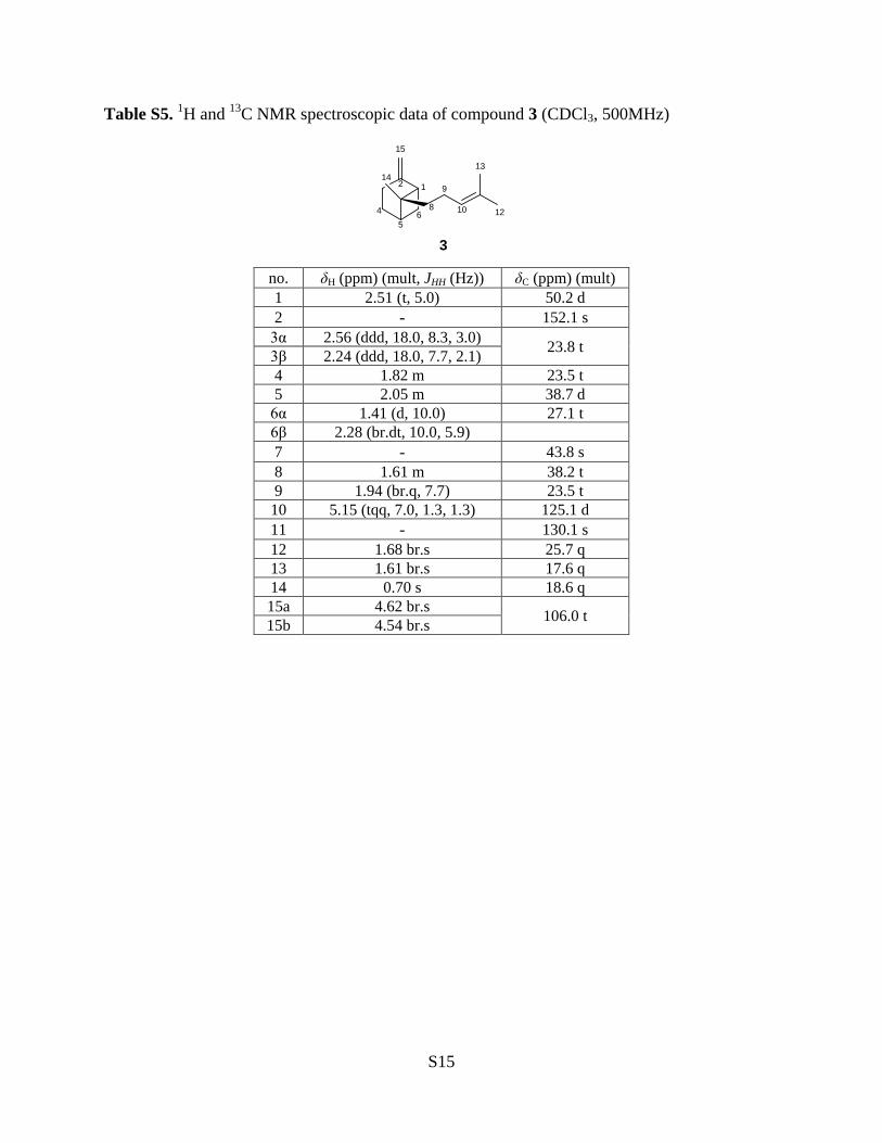
Table S5. 1H and 13C NMR spectroscopic data of compound 3 (CDCl3, 500MHz)
12
15
12
1314
45
68
9
10
3
no. δH (ppm) (mult, JHH (Hz)) δC (ppm) (mult) 1 2.51 (t, 5.0) 50.2 d 2 - 152.1 s
3α 2.56 (ddd, 18.0, 8.3, 3.0) 23.8 t 3β 2.24 (ddd, 18.0, 7.7, 2.1) 4 1.82 m 23.5 t 5 2.05 m 38.7 d
6α 1.41 (d, 10.0) 27.1 t 6β 2.28 (br.dt, 10.0, 5.9) 7 - 43.8 s 8 1.61 m 38.2 t 9 1.94 (br.q, 7.7) 23.5 t 10 5.15 (tqq, 7.0, 1.3, 1.3) 125.1 d 11 - 130.1 s 12 1.68 br.s 25.7 q 13 1.61 br.s 17.6 q 14 0.70 s 18.6 q 15a 4.62 br.s 106.0 t 15b 4.54 br.s
S15
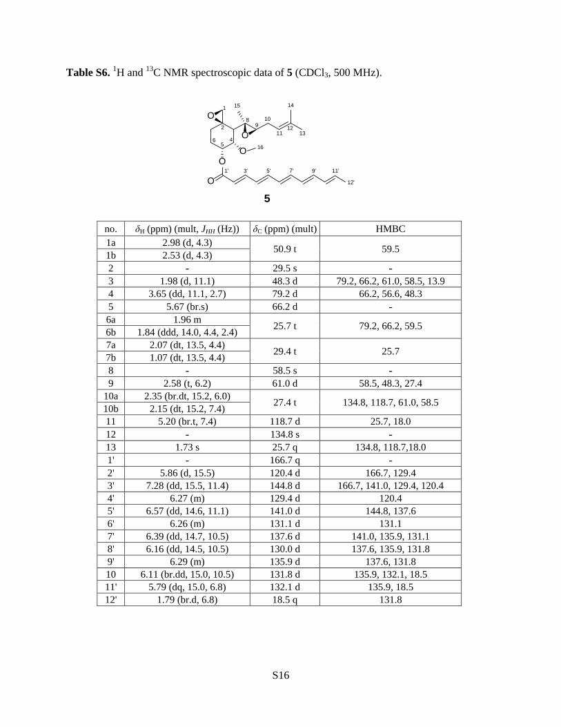
Table S6. 1H and 13C NMR spectroscopic data of 5 (CDCl3, 500 MHz).
O
O
O
O
O
1
2
6 45
89
10
1112
13
1415
16
1' 3' 5' 7' 9' 11'
12'
5
no. δH (ppm) (mult, JHH (Hz)) δC (ppm) (mult) HMBC 1a 2.98 (d, 4.3) 50.9 t 59.5 1b 2.53 (d, 4.3) 2 - 29.5 s - 3 1.98 (d, 11.1) 48.3 d 79.2, 66.2, 61.0, 58.5, 13.9 4 3.65 (dd, 11.1, 2.7) 79.2 d 66.2, 56.6, 48.3 5 5.67 (br.s) 66.2 d - 6a 1.96 m 25.7 t 79.2, 66.2, 59.5 6b 1.84 (ddd, 14.0, 4.4, 2.4) 7a 2.07 (dt, 13.5, 4.4) 29.4 t 25.7 7b 1.07 (dt, 13.5, 4.4) 8 - 58.5 s - 9 2.58 (t, 6.2) 61.0 d 58.5, 48.3, 27.4
10a 2.35 (br.dt, 15.2, 6.0) 27.4 t 134.8, 118.7, 61.0, 58.5 10b 2.15 (dt, 15.2, 7.4) 11 5.20 (br.t, 7.4) 118.7 d 25.7, 18.0 12 - 134.8 s - 13 1.73 s 25.7 q 134.8, 118.7,18.0 1' - 166.7 q - 2' 5.86 (d, 15.5) 120.4 d 166.7, 129.4 3' 7.28 (dd, 15.5, 11.4) 144.8 d 166.7, 141.0, 129.4, 120.4 4' 6.27 (m) 129.4 d 120.4 5' 6.57 (dd, 14.6, 11.1) 141.0 d 144.8, 137.6 6' 6.26 (m) 131.1 d 131.1 7' 6.39 (dd, 14.7, 10.5) 137.6 d 141.0, 135.9, 131.1 8' 6.16 (dd, 14.5, 10.5) 130.0 d 137.6, 135.9, 131.8 9' 6.29 (m) 135.9 d 137.6, 131.8 10 6.11 (br.dd, 15.0, 10.5) 131.8 d 135.9, 132.1, 18.5 11' 5.79 (dq, 15.0, 6.8) 132.1 d 135.9, 18.5 12' 1.79 (br.d, 6.8) 18.5 q 131.8
S16

Supplementary Figures
Figure S1. Targeted deletion of AFUA_8G00370 (Af370) and Southern analysis. A
B
Figure S2. Recombinant fma-PKS (Af370) and fma-AT (Af380) purified from S. cerevisiae. (A) SDS-PAGE analysis of purified fma-PKS (264 kDa) and fma-AT (33 kDa). (B) in vitro reaction of fma-PKS with (left) or without (right) addition of fma-AT (see Supplementary Methods).
S17
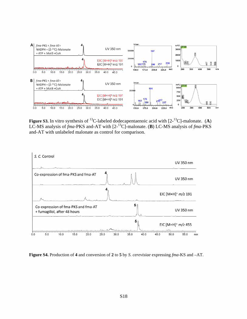
Figure S3. In vitro synthesis of 13C-labeled dodecapentaenoic acid with [2-13C]-malonate. (A) LC-MS analysis of fma-PKS and-AT with [2-13C]-malonate. (B) LC-MS analysis of fma-PKS and-AT with unlabeled malonate as control for comparison.
Figure S4. Production of 4 and conversion of 2 to 5 by S. cerevisiae expressing fma-KS and –AT.
S18
A
B
Homology model based on template c3ts7B: Chain B, farnesyl diphosphate synthase from Methylococcus capsulatus.
188 residues (65% of Af520 (fma-TC) 289 residues protein sequence, aligned region shown above) have been modeled with 92.4% confidence by the single highest scoring template.
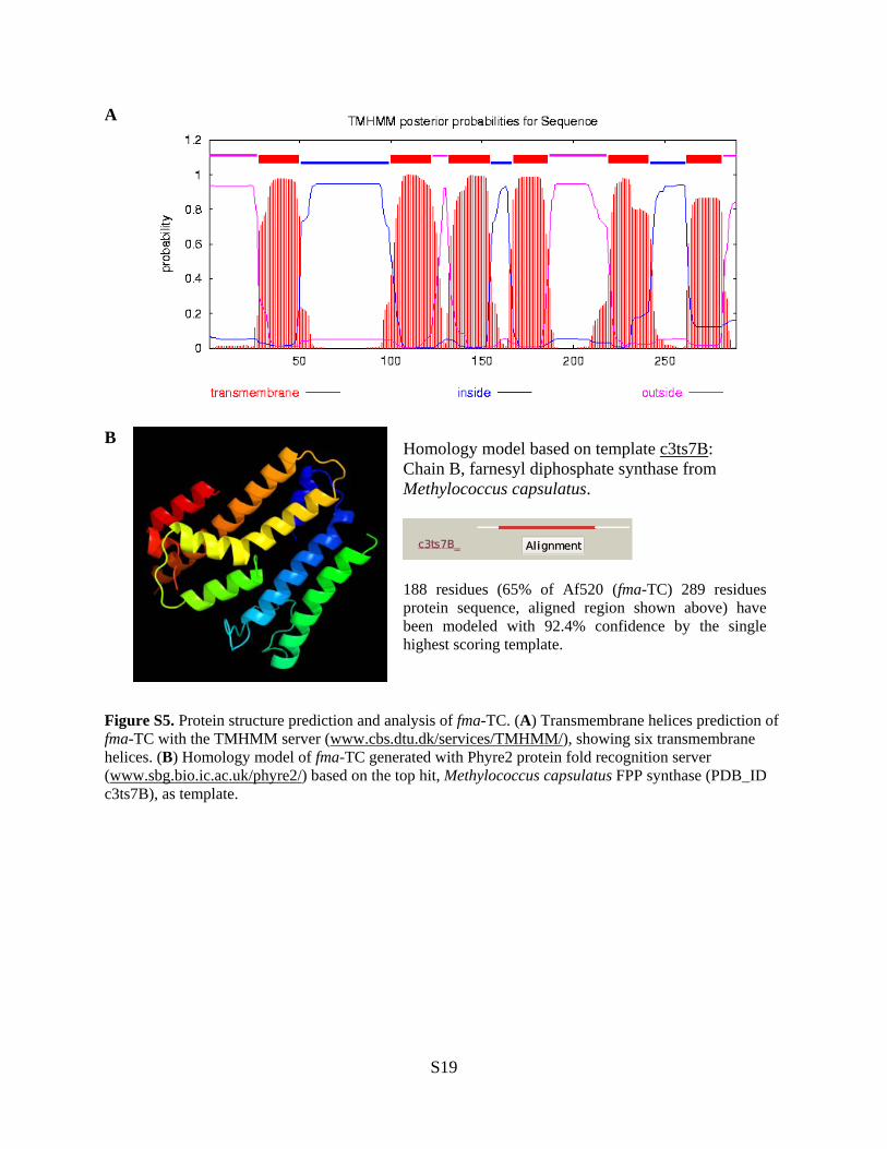
Figure S5. Protein structure prediction and analysis of fma-TC. (A) Transmembrane helices prediction of fma-TC with the TMHMM server (www.cbs.dtu.dk/services/TMHMM/), showing six transmembrane helices. (B) Homology model of fma-TC generated with Phyre2 protein fold recognition server (www.sbg.bio.ic.ac.uk/phyre2/) based on the top hit, Methylococcus capsulatus FPP synthase (PDB_ID c3ts7B), as template.
S19
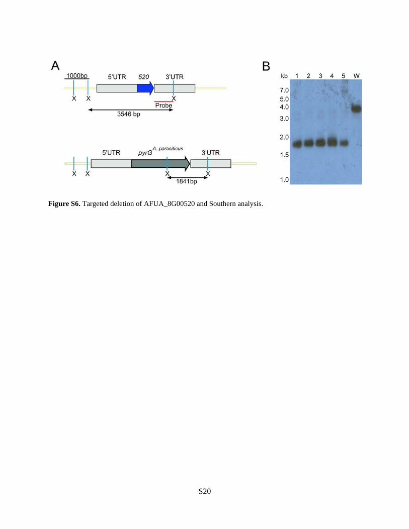
Figure S6. Targeted deletion of AFUA_8G00520 and Southern analysis.
S20

Figure S7. 1H NMR spectrum of 3 (CDCl3, 500 MHz).
S21
7.5 7.0 6.5 6.0 5.5 5.0 4.5 4.0 3.5 3.0 2.5 2.0 1.5 1.0 0.5 ppm
0.6985
1.3981
1.4180
1.5865
1.5923
1.6076
1.6258
1.6818
1.6834
1.8053
1.8255
1.9280
1.9446
2.0401
2.0468
2.0514
2.2135
2.2350
2.2622
2.2730
2.2932
2.5130
2.5361
2.5557
2.5719
4.5439
4.6150
4.6171
5.1279
5.1306
5.1333
5.1364
5.1421
5.1448
5.1475
5.1533
5.1563
5.1590
5.1617
7.2400
1.02
5.10
3.04
1.91
2.08
1.08
2.05
1.98
0.99
1.02
1.00
hcl-20121202-AF520, 1H spectrum, AF520-cell-acetone-Hex-Silica-Fr.1, in CDCl3, AV500-cryo, 2012/12/02
2.252.30 ppm
2.213
2.217
2.219
2.229
2.235
2.248
2.253
2.255
2.258
2.262
2.273
2.284
2.293
2.305
2.55 ppm
2.495
2.502
2.513
2.519
2.524
2.531
2.536
2.541
2.546
2.551
2.556
2.561
2.572
1.92.0 ppm
1.794
1.797
1.805
1.809
1.812
1.818
1.825
1.832
1.912
1.928
1.945
1.960
2.028
2.040
2.047
2.051
2.058

Figure S8. 13C NMR spectrum of 3 (CDCl3, 125 MHz).
S22
160 150 140 130 120 110 100 90 80 70 60 50 40 30 20 10 ppm
17.5647
18.5835
23.4867
23.5096
23.7546
25.7058
27.1087
38.2224
38.6643
43.7826
50.1767
76.7462
77.0002
77.2542
106.0095
125.1437
131.0607
152.1302
hcl-20121202-AF520, 13C spectrum, AF520-cel-acetone-Hex-Silica-Fr.1, in CDCl3, AV500-cryo, 2012/12/02
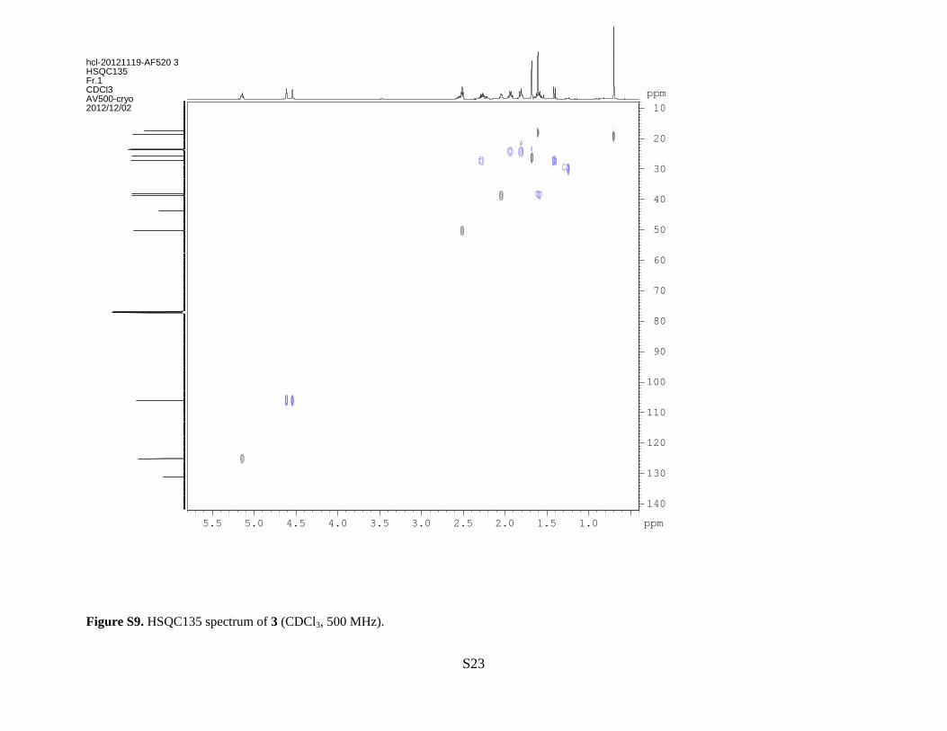
Figure S9. HSQC135 spectrum of 3 (CDCl3, 500 MHz).
S23
ppm
1.01.52.02.53.03.54.04.55.05.5 ppm
10
20
30
40
50
60
70
80
90
100
110
120
130
140
hcl-20121119-AF520 3HSQC135Fr.1CDCl3AV500-cryo2012/12/02
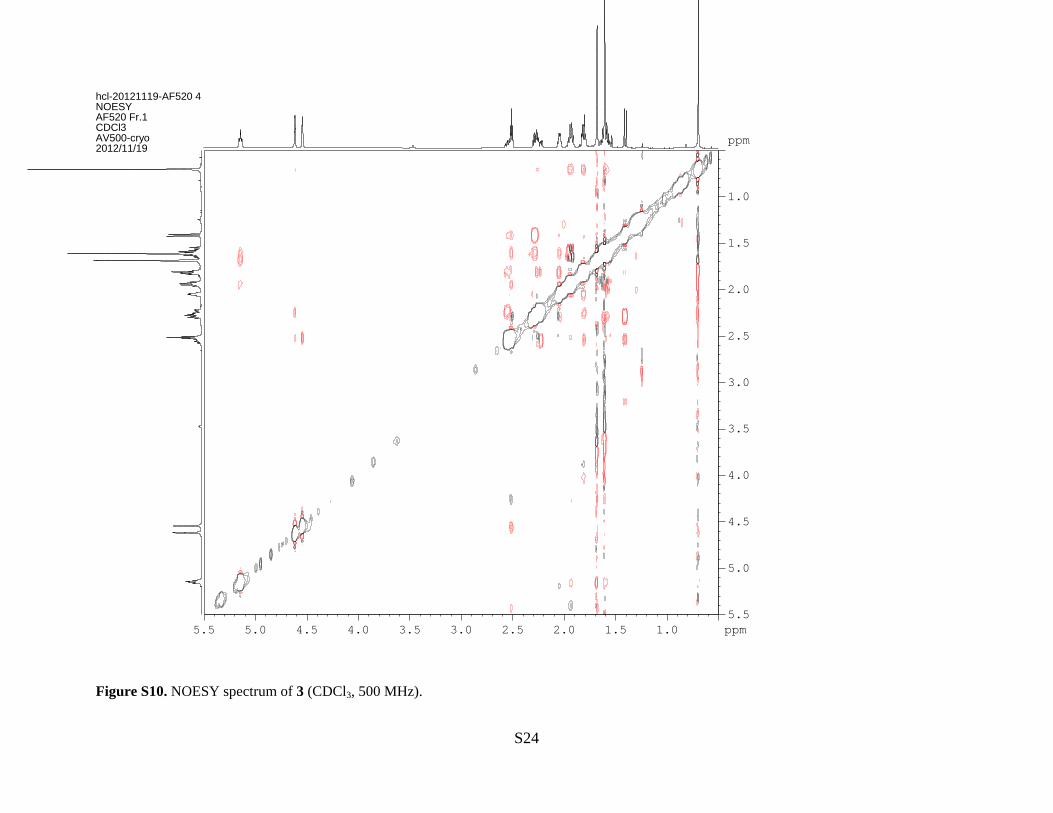
Figure S10. NOESY spectrum of 3 (CDCl3, 500 MHz).
S24
ppm
1.01.52.02.53.03.54.04.55.05.5 ppm
1.0
1.5
2.0
2.5
3.0
3.5
4.0
4.5
5.0
5.5
hcl-20121119-AF520 4NOESYAF520 Fr.1CDCl3AV500-cryo2012/11/19
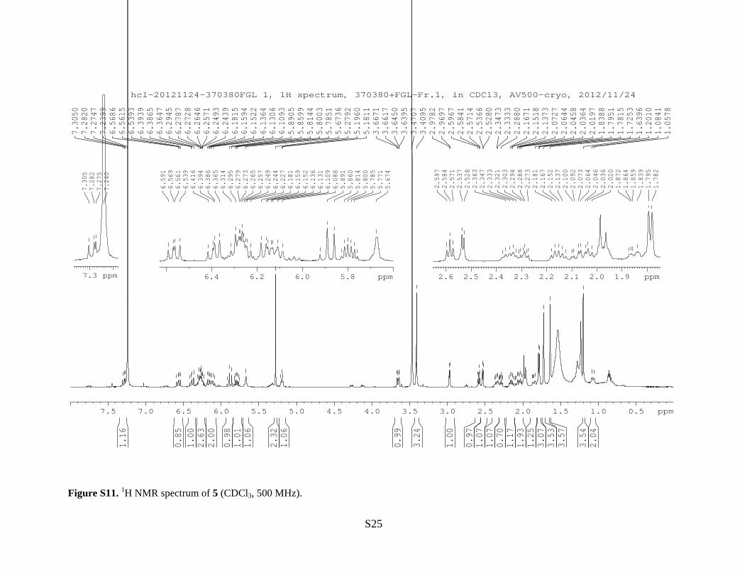
Figure S11. 1H NMR spectrum of 5 (CDCl3, 500 MHz).
S25
7.5 7.0 6.5 6.0 5.5 5.0 4.5 4.0 3.5 3.0 2.5 2.0 1.5 1.0 0.5 ppm
1.05
781.
0841
1.20
101.
6396
1.72
531.
7815
1.79
511.
8388
2.01
972.
0364
2.04
582.
0644
2.07
272.
1373
2.15
182.
1671
2.28
802.
3333
2.34
732.
5280
2.53
662.
5714
2.58
412.
5967
2.96
972.
9782
3.40
953.
4707
3.63
953.
6450
3.66
173.
6671
5.18
115.
1960
5.27
925.
6736
5.78
515.
8003
5.81
445.
8599
5.89
056.
1093
6.13
066.
1364
6.15
226.
1594
6.18
156.
2439
6.24
936.
2571
6.26
466.
2728
6.27
876.
2945
6.36
476.
3865
6.39
396.
5393
6.56
156.
5686
7.23
997.
2747
7.28
207.
3050
2.04
3.54
3.57
3.53
3.07
1.25
1.93
1.17
0.70
1.07
1.07
0.97
1.00
3.24
0.99
1.06
2.32
1.06
1.01
0.98
2.00
2.63
1.00
0.85
1.16
hcl-20121124-370380FGL 1, 1H spectrum, 370380+FGL-Fr.1, in CDCl3, AV500-cryo, 2012/11/24
1.92.02.12.22.32.42.52.6 ppm
1.78
21.
795
1.83
91.
859
1.86
41.
872
2.02
02.
036
2.04
62.
064
2.07
32.
092
2.10
02.
137
2.15
22.
167
2.18
12.
273
2.28
82.
294
2.30
32.
321
2.33
32.
347
2.36
32.
528
2.53
72.
571
2.58
42.
597
5.86.06.26.4 ppm
5.67
45.
771
5.78
55.
800
5.81
45.
860
5.89
16.
088
6.10
96.
131
6.13
66.
152
6.15
96.
181
6.22
76.
244
6.24
96.
257
6.26
56.
273
6.27
96.
295
6.31
46.
365
6.38
66.
394
6.41
66.
539
6.56
16.
569
6.59
1
7.3 ppm
7.24
07.
275
7.28
27.
305
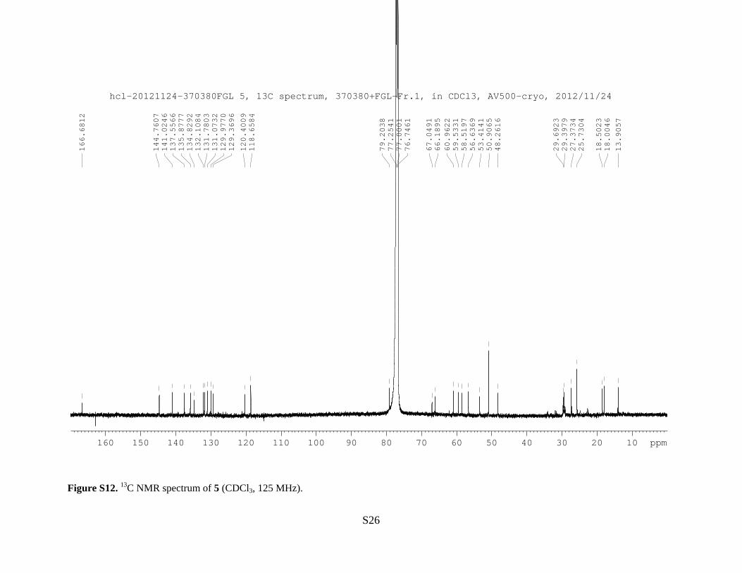
Figure S12. 13C NMR spectrum of 5 (CDCl3, 125 MHz).
S26
160 150 140 130 120 110 100 90 80 70 60 50 40 30 20 10 ppm
13.9057
18.0046
18.5023
25.7304
27.3734
29.3979
29.6923
48.2616
50.9065
53.4141
56.6369
58.5197
59.5331
60.9622
66.1895
67.0491
76.7461
77.0001
77.2541
79.2038
118.6584
120.4009
129.3696
129.9770
131.0732
131.7803
132.1084
134.8292
135.8777
137.5566
141.0246
144.7607
166.6812
hcl-20121124-370380FGL 5, 13C spectrum, 370380+FGL-Fr.1, in CDCl3, AV500-cryo, 2012/11/24
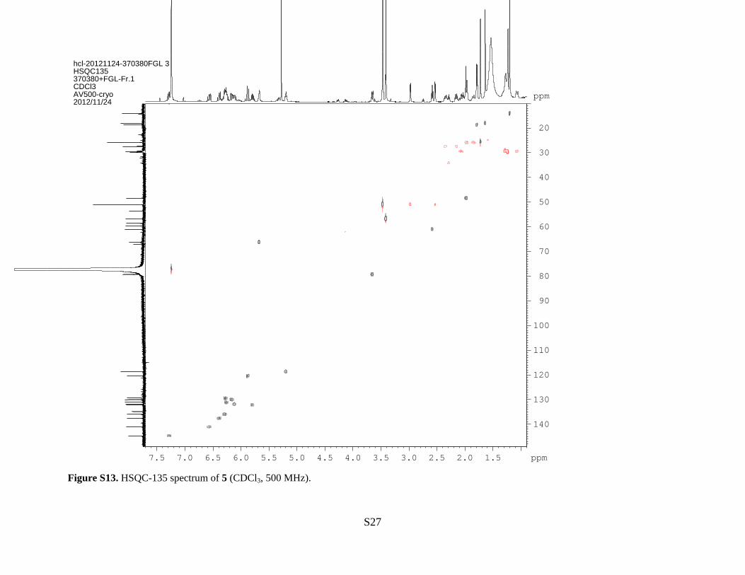
Figure S13. HSQC-135 spectrum of 5 (CDCl3, 500 MHz).
S27
ppm
1.52.02.53.03.54.04.55.05.56.06.57.07.5 ppm
20
30
40
50
60
70
80
90
100
110
120
130
140
hcl-20121124-370380FGL 3HSQC135370380+FGL-Fr.1CDCl3AV500-cryo2012/11/24
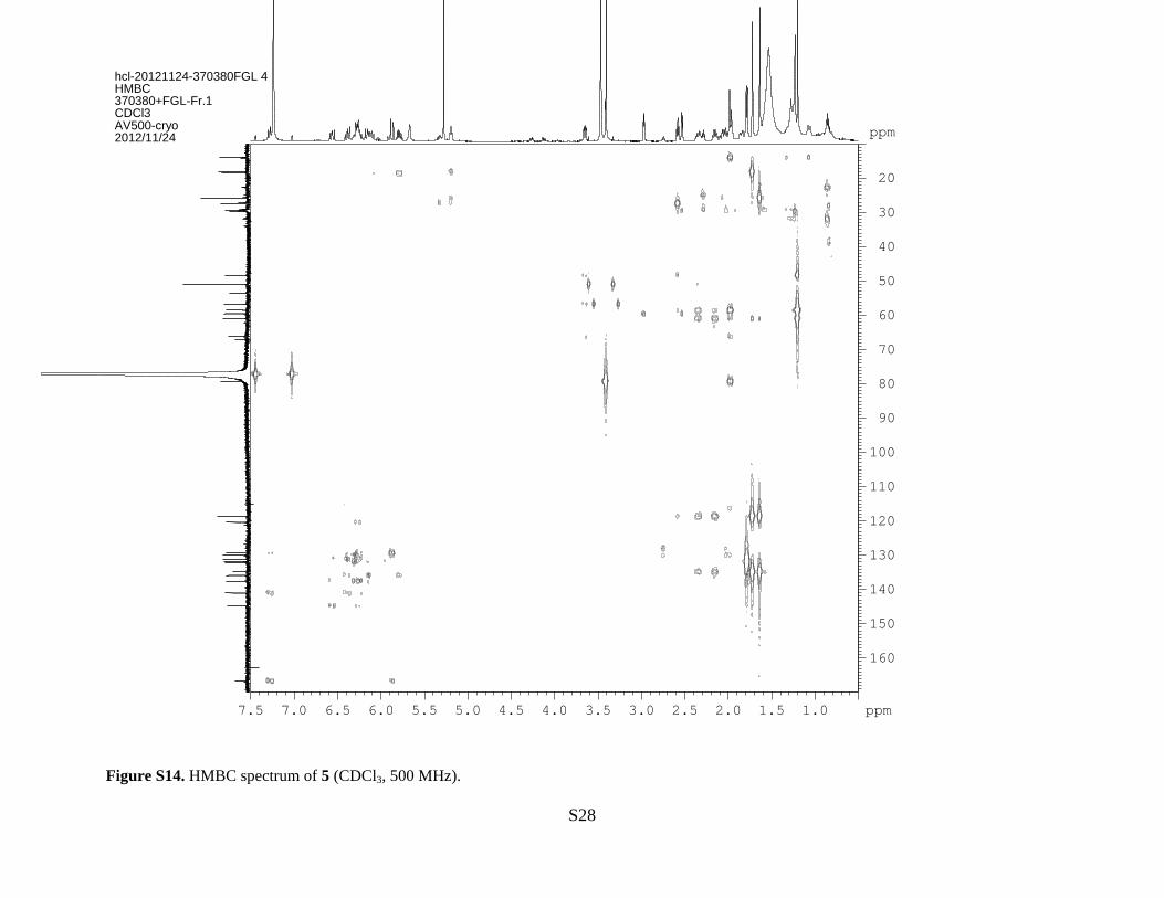
Figure S14. HMBC spectrum of 5 (CDCl3, 500 MHz).
S28
ppm
1.01.52.02.53.03.54.04.55.05.56.06.57.07.5 ppm
20
30
40
50
60
70
80
90
100
110
120
130
140
150
160
hcl-20121124-370380FGL 4HMBC370380+FGL-Fr.1CDCl3AV500-cryo2012/11/24
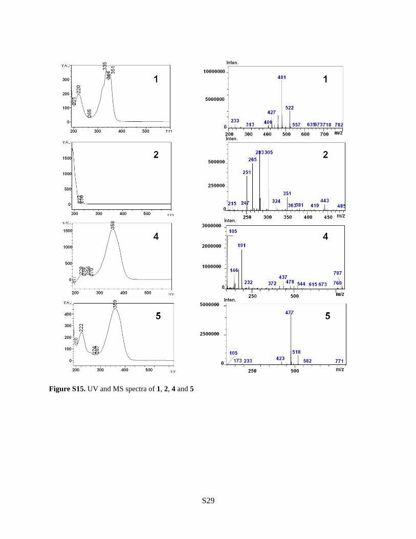
Figure S15. UV and MS spectra of 1, 2, 4 and 5
S29

Figure S16. GC-MS data of 3 purified from S. cerevisiae expressing fma-TC.
S30
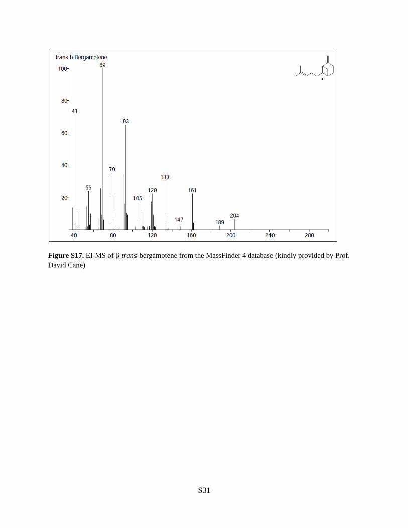
Figure S17. EI-MS of β-trans-bergamotene from the MassFinder 4 database (kindly provided by Prof. David Cane)
S31
Supplementary References (1) Kafer, E. Advances in genetics 1977, 19, 33. (2) Sambrook, J.; Russell, D. W. Molecular cloning: a laboratory manual; CSHL press, 2001; Vol. 1. (3) Kelley, L. A.; Sternberg, M. J. E. Nat. Protoc. 2009, 4, 363. (4) McGuffin, L. J.; Bryson, K.; Jones, D. T. Bioinformatics 2000, 16, 404. (5) Krogh, A.; Larsson, B.; von Heijne, G.; Sonnhammer, E. L. J. Mol. Biol. 2001, 305, 567. (6) Nugent, T.; Jones, D. T. BMC bioinformatics 2012, 13, 169. (7) Szewczyk, E.; Nayak, T.; Oakley, C. E.; Edgerton, H.; Xiong, Y.; Taheri-Talesh, N.; Osmani, S. A.; Oakley, B. R. Nat. Protoc. 2007, 1, 3111. (8) da Silva Ferreira, M. E.; Kress, M. R.; Savoldi, M.; Goldman, M. H.; Hartl, A.; Heinekamp, T.; Brakhage, A. A.; Goldman, G. H. Eukaryot. Cell 2006, 5, 207. (9) Xu, W.; Cai, X.; Jung, M. E.; Tang, Y. J. Am. Chem. Soc. 2010, 132, 13604. (10) Jez, J. M.; Ferrer, J. L.; Bowman, M. E.; Dixon, R. A.; Noel, J. P. Biochemistry 2000, 39, 890. (11) Ralston, L.; Kwon, S. T.; Schoenbeck, M.; Ralston, J.; Schenk, D. J.; Coates, R. M.; Chappell, J. Arch. Biochem. Biophys. 2001, 393, 222. (12) Cane, D. E.; Mcilwaine, D. B.; Oliver, J. S. J. Am. Chem. Soc. 1990, 112, 1285. (13) Czerson, H.; Bohlmann, F.; Stuessy, T. F.; Fischer, N. H. Phytochemistry 1979, 18, 257. (14) Nozoe, S.; Kobayashi, H.; Morisaki, N. Tetrahedron Lett. 1976, 4625. (15) Halasz, J.; Podanyi, B.; Vasvari-Debreczy, L.; Szabo, A.; Hajdu, F.; Bocskei, Z.; Hegedus-Vajda, J.; Gyorbiro, A.; Hermecz, I. Tetrahedron 2000, 56, 10081. Complete Citation for Abbreviated References in the Manuscript (18) Nierman, W. C.; Pain, A.; Anderson, M. J.; Wortman, J. R.; Kim, H. S.; Arroyo, J.; Berriman, M.; Abe, K.; Archer, D. B.; Bermejo, C.; Bennett, J.; Bowyer, P.; Chen, D.; Collins, M.; Coulsen, R.; Davies, R.; Dyer, P. S.; Farman, M.; Fedorova, N.; Feldblyum, T. V.; Fischer, R.; Fosker, N.; Fraser, A.; Garcia, J. L.; Garcia, M. J.; Goble, A.; Goldman, G. H.; Gomi, K.; Griffith-Jones, S.; Gwilliam, R.; Haas, B.; Haas, H.; Harris, D.; Horiuchi, H.; Huang, J.; Humphray, S.; Jimenez, J.; Keller, N.; Khouri, H.; Kitamoto, K.; Kobayashi, T.; Konzack, S.; Kulkarni, R.; Kumagai, T.; Lafon, A.; Latge, J. P.; Li, W.; Lord, A.; Lu, C.; Majoros, W. H.; May, G. S.; Miller, B. L.; Mohamoud, Y.; Molina, M.; Monod, M.; Mouyna, I.; Mulligan, S.; Murphy, L.; O'Neil, S.; Paulsen, I.; Penalva, M. A.; Pertea, M.; Price, C.; Pritchard, B. L.; Quail, M. A.; Rabbinowitsch, E.; Rawlins, N.; Rajandream, M. A.; Reichard, U.; Renauld, H.; Robson, G. D.; Rodriguez de Cordoba, S.; Rodriguez-Pena, J. M.; Ronning, C. M.; Rutter, S.; Salzberg, S. L.; Sanchez, M.; Sanchez-Ferrero, J. C.; Saunders, D.; Seeger, K.; Squares, R.; Squares, S.; Takeuchi, M.; Tekaia, F.; Turner, G.; Vazquez de Aldana, C. R.; Weidman, J.; White, O.; Woodward, J.; Yu, J. H.; Fraser, C.; Galagan, J. E.; Asai, K.; Machida, M.; Hall, N.; Barrell, B.; Denning, D. W. Nature 2005, 438, 1151.
(21) Ma, S. M.; Li, J. W.; Choi, J. W.; Zhou, H.; Lee, K. K.; Moorthie, V. A.; Xie, X.; Kealey, J. T.; Da Silva, N. A.; Vederas, J. C.; Tang, Y. Science 2009, 326, 589.
(30) (a) Sallaud, C.; Rontein, D.; Onillon, S.; Jabes, F.; Duffe, P.; Giacalone, C.; Thoraval, S.; Escoffier, C.; Herbette, G.; Leonhardt, N.; Causse, M.; Tissier, A. Plant Cell 2009
S32