Supporting Information Title Role of a SpoVA Protein in...
18
1 Supporting Information Title: Role of a SpoVA Protein in Dipicolinic Acid Uptake into Developing Spores of Bacillus subtilis Author affiliation: Yunfeng Li 1 , Andrew Davis 1 , George Korza 1 , Pengfei Zhang 2 , Yong-qing Li 2 , Barbara Setlow 1 , Peter Setlow 1 and Bing Hao 1 1 Department of Molecular, Microbial and Structural Biology, University of Connecticut Health Center, Farmington, CT 06030-3305 2 Department of Physics, East Carolina University, Greenville, NC 27858-4353
Transcript of Supporting Information Title Role of a SpoVA Protein in...
1
Supporting Information
Title: Role of a SpoVA Protein in Dipicolinic Acid Uptake into Developing Spores of Bacillus
subtilis
Author affiliation: Yunfeng Li1, Andrew Davis1, George Korza1, Pengfei Zhang2, Yong-qing
Li2, Barbara Setlow1, Peter Setlow1 and Bing Hao1
1Department of Molecular, Microbial and Structural Biology, University of Connecticut Health
Center, Farmington, CT 06030-3305
2Department of Physics, East Carolina University, Greenville, NC 27858-4353
2
Supporting Information List
1. Supplemental Table S1
2. Supplemental Fig. S1
3. Supplemental Fig. S2
4. Supplemental Fig. S3
5. Supplemental Fig. S4
6. Supplemental Fig. S5
7. Supplemental Fig. S6
8. Supplemental Fig. S7
9. Supplemental Fig. S8
3
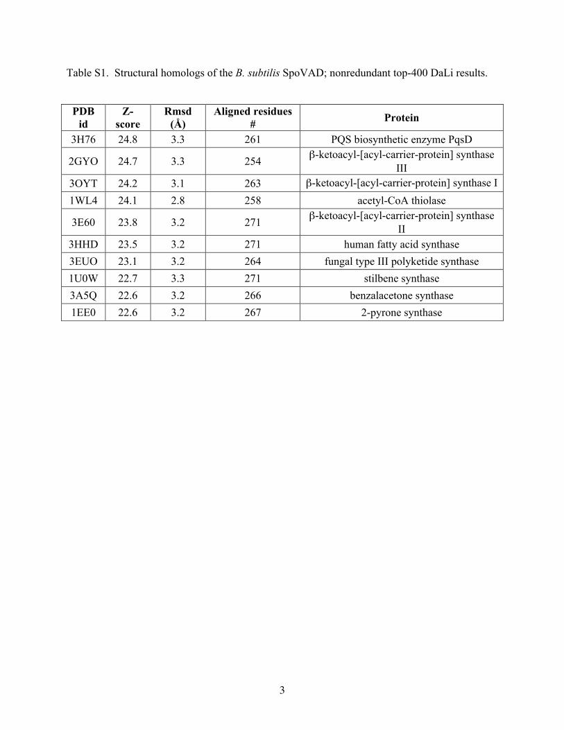
Table S1. Structural homologs of the B. subtilis SpoVAD; nonredundant top-400 DaLi results.
PDB
id Z-
score Rmsd
(Å) Aligned residues
# Protein
3H76 24.8 3.3 261 PQS biosynthetic enzyme PqsD
2GYO 24.7 3.3 254 β-ketoacyl-[acyl-carrier-protein] synthase III
3OYT 24.2 3.1 263 β-ketoacyl-[acyl-carrier-protein] synthase I 1WL4 24.1 2.8 258 acetyl-CoA thiolase
3E60 23.8 3.2 271 β-ketoacyl-[acyl-carrier-protein] synthase II
3HHD 23.5 3.2 271 human fatty acid synthase 3EUO 23.1 3.2 264 fungal type III polyketide synthase 1U0W 22.7 3.3 271 stilbene synthase 3A5Q 22.6 3.2 266 benzalacetone synthase 1EE0 22.6 3.2 267 2-pyrone synthase
4
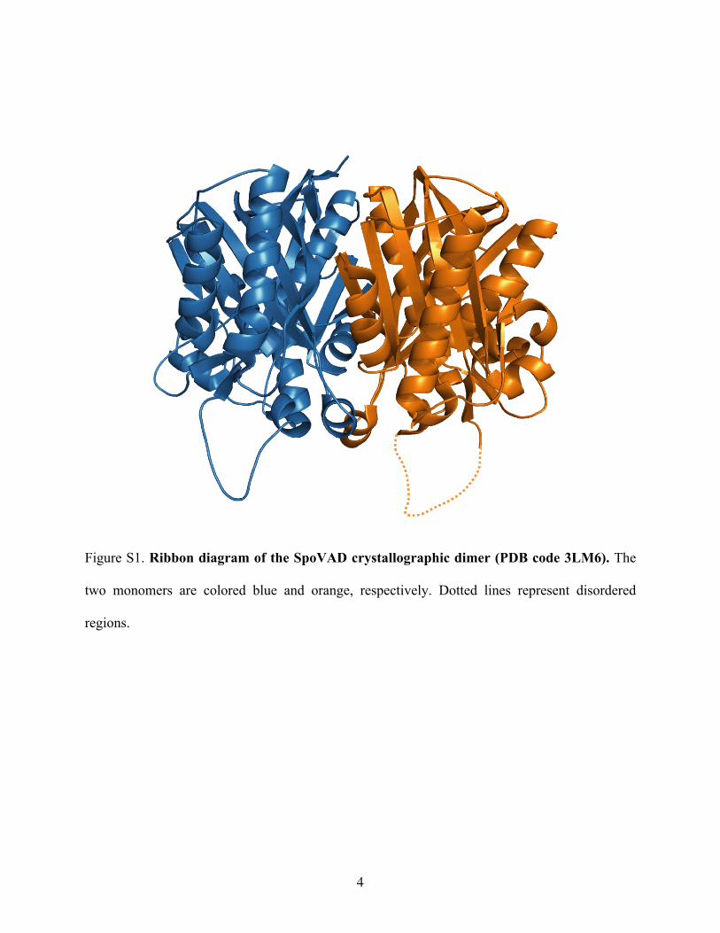
Figure S1. Ribbon diagram of the SpoVAD crystallographic dimer (PDB code 3LM6). The
two monomers are colored blue and orange, respectively. Dotted lines represent disordered
regions.
5
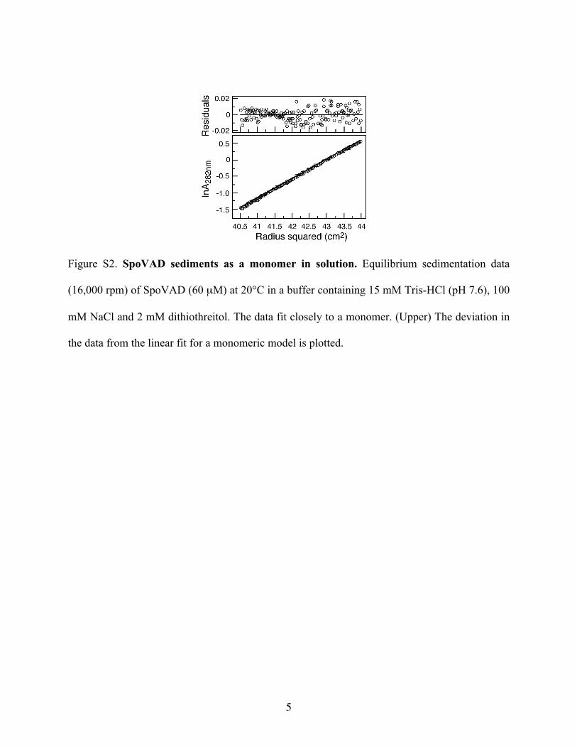
Figure S2. SpoVAD sediments as a monomer in solution. Equilibrium sedimentation data
(16,000 rpm) of SpoVAD (60 µM) at 20°C in a buffer containing 15 mM Tris-HCl (pH 7.6), 100
mM NaCl and 2 mM dithiothreitol. The data fit closely to a monomer. (Upper) The deviation in
the data from the linear fit for a monomeric model is plotted.
6
7
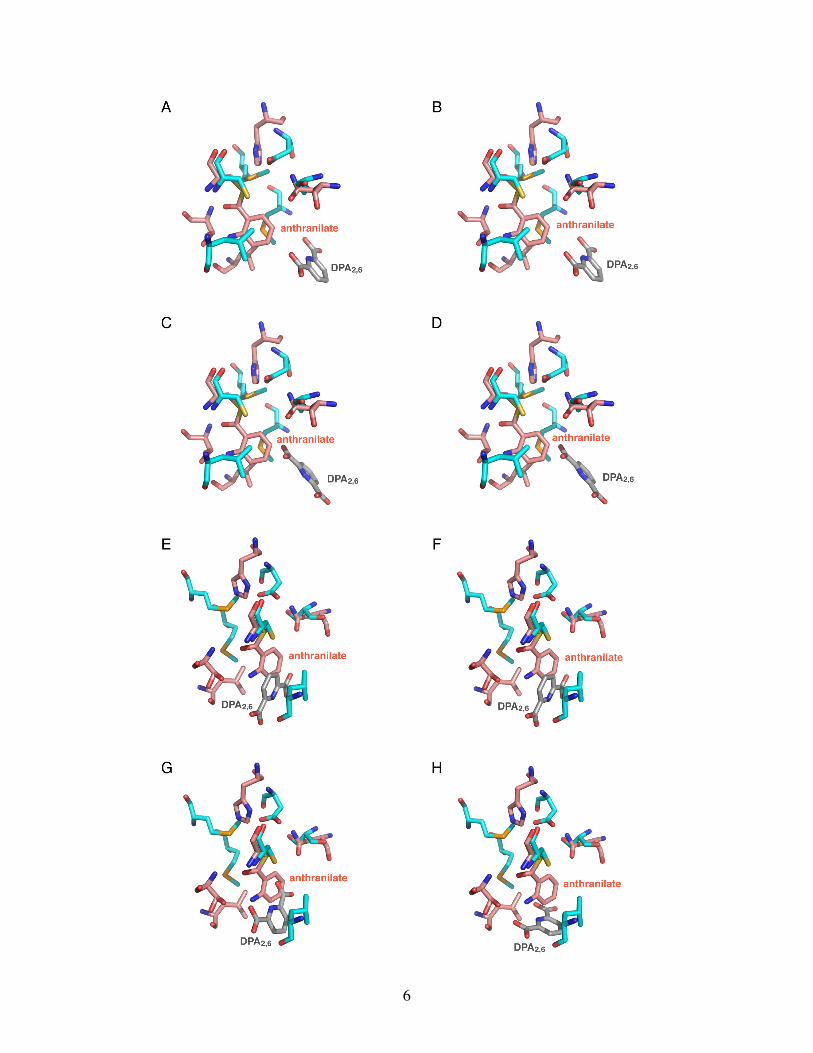
Figure S3. The docking conformations of the DPA2,6 molecule to SpoVAD. A-H, Eight of the
top nine docking solutions for the DPA2,6 binding to SpoVAD (PDB code 3LM6) using
AutoDock Vina. DPA2,6 was docked as a rigid body and all the nine solutions found the DPA2,6
molecule in the same presumed binding site in SpoVAD.
8
9
10
11
12
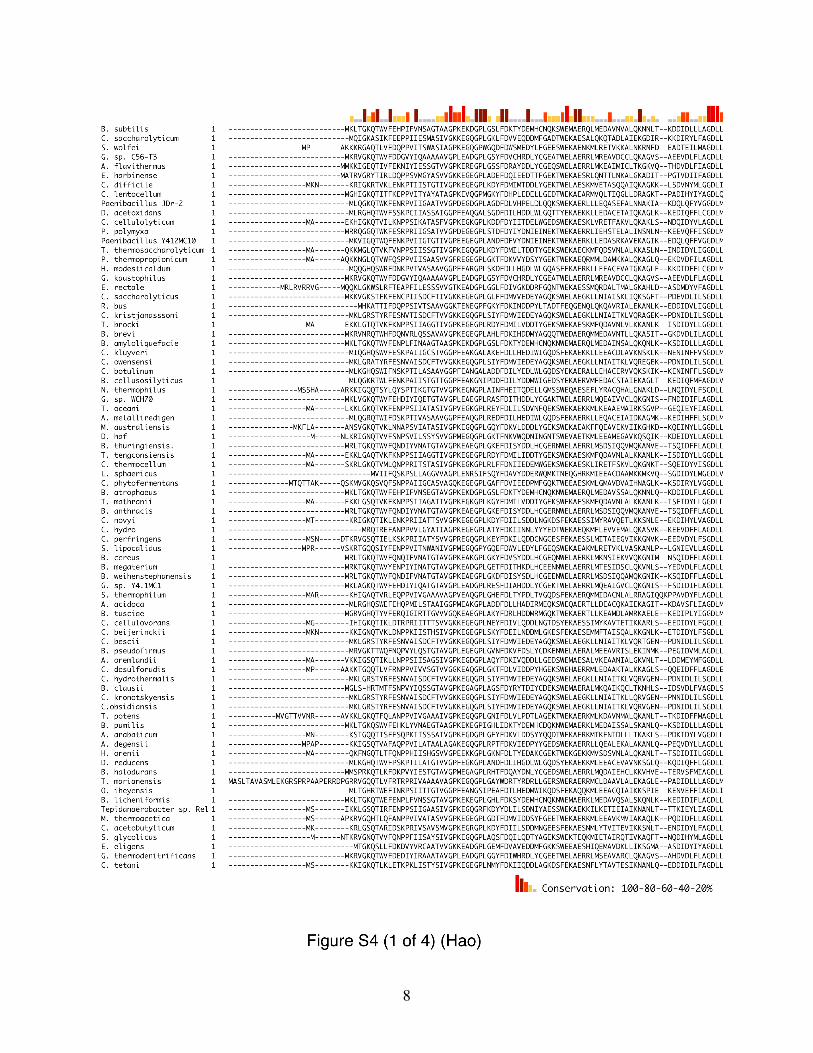
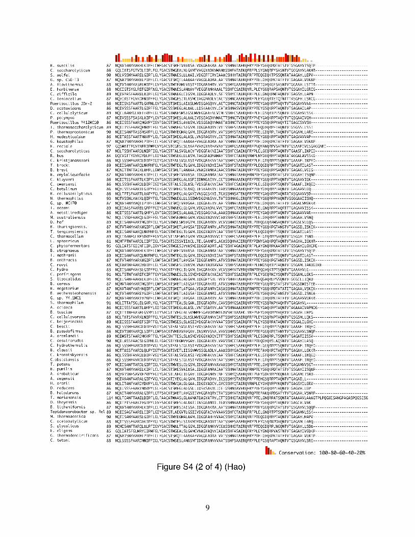
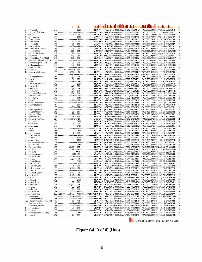
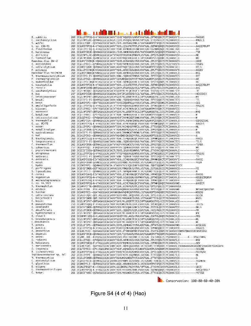
Figure S4. Sequence alignment of SpoVAD orthologs in the Bacillus and Clostridium
species. A ClustalW alignment of 80 SpoVAD orthologs was performed using DNASTAR
Lasergene suite 8 (DNASTAR Inc., Madison, WI). Sequence conservation is shown as a bar
graph, with red bars indicating identity among SpoVAD homologs.
13
14
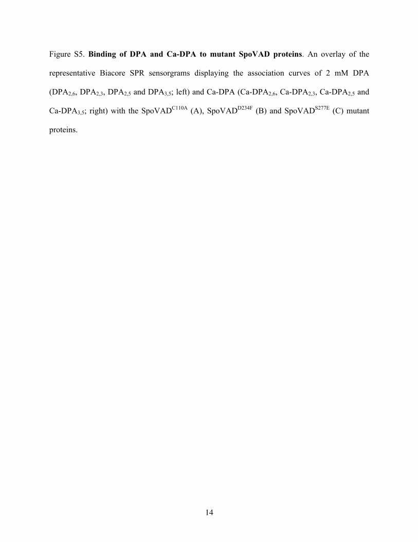
Figure S5. Binding of DPA and Ca-DPA to mutant SpoVAD proteins. An overlay of the
representative Biacore SPR sensorgrams displaying the association curves of 2 mM DPA
(DPA2,6, DPA2,3, DPA2,5 and DPA3,5; left) and Ca-DPA (Ca-DPA2,6, Ca-DPA2,3, Ca-DPA2,5 and
Ca-DPA3,5; right) with the SpoVADC110A (A), SpoVADD234F (B) and SpoVADS277E (C) mutant
proteins.
15
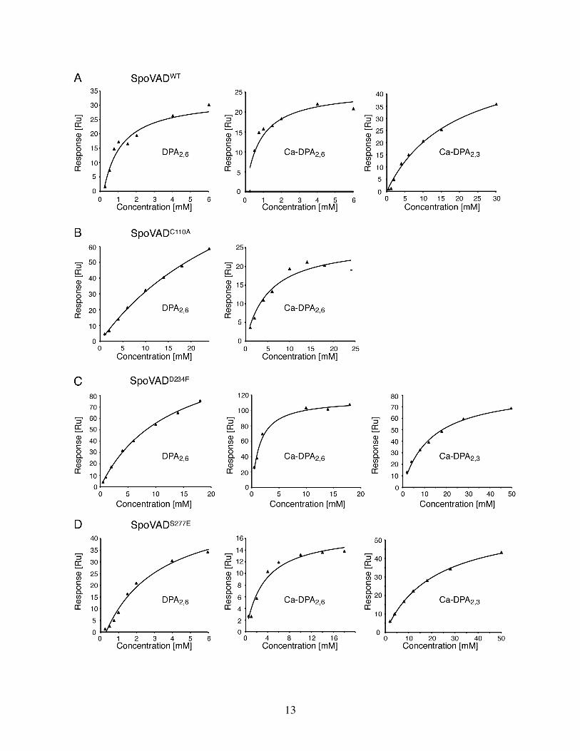
Figure S6. Measurement of binding affinity of DPA/Ca-DPA to SpoVAD by SPR. Multiple
concentrations of DPA2,6, Ca-DPA2,6 or Ca-DPA2,3 were used to determine their binding affinity
(KD) to the wild-type SpoVAD (A), SpoVADC110A (B), SpoVADD234F (C) and SpoVADS277E (D)
(also see Table 1). The titration data were analyzed by nonlinear curve fitting to a steady-state

16
model using Biacore T100 analysis software (GE Healthcare, Piscataway, NJ) and OriginPro 7.5
(OriginLab Corporation, Northampton, MA). The binding of Ca-DPA2,3 to SpoVADC110A was
too weak to be measured.
17
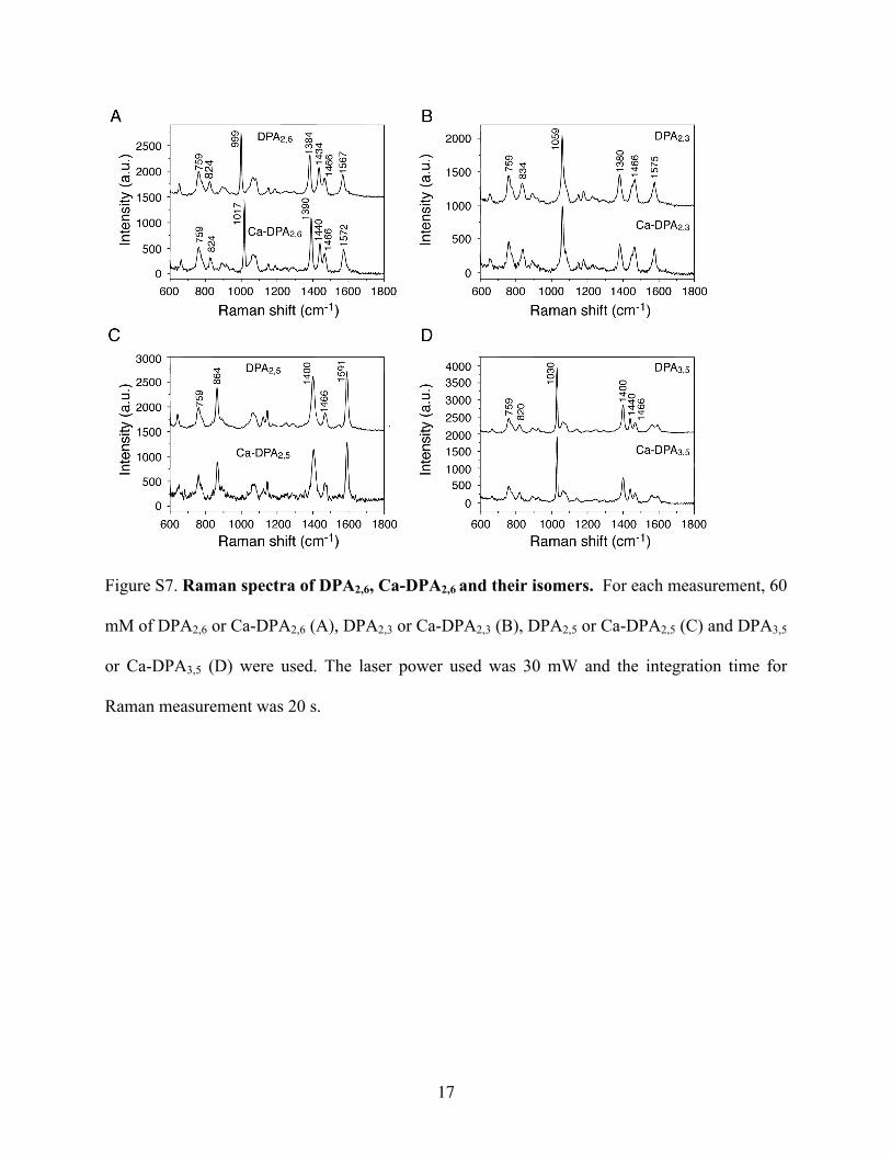
Figure S7. Raman spectra of DPA2,6, Ca-DPA2,6 and their isomers. For each measurement, 60
mM of DPA2,6 or Ca-DPA2,6 (A), DPA2,3 or Ca-DPA2,3 (B), DPA2,5 or Ca-DPA2,5 (C) and DPA3,5
or Ca-DPA3,5 (D) were used. The laser power used was 30 mW and the integration time for
Raman measurement was 20 s.
18
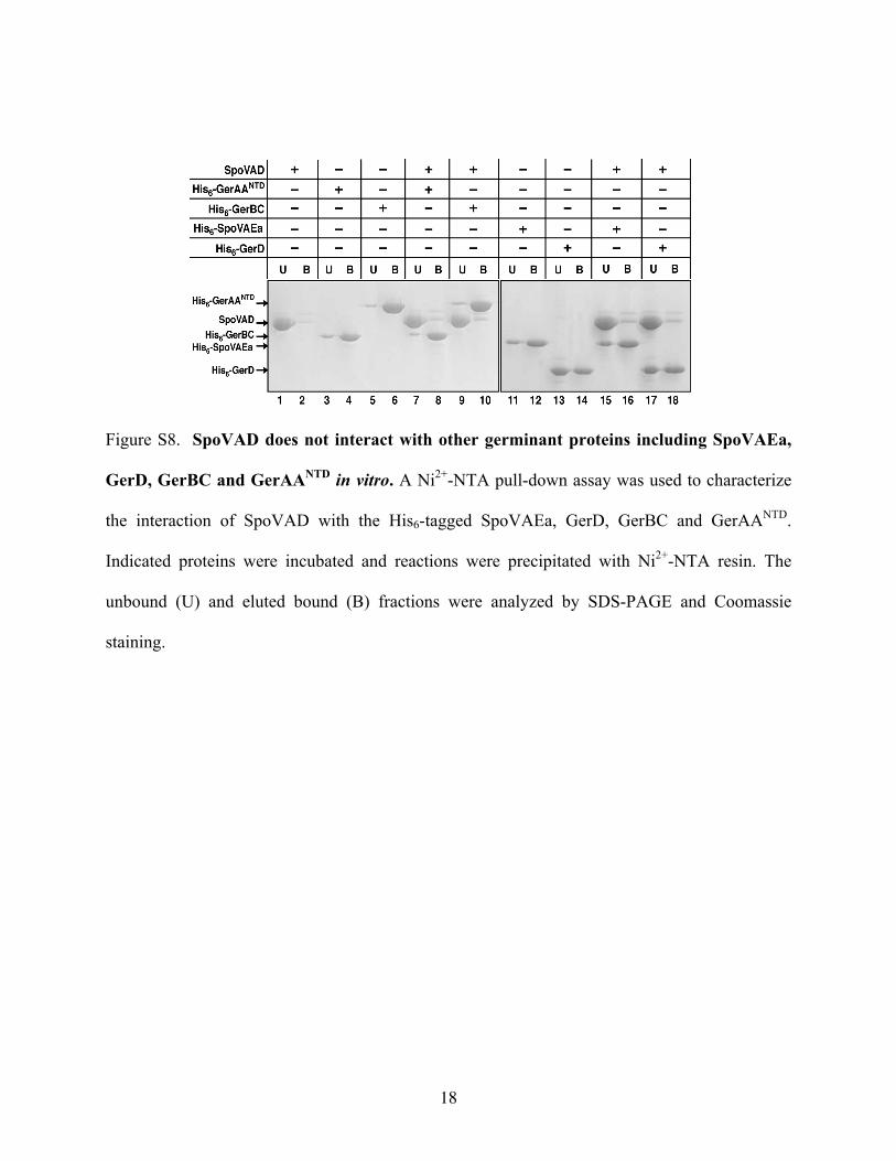
Figure S8. SpoVAD does not interact with other germinant proteins including SpoVAEa,
GerD, GerBC and GerAANTD in vitro. A Ni2+-NTA pull-down assay was used to characterize
the interaction of SpoVAD with the His6-tagged SpoVAEa, GerD, GerBC and GerAANTD.
Indicated proteins were incubated and reactions were precipitated with Ni2+-NTA resin. The
unbound (U) and eluted bound (B) fractions were analyzed by SDS-PAGE and Coomassie
staining.