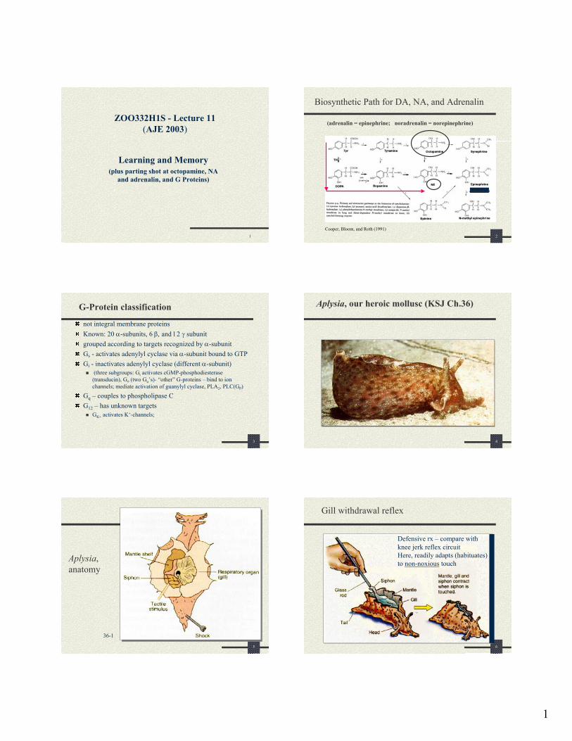
Biosynthetic Path for DA, NA, and Adrenalin
9
1 1 ZOO332H1S - Lecture 11 (AJE 2003) Learning and Memory (plus parting shot at octopamine, NA and adrenalin, and G Proteins) 2 Biosynthetic Path for DA, NA, and Adrenalin (adrenalin = epinephrine; noradrenalin = norepinephrine) Adrenalin Cooper, Bloom, and Roth (1991) 3 G-Protein classification not integral membrane proteins Known: 20 α-subunits, 6 β, and 12 γ subunit grouped according to targets recognized by α-subunit G s - activates adenylyl cyclase via α-subunit bound to GTP G i - inactivates adenylyl cyclase (different α-subunit) (three subgroups: G t activates cGMP-phosphodiesterase (transducin), Go (two G o ’s)- “other” G-proteins – bind to ion channels; mediate activation of guanylyl cyclase, PLA 2 , PLC(Gp) G q – couples to phospholipase C G 12 – has unknown targets G K+ activates K + -channels; 4 Aplysia, our heroic mollusc (KSJ Ch.36) 5 Aplysia, anatomy 36-1 6 Gill withdrawal reflex Defensive rx – compare with knee jerk reflex circuit Here, readily adapts (habituates) to non-noxious touch
Transcript of Biosynthetic Path for DA, NA, and Adrenalin
1
1
ZOO332H1S - Lecture 11(AJE 2003)
Learning and Memory(plus parting shot at octopamine, NA
and adrenalin, and G Proteins)
2
Biosynthetic Path for DA, NA, and Adrenalin
(adrenalin = epinephrine; noradrenalin = norepinephrine)
Adrenalin
Cooper, Bloom, and Roth (1991)
3
G-Protein classification
not integral membrane proteinsKnown: 20 α-subunits, 6 β, and 12 γ subunitgrouped according to targets recognized by α-subunitGs - activates adenylyl cyclase via α-subunit bound to GTPGi - inactivates adenylyl cyclase (different α-subunit)
(three subgroups: Gt activates cGMP-phosphodiesterase(transducin), Go (two Go’s)- “other” G-proteins – bind to ion channels; mediate activation of guanylyl cyclase, PLA2, PLC(Gp)
Gq – couples to phospholipase CG12 – has unknown targets
GK+ activates K+-channels;
4
Aplysia, our heroic mollusc (KSJ Ch.36)
5
Aplysia, anatomy
36-1
6
Gill withdrawal reflex
Defensive rx – compare with knee jerk reflex circuitHere, readily adapts (habituates) to non-noxious touch
2
7
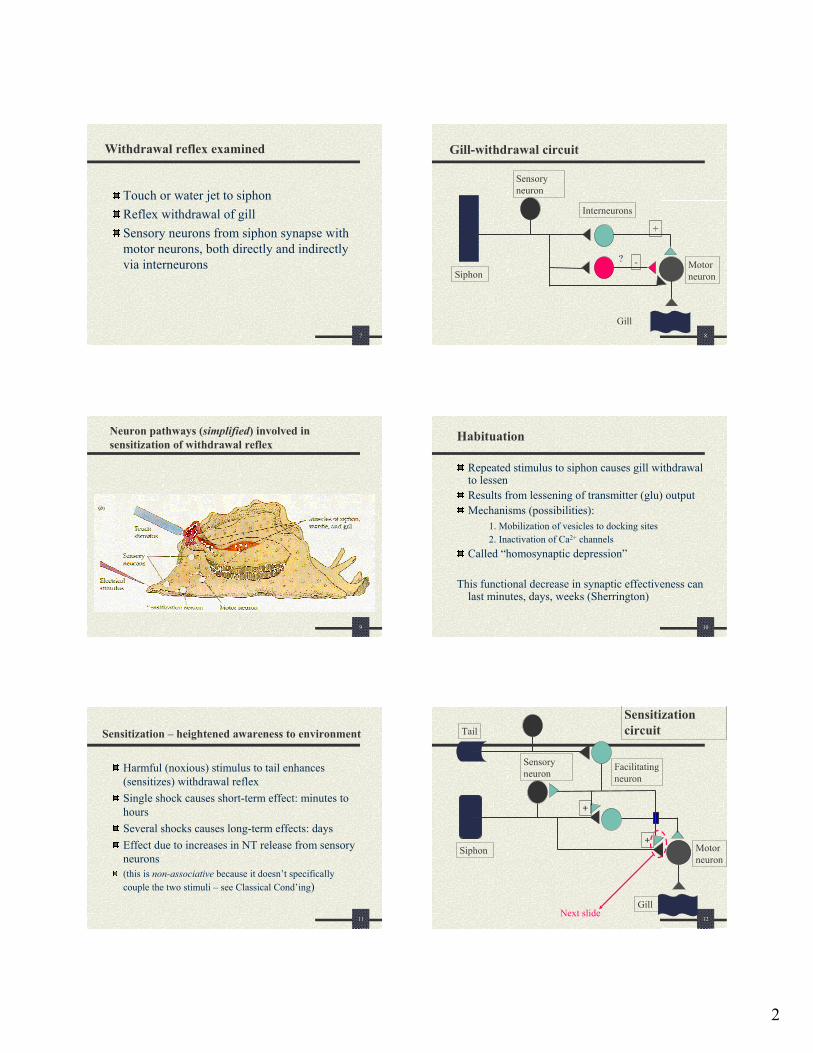
Withdrawal reflex examined
Touch or water jet to siphonReflex withdrawal of gillSensory neurons from siphon synapse with motor neurons, both directly and indirectly via interneurons
8
Gill-withdrawal circuit
Sensory neuron
Motor neuron
Gill
Siphon
Interneurons
+
-?
9
Neuron pathways (simplified) involved in sensitization of withdrawal reflex
10
Habituation
Repeated stimulus to siphon causes gill withdrawal to lessenResults from lessening of transmitter (glu) outputMechanisms (possibilities):
1. Mobilization of vesicles to docking sites2. Inactivation of Ca2+ channels
Called “homosynaptic depression”
This functional decrease in synaptic effectiveness can last minutes, days, weeks (Sherrington)
11
Sensitization – heightened awareness to environment
Harmful (noxious) stimulus to tail enhances (sensitizes) withdrawal reflexSingle shock causes short-term effect: minutes to hoursSeveral shocks causes long-term effects: daysEffect due to increases in NT release from sensory neurons(this is non-associative because it doesn’t specifically couple the two stimuli – see Classical Cond’ing)
12
Sensitization circuit
Motor neuron
Gill
Siphon
Facilitating neuron
Tail
Sensory neuron
+
+
Next slide
3
13
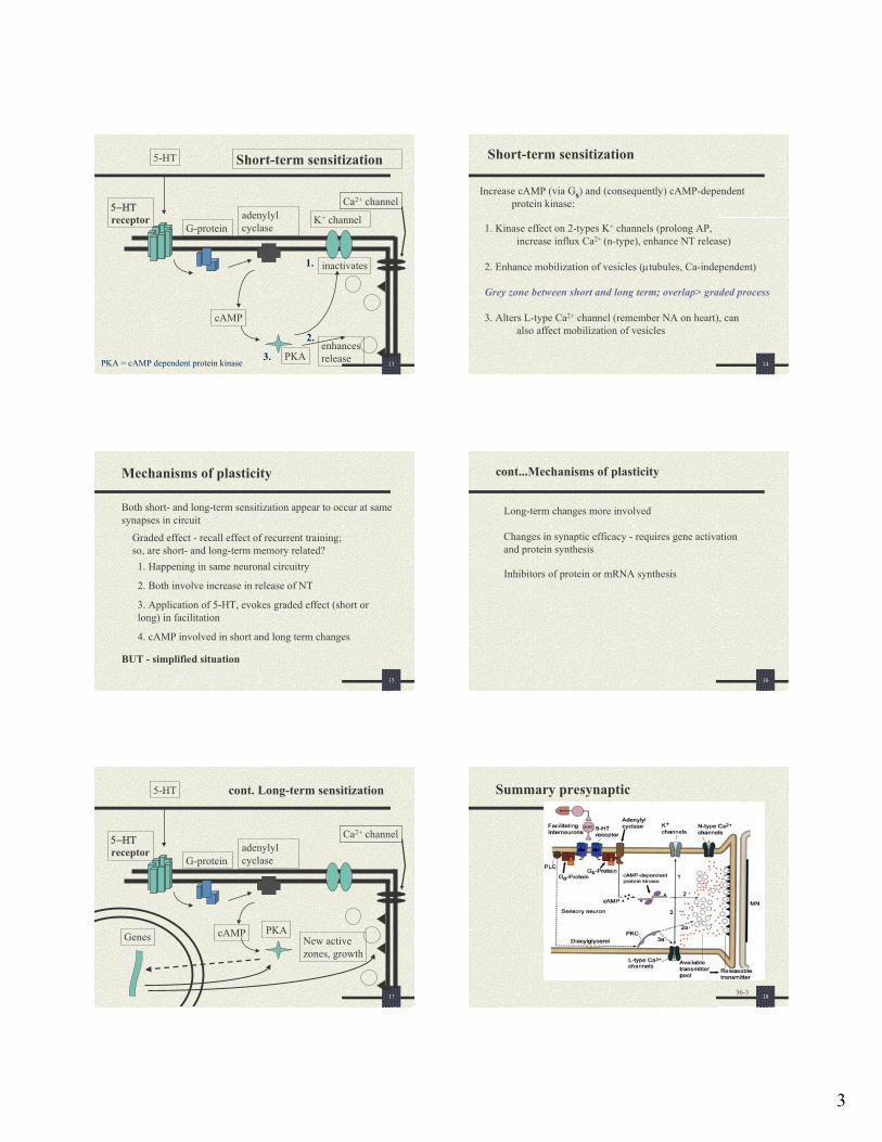
Short-term sensitization
5−ΗΤreceptor
G-proteinadenylyl cyclase
K+ channel
cAMP
PKA
inactivates
5-HT
enhancesrelease
Ca2+ channel
1.
2.
3.PKA = cAMP dependent protein kinase 14
Short-term sensitization
Increase cAMP (via Gs) and (consequently) cAMP-dependent protein kinase:
1. Kinase effect on 2-types K+ channels (prolong AP, increase influx Ca2+ (n-type), enhance NT release)
2. Enhance mobilization of vesicles (µtubules, Ca-independent)
Grey zone between short and long term; overlap> graded process
3. Alters L-type Ca2+ channel (remember NA on heart), can also affect mobilization of vesicles
15
Mechanisms of plasticity
Graded effect - recall effect of recurrent training; so, are short- and long-term memory related?
Both short- and long-term sensitization appear to occur at same synapses in circuit
BUT - simplified situation
1. Happening in same neuronal circuitry
2. Both involve increase in release of NT
3. Application of 5-HT, evokes graded effect (short or long) in facilitation
4. cAMP involved in short and long term changes
16
cont...Mechanisms of plasticity
Changes in synaptic efficacy - requires gene activation and protein synthesis
Long-term changes more involved
Inhibitors of protein or mRNA synthesis
17
cont. Long-term sensitization
5−ΗΤreceptor
G-proteinadenylyl cyclase
cAMP PKA
5-HT
New active zones, growth
Genes
Ca2+ channel
18
Summary presynaptic
36-3
4
19
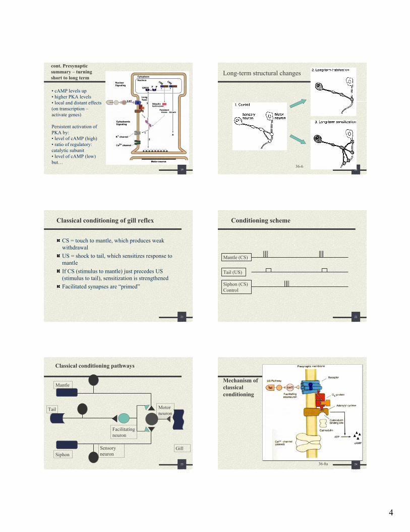
cont. Presynaptic summary – turning short to long term
• cAMP levels up• higher PKA levels• local and distant effects (on transcription –activate genes)
Persistent activation of PKA by:• level of cAMP (high) • ratio of regulatory: catalytic subunit• level of cAMP (low) but…
20
Long-term structural changes
36-6
21
Classical conditioning of gill reflex
CS = touch to mantle, which produces weak withdrawalUS = shock to tail, which sensitizes response to mantleIf CS (stimulus to mantle) just precedes US (stimulus to tail), sensitization is strengthenedFacilitated synapses are “primed”
22
Conditioning scheme
Mantle (CS)
Tail (US)
Siphon (CS) Control
23
Classical conditioning pathways
Sensory neuron
Motor neuron
GillSiphon
Tail
Facilitating neuron
Mantle
24
Mechanism of classical conditioning
36-8a
5
25
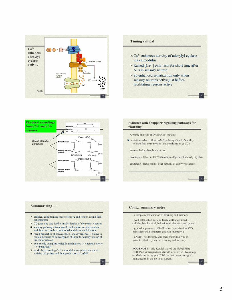
Ca2+
enhances adenylyl cyclase activity
36-8b
26
Timing critical
Ca2+ enhances activity of adenylyl cyclase via calmodulinRaised [Ca2+] only lasts for short time after APs in sensory neuronSo enhanced sensitization only when sensory neurons active just before facilitating neurons active
27
Electrical recordingsfrom CS+ and CS-neurons
Recall stimulusparadigm
28
Evidence which supports signaling pathways for “learning”
Genetic analysis of Drosophila mutants
mutations which effect cAMP pathway alter fly’s abilityto learn first year physics (and sensitization & CC)
dunce - lacks phosphodiesterase
rutabaga - defect in Ca2+/calmodulin-dependent adenylyl cyclase
amnesiac - lacks control over activity of adenylyl cyclase
29
Summarizing….
classical conditioning more effective and longer lasting than sensitizationCC goes one step further in facilitation of the sensory neuronsensory pathways from mantle and siphon are independent and thus one can be conditioned and the other left alonerecall properties of convergence (and divergence) - timing is critical because of convergence of input to sensory neuron at the motor neuronaxo-axonic synapses typically modulatory (>> neural activity >>> behaviour)works by recruiting Ca2+/calmodulin to cyclase, enhances activity of cyclase and thus production of cAMP
30
Cont…summary notes
• a simple representation of learning and memory
• well established system, fairly well understood -cellular, biochemical, behavioural, electrical and genetic
• graded appearance of facilitation (sensitization, CC), coincident with long term effects (“memory”)
• cAMP - not the only 2nd messenger involved in synaptic plasticity, and in learning and memory
FOOTNOTE: Eric Kandel shared the Nobel Prize (with Paul Greengard and Arvid Carlsson) in Physiology or Medicine in the year 2000 for their work on signal transduction in the nervous system.
6
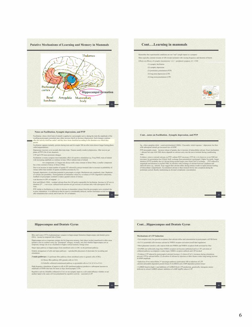
31
Putative Mechanisms of Learning and Memory in Mammals
Hippocampal formation
32
Cont….Learning in mammals
Remember that experimental conditions are not “real” (single inputs to a synapse)
More typically constant streams of APs invade terminals with varying frequency and duration of bursts
Effects on efficacy of synaptic transmission: 1,2,3 – peripheral synapses; 4,5 - CNS
(1) synaptic facilitation
(2) synaptic depression
(3) posttetanic potentiation (PTP)
(4) long term depression (LTP)
(5) long term potentiation (LTP)
33
Notes on Facilitation, Synaptic depression, and PTP
Facilitation: when a brief train of stimuli is applied to a presynaptic nerve, during the train the amplitude of the resulting postsynaptic potentials may either increase (facil) or decrease (depression). Such changes continue after the activity itself has ended` and they have been classified according to the duration over which they persist.Facilitation: appears instantly, persists during train and for couple 100 ms after train.(slower longer lasting phase called augmentation).Depression: recovery over seconds after train stops. Tetanus usually results in depression. After recover get phase of PTP (10s of min duration).LTP and LTD more persistent changes.Facilitation: at many synapses most immediate effect of repetitive stimulation (eg. Frog NMJ); train of stimuli with increasing amplitude as continue in train. Effect outlasts train of stimuli
two components: (1) largest one decays with a time constant of about 50ms; a smaller component has a time constant of decay of about 250ms.Due to an increase in mean number of quanta NT released by presyn terminal (due to increase in probability of release rather than number of quanta available).(increase by 2x)Synaptic depression: of end plate potential is presynaptic in origin. Mechanism not completely clear. Depletion of vesicles one possibility. Autoregulation of transmitter release by co-release of ATP, degraded to adenosine, feedback on presynaptic terminal to reduce quantal content of release.(can decrease to 20% of original …)Katz and Miledi (1968) – residual calcium from first AP partly responsible for facilitation. Increase in AP size or duration (2nd….) not occur; cultured leech neurons not get increase of calcium entry with subsequent APs in train.PTP similar to facilitation as it refers to increase in transmitter release from the presynaptic nerve terminal due to prior stimulation. I t is different in that its onset is considerably delayed, reaches maximum several seconds after stimulation has ceased, and it lasts for 10’s of minutes.
34
Cont…notes on Facilitation , Synaptic depression, and PTP
Eg., ciliary ganglion chick – curarized,stimulated (100Hz, 15seconds); initial response = depression, but then with subsequent stimuli get increased size of EPSP. PTP presynaptic origin – increase release of quanta, due to increase of intracellular calcium. Exact mechanism = obscure but exps with NMJ shows depends on calcium entry into the nerve terminal during conditioning train.Evidence: remove external calcium, no PTP; sodium NOT necessary (TTX & v/c)); however, at rat NMJ not necessary for potentiation but if block Na/K exchange then potentiation is prolonged. Higher Na inside, longer duration of PTP probably due to Calcium being around longer since block Na/Ca exchange ….PTP reduced in magnitude and duration at crayfish NMJ, by interfere with exchange of calcium between cytoplasm and intracell stores (eg., mitoch). Exps suggest that calcium influx during tetanus results in rapid calcium loading of intracellular compartments. Accumulated calcium is then released slowly into cytoplasm during the posttetanic period, thereby maintaining an elevated cytoplasmic concentration.
35
Hippocampus and Dentate Gyrus
Bliss and Lomo (1973) at glutamatergic synapses in hippocampal formation (hippocampus and dentate gyrus (DG))– located in temporal lobe of brain
Hippocampus acts as temporary storage site for long-term memory, later (days>weeks) transferred to other areas (putative sites in cerebral cortex for “permanent” storage); Actually, not clear whether hippocampus acts as temporary storage site or as a facilitator to higher cortical memory storage areas
Major input pathway to hippocampus from entorhinal cortex to DG via the perforant pathway
Orderly arrangement of cells and input pathways – reproducible placement of electrodes for recording and stimulation
3 main pathways: (1) perforant fibre pathway (from entorhinal cortex to granule cells of DG);
(2) Mossy fibre pathway (DG granule cells to CA3)
(3) Schaffer collateral-commissural pathway to pyramidal cells in CA1 (CA3 to CA1)
High frequency stimulation of inputs to cells in DG (perforant pathway) produces a subsequent increase in amplitude of EPSPs that lasts for hours or days (homosynaptic LTP)
Repetitive activity (Schaffer collateral to CA1) at one synaptic input to a cell could influence whether or not another input to the same cell was potentiated by repetitive activity – associative LTP
36
Cont…Hippocampus and Dentate Gyrus
Mechanisms of LTP Induction• Not complete story but general acceptance that calcium influx and concentration in postsynaptic cell NB factor
• In CA1 pyramidal cells increase calcium by NMDA receptor activation (recall dual regulation)
• Most glutamate-sensitive cells express both non-NMDA and NMDA receptors (both activated by Glu)
• If EPSPs not sufficiently large then NMDA receptors not become unblocked and no LTP; activation of collateral pathway accompanies weaker input, NMDA receptors unblock and LTP invoked
• Evidence LTP induced by postsynaptic [calcium] increase: (1) shown [Ca2+]i increases during stimulation, prevent LTP by calcium buffer; (2) elevation of calcium by injection or other means evokes long lasting increase in EPSP amplitude
• Induction of two intracellular 2nd messenger pathways particularly NB in induction of LTP: calcium/calmodulin-dependent protein kinase II (CaMKII) and cAMP dependent protein kinase
• CaMKII found in high [ ] and inhibitors of CaMKII block LTP production; genetically, transgenic mouse deficient in critical CaMKII subunit; inhibition of cAMP-depPK reduces LTP
7
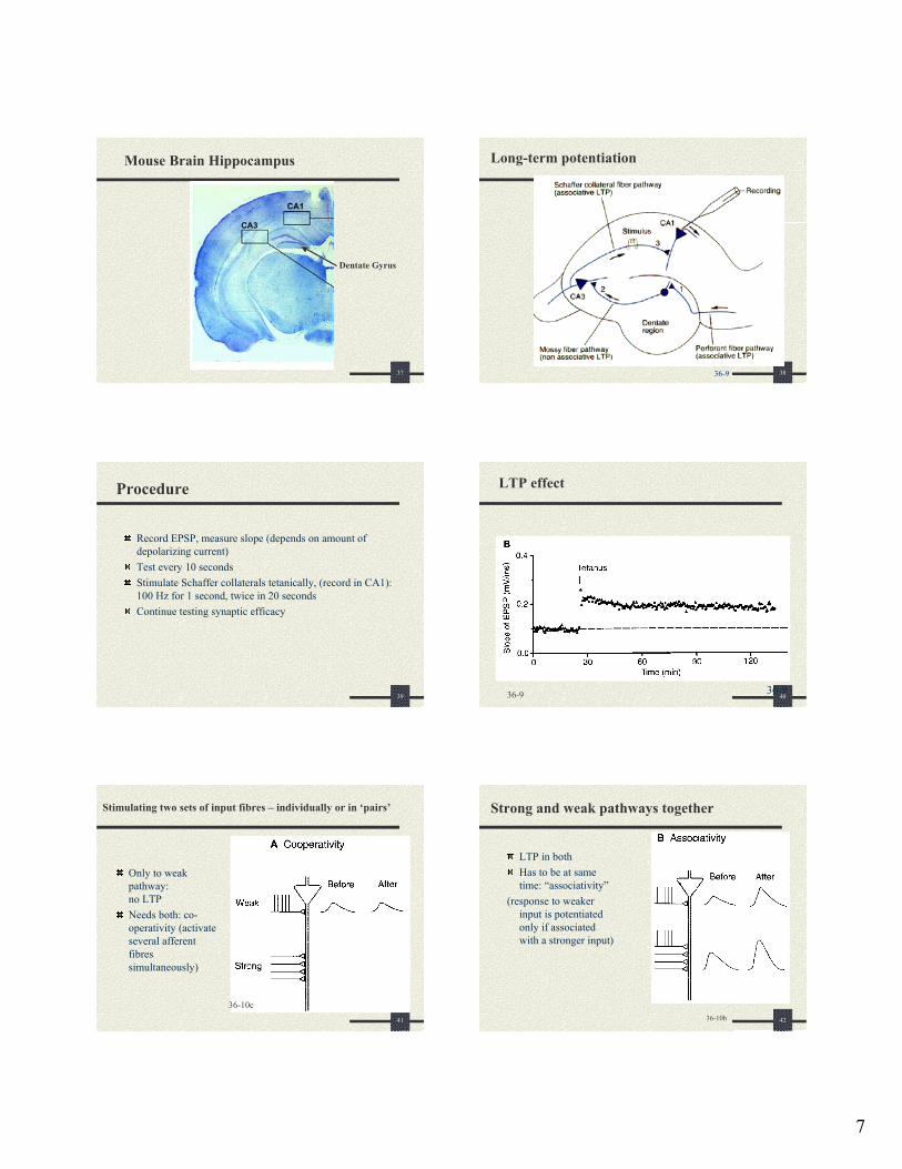
37
Mouse Brain Hippocampus
Dentate Gyrus
38
Long-term potentiation
36-9
39
Procedure
Record EPSP, measure slope (depends on amount of depolarizing current)Test every 10 secondsStimulate Schaffer collaterals tetanically, (record in CA1): 100 Hz for 1 second, twice in 20 secondsContinue testing synaptic efficacy
40
LTP effect
36-936-9
41
Stimulating two sets of input fibres – individually or in ‘pairs’
Only to weak pathway:no LTPNeeds both: co-operativity (activate several afferent fibres simultaneously)
36-10c42
Strong and weak pathways together
LTP in bothHas to be at same time: “associativity”
(response to weaker input is potentiatedonly if associated with a stronger input)
36-10b
8
43
Strong only
LTP in strong, not in weak“Specificity”
(LTP generated at one input not carry over to other synapses – LTP is specific to synapse where generated)
36-10c44
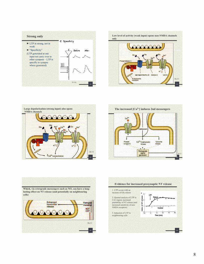
Low level of activity (weak input) opens non-NMDA channels only
36-11
45
Large depolarisation (strong input) also opensNMDA channels
36-11
46
The increased [Ca2+] induces 2nd messengers
36-11
47
Which, via retrograde messengers such as NO, can have a long-lasting effect on NT release (and potentially on neighbouring cells)
36-11
48
Evidence for increased presynaptic NT release
2. Quantal analysis of LTP in CA1 region; increased probability of NT release (and increased sensitivity of non-NMDA receptors)
1. LTP occurs with an increase of Glu release
3. Induction of LTP in neighbouring cells
9
49
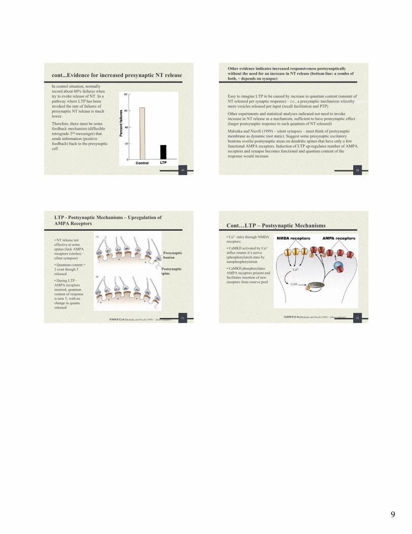
cont...Evidence for increased presynaptic NT release
In control situation, normally record about 60% failures when try to evoke release of NT. In a pathway where LTP has been invoked the rate of failures of presynaptic NT release is much lower.
Therefore, there must be some feedback mechanism (diffusible retrograde 2nd messenger) that sends information (positive feedback) back to the presynaptic cell
50
Other evidence indicates increased responsiveness postsynapticallywithout the need for an increase in NT release (bottom line: a combo of both, ~ depends on synapse)
Easy to imagine LTP to be caused by increase in quantum content (amount of NT released per synaptic response) – i.e., a presynaptic mechanism whereby more vesicles released per input (recall facilitation and PTP)
Other experiments and statistical analyses indicated not need to invoke increase in NT release as a mechanism, sufficient to have postsynaptic effect (larger postsynaptic response to each quantum of NT released)
Malenka and Nicoll (1999) – silent synapses – must think of postsynaptic membrane as dynamic (not static). Suggest some presynaptic excitatory boutons overlie postsynaptic areas on dendritic spines that have only a few functional AMPA receptors. Induction of LTP up-regulates number of AMPA receptors and synapse becomes functional and quantum content of the response would increase
51
LTP - Postsynaptic Mechanisms – Upregulation of AMPA Receptors
Presynapticbouton
Postsynapticspine
NMWF12-8 (Malenka and Nicoll (1999) – silent synapses)
• NT release not effective at some spines (lack AMPA receptors (circles) –silent synapses)
• Quantum content = 2 even though 5 released
• During LTP –AMPA receptors inserted, quantum content of response is now 5, with no change in quanta released
52
Cont…LTP – Postsynaptic Mechanisms
• Ca2+ entry through NMDA receptors;
• CaMKII activated by Ca2+
influx retains it’s active (phosphorylated) state by autophosphorylation
• CaMKII phosphorylates AMPA receptors present and facilitates insertion of new receptors from reserve pool
NMWF12-8 (Malenka and Nicoll (1999) – silent synapses)