Light and Tissue 1 - its.caltech.edubi177/private/L6_handout.pdf · ≥ λ Mie Regime • Cells,...
36
Light and tissue 1
Transcript of Light and Tissue 1 - its.caltech.edubi177/private/L6_handout.pdf · ≥ λ Mie Regime • Cells,...
Light and tissue 1
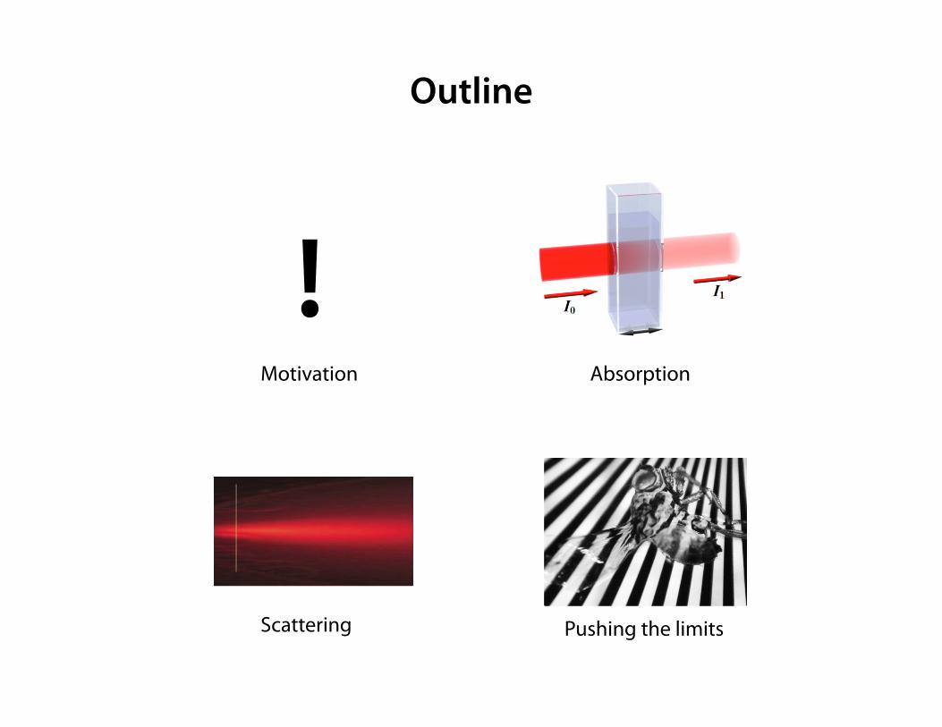
Outline
Motivation
! Absorption
Scattering Pushing the limits
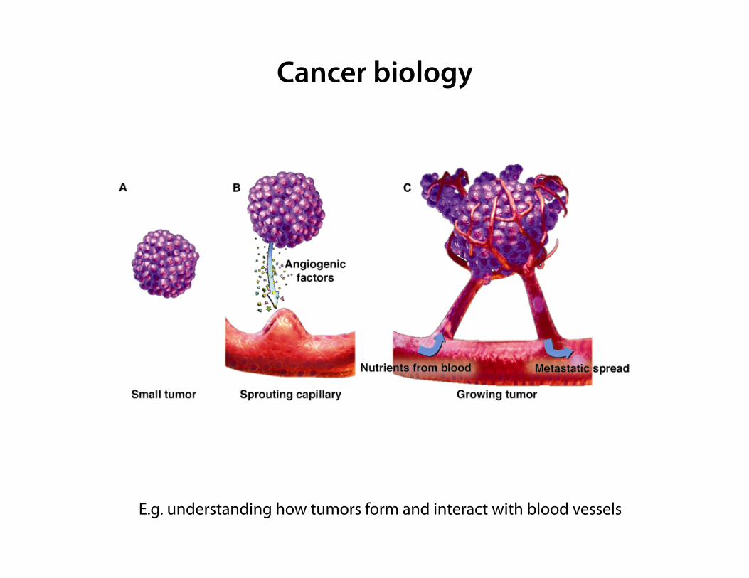
Cancer biology
E.g. understanding how tumors form and interact with blood vessels
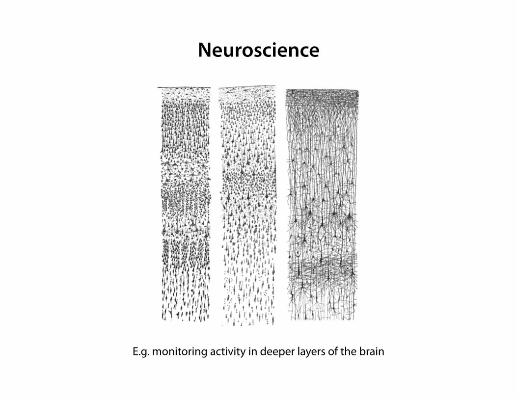
Neuroscience
E.g. monitoring activity in deeper layers of the brain
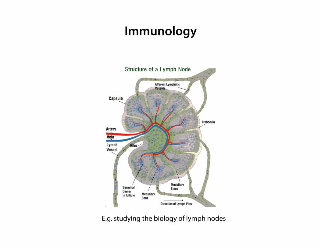
Immunology
E.g. studying the biology of lymph nodes
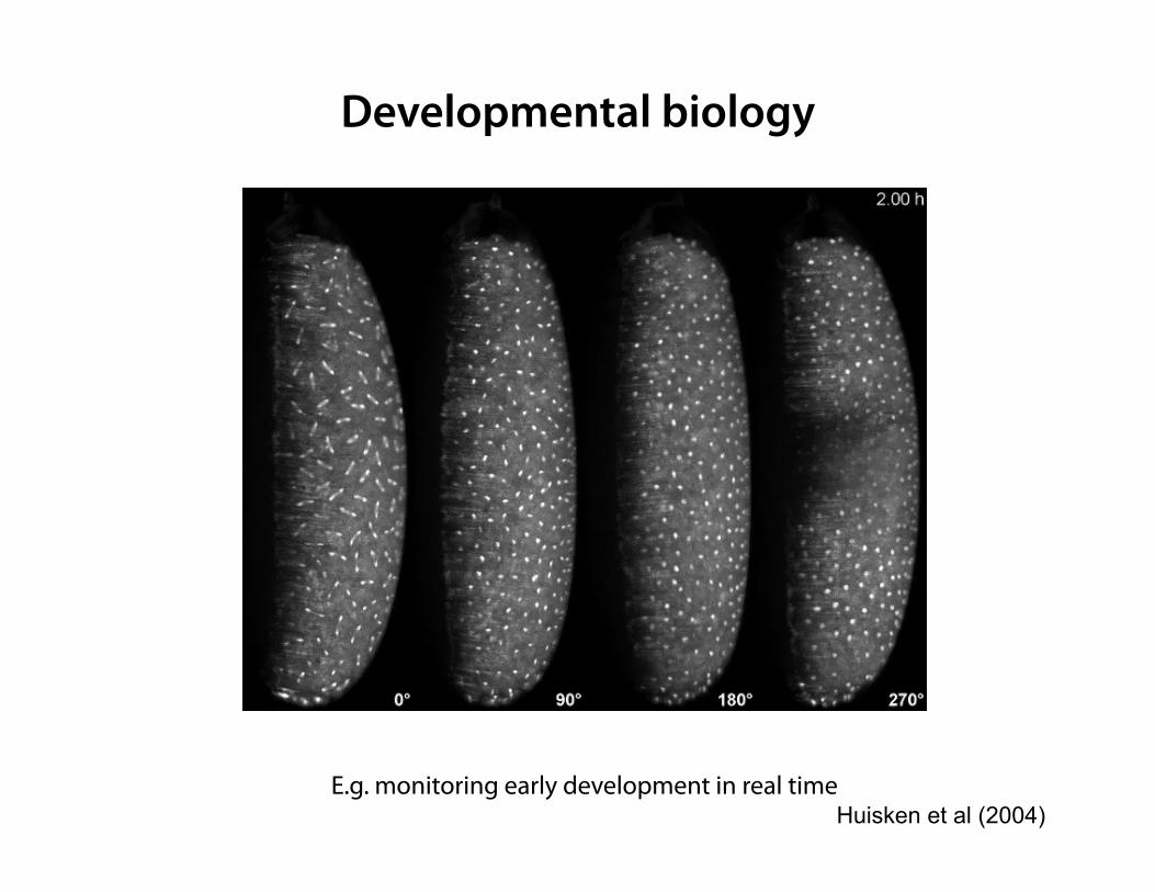
Developmental biology
E.g. monitoring early development in real time Huisken et al (2004)
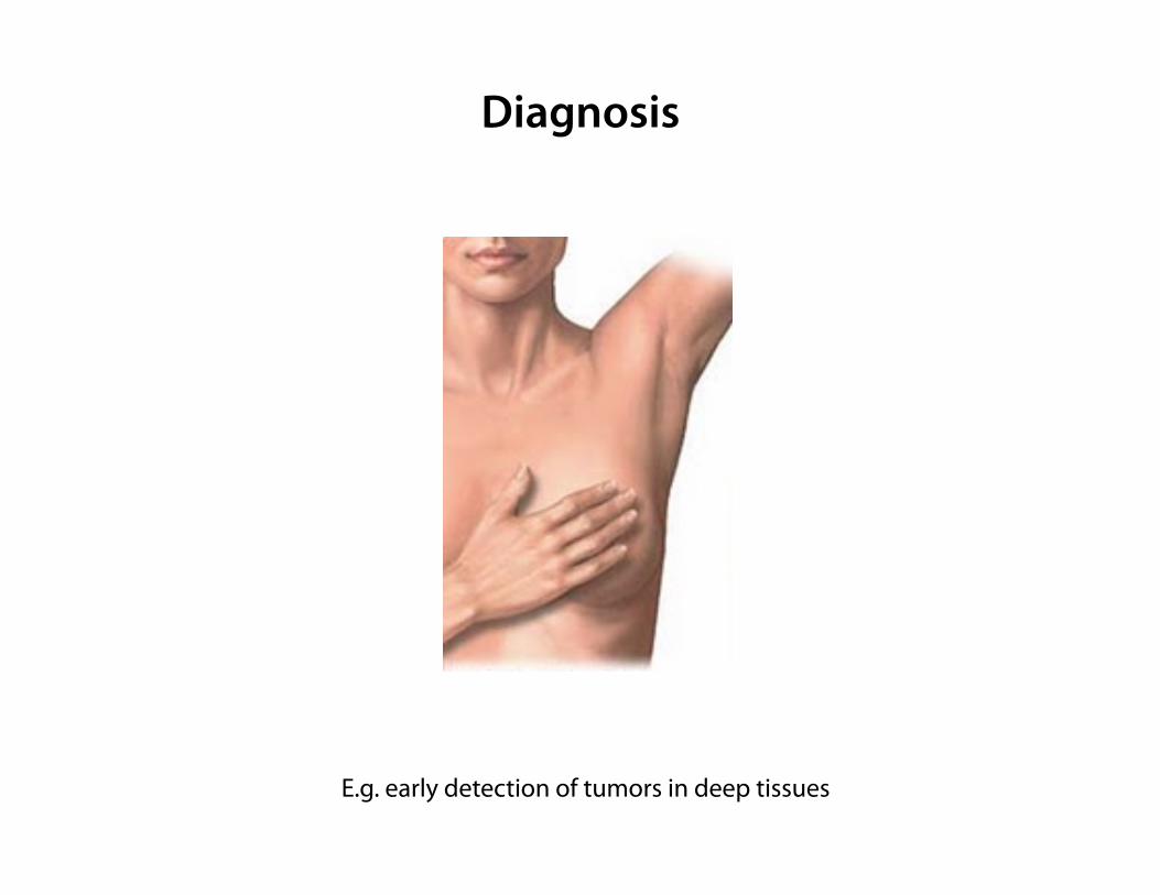
Diagnosis
E.g. early detection of tumors in deep tissues
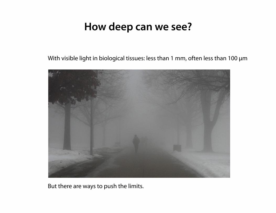
How deep can we see?
With visible light in biological tissues: less than 1 mm, often less than 100 µm But there are ways to push the limits.
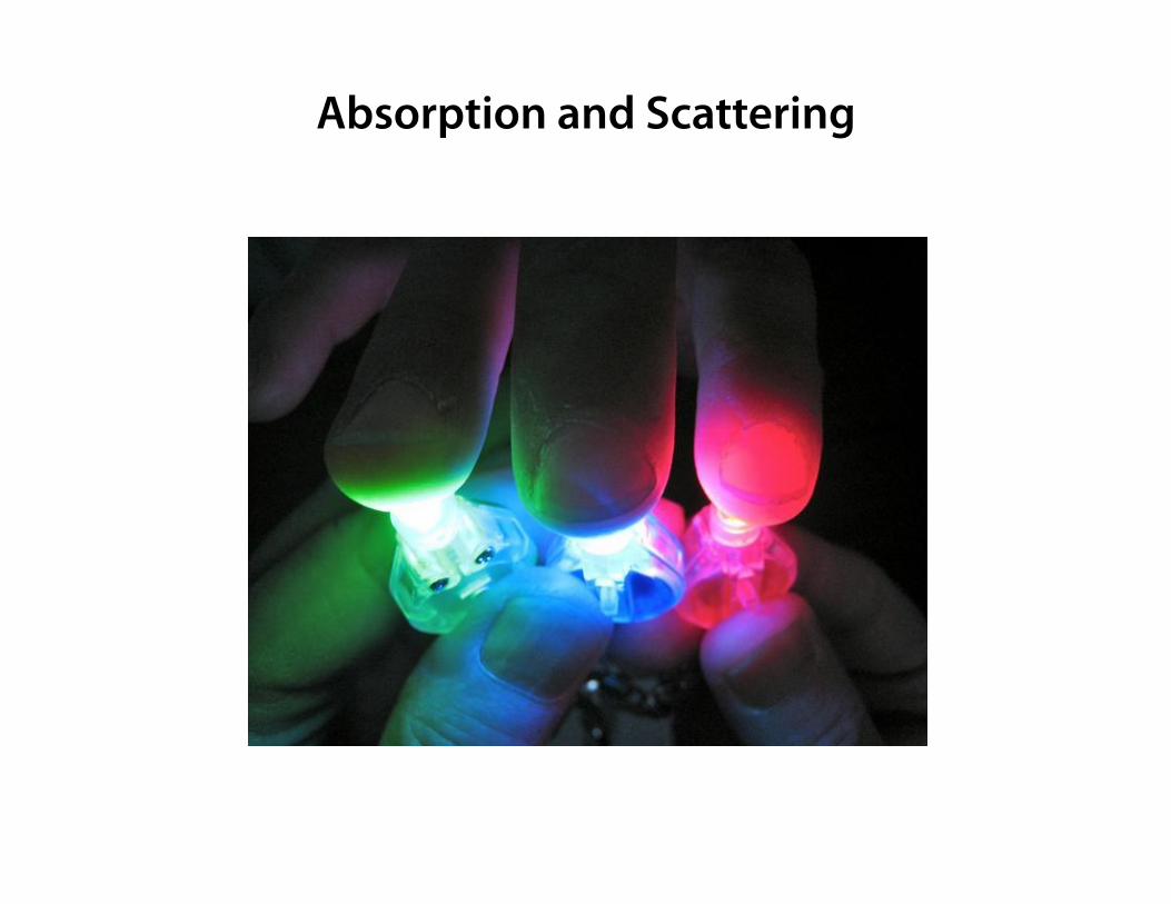
Absorption and Scattering
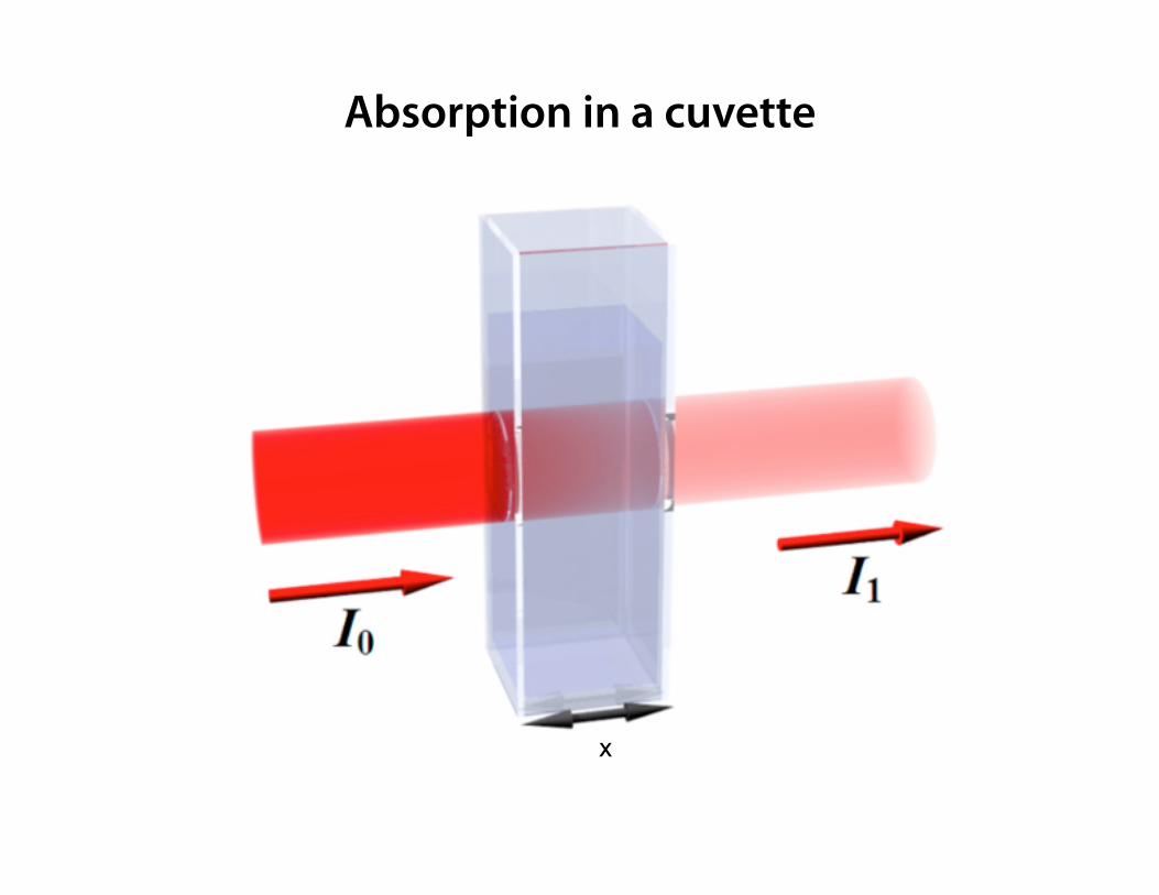
Absorption in a cuvette
x
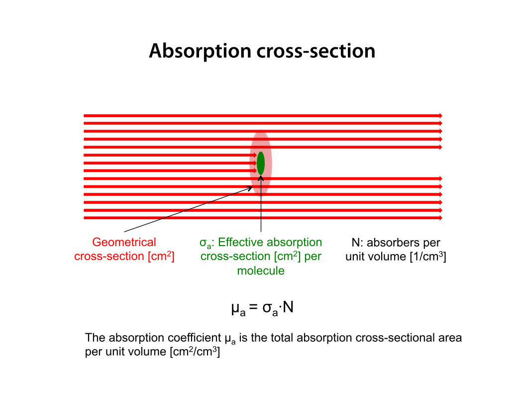
Geometrical cross-section [cm2]
σa: Effective absorption cross-section [cm2] per
molecule
µa = σa·N
N: absorbers per unit volume [1/cm3]
The absorption coefficient µa is the total absorption cross-sectional area per unit volume [cm2/cm3]
Absorption cross-section
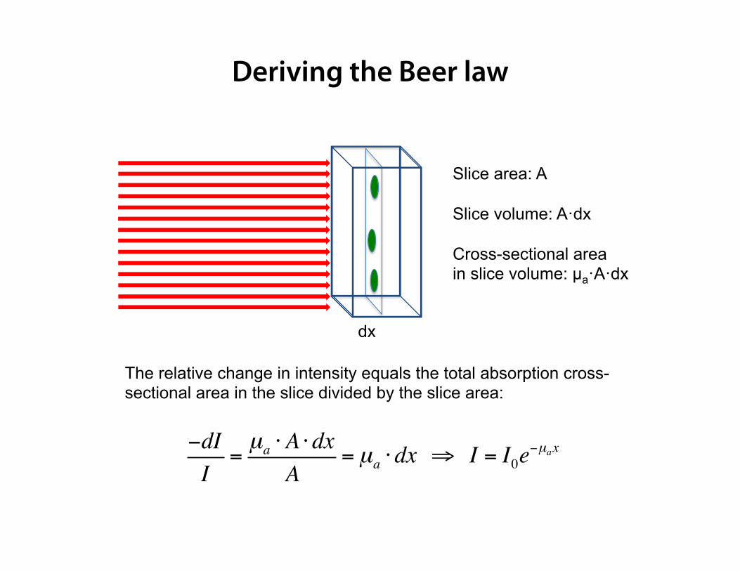
dx
Slice area: A Slice volume: A·dx Cross-sectional area in slice volume: µa·A·dx
The relative change in intensity equals the total absorption cross-sectional area in the slice divided by the slice area:
−dII
=µa ⋅A ⋅dx
A= µa ⋅dx ⇒ I = I0e
−µax
Deriving the Beer law

Absorption in a cuvette
= I0e−µax
x
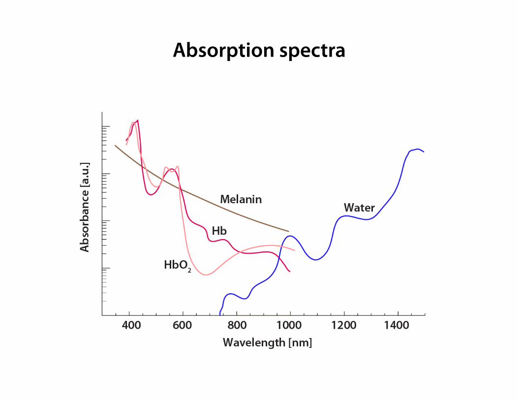
Absorption spectra

Scattering in a cuvette
x
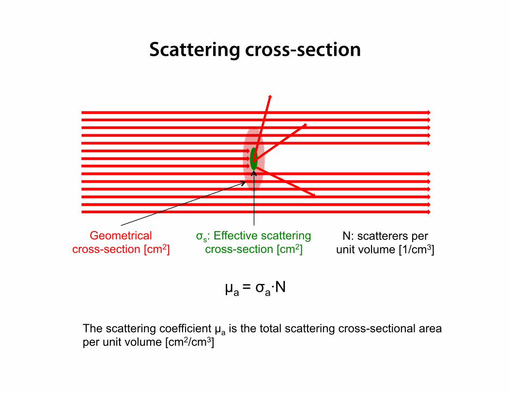
Geometrical cross-section [cm2]
σs: Effective scattering cross-section [cm2]
µa = σa·N
N: scatterers per unit volume [1/cm3]
The scattering coefficient µa is the total scattering cross-sectional area per unit volume [cm2/cm3]
Scattering cross-section
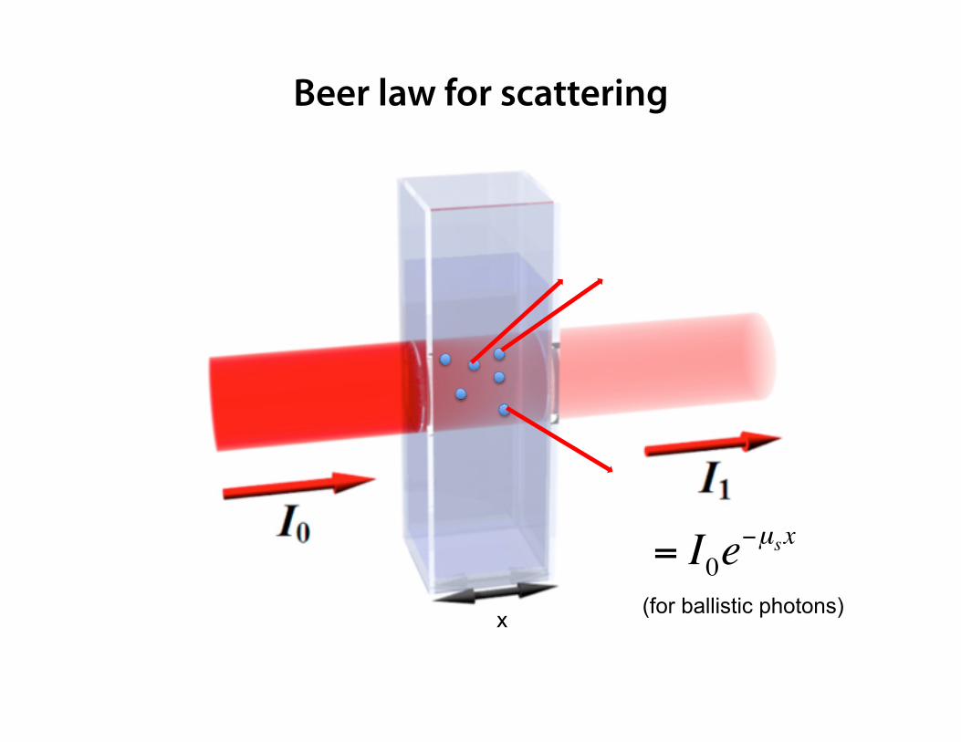
= I0e−µsx
(for ballistic photons)
Beer law for scattering
x
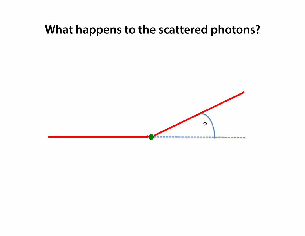
What happens to the scattered photons?
?
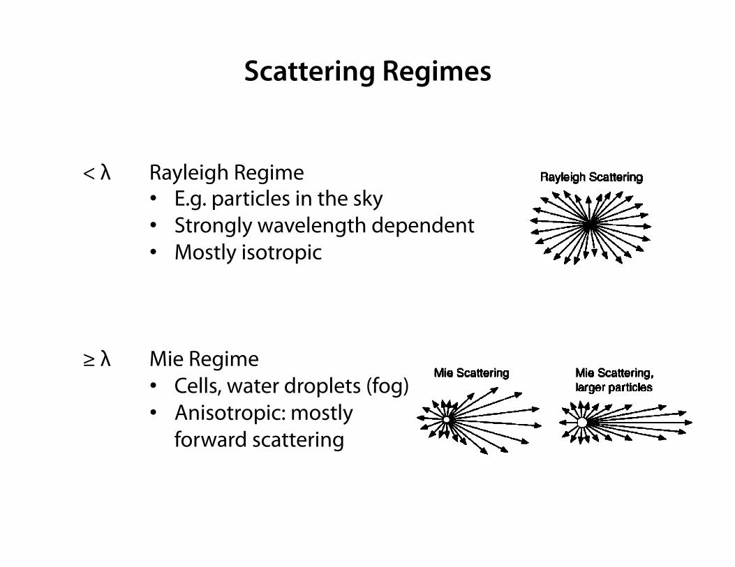
Scattering Regimes
< λ Rayleigh Regime • E.g. particles in the sky • Strongly wavelength dependent • Mostly isotropic
≥ λ Mie Regime
• Cells, water droplets (fog) • Anisotropic: mostly
forward scattering
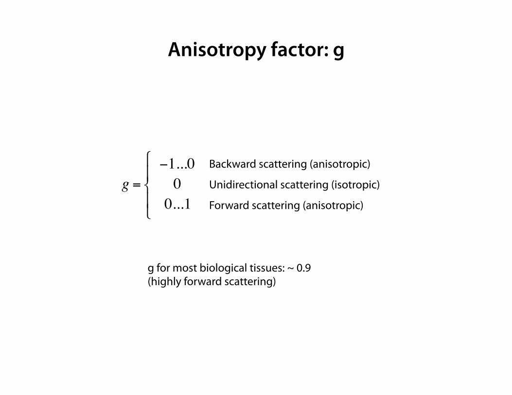
Anisotropy factor: g
g =−1...000...1
"
#$
%$
Backward scattering (anisotropic)
Unidirectional scattering (isotropic)
Forward scattering (anisotropic)
g for most biological tissues: ~ 0.9 (highly forward scattering)
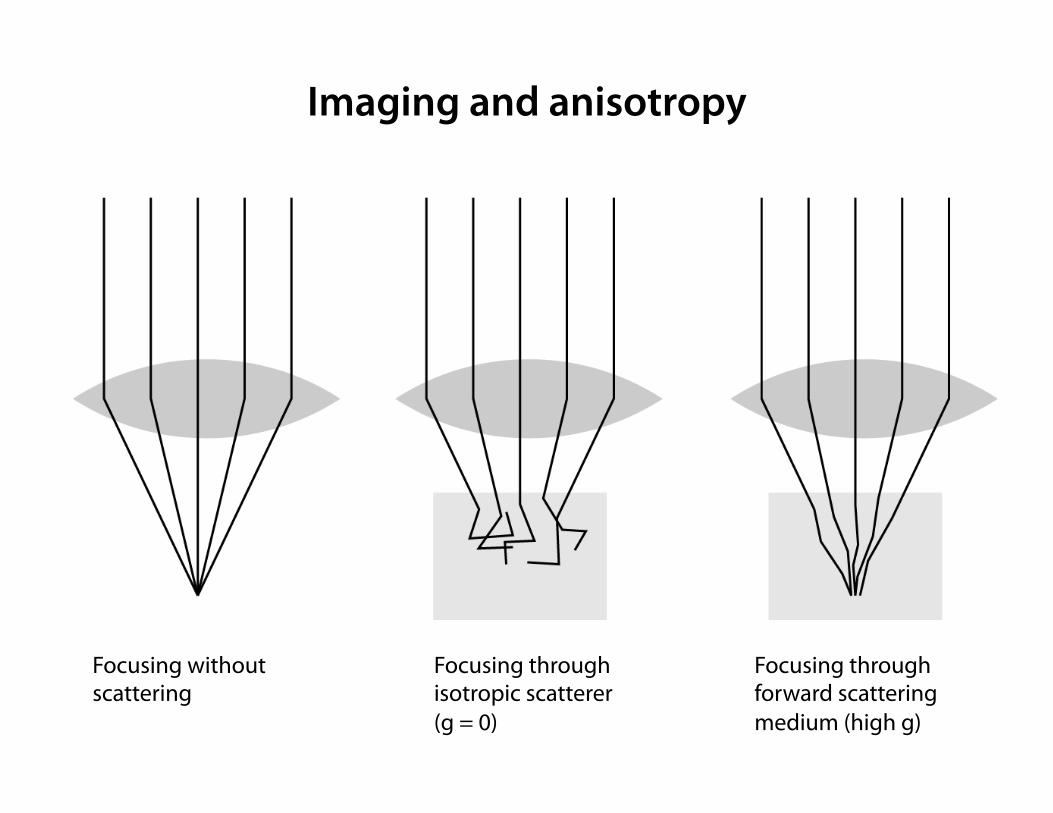
Focusing without scattering
Focusing through isotropic scatterer (g = 0)
Focusing through forward scattering medium (high g)
Imaging and anisotropy
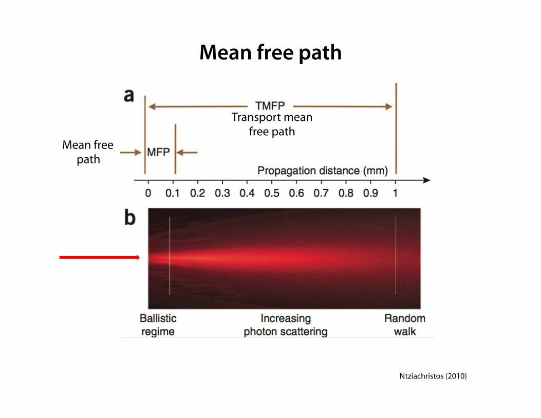
Mean free path
Mean free path
Transport mean free path
Ntziachristos (2010)
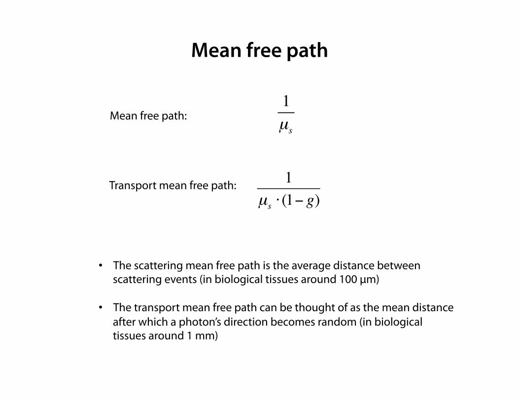
• The scattering mean free path is the average distance between scattering events (in biological tissues around 100 µm)
• The transport mean free path can be thought of as the mean distance after which a photon’s direction becomes random (in biological tissues around 1 mm)
1µs ⋅ (1− g)
Transport mean free path:
Mean free path
1µs
Mean free path:
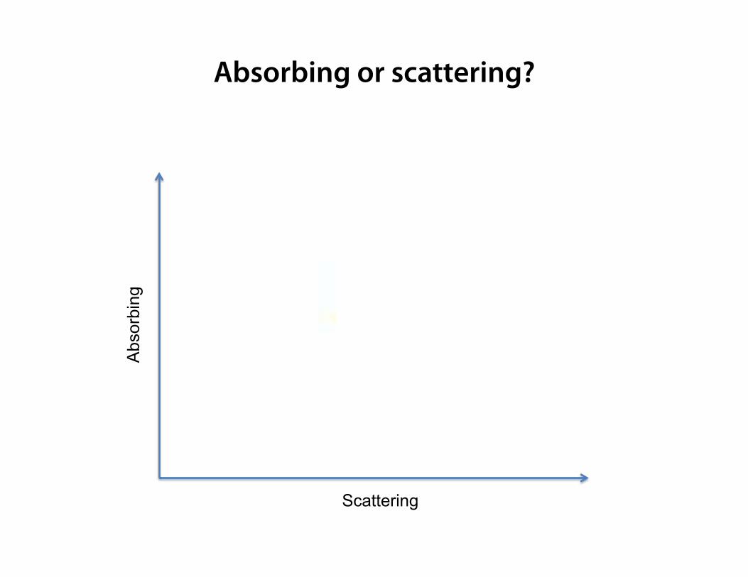
Absorbing or scattering?
Abs
orbi
ng
Scattering
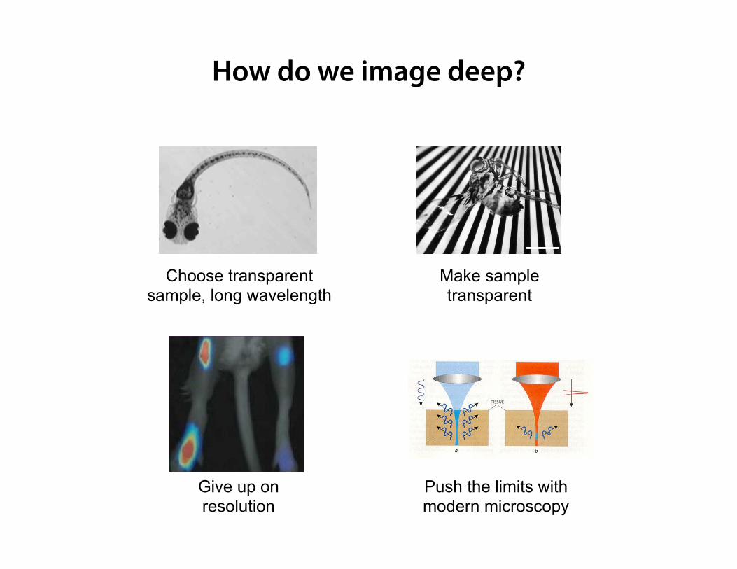
How do we image deep?
Choose transparent sample, long wavelength
Make sample transparent
Give up on resolution
Push the limits with modern microscopy
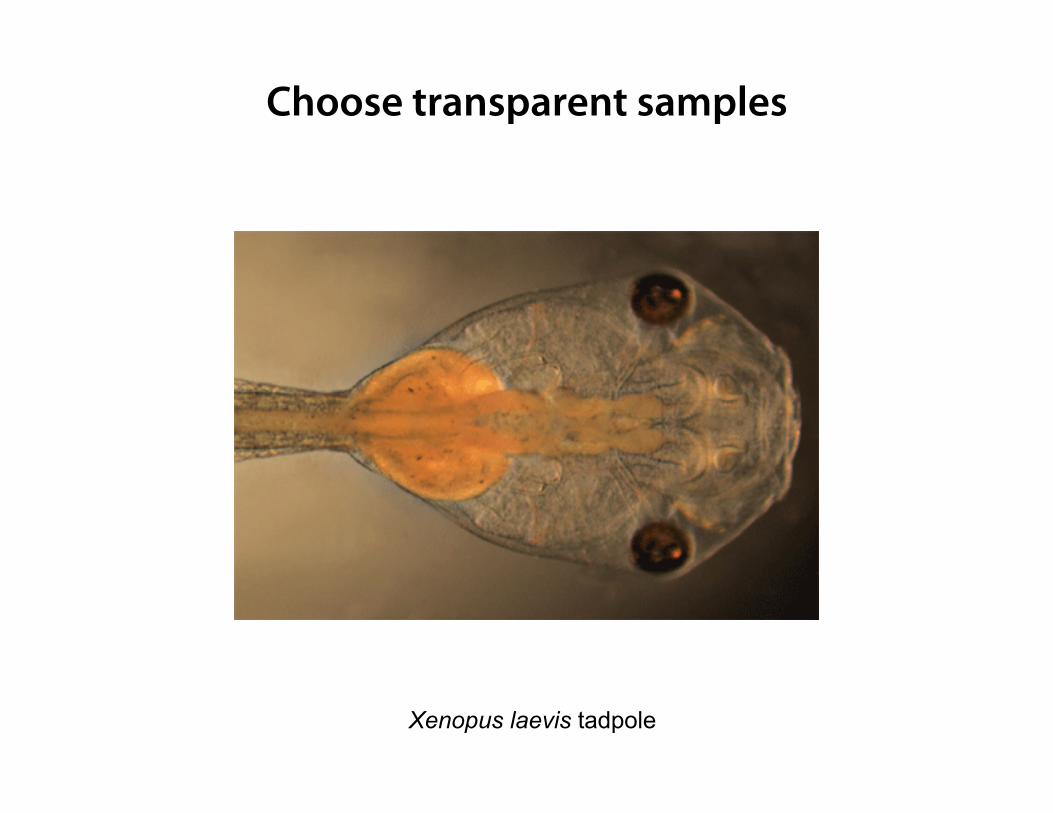
Xenopus laevis tadpole
Choose transparent samples
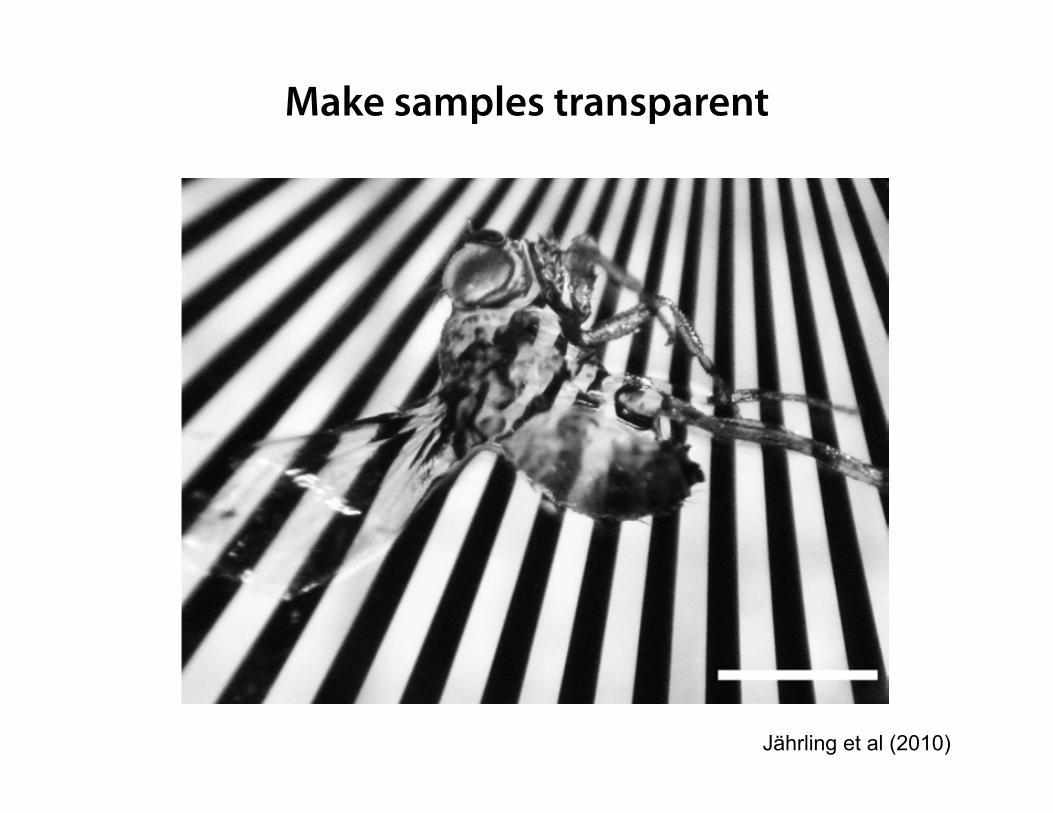
Make samples transparent
Jährling et al (2010)
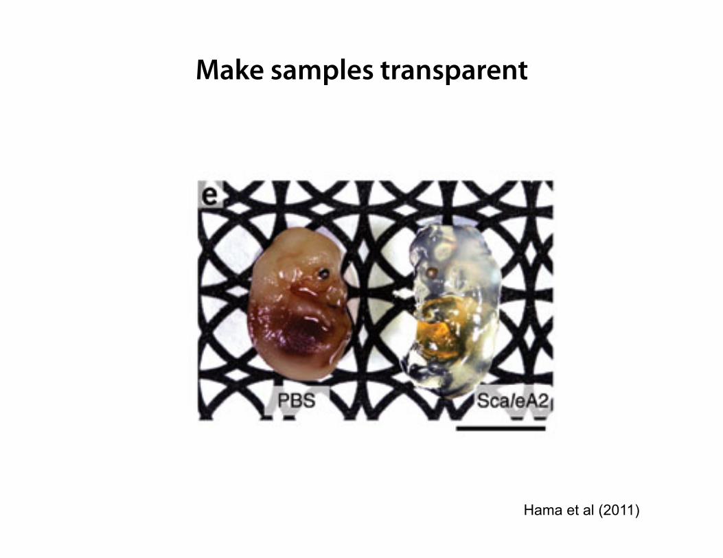
Make samples transparent
Hama et al (2011)
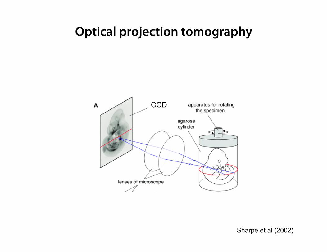
CCD
Optical projection tomography
Sharpe et al (2002)
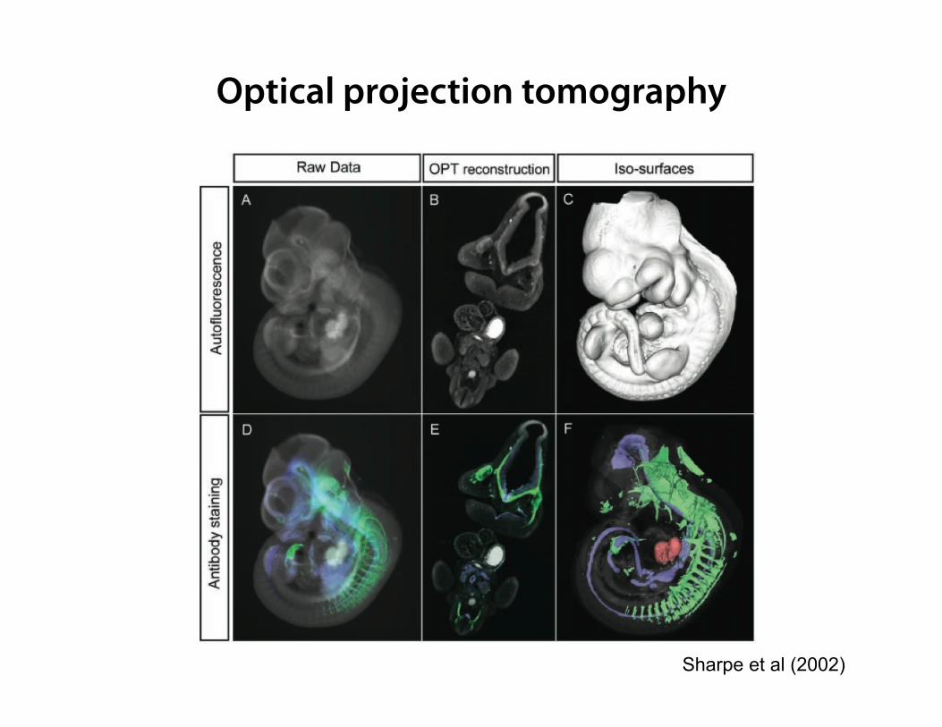
Sharpe et al (2002)
Optical projection tomography
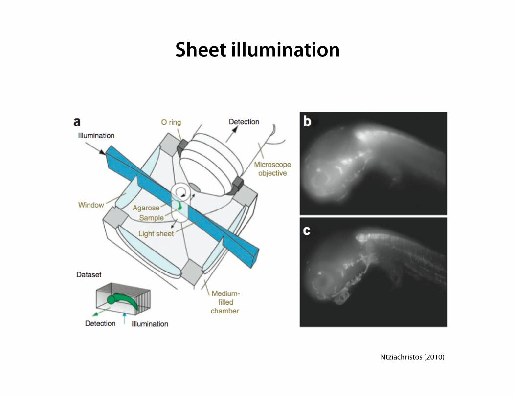
Sheet illumination
Ntziachristos (2010)
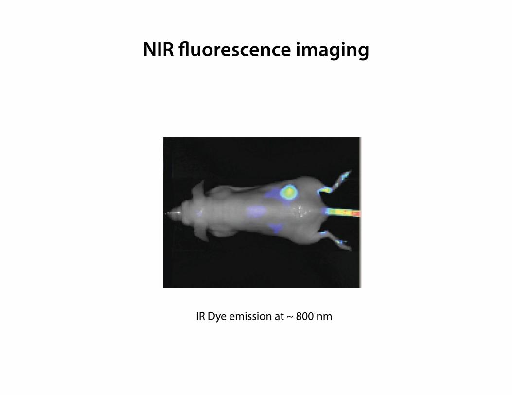
NIR !uorescence imaging
IR Dye emission at ~ 800 nm
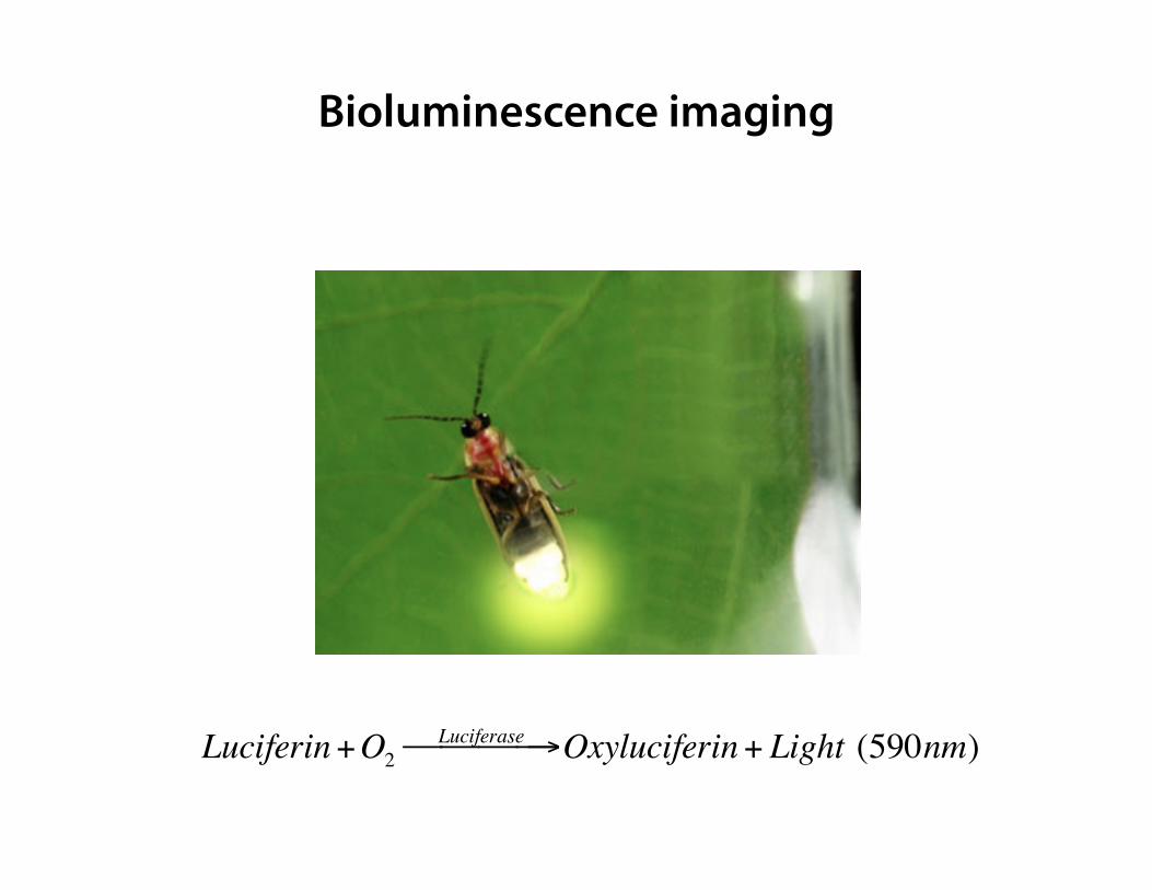
Bioluminescence imaging
Luciferin+O2Luciferase! →!!! Oxyluciferin+ Light (590nm)
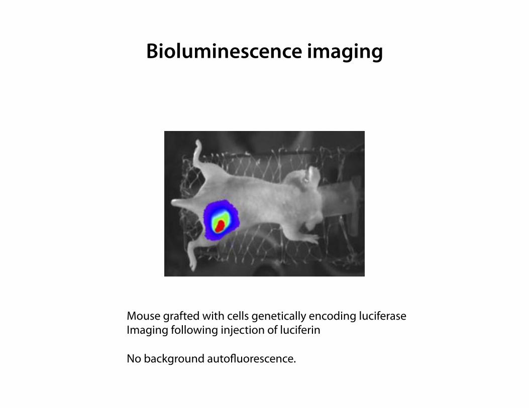
Bioluminescence imaging
Mouse grafted with cells genetically encoding luciferase Imaging following injection of luciferin No background auto#uorescence.
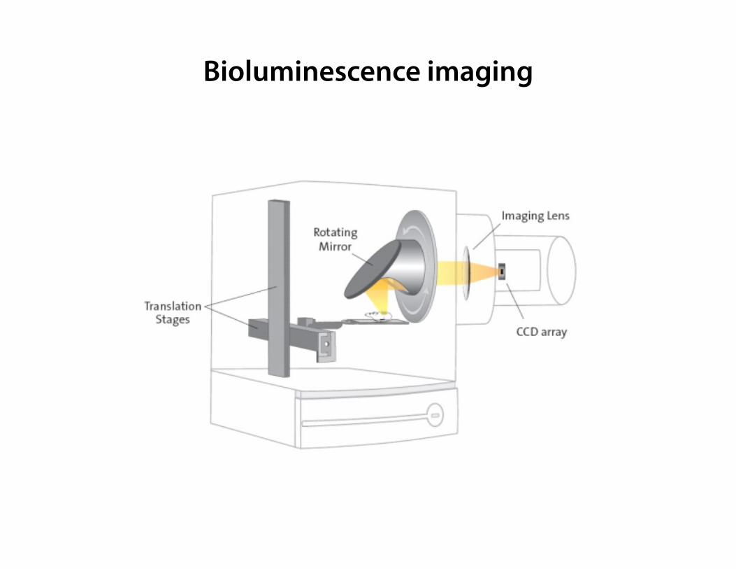
Bioluminescence imaging
Summary
• In biological tissues, scattering dominates over absorption
• Scattering in most samples is anisotropic (high g)
• Scattering mean free path and transport mean free path are a measure
of the penetration depth limit.
• Scattering and absorption are reduced at longer wavelengths
• To image deeper, the simplest solution is to use transparent samples
• Other samples can be cleared optically (but they need to be fixed)
• Optical projection tomography and sheet imaging can be used to image
large transparent samples
• NIR fluorescence imaging and bioluminescence have a penetration
depth of several mm, but sacrifice resolution.