Single Ion Channel Recordings with CMOS-Anchored … XXX, XXX−XXX. the amplifier. With popular...
5
Single Ion Channel Recordings with CMOS-Anchored Lipid Membranes Jacob K. Rosenstein,* ,†,‡ Siddharth Ramakrishnan, †,§ Jared Roseman, † and Kenneth L. Shepard* ,† † Department of Electrical Engineering, Columbia University, New York, New York 10027, United States ‡ School of Engineering, Brown University, Providence, Rhode Island 02912, United States § Department of Biology, University of Puget Sound, Tacoma, Washington 98416, United States * S Supporting Information ABSTRACT: We present single-ion-channel recordings performed with biomimetic lipid membranes which are directly attached to the surface of a complementary metal−oxide−semiconductor (CMOS) preamplifier chip. With this system we resolve single-channel currents from several types of bacterial ion channels, including fluctuations of a single alamethicin channel at a bandwidth of 1 MHz which represent the fastest single-ion-channel recordings reported to date. The platform is also used for high-resolution α-hemolysin nanopore recordings. These results illustrate the high signal fidelity, fine temporal resolution, small geometry, and multiplexed integration which can be achieved by leveraging integrated semiconductor platforms for advanced ion channel interfaces. KEYWORDS: Single-molecule, ion channels, nanopore, CMOS, lipid bilayer, low noise amplifier I on-channel proteins are ubiquitous in the cellular mem- branes of all living things. Understanding their diverse and central roles in cellular sensing, signaling, and energetics have been important areas of research for many decades, 1 and ion channels remain a major target for drug discovery. 2 Still, the stochastic behavior and delicate structure of ion channels has left challenges in understanding some of the biomolecular mechanisms and dynamics of channel gating, as well as in reliably utilizing isolated ion channels for biotechnology applications, including nanopore DNA sequencing. 3,4 Ion channels are most often studied using patch-clamp techniques, 5,6 but biophysical studies of some ion channels have employed in vitro planar reconstituted lipid bilayers as model cell membranes. Although comparatively few types of func- tional proteins have been demonstrated in artificial lipid membranes, reconstituted bilayers are attractive because their chemical makeup can be well-controlled, they involve no live cells, they can be measured outside physiological conditions, and their planar geometry is convenient for electrochemical measurements. 7 Electronic measurements of ion channels in both patch- clamp and planar bilayer experiments are usually made with voltage-clamp amplifiers, which measure the ionic current as a function of time while applying an electrochemical potential across the membrane through silver/silver-chloride (Ag/AgCl) electrodes. Ionic conductance varies between types of ion channels, but their nanoscale dimensions typically produce single-channel currents on the order of picoamperes or femtoamperes. These weak signals confront the noise floor of available electronic amplifiers, and single-channel recordings are generally constrained in both amplitude and temporal resolution by low signal-to-noise ratios. 6 Current noise amplitudes invariably increase for wider measurement bandwidths while the signal amplitude is unchanged, resulting in a signal-to-noise ratio which decreases as temporal resolution is improved. While the low-frequency noise of ion channel measurements is often dominated by the noise of the channel itself, the high- frequency noise is limited by the measurement electronics and is a strong function of experimental capacitances (see Supplementary Discussion). The voltage-clamp amplifier’s equivalent input voltage noise v n (V Hz −1/2 ) produces noise currents through all capacitances at the amplifier input (∑C), with a root-mean-squared amplitude given by: ∑ π = I B v C 2 3 n RMS 3/2 where B is the measurement bandwidth. The most commonly described capacitance in lipid bilayer experiments is that of the lipid membrane itself (C M ); lipid bilayers are very thin and exhibit a specific capacitance on the order of 0.5 μF/cm 2 . Circular planar bilayers with diameters ranging from 1 to 200 μm, therefore, have C M ranging from 4 fF to 150 pF. For larger bilayers, C M can be the single dominant parasitic in the system, and reductions to the bilayer area translate directly to lower measurement noise. 8 For smaller bilayers, however, other parasitics are equally important, in particular the input capacitance (C I ) of the voltage-clamp amplifier and wiring capacitance (C W ) from connections between the electrodes and Received: March 5, 2013 Revised: April 17, 2013 Letter pubs.acs.org/NanoLett © XXXX American Chemical Society A dx.doi.org/10.1021/nl400822r | Nano Lett. XXXX, XXX, XXX−XXX
Transcript of Single Ion Channel Recordings with CMOS-Anchored … XXX, XXX−XXX. the amplifier. With popular...
Single Ion Channel Recordings with CMOS-Anchored LipidMembranesJacob K. Rosenstein,*,†,‡ Siddharth Ramakrishnan,†,§ Jared Roseman,† and Kenneth L. Shepard*,†
†Department of Electrical Engineering, Columbia University, New York, New York 10027, United States‡School of Engineering, Brown University, Providence, Rhode Island 02912, United States§Department of Biology, University of Puget Sound, Tacoma, Washington 98416, United States
*S Supporting Information
ABSTRACT: We present single-ion-channel recordings performed withbiomimetic lipid membranes which are directly attached to the surface of acomplementary metal−oxide−semiconductor (CMOS) preamplifier chip. Withthis system we resolve single-channel currents from several types of bacterial ionchannels, including fluctuations of a single alamethicin channel at a bandwidth of1 MHz which represent the fastest single-ion-channel recordings reported todate. The platform is also used for high-resolution α-hemolysin nanoporerecordings. These results illustrate the high signal fidelity, fine temporalresolution, small geometry, and multiplexed integration which can be achievedby leveraging integrated semiconductor platforms for advanced ion channel interfaces.
KEYWORDS: Single-molecule, ion channels, nanopore, CMOS, lipid bilayer, low noise amplifier
Ion-channel proteins are ubiquitous in the cellular mem-branes of all living things. Understanding their diverse and
central roles in cellular sensing, signaling, and energetics havebeen important areas of research for many decades,1 and ionchannels remain a major target for drug discovery.2 Still, thestochastic behavior and delicate structure of ion channels hasleft challenges in understanding some of the biomolecularmechanisms and dynamics of channel gating, as well as inreliably utilizing isolated ion channels for biotechnologyapplications, including nanopore DNA sequencing.3,4
Ion channels are most often studied using patch-clamptechniques,5,6 but biophysical studies of some ion channels haveemployed in vitro planar reconstituted lipid bilayers as modelcell membranes. Although comparatively few types of func-tional proteins have been demonstrated in artificial lipidmembranes, reconstituted bilayers are attractive because theirchemical makeup can be well-controlled, they involve no livecells, they can be measured outside physiological conditions,and their planar geometry is convenient for electrochemicalmeasurements.7
Electronic measurements of ion channels in both patch-clamp and planar bilayer experiments are usually made withvoltage-clamp amplifiers, which measure the ionic current as afunction of time while applying an electrochemical potentialacross the membrane through silver/silver-chloride (Ag/AgCl)electrodes. Ionic conductance varies between types of ionchannels, but their nanoscale dimensions typically producesingle-channel currents on the order of picoamperes orfemtoamperes. These weak signals confront the noise floor ofavailable electronic amplifiers, and single-channel recordings aregenerally constrained in both amplitude and temporalresolution by low signal-to-noise ratios.6 Current noise
amplitudes invariably increase for wider measurementbandwidths while the signal amplitude is unchanged, resultingin a signal-to-noise ratio which decreases as temporal resolutionis improved.While the low-frequency noise of ion channel measurements
is often dominated by the noise of the channel itself, the high-frequency noise is limited by the measurement electronics andis a strong function of experimental capacitances (seeSupplementary Discussion). The voltage-clamp amplifier’sequivalent input voltage noise vn (V Hz−1/2) produces noisecurrents through all capacitances at the amplifier input (∑C),with a root-mean-squared amplitude given by:
∑π=I B v C2
3 nRMS3/2
where B is the measurement bandwidth. The most commonlydescribed capacitance in lipid bilayer experiments is that of thelipid membrane itself (CM); lipid bilayers are very thin andexhibit a specific capacitance on the order of 0.5 μF/cm2.Circular planar bilayers with diameters ranging from 1 to 200μm, therefore, have CM ranging from 4 fF to 150 pF. For largerbilayers, CM can be the single dominant parasitic in the system,and reductions to the bilayer area translate directly to lowermeasurement noise.8 For smaller bilayers, however, otherparasitics are equally important, in particular the inputcapacitance (CI) of the voltage-clamp amplifier and wiringcapacitance (CW) from connections between the electrodes and
Received: March 5, 2013Revised: April 17, 2013
Letter
pubs.acs.org/NanoLett
© XXXX American Chemical Society A dx.doi.org/10.1021/nl400822r | Nano Lett. XXXX, XXX, XXX−XXX
the amplifier. With popular electrophysiology amplifiers (suchas the Axopatch 200B or HEKA EPC-10), CI is on the order of10−20 pF, limiting the noise benefits that would be seen fromreducing the bilayer diameter below 30 μm. Yet methods toproduce smaller bilayers exist,8−11 and if paired with amplifierswith appropriately reduced input capacitance, these bilayer areareductions can continue to yield noise reductions.In the literature there are several examples of patch-clamp
studies using aftermarket modifications of an Axopatch 200Bamplifier.12,13 These customizations yielded lower noise floorsand higher signal bandwidths, with the fastest previouslyreported single-channel data reaching a bandwidth of 250 kHzwith corresponding noise of 12.7 pARMS.
13 There are alsoseveral examples of integrated current preamplifiers for ionchannel recordings,14−17 an approach which offers the flexibilityto reduce CI below what is practical with discrete transistors.We previously demonstrated a voltage-clamp preamplifier
integrated circuit (IC) in a commercial 130 nm CMOS process,in which each amplifier occupies just 0.4 mm2 and has CI = 1pF. Very low parasitic capacitances allowed us to resolve ioniccurrent signals through solid-state nanopores at a bandwidth of1 MHz,17 more than 10 times faster than can be recorded bycommercially available instruments. In this Letter, we apply thisplatform to similarly reduce parasitic capacitances in biologicalion channel recordings, by direct attachment of a lipid bilayer tothe surface of the IC. This arrangement enables noise-limitedbandwidths in excess of 1 MHz to be achieved on an integratedplatform which will ultimately be scalable to the independentrecording of thousands of channels.Starting with the custom CMOS amplifier chip,17 we
chemically remove the standard aluminum metallization fromthe amplifier input pads, followed by vacuum deposition of 5nm Ti and 200 nm Ag and patterning of 20 μm or 30 μmdiameter microwells in 5-μm-thick SU-8 (see SupplementaryDiscussion). A micrograph of the final structure for a singleamplifier channel is shown in Figure 1b. After wirebonding and
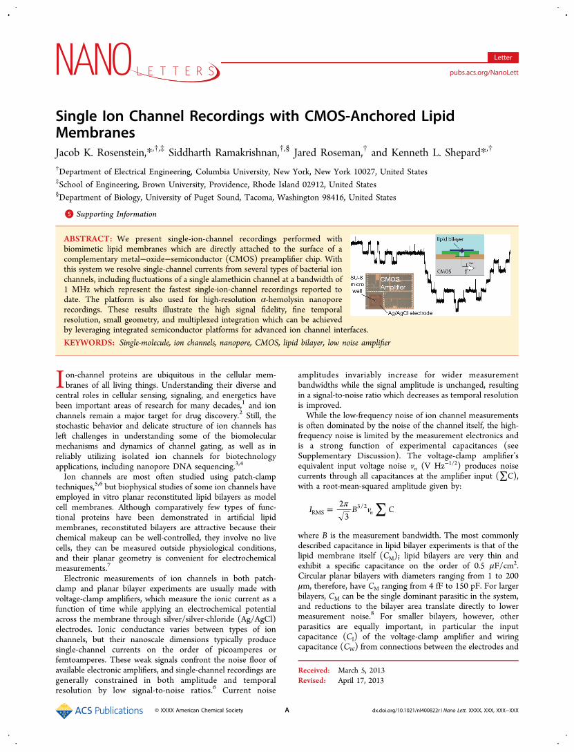
epoxy encapsulation, a 1 mL fluid chamber is assembled aroundthe amplifier chip. With the amplifier mounted on a circuitboard, we fill the fluid chamber with electrolyte and chlorinatethe silver microelectrode. We then apply ∼0.5 μL of DPhPCsolution (10 mg/mL diphytanoyl phosphatidylcholine in n-decane) over the SU-8 surface using an air bubble at the tip ofmicropipet. The lipids spontaneously form bilayers spanningthe surface of the microwells, as illustrated in the cross-sectionof Figure 1a.
These lipid-sealed microwells are similar to lipid bilayerarrays which were previously demonstrated on passive glasssubstrates10,18 The one-sided geometry of these microwellsmeans that after each is covered by a lipid membrane, it iselectrically isolated; multiple wells can be addressed in parallel,with independent trans Ag/AgCl electrodes and one shared cisreservoir.A common concern with microscale Ag/AgCl electrodes is
their depletion over time; the mass of available silverdetermines the net charge (Q = IDC × t) which can bemeasured over the lifetime of the sensor.19 Due to the lowerDC currents required for ion channels as compared to priordemonstrations with solid-state nanopores, we elected to use athin evaporated silver film rather than a thicker electroplatedlayer. The entire 100 μm × 100 μm × 200 nm electrodecontains approximately 21 ng of silver, which, when fullyconverted to AgCl, corresponds to a charge transfer of 19 μC.This places an upper bound on the electrode lifetime of ∼2days at 100 pA bias. (Much of the electrode is covered by SU-8,but at least some the covered silver is likely still electrochemi-cally available.20) The small volume of the trans chamber (∼1.6pL) helps to limit dissolution of the AgCl; although AgCl ishighly soluble in water (KSP = 1.8 × 10⁻¹⁰), 2 pL of waterbecomes saturated with AgCl after dissolving just femtogramsof AgCl. During testing, we found that each electrodecommonly lasted for several hours.Knowing the geometry of the amplifier and bilayer support,
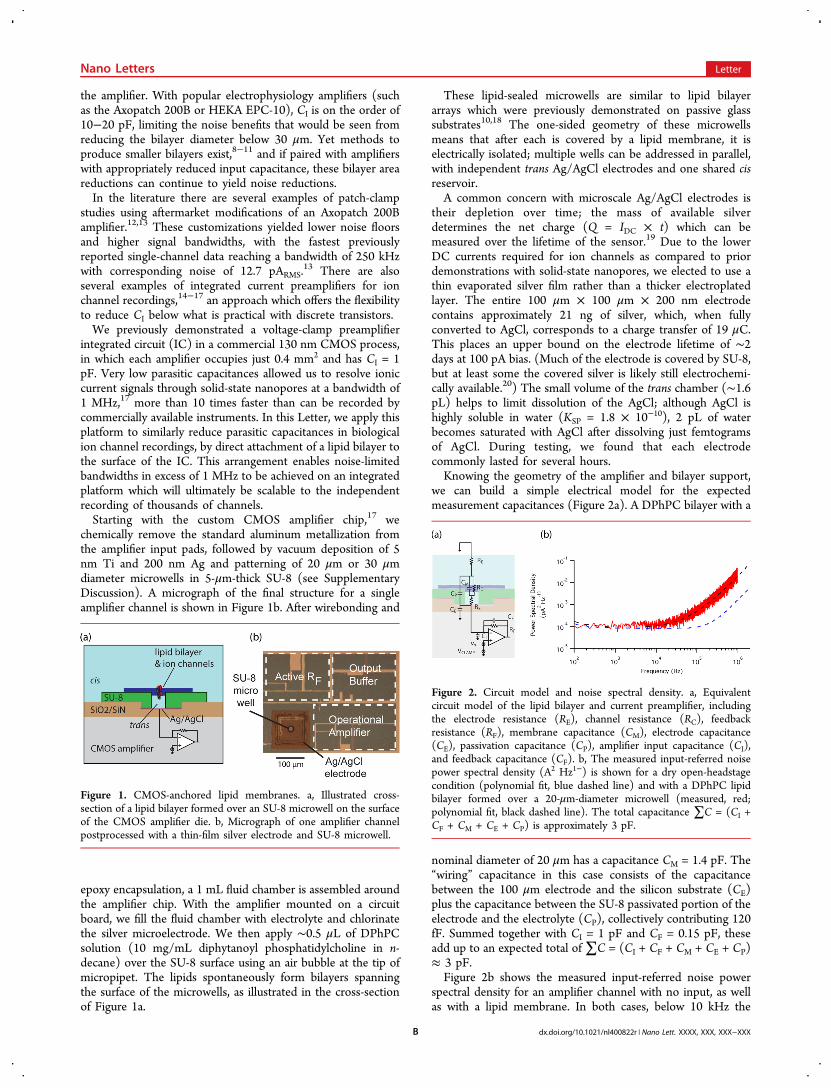
we can build a simple electrical model for the expectedmeasurement capacitances (Figure 2a). A DPhPC bilayer with a
nominal diameter of 20 μm has a capacitance CM = 1.4 pF. The“wiring” capacitance in this case consists of the capacitancebetween the 100 μm electrode and the silicon substrate (CE)plus the capacitance between the SU-8 passivated portion of theelectrode and the electrolyte (CP), collectively contributing 120fF. Summed together with CI = 1 pF and CF = 0.15 pF, theseadd up to an expected total of∑C = (CI + CF + CM + CE + CP)≈ 3 pF.Figure 2b shows the measured input-referred noise power
spectral density for an amplifier channel with no input, as wellas with a lipid membrane. In both cases, below 10 kHz the
Figure 1. CMOS-anchored lipid membranes. a, Illustrated cross-section of a lipid bilayer formed over an SU-8 microwell on the surfaceof the CMOS amplifier die. b, Micrograph of one amplifier channelpostprocessed with a thin-film silver electrode and SU-8 microwell.
Figure 2. Circuit model and noise spectral density. a, Equivalentcircuit model of the lipid bilayer and current preamplifier, includingthe electrode resistance (RE), channel resistance (RC), feedbackresistance (RF), membrane capacitance (CM), electrode capacitance(CE), passivation capacitance (CP), amplifier input capacitance (CI),and feedback capacitance (CF). b, The measured input-referred noisepower spectral density (A2 Hz1−) is shown for a dry open-headstagecondition (polynomial fit, blue dashed line) and with a DPhPC lipidbilayer formed over a 20-μm-diameter microwell (measured, red;polynomial fit, black dashed line). The total capacitance ∑C = (CI +CF + CM + CE + CP) is approximately 3 pF.
Nano Letters Letter
dx.doi.org/10.1021/nl400822r | Nano Lett. XXXX, XXX, XXX−XXXB
noise is determined by the white noise of the amplifier feedbacknetwork. With the bilayer present, at higher frequencies thenoise density is fit by Sn( f) ≈ (2πf × 3 pF × 5 nV Hz−1/2)2,consistent with the estimate above. The time-domain currentnoise is evaluated from the integral of the noise spectrum up toa given bandwidth. If a fourth-order low-pass Bessel filter isapplied at various cutoff frequencies ( fc), this integratedbaseline noise current in the presence of the bilayercorresponds to 1 pARMS for fc = 10 kHz, 4.4 pARMS at 100kHz, 11.8 pARMS at 250 kHz, and 61 pARMS at 1 MHz.Since the amplifier was originally designed to support DC
bias currents as large as 15 nA while operating from a 1.5 Vpower supply, it uses an active feedback network whose whitenoise contribution (∼ 10 fA Hz−1/2) determines the low-frequency noise floor of the system. Phenomena which requirenoise amplitudes below 1 pARMS may be better served byhigher-gain preamplifiers with a smaller dynamic range, but forthe many applications in which 1 pARMS or more is acceptable,reduced high-frequency noise density provides an opportunityto acquire meaningful data at much finer temporal resolution.This is a common trade-off associated with integratedmicroelectronics; low-frequency noise reductions can be morechallenging because of difficulties integrating high-valueresistors and low-noise devices such as junction field-effecttransistors, while high-frequency noise is naturally lower due toreduced capacitive parasitics.To verify the bilayer structure of our CMOS-anchored lipid
membranes, we perform measurements of gramicidin, anantibiotic peptide which increases the permeability of cellmembranes.21−24 Gramicidin produces transient dimer chan-nels in lipid bilayers, formed by junctions between peptides inopposing halves of the membrane. Gramicidin ion channelsform and dissociate stochastically irrespective of appliedvoltage, yielding stepwise current changes in voltage-clamprecordings. Prior to forming a bilayer, < 0.5 μL of gramicidinsolution (1 μg/mL in ethanol) is added to the electrolyte suchthat it is present in both the cis and the trans chambers. Shortlyafter painting the lipids, random stepwise current fluctuationsare observed. An example recording of gramicidin is shown inFigure 3, showing several simultaneously active channelsproducing discrete current levels separated by approximately4 pA.To explore biotechnology applications of this integrated
platform, we consider α-hemolysin (α-HL), a well-studiedbacterial toxin which forms heptameric ion channels in cell
membranes.25 In recent years, α-HL has attracted considerableinterest as a nanopore sensor with the potential to enable high-speed DNA sequencing, among other applications.4,26 Thechannel of an α-HL pore has a diameter of approximately 1.4nm at its smallest constriction, and the presence of even verysmall molecules within a single channel can measurablymodulate its ionic conductance.Figure 4 shows a current trace measured with a single α-HL
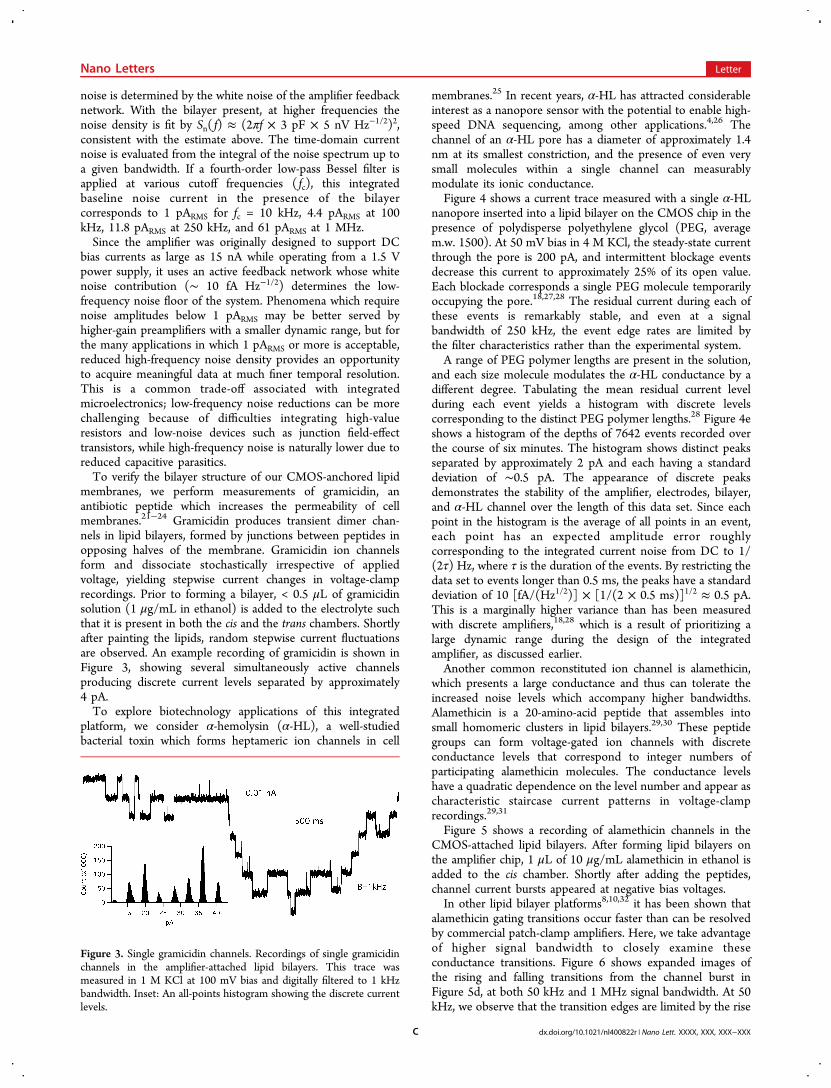
nanopore inserted into a lipid bilayer on the CMOS chip in thepresence of polydisperse polyethylene glycol (PEG, averagem.w. 1500). At 50 mV bias in 4 M KCl, the steady-state currentthrough the pore is 200 pA, and intermittent blockage eventsdecrease this current to approximately 25% of its open value.Each blockade corresponds a single PEG molecule temporarilyoccupying the pore.18,27,28 The residual current during each ofthese events is remarkably stable, and even at a signalbandwidth of 250 kHz, the event edge rates are limited bythe filter characteristics rather than the experimental system.A range of PEG polymer lengths are present in the solution,
and each size molecule modulates the α-HL conductance by adifferent degree. Tabulating the mean residual current levelduring each event yields a histogram with discrete levelscorresponding to the distinct PEG polymer lengths.28 Figure 4eshows a histogram of the depths of 7642 events recorded overthe course of six minutes. The histogram shows distinct peaksseparated by approximately 2 pA and each having a standarddeviation of ∼0.5 pA. The appearance of discrete peaksdemonstrates the stability of the amplifier, electrodes, bilayer,and α-HL channel over the length of this data set. Since eachpoint in the histogram is the average of all points in an event,each point has an expected amplitude error roughlycorresponding to the integrated current noise from DC to 1/(2τ) Hz, where τ is the duration of the events. By restricting thedata set to events longer than 0.5 ms, the peaks have a standarddeviation of 10 [fA/(Hz1/2)] × [1/(2 × 0.5 ms)]1/2 ≈ 0.5 pA.This is a marginally higher variance than has been measuredwith discrete amplifiers,18,28 which is a result of prioritizing alarge dynamic range during the design of the integratedamplifier, as discussed earlier.Another common reconstituted ion channel is alamethicin,
which presents a large conductance and thus can tolerate theincreased noise levels which accompany higher bandwidths.Alamethicin is a 20-amino-acid peptide that assembles intosmall homomeric clusters in lipid bilayers.29,30 These peptidegroups can form voltage-gated ion channels with discreteconductance levels that correspond to integer numbers ofparticipating alamethicin molecules. The conductance levelshave a quadratic dependence on the level number and appear ascharacteristic staircase current patterns in voltage-clamprecordings.29,31
Figure 5 shows a recording of alamethicin channels in theCMOS-attached lipid bilayers. After forming lipid bilayers onthe amplifier chip, 1 μL of 10 μg/mL alamethicin in ethanol isadded to the cis chamber. Shortly after adding the peptides,channel current bursts appeared at negative bias voltages.In other lipid bilayer platforms8,10,32 it has been shown that
alamethicin gating transitions occur faster than can be resolvedby commercial patch-clamp amplifiers. Here, we take advantageof higher signal bandwidth to closely examine theseconductance transitions. Figure 6 shows expanded images ofthe rising and falling transitions from the channel burst inFigure 5d, at both 50 kHz and 1 MHz signal bandwidth. At 50kHz, we observe that the transition edges are limited by the rise
Figure 3. Single gramicidin channels. Recordings of single gramicidinchannels in the amplifier-attached lipid bilayers. This trace wasmeasured in 1 M KCl at 100 mV bias and digitally filtered to 1 kHzbandwidth. Inset: An all-points histogram showing the discrete currentlevels.
Nano Letters Letter
dx.doi.org/10.1021/nl400822r | Nano Lett. XXXX, XXX, XXX−XXXC
time of the low-pass filter (∼ 10 μs), and as a result short-livedconductance levels briefer than ∼20 μs are not captured. In thesame trace at 1 MHz, the transition times are indeed muchfaster than 10 μs. Flickering conductance levels as brief as 3 μsare evident during some channel openings, while during thechannel closures the steps usually appear sharp even at thishigher bandwidth; thus, we can infer that these transitionsoccur in less than 500 ns. To the best of our knowledge, thesemeasurements represent the fastest single ion channel record-ings achieved to date.
In this Letter we have shown that an active CMOS amplifierchip can simultaneously serve as the physical support andelectronic interface for microscale lipid bilayers,7 creating apowerful platform for high-throughput biotechnology applica-tions such as DNA sequencing and drug discovery. Scaling thesensor and electronic signal chain to submillimeter dimensions
Figure 4. Nanopore mass spectrometry. a, A single α-hemolysin channel in the presence of polydisperse polyethylene glycol (PEG, average m.w.1500). Each rectangular current blockade corresponds to a single PEG molecule occupying the channel. The trace is shown at B = 100 kHz. b, All-points histogram of the trace, showing clear separation between open and blocked current levels. c, One of the PEG events, digitally filtered to 50kHz and 250 kHz bandwidths. d, Magnified view of the falling edge of the event, contrasting the signal fall-time at 10 kHz, 50 kHz, and 250 kHzbandwidths. e, Histogram of the average residual current in 7642 events with durations of at least 0.5 ms. Each peak in the histogram corresponds toa unique PEG polymer length.
Figure 5. Single alamethicin channels. a, Recordings of singlealamethicin channel bursts in 3 M KCl at 150 mV bias, digitallyfiltered to 50 kHz bandwidth. b, All-points histogram of the trace,showing three distinct conductance levels. c, The discrete currentlevels identified in the histogram have a quadratic dependence on thelevel number. d, A magnified view of one of the channel bursts,identified by the red arrow.
Figure 6. Fast ion channel gating. a, Rising edge of the alamethicinchannel burst shown in Figure 5d, sampled at 4 MS/s and digitallyfiltered to 50 kHz and 1 MHz bandwidths. At 1 MHz it is evident thattransition between L1 and L3 includes two stays at L2, lasting 6 and 3μs, respectively. b, Falling edge of the same channel burst. The closingtransitions are briefer than can be resolved in our measurements, evenat 1 MHz bandwidth, suggesting that the channel’s actual ionicconductance changes in less than 500 ns.
Nano Letters Letter
dx.doi.org/10.1021/nl400822r | Nano Lett. XXXX, XXX, XXX−XXXD
decreases parasitic capacitances and enables measurements ofion channel gating transitions faster than 1 μs. In addition tothe parallelism that will be enabled by integrating ion channelswith microelectronics, these newly accessible time scalesoverlap with the durations of molecular dynamics simulations,33
making it conceivable that soon it will be possible to directlycorrelate kinetic processes within simulations and measure-ments of single-molecule ion channel gating.
■ ASSOCIATED CONTENT*S Supporting InformationExperimental methods and electronic circuit description. Thismaterial is available free of charge via the Internet at http://pubs.acs.org.
■ AUTHOR INFORMATIONCorresponding Author*E-mail: [email protected] or [email protected] authors declare no competing financial interest.
■ ACKNOWLEDGMENTSThe authors thank Daniel Fleischer for his assistance. Thiswork was supported by the National Institutes of Health underGrant R01-HG006879.
■ REFERENCES(1) Hille, B. Ion Channels of Excitable Membranes, 3rd ed.; SinauerAssociates: Sunderland, MA, 2001.(2) Wood, C.; Williams, C.; Waldron, G. J. Drug Discovery Today2004, 9, 434−41.(3) Branton, D.; Deamer, D. W.; Marziali, A.; Bayley, H.; Benner, S.A.; Butler, T.; Di Ventra, M.; Garaj, S.; Hibbs, A.; Huang, X.;Jovanovich, S. B.; Krstic, P. S.; Lindsay, S.; Ling, X. S.; Mastrangelo, C.H.; Meller, A.; Oliver, J. S.; Pershin, Y. V.; Ramsey, J. M.; Riehn, R.;Soni, G. V.; Tabard-Cossa, V.; Wanunu, M.; Wiggin, M.; Schloss, J. A.Nat. Biotechnol. 2008, 26, 1146−53.(4) Bayley, H.; Cremer, P. S. Nature 2001, 413, 226−30.(5) Hamill, O. P.; Marty, A.; Neher, E.; Sakmann, B.; Sigworth, F. J.Pflugers Arch. 1981, 391, 85−100.(6) Sakmann, B.; Neher, E. Single-Channel Recording; Springer: NewYork, 2009.(7) Zagnoni, M. Lab Chip 2012, 12, 1026−39.(8) Mayer, M.; Kriebel, J. K.; Tosteson, M. T.; Whitesides, G. M.Biophys. J. 2003, 85, 2684−95.(9) Fertig, N.; Meyer, C.; Blick, R.; Trautmann, C.; Behrends, J. Phys.Rev. E 2001, 64, 1−4.(10) Baaken, G.; Sondermann, M.; Schlemmer, C.; Ruhe, J.;Behrends, J. C. Lab Chip 2008, 8, 938−44.(11) White, R. J.; Ervin, E. N.; Yang, T.; Chen, X.; Daniel, S.;Cremer, P. S.; White, H. S. J. Am. Chem. Soc. 2007, 129, 11766−75.(12) Parzefall, F.; Wilhelm, R.; Heckmann, M.; Dudel, J. J. Physiol.1998, 181−188.(13) Shapovalov, G.; Lester, H. A. J. Gen. Physiol. 2004, 124, 151−61.(14) Kim, D.; Goldstein, B.; Tang, W.; Sigworth, F. J.; Culurciello, E.IEEE Trans. Biomed. Circuits Syst. 2012, 1−11.(15) Goldstein, B.; Kim, D.; Xu, J.; Vanderlick, T. K.; Culurciello, E.IEEE Trans. Biomed. Circuits Syst. 2012, 6, 111−119.(16) Kim, J.; Maitra, R.; Pedrotti, K.; Dunbar, W. IEEE Trans.Biomed. Circuits Syst. 2012, 1−1.(17) Rosenstein, J. K.; Wanunu, M.; Merchant, C. A.; Drndic, M.;Shepard, K. L. Nat. Methods 2012, 9.(18) Baaken, G.; Ankri, N.; Schuler, A.-K.; Ruhe, J.; Behrends, J. C.ACS Nano 2011, 5, 8080−88.
(19) Polk, B. J.; Stelzenmuller, A.; Mijares, G.; MacCrehan, W.;Gaitan, M. Sens. Actuators B 2006, 114, 239−247.(20) Suzuki, H.; Hiratsuka, A.; Sasaki, S.; Karube, I. Sens. Actuators1998, 46, 104−113.(21) Hladsky, S. B.; Haydon, D. A. Biochim. Biophys. Acta 1972, 274,294−312.(22) Bamberg, E.; Noda, K.; Gross, E.; Lauger, P. Biochim.Biophys.Acta 1976, 419, 223−228.(23) Kelkar, D. A.; Chattopadhyay, A. Biochim. Biophys. Acta 2007,1768, 2011−25.(24) Andersen, O. S. Biophys. J. 1983, 41, 119−33.(25) Bhakdi, S.; Tranum-Jensen, J. Microbiol. Rev. 1991, 55.(26) Kasianowicz, J. J.; Brandin, E.; Branton, D.; Deamer, D. W. Proc.Natl. Acad. Sci. U.S.A. 1996, 93, 13770−3.(27) Reiner, J. E.; Kasianowicz, J. J.; Nablo, B. J.; Robertson, J. W. F.Proc. Natl. Acad. Sci. U.S.A. 2010, 107, 6−11.(28) Robertson, J. W. F.; Rodrigues, C. G.; Stanford, V. M.;Rubinson, K. A.; Krasilnikov, O. V.; Kasianowicz, J. J. Proc. Natl. Acad.Sci. U.S.A. 2007, 104, 8207−11.(29) Cafiso, D. S. Annu. Rev. Biophys. Biomol. Struct. 1994, 23, 141−65.(30) Ramakrishnan, N.; Balaram, P. Acc. Chem. Res. 1981, 356−362.(31) Gordon, L. G.; Haydon, D. A. Philos. Trans. R. Soc. London, Ser.Bs 1975, 270, 433−47.(32) Sondermann, M.; George, M.; Fertig, N.; Behrends, J. C.Biochim. Biophys. Acta 2006, 1758, 545−51.(33) Klepeis, J. L.; Lindorff-Larsen, K.; Dror, R. O.; Shaw, D. E. Curr.Opin. Struct. Biol. 2009, 19, 120−7.
Nano Letters Letter
dx.doi.org/10.1021/nl400822r | Nano Lett. XXXX, XXX, XXX−XXXE

![B C H B F P F U : ] $POUBDU XXX BUIFOTWJEFPBSUGFTUJWBM … · Κωνσταντίνος Καβάφης (∆7) και Κώστας Βάρναλης (Α8). Σύµβολο της το](https://static.fdocument.org/doc/165x107/5e472a07b68e936fc83a4dea/b-c-h-b-f-p-f-u-poubdu-xxx-buifotwjefpbsugftujwbm-f.jpg)