Transcription Regulation and Gene Expression in Eukaryotes ...

Regulation of conformation and activity of
nuclear NF-κB p65 by phosphorylation,
chaperones and p65 DNA-binding
Dissertation
zur Erlangung des Doktorgrades
der Naturwissenschaften
-Dr. rer. nat.-
angefertigt am Institut für Biochemie
Fachbereich Medizin und dem Fachbereich Biologie und Chemie
Justus-Liebig-Universität Gießen
vorgelegt von
Maja Milanovic
Gießen, März 2014


Dekan: Prof. Dr. Holger Zorn
Institut für Lebensmittelchemie und Lebensmittelbiotechnologie
Fachbereich für Biologie und Chemie
Justus-Liebig-Universität Giessen
1. Gutachter: Prof. Dr. Michael U. Martin
Institut für Immunologie
Fachbereich für Biologie und Chemie
Justus-Liebig-Universität Giessen
2. Gutachter: Prof. Dr. M.L. Schmitz
Biochemisches Institut
Fachbereich Medizin
Justus-Liebig-Universität Giessen

“Ich erkläre: Ich habe die vorgelegte Dissertation selbstständig und ohne unerlaubte
fremde Hilfe und nur mit den Hilfen angefertigt, die ich in der Dissertation angegeben
habe. Alle Textstellen, die wörtlich oder sinngemäß aus veröffentlichen Schriften
entnommen sind, und alle Angaben, die auf mündlichen Auskünften beruhen, sind als
solche kenntlich gemacht. Bei der von mir durchgeführten und in der Dissertation
erwähnten Untersuchungen habe ich die Grundsätze guter wissenschaftlicher Praxis, wie
sie in der „Satzung der Justus-Liebig-Universität Gießen zur Sicherung guter
wissenschaftlicher Praxis“ niedergelegt sind, eingehalten.“
Gießen, den 03.03.2014 ……………………………………
Maja Milanovic

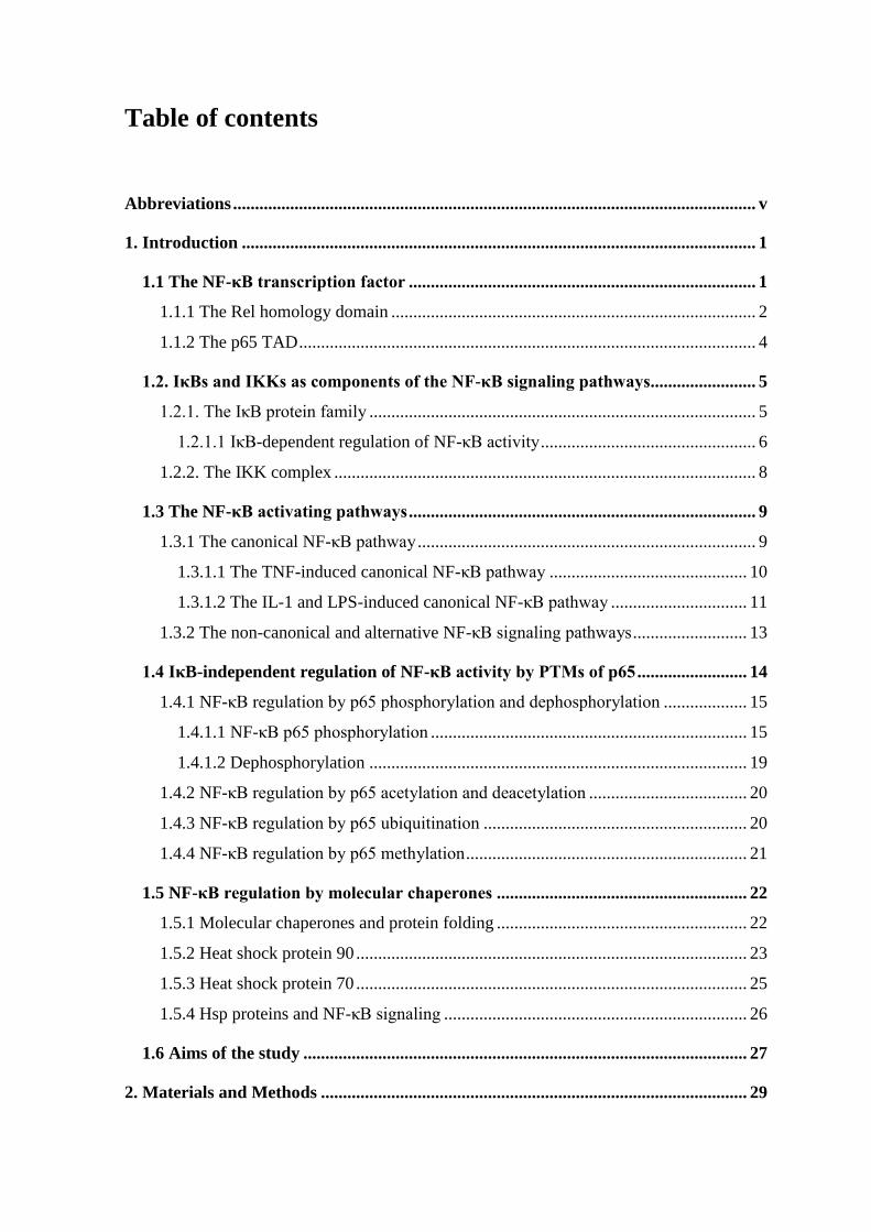
Table of contents
Abbreviations ....................................................................................................................... v
1. Introduction ..................................................................................................................... 1
1.1 The NF-κB transcription factor ............................................................................... 1
1.1.1 The Rel homology domain ................................................................................... 2
1.1.2 The p65 TAD ........................................................................................................ 4
1.2. IκBs and IKKs as components of the NF-κB signaling pathways ........................ 5
1.2.1. The IκB protein family ........................................................................................ 5
1.2.1.1 IκB-dependent regulation of NF-κB activity ................................................. 6
1.2.2. The IKK complex ................................................................................................ 8
1.3 The NF-κB activating pathways ............................................................................... 9
1.3.1 The canonical NF-κB pathway ............................................................................. 9
1.3.1.1 The TNF-induced canonical NF-κB pathway ............................................. 10
1.3.1.2 The IL-1 and LPS-induced canonical NF-κB pathway ............................... 11
1.3.2 The non-canonical and alternative NF-κB signaling pathways .......................... 13
1.4 IκB-independent regulation of NF-κB activity by PTMs of p65 ......................... 14
1.4.1 NF-κB regulation by p65 phosphorylation and dephosphorylation ................... 15
1.4.1.1 NF-κB p65 phosphorylation ........................................................................ 15
1.4.1.2 Dephosphorylation ...................................................................................... 19
1.4.2 NF-κB regulation by p65 acetylation and deacetylation .................................... 20
1.4.3 NF-κB regulation by p65 ubiquitination ............................................................ 20
1.4.4 NF-κB regulation by p65 methylation ................................................................ 21
1.5 NF-κB regulation by molecular chaperones ......................................................... 22
1.5.1 Molecular chaperones and protein folding ......................................................... 22
1.5.2 Heat shock protein 90 ......................................................................................... 23
1.5.3 Heat shock protein 70 ......................................................................................... 25
1.5.4 Hsp proteins and NF-κB signaling ..................................................................... 26
1.6 Aims of the study ..................................................................................................... 27
2. Materials and Methods ................................................................................................. 29
2.1 Materials .................................................................................................................. 29
2.1.1 Antibodies .......................................................................................................... 29
2.1.2 Bacterial strains .................................................................................................. 30
2.1.3 Eukaryotic cell lines ........................................................................................... 30
2.1.4 Plasmids ............................................................................................................. 31
2.1.4.1 Expression plasmids .................................................................................... 31
2.1.4.2 Plasmids for lentiviral production ............................................................... 32
2.1.4.3 Plasmids for luciferase reporter gene assays ............................................... 32
2.1.5 Oligonucleotides ................................................................................................. 32
2.1.6 Antibiotics .......................................................................................................... 34
2.1.7 Enzymes ............................................................................................................. 34
2.1.8 Inhibitors ............................................................................................................ 34
2.1.9 Kits ..................................................................................................................... 35
2.1.10 Chemicals ......................................................................................................... 35
2.1.11 Other reagents .................................................................................................. 37
2.1.12 Buffers and other solutions .............................................................................. 37
2.2 Methods in molecular biology ................................................................................ 38
2.2.1 Transformation of chemically competent E.coli ................................................ 38
2.2.2 Storage of E.coli ................................................................................................. 38
2.2.3 Preparation of chemically competent E.coli ...................................................... 38
2.2.4 Isolation of plasmid DNA from transformed E.coli ........................................... 39
2.2.5 Polymerase Chain Reaction (PCR) .................................................................... 40
2.2.5.1 Amplification of DNA fragments for cloning ............................................. 40
2.2.5.2 Site-directed point mutagenesis .................................................................. 41
2.2.6 Agarose gel electrophoresis ............................................................................... 42
2.2.6.1 DNA extraction from agarose gel ............................................................... 42
2.2.7 Digestion of DNA with restriction enzymes ...................................................... 43
2.2.8 Ligation of DNA fragments ............................................................................... 43
2.2.10 Complementary DNA (cDNA) synthesis ......................................................... 44
2.3 Methods in cell biology ........................................................................................... 46
2.3.1 Cultivation of eukaryotic cell lines .................................................................... 46
2.3.2 Freezing and thawing of cells ............................................................................. 47
2.3.3 Transfection of mammalian cells ....................................................................... 47
2.3.4 Production of lentiviruses and infection of cells ................................................ 48
2.3.5 Lysate preparation .............................................................................................. 50
2.3.5.1 Lysis under denaturing conditions ............................................................... 50
2.3.5.2 Lysis under native conditions - whole cell lysate ........................................ 50
2.3.5.3 Lysis under native conditions - subcellular fractionation ............................ 50
2.3.6 Immunofluorescence (IF) staining...................................................................... 51
2.3.7 Luciferase reporter gene assays .......................................................................... 52
2.4 Biochemical methods ............................................................................................... 52
2.4.1 SDS polyacrylamide gel electrophoresis (SDS-PAGE) ..................................... 52
2.4.2 Coomassie brilliant blue staining of polyacrylamide gels .................................. 54
2.4.3 Western blot and immune detection ................................................................... 54
2.4.4 IP ......................................................................................................................... 55
2.4.5 EMSA ................................................................................................................. 56
2.4.6 Ni-NTA affinity purification .............................................................................. 58
2.4.7 Limited proteolysis assay ................................................................................... 58
3. Results ............................................................................................................................. 61
3.1 The cytokine-induced conformational change of the NF-κB p65 subunit is
mediated by phosphorylation ....................................................................................... 61
3.1.1 Phosphorylation regulates the structural flexibility of NF-κB p65 .................... 61
3.1.2 Individual phosphorylation sites control regulatory and degradative
ubiquitination of p65 ................................................................................................... 63
3.1.3 Cytokines induce a phosphorylation-dependent conformational change of
nuclear p65 .................................................................................................................. 65
3.1.4 The TNF-induced NF-κB p65 structural rearrangement unmasks an epitope
localised in the TAD2 subdomain ............................................................................... 69
3.1.5 Phosphorylation-dependent changes of NF-κB p65 conformation regulate p65
interactions with transcriptional cofactors ................................................................... 70
3.2. Regulation of p65 refolding and activity by chaperones ..................................... 71
3.2.1 NF-κB p65 physically interacts with Hsc/Hsp70 and Hsp90α ........................... 71
3.2.2 Phosphorylation-dependent structural changes of p65 regulate p65 interaction
with Hsps ..................................................................................................................... 73
3.2.3 Chaperones contribute to the control of the TNF-induced p65 refolding .......... 75

3.2.4 Hsc/Hsp70 proteins control the level of p65 PTMs and p65-mediated gene
expression .................................................................................................................... 77
3.2.4.1 Hsc/Hsp70 proteins regulate p65 PTMs...................................................... 77
3.2.4.2 Hsps modulate p65-mediated gene expression ........................................... 80
3.3 Influence of p65 DNA-binding ability on its conformation and activity ............ 81
3.3.1 Characterization of stably reconstituted p65-/- MEFs ....................................... 81
3.3.2 DNA-binding of p65 affects the kinetics of its nuclear export and Ser468-
phosphorylation ........................................................................................................... 82
3.3.3 The impact of p65 DNA-binding on TNF-induced p65 conformation .............. 84
3.3.4 Expression of some TNF-induced p65 dependent genes is not dependent on p65
DNA-binding ............................................................................................................... 85
4. Discussion ....................................................................................................................... 89
4.1 Phosphorylation is important for the conformation of the active nuclear p65 . 89
4.2 Phosphorylation-induced p65 refolding regulates p65 ubiquitination ............... 93
4.3 Phosphorylation-induced conformational changes of p65 influence its
association with other proteins ..................................................................................... 94
4.4 Chaperones as coregulators of p65 refolding ....................................................... 96
4.5 Chaperones as coregulators of p65 PTMs and p65-mediated gene expression . 98
4.6 Cytokine-induced conformation change of p65 occurs independently of its
binding to DNA ............................................................................................................ 100
4.7 Non-genomic functions of NF-κB p65 ................................................................. 102
5. Summary ...................................................................................................................... 107
6. Zusammenfassunf........................................................................................................ 109
7. References .................................................................................................................... 111
Publications ...................................................................................................................... 135
Acknowledgements .......................................................................................................... 136
v
Abbreviations
% (v/v) volume/volume percentage
% (w/v) weight/volume percentage
ºC degree Celsius
3D three-dimensional
17-AAG 17-N-allylamino-17-demethoxygeldanamycin
Ab antibody
ABIN A20 binding inhibitor of NF-κB 2
ADP adenosine diphosphate
AES ` amino enhancer of split
ANK ankyrin repeat
Apaf-1 apoptotic protease activating factor 1
Arg arginine
ARF alternate reading frame
Asp aspartic acid
ATM ataxia telangiectasia mutated
ATP adenosine triphosphate
BAFFR B-cell activating factor of the TNF family receptor
Bcl-xL B-cell lymphoma-extra large
bp base pairs
β-TrCP β-transducin repeat-containing protein
Ct threshold cycle
CaMKIV calmodulin-dependent protein kinase IV
CBP CREB-(c-AMP-response element-binding protein) -binding protein
CC coiled-coil
CCT chaperonin-containing t-complex polypeptide 1 (TCP1)
CD circular dichroism
cDNA complementary DNA
CDK6 cyclin dependent kinase 6
ChIP chromatin immunoprecipitation
CHIP carboxyl-terminus of Hsp70 interacting protein
Chk1 checkpoint kinase 1
vi
cIAP cellular inhibitor of apoptosis protein
CIAP calf intestine alkaline phosphatase
COMMD1 copper metabolism MURR1 domain-containing protein 1
CoREST corepressor for REST (RE1 silencing TF)
Crm-1 chromosomal region maintenance-1
Cul2 cullin-2
DD dimerisation domain
DISC death-inducing signaling complex
DNA deoxyribonucleic acid
ds double-stranded
E3 ubiquitin ligase enzyme
E2 ubiquitin conjugating enzyme
Elk-1 ETS domain-containing protein1
ELKS protein rich in glutamate, leucine, lysine and serine
EMSA electrophoretic mobility shift assay
EPR electron paramagnetic resonance
FADD Fas-associated protein with death domain
FBXL11 F-box and leucine-rich repeat protein 11
FoxO1 forkhead box O1
GA geldanamycin
GCN5 general control nonderepressible 5
GFP green fluorescent protein
GLP G9A-like protein
Glu glutamic acid
GR glucocorticoid receptor
GRR glycine-rich region
GSK-3β glycogen-synthase kinase-3beta
h hour
HA hemagglutinin
HLH helix-loop-helix
HDAC histone deacetylase
HEK human embryonic kidney
His histidine
HOIL-1 heme-oxidized IRP2 ubiquitin ligase-1
vii
HOIP HOIL-1 interacting protein
HOP Hsp organizing protein
HSF-1 heat shock transcription factor-1
Hsp heat shock protein
IAP inhibitor of apoptosis
IB immunoblotting
Ig immunoglobulin
ID intrinsic disorder
IFNβ interferon β
IL-1 interleukin-1
IL-1R interleukin-1 receptor
IκB inhibitor of NF-κB
IKK IκB kinase
IP immunoprecipitation
IRAK1 IL-1 receptor associated kinase 1
IRF3 interferon regulatory transcription factor
ITC Isothermal calorimetry
kb kilo base
KD kinase domain
kDa kilo dalton
Leu leucine
LPS lipopolysaccharide
LTβR lymphotoxin-β receptor
LUBAC linear ubiquitin chain assembly complex
LZ leucine zipper
mAb monoclonal antibody
Mal MyD88 adaptor like
MDM2 mouse double minute 2 homolog
min minutes
MEF mouse embryonic fibroblast
MEKK mitogen-activated protein kinase kinase kinase
MS mass spectrometry
MSK-1 mitogen- and stress-activated protein kinase-1
MYBBP1a Myb-binding protein 1a
viii
MyD88 myeloid differentiation factor 88
NBD NEMO-binding domain
NCoR nuclear receptor corepressor
NEF nucleotide exchange factor
NEMO NF-κB essential modulator
NES nuclear export signal
NF-κB nuclear factor-κB
NFAT nuclear factor of activated T cells
NIK NF-κB-inducing kinase
Ni-NTA nickel-nitrilotriacetic acid
NLS nuclear localization signal
NMR nuclear magnetic resonance
NSD1 nuclear receptor-binding SET domain protein 1
NTD N-terminal domain
Nurr1 nuclear receptorrelated-1 protein
Pro proline
pAb polyclonal antibody
P-TEFb positive transcription elongation factor b
PARP-1 poly (ADP-ribose) polymerase-1
PAMP pathogen-associated molecular pattern
PCR polymerase chain reaction
PDLIM2 PDZ and LIM domain 2
PEST proline-glutamic acid-serine-threonine sequence
PHF20 PHD finger protein 20
PIASy protein inhibitor of activated STAT4
Pim-1 proviral integration site for the Moloney-murine leukemia virus-1
Pin-1 peptidyl-prolyl cis-trans isomerase NIMA-interacting-1
PKAc catalytic subunit of protein kinase A
PKC protein kinase C
PKCδ protein kinase C, zeta
PMA phorbol myristoylacetate
PML promyelocytic leukemia protein
polyUb polyubiquitination
PP2A protein phosphatase 2A

ix
PPA4 protein phosphatase 4
PRMT5 protein arginine methyltransferase 5
PTM posttranslational modification
qRT-PCR quantitative real-time PCR
RANK receptor activator of NF-κB
RDH Rel homology domain
RIP1 receptor interacting protein 1
RNA ribonucleic acid
RNAi RNA interference
RNA Pol II RNA polymerase II
RSK-1 ribosomal protein S6 kinase 1
SAP Shrimp alkaline phosphatase
sec seconds
SETD6 SET domain-containing protein 6
Ser serine
SET suppressor of variegation-enhancer of zeste-trithorax
Seq sequencing
shRNA small-hairpin RNA
SHARPIN SHANK-associated RH domain interacting protein
SMRT silencing mediator for retinoic acid receptor and thyroid hormone receptor
SOCS-1 suppressor of cytokine signaling-1
SPINE-D sequence based prediction with integrated neural network for
disordered residues
SPR surface plasmon resonance
SRC steroid receptor cofactors
STAT signal transducer and activator of transcription
TAB TAK1 binding protein
TAD transactivation domain
TAFII31 TATA-binding-protein-associated factor II31
TAK1 transforming growth factor beta-activated kinase 1
TANK TRAF family member-associated NF-κB activator
TAZ1 transcriptional adaptor zinc binding 1
TBK1 TANK-binding kinase 1
TF transcription factor
x
Thr threonine
Tip60 60 kDa trans-acting regulatory protein of HIV type 1-interacting protein
TIR Toll/interleukin-1 receptor
TIRAP TIR-domain-containing adaptor protein
TLR Toll-like receptors
TNF tumor necrosis factor
TNFR TNF receptor
TRADD TNF receptor associated protein with a death domain
TRAF TNF receptor associated factor
TRAM TRIF-related adapter molecule
TRIF TIR domain-containing adapter-inducing IFN-β
TRP tetratricopeptide repeat
TSS transcription start site
Tyr tyrosine
Ub ubiquitin
UBAN ubiquitin binding in ABIN and NEMO
Ubc13 ubiquitin conjugating enzyme 13
Uev1A E2 variant 1 isoform A
ULD ubiquitin-like domain
uPA urinary plasminogen activator
UV ultraviolet light
VER VER155008
WIP1 wild-type p53-induced phosphatase
YFP yellow fluorescent protein
ZF zinc finger

Introduction
1
1. Introduction
1.1 The NF-κB transcription factor
NF-κB (nuclear factor-κB) is a collective term for a family of eukaryotic transcription
factors (TFs) that play a critical role in inflammation, immunity, cell proliferation,
differentiation and survival. It exists in all cell types with a nucleus [1, 2]. NF-κB is
composed of various combinations of different DNA-binding subunits: p105/p50 (NF-
κB1), p100/p52 (NF-κB2), RelA (p65), RelB and c-Rel [2-4] (Fig.1.1). All of subunits
possess a homologous sequence in their N-termini referred to as the Rel homology domain
(RHD). This region is approximately 300 amino acids long and is responsible for critical
functions including dimerisation, DNA binding, nuclear localization and association with a
family of inhibitory proteins called inhibitors of κB (IκBs). The RHD can be divided into
three structural regions: the N-terminal domain (NTD) involved in binding to DNA, the
dimerisation domain (DD) and the nuclear localization signal (NLS). Between the NTD
and the DD there is a short flexible region which participates in NF-κB DNA binding. The
DD alone mediates the association of individual NF-κB subunits to form combinatorial
dimers. The Rel/NF-κB family members can be grouped into two classes (Fig. 1.1, upper
panel). The first class consists of p105 and p100 precursor proteins which are
proteolytically processed to the NF-κB subunits p50 and p52, respectively. The mature p50
and p52 proteins contain the RHD followed by a 23-amino acid glycine-rich region (GRR),
a region that is essential for directing the cleavage and proteolytic processing of a long
IκB-like C-terminal part of the precursors [5]. The other class consists of p65, RelB and c-
Rel which all contain transactivation domains (TADs) in their C-termini. The TAD regions
are not conserved between the NF-κB subunits [6, 7]. They are rather functionally defined,
as they activate transcription by recruitment of transcriptional coregulators and
components of the basal transcriptional machinery [4]. Due to the lack of C-terminal
TADs, NF-κB dimers composed only of p50 and/or p52 subunits are transcriptionally
inactive [8-10].
Introduction
2
1.1.1 The Rel homology domain
To date, X-ray crystal structures of the DNA-bound RHDs of the p50/p50, p52/p52 and
p65/65 homodimers are known [10-13]. The structure of the p50/p65 heterodimer-DNA
complex has also been reported [14].
Figure 1.1 Schematic representations of NF-κB, IκB and IκB kinase (IKK) family proteins. The upper
panel represents subunits of the NF-κB TF family. In the middle panel are members of the IκB protein
family. The lower panel represents subunits of the IKK complex. Abbreviations: LZ (leucine zipper), ANK
(ankyrin repeat), NES (nuclear export signal), PEST (proline-glutamic acid-serine-threonine sequence), Lys
(lysine), Ser (serine), KD (kinase domain), ULD (ubiquitin (Ub)-like domain), HLH (helix-loop-helix), NBD
(NEMO (NF-κB essential modulator)-binding domain), CC (coiled-coil), ZF (zinc finger).
The crystal structure of the murine p65 RHD (construct containing residues 9-291) bound
to a specific DNA target revealed that the RHD region is folded into two immunoglobulin
(Ig)-like domains which are connected by a 10 amino acid long flexible region [13]. The
N-terminal Ig-like domain is responsible for sequence-specific DNA recognition. The C-
terminal Ig-like fold is responsible for subunit dimerisation and non-specific DNA binding.
1 551
Rel Homology Domain (RHD)
NL
S
TADDDNTD
1 560NL
S
TADDDNTD
p65
RelB
1 620NL
S
TADDDNTDc-Rel
LZ
IκB-like C- terminal domain
1 970NL
S
GRRDDNTDp105/ p50 (NF-κB1)
AN
K
AN
K
AN
K
AN
K
AN
K
AN
K
AN
K
1 NL
S
GRRDDNTDp50
433
1 899NL
S
GRRDDNTDp100/ p52 (NF-κB2)
AN
K
AN
K
AN
K
AN
K
AN
K
AN
K
AN
K
1 NL
S
GRRDDNTDp52
447
1 317PEST
IκBα
AN
K
AN
K
AN
K
AN
K
AN
K
AN
K
1 356PESTIκBβ
AN
K
AN
K
AN
K
AN
K
AN
K
AN
K
1 361
IκBε
AN
K
AN
K
AN
K
AN
K
AN
K
AN
K
1 447
Bcl-3
AN
K
AN
K
AN
K
AN
K
AN
K
AN
K
AN
K
PEST
NE
S
Lys
Se
rL
ys
Se
rL
ys
Se
r
1 716
IκB-δ
AN
K
AN
K
AN
K
AN
K
AN
K
AN
K
AN
K1 327
IκBNS
AN
K
AN
K
AN
K
AN
K
AN
K
AN
K
AN
K
Classical IκBs
Nuclear IκBs
IKKα1 745NBDHLHKD LZ
IKKβ1 756NBDHLHKD LZULD
1 ZFLZCCNEMO
419

Introduction
3
Unlike most DNA-binding proteins, which use α-helices for base-pair recognition, Rel
family dimers use loops from the edge of the N- and C- terminal parts of the RHD to
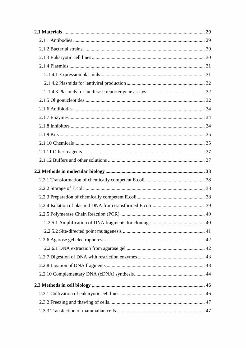
mediate DNA contacts [14]. In case of p65 subunit, five loops are involved in DNA
binding (Fig. 1.2). p65 makes DNA base-specific contacts via Tyr36, Glu39, Arg33 and
Arg35 that arise from the loop L1 (residues 30-50) and Arg187 located in the loop L3
which links the N-terminal domain to the DD. The loop L2 is located in the N-terminal
domain, while loops L4 and L5 arise from the DD. Amino acids from the L2, L4 and L5
loops contact only the phosphodiester backbone of the target DNA [15].
Figure 1.2 Sequence of a murine p65 RHD with shown secondary structure. The scheme is
modified from [15].
The DNA target sequence - NF-κB recognizes 9-11 bp (base pairs) long double-stranded
(ds) DNA-elements termed as κB elements. They are often located within promoters and
enhancers of NF-κB target genes[16, 17], but chromatin immunoprecipitation (ChIP)
experiments with parallel DNA sequencing (ChIP-Seq) identified κB sites in exons and
intergenic space as well [18-22]. The first NF-κB binding site was identified as a B-cell
specific element in the intronic enhancer of the Ig κ light chain gene, with the sequence 5’-
GGGACTTTCC-3’ [23]. Comparison of several different κB sequences recognized by the
NF-κB dimers, allowed the delineation of a consensus sequence 5’-GGGRNWYYCC-3’,
where R denotes a purine base, N means any base, W stands for adenine or thymine and Y
represents a pyrimidine base [24]. Hundreds of such sequences have been confirmed
experimentally. Non-consensus κB sites that hold a significant variation in comparison to
Transcription factor p65 isoform 1 [Mus musculus; AF199371.1]
19 PY VEIIEQPKQR GMRFRYKCEG RSAGSIPGER STDTTKTHPT IKINGYTGPG TVRISLVTKD 80
PPHRPHPHEL VGKDCRDGYY EADLCPDRSI HSFQNLGIQC VKKRDLEQAI SQRIQTNNNP 140
FHVPIEEQRG DYDLNAVRLC FQVTVRDPAG RPLLLTPVLS HPIFDNRAPN TAELKICRVN 200
RNSGSCLGGD EIFLLCDKVQ KEDIEVYFTG PGWEARGSFS QADVHRQVAI VFRTPPYADP 260
SLQAPVRVSM QLRRPSDREL SEPMEFQYLP DTDDRHRIEE KRKR 304
L1
L2
L3
L4 L5
β Strands α Helix
DNA backbone contact Dimer interface Dimer DNA contact
DNA base contacts

Introduction
4
the original consensus κB sequence, were also identified [18-22]. In general, the κB site is
pseudo-symmetric and each NF-κB monomer binds to one DNA half site. The NF-κB p50
and p52 subunits prefer the first half of the κB sequence that begin with 5’-GGG and is
five bp in length. The second half of the κB DNA is four bp in length (5’-YYCC-3’) and is
preferentially occupied by the TAD-containing subunits (p65, RelB or c-Rel). Homodimers
of p50 or p52 would bind optimally to an 11 bp κB DNA, while p65, RelB and c-Rel prefer
binding to 9 bp κB DNA [4, 13].
The NF-κB DD -The DD domain of NF-κB consists of approximately 100 amino acids
near the C-terminal end of the RHD. The corresponding segment of p65 includes 101
residues from Thr191 to Asp291 [25]. About 12-14 residues of each monomer are directly
involved in formation of a dimer interface. A close inspection of these structures revealed
that differential selectivity and stability of NF-κB dimers are influenced by variations in
residues across the dimer interface and by variations of residues located outside of the
dimer interface that influence folding stability of the DD [4].
The NLS – The NF-κB NLS is located next to the DD and is recognized by the stretch of
the basic amino acids KRKR (301
KRKR304
, respectively for human p65). It regulates the
nuclear localization of the NF-κB dimers via recognition by the import receptor [26]. The
p65 NLS peptide and surrounding sequence (residues 293-321) are unfolded in the free
state, while they gain structure upon binding to IκBα [27].
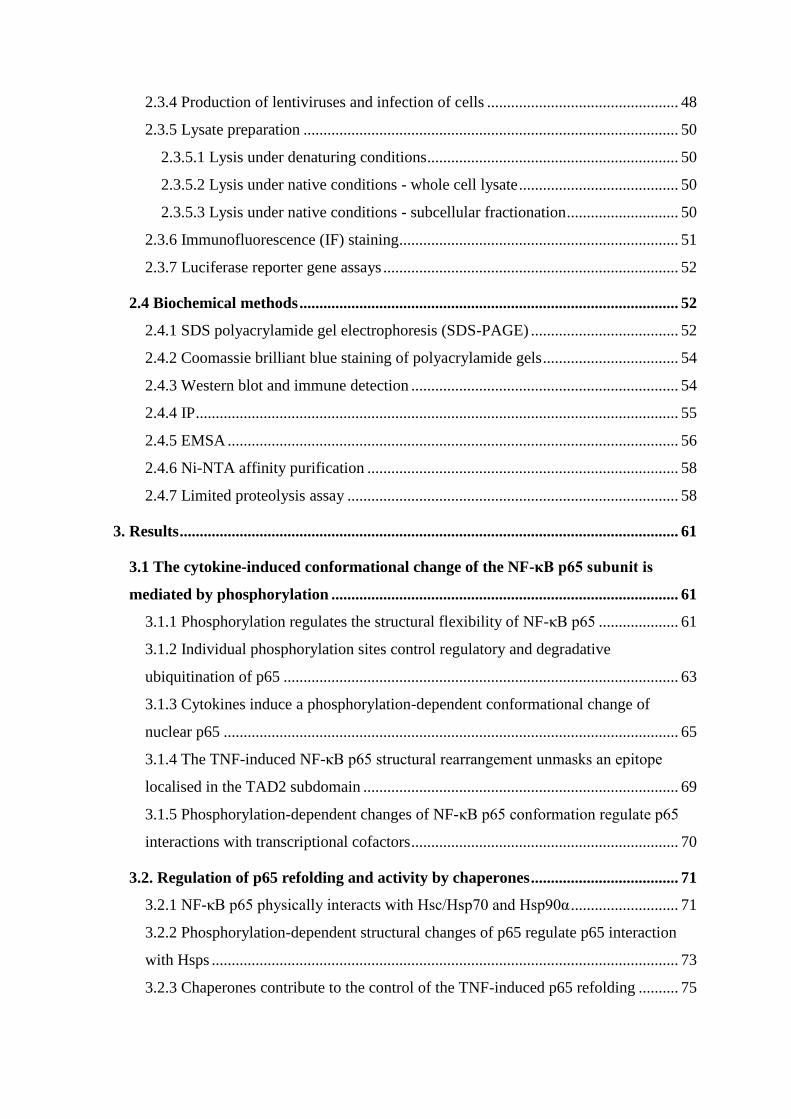
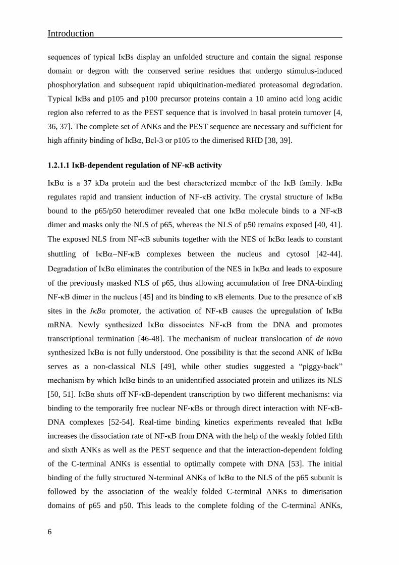
1.1.2 The p65 TAD
The three NF-κB subunits p65, RelB and c-Rel contain non-homologous TADs in their C-
termini which enables them to trigger gene expression [7]. As the p65 subunit is the most
abundantly studied member of this protein family, the architecture and properties of p65 C-
terminal TAD will be described in more detail. The C-terminal 120 amino acids contain
two strong and fully independent TADs: TAD1 in the last C-terminal 30 amino acids
(residues 521-551 of human p65) and TAD2 directly adjacent to TAD1 (residues 428-521
of human p65) (Fig. 1.3) [28-31]. NMR (nuclear magnetic resonance) studies of the
polypeptide corresponding to p65 amino acids 428-551, which comprises both TAD1 and
TAD2, showed that the p65 transactivating C-terminus is unstructured under physiological
conditions [28]. TAD1 and TAD2 of p65 belong to the class of acidic TAD domains. They
are characterized by the presence of evolutionary conserved regions (TAD1 and TAD1’
Introduction
5
located within TAD2, Fig. 1.3), which contain high percentages of acidic and hydrophobic
amino acid residues and can form amphipatic α-helical structures in the hydrophobic
solvent [6, 32]. The N-terminus of TAD2 possesses a mini-LZ (Leu436, Leu443 and
Leu450) which contributes to the transactivation activity of the TAD2 subdomain [28].
The N-terminus of TAD2 also harbors a NES-like sequence [33].
Figure 1.3 Schematic representation of human p65 C-terminal TAD. Upper: underlined NES-like
sequence in p65, stars indicate lysines of the mini LZ-like motif. Lower: the TAD2region with
homology to TAD1 is designated as TAD1’. Identical positions are shown by solid bars, the conserved
hydrophobic amino acids by dashed bars. Figure modified from [6].
1.2. IκBs and IKKs as components of the NF-κB signaling pathways
1.2.1. The IκB protein family
The activity of NF-κB dimers is directly controlled by a set of IκBs through the formation
of stable IκB-NF-κB complexes. Within those complexes, IκB protein masks the NLS of
NF-κB subunits, thereby preventing NF-κB translocation into the nucleus. IκBs are the
coevolved functional partners of NF-κB and regulate NF-κB-dependent gene expression
under a variety of different conditions [34]. The IκB protein family is characterized by the
presence of six to seven conserved ANK motifs which mediate IκB interaction with the
RHD of NF-κB dimers [4]. These motifs are known to play an important role in protein-
protein interactions while lacking any enzymatic activity [35]. The IκB protein family
contains the classical IκBs (IκBα, IκBβ and IκBε), the non-classical IκBs (NF-κB
precursors p105 and p100) and the nuclear IκBs ( Bcl-3, IκBδ, IκBNS and IκBε) (Fig. 1.1)
[36]. Classical IκBs are defined by the presence of six ANKs [36]. The N-terminal
551
428521
Human p65 (428-450) PTQAGEGTLSEALLQLQFDDEDLNES
* * *436 443 450
Human p65 TA1 (526-551) GLLSGDEDFSSIADMDFSALLSQISS
Human p65 TA1’ (458-483) TDPAVFTDLASVDNSEFQQLLNQGIP
Introduction
6
sequences of typical IκBs display an unfolded structure and contain the signal response
domain or degron with the conserved serine residues that undergo stimulus-induced
phosphorylation and subsequent rapid ubiquitination-mediated proteasomal degradation.
Typical IκBs and p105 and p100 precursor proteins contain a 10 amino acid long acidic
region also referred to as the PEST sequence that is involved in basal protein turnover [4,
36, 37]. The complete set of ANKs and the PEST sequence are necessary and sufficient for
high affinity binding of IκBα, Bcl-3 or p105 to the dimerised RHD [38, 39].
1.2.1.1 IκB-dependent regulation of NF-κB activity
IκBα is a 37 kDa protein and the best characterized member of the IκB family. IκBα
regulates rapid and transient induction of NF-κB activity. The crystal structure of IκBα
bound to the p65/p50 heterodimer revealed that one IκBα molecule binds to a NF-κB
dimer and masks only the NLS of p65, whereas the NLS of p50 remains exposed [40, 41].
The exposed NLS from NF-κB subunits together with the NES of IBleads to constant
shuttling of IBNF-B complexes between the nucleus and cytosol [42-44].
Degradation of IκBα eliminates the contribution of the NES in IBand leads to exposure
of the previously masked NLS of p65, thus allowing accumulation of free DNA-binding
NF-κB dimer in the nucleus [45] and its binding to κB elements. Due to the presence of κB
sites in the IκBα promoter, the activation of NF-κB causes the upregulation of IκBα
mRNA. Newly synthesized IκBα dissociates NF-κB from the DNA and promotes
transcriptional termination [46-48]. The mechanism of nuclear translocation of de novo
synthesized IκBα is not fully understood. One possibility is that the second ANK of IκBα
serves as a non-classical NLS [49], while other studies suggested a “piggy-back”
mechanism by which IκBα binds to an unidentified associated protein and utilizes its NLS
[50, 51]. IκBα shuts off NF-κB-dependent transcription by two different mechanisms: via
binding to the temporarily free nuclear NF-κBs or through direct interaction with NF-κB-
DNA complexes [52-54]. Real-time binding kinetics experiments revealed that IκBα
increases the dissociation rate of NF-κB from DNA with the help of the weakly folded fifth
and sixth ANKs as well as the PEST sequence and that the interaction-dependent folding
of the C-terminal ANKs is essential to optimally compete with DNA [53]. The initial
binding of the fully structured N-terminal ANKs of IκBα to the NLS of the p65 subunit is
followed by the association of the weakly folded C-terminal ANKs to dimerisation
domains of p65 and p50. This leads to the complete folding of the C-terminal ANKs,

Introduction
7
allowing the correct positioning of the negatively charged PEST sequence to efficiently
displace IκBα from DNA [54]. The nuclear export of IκBα-NF-κB complex requires the
IκBα NES between residues 45-54. This mechanism was suggested to be required for
proper termination of TNF (tumor necrosis factor)-induced NF-κB activity [42].
IκBβ is a 45 kDa protein that is constitutively phosphorylated in resting cells [55]. In
contrast to IκBα, IκBβ lacks the NES and is not an NF-κB target gene, even though the
IκBβ promoter contains a κB site [56, 57]. Nevertheless, IκBβ can be resynthesized
following stimulation with LPS (lipopolysaccharide) for four hours in a stimulus-
dependent manner and appears as a hypophosphorylated protein which enters the nucleus
and forms a stable complex with DNA-bound NF-κB [58, 59]. Therefore, besides its role
as a cytoplasmic NF-κB inhibitor in resting cells, IκBβ also acts as a target gene-specific
nuclear coactivator in TLR (Toll-like receptor) signaling.
The specific role of the 45 kDa protein IκBε is less well established. It is most likely that
the combined action of IκBα and IκBε is required to regulate distinct expression dynamics
of NF-κB-dependent target genes [34]. Stimulus-induced degron phosphorylation and
subsequent ubiquitination-mediated proteasomal degradation of IκBε occurs with slower
kinetics in comparison to IκBα and IκBβ. The transcription of the IκBε gene is NF-κB
dependent but the resynthesis of IκBε occurs with a significantly delayed kinetics
compared to IκBα [60]. Also, the nuclear import of IκBε occurs with a lower efficiency
compared to IκBα. The nuclear export is mediated by a short NES-like sequence located
between residues 343-352 within IκBε [61].
The C-termini of p100 and p105contain a long IκB-like domain containing seven ANKs,
which blocks nuclear translocation and DNA-binding of the NF-κB precursor proteins [62-
64]. The p105 precursor undergoes IKKβ-mediated phosphorylation at Ser927 and
Ser932and subsequent ubiquitination-dependent proteolytic degradation, releasing the NF-
κB p50-containing dimers [65]. The precursor p100 is phosphorylated by IKKα at
conserved Ser866 and Ser870 and this promotes p100 polyubiquitination (polyUb) and
processing by the proteasome, generating active p52 [66, 67]. The NF-κB precursor
proteins are responsible for the inhibition of nearly half of the NF-κB dimers in resting
cells. The precursors can assemble more than one NF-κB dimer into a high-molecular-
weight complex which might function as a dynamic buffering system for NF-κB subunits

Introduction
8
that are not bound to typical IκBs [68]. As targets of NF-κB, the precursor proteins p105
and p100 together with a newly synthesized classical IκBs serve to block NF-κB activity
[69].
The atypical IκBs Bcl-3, IκBδ, IκBNS and IκBε are located in the nucleus and exhibit a
variety of functions. In resting cells they display low expression levels, but upon
stimulation with NF-κB-inducing agents their expression increases significantly [70-72].
They prevent the degradation of DNA-bound NF-κB dimers by competing with
cytoplasmic IκBs for binding and provide interactions with other TFs. They can
transactivate or inhibit transcription and provide a fine-tuning mechanism for late NF-κB-
dependent gene regulation [73]. For example, Bcl-3 exhibits a coactivator function via
interaction with DNA-bound p50 homodimer [74, 75] and provides an interaction platform
for diverse multi-protein complexes involved in transcriptional regulation of NF-κB target
genes [76-78].
1.2.2. The IKK complex
The common feature of all signaling pathways leading to NF-κB activation is the induction
of IKKs. The IKK complex consists of two highly homologous kinases IKKα/IKK1 and
IKKβ/IKK2 as well as the regulatory subunit IKKγ/NEMO [79] (Fig.1.1). IKKα and IKKβ
are serine/threonine kinases characterized by the presence of an N-terminal kinase domain,
followed by an ULD, a LZ and the C-terminal HLH domain. The C-terminus contains a
stretch of six amino acids termed the NBD, which mediates interaction of IKKα/β with
NEMO. In addition, IKKα contains a putative NLS [80] which possibly allows its
translocation to the nucleus. NEMO is not related to IKKα and IKKβ and contains a CC,
LZ and a C-terminal ZF-like domain [36] (Fig. 1.1). The activation of IKK proteins is
mediated by phosphorylation of either IKKα or IKKβ at two specific serine residues
(Ser177 and Ser181 for IKKα, and Ser176 and Ser180 for IKKβ) within the activation loop
of the catalytic domain, yet its regulation is poorly understood [79]. Tang et al. showed
that the activation of IKKβ depends on ligand-induced homotypic interactions between
IKKβ molecules that result in its phosphorylation and consequently IKK activation [81].
On the other hand, it has been shown that TAK1 (transforming growth factor beta-
activated kinase 1) directly phosphorylates IKKwithin the activation loop, leading to
activation of the IKK complex [82]. NEMO acts as a scaffold protein that promotes the

Introduction
9
assembly of the IKK complex and contributes to the recruitment of the IKK complex to
upstream signaling molecules [84-86]. IKK proteins, although similar in structure, have
relatively distinct substrates and functions that relate to the existence of different NF-κB
activation pathways.
1.3 The NF-κB activating pathways
The NF-κB activating pathways can be grouped depending on the set of stimuli and signal
transducing molecules as:
- The canonical NF-κB activation pathway
- The non-canonical NF-κB activation pathway
- The alternative NF-κB activation pathway
1.3.1 The canonical NF-κB pathway
Activation of the canonical pathway occurs in response to inflammatory cytokines, such as
TNF and interleukin-1 (IL-1). These cytokines are recognized by corresponding membrane
receptors: the TNF receptor (TNFR) and interleukin-1 receptor (IL-1R) [87]. The canonical
pathway is also activated in response to PAMPs (pathogen-associated molecular patterns)
such as LPS, flagellin, viral dsRNA and unmethylated CpG motifs recognized by TLRs.
NF-κB activated by canonical pathway is involved in the control of innate immune
responses and apoptosis [88, 89]. Depending on the nature of the stimulus and activated
receptor, posttranslational modifications (PTMs) of signaling cascade components lead to
the activation of the IKK complex. These modifications include ubiquitination of NEMO
and phosphorylation of two serine residues in the activation loop of IKKβ [85, 90]. In most
of the stimulus-initiated canonical pathways, IKKβ is necessary and sufficient for
phosphorylation of IκBα at residues Ser32 and Ser36 and IκBβ at Ser19 and Ser23 [91].
This site-specific phosphorylation leads to IκBα/β ubiquitination, which tags IκBs for
degradation by the proteasome [92]. Free DNA-binding p50/p65 dimers rapidly translocate
to the nucleus within several minutes (min) [69]. Full activation of gene expression
requires a number of further PTMs of the p65 subunit, including phosphorylation,
acetylation, ubiquitination, methylation and prolyl-isomerisation [93, 94].
Introduction
10
1.3.1.1 The TNF-induced canonical NF-κB pathway
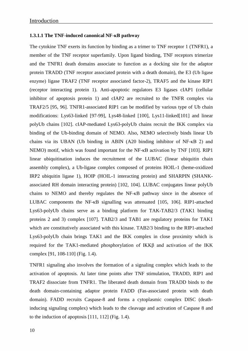
The cytokine TNF exerts its function by binding as a trimer to TNF receptor 1 (TNFR1), a
member of the TNF receptor superfamily. Upon ligand binding, TNF receptors trimerize
and the TNFR1 death domains associate to function as a docking site for the adaptor
protein TRADD (TNF receptor associated protein with a death domain), the E3 (Ub ligase
enzyme) ligase TRAF2 (TNF receptor associated factor-2), TRAF5 and the kinase RIP1
(receptor interacting protein 1). Anti-apoptotic regulators E3 ligases cIAP1 (cellular
inhibitor of apoptosis protein 1) and cIAP2 are recruited to the TNFR complex via
TRAF2/5 [95, 96]. TNFR1-associated RIP1 can be modified by various type of Ub chain
modifications: Lys63-linked [97-99], Lys48-linked [100], Lys11-linked[101] and linear
polyUb chains [102]. cIAP-mediated Lys63-polyUb chains recruit the IKK complex via
binding of the Ub-binding domain of NEMO. Also, NEMO selectively binds linear Ub
chains via its UBAN (Ub binding in ABIN (A20 binding inhibitor of NF-κB 2) and
NEMO) motif, which was found important for the NF-κB activation by TNF [103]. RIP1
linear ubiquitination induces the recruitment of the LUBAC (linear ubiquitin chain
assembly complex), a Ub-ligase complex composed of proteins HOIL-1 (heme-oxidized
IRP2 ubiquitin ligase 1), HOIP (HOIL-1 interacting protein) and SHARPIN (SHANK-
associated RH domain interacting protein) [102, 104]. LUBAC conjugates linear polyUb
chains to NEMO and thereby regulates the NF-κB pathway since in the absence of
LUBAC components the NF-κB signalling was attenuated [105, 106]. RIP1-attached
Lys63-polyUb chains serve as a binding platform for TAK-TAB2/3 (TAK1 binding
proteins 2 and 3) complex [107]. TAB2/3 and TAB1 are regulatory proteins for TAK1
which are constitutively associated with this kinase. TAB2/3 binding to the RIP1-attached
Lys63-polyUb chain brings TAK1 and the IKK complex in close proximity which is
required for the TAK1-mediated phosphorylation of IKKβ and activation of the IKK
complex [91, 108-110] (Fig. 1.4).
TNFR1 signaling also involves the formation of a signaling complex which leads to the
activation of apoptosis. At later time points after TNF stimulation, TRADD, RIP1 and
TRAF2 dissociate from TNFR1. The liberated death domain from TRADD binds to the
death domain-containing adaptor protein FADD (Fas-associated protein with death
domain). FADD recruits Caspase-8 and forms a cytoplasmic complex DISC (death-
inducing signaling complex) which leads to the cleavage and activation of Caspase 8 and
to the induction of apoptosis [111, 112] (Fig. 1.4).

Introduction
11
Figure 1.4 The canonical TNFR1-mediated signaling pathway to NF-κB. TNF-induced NF-κB signaling
pathway mediated by the regulatory ubiquitination of RIP1 and activation of the IKK complex via LUBAC
and TAK1/TABs complexes that are attached to RIP1 polyUb chains. TNF also induces the formation of
apoptosis signaling complexes. Scheme is modified from [113].
1.3.1.2 The IL-1 and LPS-induced canonical NF-κB pathway
NF-κB activation in response to cytokine IL-1 or PAMPs is mediated via IL-1R or
TLRsrespectively (Fig. 1.5). Upon binding of IL-1 to the IL-1R1, the adaptor protein
MyD88 (myeloid differentiation factor 88) is recruited to the receptor via its own TIR
(Toll/interleukin-1 receptor) domain [114, 115]. This leads to the assembly of a signaling
complex that includes the serine/threonine kinase IRAK1 (IL-1 receptor associated kinase-
1), IRAK4 and the E3 ligase TRAF6 (TNF receptor associated factor-6) [116, 117].
IRAK4 becomes activated by intramolecular auto-phosphorylation within its activation
loop [118] and phosphorylates IRAK1. This further promotes dissociation of IRAK1 and
TRAF6 from MyD88 [119, 120]. The E3 ligase β-TrCP (β-transducin repeat-containing
protein) mediates Lys48-linked IRAK1 polyubiquitination (Lys48-polyUb) and subsequent
IRAK1 degradation which has a critical role in dissociation of TRAF6 from IRAK1 [121].
In the cytoplasm, TRAF6 interacts with Ubc13 (Ub conjugating enzyme 13) and Uev1A
plasma membrane
apoptosis
TNFR1
TNFα
casp
ase
-8
Lys63-polyUb
Lys48-polyUb
Linear polyUb
UbUbUb
UbUbUb
Ub
UbUbUbUb
UbUbUb
Ub
Ub
Ub
Ub
Ub
Ub
Ub
Ub
Ub
Ub
PUbUbUbUb
LUB
AC
P
P Ub Ub
UbUb
Ub
proteasome
cytoplasm
nucleus
P
IκBα
Introduction
12
(E2 variant 1 isoform A), resulting in the regulatory ubiquitination of TRAF6 [122].
TRAF6-attached Lys63-polyUb chains provide a platform for recruitment of the TAK1-
TABs, the LUBAC and IKK complex via NEMO. Finally, TAK1 phosphorylates IKKβ,
leading to the activation of IKK complex and NF-κB [123, 124].
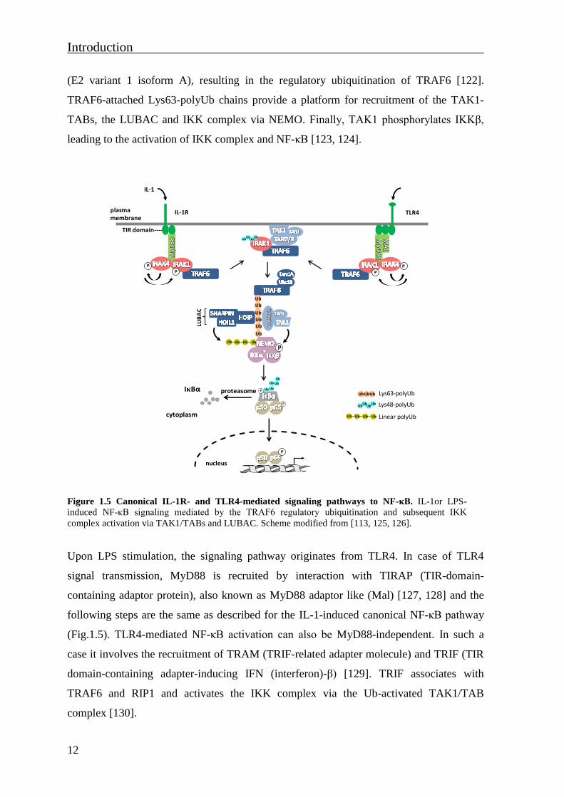
Figure 1.5 Canonical IL-1R- and TLR4-mediated signaling pathways to NF-κB. IL-1or LPS-
induced NF-κB signaling mediated by the TRAF6 regulatory ubiquitination and subsequent IKK
complex activation via TAK1/TABs and LUBAC. Scheme modified from [113, 125, 126].
Upon LPS stimulation, the signaling pathway originates from TLR4. In case of TLR4
signal transmission, MyD88 is recruited by interaction with TIRAP (TIR-domain-
containing adaptor protein), also known as MyD88 adaptor like (Mal) [127, 128] and the
following steps are the same as described for the IL-1-induced canonical NF-κB pathway
(Fig.1.5). TLR4-mediated NF-κB activation can also be MyD88-independent. In such a
case it involves the recruitment of TRAM (TRIF-related adapter molecule) and TRIF (TIR
domain-containing adapter-inducing IFN (interferon)-β) [129]. TRIF associates with
TRAF6 and RIP1 and activates the IKK complex via the Ub-activated TAK1/TAB
complex [130].
plasma membrane
IL-1R
IL-1
TIR domain----
TLR4
PP
PP
UbUbUb
Ub
P
Ub
Ub
Ub
Ub
Ub
Ub
Lys63-polyUb
Lys48-polyUb
Linear polyUb
UbUbUb
UbUbUb
Ub
UbUbUbUb
UbUbUbUb
LUB
AC
P
P Ub Ub
UbUb
Ub
proteasome
cytoplasm
nucleus
P
IκBα

Introduction
13
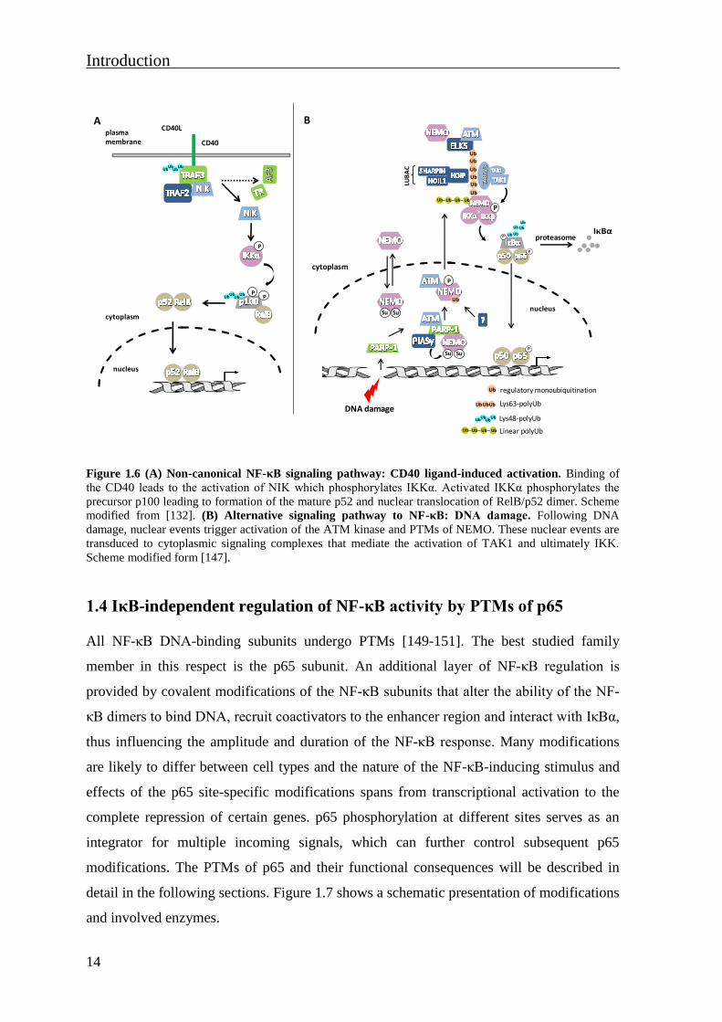
1.3.2 The non-canonical and alternative NF-κB signaling pathways
The non-canonical NF-κB pathway is mediated by a specific subset of TNFR superfamily
members including: LTβR (lymphotoxin-β receptor), BAFFR (B-cell activating factor of
the TNF family receptor), CD40 and CD27 receptors, RANK (receptor activator of NF-κB)
receptor or TNFR2. The key feature of the non-canonical pathway is the processing of the
precursor p100 protein by the proteasome. This processing results in the release of the NF-
κB subunit p52 and generation of p52-containing NF-κB dimers, in most cases p52/RelB
heterodimers [131]. In comparison to the canonical pathway, this pathway is independent
of IKKβ or NEMO, whereas the activation of NIK (NF-κB-inducing kinase) and IKKα are
essential. For example, in response to CD40 ligand, NIK is activated and phosphorylates
IKKα. Activated IKKα phosphorylates p100 at Ser866 and Ser870, leading to processing
of precursor p100 to mature p52 and nuclear translocation of p52/RelB dimer [132-135]
(Fig. 1.6A).
Activation of the NF-κB response can be induced by alternative mechanisms which are
characterized by IKK activity in a manner distinct from those found in canonical and non-
canonical pathways. Alternative pathways of NF-κB activation are initiated in response to
short-wavelength ultraviolet light (UV) or DNA damage [79, 136-141]. As an example, the
NF-κB response to DNA damage will be described. NF-κB activation upon DNA-damage
is still poorly understood. A series of posttranslational events, including sumoylation,
ubiquitination, phosphorylation and nuclear-cytoplasmic shuttling of NEMO appear
critical [142], but the order and which molecules fine-tuning these events need to be
ravealed. A nuclear poly (ADP-ribose) polymerase 1 (PARP-1), ataxia telangiectasia
mutated (ATM)-kinase, protein inhibitor of activated STAT4 (PIASy) and NEMO
signalosome are defined as critical nuclear components of the DNA damage-induced NF-
κB signaling pathway [143-145]. The required cytoplasmic proteins involve ATM that is
translocated to the cytoplasm, TRAF6 and/or ELKS (protein rich in glutamate, leucine,
lysine and serine), as well as the E2 ligase Ubc13 and members of the inhibitor of
apoptosis (IAP) family [146, 147]. The latter act as E3 ligases conducting assembly of
Lys63-polyUb scaffolds thereby facilitating TAK1-TAB2/3-mediated activation of a
functional IKK complex [144, 145]. In addition, linear Ub chains assembled on NEMO by
LUBAC were shown to be essential for the DNA damage induced NF-κB activation [148]
(Fig. 1.6B).
Introduction
14
Figure 1.6 (A) Non-canonical NF-κB signaling pathway: CD40 ligand-induced activation. Binding of
the CD40 leads to the activation of NIK which phosphorylates IKKα. Activated IKKα phosphorylates the
precursor p100 leading to formation of the mature p52 and nuclear translocation of RelB/p52 dimer. Scheme
modified from [132]. (B) Alternative signaling pathway to NF-κB: DNA damage. Following DNA
damage, nuclear events trigger activation of the ATM kinase and PTMs of NEMO. These nuclear events are
transduced to cytoplasmic signaling complexes that mediate the activation of TAK1 and ultimately IKK.
Scheme modified form [147].
1.4 IκB-independent regulation of NF-κB activity by PTMs of p65
All NF-κB DNA-binding subunits undergo PTMs [149-151]. The best studied family
member in this respect is the p65 subunit. An additional layer of NF-κB regulation is
provided by covalent modifications of the NF-κB subunits that alter the ability of the NF-
κB dimers to bind DNA, recruit coactivators to the enhancer region and interact with IκBα,
thus influencing the amplitude and duration of the NF-κB response. Many modifications
are likely to differ between cell types and the nature of the NF-κB-inducing stimulus and
effects of the p65 site-specific modifications spans from transcriptional activation to the
complete repression of certain genes. p65 phosphorylation at different sites serves as an
integrator for multiple incoming signals, which can further control subsequent p65
modifications. The PTMs of p65 and their functional consequences will be described in
detail in the following sections. Figure 1.7 shows a schematic presentation of modifications
and involved enzymes.
CD40
CD40Lplasma membrane
A
UbUbUb
Ub
UbUbUb
Ub
P
PP
cytoplasm
nucleus
DNA damage
B
cytoplasm
P
Su Su
P
Ub
nucleus
Su Su
P
Ub
Ub
Ub
Ub
Ub
Ub
UbUbUbUb
LUB
AC
P
P Ub Ub
UbUb
Ub
proteasome
Lys63-polyUb
Lys48-polyUb
Linear polyUb
UbUbUb
UbUbUb
Ub
UbUbUbUb
regulatory monoubiquitinationUb
IκBα
Introduction
15
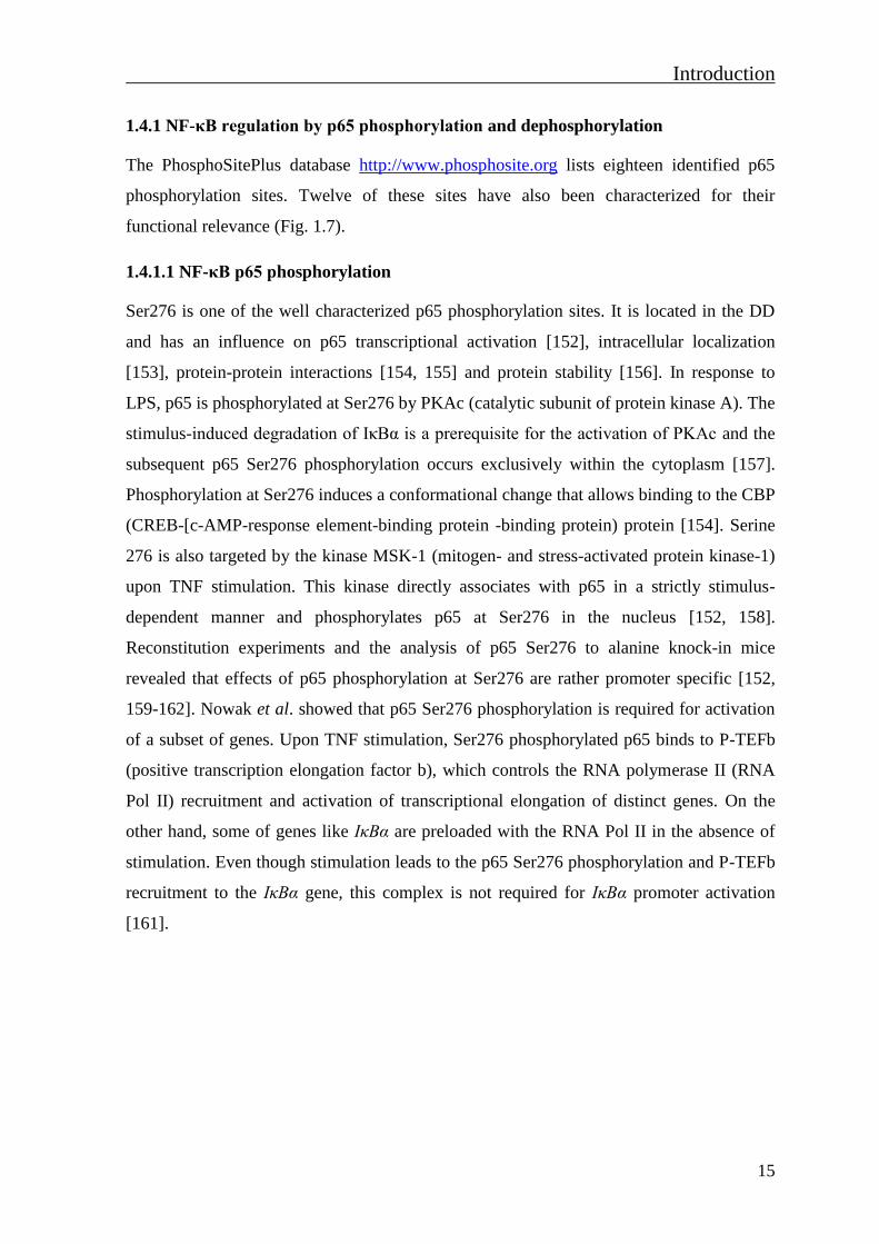
1.4.1 NF-κB regulation by p65 phosphorylation and dephosphorylation
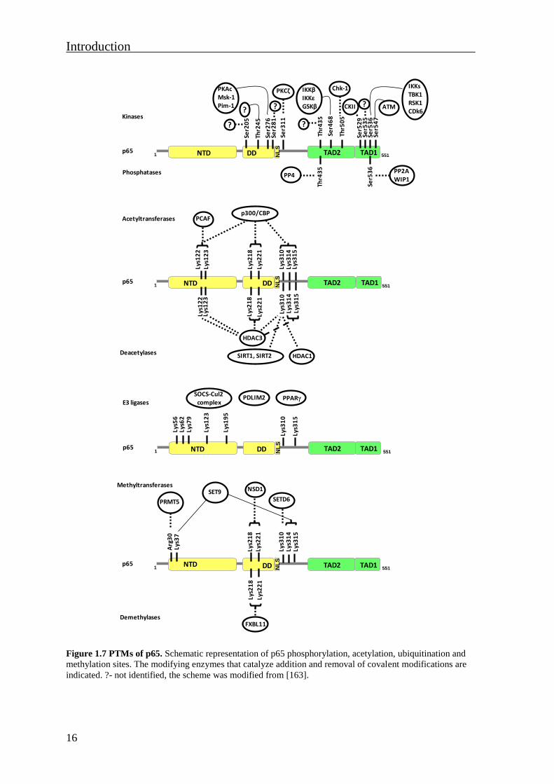
The PhosphoSitePlus database http://www.phosphosite.org lists eighteen identified p65
phosphorylation sites. Twelve of these sites have also been characterized for their
functional relevance (Fig. 1.7).
1.4.1.1 NF-κB p65 phosphorylation
Ser276 is one of the well characterized p65 phosphorylation sites. It is located in the DD
and has an influence on p65 transcriptional activation [152], intracellular localization
[153], protein-protein interactions [154, 155] and protein stability [156]. In response to
LPS, p65 is phosphorylated at Ser276 by PKAc (catalytic subunit of protein kinase A). The
stimulus-induced degradation of IκBα is a prerequisite for the activation of PKAc and the
subsequent p65 Ser276 phosphorylation occurs exclusively within the cytoplasm [157].
Phosphorylation at Ser276 induces a conformational change that allows binding to the CBP
(CREB-[c-AMP-response element-binding protein -binding protein) protein [154]. Serine
276 is also targeted by the kinase MSK-1 (mitogen- and stress-activated protein kinase-1)
upon TNF stimulation. This kinase directly associates with p65 in a strictly stimulus-
dependent manner and phosphorylates p65 at Ser276 in the nucleus [152, 158].
Reconstitution experiments and the analysis of p65 Ser276 to alanine knock-in mice
revealed that effects of p65 phosphorylation at Ser276 are rather promoter specific [152,
159-162]. Nowak et al. showed that p65 Ser276 phosphorylation is required for activation
of a subset of genes. Upon TNF stimulation, Ser276 phosphorylated p65 binds to P-TEFb
(positive transcription elongation factor b), which controls the RNA polymerase II (RNA
Pol II) recruitment and activation of transcriptional elongation of distinct genes. On the
other hand, some of genes like IκBα are preloaded with the RNA Pol II in the absence of
stimulation. Even though stimulation leads to the p65 Ser276 phosphorylation and P-TEFb
recruitment to the IκBα gene, this complex is not required for IκBα promoter activation
[161].
Introduction
16
Figure 1.7 PTMs of p65. Schematic representation of p65 phosphorylation, acetylation, ubiquitination and
methylation sites. The modifying enzymes that catalyze addition and removal of covalent modifications are
indicated. ?- not identified, the scheme was modified from [163].
Ser2
76
1 551NL
S
TAD2DDNTDp65 TAD1
Ser3
11
Ser4
68
Thr2
45
?
PKAcMsk-1Pim-1
PKCζ IKKβIKKεGSKβ
Thr5
05
Ser2
05
?
Ser2
81
?
Thr4
35
?
Chk-1
Thr4
35
Ser5
29
Ser5
35
Ser5
36
Ser5
47
IKKsTBK1RSK1CDk6
? ATM
Ser5
36
PP4PP2AWIP1
Kinases
Phosphatases
Lys1
22
1 551NL
S
TAD2DDNTDp65 TAD1Ly
s31
0
Lys2
21
HDAC3
Lys2
18
Lys1
23
SIRT1, SIRT2
PCAFp300/CBP
Acetyltransferases
Deacetylases
Lys3
14
Lys3
15
Lys1
22
Lys3
10
Lys2
21
Lys2
18
Lys1
23
HDAC1
Lys3
14
Lys3
15
1 551NL
S
TAD2DDNTDp65 TAD1
Lys3
10
Lys2
21
Lys2
18
NSD1SET9Methyltransferases
Demethylases
Lys3
14
Lys3
15
Lys2
21
Lys2
18
Arg
30
Lys3
7
PRMT5 SETD6
FXBL11
Lys1
23
1 551NL
S
TAD2DDNTDp65 TAD1
Lys3
10
Lys5
6
SOCS-Cul2complexE3 ligases
Lys3
15
Lys6
2Ly
s79
Lys1
95
PDLIM2 PPARγ
Introduction
17
The serine/threonine kinase Pim-1 (proviral integration site for the Moloney-murine
leukemia virus-1) was also identified as a p65 kinase that mediates phosphorylation of
Ser276 and subsequent activation of p65. This study suggested that Ser276
phosphorylation protects p65 from proteasomal degradation [156].
In response to TNF, p65 is phosphorylated at Thr245 by an unknown kinase [164].
Phosphorylation at this site creates a phospho-Thr/Pro motif which is a target of another
PTM catalysed by the enzyme peptidyl-prolyl isomerase Pin-1 (peptidyl-prolyl cis-trans
isomerase NIMA-interacting-1). Pin-1 isomerisation disrupts p65 association with IκBα
and induces its translocation to the nucleus. This Pin-1-dependent mechanism also protects
p65 from SOCS-1 (suppressor of cytokine signaling-1)-mediated ubiquitination and
subsequent proteolysis. The effect of p65 isomerization on DNA-binding or transcriptional
activity is still unknown.
PKCδ (protein kinase C, zeta) phosphorylates p65 at Ser311 in a TNF-dependent manner.
Phosphorylaytion at this site enhances the recruitment of CBP to the Il-6 promoter, thus
enhancing the transcriptional activity of NF-κB [165]. On the other hand, a study using a
site-directed mutagenesis approach revealed that Ser311 phosphorylation is not essential
for p65 transcriptional activity, but it is required for transcription of a minority of NF-κB
genes [162].
Residues Ser468 and Ser536 are the most studied C-terminal phosphosites.
Phosphorylation of p65 at Ser536 is mediated by different kinases: IKKα, IKKβ, TBK1
(TANK (TRAF family member-associated NF-κB activator)-binding kinase 1), CDK6
(cyclin dependent kinase 6) and RSK-1 (ribosomal protein S6 kinase 1) and is induced by
various agents, such as TNF, IL-1, T-cell costimulation, human T lymphotrophic virus-1
(HTLV-1)-encoded Tax protein and cytotoxic agents [166-173]. Upon TNF treatment or T-
cell costimulation, NF-κB phosphorylated at p65 Ser536 has been mainly detected in the
cytosol, in particular in the area surrounding the nucleus. Those findings implicate the role
of Ser536 phosphorylated p65 in the control of NF-κB nuclear import kinetics and allows a
fine tuning of the NF-κB mediated transcriptional response [166, 168]. In contrast, RSK-1-
and TBK-1-mediated phosphorylation of Ser536 decreases the affinity between p65 and
IκBα and reduces IκBα-mediated nuclear export of NF-κB, thereby promoting the binding
and action of NF-κB on cognate κB enhancers [170, 172]. Functional characterization of

Introduction
18
phospho-Ser536 in Il-8 transcription revealed that phosphorylation at this site modulates
the balance between binding of TAFII31 (TATA-binding-protein-associated factor II31), a
component of the general TF IID complex, and the corepressor AES (amino enhancer of
split) [170]. The IKKα-mediated phosphorylation of p65 at Ser536 and of corepressor
SMRT (silencing mediator for retinoic acid receptor and thyroid hormone receptor) at
Ser2410 prevents repressor complex recruitment and its association to the NF-κB
promoter. This dual phosphorylation thus allows the loading of p300 to the promoter and
subsequent p65 acetylation at Ser310, which is required for full NF-κB transcriptional
activity [174, 175]. On the other hand, IKKα-dependent phosphorylation of p65 at Ser536
in macrophages results in accelerated turnover of this subunit, thereby facilitating their
removal from the promoters and terminating NF-κB-mediated gene induction [176].
Phosphorylation of Ser536 and Ser468 is not mediated by same kinases, except IKKβ
which has been found to phosphorylates both sites after the T cell costimulation [166, 177].
Ser468 is the target of at least three protein kinases: IKKβ, IKKε and GSK-3β (glycogen-
synthase kinase-3beta) [177-179]. Loss-of-function experiments using small-hairpin RNA
(shRNA)-mediated IKKε knock-down showed that TNF-induced Ser536 phosphorylation
was independent from IKKε, while Ser468 phosphorylation was largely impaired in the
absence of this kinase [166]. Serine 468 phosphorylation is described as a nuclear event
and NF-κB phosphorylated at p65 Ser468 is predominantly located within the nucleus
[166, 179, 180]. On the other hand, IKKβ-mediated phosphorylation at this site was
reported to be cytoplasmic while the NF-κB is still bound to IκBα [177]. Ser468
phosphorylation has been described as both stimulating and inhibiting p65 transactivation
[166, 177, 179]. Different outcomes could be explained by a NF-κB barcode hypothesis,
according to which phosphorylation at Ser468 alone or in combination with other PTMs
could generate distinct patterns that function to direct transcription in a target gene-specific
fashion [166]. Also, phosphorylation by GSK-3β enables p65 to recruit Nurr1 (nuclear
receptor related-1 protein) to the NF-κB on the target gene promoters. This is followed by
recruitment of the CoREST (corepressor for REST (RE1 silencing TF)) corepressor
complex, resulting in clearance of p65 and inhibition of basal NF-κB activity [178, 181].
Phosphorylation of Ser468 by IKKβ or IKKε in response to TNF or IL-1 attenuates the
activity of NF-κB by enhancing the binding of a COMMD1 (copper metabolism MURR1
domain-containing protein 1)-containing E3 ligase complex, resulting in Lys48-linked

Introduction
19
ubiquitination and target gene-specific proteasomal degradation of NF-κB [182, 183]. In
response to proapoptotic stimuli, COMMD1 also mediates p65 ubiquitination through
interaction with p65. This acts as a signal for nucleolar translocation of the p65, but this
recruitment of the COMMD1-containing E3 ligase complex is independent from p65
Ser468 phosphorylation [184].
TNF-induced Thr435 phosphorylation by an unknown kinase disrupts the interaction of
p65 with HDAC1 (histone deacetylase 1) and enhances histone acetylation associated with
decreased recruitment of HDAC1 on target-gene promoters [185]. On the other hand,
tumor suppressor ARF (alternate reading frame) or cisplatin-induced phosphorylation of
Thr505 by Chk1 (checkpoint kinase 1) inhibits p65 transactivation. Thr505
phosphorylation increases p65 association with HDAC1, resulting in transcriptional
repression of some NF-κB target genes, like Bcl-xL (B-cell lymphoma-extra large) [186,
187]. In response to genotoxic stimuli, activated ATM kinase directly phosphorylates p65
at Ser547 resulting in decreased expression of a specific set of inflammatory NF-κB target
genes by a mechanism involving HDAC recruitment [188]. Phosphorylation at Ser529 only
moderately enhances the NF-κB-dependent transcription [189, 190], while phosphorylation
of Ser535 mediated by CaMKIV (calmodulin-dependent protein kinase IV) increases NF-
κB-dependent transcription [191].
1.4.1.2 Dephosphorylation
Dephosporylation is an important step in re-establishing the normal responsiveness of NF-
κB. Protein phosphatase 2A (PP2A) interacts with p65 and directly dephosphorylates p65
under basal conditions [192]. A systematic RNAi (RNA interference) screen of
phosphatases also identified PPA2 as a phosphatase responsible for Ser536 and Ser276
dephosphorylation, leading to inhibition of NF-κB transcriptional activity [193]. WIP1
(wild-type p53-induced phosphatase) was identified as another Ser536 phosphatase,
reducing the interaction between p65 and p300 and thereby target gene transcription [194].
The function of NF-κB is also regulated through dephosphorylation of p65 at Thr435 by
protein phosphatase 4 (PPA4) in response to cisplatin treatment [195].

Introduction
20
1.4.2 NF-κB regulation by p65 acetylation and deacetylation
Acetylation of different lysines leads to different effects on p65 DNA binding,
transcriptional activity, interaction with IκBα proteins and subcellular localization [94,
196, 197]. Acetylation of Lys122 and Lys123 reduces p65 binding to the κB element and
seems to negatively regulate NF-κB mediated transcription [197]. Acetylation at Lys221
enhances p65 binding to DNA and together with acetylated Lys218 impairs the p65
association with newly synthesized IκBα, preventing the relocation of the NF-κB complex
to the cytoplasm. The same study emphasizes the positive role of Lys310 acetylation on
the transactivation potency of p65 [198]. TNF-induced p65 acetylation at Lys314 and
Lys315 by p300 neither affects NF-κB shuttling, DNA binding nor the induction of anti-
apoptotic genes, but differentially regulates the expression of specific sets of NF-κB target
genes [199, 200]. NF-κB p65 knockout mouse embryonic fibroblasts (MEFs) reconstituted
with wild type p65 or its acetylation-mimicking mutant forms where five acetylation
acceptor sites Lys122, Lys123, Lys314, Lys315 and Lys310 were changed to glutamine
where tested for their capacity to regulate gene expression. These showed that acetylation
inhibits the expression of most IL-1-induced p65 target genes such as Vcam1, Il-6, Lamb3.
This finding indicates that the effect of p65 acetylation is rather gene specific and that
acetylation should not be considered as a PTM that amplifies the activation signal [201].
The histone deacetylases HDAC1, HDAC2, HDAC3, SIRT1 and SIRT2 deacetylate p65
and regulate functions of NF-κB [197, 202-206]. Early studies showed that deacetylation
of p65 by HDAC3 inhibits the transcriptional activity of NF-κB and also enhance the
nuclear export of the NF-κB-IκBα complex by promoting the interaction between NF-κB
and IκBα [198, 207]. In contrast to this, a recent study showed that HDAC3 functions as a
coactivator by binding to p65 and removing the inhibitory p65 acetylations at Lys122,
Lys123, Lys134 and Lys135 [197, 206]. Deacetylation of Lys310, mediated by SIRT1 and
SIRT2, inhibits the transcriptional activity of NF-κB and sensitizes cells for TNF-induced
apoptosis [204, 205].
1.4.3 NF-κB regulation by p65 ubiquitination
Seven Ub acceptor sites in the N-terminus of p65 were identified by mass spectrometry
(MS): Lys56, Lys62, Lys79, Lys123, Lys195, Lys310 and Lys315. Additionally, the MS
data confirmed the p65 modification by Lys48-polyUb chains and also showed that this
Introduction
21
subunit can be modified by Lys29-, Lys33- and Lys63-polyUb chains [202]. The covalent
conjugation of Ub to cellular proteins regulates various cell processes. Ubiquitination of
p65 mainly favours the termination of NF-κB activity by promoting the degradation of a
fraction of DNA-bound and active p65 in a gene-specific manner [208, 209]. NF-κB p65
ubiquitination is mediated by a SOCS-Cul2 (cullin-2) containing E3 ligase complex in
which SOCS1 functions as a substrate receptor. This complex contains two regulators of
SOCS1 for the ubiquitination of p65-COMMD1 and the histone acetyltransferase GCN5
(general control nonderepressible 5). COMMD1, first identified as an NF-κB inhibitor,
provides ubiquitination and degradation of nuclear p65 by stabilizing the interaction
between SOCS1 and p65. Phosphorylation at Ser468 facilitates p65 ubiquitination by
promoting the interaction with GCN5, which mediates p65 interaction with the
COMMD1/Cul2-containing E3 ligase complex [182, 183, 210]. Recent studies showed that
upon IL-1 stimulation, SOCS1 exclusively binds to p65 within the nucleus and has access
to p65 only when it is bound to DNA, since the DNA binding mutant did not interact with
SOCS1 [211]. In response to aspirin, COMMD1-mediated p65 ubiquitination targets p65
for nucleolar translocation [212]. PDLIM2 (PDZ and LIM domain 2)-mediated p65
ubiquitination shuttles nuclear p65 into PML (promyelocytic leukemia protein) nuclear
bodies where it is degraded by the proteasome .
1.4.4 NF-κB regulation by p65 methylation
Lysine methylation has recently emerged as another important modification which
regulates the transcriptional activity of NF-κB depending on the position of the
methylation site. The SET (suppressor of variegation-enhancer of zeste-trithorax) domain
histone lysine methyltransferase Set9 has been identified as a p65 methyltransferase. In
response to TNF, Set9 interacts with p65 and monomethylates p65 at Lys314 and Lys315.
This dual monomethylation induces the proteasome-mediated degradation of a promoter-
associated p65 and terminates NF-κB activity [213]. On the other hand, Set9-mediated p65
monomethylation at Lys37 appears to be important for the activation of a subset of NF-κB
target genes by stabilizing the NF-κB binding to DNA [214]. The kinetics of p65
methylation by Set9 in response to TNF showed that maximal Lys37 methylation appears
at 30 min [214], whereas Lys314 and Lys315 modification appears after 60 min [213],
implying that Set9 sequentially methylates different lysines during the course of NF-κB
activation which exert different effects [214]. The NSD1 (nuclear receptor-binding SET
Introduction
22
domain protein 1) methyltransferase methylates p65 at Lys218 and Lys221. Methylation of
p65 by NSD1 enhances the transcriptional activity of NF-κB and expression of NF-κB
target genes. Demethylation of Lys218 and Lys221 by FBXL11 (F-box and leucine-rich
repeat protein 11) negatively regulates the transcriptional activity of NF-κB [215].
Recently, Hur et al. reported that glioma-expressed antigen-2 PHF20 (PHD finger protein
20) interacts with p65 by recognizing methylated Lys218 and Lys221 [216]. This
methylation-dependent interaction between PHF20 and p65 leads to persistent NF-κB
phosphorylation and limits the recruitment of protein phosphatase PP2A to p65. A screen
of 40 candidate p65 methyltransferases identified SETD6 (SET domain-containing protein
6) as an enzyme that monomethylates chromatin-associated NF-κB subunit p65 at Ser310.
Monomethylation of nuclear p65 at Lys310 attenuates NF-κB signaling through
recruitment of another methyltransferase GLP (G9A-like protein). Under basal conditions
GLP promotes a repressed chromatin state at p65 target genes by methylation of histone
H3 at Lys9. SETD6 p65 Lys310 monomethylation occurs in the absence of stimulation and
is functionally suppressed by TNF-induced phosphorylation of p65 at the neighboring
Ser311 [217, 218]. Very recently it has been found that the p65 subunit is dimethylated on
Arg30 by PRMT5 (protein arginine methyltransferase 5) in response to IL-1. A microarray
analysis using HEK-293IL-1R cells overexpressing wild type p65 or the Arg30 to alanine
mutant p65 protein showed that p65 Arg30 dimethylation is the prerequisite for activation
of 75% of all p65-dependent genes. Structural data suggest that dimethylation at Arg30
increases the ability of p65 to bind DNA and thus affects gene expression [219].
1.5 NF-κB regulation by molecular chaperones
1.5.1 Molecular chaperones and protein folding
The biological functions of proteins are governed by their three-dimensional (3D) folding.
Following synthesis on ribosomes as linear sequences of amino acids, the vast majority of
proteins must fold into well-defined 3D structures (their native state) to attain
functionality. The folded 3D structures of most proteins represent a compromise between
thermodynamic stability and the conformational flexibility required for function. Proteins
are often marginally stable under the physiological conditions inside the cell and thus
susceptible for misfolding and aggregation [220]. In addition, a substantial fraction of
proteins in eukaryotic cells (~30%) are classified as intrinsically unstructured and contain
Introduction
23
regions thought to adopt ordered structure only upon interaction with binding partners
[221]. Because of a great variety of possible conformations that protein chain can adopt,
folding reactions are highly complex and heterogeneous and rely on the cooperation of
multiple weak, noncovalent interactions. Among these, hydrophobic forces are critical in
driving chain collapse and the burial of nonpolar amino acid residues within the interior of
the folded protein. In particular, proteins with complex structure may expose hydrophobic
amino acid residues to the solvent during folding, rendering them susceptible to nonnative
interactions that lead to aggregation [220]. To counteract these nonnative interactions, cells
have a network of molecular chaperones that assist in de novo folding and also maintain
pre-existing proteins in their native states. By definition, a molecular chaperone is any
protein that interacts with and aids in the folding or assembly of another protein without
being part of its final structure. Many chaperones are members of the heat shock protein
(Hsp) families. Synthesis of the Hsp is induced under the stress conditions, like heat shock
or oxidative stress, when a subset of cellular proteins are structurally destabilized [222].
Mammalian Hsps have been classified mainly in four families according to their molecular
weight: Hsp90, Hsp70, Hsp60 and small (15-30 kDa) Hsps that include Hsp27. Members
of the Hsp families are either constitutively or inducibly expressed. They are present in
different cellular compartments, including cytosol, mitochondria, endoplasmic reticulum
and the nucleus [223]. Besides their role in proper protein folding, stress-inducible Hsps
exhibit their cytoprotective effect by interfering with apoptosis at different stages. This
function is mediated through associations with key effectors of anti-apoptotic proteins. As
an example, Hsp90 and Hsp70 binds to the apoptotic protease activating factor-1 (Apaf-1),
thereby inhibiting caspase activation and apoptotic cell death [224]. Additionally, Hsps
contribute to cell survival through stabilisation or CHIP (carboxyl-terminus
of Hsp70 interacting protein)-mediated proteasomal degradation of selected proteins under
stress conditions [225-227].
1.5.2 Heat shock protein 90
Hsp90 is a highly abundant chaperone protein expressed in all eukaryotic cells. In humans
the most prominent members are the Hsp90 alpha (Hsp90α) and Hsp90 beta (Hsp90β)
isoforms. They are expressed by two different genes and share 86% of homology. Hsp90
functions as a part of a multichaperone complex via association with a variety of co-
Introduction
24
chaperones and client proteins that rely on the complex for maturation and stability. Hsp90
exists as a homodimer and contains three relevant domains (Fig. 1.8). The N-terminal
domain contains the ATP (adenosine triphosphate)-binding pocket, which is responsible
for the ATPase activity. A charged middle linker region has a high affinity for co-
chaperones (Cdc37, p23, Aha1) and client proteins. The C-terminus contains the DD that
harbors the tetratricopeptide repeat (TRP)-binding motif. This motif mediates the
interaction of Hsp90 with TRP-containing co-chaperones such as HOP (Hsp organizing
protein), a protein responsible for the proper ATPase function of Hsp90 [223, 228].
Figure 1.8 Schematic representations of Hsp90α and its client proteins. Upper: Hsp90α protein with the
indicated functional domains and regions responsible for interaction with the co-chaperones, TRP-
containing proteins and inhibitors. Lower: Hsp90α is represented as a dimer with some indicated Hsp90
client partners. Abbreviations: ADP (adenosine diphosphate).
In eukaryotes, many signal proteins are transferred from Hsp70 to the ATP-dependent
Hsp90 chaperone system to further take care of the folding and conformational regulation
[229]. Various proteins implicated in almost all cellular processes are regulated by Hsp90,
most of them being involved in signal transduction and functioning as TFs and kinases
[230, 231] (Fig. 1.8). Therefore, Hsp90 affects key cellular processes, including cell cycle
progression, steroid signaling, protein trafficking, protein secretion, the immune response
and the heat shock response. Pharmacological inhibition of Hsp90 with geldanamycin
(GA) or its derivatives results in an unfolded or partially folded state of many kinases,
reducing their activity and/or increasing their degradation [232]. Hsp90 inhibitors are in
clinical development for cancer therapy [223].
1TRPDDATPase domain
Hsp90α854middle linker
ATP-ADP;GA, radicicol
co-chaperones(Cdc37, p27, Aha1);client proteins
TRP-containingproteins (Hop)
Hsp90α
AktCasein kinase 2Pim1IKKsCdk4Cdk6
steroid hormone receptors
p53 mutant
Heat-shock factor
NF-κB subunit p65
KinasesTranscription
factors
Introduction
25
1.5.3 Heat shock protein 70
The Hsp70 family consists of several highly related members. Among them the highly
stress-inducible Hsp70/Hsp72 proteins and the constitutively expressed Hsc70/Hsp73
proteins are best studied [233]. The Hsp70s are an important part of cells machinery for the
protein folding and help to protect cells from stress [234]. Hsp70 contains two distinct
functional regions: the N-terminal ATPase domain and the C-terminal peptide binding
domain with TRP-binding motif (Fig. 1.9). The peptide-binding domain is responsible for
substrate binding and refolding of the Hsp70 client proteins. The ATPase domain, in turn,
facilitates the release of the client protein after ATP hydrolysis. The C-terminal TRP
domain mediates interaction with the TRP-containing proteins, such as co-chaperones Hop
(Hsp70-Hsp90 organizing protein) and CHIP [223, 235].
Figure 1.9 Schematic representation of Hsp70. The Hsp70 protein is shown with the indicated functional
domains and regions responsible for interaction with the co-chaperones, TRP-containing proteins and
inhibitor VER155008 (VER). It is still not known whether the Hsp40 make a contact with ATPase domain
or peptide-binding domain. Abbreviations: NEF (nucleotide exchange factor).
The Hsp70 chaperone function is regulated by distinct co-chaperones. The J-domain co-
chaperone, also known as Hsp40, accelerates hydrolysis of ATP to ADP and mediates
interaction between the substrate and Hsp70. Hsp40 also can interact directly with
unfolded polypeptides and recruits Hsp70 when needed [236, 237]. The NEF co-
chaperones (e.g. Bag-1, Hsp110, HspBP1) catalyze the release of ADP required for
completion of the Hsp70 ATPase cycle. TRP-containing co-chaperones (Hop, CHIP) are
essential for combinatorial assembly of Hsp70 and Hsp90 complexes. Additionally, CHIP
as a Ub ligase, ubiquitinates Hsp/Hsc70 substrates promoting their degradation by the
proteasome [228, 238]. Degradation-independent functions of CHIP are also observed
[239, 240]. Hsp70 also regulates the conformation of TFs and various kinases [241-243].
Constitutively high expression of Hsp70 is essential for the survival of most cancer cells
1TRPATPase domainHsp70
646Peptide binding domain
ATP-ADP;NEF co-chaperonesVER155008 Hsp40
?
TRP-containingproteins (Hop, CHIP)
Introduction
26
[244]. In experimental models, Hsp70 overexpression increases the tumorigenicity of
cancer cells while Hsp70 down-regulation strongly decreases tumorigenicity [245].
Neutralizing Hsp70 is therefore an attractive strategy for anticancer therapy. Recently it
has been shown that Hsp70 inhibitor VER significantly reduces the division of myeloma
cells with limited effects on normal blood cells [246]. Furthermore, VER in combination
with the GA analogue 17-AAG (17-N-Allylamino-17-demethoxygeldanamycin) blocks
myeloma cell division more effectively than either inhibitor alone. Also, treatment with
both VER and 17-AAG induced apoptosis in HCT116 colon carcinoma cells [247].
1.5.4 Hsp proteins and NF-κB signaling
Hsp90 and its co-chaperone Cdc37 are constitutively associated with the IKK complex.
Hsp90 is recruited to the IKK complex via direct interaction between co-chaperone Cdc37
and the catalytic regions of IKKα and IKKβ [248]. The Hsp90 inhibitor GA inhibits TNF-
induced activation of IKK and NF-κB. The inhibitor also prevented the recruitment of the
IKK complex to the TNFR1, indicating a role of Hsp90/Cdc37 in activation and trafficking
of IKKs from the cytoplasm to the membrane. Another study showed that Hsp90 is also
required for stabilization or folding of newly synthesized IKKα and IKKβ proteins during
translation [249]. Hsp90 also associates with other kinases involved in the NF-κB pathway:
RIP1, MEKK3 (mitogen-activated protein kinase kinase kinase 3), MEKK1, NIK, Akt,
TBK1 [250-253]. Functional inhibition of Hsp90 by GA efficiently disrupts its direct
interaction with NIK, resulting in ubiquitination-proteasome system-independent NIK
degradation and subsequent blockage of p100 processing. Inhibition with GA does not
affect Nik mRNA transcription and translation, which indicates that Hsp90 is involved in
the folding/stabilization of NIK protein [254]. It has been shown that Hsp70 directly
interacts with NEMO through NEMO´s CC domain, precluding NEMO oligomerisation
and the assembly of the IKK complex [255]. Overexpression of the co-chaperone Hsp27
stimulates the ubiquitination-mediated proteasomal degradation of phosphorylated IκBα
and thereby increases the activity of NF-κB. Hsp27 is a Ub-binding protein and under
stress conditions it interacts with the 26S proteasome and favors the degradation of
ubiquitinated IκBα [256]. The major stress protein Hsp70 interacts with p65, p50 and c-
Rel, where the interaction of p65 and p50 with Hsp70 is constitutive and further enhanced
upon stimulation with phorbol myristoylacetate (PMA) after the exposure to heat shock
[257]. Recently, MS analysis of extracts from neurons revealed an interaction between the
Introduction
27
NF-κB subunit p65 and Hsp90α and Hsp90β and identified Hsc70 as a novel interaction
partner of p65 [242]. Mortalin, a mitochondrial Hsp70, has been also identified as a p65
interacting partner and mediates the mitochondrial localization of p65 [258].
1.6 Aims of the study
PTMs provide an additional layer of regulation for the activity of TFs. Previous data in the
lab have shown that mutation of a single phosphorylation site in p65 (Ser468) largely
prevented association with COMMD1 and further members of COMMD1-containig E3
ligase complex, suggesting that this mutation alters the overall conformation of NF-κB p65
[182]. Also, as literature data raised the possibility that p65 undergoes a stimulus-induced
conformational change [259], this study aimed to address the question whether p65
conformation is regulated in a stimulus-dependent fashion. For that purpose assays that
allow the determination of p65 conformation in vitro should be developed. To examine the
impact of a single phosphosite on p65 conformation, point mutants of phosphorylated
residues should be created. A possible contribution of chaperones on p65 folding and
activity should be investigated. As several studies suggested a p65 conformational change
upon binding to a κB site [259, 260], the possible contribution of p65 DNA-binding should
be analysed. To address this question, p65 DNA-binding mutants should be produced and
used to reconstitute p65-deficient MEFs. These mutants should also allow investigations of
non-genomic functions of the p65 protein.

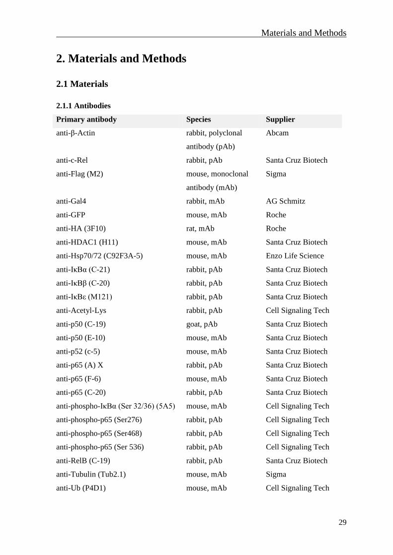
Materials and Methods
29
2. Materials and Methods
2.1 Materials
2.1.1 Antibodies
Primary antibody Species Supplier
anti-β-Actin rabbit, polyclonal
antibody (pAb)
Abcam
anti-c-Rel rabbit, pAb Santa Cruz Biotech
anti-Flag (M2) mouse, monoclonal
antibody (mAb)
Sigma
anti-Gal4 rabbit, mAb AG Schmitz
anti-GFP mouse, mAb Roche
anti-HA (3F10) rat, mAb Roche
anti-HDAC1 (H11) mouse, mAb Santa Cruz Biotech
anti-Hsp70/72 (C92F3A-5) mouse, mAb Enzo Life Science
anti-IκBα (C-21) rabbit, pAb Santa Cruz Biotech
anti-IκBβ (C-20) rabbit, pAb Santa Cruz Biotech
anti-IκBε (M121) rabbit, pAb Santa Cruz Biotech
anti-Acetyl-Lys rabbit, pAb Cell Signaling Tech
anti-p50 (C-19) goat, pAb Santa Cruz Biotech
anti-p50 (E-10) mouse, mAb Santa Cruz Biotech
anti-p52 (c-5) mouse, mAb Santa Cruz Biotech
anti-p65 (A) X rabbit, pAb Santa Cruz Biotech
anti-p65 (F-6) mouse, mAb Santa Cruz Biotech
anti-p65 (C-20) rabbit, pAb Santa Cruz Biotech
anti-phospho-IκBα (Ser 32/36) (5A5) mouse, mAb Cell Signaling Tech
anti-phospho-p65 (Ser276) rabbit, pAb Cell Signaling Tech
anti-phospho-p65 (Ser468) rabbit, pAb Cell Signaling Tech
anti-phospho-p65 (Ser 536) rabbit, pAb Cell Signaling Tech
anti-RelB (C-19) rabbit, pAb Santa Cruz Biotech
anti-Tubulin (Tub2.1) mouse, mAb Sigma
anti-Ub (P4D1) mouse, mAb Cell Signaling Tech

Materials and Methods
30
Primary antibodies used for the
p65 conformational study Species
Supplier
(catalog number)
anti-p65 rabbit, pAb Imgenex (IMG-5915A)
anti-p65 rabbit, pAb Imgenex (IMG-5098)
anti-p65 (6H7) mouse, mAb Abcam (ab91626)
anti-p65 rabbit, pAb Imgenex (IMG-5238)
anti-p65 rabbit, pAb Imgenex (IMG-6066A)
anti-p65 (EP2161Y) rabbit, mAb Abcam (ab76311)
anti-p65 (112A1021) mouse, mAb Imgenex (IMG-150A)
Secondary antibody Conjugated to Supplier
goat-anti-mouse IgG Horseradish peroxidase Dianova
goat-anti-rabbit IgG Horseradish peroxidase Dianova
goat-anti-rabbit IgG CyTM
3 Dianova
goat-anti-rat IgG Horseradish peroxidase Dianova
2.1.2 Bacterial strains
Strain Genotype Supplier
BL21 F- ompT hsdSB(rB-, mB-) gal dcm Novagene
BL21pLysS F-ompT hsdSB(rB-, mB-)galdcm pLysS (CamR) Invitrogen
GM2163 F-ara-14 leuB6 fhuA31 lacY1 tsx78 glnV44 galK2 galT22 mcrA
dcm-6 hisG4 rfbD1 rpsL136 dam-13::Tn9(CamR) xylA5 mtl-1
thi-1 mcrB1 hsdR2
New England
Biolabs
Top10 F-mcrA Δ(mrr-hsdRMS-mcrBC) φ80lacZΔM15 ΔlacX74 recA1
araD139 Δ(araleu) 7697 galU galK rpsL (StrR) endA1 nupG
Invitrogen
2.1.3 Eukaryotic cell lines
Strain Cell type
HEK-293 Human embryonic kidney cells
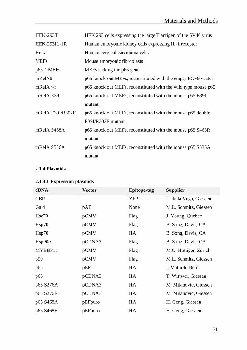
Materials and Methods
31
HEK-293T HEK 293 cells expressing the large T antigen of the SV40 virus
HEK-293IL-1R Human embryonic kidney cells expressing IL-1 receptor
HeLa Human cervical carcinoma cells
MEFs Mouse embryonic fibroblasts
p65 -/-
MEFs MEFs lacking the p65 gene
mRelAζ p65 knock-out MEFs, reconstituted with the empty EGF9 vector
mRelA wt p65 knock out MEFs, reconstituted with the wild type mouse p65
mRelA E39I p65 knock out MEFs, reconstituted with the mouse p65 E39I
mutant
mRelA E39I/R302E p65 knock out MEFs, reconstituted with the mouse p65 double
E39I/R302E mutant
mRelA S468A p65 knock out MEFs, reconstituted with the mouse p65 S468R
mutant
mRelA S536A p65 knock out MEFs, reconstituted with the mouse p65 S536A
mutant
2.1.4 Plasmids
2.1.4.1 Expression plasmids
cDNA Vector Epitope-tag Supplier
CBP YFP L. de la Vega, Giessen
Gal4 pAB None M.L. Schmitz, Giessen
Hsc70 pCMV Flag J. Young, Quebec
Hsp70 pCMV Flag B. Song, Davis, CA
Hsp70 pCMV HA B. Song, Davis, CA
Hsp90α pCDNA3 Flag B. Song, Davis, CA
MYBBP1a pCMV Flag M.O. Hottiger, Zurich
p50 pCMV Flag M.L. Schmitz, Giessen
p65 pEF HA I. Mattioli, Bern
p65 pCDNA3 HA T. Wittwer, Giessen
p65 S276A pCDNA3 HA M. Milanovic, Giessen
p65 S276E pCDNA3 HA M. Milanovic, Giessen
p65 S468A pEFpuro HA H. Geng, Giessen
p65 S468E pEFpuro HA H. Geng, Giessen
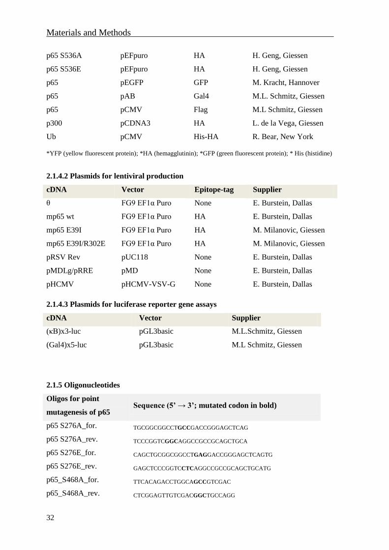
Materials and Methods
32
p65 S536A pEFpuro HA H. Geng, Giessen
p65 S536E pEFpuro HA H. Geng, Giessen
p65 pEGFP GFP M. Kracht, Hannover
p65 pAB Gal4 M.L. Schmitz, Giessen
p65 pCMV Flag M.L Schmitz, Giessen
p300 pCDNA3 HA L. de la Vega, Giessen
Ub pCMV His-HA R. Bear, New York
*YFP (yellow fluorescent protein); *HA (hemagglutinin); *GFP (green fluorescent protein); * His (histidine)
2.1.4.2 Plasmids for lentiviral production
cDNA Vector Epitope-tag Supplier
ζ FG9 EF1α Puro None E. Burstein, Dallas
mp65 wt FG9 EF1α Puro HA E. Burstein, Dallas
mp65 E39I FG9 EF1α Puro HA M. Milanovic, Giessen
mp65 E39I/R302E FG9 EF1α Puro HA M. Milanovic, Giessen
pRSV Rev pUC118 None E. Burstein, Dallas
pMDLg/pRRE pMD None E. Burstein, Dallas
pHCMV pHCMV-VSV-G None E. Burstein, Dallas
2.1.4.3 Plasmids for luciferase reporter gene assays
cDNA Vector Supplier
(κB)x3-luc pGL3basic M.L.Schmitz, Giessen
(Gal4)x5-luc pGL3basic M.L Schmitz, Giessen
2.1.5 Oligonucleotides
Oligos for point
mutagenesis of p65 Sequence (5’ → 3’; mutated codon in bold)
p65 S276A_for. TGCGGCGGCCTGCCGACCGGGAGCTCAG
p65 S276A_rev. TCCCGGTCGGCAGGCCGCCGCAGCTGCA
p65 S276E_for. CAGCTGCGGCGGCCTGAGGACCGGGAGCTCAGTG
p65 S276E_rev. GAGCTCCCGGTCCTCAGGCCGCCGCAGCTGCATG
p65_S468A_for. TTCACAGACCTGGCAGCCGTCGAC
p65_S468A_rev. CTCGGAGTTGTCGACGGCTGCCAGG
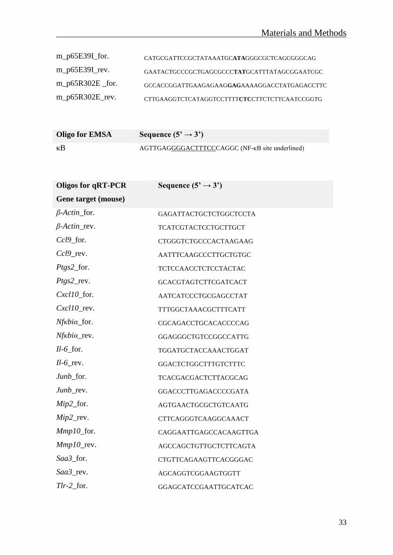
Materials and Methods
33
m_p65E39I_for. CATGCGATTCCGCTATAAATGCATAGGGCGCTCAGCGGGCAG
m_p65E39I_rev. GAATACTGCCCGCTGAGCGCCCTATGCATTTATAGCGGAATCGC
m_p65R302E _for. GCCACCGGATTGAAGAGAAGGAGAAAAGGACCTATGAGACCTTC
m_p65R302E_rev. CTTGAAGGTCTCATAGGTCCTTTTCTCCTTCTCTTCAATCCGGTG
Oligo for EMSA Sequence (5’ → 3’)
κB AGTTGAGGGGACTTTCCCAGGC (NF-κB site underlined)
Oligos for qRT-PCR Sequence (5’ → 3’)
Gene target (mouse)
β-Actin_for. GAGATTACTGCTCTGGCTCCTA
β-Actin_rev. TCATCGTACTCCTGCTTGCT
Ccl9_for. CTGGGTCTGCCCACTAAGAAG
Ccl9_rev. AATTTCAAGCCCTTGCTGTGC
Ptgs2_for. TCTCCAACCTCTCCTACTAC
Ptgs2_rev. GCACGTAGTCTTCGATCACT
Cxcl10_for. AATCATCCCTGCGAGCCTAT
Cxcl10_rev. TTTGGCTAAACGCTTTCATT
Nfκbiα_for. CGCAGACCTGCACACCCCAG
Nfκbiα_rev. GGAGGGCTGTCCGGCCATTG
Il-6_for. TGGATGCTACCAAACTGGAT
Il-6_rev. GGACTCTGGCTTTGTCTTTC
Junb_for. TCACGACGACTCTTACGCAG
Junb_rev. GGACCCTTGAGACCCCGATA
Mip2_for. AGTGAACTGCGCTGTCAATG
Mip2_rev. CTTCAGGGTCAAGGCAAACT
Mmp10_for. CAGGAATTGAGCCACAAGTTGA
Mmp10_rev. AGCCAGCTGTTGCTCTTCAGTA
Saa3_for. CTGTTCAGAAGTTCACGGGAC
Saa3_rev. AGCAGGTCGGAAGTGGTT
Tlr-2_for. GGAGCATCCGAATTGCATCAC
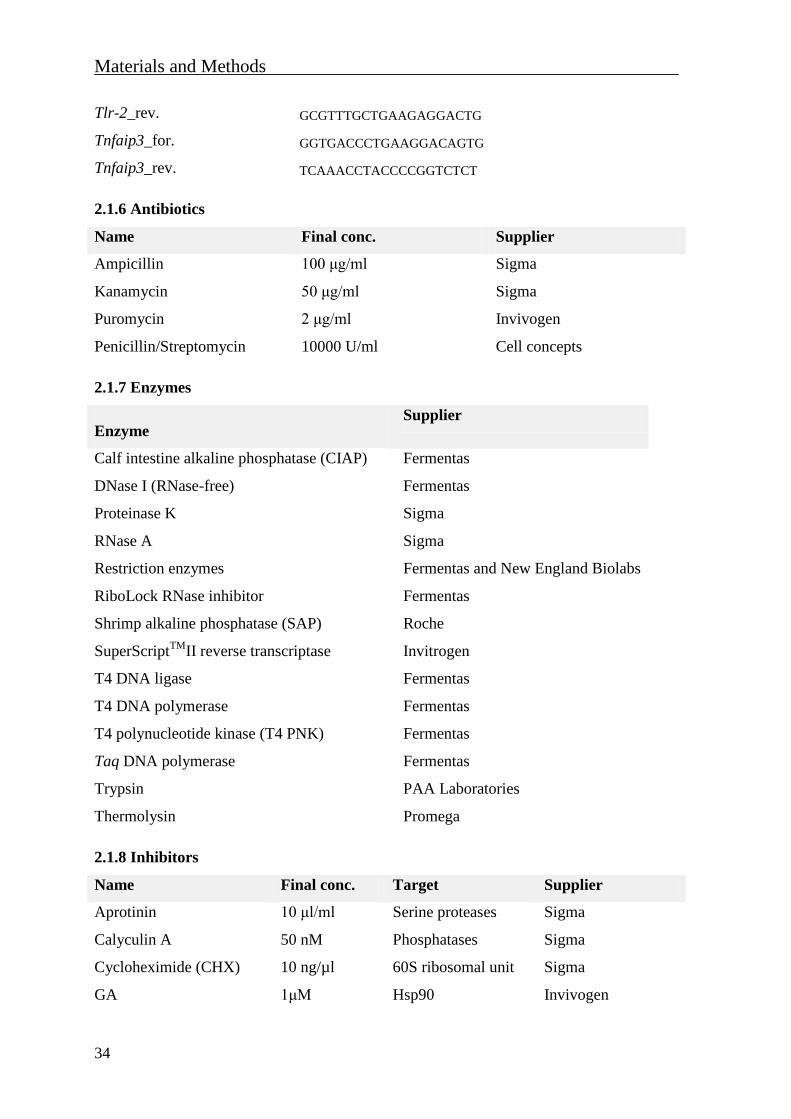
Materials and Methods
34
Tlr-2_rev. GCGTTTGCTGAAGAGGACTG
Tnfaip3_for. GGTGACCCTGAAGGACAGTG
Tnfaip3_rev. TCAAACCTACCCCGGTCTCT
2.1.6 Antibiotics
Name Final conc. Supplier
Ampicillin 100 μg/ml Sigma
Kanamycin 50 μg/ml Sigma
Puromycin 2 μg/ml Invivogen
Penicillin/Streptomycin 10000 U/ml Cell concepts
2.1.7 Enzymes
Enzyme Supplier
Calf intestine alkaline phosphatase (CIAP) Fermentas
DNase I (RNase-free) Fermentas
Proteinase K Sigma
RNase A Sigma
Restriction enzymes Fermentas and New England Biolabs
RiboLock RNase inhibitor Fermentas
Shrimp alkaline phosphatase (SAP) Roche
SuperScriptTM
II reverse transcriptase Invitrogen
T4 DNA ligase Fermentas
T4 DNA polymerase Fermentas
T4 polynucleotide kinase (T4 PNK) Fermentas
Taq DNA polymerase Fermentas
Trypsin PAA Laboratories
Thermolysin Promega
2.1.8 Inhibitors
Name Final conc. Target Supplier
Aprotinin 10 μl/ml Serine proteases Sigma
Calyculin A 50 nM Phosphatases Sigma
Cycloheximide (CHX) 10 ng/µl 60S ribosomal unit Sigma
GA 1μM Hsp90 Invivogen
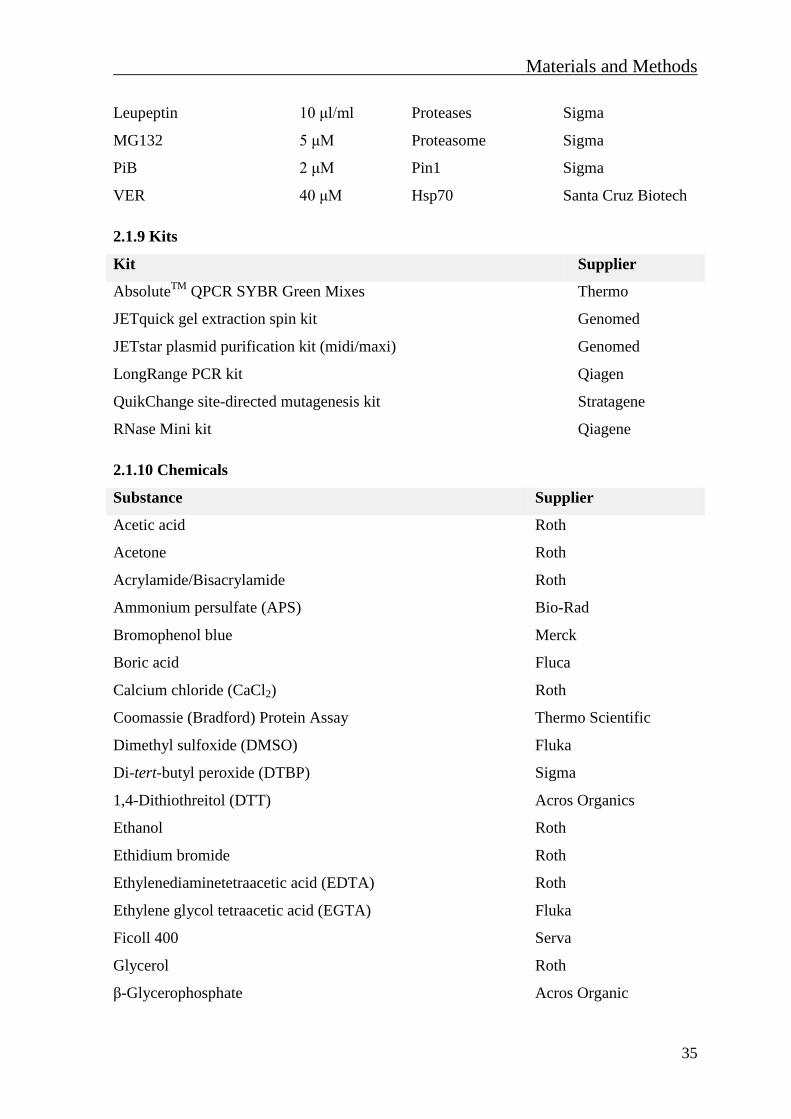
Materials and Methods
35
Leupeptin 10 μl/ml Proteases Sigma
MG132 5 μM Proteasome Sigma
PiB 2 μM Pin1 Sigma
VER 40 μM Hsp70 Santa Cruz Biotech
2.1.9 Kits
Kit Supplier
AbsoluteTM
QPCR SYBR Green Mixes Thermo
JETquick gel extraction spin kit Genomed
JETstar plasmid purification kit (midi/maxi) Genomed
LongRange PCR kit Qiagen
QuikChange site-directed mutagenesis kit Stratagene
RNase Mini kit Qiagene
2.1.10 Chemicals
Substance Supplier
Acetic acid Roth
Acetone Roth
Acrylamide/Bisacrylamide Roth
Ammonium persulfate (APS) Bio-Rad
Bromophenol blue Merck
Boric acid Fluca
Calcium chloride (CaCl2) Roth
Coomassie (Bradford) Protein Assay Thermo Scientific
Dimethyl sulfoxide (DMSO) Fluka
Di-tert-butyl peroxide (DTBP) Sigma
1,4-Dithiothreitol (DTT) Acros Organics
Ethanol Roth
Ethidium bromide Roth
Ethylenediaminetetraacetic acid (EDTA) Roth
Ethylene glycol tetraacetic acid (EGTA) Fluka
Ficoll 400 Serva
Glycerol Roth
β-Glycerophosphate Acros Organic

Materials and Methods
36
Glycine Roth
Guanidine hydrochloride (Gu-HCl) Roth
HEPES Sigma
Hexadimethrine bromide (Polybrene) Sigma
Hoechst 33342 Invitrogen
Hydrochloride acide 37% (HCl) Roth
IPTG Roth
Imidazole Fluka
Isopropanol Merck
Kaiser’s glycerol gelatine Merck
Magnesium carbonate (MgCO3) Sigma
Magnesium chloride (MgCl2) Merck
Magnesium sulfate (MgSO4) Fluka
β-Mercaptoethanol Fluka
Methanol Roth
Nonidet P40 (NP-40) Roche
Paraphormadehyde Roth
Phenylmethanesulfonyl fluoride (PMSF) Fluka
Polyethylenimine (PEI) Sigma
Potassium acetate Roth
Potassium chloride (KCl) Roth
Potassium dihydrogene phosphate (KH2PO4) Fluka
Potassium hydroxide (KOH) Merck
Sodium azide (NaN3) Roth
Sodium chloride (NaCl) Roth
Sodium dihydrogen phosphate (NaH2PO4) Merck
Sodium dodecyl sulfate (SDS) Bio-Rad
Sodium fluoride (NaF) Roth
Sodium hydroxide (NaOH) Merck
Sodium orthovanadate (Na3VO4) Sigma
N,N,N’,N’-Tetramethylethylendiamine (TEMED) Bio-Rad
Tris base Acros Organics
Triton X-100 Sigma
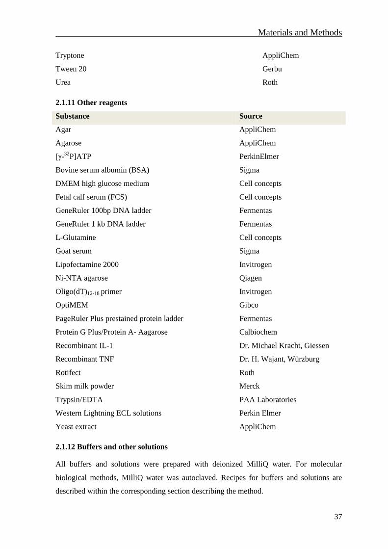
Materials and Methods
37
Tryptone AppliChem
Tween 20 Gerbu
Urea Roth
2.1.11 Other reagents
Substance Source
Agar AppliChem
Agarose AppliChem
[γ-32
P]ATP PerkinElmer
Bovine serum albumin (BSA) Sigma
DMEM high glucose medium Cell concepts
Fetal calf serum (FCS) Cell concepts
GeneRuler 100bp DNA ladder Fermentas
GeneRuler 1 kb DNA ladder Fermentas
L-Glutamine Cell concepts
Goat serum Sigma
Lipofectamine 2000 Invitrogen
Ni-NTA agarose Qiagen
Oligo(dT)12-18 primer Invitrogen
OptiMEM Gibco
PageRuler Plus prestained protein ladder Fermentas
Protein G Plus/Protein A- Aagarose Calbiochem
Recombinant IL-1 Dr. Michael Kracht, Giessen
Recombinant TNF Dr. H. Wajant, Würzburg
Rotifect Roth
Skim milk powder Merck
Trypsin/EDTA PAA Laboratories
Western Lightning ECL solutions Perkin Elmer
Yeast extract AppliChem
2.1.12 Buffers and other solutions
All buffers and solutions were prepared with deionized MilliQ water. For molecular
biological methods, MilliQ water was autoclaved. Recipes for buffers and solutions are
described within the corresponding section describing the method.
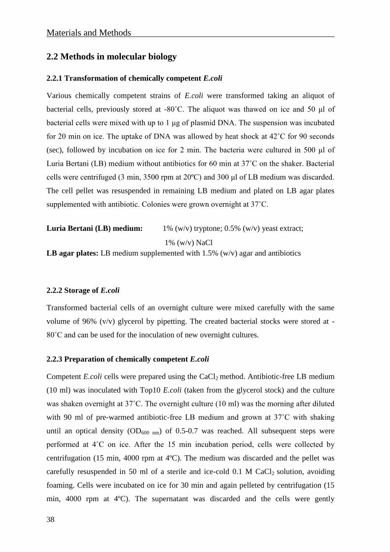
Materials and Methods
38
2.2 Methods in molecular biology
2.2.1 Transformation of chemically competent E.coli
Various chemically competent strains of E.coli were transformed taking an aliquot of
bacterial cells, previously stored at -80˚C. The aliquot was thawed on ice and 50 μl of
bacterial cells were mixed with up to 1 μg of plasmid DNA. The suspension was incubated
for 20 min on ice. The uptake of DNA was allowed by heat shock at 42˚C for 90 seconds
(sec), followed by incubation on ice for 2 min. The bacteria were cultured in 500 μl of
Luria Bertani (LB) medium without antibiotics for 60 min at 37˚C on the shaker. Bacterial
cells were centrifuged (3 min, 3500 rpm at 20ºC) and 300 μl of LB medium was discarded.
The cell pellet was resuspended in remaining LB medium and plated on LB agar plates
supplemented with antibiotic. Colonies were grown overnight at 37˚C.
Luria Bertani (LB) medium: 1% (w/v) tryptone; 0.5% (w/v) yeast extract;
1% (w/v) NaCl
LB agar plates: LB medium supplemented with 1.5% (w/v) agar and antibiotics
2.2.2 Storage of E.coli
Transformed bacterial cells of an overnight culture were mixed carefully with the same
volume of 96% (v/v) glycerol by pipetting. The created bacterial stocks were stored at -
80˚C and can be used for the inoculation of new overnight cultures.
2.2.3 Preparation of chemically competent E.coli
Competent E.coli cells were prepared using the CaCl2 method. Antibiotic-free LB medium
(10 ml) was inoculated with Top10 E.coli (taken from the glycerol stock) and the culture
was shaken overnight at 37˚C. The overnight culture (10 ml) was the morning after diluted
with 90 ml of pre-warmed antibiotic-free LB medium and grown at 37˚C with shaking
until an optical density (OD600 nm) of 0.5-0.7 was reached. All subsequent steps were
performed at 4˚C on ice. After the 15 min incubation period, cells were collected by
centrifugation (15 min, 4000 rpm at 4ºC). The medium was discarded and the pellet was
carefully resuspended in 50 ml of a sterile and ice-cold 0.1 M CaCl2 solution, avoiding
foaming. Cells were incubated on ice for 30 min and again pelleted by centrifugation (15
min, 4000 rpm at 4ºC). The supernatant was discarded and the cells were gently

Materials and Methods
39
resuspended in 3 ml of sterile 0.1 M CaCl2 containing 10% glycerol (v/v). Competent
E.coli cells were aliquoted (100μl) into the sterile 1.5 ml tubes, shock frozen in liquid
nitrogen and stored at -80˚C until use.
2.2.4 Isolation of plasmid DNA from transformed E.coli
For the isolation of plasmid DNA a method based on alkaline lysis was used. Bacterial
E.coli cells containing the plasmid of interest were first cultured overnight in LB medium
containing the appropriate antibiotic. For Mini-preps, 2 ml of overnight culture was
centrifuged, the supernatant was discarded and the pellet was suspended in 200 μl of
EDTA-containing physiological buffer P1, supplemented with RNase A. The following
step included the lysis of bacterial cells by adding the 200 μl of lysis buffer P2. During the
5 min incubation at room temperature (20ºC), SDS contained in the P2 buffer dissolves the
lipid components from the cell membrane, as well as cellular proteins. The high alkaline
conditions denature chromosomal and plasmid DNA. To neutralize the solution, 200 μl of
pre-chilled buffer P3 was added and the tubes were mixed by inversion. Potassium acetate
returns the pH to neutral allowing the renaturation of DNA strands. Large chromosomal
strands partially renature, while the single-stranded plasmid DNA completely renaturates
into dsDNA molecules and cellular proteins precipitate. Lysates were incubated for 20 min
on ice and cleared from debris by centrifugation (10 min, 13000 rpm at 4ºC). The
supernatant was transferred into a new tube and iso-propanol was added (final
concentration 70%). The DNA was pelleted by centrifugation (10 min, 13000 rpm at 4ºC)
and washed with 300 μl 70% (v/v) ethanol which removed some remaining salts and SDS
from the preparation. After the centrifugation step (10 min, 13000 rpm at 4ºC), pelleted
DNA was air-dried at 20ºC for 10 min. Plasmid DNA was disolved in 30 μl sterile water
and stored at -20˚C.
Medium and large scale plasmid DNA purifications were performed using the JetStar 2.0
Plasmid Purification Kit (Genomed) according to manufactures instructions. The
purification is also based on alkaline lysis. Lysates were centrifuged and supernatants were
passed through JetStar 2.0 anion-exchange columns. The negatively charged phosphates on
the DNA-backbone interact with the positive charges on the resin surface. Once the lysate
ran through the column by gravity flow, the column was washed with washing buffer
allowing the removal of RNA, proteins, carbohydrates and other impurities, while the
Materials and Methods
40
plasmid DNA remains bound to the resin. The plasmid DNA was eluted under high salt
conditions and precipitated from the eluate with isopropanol and 70% ethanol.
Buffer P1: 50 mM Tris/HCl (pH 8.0); 10 mM EDTA; 100 μg/ml RNase A
Buffer P2: 200 mM NaOH; 1% (w/v) SDS
Buffer P3: 3 M potassium acetate (pH 5.5)
The concentration of a DNA purified by columns was quantified in a spectrophotometer by
measuring the absorbance of the sample at a wavelength of 260 nm. The ratio of the
absorbance at 260 and 280 nm (OD260/280) was used to assess DNA purity. An OD260/280
ratio between 1.7 and 2.0 represented a high-quality sample.
2.2.5 Polymerase Chain Reaction (PCR)
2.2.5.1 Amplification of DNA fragments for cloning
PCR was used for amplification of desired DNA fragments. The method is based on the
usage of two oligonucleotide primers that hybridize to complementary target regions on
opposite DNA strands and flank the target sequence. Oligonucleotide primers were
designed with additional non-annealing DNA sequence containing specific endonuclease
restriction sites at the 5’ ends of each primer, allowing the direct cloning of DNA
fragment. For efficient amplification of DNA fragments, the QIAGEN LongRange PCR
Kit was used according to manufacturers suggestions. PCR reaction mix consistent of:
Reagent
Template DNA 20 – 100 ng
LongRange PCR buffer (10x) 5 μl
dNTP mix 500 μM
forward primer 0.4 μM
reverse primer 0.4 μM
LongRange PCR enzyme mix 2 units
MilliQ water up to 50 μl
PCR reaction was performed in thermal cycler using the following program:
Materials and Methods
41
Initial denaturation 93˚C, 3 min
Denaturation 93˚C, 20 sec
Primer annealing 55 - 60˚C; ~ 5˚C below Tm of primers, 30 sec 35 cycles
Elongation 68˚C, 1 min/kb
Final elongation 68˚C, 5 min
Storage 4˚C until use or -20˚C for long term storage
After amplification, the PCR product was analysed by agarose gel electrophoresis, purified
from the agarose gel (2.2.1.7) and digested with restriction enzymes (2.2.1.8).
2.2.5.2 Site-directed point mutagenesis
In order to examine the importance of single amino acids and their PTMs for protein
functions, site-directed point mutants were produced. Single amino acids were changed
using the QuikChange site-directed mutagenesis kit. Specific oligonucleotides, carrying the
changed sequence, were previously designed. PCR reaction mix for point-mutagenesis
contained:
Reagent
Template DNA 20-100 ng
10x reaction buffer 5 μl
Quik solution 3 μl
10 mM dNTP mix 1 μl
forward primer 125 ng
reverse primer 125 ng
Pfu ultra DNA polymerase 2.5 units
MilliQ water up to 50 μl
Mutagenesis PCR was performed using the following program:
Initial denaturation 95˚C, 2 min
Denaturation 95˚C, 30 sec
Primer annealing 55˚C, 1 min 18 cycles
Elongation 68°C, 1 min/kb
Final elongation step 68ºC, 7 min
Materials and Methods
42
Non-mutated template DNA from the PCR reaction was removed by adding 1 μl of DpnI
(10 U/μl) to the reaction mix, followed by incubation for at least 2 h at 37˚C. To increase
the purity of mutated plasmid DNA, the DNA was separated on an agarose gel and purified
(see 2.2.1.7). The amplified plasmid DNA was transformed into Top10 E.coli cells (see
2.2.1.1) and cells were plated on LB agar containing the appropriate antibiotic. Plasmid
DNA was purified (see 2.2.4.1) and the presence of desired point mutation(s) was
confirmed by DNA sequencing or by restriction digestion, in case that diagnostic
restriction sites were included in primers sequences.
2.2.6 Agarose gel electrophoresis
Agarose gel electrophoresis was performed for the analysis of digested vectors and PCR
products. Agarose gels (0.8 - 2.0% (w/v)) were prepared dissolving the powdered agarose
in a TRIS-acetate-EDTA (TAE) buffer. The agarose solution was heated in a microwave
until it was completely melted, left for about 5 min to cool down and ethidium bromide
was added (final concentration 0.2 μg/ml) to the agarose solution. This fluorescent dye
intercalates between bases of nucleic acids and allows the detection of a DNA under the
UV light. DNA samples were mixed with 6x DNA sample buffer before gel loading. Gels
were run in TAE buffer at a constant voltage of 80 V or maximally 70 V for preparative
agarose gels. Separation of DNA fragments was analysed on an UV-transilluminator.
50x TAE buffer: 2M Tris (pH 8.3); 1 M acetic acid; 50 mM EDTA
6x DNA sample buffer: 60 mM EDTA (pH 8.0); 60% (w/v) glycerol;
0.09% (w/v) bromphenol blue
2.2.6.1 DNA extraction from agarose gel
The DNA fragment of interest was located at low UV radiation (reduced intensity), excised
from the agarose gel with a razor blade and the DNA was extracted using the JETquick gel
extraction kit (Genomed) according to manufacturers instructions. Briefly, the gel slice
was solubilised by adding solution L1 and heated to 50°C for 15 min. The mixture was
loaded into a JETquick spin column where the DNA bound to the resin. After several
washing steps with the solution L2, DNA fragments were eluted from the resin with 30 μl
of sterile water. The yield and purity of the extracted DNA was tested by agarose gel
electrophoresis.
Materials and Methods
43
2.2.7 Digestion of DNA with restriction enzymes
For the insertion of amplified DNA fragment into the vector, both DNA plasmid and PCR
products were digested with restriction enzymes to generate complementary end with
single-stranded overhangs. In most cases the digestion was performed with two restriction
enzymes in the recommended restriction buffer. A reaction mix for the analytical DNA
digestion included:
Reagent
DNA (1 μg/μl) 2 μl
10x restriction buffer 1 μl
Restriction enzyme 1 (10 U/μl) 5 units
Restriction enzyme 2 (10 U/μl) 5 units
MilliQ water up to 10 μl
After the incubation for at least 2 h at the optimal temperature for restriction enzyme(s),
products were analysed by agarose gel electrophoresis (see 2.2.1.7) and purified for
cloning when necessary (see 2.2.1.8). If a single restriction enzyme was used, 5’-
phosphate ends of linearised DNA plasmid were dephosphorylated using CIAP in order to
avoid re-ligation of the plasmid DNA. A reaction mix for the DNA dephosphorylation
included:
Reagent
Linearized plasmid DNA (1 μg/μl) 2 μl
10x CIAP reaction buffer 1 μl
CIAP (1 U/μl) 1 unit
MilliQ water up to 10 μl
After the incubation on 37˚C for 30 min, reaction was stopped by heating for 10 min at
85°C.
2.2.8 Ligation of DNA fragments
To assure the best outcome, ligation mixtures were prepared with a molar ratio of vector to
insert of 1:3 and 1:5. The amount of DNA insert was calculated by converting the molar
ratios to mass ratios using the formula:
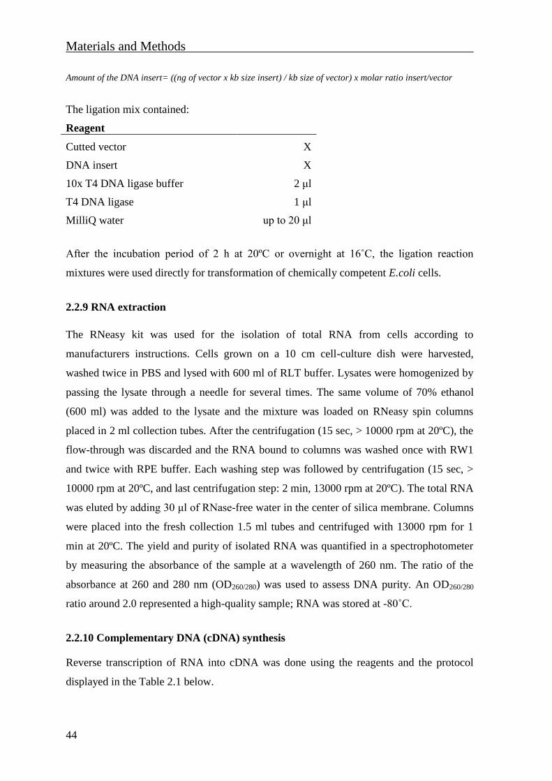
Materials and Methods
44
Amount of the DNA insert= ((ng of vector x kb size insert) / kb size of vector) x molar ratio insert/vector
The ligation mix contained:
Reagent
Cutted vector X
DNA insert X
10x T4 DNA ligase buffer 2 μl
T4 DNA ligase 1 μl
MilliQ water up to 20 μl
After the incubation period of 2 h at 20ºC or overnight at 16˚C, the ligation reaction
mixtures were used directly for transformation of chemically competent E.coli cells.
2.2.9 RNA extraction
The RNeasy kit was used for the isolation of total RNA from cells according to
manufacturers instructions. Cells grown on a 10 cm cell-culture dish were harvested,
washed twice in PBS and lysed with 600 ml of RLT buffer. Lysates were homogenized by
passing the lysate through a needle for several times. The same volume of 70% ethanol
(600 ml) was added to the lysate and the mixture was loaded on RNeasy spin columns
placed in 2 ml collection tubes. After the centrifugation (15 sec, > 10000 rpm at 20ºC), the
flow-through was discarded and the RNA bound to columns was washed once with RW1
and twice with RPE buffer. Each washing step was followed by centrifugation (15 sec, >
10000 rpm at 20ºC, and last centrifugation step: 2 min, 13000 rpm at 20ºC). The total RNA
was eluted by adding 30 μl of RNase-free water in the center of silica membrane. Columns
were placed into the fresh collection 1.5 ml tubes and centrifuged with 13000 rpm for 1
min at 20ºC. The yield and purity of isolated RNA was quantified in a spectrophotometer
by measuring the absorbance of the sample at a wavelength of 260 nm. The ratio of the
absorbance at 260 and 280 nm (OD260/280) was used to assess DNA purity. An OD260/280
ratio around 2.0 represented a high-quality sample; RNA was stored at -80˚C.
2.2.10 Complementary DNA (cDNA) synthesis
Reverse transcription of RNA into cDNA was done using the reagents and the protocol
displayed in the Table 2.1 below.
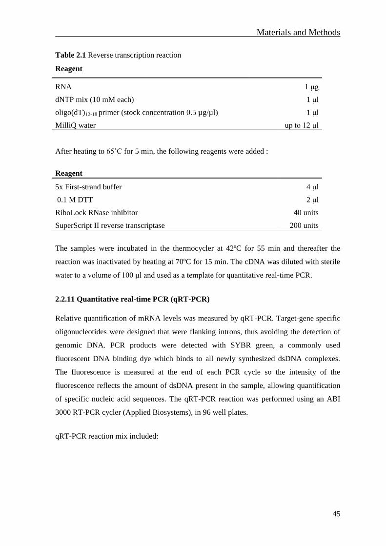
Materials and Methods
45
Table 2.1 Reverse transcription reaction
Reagent
RNA 1 μg
dNTP mix (10 mM each) 1 μl
oligo(dT)12-18 primer (stock concentration 0.5 µg/µl) 1 μl
MilliQ water up to 12 μl
After heating to 65˚C for 5 min, the following reagents were added :
Reagent
5x First-strand buffer 4 μl
0.1 M DTT 2 μl
RiboLock RNase inhibitor 40 units
SuperScript II reverse transcriptase 200 units
The samples were incubated in the thermocycler at 42ºC for 55 min and thereafter the
reaction was inactivated by heating at 70ºC for 15 min. The cDNA was diluted with sterile
water to a volume of 100 μl and used as a template for quantitative real-time PCR.
2.2.11 Quantitative real-time PCR (qRT-PCR)
Relative quantification of mRNA levels was measured by qRT-PCR. Target-gene specific
oligonucleotides were designed that were flanking introns, thus avoiding the detection of
genomic DNA. PCR products were detected with SYBR green, a commonly used
fluorescent DNA binding dye which binds to all newly synthesized dsDNA complexes.
The fluorescence is measured at the end of each PCR cycle so the intensity of the
fluorescence reflects the amount of dsDNA present in the sample, allowing quantification
of specific nucleic acid sequences. The qRT-PCR reaction was performed using an ABI
3000 RT-PCR cycler (Applied Biosystems), in 96 well plates.
qRT-PCR reaction mix included:
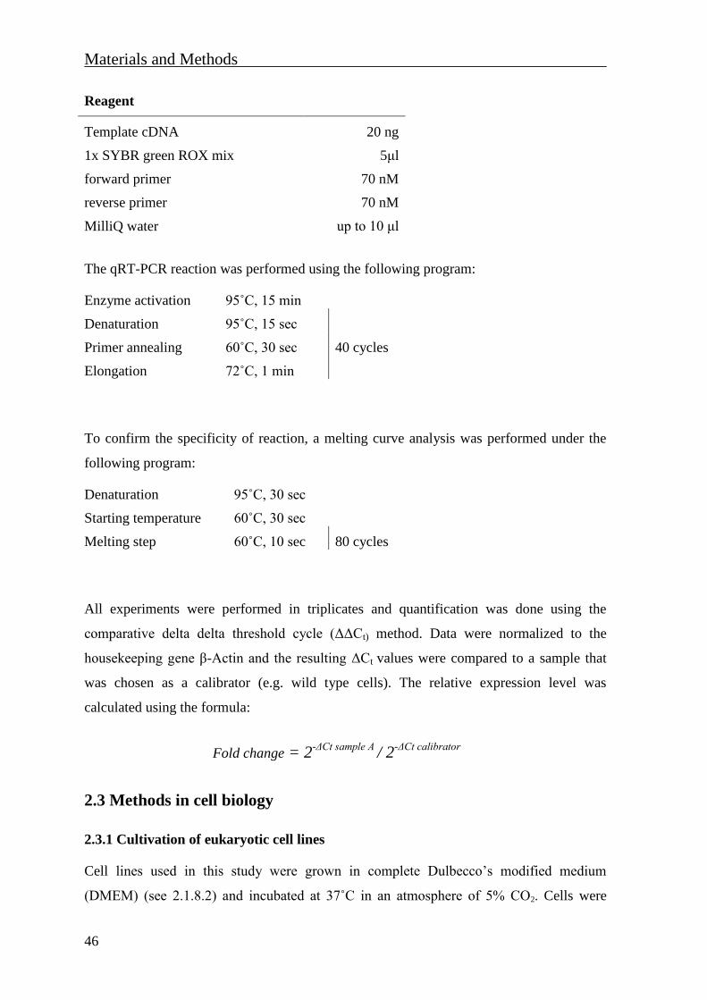
Materials and Methods
46
Reagent
Template cDNA 20 ng
1x SYBR green ROX mix 5μl
forward primer 70 nM
reverse primer 70 nM
MilliQ water up to 10 μl
The qRT-PCR reaction was performed using the following program:
Enzyme activation 95˚C, 15 min
Denaturation 95˚C, 15 sec
Primer annealing 60˚C, 30 sec 40 cycles
Elongation 72˚C, 1 min
To confirm the specificity of reaction, a melting curve analysis was performed under the
following program:
Denaturation 95˚C, 30 sec
Starting temperature 60˚C, 30 sec
Melting step 60˚C, 10 sec 80 cycles
All experiments were performed in triplicates and quantification was done using the
comparative delta delta threshold cycle (ΔΔCt) method. Data were normalized to the
housekeeping gene β-Actin and the resulting ΔCt values were compared to a sample that
was chosen as a calibrator (e.g. wild type cells). The relative expression level was
calculated using the formula:
Fold change = 2-ΔCt sample A
/ 2-ΔCt calibrator
2.3 Methods in cell biology
2.3.1 Cultivation of eukaryotic cell lines
Cell lines used in this study were grown in complete Dulbecco’s modified medium
(DMEM) (see 2.1.8.2) and incubated at 37˚C in an atmosphere of 5% CO2. Cells were
Materials and Methods
47
cultured in 75 or 175 cm2γ-irrediated cell-culture flasks for 2-3 days to reach a confluency
of 70-80%. The adherent cells that stick to the surface were washed once with pre-warmed
PBS and detached from the flask surface by adding Trypsin/EDTA, followed by incubation
for 2-3 min at 37˚C. Trypsin activity was stopped by adding complete DMEM. Cells were
seeded into a new flask in an appropriate dilution. Easily detached adherent cell lines such
as HEK-293 and HEK-293T cells were detached by intensive pipetting and seeded into
new flask. If necessary, the cells were pelleted by centrifugation (1300 rpm, 3min at 20˚C),
resuspended in the desired volume of complete DMEM and seeded.
PBS: 137mM NaCl; 1.5 mM KH2PO4; 2.7mM KCl; 8.1 mM Na2HPO4 (pH 7.4)
DMEM complete: DMEM high glucose (4.5 g/L); 2 mM L-glutamine;
1% (v/v) penicillin/streptomycin;
10% (v/v) heat inactivated (56˚C, 30 min) FCS
2.3.2 Freezing and thawing of cells
For long-term storage, exponentially grown cells were collected by centrifugation (1300
rpm, 3 min at 20˚C), washed once in PBS, collected again by centrifugation and
resuspended in 1ml of freezing medium. Cells were aliquoted in labeled cryo tubes,
transferred to a cooling device ensuring slow cooling and transferred to -80˚C for one day.
The next day, cells were transferred for long-term storage at -150˚C.
Frozen cryo tubes were thawed at 37˚C in the water bath. Cells were washed in 10 ml of
pre-warmed complete DMEM, followed by centrifugation (1300rpm, 3 min at 20˚C). After
removal of the supernatant the pellet containing the collected cells were resuspended in 10
ml of complete DMEM and seeded into a 75 cm2
cell-culture flask.
Freezing medium: 90% (v/v) FCS; 10% (v/v) DMSO
2.3.3 Transfection of mammalian cells
Introduction of plasmid DNA into eukaryotic cells was performed using polyethylenimine
(PEI). As a polycationic transfection reagent, it binds to DNA, neutralizes its negative
charge, condenses its structure and forms a PEI-DNA complex that is easily introduced
into the cell by endocytosis.

Materials and Methods
48
One day prior to transfection, cells were seeded in 10 cm dishes in order to reach 50-60%
confluency at the day of transfection. For the transfection, the ratio of DNA and
transfection reagent was always 2 μl of PEI per 1 μg of DNA (PEI stock concentration1
µg/µl). Total DNA amounts were kept equal in all transfections by adding empty vector.
All the steps were performed under the hood.DNA was mixed with serum- and antibiotic
free DMEM (50 μl per 1 μg of DNA) and incubated for 5 min at 20ºC. In the other 1.5 ml
tube, PEI was diluted with the serum- and antibiotic free DMEM and incubated for 5 min
at 20ºC. After 5 min, diluted PEI was added to DNA mixture and incubated for 20 min at
20ºC allowing formation of PEI-DNA complex. In the meantime, the complete medium
from the cultivated 10 cm dishes was aspirated off, cells were washed twice with pre-
warmed PBS and 5 ml of the antibiotic free DMEM (transfection medium) was put over
cultured cells. The PEI-DNA mixture was added in a drop-wise manner to cells. After 3-6
h, the transfection medium was aspirated off from plates and replaced by 6 ml of complete
DMEM. After 36-48 h of incubation at 37˚C, cells were ready for the lysis.
DMEM antibiotic-free: DMEM high glucose (4.5 g/L); 2 mM L-glutamine;
10% (v/v) heat inactivated (56˚C, 30 min) FCS
Transfection mix for protein overexpression contained:
Reagent
plasmid DNA 1-3 µg
PEI (stock concentration 1 µg/µl) 2-9 µl
DMEM serum reduced 50 µl per 1 µg of plasmid DNA
DMEM antibiotic-free 2 or 5 ml, for 6 or 10 cm cell culture dish
2.3.4 Production of lentiviruses and infection of cells
Production of lentiviral particles of the 3rd
generations was done in HEK-293T packaging
cells. The FG9EF-1a-Puro expression vector encoding p65 or its mutants were transfected
together with packaging vectors pMDLg/pRRE, pRSV-Rev and pHCMVG, in a ratio of
1:1:1:1 (5µg of the each construct). Cells were seeded in 10 cm dishes to reach a
confluence of 80% on the day of transfection. As a transfection reagent was used
Lipofectamine2000according to the procedure suggested by the manufacturer.

Materials and Methods
49
Lipofectamine is a cationic lipid and facilitate transfection during the early steps of the
process by mediating DNA condensation and DNA/cellular interactions. In one sterile tube
five micrograms of each of four vectors (in total 20 μg of DNA) were mixed with 1.5 ml
OptiMEM. In the other sterile tube 40 μl of Lipofectamine2000 (DNA –
Lipofectamine2000 ratio of 1:2) was mixed with 1.5 ml OptiMEM. After the 5 min
incubation at 20ºC, the diluted DNA and Lipofectamine2000 were combined and incubated
for 20 min to allow formation of Lipofectamine-DNA complexes. Complete medium was
removed from each 10 cm dish and replaced with antibiotic-free DMEM containing 2.5%
(v/v) FCS and 3 ml of transfection mixture were added to the cells. After 4-6 h, the
transfection medium was totally replaced by DMEM complete medium. Viruses were
collected 48 h post-transfection, filtered through a 0.45 μm filter and mixed with polybrene
(final concentration 5µg/ml) to increase the efficiency of infection. Viruses were added
over cells. One day after infection, medium with viruses was removed and replaced with
the complete DMEM medium containing 2 μg/ml of puromycin (final concentration) for
cell selection. Cells were kept under the puromycin selection for at least one week.
DMEM serum reduced
for the lentivirus production: DMEM high glucose (4.5 g/L); 2 mM L-glutamine;
1.5% (v/v) heat inactivated (56˚C, 30 min) FCS
Transfection mixture for lentivirus production included (for 10 cm cell culture dish):
Reagent
lentiviral vector 5 µg
packaging vectors: pMDLg/pRRE 5 µg
pRSV-Rev 5 µg
pHCMVG 5 µg
Lipofectamine2000 (stock concentration 1 µg/µl) 40 µl
OPTI-MEM serum reduced 3 ml
DMEM serum reduced
7 ml

Materials and Methods
50
2.3.5 Lysate preparation
2.3.5.1 Lysis under denaturing conditions
Cells were scraped off, washed twice in ice-cold PBS and collected by centrifugation for 2
min, 2000 rpm at 4ºC). Cells were lysed in 100-200 μl of 1x SDS sample buffer and the
lysate was boiled for 2 min at 95˚C. The DNA was fragmented by shearing (2 x 20 sec)
using a Branson 250 sonifier. Lysates were boiled again at 95˚C for 5 min and used for
Western blotting or frozen at -80˚C.
5x SDS sample buffer: 312.5 mM Tris/HCl (pH 6.8); 50% (v/v) glycerol;
25% (v/v) β-mercaptoethanol; 10% (w/v) SDS;
0.01% (w/v) bromphenol blue
2.3.5.2 Lysis under native conditions - whole cell lysate
Whole cell lysates were prepared by lysing cells in the lysis buffer containing the non-ionic
detergent NP-40. Cells were harvested and washed twice in ice-cold PBS. Cell pellets were
resuspended in an appropriate volume of the NP-40 lysis buffer (approximately 4 volumes
of NP-40 lysis buffer for 1 volume of cell pellet) and incubated 20 min on ice. Lysates
were cleared by centrifugation (15 min, 13200 rpm, 4˚C) and supernatants were transferred
into fresh tubes. Lysates were used for limited proteolysis assay, immunoprecipitation (IP),
GST pull-down, luciferase reporter assay or were directly mixed with 5x SDS sample
buffer at a ratio of 1:4 (5x SDS sample buffer : lysate), boiled for 5 min at 95˚C and
analysed by Western blotting.
NP-40 lysis buffer: 20 mM Tris/HCl (pH 7.5); 150 mM NaCl; 1% (v/v) NP-40;
freshly added: 0.5 mM Na3VO4; 10 mM NaF; 1 mM PMSF;
10 μg/ml aprotinin; 10 μg/ml leupeptin
2.3.5.3 Lysis under native conditions - subcellular fractionation
For preparation of cytosolic and nuclear protein extracts, cells were harvested after
washing in cold PBS and collected by centrifugation. The pellet was suspended in cold
extraction buffer A. After incubation on ice for 10 min, NP-40 was added in a final
concentration of 0.25% (v/v) and cells were vortexed gently for 5 sec. Samples were
centrifuged (10 min, 4200 rpm at 4˚C) and the supernatants (corresponding to cytosolic
extracts) were collected and transfered to a fresh tubes. Cell pellets were washed once in

Materials and Methods
51
buffer A and cleared by centrifugation (10 min, 4200 rpm at 4˚C). For the preparation of
nuclear extracts, pellets were resuspended very gently in extraction buffer C. After
incubation on ice for 20 min and centrifugation (10 min, 13200 rpm at 4˚C), supernatants
corresponding to nuclear extracts were transferred to fresh tubes. Cytosolic and nuclear
extracts were further used for limited proteolysis assay, IP, electrophoretic mobility shift
assay (EMSA) or they were mixed with 5x SDS sample buffer, boiled at 95˚C for 5 min
and analysed by Western blotting.
Buffer A: 10 mM HEPES/KOH (pH 7.9); 10 mM KCl; 0.1 mM EDTA;
0.1 mM EGTA; 1 mM β-mercaptoethanol
Buffer C: 20 mM HEPES/KOH (pH 7.9), 400 mM NaCl; 1 mM EDTA;
1 mM EGTA; 1 mM β-mecaptoethanol
10% NP-40 solution: 10% (v/v) NP-40
2.3.6 Immunofluorescence (IF) staining
Intracellular localization of the proteins was analysed by IF. Adherent MEFs were grown
on cover slips in 12-well plates to reach the confluence of 70% on the day of experiment.
All washing and incubation steps were done under gentle shaking on 20ºC. The medium
was aspirated off and the cells were washed twice with PBS for 5 min. Cells were fixed by
adding 500 μl of freshly made 4% (w/v) paraformaldehyde in PBS and incubated for 15
min at 20ºC. After removal of the paraformaldehyde solution, cells were washed twice for
5 min with PBS. For cell permeability, cells were incubated with PBS containing 0.25%
(v/v) Triton X-100 for 10 min. After the 3 washing steps with PBS, cells were blocked for
60 min by shaking in PBS containing 10% (v/v) goat serum. The blocking solution was
removed and replaced with the primary antibody diluted in PBS containing 1% (v/v) goat
serum. Cells were incubated overnight at 4˚C. Cells were washed four times with PBS after
which were incubated for 60 min with fluorescence dye-coupled secondary antibodies
diluted in PBS containing 1% (v/v) goat serum. Of note, cells were protected from light.
Cells were again washed four times with PBS, still keeping them protected from light and a
nuclear DNA was stained by incubation with Hoechst (1:1000 dilution in PBS) for 10 min.
Cells were washed two times with PBS. Cover slips were mounted with one drop of pre-
warmed Kaiser’s glycerol gelatin on a surface of a microscope slide and gently pressed.
Materials and Methods
52
Slides were left at 4˚C overnight. The next day, cover slips and slides were cleaned and
sealed with the nail polish. Microscope analysis was done using an inverted Nikon Eclipse
2000E microscope.
2.3.7 Luciferase reporter gene assays
To study transcriptional activity in cells, luciferase reporter assays were performed. Cells
were transiently transfected with constructs in which a specific promoter sequence is fused
to a luciferase gene. The luciferase gene encodes the enzyme luciferase, which catalyzes
the oxidation of its substrate luciferin in a light producing reaction. Since the transcription
of the luciferase gene is controlled by DNA elements allowing binding of a TF of interest,
the amount of synthesized luciferase and hence the intensity of bioluminiscence represents
the TF activity.
Cells were grown on 6 well plates and transfected with plasmids encoding the TF-reporter
construct and if necessary, the corresponding TF. After 24-48 h post-transfection cells
were stimulated with cytokines for 6 h or left untreated. Cells were washed with PBS and
lysed in 100μl cold NP-40 buffer. Lysates were cleared by centrifugation (10 min, 13200
rpm at 4ºC), supernatants (10 μl) were mixed with luciferase buffer (10 μl) and
bioluminiscence was immediately measured for 10 sec in a luminometer (Berthold Lumat
LB 9507).
Firefly luciferase
assay buffer: 0.47 mM Photinius-Luciferin; 530 μM ATP; 2.67 mM MgSO4;
20 mM Tricine; 1.07mM (MgCO3)4Mg(OH)2 x 5H2O;
0.1 mM EDTA; 270 μM Coenzyme A; 33.3 mM DTT
2.4 Biochemical methods
2.4.1 SDS polyacrylamide gel electrophoresis (SDS-PAGE)
SDS-PAGE allows the separation of proteins in a gel matrix according to their molecular
weight. Samples were mixed with 5x SDS sample buffer containing the anionic detergent
SDS and β-mercaptoethanol in order to disrupt intra- and intermolecular protein
interactions without breaking peptide bonds. The discontinuous SDS-PAGE employs two
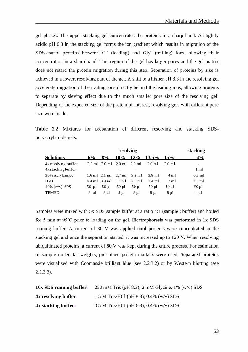
Materials and Methods
53
gel phases. The upper stacking gel concentrates the proteins in a sharp band. A slightly
acidic pH 6.8 in the stacking gel forms the ion gradient which results in migration of the
SDS-coated proteins between Cl- (leading) and Gly
- (trailing) ions, allowing their
concentration in a sharp band. This region of the gel has larger pores and the gel matrix
does not retard the protein migration during this step. Separation of proteins by size is
achieved in a lower, resolving part of the gel. A shift to a higher pH 8.8 in the resolving gel
accelerate migration of the trailing ions directly behind the leading ions, allowing proteins
to separate by sieving effect due to the much smaller pore size of the resolving gel.
Depending of the expected size of the protein of interest, resolving gels with different pore
size were made.
Table 2.2 Mixtures for preparation of different resolving and stacking SDS-
polyacrylamide gels.
Samples were mixed with 5x SDS sample buffer at a ratio 4:1 (sample : buffer) and boiled
for 5 min at 95˚C prior to loading on the gel. Electrophoresis was performed in 1x SDS
running buffer. A current of 80 V was applied until proteins were concentrated in the
stacking gel and once the separation started, it was increased up to 120 V. When resolving
ubiquitinated proteins, a current of 80 V was kept during the entire process. For estimation
of sample molecular weights, prestained protein markers were used. Separated proteins
were visualized with Coomassie brilliant blue (see 2.2.3.2) or by Western blotting (see
2.2.3.3).
10x SDS running buffer: 250 mM Tris (pH 8.3); 2 mM Glycine, 1% (w/v) SDS
4x resolving buffer: 1.5 M Tris/HCl (pH 8.8); 0.4% (w/v) SDS
4x stacking buffer: 0.5 M Tris/HCl (pH 6.8); 0.4% (w/v) SDS
resolving stacking
Solutions 6% 8% 10% 12% 13.5% 15% 4%
4x resolving buffer 2.0 ml 2.0 ml 2.0 ml 2.0 ml 2.0 ml 2.0 ml -
4x stacking buffer - - - - - - 1 ml
30% Acrylamide 1.6 ml 2.1 ml 2.7 ml 3.2 ml 3.8 ml 4 ml 0.5 ml
H2O 4.4 ml 3.9 ml 3.3 ml 2.8 ml 2.4 ml 2 ml 2.5 ml
10% (w/v) APS 50 μl 50 μl 50 μl 50 μl 50 μl 50 μl 50 μl
TEMED 8 μl 8 μl 8 μl 8 μl 8 μl 8 μl 4 μl
Materials and Methods
54
2.4.2 Coomassie brilliant blue staining of polyacrylamide gels
To visualize proteins on polyacrylamide gels, protein samples were electrophoretically
separated, washed in 100 ml deionized water and stained by incubating the gel with
Coomassie staining solution for 20 min under gentle agitation. To visualize proteins, the
gel was rinsed in destaining solution several times.
Coomassie staining solution: 0.1% (w/v) Coomassie brilliant blue G-250;
40% (v/v) Methanol, 10% (v/v) Acetic acid
Coomassie destaining solution: 30% (v/v) Methanol; 20% (v/v) Acetic acid
2.4.3 Western blot and immune detection
Proteins separated by SDS-PAGE (see 2.2.3.1) were immobilized on polyvinylidene
fluoride (PVDF) membranes by the Western blot method, which allows their detection by
specific antibodies. For this aim, PVDF membrane was immersed in 100% methanol for a
few sec until the entire membrane was translucent. The wet membrane was transferred to a
vessel containing the transfer buffer and incubated for at least 2-3 min to allow membrane
equilibration. Once equilibrated, the membrane was ready to bind proteins. In the
meantime, Whatman papers were soaked in the transfer buffer. A blot “sandwich”
consisting of two Whatman papers, one PVDF membrane, the gel and an additional
Wathman paper placed on the top of the gel, was assembled on a semi-dry blotting
apparatus (Bio-Rad). Air bubbles were carefully removed by pressing the blot “sandwich”
very gently and the transfer was performed at the constant voltage of 24 V for 2 h.
Afterwards, the membrane was incubated in blocking solution for 30 min at 20ºC, followed
by incubation with appropriately diluted primary antibody for 2 h at 20ºC or overnight at
4˚C. Primary antibodies were diluted in 2% (w/v) skim milk powder or, in case of
phospho-specific or acetyl-specific antibodies, in 1x Tris buffered saline (TBS-T)
containing 3% (w/v) BSA. After washing the membrane 5 times in TBS-T buffer, the
membrane was incubated with corresponding horseradish peroxidase (HRP)-coupled
secondary antibodies for 2 h at 20ºC. All blocking, washing and antibody-incubation steps
were performed under gentle shaking. Unbound secondary antibody was removed by
washing the membrane five times in TBS-T and proteins were detected by autoradiography
using an enhanced chemoluminiscence system.
Materials and Methods
55
Transfer buffer: 50 mM Tris/HCl, 40 mM Glycine, 20% (v/v) Methanol;
0.038% (w/v) SDS
10x TBS-T: 250 mM Tris (pH 7.4); 1.37 mM NaCl; 50 mM KCl;7 mM CaCl2;
1 mM MgCl2 x 2H2O; 0.1% (v/v) Tween 20
2.4.4 IP
IP was used to purify proteins or to study protein-protein interactions. Proteins of interest
are precipitated by a specific antibody that is immobilized on a resin. Cells were harvested
as described (see 2.2.2.5.1) and lysed under native conditions (see 2.2.2.5.2). Lysates were
transferred to new tubes. An aliquot of the lysate (10%) was kept as an input control. The
remaining lysate was pre-cleared adding 15 μl of A/G sepharose and incubated for 60 min
at 4˚C on a rotating wheel. Samples were centrifuged (5 min, 2000 rpm at 4ºC) and cleared
cell extracts were transferred to fresh tubes and supplemented with 1 μg precipitating
antibody or control IgG. After incubation for 4 h at 4˚C on a rotation wheel, 30 μl of A/G
sepharose were added to lysates. Samples were additionally incubated for 60 min (4˚C,
rotation wheel) to allow binding of protein-antibody complexes to beads. Afterwards,
beads were washed four times with 1 ml cold NP-40 lysis buffer by inverting tubes. After
each washing step beads were collected by centrifugation (1 min, 2000 rpm at 4ºC). After
the last washing step, 1 ml of a cold NP-40 lysis buffer was added over the beads and the
slurry was transferred to a new eppendorf tube. The beads were collected by
centrifugation, the supernatant was removed and precipitated proteins were eluted from
beads by boiling in 1.5x SDS sample buffer for 5 min. Again, the samples were centrifuged
(5 min, 13200 rpm at 4ºC) and the supernatants were transferred to fresh tubes and stored
at -80˚C or directly analysed by Western blotting.
To detect weak protein-protein interactions, cells were treated the cross-linker DTBP. Cells
were first washed once with cold PBS on the dish. A freshly prepared 0.5 mM DTBP
solution in PBS was added to the cells and incubated for 30 min at 20ºC. After removing
the DTBP solution cells were washed two times with PBS containing 200 mM TRIS/HCl
(pH 7.5) for 10 min to stop the cross-linking reaction. Then, cells were harvested by
scraping in PBS containing 200 mM TRIS/HCl and transferred into fresh tubes. After the
two washing steps with PBS and collection by centrifugation (1 min, 2000 rpm at 4ºC)
cells were lysed under native conditions by NP-40 buffer.
Materials and Methods
56
2.4.5 EMSA
EMSAs were used to study protein-DNA interactions. With this method it is possible to
detect if a protein of interest is capable of binding to a specific DNA sequence. A
radioactively labeled oligonucleotide is incubated with the protein of interest. As protein-
free DNA oligonucleotides migrate faster on a native polyacrylamide gel, binding of the
protein to the oligonucleotide will cause an upshift in the gel.
Cells were seeded on 10 cm cell culture plates and if necessary transfected with plasmids
encoding wild type or mutated version of the protein of interest. After 24-48h post-
transfection, cell lysates were prepared with TOTEX lysis buffer. Equal amounts of lysates
were mixed with poly (dI-dC), BSA, 32
P-labeled oligonucleotide (hot probe) and the 5x
EMSA binding buffer in a final volume of 20 μl. The final NaCl concentration in the
reaction mix was set to 100 mM. EMSA reaction mix contained:
Reagent
lysate 10 μl
hot probe 3 µl
poly (dI-dC) 2 μg
BSA 2 μg
5x EMSA binding buffer 4 μl
MilliQ water up to 20 μl
The mixture was incubated at 20ºC for 60 min. During that time the 4% (v/v) native
polyacrylamide gel was prepared and pre-runned at 80 V for 30 min. After the incubation
period, the reaction mixture was loaded onto a native 4% (v/v) polyacrylamide gel.
Electrophoresis was performed in 0.5x Tris-borate-EDTA (TBE) buffer. First 30 min the
gel was run at 100 V and afterwards the voltage was set to 150 V. The gel was run at 150
V for approximately 3 h. The apparatus was carefully disassembled and the gel was gently
placed on Whatman paper, covered with transparent folia and dried for 2 h in Bio-Rad Gel
Dryer. Without removing the folia, dry gel was exposed to an X-ray film over night on -
80ºC.
Prior to EMSA experiment, oligonucleotides with NF-κB binding site (underlined) were
labeled using polynucleotide kinase (PNK, Fermentas).
Materials and Methods
57
5’ –AGTTGAGGGGACTTTCCCAGGC- 3’
3’ –TCAACTCCCCTGAAAGGGTCCG- 5’
Reaction mix for radioactive labeling of oligos contained:
Reagent
Annealed oligos 100 ng
[γ-32
P] ATP (6000 Ci/mM, 10 μCi/μl) 5 μl
10x PNK Buffer A 5 μl
PNK (10 U/μl) 10 units
MilliQ water up to 50 μl
The mixture was incubated for 30 min at 37˚C. Excess [γ-32
P] ATP was removed using a
Sephadex G50 spin column equilibrated with Tris-EDTA (TE) buffer. The column was
placed in a fresh collection tube. The reaction mix was added, centrifuged for 30 sec at
13200 rpm and flow through contained labeled product.
TOTEX buffer: 20 mM Hepes/KOH (pH 7.9); 350 mM NaCl; 20% (v/v) glycerol;
1% (v/v) NP-40; 1 mM MgCl2; 0.5 mM EDTA; 0.1 mM EGTA;
freshly added: 0.5 mM Na3VO4; 10 mM NaF; 1 mM PMSF
TE buffer: 10 mM Tris-HCl (pH 8.0); 1 mM EDTA
5x EMSA binding buffer: 100 mM HEPES/KOH (pH 7.9); 300 mM NaCl;
1 mM DTT; 20% (v/v) Ficoll 400
5x TBE buffer: 445 mM Tris; 445 mM Boric acid; 10 mM EDTA
A mixture for a native 4% (v/v) polyacrylamide gel :
Solutions Volume
5x TBE 4.5 ml
30% Acrylamide 6 ml
dH2O 34.5 ml
APS (10%) 300 μl
TEMED 37.5 μl

Materials and Methods
58
2.4.6 Ni-NTA affinity purification
Binding of His residues on nickel nitrilotriacetic acid (Ni-NTA) allows the affinity
purification of proteins tagged with 6 consecutive His residues (His-tag) under denaturing
conditions. Ni-NTA purification was used to analyse protein ubiquitination. HEK-293T
cells were transfected with expression vectors encoding the target protein together with a
vector encoding the His-tagged Ub. Cells were harvested 48 h post-transfection and
washed two times with cold PBS. To confirm protein expression, 20% of cells were
directly lysed in 1x SDS sample buffer. The remaining cells were lysed in 800 μl NiNTA
lysis buffer and the DNA was shared by sonication (2 x 20 sec). After centrifugation (10
min, 13200 rpm, 4˚C), supernatants were transferred to fresh tubes, mixed with 50 μl
prewashed Ni-NTA beads and incubated for 2-4 h at 20ºC on a rotating wheel. Beads were
first washed three times by resuspending beads in 1 ml Ni-NTA lysis buffer, followed by
two washing steps in 1 ml Ni-NTA washing buffer. During washing steps, both buffers
were supplemented with 10 mM β-mercaptoethanol and 25 mM imidazole. Between
washing steps cells beads were collected by centrifugation (3 min, 2000 rpm at 20ºC).
Bound proteins were eluted by boiling the beads in 50 μl elution buffer. The eluates were
centrifuged (5 min, 13200 rpm at 4ºC), transferred into a fresh tube and analysed by
Western blotting (see 2.2.3.3).
Ni-NTA lysis buffer: 50 mM Tris/HCl (pH 8.0); 8 M Urea; 300 mM NaCl;
50 mM Na2HPO4; 0.5% (v/v) NP-40
NiNTA washing buffer: 50 mM Tris/HCl (pH 8.0); 8 M Urea; 200 mM NaCl;
0.2% (w/v) SDS
Elution buffer: 2.5x SDS sample buffer; 200 mM imidazole
2.4.7 Limited proteolysis assay
Proteolysis is the breakdown of proteins into smaller polypeptides and amino acids by
proteases. For limited proteolysis assays, the digestion reaction was performed under
conditions which would enable just the partial digestions of proteins. This gives the
possibility to indirectly measure possible changes in protein conformation. Trypsin and
thermolysin were proteases used in these assays. The method is based on the property of
protease to hydrolyse the accessible peptide bonds between specific amino acids. Trypsin
Materials and Methods
59
specifically cleaves the peptide bond at the C-terminus of the lysine or arginine.
Thermolysin preferentially cleaves at the N-terminus of the hydrophobic residues leucine,
phenylalanine, valine, isoleucine, alanine and methionine.
Cells were harvested and washed twice in ice-cold PBS. Cell lysates were prepared in NP-
40 buffer or by fractionation into cytosolic and nuclear extracts. All buffers lacked protease
inhibitors and limited proteolysis was conducted upon addition of trypsin and thermolysin.
Depending on the cell type, different enzyme concentrations were needed (Table 2.3).
Table 2.3 Conditions for limited proteolysis
Cell type
Trypsin
(final concentration)
Thermolysin
(final concentration)
293 T 10 mM 10 ng/μl
293 10 mM 10 ng/μl
MEF 5 mM 7 ng/μl
HeLa
2.5 mM 5 ng/μl
incubation conditions 37˚C, 30 min 37˚C, 20 min
Proteolysis was terminated upon addition of 5x SDS sample buffer and boiling for 5 min at
95ºC. The digested material was separated by SDS-PAGE and analysed by
immunoblotting.

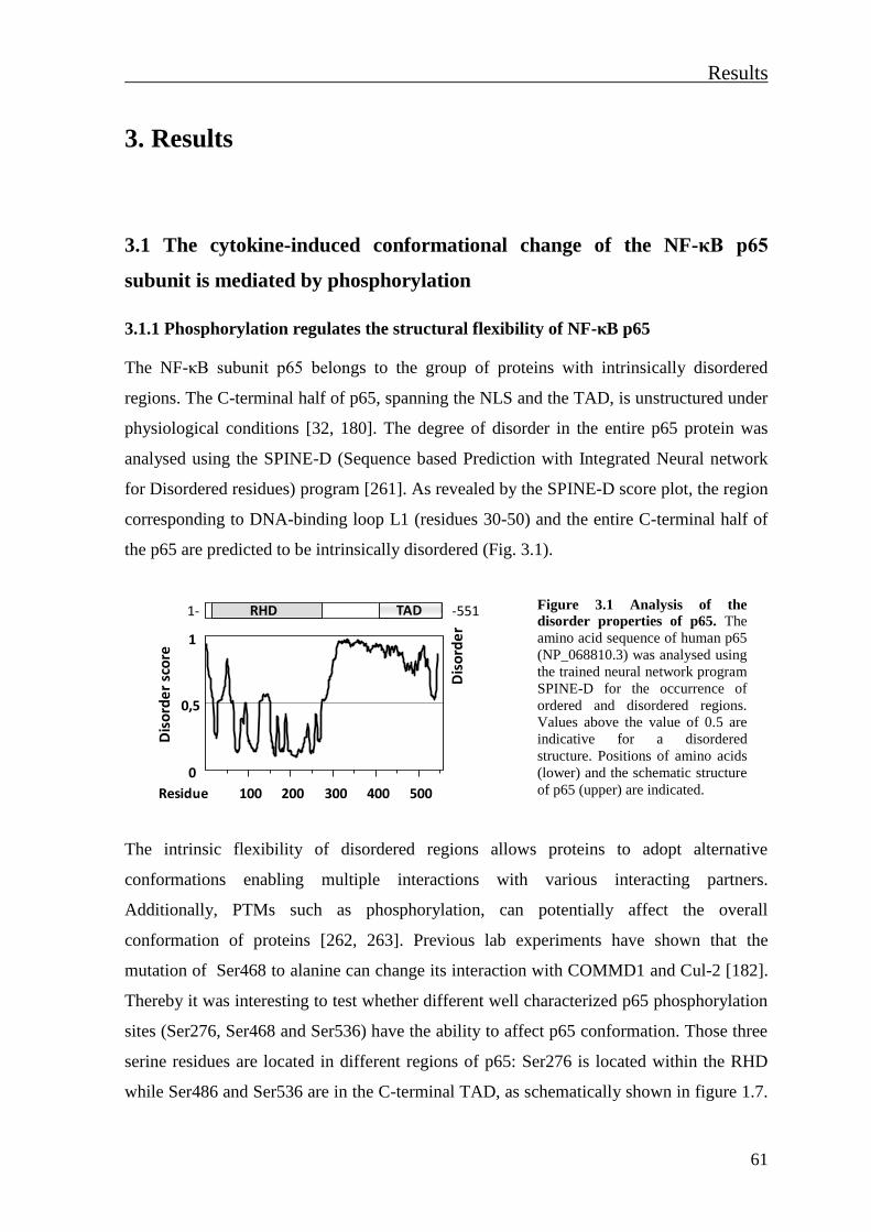
Results
61
3. Results
3.1 The cytokine-induced conformational change of the NF-κB p65
subunit is mediated by phosphorylation
3.1.1 Phosphorylation regulates the structural flexibility of NF-κB p65
The NF-κB subunit p65 belongs to the group of proteins with intrinsically disordered
regions. The C-terminal half of p65, spanning the NLS and the TAD, is unstructured under
physiological conditions [32, 180]. The degree of disorder in the entire p65 protein was
analysed using the SPINE-D (Sequence based Prediction with Integrated Neural network
for Disordered residues) program [261]. As revealed by the SPINE-D score plot, the region
corresponding to DNA-binding loop L1 (residues 30-50) and the entire C-terminal half of
the p65 are predicted to be intrinsically disordered (Fig. 3.1).
Figure 3.1 Analysis of the
disorder properties of p65. The
amino acid sequence of human p65
(NP_068810.3) was analysed using
the trained neural network program
SPINE-D for the occurrence of
ordered and disordered regions.
Values above the value of 0.5 are
indicative for a disordered
structure. Positions of amino acids
(lower) and the schematic structure
of p65 (upper) are indicated.
The intrinsic flexibility of disordered regions allows proteins to adopt alternative
conformations enabling multiple interactions with various interacting partners.
Additionally, PTMs such as phosphorylation, can potentially affect the overall
conformation of proteins [262, 263]. Previous lab experiments have shown that the
mutation of Ser468 to alanine can change its interaction with COMMD1 and Cul-2 [182].
Thereby it was interesting to test whether different well characterized p65 phosphorylation
sites (Ser276, Ser468 and Ser536) have the ability to affect p65 conformation. Those three
serine residues are located in different regions of p65: Ser276 is located within the RHD
while Ser486 and Ser536 are in the C-terminal TAD, as schematically shown in figure 1.7.
0
0,5
1
Dis
ord
er
sco
re
100 200 300 400 500
Dis
ord
er
Residue
RHD TAD1- -551
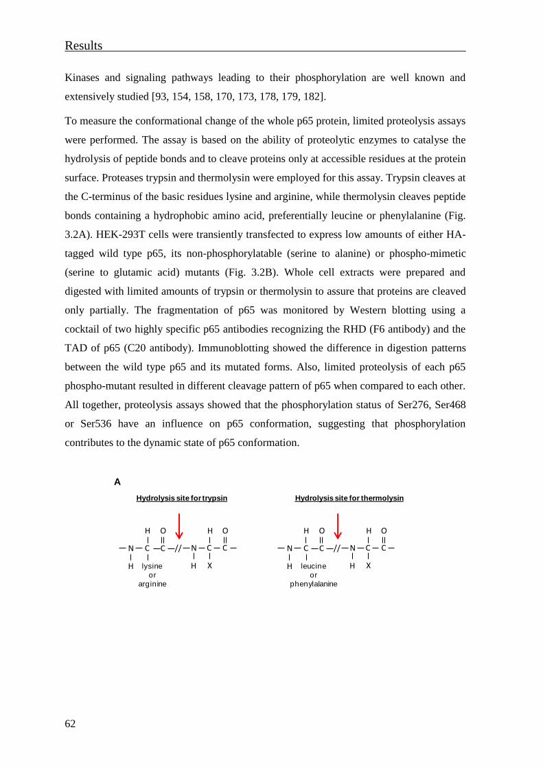
Results
62
Kinases and signaling pathways leading to their phosphorylation are well known and
extensively studied [93, 154, 158, 170, 173, 178, 179, 182].
To measure the conformational change of the whole p65 protein, limited proteolysis assays
were performed. The assay is based on the ability of proteolytic enzymes to catalyse the
hydrolysis of peptide bonds and to cleave proteins only at accessible residues at the protein
surface. Proteases trypsin and thermolysin were employed for this assay. Trypsin cleaves at
the C-terminus of the basic residues lysine and arginine, while thermolysin cleaves peptide
bonds containing a hydrophobic amino acid, preferentially leucine or phenylalanine (Fig.
3.2A). HEK-293T cells were transiently transfected to express low amounts of either HA-
tagged wild type p65, its non-phosphorylatable (serine to alanine) or phospho-mimetic
(serine to glutamic acid) mutants (Fig. 3.2B). Whole cell extracts were prepared and
digested with limited amounts of trypsin or thermolysin to assure that proteins are cleaved
only partially. The fragmentation of p65 was monitored by Western blotting using a
cocktail of two highly specific p65 antibodies recognizing the RHD (F6 antibody) and the
TAD of p65 (C20 antibody). Immunoblotting showed the difference in digestion patterns
between the wild type p65 and its mutated forms. Also, limited proteolysis of each p65
phospho-mutant resulted in different cleavage pattern of p65 when compared to each other.
All together, proteolysis assays showed that the phosphorylation status of Ser276, Ser468
or Ser536 have an influence on p65 conformation, suggesting that phosphorylation
contributes to the dynamic state of p65 conformation.
C // N C C
H
N C
O H O
H H Xleucine
or
phenylalanine
Hydrolysis site for thermolysin
C // N C C
H
N C
O H O
H H Xlysine
or
arginine
Hydrolysis site for trypsin
A
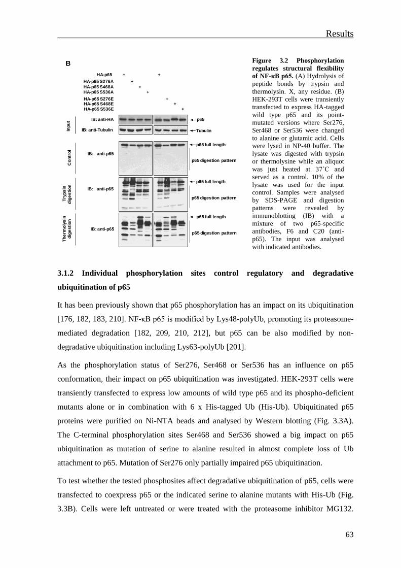
Results
63
Figure 3.2 Phosphorylation
regulates structural flexibility
of NF-κB p65. (A) Hydrolysis of
peptide bonds by trypsin and
thermolysin. X, any residue. (B)
HEK-293T cells were transiently
transfected to express HA-tagged
wild type p65 and its point-
mutated versions where Ser276,
Ser468 or Ser536 were changed
to alanine or glutamic acid. Cells
were lysed in NP-40 buffer. The
lysate was digested with trypsin
or thermolysine while an aliquot
was just heated at 37˚C and
served as a control. 10% of the
lysate was used for the input
control. Samples were analysed
by SDS-PAGE and digestion
patterns were revealed by
immunoblotting (IB) with a
mixture of two p65-specific
antibodies, F6 and C20 (anti-
p65). The input was analysed
with indicated antibodies.
3.1.2 Individual phosphorylation sites control regulatory and degradative
ubiquitination of p65
It has been previously shown that p65 phosphorylation has an impact on its ubiquitination
[176, 182, 183, 210]. NF-κB p65 is modified by Lys48-polyUb, promoting its proteasome-
mediated degradation [182, 209, 210, 212], but p65 can be also modified by non-
degradative ubiquitination including Lys63-polyUb [201].
As the phosphorylation status of Ser276, Ser468 or Ser536 has an influence on p65
conformation, their impact on p65 ubiquitination was investigated. HEK-293T cells were
transiently transfected to express low amounts of wild type p65 and its phospho-deficient
mutants alone or in combination with 6 x His-tagged Ub (His-Ub). Ubiquitinated p65
proteins were purified on Ni-NTA beads and analysed by Western blotting (Fig. 3.3A).
The C-terminal phosphorylation sites Ser468 and Ser536 showed a big impact on p65
ubiquitination as mutation of serine to alanine resulted in almost complete loss of Ub
attachment to p65. Mutation of Ser276 only partially impaired p65 ubiquitination.
To test whether the tested phosphosites affect degradative ubiquitination of p65, cells were
transfected to coexpress p65 or the indicated serine to alanine mutants with His-Ub (Fig.
3.3B). Cells were left untreated or were treated with the proteasome inhibitor MG132.
HA-p65
HA-p65 S276A
HA-p65 S468AHA-p65 S536A
+
HA-p65 S276EHA-p65 S468EHA-p65 S536E
+
++
++
+
+
Inp
ut IB: anti-HA p65
IB: anti-Tubulin Tubulin
IB: anti-p65
Co
ntr
ol
Try
ps
ind
ige
sti
on
p65 digestion pattern
p65 full length
p65 digestion pattern
p65 full length
B
IB: anti-p65
IB: anti-p65p65 digestion pattern
p65 full length
Th
erm
oly
sin
dig
es
tio
n
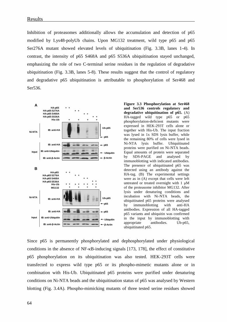
Results
64
Inhibition of proteasomes additionally allows the accumulation and detection of p65
modified by Lys48-polyUb chains. Upon MG132 treatment, wild type p65 and p65
Ser276A mutant showed elevated levels of ubiquitination (Fig. 3.3B, lanes 1-4). In
contrast, the intensity of p65 S468A and p65 S536A ubiquitination stayed unchanged,
emphasizing the role of two C-terminal serine residues in the regulation of degradative
ubiquitination (Fig. 3.3B, lanes 5-8). These results suggest that the control of regulatory
and degradative p65 ubiquitination is attributable to phosphorylation of Ser468 and
Ser536.
Figure 3.3 Phosphorylation at Ser468
and Ser536 controls regulatory and
degradative ubiquitination of p65. (A)
HA-tagged wild type p65 or p65
phosphorylation-deficient mutants were
expressed in HEK-293T cells alone or
together with His-Ub. The input fraction
was lysed in 1x SDS lysis buffer, while
the remaining 80% of cells were lysed in
Ni-NTA lysis buffer. Ubiquitinated
proteins were purified on Ni-NTA beads.
Equal amounts of protein were separated
by SDS-PAGE and analysed by
immunoblotting with indicated antibodies.
The presence of ubiquitinated p65 was
detected using an antibody against the
HA-tag. (B) The experimental settings
were as in (A) except that cells were left
untreated or treated overnight with 1 μM
of the proteasome inhibitor MG132. After
lysis under denaturing conditions and
incubation with Ni-NTA beads, the
ubiquitinated p65 proteins were analysed
by immunoblotting with anti-HA
antibodies. Expression of all HA-tagged
p65 variants and ubiquitin was confirmed
in the input by immunoblotting with
appropriate antibodies. Ub-p65,
ubiquitinated p65.
Since p65 is permanently phosphorylated and dephosphorylated under physiological
conditions in the absence of NF-κB-inducing signals [173, 178], the effect of constitutive
p65 phosphorylation on its ubiquitination was also tested. HEK-293T cells were
transfected to express wild type p65 or its phospho-mimetic mutants alone or in
combination with His-Ub. Ubiquitinated p65 proteins were purified under denaturing
conditions on Ni-NTA beads and the ubiquitination status of p65 was analysed by Western
blotting (Fig. 3.4A). Phospho-mimicking mutants of three tested serine residues showed
HA-p65
HA-p65 S276A
HA-p65 S468A
HA-p65 S536A
+ +
+ +
+ +
+ + ++
++
p65
β-Actin
p65
Ubiquitin
Ub-p65IB: anti-HA
Ni-NTA
Input
IB: anti-HA
IB: anti-β-Actin
IB: anti-Ubiquitin
His-Ub
A
HA-p65
HA-p65 S276A
HA-p65 S468A
HA-p65 S536AHis-Ub
MG132
+ +
+ +
+ +
+
+ +++
+
+ +
+ + + + + +
IB: anti-HANi-NTA
Input
IB: anti-HA
IB: anti-β-Actin
IB: anti-Ubiquitin
p65
β-Actin
p65
Ubiquitin
Ub-p65
B

Results
65
impaired regulatory p65 ubiquitination in comparison to wild type p65. To detect the
impact of permanent phosphorylation on degradative ubiquitination, cells expressing wild
type p65 or the indicated p65 phospho-mimetic mutants were co-transfected with His-Ub
and left untreated or treated with MG132 (Fig. 3.4B). Immunoblotting with anti-HA
antibody showed that MG132 treatment merely increased p65 S468E ubiquitination level,
indicating that permanent phosphorylation at Ser468 rather protects p65 from degradative
ubiquitination.
Figure 3.4 An impact of constitutive
p65 phosphorylation on regulatory
and degradative ubiquitination. (A)
HA-tagged wild type p65 or p65
phospho-mimetic mutants were
expressed in HEK-293T cells alone or
together with His-Ub. Cells were lysed
under denaturing conditions. An aliquot
of cells was taken for an input and lysed
in 1x SDS lysis buffer. The remaining
cells were lysed in Ni-NTA lysis buffer,
followed by purification of ubiquitinated
proteins using Ni-NTA beads. Equal
amounts of protein were separated by
SDS-PAGE. The presence of
ubiquitinated p65 was detected using
anti-HA antibodies. The input fraction
was immunoblotted with indicated
antibodies. (B) The experimental
settings were as in (A) except that cells
were left untreated or treated overnight
with the proteasome inhibitor MG132.
After cell lysis under denaturing
conditions and incubation with Ni-NTA
beads, precipitated ubiquitinated p65
proteins were separated by SDS-PAGE
and detected by immunoblotting with
indicated antibodies.
In agreement with the limited proteolysis assays, these experiments show that the
phosphorylation status of individual serine residues affects p65 conformation and
consequently also p65 ubiquitination.
3.1.3 Cytokines induce a phosphorylation-dependent conformational change of
nuclear p65
In order to detect the occurrence of p65 conformational change under physiological
conditions, cells were stimulated with TNF to trigger activation of NF-κB. HEK-293 cells
HA-p65
HA-p65 S276E
HA-p65 S468E
HA-p65 S536E
His-Ub
MG132
+ +
+ +
+ +
+
+ +++
+
+ +
+ + + + + +
IB: anti-HANi-NTA
Input
IB: anti-HA
IB: anti-β-Actin
IB: anti-Ubiquitin
p65
β-Actin
p65
Ubiquitin
Ub-p65
B
HA-p65
HA-p65 S276E
HA-p65 S468E
HA-p65 S536E
+ +
+ +
+ +
+ + ++
++
p65
β-Actin
p65
Ubiquitin
Ub-p65IB: anti-HA
Ni-NTA
Input
IB: anti-HA
IB: anti-β-Actin
IB: anti-Ubiquitin
His-Ub
A
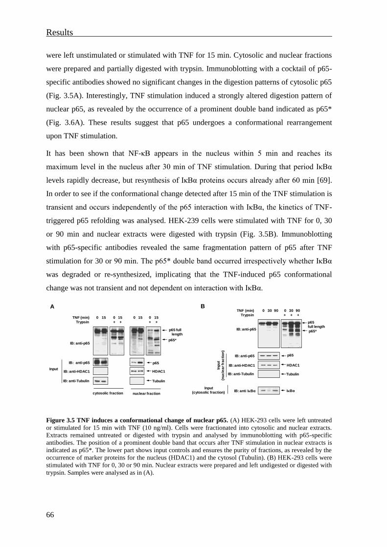
Results
66
were left unstimulated or stimulated with TNF for 15 min. Cytosolic and nuclear fractions
were prepared and partially digested with trypsin. Immunoblotting with a cocktail of p65-
specific antibodies showed no significant changes in the digestion patterns of cytosolic p65
(Fig. 3.5A). Interestingly, TNF stimulation induced a strongly altered digestion pattern of
nuclear p65, as revealed by the occurrence of a prominent double band indicated as p65*
(Fig. 3.6A). These results suggest that p65 undergoes a conformational rearrangement
upon TNF stimulation.
It has been shown that NF-κB appears in the nucleus within 5 min and reaches its
maximum level in the nucleus after 30 min of TNF stimulation. During that period IκBα
levels rapidly decrease, but resynthesis of IκBα proteins occurs already after 60 min [69].
In order to see if the conformational change detected after 15 min of the TNF stimulation is
transient and occurs independently of the p65 interaction with IκBα, the kinetics of TNF-
triggered p65 refolding was analysed. HEK-239 cells were stimulated with TNF for 0, 30
or 90 min and nuclear extracts were digested with trypsin (Fig. 3.5B). Immunoblotting
with p65-specific antibodies revealed the same fragmentation pattern of p65 after TNF
stimulation for 30 or 90 min. The p65* double band occurred irrespectively whether IκBα
was degraded or re-synthesized, implicating that the TNF-induced p65 conformational
change was not transient and not dependent on interaction with IκBα.
Figure 3.5 TNF induces a conformational change of nuclear p65. (A) HEK-293 cells were left untreated
or stimulated for 15 min with TNF (10 ng/ml). Cells were fractionated into cytosolic and nuclear extracts.
Extracts remained untreated or digested with trypsin and analysed by immunoblotting with p65-specific
antibodies. The position of a prominent double band that occurs after TNF stimulation in nuclear extracts is
indicated as p65*. The lower part shows input controls and ensures the purity of fractions, as revealed by the
occurrence of marker proteins for the nucleus (HDAC1) and the cytosol (Tubulin). (B) HEK-293 cells were
stimulated with TNF for 0, 30 or 90 min. Nuclear extracts were prepared and left undigested or digested with
trypsin. Samples were analysed as in (A).
nuclear fraction
p65IB: anti-p65
HDAC1IB: anti-HDAC1
Trypsin + +
p65 fulllength
p65*IB: anti-p65
TNF (min) 15 15
TubulinIB: anti-Tubulin
+ +
15 15
cytosolic fraction
A
Input
0 0 0 0
IB: anti-p65
Trypsin ++ +
p65IB: anti-p65
IB: anti-Tubulin Tubulin
p65 full length
p65*
TNF (min)
HDAC1IB: anti-HDAC1
IB: anti IκBα IκBα
30 90 30 90
Inp
ut
(nu
cle
ar
fra
cti
on
)
B
Input(cytosolic fraction)
0 0

Results
67
It was interesting to test whether the cytokine IL-1 induces the same conformational
rearrangement as TNF. As HEK-293 cells hardly respond to IL-1, HEK-293 cells
expressing the IL-1 receptor (HEK-293IL-1R) were used for these experiments. Cells were
stimulated with TNF or IL-1 for 0 or 15 min. Cytosolic and nuclear extracts were prepared
and digested with trypsin (Fig. 3.6A). Stimulation with IL-1 induced the same pattern of
p65 fragmentation when compared to TNF-treated nuclear p65. This suggests that both
cytokines induce the same configuration of p65. The double p65* marker band was
detected after 90 min of IL-1 stimulation, confirming that rearrangement of nuclear p65 is
not a transient event (Fig. 3.6B).
Figure 3.6 Comparison between IL-1- and TNF-induced conformational changes of nuclear p65. (A)
HEK-293IL-1R cells were left untreated or stimulated for 15 min with TNF (10 ng/ml) or IL-1 (10 ng/ml).
Cells were fractionated into cytosolic and nuclear extracts. These extracts remained untreated or partially
digested with trypsin. Fragmentation of p65 was analysed by immunoblotting with p65-specific antibodies.
An aliquot was used to ensure the purity of lysates (Input). (B) HEK-293IL-1R cells were stimulated with
TNF or IL-1 for 0, 30 or 90 min. Nuclear extracts were prepared and digested with trypsin. Samples were
analysed as in (A).
The relative contribution of phosphorylation for the cytokine-induced conformational
change of nuclear p65 was tested. For this purpose HEK-293 cells were treated with
calyculin A to induce phosphorylation of endogenous p65 by strong inhibition of
serine/threonine protein phosphatases [177, 178, 185, 264]. Also treatment with calyculin
A induced the appearance of the p65* double band, as revealed by limited proteolysis
assays of nuclear extracts (Fig. 3.7A). To address the contribution of individual
phosphorylation sites on cytokine-induced p65 folding, mouse embryonic fibroblasts
lacking the p65 gene (p65-/- MEFs) were reconstituted with lentiviral constructs to stably
express HA-tagged wild type p65 or its phospho-deficient mutants (p65 S468A or p65
S536A). Cells were stimulated with TNF and nuclear extracts were tested for p65
p65full length
nuclear fraction
p65IB: anti-p65
HDAC1IB: anti-HDAC1
Trypsin
p65*IB: anti-p65
IL-1 (min)
TubulinIB: anti-Tubulin
+ +
15 15
cytosolic fraction
TNF (min)
15 15
+ + +
15 15
15 15
+
Inp
ut
0
00
0
0
0
0
0
A
p65IB: anti-p65
HDAC1IB: anti-HDAC1
Trypsin
IB: anti-p65
IL-1 (min)
TubulinIB: anti-Tubulin
TNF (min)
IB: anti IκBα(cytosolic fraction)
IκBα
+ +
30
30
90
90
+ + +
Inp
ut
(nu
cle
ar
fra
cti
on
)
p65full length
p65*
0
0
B
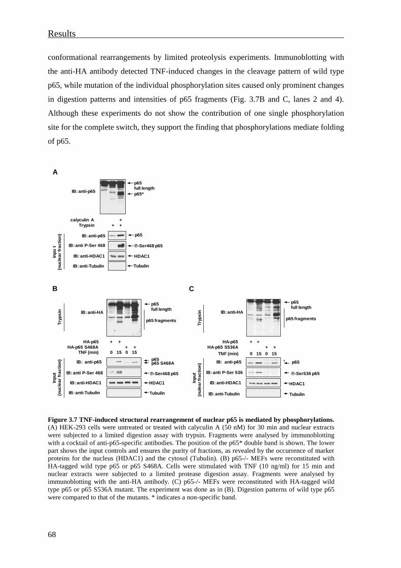
Results
68
conformational rearrangements by limited proteolysis experiments. Immunoblotting with
the anti-HA antibody detected TNF-induced changes in the cleavage pattern of wild type
p65, while mutation of the individual phosphorylation sites caused only prominent changes
in digestion patterns and intensities of p65 fragments (Fig. 3.7B and C, lanes 2 and 4).
Although these experiments do not show the contribution of one single phosphorylation
site for the complete switch, they support the finding that phosphorylations mediate folding
of p65.
Figure 3.7 TNF-induced structural rearrangement of nuclear p65 is mediated by phosphorylations.
(A) HEK-293 cells were untreated or treated with calyculin A (50 nM) for 30 min and nuclear extracts
were subjected to a limited digestion assay with trypsin. Fragments were analysed by immunoblotting
with a cocktail of anti-p65-specific antibodies. The position of the p65* double band is shown. The lower
part shows the input controls and ensures the purity of fractions, as revealed by the occurrence of marker
proteins for the nucleus (HDAC1) and the cytosol (Tubulin). (B) p65-/- MEFs were reconstituted with
HA-tagged wild type p65 or p65 S468A. Cells were stimulated with TNF (10 ng/ml) for 15 min and
nuclear extracts were subjected to a limited protease digestion assay. Fragments were analysed by
immunoblotting with the anti-HA antibody. (C) p65-/- MEFs were reconstituted with HA-tagged wild
type p65 or p65 S536A mutant. The experiment was done as in (B). Digestion patterns of wild type p65
were compared to that of the mutants. * indicates a non-specific band.
IB: anti-p65
Trypsin +
+
+
Inp
ut
(nu
cle
ar
fra
cti
on
) p65IB: anti-p65
IB: anti-Tubulin Tubulin
p65full length
p65*
calyculin A
HDAC1IB: anti-HDAC1
IB: anti P-Ser 468 ℗-Ser468 p65
A
HA-p65
HA-p65 S468A TNF (min)
HDAC1
Tubulin
15 15
+ +
+ +
IB: anti-p65
IB: anti-HDAC1
IB: anti P-Ser 468
IB: anti-Tubulin
Inp
ut
(nu
cle
ar
fra
cti
on
)
IB: anti-HA
p65 full length
Try
ps
in
p65 fragments
p65 S468Ap65
B
℗-Ser468 p65
0 0
HA-p65
HA-p65 S536A
HDAC1
Tubulin
+ +
+ +
IB: anti-p65
IB: anti-HDAC1
IB: anti P-Ser 536
IB: anti-Tubulin
Inp
ut
(nu
lea
rfr
ac
tio
n)
p65 full length
Try
ps
in
p65 fragments
p65 *
℗-Ser536 p65
C
TNF (min) 15 150 0
IB: anti-HA

Results
69
3.1.4 The TNF-induced NF-κB p65 structural rearrangement unmasks an epitope
localised in the TAD2 subdomain
The cytokine-induced conformational change of nuclear p65 was tested by an alternative
approach. Seven p65-specific antibodies were tested for their ability to immunoprecipitate
p65 in its active conformation after TNF stimulation (Fig. 3.8). Each tested antibody
recognizes a different p65 epitope, as schematically illustrated in green and grey in figure
3.8A. The antibodies F6 and C20 were used as a control (Fig. 3.8A, represented in red).
HEK-293 cells were left unstimulated or exposed to TNF for 15 min after which the
nuclear extract was prepared and pre-cleared with agarose beads. Immunoprecipitation was
performed with antibodies indicated in figure 3.8A and corresponding control IgG
antibodies. Eluted p65 proteins were detected by immunoblotting with the p65-specific
antibody C20 (Fig. 3.8B).
Figure 3.8 Screening of antibodies specifically
recognising the refolded p65 protein. (A) Amino
acid sequence of human NF-κB p65 subunit with
marked regions recognized by p65-specific
antibodies. Tested antibodies are indicated in gray
and green. Antibodies that were able to
immunoprecipitate (IP) nuclear p65 after TNF
stimulation are indicated in green, while those that
were not are labeled in grey. Antibodies indicated in
red were used as a control. (B) HEK-293 cells were
left unstimulated or stimulated for 15 min with TNF
(10 ng/ml). Nuclear extracts containing the activated
p65 protein were immunoprecipitated with different
p65-specific and corresponding control IgG
antibodies. Precipitated nuclear p65 proteins were
detected by immunoblotting with an antibody
recognizing the C-terminal part of the p65 (C20). An
aliquot was used to check purity of the nuclear
fraction and equal sample loading (Input). * non-
specific band.
112A1021526-539 aa
EP2161Ysurrounding Ser468 6H7
136 – 224 aa
F6, 1-286 aa
IMG-5915A2-17 aa
LS-C14790187-97 aa
IMX-5238273 – 284 aa
IMG – 6066A290-305 aa C20, Peptide maping at the C-terminus
1 551
100 300 aa 500
A
TNF (min) 0 15
IB: anti-p65
IB: anti-HDAC1
IB: anti-Tubulin
Inp
ut
(nu
cle
ar
fra
cti
on
)
p65
HDAC1
Tubulin
IP: 112A1021 p65
IP: EP2161Y p65
IP: IMG-6066A p65
*
IP: IMX-5238 p65
IP: 6H7 p65
IP: LS-C147901 p65
IP: IMG-5915A p65
IB: a
nti
-p
65
(C
20
)
IP: C20 p65
IP: F6 p65
IP: anti-p65
IP: IgG
B
Results
70
These experiments showed that an antibody recognizing a region surrounding Ser468
(Abcam EP2161Y) was able to immunoprecipitate the active, nuclear form of p65. An
antibody recognizing the NLS of p65 (Imgenex IMG-6066A) was also able to
immunoprecipitate TNF-activated p65 as reported previously [265]. However, the
specificity of this antibody relies on a different principle, as it can only recognize the NLS
of p65 when IκBα is degraded.
3.1.5 Phosphorylation-dependent changes of NF-κB p65 conformation regulate p65
interactions with transcriptional cofactors
Several studies have shown that phosphorylation of p65 governs the outcome of NF-κB
responses through differential interaction with various enzymes [154, 166, 177, 182, 183,
266]. Therefore it was interesting to investigate the impact of p65 phosphorylation-
dependent structural changes on protein-protein interactions. Phosphorylation at the N-
terminal Ser276 and the C-terminal Ser468 leads to different rearrangement of p65 (Fig.
3.2B). Therefore, coimmunoprecipitation experiments were performed to compare the
binding of known p65 interactors to wild type p65 and phospho-mimetic p65 S468E and
p65 S276E mutants. The interaction with the p65 interacting partner CBP was tested since
it can interact with both the N- and C-terminal half of the p65 [154]. Another tested
interaction partner was the NF-κB corepressor MYBBP1a (Myb-binding protein 1a), as
this protein interacts with the C-terminal half of p65 [267, 268]. Cells were transfected to
express p65 variants alone or together with CBP, MYBBP1a or the NF-κB p50 subunit.
NF-κB interacting partners were immunoprecipitated with anti-GFP or anti-Flag antibodies
and the amount of a bound p65 protein was revealed by immunoblotting with anti-HA
antibodies. Permanent phosphorylation at Ser468 reduced the interaction with CBP and
MYBBP1a proteins and also led to reduced binding of p65 to NF-κB subunit p50 (Fig.
3.9A). In contrast to Ser468, permanent phosphorylation at Ser276 did not change the
ability of p65 to interact with the tested interacting partners (Fig. 3.9B). These
coimmunoprecipitation experiments implicate that a phosphorylation-induced
conformational change affects the association of p65 with transcriptional coactivators and
corepressors, as exemplified for p65 phospho-mimetic Ser468 and Ser276 mutants.
Additionally, these experiments emphasize the role of Ser468 for the conformation of p65
and its affinity to interaction partners.
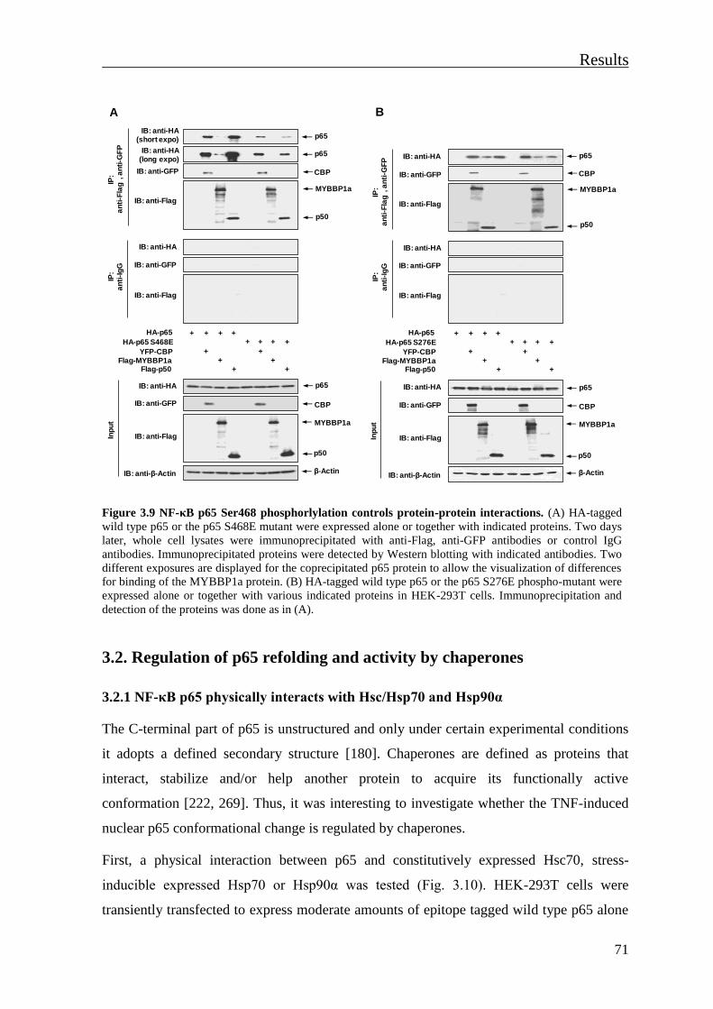
Results
71
Figure 3.9 NF-κB p65 Ser468 phosphorlylation controls protein-protein interactions. (A) HA-tagged
wild type p65 or the p65 S468E mutant were expressed alone or together with indicated proteins. Two days
later, whole cell lysates were immunoprecipitated with anti-Flag, anti-GFP antibodies or control IgG
antibodies. Immunoprecipitated proteins were detected by Western blotting with indicated antibodies. Two
different exposures are displayed for the coprecipitated p65 protein to allow the visualization of differences
for binding of the MYBBP1a protein. (B) HA-tagged wild type p65 or the p65 S276E phospho-mutant were
expressed alone or together with various indicated proteins in HEK-293T cells. Immunoprecipitation and
detection of the proteins was done as in (A).
3.2. Regulation of p65 refolding and activity by chaperones
3.2.1 NF-κB p65 physically interacts with Hsc/Hsp70 and Hsp90α
The C-terminal part of p65 is unstructured and only under certain experimental conditions
it adopts a defined secondary structure [180]. Chaperones are defined as proteins that
interact, stabilize and/or help another protein to acquire its functionally active
conformation [222, 269]. Thus, it was interesting to investigate whether the TNF-induced
nuclear p65 conformational change is regulated by chaperones.
First, a physical interaction between p65 and constitutively expressed Hsc70, stress-
inducible expressed Hsp70 or Hsp90α was tested (Fig. 3.10). HEK-293T cells were
transiently transfected to express moderate amounts of epitope tagged wild type p65 alone
HA-p65
HA-p65 S468E
YFP-CBP
Flag-MYBBP1aFlag-p50
+ + + ++ + ++
+
+ +
p65
β-Actin
CBP
MYBBP1a
IB: anti-HA(short expo)
IP:
an
ti-F
lag
, a
nti
-GF
P
Inp
ut
IB: anti-β-Actin
+
+
IB: anti-Flag
IB: anti-HA
IB: anti-Flag
IP:
an
ti-I
gG
p50
p65
CBP
MYBBP1a
p50
IB: anti-HA
IB: anti-Flag
IB: anti-GFP
IB: anti-GFP
IB: anti-GFP
+
IB: anti-HA(long expo)
p65
A
Inp
ut
HA-p65
HA-p65 S276E
YFP-CBP
Flag-MYBBP1aFlag-p50
+ + + ++ + ++
+
+ +
β-Actin
CBP
MYBBP1a
IP:
an
ti-F
lag
, a
nti
-GF
P
IB: anti-β-Actin
+
+
IB: anti-Flag
IB: anti-HA
IB: anti-Flag
IP:
an
ti-I
gG
p50
p65
CBP
MYBBP1a
p50
IB: anti-HA
IB: anti-Flag
IB: anti-GFP
IB: anti-GFP
IB: anti-GFP
+
IB: anti-HA p65
B

Results
72
or together with various Hsps or p50 as a positive control. Coimmunoprecipitation
experiments showed an interaction between p65 and Hsc70 or Hsp70, which is in line with
previous studies [242, 257]. The interaction with Hsp90α was not observed (Fig. 3.10A).
Hsp90 interacts with its client proteins with a weak affinity and only transiently [229]. To
test a potential interaction between p65 and Hsp90α by a more sensitive approach,
coimmunoprecipitation experiments were performed in the presence of the membrane
permeable cross-linker DTBP, which reversibly cross-links and stabilizes protein-protein
interactions which occur in close proximity. Cells were transfected to express HA-tagged
p65 alone or together with Flag-tagged Hsp90α. Before lysis, cells were incubated with
vehicle or DTBP. Coimmunoprecipitation detected an interaction between p65 and Hsp90α
only after the treatment with DTBP (Fig. 3.10B, lane 3), revealing that this interaction is
rather weak.
Figure 3.10 p65 physically interacts with Hsps. (A) HEK-293T cells were transiently transfected to
express HA-tagged p65 together with the Flag-tagged Hsc70, Hsp70 or Hsp90α. Coexpression with Flag-
tagged p50 served as a positive control. Cells were lysed in NP-40 buffer and an aliquot was used for the
analysis of protein expression (Input). The remaining lysate was used for immunoprecipitation with anti-
Flag or control IgG antibodies. Precipitated proteins were detected by immunoblotting with indicated
antibodies. (B) HEK-293T cells were transfected with indicated plasmids and incubated with the cross-
linker DTBP. After NP-40 lysis, an aliquot was used for the input control, while the rest of the lysate was
immunopreciptated with either anti-Flag or control IgG antibodies; precipitated proteins were detected by
immunoblotting with indicated antibodies.
The proinflammatory cytokine TNF is produced under various stress conditions and leads
to increased Hsp70 expression in cardiac myocytes [270]. It has been demonstrated that
IB: anti-Flag
IB: anti-HA p65
Hsp 90α
IP:
an
ti-H
A
IB: anti-HA
IB: anti-Flag
IP:
an
ti-I
gG
IB: anti-Flag
IB: anti-HA
IB: anti-β-Actin
Inp
ut
p65
Hsp 90α
β-Actin
HA-p65 + ++
+Flag-Hsp 90α +
DTBP +
B
Hsc 70
IB: anti-HA
IB: anti-Flag
p65
Hsp 70
p50
Hsp 90α
IB: anti-HA
IB: anti-Flag
IP:
an
ti-I
gG
IB: anti-Flag
IB: anti-HA
IB: anti-β-Actin
Inp
ut
p65
Hsp 90αHsp 70
Hsc 70
p50
β-Actin
HA-p65
Flag-Hsc 70
Flag-Hsp 70
+ ++ ++
Flag-Hsp 90α
Flag-p50
+
+
+
+
IP:
an
ti-F
lag
IB: anti-Flag
A
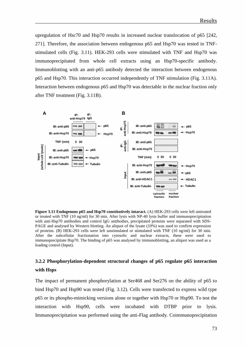
Results
73
upregulation of Hsc70 and Hsp70 results in increased nuclear translocation of p65 [242,
271]. Therefore, the association between endogenous p65 and Hsp70 was tested in TNF-
stimulated cells (Fig. 3.11). HEK-293 cells were stimulated with TNF and Hsp70 was
immunoprecipitated from whole cell extracts using an Hsp70-specific antibody.
Immunoblotting with an anti-p65 antibody detected the interaction between endogenous
p65 and Hsp70. This interaction occurred independently of TNF stimulation (Fig. 3.11A).
Interaction between endogenous p65 and Hsp70 was detectable in the nuclear fraction only
after TNF treatment (Fig. 3.11B).
Figure 3.11 Endogenous p65 and Hsp70 constitutively intaract. (A) HEK-293 cells were left untreated
or treated with TNF (10 ng/ml) for 30 min. After lysis with NP-40 lysis buffer and immunoprecipitation
with anti-Hsp70 antibodies and control IgG antibodies, precipitated proteins were separated with SDS-
PAGE and analysed by Western blotting. An aliquot of the lysate (10%) was used to confirm expression
of proteins. (B) HEK-293 cells were left unstimulated or stimulated with TNF (10 ng/ml) for 30 min.
After the subcellular fractionation into cytosolic and nuclear extracts, these were used to
immunoprecipitate Hsp70. The binding of p65 was analysed by immunoblotting, an aliquot was used as a
loading control (Input).
3.2.2 Phosphorylation-dependent structural changes of p65 regulate p65 interaction
with Hsps
The impact of permanent phosphorylation at Ser468 and Ser276 on the ability of p65 to
bind Hsp70 and Hsp90 was tested (Fig. 3.12). Cells were transfected to express wild type
p65 or its phospho-mimicking versions alone or together with Hsp70 or Hsp90. To test the
interaction with Hsp90, cells were incubated with DTBP prior to lysis.
Immunoprecipitation was performed using the anti-Flag antibody. Coimmunoprecipitation
TNF (min) 0 30
IB: anti-Hsp70
IB: anti-p65 p65
IB: anti-p65
IB: anti-Hsp70
IB: anti-Tubulin
Inp
ut
(wh
ole
ce
ll l
ys
ate
)
p65
Hsp70
Tubulin
IP: anti-Hsp70
IP: IgG
Hsp70
A
TNF (min) 30
IB: anti-p65
IB: anti-Hsp70
IP:
an
ti-H
sp
70
IB: anti-p65
IB: anti-Hsp70
IP:
an
ti-I
gG
Hsp70
p65
IB: anti-Hsp70
IB: anti-p65
IB: anti-Tubulin
Inp
ut
Hsp70
p65
Tubulin
30
cytosolic
fraction
nuclear
fraction
IB: anti-HDAC1 HDAC1
B
0 0

Results
74
of p65 with Hsp70 (Fig. 3.12A/C) or Hsp90 (Fig. 3.12B/D) showed that permanent Ser468
phosphorylation strongly diminished the association with Hsp90, while it slightly enhanced
the interaction of p65 with Hsp70. Permanent phosphorylation at Ser276 slightly reduced
the interaction of p65 with both tested chaperones. These protein-protein interaction
studies support the idea that the phosphorylation-dependent p65 conformational change
affects affinities of p65 to its interaction partners.
Figure 3.12 Permanent Ser468-phosphorylation affects the interaction of p65 with Hsps. (A) HA-
tagged wild type p65 or its phospho-mimetic mutant HA-p65 S468E were transfected alone or together
with Flag-tagged Hsp70. HEK-293T cells were lysed in NP-40 buffer. An aliquot was analysed for
correct protein expression (Input). Immunoprecipitation was performed using an anti-Flag antibody or a
control IgG antibody. Precipitated proteins and input samples were analysed by SDS-PAGE and Western
blotting using appropriate antibodies. (B) HEK-293T cells were transfected to express epitope-tagged
versions of p65 and Hsp90α as shown and treated with vehicle or the cross-linker DTBP. Cells were lysed
and subjected to immunoprecipitation using an anti-Flag antibody. An aliquot was used for input control.
The precipitates and input controls were analysed by immunoblotting with indicated antibodies. (C) and
(D) Experiments were done as in (A) and (B) respectively, except that the HA-p65 S276E mutant was
used.
Hsp70
HA-p65 + +
+Flag Hsp70 +
+HA-p65 S468E
IB: anti-Flag
IB: anti-HA
IP:
an
ti-F
lag
IB: anti-HA
IB: anti-Flag
IP:
an
ti-I
gG
IB: anti-HA
IB: anti-Flag
IB: anti-Tubulin
Inp
ut
Hsp70
p65
p65
Tubulin
A
HA-p65 + ++
+Flag-Hsp 90α +
+
+DTBP
HA-p65 S468E + +
+ +
+
IB: anti-HA
IB: anti-Flag
IP:
an
ti-F
lag
IB: anti-HA
IB: anti-Flag
IP:
an
ti-I
gG
IB: anti-HA
IB: anti-Flag
IB: anti-β-Actin
Inp
ut
p65
Hsp 90α
p65
Hsp 90α
β-Actin
B
HA-p65 + ++
+Flag-Hsp 90α +
+
+DTBP
HA-p65 S276E + +
+ +
+
IB: anti-HA
IB: anti-Flag
IP:
an
ti-F
lag
IB: anti-HA
IB: anti-Flag
IP:
an
ti-I
gG
IB: anti-HA
IB: anti-Flag
IB: anti-β-Actin
Inp
ut
p65
Hsp 90α
p65
Hsp 90α
β-Actin
D
Hsp70
C
HA p65 + +
+Flag-Hsp70 +
+HA-p65 S276E
IB: anti-Flag
IB: anti-HA
IP:
an
ti-F
lag
IB: anti-HA
IB: anti-Flag
IP:
an
ti-I
gG
IB: anti-HA
IB: anti-Flag
IB: anti-Tubulin
Inp
ut
Hsp70
p65
p65
Tubulin

Results
75
3.2.3 Chaperones contribute to the control of the TNF-induced p65 refolding
A substantial fraction of all proteins in eukaryotic cells adopt a folded conformation only
after interaction with a cognate binding partner [222]. In order to gain a functional
conformation, some proteins need further interaction with the Hsp90 and/or Hsp70 [229,
231]. To address whether Hsps contribute to basal p65 conformation, Hsps were
pharmacologically inhibited. Hsp90 was inhibited by GA and Hsc/Hsp70 by the use of
VER. Both inhibitors block the ATPase activity of Hsps, thereby inhibiting their chaperone
activity [223, 247, 272, 273]. HEK-293 cells were incubated with vehicle, GA and VER
for 17 h and whole cell lysates were subjected to limited proteolysis assays.
Immunoblotting with p65-specific antibodies, revealed no difference in p65 digestion
patterns after the inhibition of Hsp90 or Hsc/Hsp70 in comparison to untreated cells (Fig.
3.13).
Figure 3.13 Chaperones have no
influence on basal conformation of p65.
HEK-293 cells were left untreated or treated
with inhibitors GA (1 μM) or VER (30 μM).
Whole cell lysate was prepared and 10%
was used for the input control. The rest of
the lysate was used for the limited
proteolysis assay. Extracts were left
untreated or treated with trypsin. p65
fragments and input controls were separated
by SDS-PAGE and analysed by
immunoblotting with p65-specific and anti-
Tubulin antibodies.
With the aim to test the role of Hsp70 and Hsp90 in folding of newly synthesized p65, it
was necessary to induce the degradation of p65 proteins that were present inside cells. For
that aim, HeLa cells were first stimulated with a TNF pulse for 15 min, followed by
blockage of new protein synthesis by CHX for indicated periods. As CHX prevents the
resynthesis of the eliminated protein and thus allows the detection of TNF-induced p65
diminishment (Fig. 3.14A). After the CHX treatment for 24 h which caused almost
complete loss of p65, cells were washed with PBS to remove the reversible translation
inhibitor CHX [274, 275] and to allow synthesis of new proteins. After an incubation
period of 3 h, cells were treated with vehicle, VER and GA for the next 17 h in order to
detect a possible contribution of Hsps on folding of newly synthesized p65 (Fig. 3.14B).
Limited proteolysis assays followed by immunoblotting did not show a significant
p65 full length
IB: anti-p65
GA
TrypsinVER
+ +
+ +
+ + +
p65 fragments
Inp
ut p65IB: anti-p65
IB: anti-Tubulin Tubulin
GA
VER
+
+
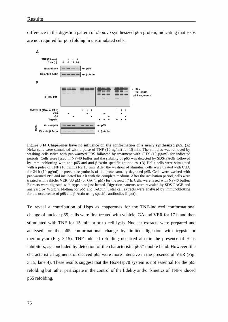
Results
76
difference in the digestion pattern of de novo synthesized p65 protein, indicating that Hsps
are not required for p65 folding in unstimulated cells.
Figure 3.14 Chaperones have no influence on the conformation of a newly synthesized p65. (A)
HeLa cells were stimulated with a pulse of TNF (10 ng/ml) for 15 min. The stimulus was removed by
washing cells twice with pre-warmed PBS followed by treatment with CHX (10 µg/ml) for indicated
periods. Cells were lysed in NP-40 buffer and the stability of p65 was detected by SDS-PAGE followed
by immunoblotting with anti-p65 and anti-β-Actin specific antibodies. (B) HeLa cells were stimulated
with a pulse of TNF (10 ng/ml) for 15 min. After the washout of stimulus, cells were treated with CHX
for 24 h (10 μg/ml) to prevent resynthesis of the proteosomally degraded p65. Cells were washed with
pre-warmed PBS and incubated for 3 h with the complete medium. After the incubation period, cells were
treated with vehicle, VER (30 μM) or GA (1 μM) for the next 17 h. Cells were lysed with NP-40 buffer.
Extracts were digested with trypsin or just heated. Digestion patterns were revealed by SDS-PAGE and
analysed by Western blotting for p65 and β-Actin. Total cell extracts were analysed by immunoblotting
for the occurrence of p65 and β-Actin using specific antibodies (Input).
To reveal a contribution of Hsps as chaperones for the TNF-induced conformational
change of nuclear p65, cells were first treated with vehicle, GA and VER for 17 h and then
stimulated with TNF for 15 min prior to cell lysis. Nuclear extracts were prepared and
analysed for the p65 conformational change by limited digestion with trypsin or
thermolysin (Fig. 3.15). TNF-induced refolding occurred also in the presence of Hsps
inhibitors, as concluded by detection of the characteristic p65* double band. However, the
characteristic fragments of cleaved p65 were more intensive in the presence of VER (Fig.
3.15, lane 4). These results suggest that the Hsc/Hsp70 system is not essential for the p65
refolding but rather participate in the control of the fidelity and/or kinetics of TNF-induced
p65 refolding.
Inp
ut
Trypsin
+ + +
p65 full length
p65 fragmentsIB: anti-p65
GA+
TNF/CHX (15 min/ 24 h)
IB: anti- β Actin
VER +
IB: anti-p65
+ +
+ + +
++
+ + + + + +
++
B
p65
β Actin
++TNF (15 min)
CHX (h) 0 0 24
IB: anti-p65 p65
IB: anti-β Actin β Actin
+
12
A
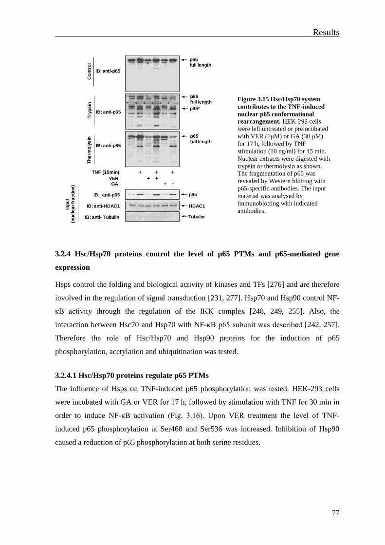
Results
77
Figure 3.15 Hsc/Hsp70 system
contributes to the TNF-induced
nuclear p65 conformational
rearrangement. HEK-293 cells
were left untreated or preincubated
with VER (1μM) or GA (30 μM)
for 17 h, followed by TNF
stimulation (10 ng/ml) for 15 min.
Nuclear extracts were digested with
trypsin or thermolysin as shown.
The fragmentation of p65 was
revealed by Western blotting with
p65-specific antibodies. The input
material was analysed by
immunoblotting with indicated
antibodies.
3.2.4 Hsc/Hsp70 proteins control the level of p65 PTMs and p65-mediated gene
expression
Hsps control the folding and biological activity of kinases and TFs [276] and are therefore
involved in the regulation of signal transduction [231, 277]. Hsp70 and Hsp90 control NF-
κB activity through the regulation of the IKK complex [248, 249, 255]. Also, the
interaction between Hsc70 and Hsp70 with NF-κB p65 subunit was described [242, 257].
Therefore the role of Hsc/Hsp70 and Hsp90 proteins for the induction of p65
phosphorylation, acetylation and ubiquitination was tested.
3.2.4.1 Hsc/Hsp70 proteins regulate p65 PTMs
The influence of Hsps on TNF-induced p65 phosphorylation was tested. HEK-293 cells
were incubated with GA or VER for 17 h, followed by stimulation with TNF for 30 min in
order to induce NF-κB activation (Fig. 3.16). Upon VER treatment the level of TNF-
induced p65 phosphorylation at Ser468 and Ser536 was increased. Inhibition of Hsp90
caused a reduction of p65 phosphorylation at both serine residues.
GA
TNF (15min)
VER + +
+ + +
+ +
Inp
ut
(nu
cle
ar
fra
cti
on
)
IB: anti-p65
IB: anti-HDAC1
IB: anti- Tubulin
p65
HDAC1
Tubulin
p65 full length
p65*
Try
ps
in
IB: anti-p65
IB: anti-p65
IB: anti-p65
p65 full length
p65 full length
Th
erm
oly
sin
Co
ntr
ol
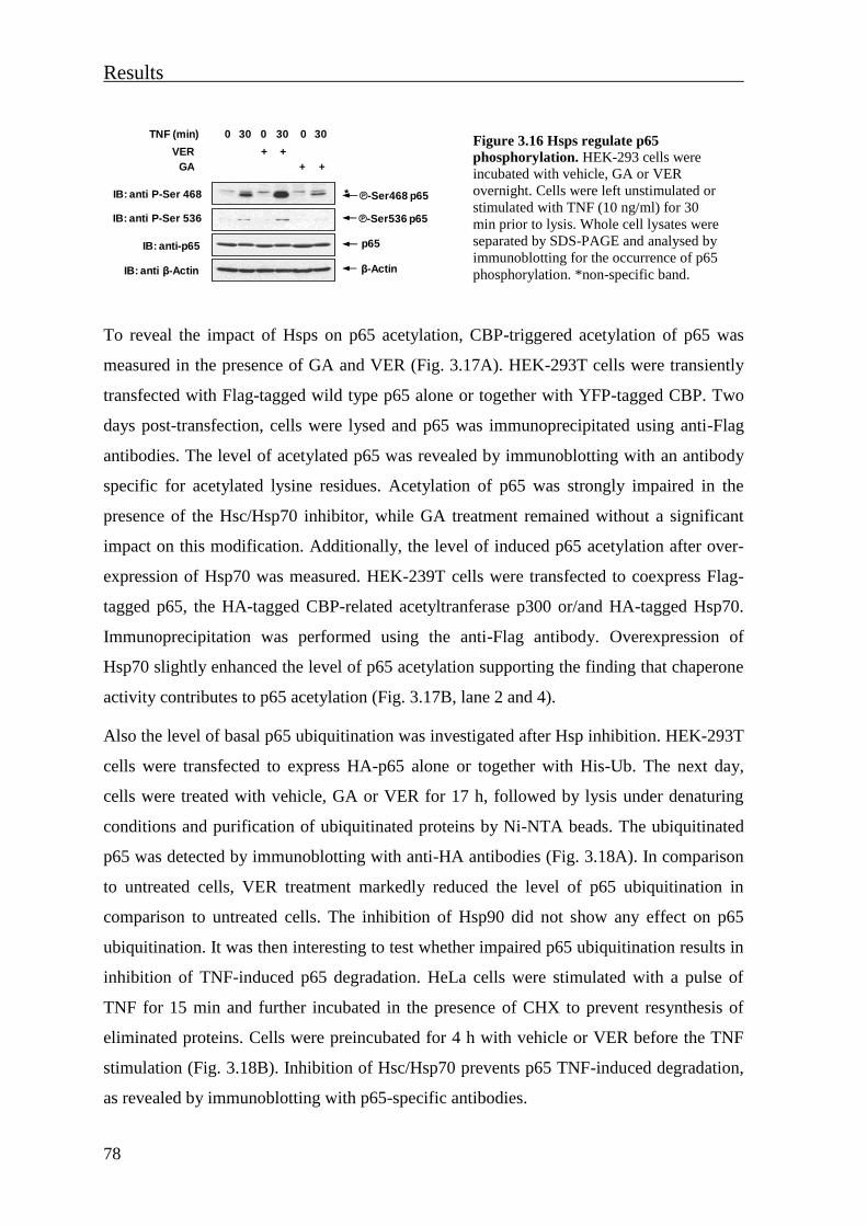
Results
78
Figure 3.16 Hsps regulate p65
phosphorylation. HEK-293 cells were
incubated with vehicle, GA or VER
overnight. Cells were left unstimulated or
stimulated with TNF (10 ng/ml) for 30
min prior to lysis. Whole cell lysates were
separated by SDS-PAGE and analysed by
immunoblotting for the occurrence of p65
phosphorylation. *non-specific band.
To reveal the impact of Hsps on p65 acetylation, CBP-triggered acetylation of p65 was
measured in the presence of GA and VER (Fig. 3.17A). HEK-293T cells were transiently
transfected with Flag-tagged wild type p65 alone or together with YFP-tagged CBP. Two
days post-transfection, cells were lysed and p65 was immunoprecipitated using anti-Flag
antibodies. The level of acetylated p65 was revealed by immunoblotting with an antibody
specific for acetylated lysine residues. Acetylation of p65 was strongly impaired in the
presence of the Hsc/Hsp70 inhibitor, while GA treatment remained without a significant
impact on this modification. Additionally, the level of induced p65 acetylation after over-
expression of Hsp70 was measured. HEK-239T cells were transfected to coexpress Flag-
tagged p65, the HA-tagged CBP-related acetyltranferase p300 or/and HA-tagged Hsp70.
Immunoprecipitation was performed using the anti-Flag antibody. Overexpression of
Hsp70 slightly enhanced the level of p65 acetylation supporting the finding that chaperone
activity contributes to p65 acetylation (Fig. 3.17B, lane 2 and 4).
Also the level of basal p65 ubiquitination was investigated after Hsp inhibition. HEK-293T
cells were transfected to express HA-p65 alone or together with His-Ub. The next day,
cells were treated with vehicle, GA or VER for 17 h, followed by lysis under denaturing
conditions and purification of ubiquitinated proteins by Ni-NTA beads. The ubiquitinated
p65 was detected by immunoblotting with anti-HA antibodies (Fig. 3.18A). In comparison
to untreated cells, VER treatment markedly reduced the level of p65 ubiquitination in
comparison to untreated cells. The inhibition of Hsp90 did not show any effect on p65
ubiquitination. It was then interesting to test whether impaired p65 ubiquitination results in
inhibition of TNF-induced p65 degradation. HeLa cells were stimulated with a pulse of
TNF for 15 min and further incubated in the presence of CHX to prevent resynthesis of
eliminated proteins. Cells were preincubated for 4 h with vehicle or VER before the TNF
stimulation (Fig. 3.18B). Inhibition of Hsc/Hsp70 prevents p65 TNF-induced degradation,
as revealed by immunoblotting with p65-specific antibodies.
TNF (min) 30
GA
30
++
IB: anti-p65 p65
IB: anti P-Ser 468
IB: anti P-Ser 536
IB: anti β-Actin β-Actin
++
30
VER
℗-Ser536 p65
℗-Ser468 p65*
000
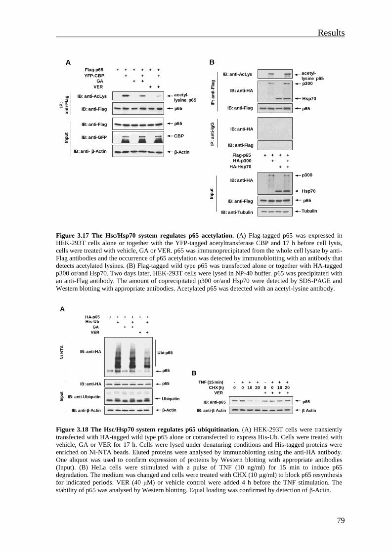
Results
79
Figure 3.17 The Hsc/Hsp70 system regulates p65 acetylation. (A) Flag-tagged p65 was expressed in
HEK-293T cells alone or together with the YFP-tagged acetyltransferase CBP and 17 h before cell lysis,
cells were treated with vehicle, GA or VER. p65 was immunoprecipitated from the whole cell lysate by anti-
Flag antibodies and the occurrence of p65 acetylation was detected by immunoblotting with an antibody that
detects acetylated lysines. (B) Flag-tagged wild type p65 was transfected alone or together with HA-tagged
p300 or/and Hsp70. Two days later, HEK-293T cells were lysed in NP-40 buffer. p65 was precipitated with
an anti-Flag antibody. The amount of coprecipitated p300 or/and Hsp70 were detected by SDS-PAGE and
Western blotting with appropriate antibodies. Acetylated p65 was detected with an acetyl-lysine antibody.
Figure 3.18 The Hsc/Hsp70 system regulates p65 ubiquitination. (A) HEK-293T cells were transiently
transfected with HA-tagged wild type p65 alone or cotransfected to express His-Ub. Cells were treated with
vehicle, GA or VER for 17 h. Cells were lysed under denaturing conditions and His-tagged proteins were
enriched on Ni-NTA beads. Eluted proteins were analysed by immunoblotting using the anti-HA antibody.
One aliquot was used to confirm expression of proteins by Western blotting with appropriate antibodies
(Input). (B) HeLa cells were stimulated with a pulse of TNF (10 ng/ml) for 15 min to induce p65
degradation. The medium was changed and cells were treated with CHX (10 μg/ml) to block p65 resynthesis
for indicated periods. VER (40 μM) or vehicle control were added 4 h before the TNF stimulation. The
stability of p65 was analysed by Western blotting. Equal loading was confirmed by detection of β-Actin.
Flag-p65
YFP-CBP
+
+
+
+ +
++ ++
+ +GA
++VER
IB: anti-Flag
IB: anti-AcLys
IP:
an
ti-F
lag
IB: anti-Flag
IB: anti-GFP
Inp
ut
acetyl-lysine p65
β-Actin
p65
p65
CBP
IB: anti- β-Actin
A
Flag-p65
HA-p300
HA-Hsp70
+ + + +
+
+
p300
Tubulin
Hsp70
p65
IP:
an
ti-F
lag
Inp
ut
IB: anti-Tubulin
+
IB: anti-Flag
IB: anti-HA
IB: anti-FlagIP:
an
ti-I
gG
p65
p300
Hsp70
IB: anti-HA
IB: anti-Flag
+
IB: anti-HA
IB: anti-AcLys acetyl-lysine p65
B
HA-p65His-Ub
+
+
+
+ +
++ ++
+ +GA
++VER
p65
β-Actin
p65
Ubiquitin
IB: anti-HA
Ni-
NT
AIn
pu
t
IB: anti-HA
IB: anti-β-Actin
IB: anti-Ubiquitin
Ubi-p65
A
- ++ + + +TNF (15 min)
CHX (h) 0 0 20 0 10 20
VER
IB: anti-p65 p65
IB: anti-β Actin β Actin
+
10
-
0
+ ++ +
B

Results
80
In summary, these experiments show that variety of PTMs acting on the NF-κB p65
protein can be modulated upon interference with the chaperone function of Hsc/Hsp70
proteins.
3.2.4.2 Hsps modulate p65-mediated gene expression
To test whether Hsps regulate the transcriptional activity of p65, reporter gene assays were
performed. HEK-293T cells were transfected with a luciferase reporter gene controlled by
3 κB sites along with a p65 expression vector. The next day, cells were further grown in
the absence or presence of Hsp inhibitors for 17 h and reporter gene activity was measured.
The presence of GA only slightly diminished p65-triggered luciferase activity, while VER
treatment significantly impaired p65-driven gene expression (Fig. 3.19A). Alternatively,
luciferase gene expression was induced by Gal4-p65 fusion proteins that are independent
of NF-κB DNA binding, as the contact with DNA is mediated by Gal4. Again, Gal4-p65
induced luciferase activity was significantly impaired in the presence of the Hsc/Hsp70
inhibitor, showing that their chaperone activity contributes to p65-mediated transcription
(Fig. 3.19B).
Figure 3.19 Hsc/Hsp70 chaperones are important for p65-mediated transcription. (A) HEK-293T
cells were transfected with NF-κB-dependent luciferase reporter gene and an expression plasmid for HA-
tagged wild type p65. Cells were treated with vehicle, GA (1 μM) or VER (40 μM) for one day, followed
by cell lysis and determination of luciferase activity (upper panel) and protein expression (lower panel).
Error bars show standard error of the mean (SEM) from three independent experiments. (B) HEK-293T
cells were transfected with a luciferase reporter gene controlled by five Gal4-binding sites along with
expression vectors for the DNA-binding domain of Gal4 fused to p65 (Gal-p65). Cells were treated with
Hsp inhibitors and analysed for luciferase activity and protein expression as in (A). Error bars represent
SEM from three independent experiments.
Gal4-p65 GA
VER
+ + ++
+
+
+
IB: anti-Gal4
IB: anti-Tubulin Tubulin
Gal4
Gal4-p65
0
50
100
150
200
250
300
350
400
Re
lati
ve
ind
uc
tio
n o
f (G
al4
)5-
luc
ife
ras
e
B
HA-p65GA
++
++
p65IB: anti-HA
IB: anti-β-Actin β-Actin
VER
+
+ +
0
50
100
150
200
250
300
350
400
450
Re
lati
ve
ind
uc
tio
n o
f (κ
B)3
-lu
cif
era
se
A
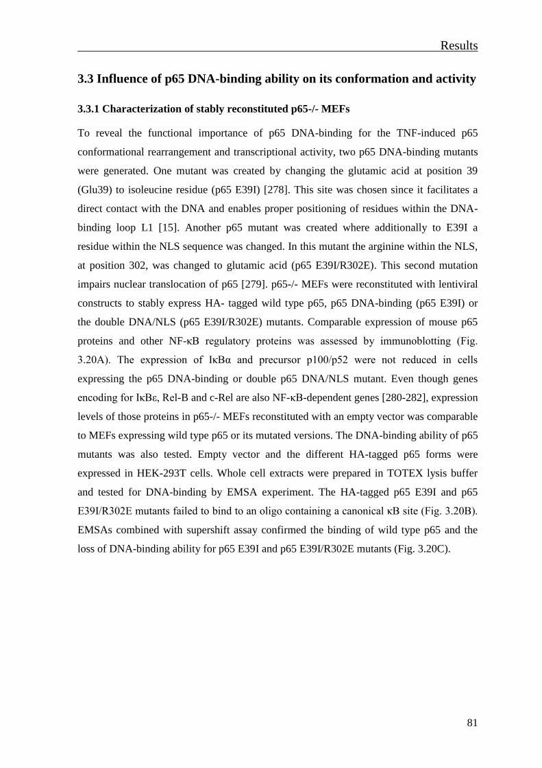
Results
81
3.3 Influence of p65 DNA-binding ability on its conformation and activity
3.3.1 Characterization of stably reconstituted p65-/- MEFs
To reveal the functional importance of p65 DNA-binding for the TNF-induced p65
conformational rearrangement and transcriptional activity, two p65 DNA-binding mutants
were generated. One mutant was created by changing the glutamic acid at position 39
(Glu39) to isoleucine residue (p65 E39I) [278]. This site was chosen since it facilitates a
direct contact with the DNA and enables proper positioning of residues within the DNA-
binding loop L1 [15]. Another p65 mutant was created where additionally to E39I a
residue within the NLS sequence was changed. In this mutant the arginine within the NLS,
at position 302, was changed to glutamic acid (p65 E39I/R302E). This second mutation
impairs nuclear translocation of p65 [279]. p65-/- MEFs were reconstituted with lentiviral
constructs to stably express HA- tagged wild type p65, p65 DNA-binding (p65 E39I) or
the double DNA/NLS (p65 E39I/R302E) mutants. Comparable expression of mouse p65
proteins and other NF-κB regulatory proteins was assessed by immunoblotting (Fig.
3.20A). The expression of IκBα and precursor p100/p52 were not reduced in cells
expressing the p65 DNA-binding or double p65 DNA/NLS mutant. Even though genes
encoding for IκBε, Rel-B and c-Rel are also NF-κB-dependent genes [280-282], expression
levels of those proteins in p65-/- MEFs reconstituted with an empty vector was comparable
to MEFs expressing wild type p65 or its mutated versions. The DNA-binding ability of p65
mutants was also tested. Empty vector and the different HA-tagged p65 forms were
expressed in HEK-293T cells. Whole cell extracts were prepared in TOTEX lysis buffer
and tested for DNA-binding by EMSA experiment. The HA-tagged p65 E39I and p65
E39I/R302E mutants failed to bind to an oligo containing a canonical κB site (Fig. 3.20B).
EMSAs combined with supershift assay confirmed the binding of wild type p65 and the
loss of DNA-binding ability for p65 E39I and p65 E39I/R302E mutants (Fig. 3.20C).

Results
82
Figure 3.20 Characterization of stably reconstituted p65-/- MEFs. (A) The indicated murine HA-tagged
wild type p65 and its mutated versions were stably expressed after lentiviral infection. Expression of p65 and
other NF-κB regulatory proteins were assessed by immunoblotting as indicated. (B) HA-tagged wild type
p65, p65 E39I or p65 E39I/R302E were expressed in HEK-293T cells. Whole cell extracts were prepared
and tested by EMSA experiment for the ability to bind to a κB-oligonucleotide. Upper panel: an
autoradiograph with the NF-κB/DNA binding complex, indicated by an arrow. Lower panel: an immunoblot
with anti-HA antibody ensuring the correct expression of HA-tagged proteins. (C) p65-/- MEFs transduced
with lentiviruses to stably express similar levels of HA-tagged p65 and its indicated mutated forms were left
untreated or were treated with TNF (10 ng/ml) for 30 min. Total cell extracts were prepared. A p65-specific
antibody used for supershift assay was added as indicated and the DNA binding ability of p65 versions was
assayed by EMSA. After separation of free and NF-κB-bound oligos by native gel electrophoresis, the gel
was dried and autoradiographed (lower panel). The upper panel represents a control Western blot ensuring
that comparable amounts of protein were contained in the EMSA samples. Ab, antibody.
3.3.2 DNA-binding of p65 affects the kinetics of its nuclear export and Ser468-
phosphorylation
Kinetics of NF-κB nuclear translocation was tested in stably reconstituted p65-/- MEFs.
The reconstituted MEFs were stimulated with TNF for 0, 30, 60 or 120 min and the
kinetics of the nuclear import and export was analysed by IF experiments (Fig. 3.21 A-C).
In cells reconstituted with wild type p65, p65 was detected inside the nucleus after 30 min
TNF stimulation and the nuclear export was visible after 60 min post-stimulation (Fig.
3.21A). Unexpectedly, TNF-activated p65 E39I mutants retained inside the nucleus even
120 min after stimulation (Fig. 3.21B). IF detected a slight nuclear translocation of the
empty vector
HA-p65
HA-p65 E39I
+
+
+
IB: anti- p65
IB: anti- IκBα
IB: anti- IκBε
IB: anti- IκBβ
IB: anti- p105/p50
IB: anti- p100/p52
IB: anti- β-Actin
p65
IκBα
IκBβ
RelB
p105
p50
HA-p65 E39I/R302E +
IB: anti- HA p65
IκBε
p100
p52
IB: anti- RelB
c-RelIB: anti- c-Rel
β-Actin
A+
+
+
DNA/p65
IB: anti- anti-HA p65
EMSA
empty vector
HA-p65
HA-p65 E39I
+
B
HA-p65 E39I/R302E
C+
+
+
empty vector
HA-p65
HA-p65 E39I
+
p65IB: anti- anti-HA
HA-p65 E39I/R302E
DNA/ NF-κB
EMSA
empty vector
HA-p65
HA-p65 E39I
+
+ + +
+ +
+
+
TNF (30 min)
anti-p65 Ab
+ +
+
++
+
Supershift
β-ActinIB: anti- β Actin
++ +
+
+ + +HA-p65 E39I/R302E

Results
83
double mutant p65 E39I/R302E upon TNF stimulation which probably occurs by a “piggy
back” mechanism (Fig. 3.23C). Additionally, the kinetics of the TNF-induced p65 nuclear
translocation and p65 phosphorylation was tested by a Western blot analysis. Reconstituted
MEFs were stimulated with TNF for indicated periods. Immunoblotting of nuclear extracts
revealed prolonged nuclear localization of p65 DNA-mutants in comparison to wild type
p65. Upon TNF treatment, phosphorylation of wild type at Ser468 reaches its maximum at
15 min, followed by a gradual decrease of the Ser468-phosphorylation signal.
Interestingly, in cells expressing the p65 DNA-binding mutant, Ser468 phosphorylation
reached its maximum at 15 min TNF stimulation and did not change during the following
time course of stimulation (Fig. 3.21D). Kinetics of Ser563 phosphorylation remained
unchanged. All together, results presented in Fig. 3.21 imply that the p65 DNA-binding is
involved in regulation of the p65 nuclear export and affects phosphorylation at Ser468.
Hoechst Mergeanti-p65
MEF p65 E39I reconstitutedB
Hoechst Merge
TNF 0 min
anti-p65
TNF 30 min
TNF 60 min
TNF 120 min
ctrl
MEF p65 reconstitutedA
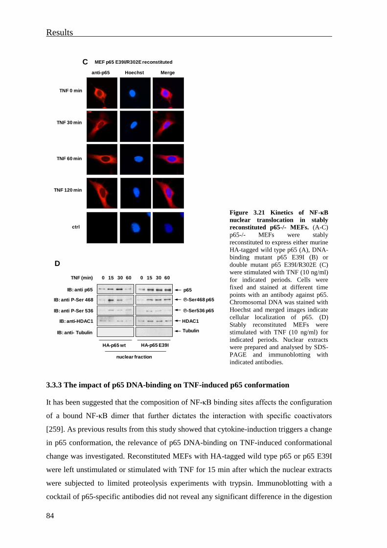
Results
84
Figure 3.21 Kinetics of NF-κB
nuclear translocation in stably
reconstituted p65-/- MEFs. (A-C)
p65-/- MEFs were stably
reconstituted to express either murine
HA-tagged wild type p65 (A), DNA-
binding mutant p65 E39I (B) or
double mutant p65 E39I/R302E (C)
were stimulated with TNF (10 ng/ml)
for indicated periods. Cells were
fixed and stained at different time
points with an antibody against p65.
Chromosomal DNA was stained with
Hoechst and merged images indicate
cellular localization of p65. (D)
Stably reconstituted MEFs were
stimulated with TNF (10 ng/ml) for
indicated periods. Nuclear extracts
were prepared and analysed by SDS-
PAGE and immunoblotting with
indicated antibodies.
3.3.3 The impact of p65 DNA-binding on TNF-induced p65 conformation
It has been suggested that the composition of NF-κB binding sites affects the configuration
of a bound NF-κB dimer that further dictates the interaction with specific coactivators
[259]. As previous results from this study showed that cytokine-induction triggers a change
in p65 conformation, the relevance of p65 DNA-binding on TNF-induced conformational
change was investigated. Reconstituted MEFs with HA-tagged wild type p65 or p65 E39I
were left unstimulated or stimulated with TNF for 15 min after which the nuclear extracts
were subjected to limited proteolysis experiments with trypsin. Immunoblotting with a
cocktail of p65-specific antibodies did not reveal any significant difference in the digestion
TNF 0 min
TNF 30 min
TNF 60 min
TNF 120 min
ctrl
MEF p65 E39I/R302E reconstituted
Hoechst Mergeanti-p65
C
TNF (min) 15 30 600
HA-p65 wt
IB: anti-HDAC1
IB: anti- Tubulin
IB: anti P-Ser 468
IB: anti P-Ser 536
15 30 600
HA-p65 E39I
HDAC1
Tubulin
℗-Ser468 p65
℗-Ser536 p65
IB: anti p65 p65
nuclear fraction
D
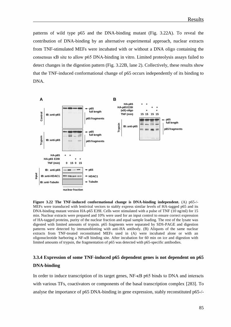
Results
85
patterns of wild type p65 and the DNA-binding mutant (Fig. 3.22A). To reveal the
contribution of DNA-binding by an alternative experimental approach, nuclear extracts
from TNF-stimulated MEFs were incubated with or without a DNA oligo containing the
consensus κB site to allow p65 DNA-binding in vitro. Limited proteolysis assays failed to
detect changes in the digestion pattern (Fig. 3.22B, lane 2). Collectively, these results show
that the TNF-induced conformational change of p65 occurs independently of its binding to
DNA.
Figure 3.22 The TNF-induced conformational change is DNA-binding independent. (A) p65-/-
MEFs were transduced with lentiviral vectors to stably express similar levels of HA-tagged p65 and its
DNA-binding mutant version HA-p65 E39I. Cells were stimulated with a pulse of TNF (10 ng/ml) for 15
min. Nuclear extracts were prepared and 10% were used for an input control to ensure correct expression
of HA-tagged proteins, purity of the nuclear fraction and equal sample loading. The rest of the lysate was
digested with limited amounts of trypsin. p65 fragments were separated by SDS-PAGE and digestion
patterns were detected by immunoblotting with anti-HA antibody. (B) Aliquots of the same nuclear
extracts from TNF-treated reconstituted MEFs used in (A) were incubated alone or with an
oligonucleotide harboring a NF-κB binding site. After incubation for 60 min on ice and digestion with
limited amounts of trypsin, the fragmentation of p65 was detected with p65-specific antibodies.
3.3.4 Expression of some TNF-induced p65 dependent genes is not dependent on p65
DNA-binding
In order to induce transcription of its target genes, NF-κB p65 binds to DNA and interacts
with various TFs, coactivators or components of the basal transcription complex [283]. To
analyse the importance of p65 DNA-binding in gene expression, stably reconstituted p65-/-
HDAC1
p65 full length
IB: anti-p65
IB: anti-p65
p65 full length
Try
ps
inC
on
tro
l
p65 fragments
p65 fragments
HA-p65
HA-p65 E39I
TNF (min)
p65
Tubulin
15 15
+ +
+ +
IB: anti-p65
IB: anti-HDAC1
IB: anti-Tubulin
Inp
ut
0 0
nuclear fraction
A
15 15
+ +
+++ +
TNF (min)
HA-p65
(κB)-oligo
1515
p65 full length
IB: anti-p65
Try
ps
in
p65 fragments
B
HA-p65 E39I
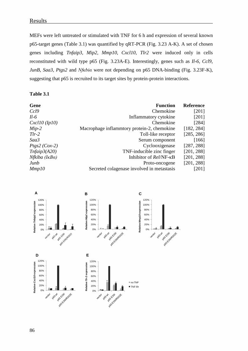
Results
86
MEFs were left untreated or stimulated with TNF for 6 h and expression of several known
p65-target genes (Table 3.1) was quantified by qRT-PCR (Fig. 3.23 A-K). A set of chosen
genes including Tnfaip3, Mip2, Mmp10, Cxcl10, Tlr2 were induced only in cells
reconstituted with wild type p65 (Fig. 3.23A-E). Interestingly, genes such as Il-6, Ccl9,
JunB, Saa3, Ptgs2 and Nfκbiα were not depending on p65 DNA-binding (Fig. 3.23F-K),
suggesting that p65 is recruited to its target sites by protein-protein interactions.
Table 3.1
Gene Function Reference
Ccl9 Chemokine [201]
Il-6 Inflammatory cytokine [201]
Cxcl10 (Ip10) Chemokine [284]
Mip-2 Macrophage inflammtory protein-2, chemokine [182, 284]
Tlr-2 Toll-like receptor [285, 286]
Saa3 Serum component [166]
Ptgs2 (Cox-2) Cyclooxigenase [287, 288]
Tnfaip3(A20) TNF-inducible zinc finger [201, 288]
Nfkiba (IκBα) Inhibitor of Rel/NF-κB [201, 288]
Junb Proto-oncogene [201, 288]
Mmp10 Secreted colagenase involved in metastasis [201]
0%
20%
40%
60%
80%
100%
120%
Re
lati
ve
Mm
p1
0e
xp
res
sio
n
C
0%
20%
40%
60%
80%
100%
120%
Re
lati
ve
Cx
cl1
0 e
xp
res
sio
n
D
0%
20%
40%
60%
80%
100%
120%
Re
lati
ve
Tlr
-2e
xp
res
sio
n
no TNF
TNF 6h
E
A B
0%
20%
40%
60%
80%
100%
120%
Re
lati
ve
Tn
faip
3e
xp
res
sio
n
0%
20%
40%
60%
80%
100%
120%
Re
lati
ve
Mip
2e
xp
res
sio
n
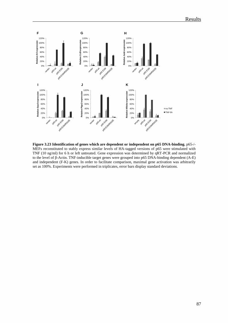
Results
87
Figure 3.23 Identification of genes which are dependent or independent on p65 DNA-binding. p65-/-
MEFs reconstituted to stably express similar levels of HA-tagged versions of p65 were stimulated with
TNF (10 ng/ml) for 6 h or left untreated. Gene expression was determined by qRT-PCR and normalized
to the level of β-Actin. TNF-inducible target genes were grouped into p65 DNA-binding dependent (A-E)
and independent (F-K) genes. In order to facilitate comparison, maximal gene activation was arbitrarily
set as 100%. Experiments were performed in triplicates, error bars display standard deviations.
0%
20%
40%
60%
80%
100%
120%R
ela
tiv
e Il
-6e
xp
res
sio
n
F
0%
20%
40%
60%
80%
100%
120%
Re
lati
ve
Cc
l9e
xp
res
sio
n
G
0%
20%
40%
60%
80%
100%
120%
Re
lati
ve
Ju
nb
ex
pre
ss
ion
H
I
0%
20%
40%
60%
80%
100%
120%
Re
lati
ve
Ptg
s2
ex
pre
ss
ion
J
0%
20%
40%
60%
80%
100%
120%
Re
lati
ve
Sa
a3
ex
pre
ss
ion
K
0%
20%
40%
60%
80%
100%
120%
Re
lati
ve
Nfiκ
bα
ex
pre
ss
ion
no TNF
TNF 6h


Discussion
89
4. Discussion
4.1 Phosphorylation is important for the conformation of the active
nuclear p65
The NF-κB transcription factor is involved in the control of a large number of cellular
processes, such as immune and inflammatory responses, developmental processes, cell
growth and apoptosis [1, 289]. The cellular localization, activity and termination of NF-κB
activity must be carefully controlled in response to intrinsic and environmental stimuli.
Many aspects of these processes rely on changes in the folding state of the transcriptionally
highly active NF-κB p65 subunit. Inside the nucleus, the function of NF-κB is greatly
regulated by various stimulus-induced PTMs of the p65 subunit. Early studies on p65
showed that the TAD of p65 is binding its own RHD through intramolecular associations
[154, 290]. Stable physical interaction between the p65 N- and C-termini was detected by
GST pull-down assays. Protein kinase A-mediated phosphorylation of Ser276 abrogated
this interaction, implicating the role of phosphorylation in the regulation of p65
conformation [154]. A previous study from the Schmitz lab showed that mutation of a
single phosphorylation site in p65 (Ser468) largely prevents the association with
components of a multimeric ubiquitin ligase complex and subsequent p65 ubiquitination,
suggesting that this mutation alters the overall conformation of NF-κB p65 [182]. In an
effort to measure the conformational changes of p65 mediated by site-specific
phosphorylation, limited proteolysis assays were performed using cells expressing either
the wild type p65 or individual p65 Ser276, Ser468 and Ser536 mutated forms.
Interestingly, proteolysis resulted in a different fragmentation of each p65 version (Fig.
3.2B). These results suggest that the phosphorylation status of individual serine residues
affects the overall conformation of p65, indicating a highly dynamic behavior of the
protein. Since NF-B p65 undergoes simultaneous phosphorylation at different sites upon
stimulation, these results raise the possibility that p65 can adopt more than one
conformation. It remains to be elucidated whether there are only two or even more
different conformations. It has been suggested that phosphorylation at Ser276 indirectly
regulates the DNA-binding affinity of p65 [24] and its dimerisation with other NF-κB
monomers [4]. Also, site-specific phosphorylation of p65 regulates p65 affinity to

Discussion
90
modifying enzymes and/or transcriptional cofactors [154, 164, 165, 183, 291]. Thereby, in
agreement with the proteolysis assays, all these results could be explained by a highly
dynamic flexibility of p65 that allows p65 to respond under diverse conditions. The
intrinsic disorder of the C-terminal portion also contributes to the dynamic behavior of the
p65 protein (Fig. 3.1).
Conformational changes induced by a site-specific or multiple-site phosphorylations were
detected for various TFs such as p53 [263], the glucocorticoid receptor (GR) [292], signal
transducer and activator of transcription 1 (STAT1) [293, 294], FoxO1 (Forkhead box O1)
[295] and Elk-1 (ETS domain-containing protein1) [296]. For example, a certain
phosphorylation of FoxO1 within its DBD induces a conformational change that blocks
FoxO1 DNA-binding [295]. Phosphorylation of the Elk-1 C-terminal TAD induces a
change in the intramolecular interaction between the C-terminal TAD and the N-terminal
DBD that accompanies the stimulation of DNA binding [296]. The exact contact sites
between the N- and C-terminal halves of p65 remain to be identified. As the intrinsically
disordered C-terminal half of the p65 protein does not allow structural data for the entire
p65 protein [32], biochemical experiments with purified parts of p65 could determine
contact sites or protein regions that are responsible for intramolecular interaction.
Limited proteolysis experiments with comparable amounts of TNF or IL-1-induced p65
showed no significant cytokine-inducible changes for cytosolic p65, while nuclear p65
displayed a major conformational rearrangement (Fig. 3.5A and 3.6A). Treatment with the
phosphatase inhibitor calyculin A in the absence of cytokine stimulation was sufficient to
cause a p65 rearrangement highly similar to cytokine-induced refolding, suggesting that
phosphorylations are major contributors of p65 rearrangement (Fig. 3.7A). The limited
proteolysis experiments with p65-/- MEFs stably reconstituted with non-phosphorylatable
Ser468 or Ser536 p65 mutants did not show a significant effect of those two phosphosites
on p65 conformation (Fig. 3.7B). This indicates that phosphorylation at Ser 468 or 536
alone is not sufficient to induce a conformational change, but other modifications need to
occur as well. Besides phosphorylation, other induced PTMs such as acetylation [297]
methylation [298], ubiquitination [299, 300], sumoylation [301] and prolyl-isomerisation
[164, 263] can affect protein conformation. The phosphorylation site(s) responsible for a
cytokine-induced conformational switch needs to be revealed in the future by identifying
p65 mutants refractory to the cytokine-induced refolding. As TNF and IL-1 induce a

Discussion
91
variety of different PTMs [164, 170, 182, 214, 302, 303], their contribution to refolding of
nuclear p65 should be investigated. It is presently also completely unclear whether the
other NF-B activating stimuli such as LPS, UV light and genotoxic stress induce the same
conformational switch or possibly allow alternative configurations. It has been shown that
TNF stimulation induces the COMMD1-mediated p65 ubiquitination through Ser468
phosphorylation [182], while the aspirin-induced COMMD1-mediated p65 ubiquitination
occurs independently of Ser468-phosphorylation [184]. Cell stimulations with other NF-κB
activating stimuli followed by a limited proteolysis should be performed in order to reveal
whether a configuration of an active nuclear p65 is stimulus-specific.
In this study limited proteolysis assays were used to reveal conformational features of p65.
Unlike other methods it allows the analysis of protein conformations using low protein
concentrations. Protein regions with a high structural flexibility, such as loops and hinges,
are usually most susceptible to proteolytic cleavage [304, 305]. As many phosphorylation,
methylation, acetylation and ubiquitination sites predominantly locate in flexible,
disordered regions [306-308], the outcome of a limited proteolytic protein can be
influenced by PTMs. Of note, the modification sites mutated in this study did not change
the predicted cleavage by trypsin or thermolysin. Also protein-protein interactions could
affect protease susceptibility by masking the cleavage sites. The kinetics of a TNF- or IL-
1-induced p65 refolding showed that characteristic conformational change of nuclear p65
is not transient, at least for the time points that were measured (Fig. 3.6B). It still can be
detected at time points when the first wave of NF-κB activity is terminated by newly
synthesized IκBα [69]. Thus, it would be interesting to investigate whether the changed
digestion pattern disappears at later time points. To investigate the role of IκBα in p65
refolding in more detail, the kinetics of cytokine-induced p65 refolding should be
measured in IκBα-/- cells or alternatively in cells with silenced IκBα. A previous study
showed that cytokine stimulation induces the phosphorylation at Thr254 and leads to the
binding of the prolyl isomerase Pin-1 to the phospho-Thr254/Pro motif in p65 [164]. Ryo
et al. further showed that the subsequent phosphorylation-dependent isomerisation of p65
by Pin1 inhibits p65 binding to IκBα and results in increased nuclear accumulation and
protein stability of p65. Thereby the detected rearrangement of p65 could be influenced by
interaction with Pin1. The treatment of cells with a Pin1 inhibitor did not show any effect
on TNF-induced p65 rearrangement (data not shown). Alternatively, the kinetics of a

Discussion
92
cytokine-induced p65 refolding could be performed in Pin1-deficient cells. Further
identification of digested fragments, especially p65* double band, by MS would give
information about the trypsin cleavage sites of p65 which become accessible after
refolding. Also, this could identify functional subregions within p65.
Immunoprecipitation of nuclear extracts from TNF stimulated cells with a set of p65-
specific antibodies allowed the identification of a conformation-specific p65 antibody
(EP2161Y) (Fig. 3.8B). Interestingly, cytokine stimulation leads to the exposure of a
Ser468 surrounding region, emphasizing the regulatory role of the TAD2 subdomain
containing Ser468 as a dynamic region within nuclear p65. It would be desirable to
generate more conformation-specific antibodies, as they were successfully developed for
the p53 protein [309]. Identification of antibodies that recognize constitutively exposed
epitopes (e.g. IMG-5915A) and the epitope that become hidden after the TNF-stimulation
would contribute to a better characterization of the observed cytokine-induced
rearrangement (Fig. 4.1). The conformation-specific p53 antibody PAb240 that recognizes
the mutant-specific epitope was able to immunoprecipitate p53 bound to Hsp70 or MDM2
(Mouse double minute 2 homolog), while the antibodies that were able to recognize the
wild type-specific epitope did not [241, 300]. Thereby, conformation-specific p65
antibodies could be further used for the identification and characterization of
conformation-specific p65 protein-protein interactions as well as interactions of NF-κB
with chromatin and DNA.
Figure 4.1 Conformation-specific antibodies
to monitor cytokine-induced rearrangement
of p65. IMG-5915A recognizes a constitutively
exposed epitope, EP2161Y preferentially
recognizes TNF-inducible exposed epitope. An
hypothetical antibody (?) recognizing the
epitope that would be concealed after the TNF
stimulation.
The detailed role of site-specific PTMs (e.g. phosphorylation) in the function of individual
proteins remains poorly understood. Structural information is particularly limited, as it is
difficult to obtain sufficient amounts of purified protein in a specific modification state.
Several methods could be further employed in order to examine protein structural changes
induced by phosphorylation. Atomic-resolution structures for the phosphorylated proteins
can be determined by X-ray crystallography. For phosphorylated peptides and small
TNFαp65
?EP2161Y
p65
IMG-5915A IMG-5915A

Discussion
93
peptides in solution NMR is the method of choice. Several NMR studies of
phosphorylation-induced conformational changes have been reported [262, 292, 310-313].
Electron paramagnetic resonance (EPR) and circular dichroism (CD) experiments can also
provide information about the conformational change due to the phosphorylation, but do
not provide atomic details. Computational modeling studies can be helpful to enlighten the
principles and mechanisms of PTMs [262]. Size exclusion chromatography in combination
with analytical ultracentrifugation can be also applied to characterize hydrodynamic and
conformational changes in proteins [314, 315]. Hydrogen exchange coupled to MS has
become a valuable analytical tool for the study of protein dynamics and can provide
information on the protein structure [316].
4.2 Phosphorylation-induced p65 refolding regulates p65 ubiquitination
The phosphorylation-dependent dynamic behavior of p65 could serve to explain the
differences in the level and type of ubiquitination of wild type p65 and its phospho-mutants
(Fig. 3.3 and 3.4). Mutation of a single phosphorylation site in the disordered C-terminal
TAD resulted in a strongly decreased p65 ubiquitination in comparison to wild type p65.
These results suggest that a serine to alanine mutation of C-terminal residues could induce
an intramolecular rearrangement that affects the exposure of ubiquitination sites. On the
other hand, it cannot be excluded that phosphorylation at Ser468 and Ser536 induces
exposure of docking motifs through which the interaction with the Cul2/COMMD1
complex occurs, facilitating the SOCS1-p65 interaction [182, 183, 210]. Also, it can be
possible that the binding of a Cul2/COMMD1 complex itself, induces the change in p65
conformation. A phosphorylation-dependent control of ubiquitination was also observed
for the p53 protein. Binding of the E3 ligase MDM2 to the intrinsically disordered p53 N-
terminus induces a conformational change in wild type p53, as revealed by the p53
immunoprecipitation with a mutant conformation-specific p53 antibody [300].
Phosphorylation of N-terminal Ser20 contributes to disruption of p53-MDM2 interaction,
especially in combination with Ser15 and Thr18 phosphorylation [317-319]. Still, mutation
of Ser15 to alanine had little effect on the ubiquitination and degradation of p53. In
contrast, replacement of Ser20 with alanine makes p53 highly sensitive to MDM2-
mediated turnover [320]. The cross-talk between the p65 C-terminal phosphorylation and
ubiquitination requires further detailed investigation. Serine to alanine mutation of Ser276
led to the p65 ubiquitination, while the permanent phosphorylation inhibited p65
Discussion
94
ubiquitination. In agreement with a Ser276 phosphorylation-induced p65 refolding, it is
possible that mutation to alanine makes ubiquitin acceptor sites available for modification.
This result is in agreement with a previous study where it has been shown that Pim-1-
mediated phosphorylation at Ser276 prevents SOCS1-dependent ubiquitination and
subsequent degradation [321]. Lysines are also subjected to acetylation and methylation
[200, 201, 214, 218]. The observed effect of phosphorylation at Ser276 on p65
ubiquitination could be a consequence of a p65 interaction with acetyl- or
methyltransferase. It is well documented that Ser276-phosphorylation stabilizes the
interaction with the CBP/p300 [154, 322]. This interaction could induce further lysine
modifications and/or protects the other ubiquitin acceptor lysines, leading to changes of
p65 conformation. The highly dynamic p65 flexibility would also help to explain previous
experiments that characterized a p65 variant mutated in four out of seven ubiquitinated
lysines. While the exchange of four lysines largely prevented p65 ubiquitination, mutation
of further lysines paradoxically further increased p65 ubiquitination [201], a finding that
may reflect large conformational changes leading to the surface exposure of additional
lysines.
4.3 Phosphorylation-induced conformational changes of p65 influence its
association with other proteins
Coimmunoprecipitation experiments showed that a conformational change induced by
Ser468 phosphorylation elicits significant effects on p65 protein-protein interactions (Fig.
3.9 and 3.12). This supports the finding that Ser486 and its surrounding sequence in the
TAD2 subdomain are of special relevance for a configuration of active nuclear p65.
Phosphorylation-induced conformational changes in proteins are largely driven by
electrostatic perturbations that change the protein backbone structure of a surrounding
region [262] and induce the exposure of otherwise buried enzyme docking sites [323]. The
region surrounding the Ser468 (Ser467 for murine p65) contains two ΨXXΨΨ motifs (Ψ-
hydrophobic residue, X-any residue; 449
LGALL453
and 473
FQQLL477
, respectively for
murine p65) which are a generalization of a LXXLL motif that is known to mediate
protein-protein interactions in transcriptional regulation [324]. Recently it has been shown
that hydrophobic residues from those motifs create a platform for the interaction with the
CBP/p300 TAZ1 (transcriptional adaptor zinc binding 1) domain allowing the disorder-to-
order transition of the unstructured p65 TAD2 subdomain [180]. Isothermal calorimetric
Discussion
95
(ITC) experiments with p65 TAD2 fragments showed that a phospho-mimetic p65 S467D
fragment binds TAZ1 with a slightly higher affinity than wild type p65 TAD2, suggesting
that the increased negative charge of phosphoserine is not essential for the interaction but
would enhance CBP TAZ1 binding even further over nonphosphorylated p65. GST pull-
down assay revealed that the TAD1 subdomain of p65 provides the main region for
binding of the cofactor GCN5 [183]. Still, the individual or combined mutation of all six
serines in TAD1, including the Ser536, did not abrogate the inducible binding between p65
TAD and GCN5. Interestingly, the mutation of Ser468 to alanine affected the interaction
between p65 and GCN5 in response to either calyculin A or TNF treatment, suggesting
that phosphorylation of p65 at Ser468 increases its binding to GCN5 [183]. Another
LXXLL-like motif (542
FSALL546
, respectively for human p65) is positioned close to the
Ser536. Phosphorylation of Ser536 also has the ability to control protein association, as
exemplified by the increased association of the transcriptional regulator TAFII31 [170].
Future experiments could investigate in more detail the role of phosphorylations on the
exposure of the FXXLL motifs. Also, the impact of ψXXψψ motifs on the interaction with
transcriptional coregulators in general could be tested. Phosphorylation-induced
conformational changes regulating the interaction with transcriptional cofactors have been
also detected for other TFs [292, 325, 326]. Limited proteolysis assays together with CD
and fluorescence emission spectroscopy showed that single-site phosphorylation in
intrinsically disordered N-terminal TAD of the GR is sufficient to induce a functionally
active conformation of GR [292]. This phosphorylation-induced rearrangement of GR
further facilitates interaction with transcriptional coregulators and subsequently GR
transcriptional activity.
This study showed that p65 interaction with Hsp70 and Hsp90 was also influenced by
phosphorylation of Ser468 (Fig. 3.12). Exposed stretches of hydrophobic residues were
also identified as regions through which Hsp70 binds to its substrates [327] and the
conformation of substrate peptides plays a pivotal role in the binding process [328].
Further experiments could be performed with the aim to investigate whether the p65 TAD
ψXXψψ motifs participate in NF-κB p65-Hsp70 interaction. A recent study performed by
Neil Perkins and his colleagues showed that in the absence of p53, p65 was transported
into the mitochondria and recruited to the mitochondrial genome by an interaction between
the Thr505 region of the p65 TAD and Mortalin, a mitochondrial Hsp70 protein [258].
Discussion
96
Mutation of Thr505 to alanine abolished the p65 interaction with Mortalin and inhibited
p65 mitochondrial localization, suggesting that phosphorylation at this site may be required
to promote this interaction. The authors do not exclude the possibility that this can also
happen due to the structural effect of a serine to alanine mutation. While the data presented
here did not reveal the importance of Ser536 phosphorylation on the interaction of p65
with Hsp70 (data not shown), still the impact of Thr505 phosphorylation on the p65/Hsp70
association could be tested in the future. Phosphorylation-dependent binding of Hsp70 to
its substrate was detected for protein kinase C (PKC), Akt/protein kinase B and PKA
[243]. GST pull-down assays with wild type PKC or a kinase variant lacking
phosphorylated amino acids in a C-terminal turn motif revealed that Hsp70 binds to the
unphosphorylated PKC turn motif. This interaction of Hsp70 with PKC allows the re-
phosphorylation of the enzyme and sustains the activated state of protein kinase. All
together, these examples readily illustrate that phosphorylation-mediated conformational
changes of p65 can greatly extend and specify the repertoire of possible protein-protein
interactions. Methods like surface plasmon resonance (SPR) and ITC can be employed on
a series of truncated and mutated purified parts of a p65 to measure the p65 protein-protein
interactions and to understand what regions of the interface are most important for the tight
binding affinity of protein complexes. GST pull-down assays with purified Hsps and
truncated p65 fragments should be performed in order to see whether they directly interact
with p65 or via co-chaperones.
4.4 Chaperones as coregulators of p65 refolding
Hsp70 and Hsp90 are highly abundant cellular proteins that play a dominant role in the
cellular system that controls conformational states and conformational maturation of
proteins [329, 330]. In this study the pharmacological inhibition of Hsc/Hsp70 and Hsp90
revealed that chaperones have no impact on folding of newly synthesized p65 (Fig. 3.13)
and maintenance of the p65 conformation under basal conditions (Fig. 3.14). In the
presence of the Hsc/Hsp70 inhibitor VER TNF-induced refolding of p65 occurred, but the
overall digestion pattern was slightly changed (Fig. 3.15). This suggests that chaperone
activity of Hsc/Hsp70 is not crucial for the p65 refolding per se, but rather for the fidelity
of TNF-induced conformational rearrangements. The absent effect of Hsp90 inhibition on
the p65 conformational change could be a consequence of weak interactions between p65
and Hsp90. Alternatively, Hsp90 could help in the folding of p65 interaction partners.
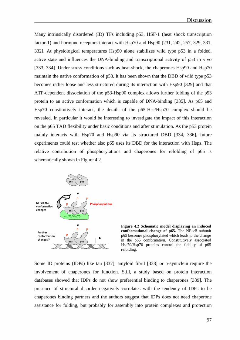
Discussion
97
Many intrinsically disordered (ID) TFs including p53, HSF-1 (heat shock transcription
factor-1) and hormone receptors interact with Hsp70 and Hsp90 [231, 242, 257, 329, 331,
332]. At physiological temperatures Hsp90 alone stabilizes wild type p53 in a folded,
active state and influences the DNA-binding and transcriptional activity of p53 in vivo
[333, 334]. Under stress conditions such as heat-shock, the chaperones Hsp90 and Hsp70
maintain the native conformation of p53. It has been shown that the DBD of wild type p53
becomes rather loose and less structured during its interaction with Hsp90 [329] and that
ATP-dependent dissociation of the p53-Hsp90 complex allows further folding of the p53
protein to an active conformation which is capable of DNA-binding [335]. As p65 and
Hsp70 constitutively interact, the details of the p65-Hsc/Hsp70 complex should be
revealed. In particular it would be interesting to investigate the impact of this interaction
on the p65 TAD flexibility under basic conditions and after stimulation. As the p53 protein
mainly interacts with Hsp70 and Hsp90 via its structured DBD [334, 336], future
experiments could test whether also p65 uses its DBD for the interaction with Hsps. The
relative contribution of phosphorylations and chaperones for refolding of p65 is
schematically shown in Figure 4.2.
Figure 4.2 Schematic model displaying an induced
conformational change of p65. The NF-κB subunit
p65 becomes phosphorylated which leads to the change
in the p65 conformation. Constitutively associated
Hsc70/Hsp70 proteins control the fidelity of p65
refolding.
Some ID proteins (IDPs) like tau [337], amyloid fibril [338] or α-synuclein require the
involvement of chaperones for function. Still, a study based on protein interaction
databases showed that IDPs do not show preferential binding to chaperones [339]. The
presence of structural disorder negatively correlates with the tendency of IDPs to be
chaperones binding partners and the authors suggest that IDPs does not need chaperone
assistance for folding, but probably for assembly into protein complexes and protection
Hsp70/Hsc70
NF-B p65conformation changes
P P
p65 p65
P
Phosphorylations
P P
P
Furtherconformation changes ?
p65 p65
p65 p65
Discussion
98
from aggregation. Furthermore, it has been detected that chaperones could promote
degradation of several IDPs, including p53, Tau, and HIF-1 [340]. In the present study
VER treatment inhibited TNF-induced p65 degradation, thus it will be interesting in future
experiments to investigate the role of Hsp70 on p65 stability (Fig. 3.18B).
Hsp70 functions in cooperation with co-chaperones (e.g. Hsp40) and various NEFs where
these proteins assist protein folding during translation or post-translationally. The recently
proposed N-terminal folding nucleation hypothesis suggests that the generation of
intrinsically folded N-terminal regions at the ribosome serve as nucleation points for the
yet unstructured C-terminal portions of the peptide chains [341]. This mechanism is likely
to regulate the folding of a newly synthesized p65 protein. According to this hypothesis, a
structured p65 N-terminal half would allow for specific intramolecular protein contacts
with the unstructured C-terminal half. This would generate a compact protein shape which
allows prevention of protein aggregation and proteolysis. On the other hand, if a protein
has a disordered N-terminus, its degradation can be potentially initiated when it starts to
emerge from the ribosomal channel. These proteins usually require chaperones for folding.
For example, p53 has an unstructured N-terminal domain and a recent study showed that
its N-terminus binds the chaperonine CCT (chaperonin-containing t-complex polypeptide 1
(TCP1)) which plays an important role in the folding of newly synthesized proteins [342-
344]. The identification of intramolecular p65 contact sites would help to investigate the
role of the N-terminal hypothesis in p65 folding.
4.5 Chaperones as coregulators of p65 PTMs and p65-mediated gene
expression
Treatment with Hsp inhibitors led to changed p65 PTMs. This effect can be due to several
reasons including effects on Hsp-assisted refolding of p65. On the other hand, Hsps could
also be important for proper conformation of the p65 modifiers themselves. It has been
shown that GA treatment reduces IKKα and IKKβ kinase activities, which in turn resulted
in decreased IκBα phosphorylation [248, 249, 255]. The loss of Ser536 phosphorylation in
the presence of GA can therefore be explained by impaired IKK complex kinase activity
(Fig. 3.16). The reason for diminished Ser468 phosphorylation in the presence of GA could
be due to impaired activity of the kinase(s) or phosphatases regulating the phosphorylation
state of this residue. As IKKε is important for Ser468 phosphorylation [179], further

Discussion
99
experiments should be performed to reveal whether inhibition of the Hsp90 system affects
IKKε activity. It is reasonable to assume that other p65 phosphorylation sites are affected
upon inhibition of the Hsp90 system as well. The Pim-1 kinase phosphorylates p65 at
Ser276 and is known to interact with Hsp90 and Hsp70 [345], thus Pim-1-mediated
phosphorylation will depend on proper protein folding.
Inhibition of Hsp70 enhanced p65 Ser468 phosphorylation (Fig. 3.16). The kinase GSK-3β
phosphorylates p65 at Ser468 under basal conditions [178], but it is also implicated in
TNF-induced NF-κB activation [346]. GSK-3β is inactivated through Akt-mediated
phosphorylation [347], while the dephosphorylation by the phosphatase PP1 converts
GSK-3β into its active form [348]. As Hsp70 regulates the activity of the Akt kinase [243],
inhibition of the Hsp70 ATPase activity could causes impaired Akt kinase activity. Loss of
Akt-mediated phosphorylation-dependent GSK-3β inactivation would lead to permanent
GSK-3β activity and enhanced p65 Ser468 phosphorylation. On the other hand, a higher
p65 phosphorylation at Ser468 and Ser536 in the presence of VER treatment could be a
consequence of impaired Hsp70/Hsc70-mediated regulation of serine/threonine
phosphatases. One of the Hsp70 client proteins is the phosphatase slingshot, which
regulates the phosphorylation-induced activity of the actin-binding protein cofilin [349].
Reduced p65 acetylation and ubiquitination in the presence of Hsp inhibitors could be a
consequence of impaired p65 phosphorylations which can further affect p65 binding
affinities to acetyltransferases, HDACs [154, 174, 180] or assembly with the COMMD1-
E3 ligase complex [182, 183]. The role of the Hsc/Hsp70 system in regulation of p65
phosphorylation/dephosphorylation cycles and associated p65 refolding should be
investigated in more detail in the future.
Decreased p65 acetylation caused by the Hsp inhibition can be a consequence of a direct
effect of chaperones on acetyltansferase activity. Recently it has been shown that the
chaperonin containing TCP1 subunit eta (CCTε) regulates p65 acetylation and NF-κB
transcriptional activity by controlling CBP activity. CBP activity is regulated by auto-
acetylation which contributes to its enzymatic activity [350]. Knockdown of CCTε
reduced CBP acetylation and thus its activity. In CCTε knockdown cells the level of p65
acetylation was also reduced. The role of Hsp90 in regulation of GR-mediated gene
transcription was also detected. Treatment with GA diminished GR-mediated gene
transcription by blocking the GR and CBP/p300 recruitment to promoters [351].
Discussion
100
Reporter gene experiments showed that chaperone activity provides an important
contribution to p65-induced transcription (Fig. 3.19). The p65-triggered gene activity was
only slightly diminished in the presence of GA, while VER significantly impaired p65-
driven gene expression. These results are in agreement with a recent study where siRNA-
mediated knock-down of Hsc70 was sufficient for strongly reduced NF-κB transcriptional
activity [242]. Klenke et al. showed that interaction between Hsc70 and p65 is dependent
on Hsc70 ATPase activity, since p65 interaction with Hsc70 mutant carrying a point
mutation in the ATPase domain resulted in a loss of interaction. Chaperone inhibitors can
affect the outcome of p65-dependent transactivation by preventing the proper folding of
accessory proteins such as RNA Pol II and its numerous subunits and coregulators [352]. It
was recently shown that Hsp90 binds the transcription start site (TSS) and maintains and
optimizes RNA pol II pausing via stabilization of the negative elongation factor complex
over the gene body [353]. Hsp90 and Hsp70 inhibition may also contribute to gene
induction by removing promoter nucleosomes, as shown for galactose genes in budding
yeast [354]. Currently, much more is known about the role of Hsp90 in transcriptional
regulation due to a global and high-resolution chromatin-binding profile of Hsp90 by
ChIP-Seq experiments [353]. Because of a strong effect of Hsp70 inhibition on
transactivation potential of p65, the same approach should be performed for comparison of
Hsp70 chromatin-binding with other transcriptional features.
4.6 Cytokine-induced conformation change of p65 occurs independently
of its binding to DNA
Previous studies showed that the composition of the κB site highly regulates the activity of
NF-κB dimers through conformational changes of its DNA-bound subunits. A comparison
of p50/p65 dimers bound to HIV-κB, uPA (urinary plasminogen activator)-κB or IFNβ-κB
sites revealed significant differences in their conformation [260]. Also, it has been
suggested that the change of only one nucleotide in a κB site is enough to induce such a
conformational change of a bound NF-κB that would further dictate differential assembly
of co-activators to the κB site-bound dimer [259]. Interestingly, the limited proteolysis
assay with TNF-stimulated p65-/-
MEFs expressing the wild type p65 or p65 DNA-binding
mutant did not detect any difference in the digestion pattern (Fig. 3.22) showing that the
cytokine-induced p65 conformational change occurs independently from its contact with
Discussion
101
DNA. It is thus possible that p65 binding to its cognate DNA causes further structural
changes that serve to fine tune regulation of target gene expression.
Several recent studies showed that promoter regions of NF-κB target genes are occupied
with cofactors which recruit the stimulus-induced NF-κB. ChIP experiments against
CBP/p300 revealed the constitutive enrichment of this cofactor on a set of p65-dependent
NF-κB target gene promoters [180]. TNF stimulation can induce recruitment of the
cofactor Tip60 (60 kDA trans-acting regulatory protein of HIV type 1-interacting protein)
to promoters of NF-κB target genes. Tip60 acetylates histones H3 and H4 and the resulting
chromatin remodelling further leads to the recruitment of acetyl-Lys310 p65 to its target
gene promoters [302]. A recent study from the Kracht lab showed that IL-1 induces CDK6
recruitment to distinct chromatin regions of inflammatory NF-κB target genes which was
found to be essential for proper NF-κB p65 loading to its cognate sites [22]. Thereby it is
possible that under those conditions the cytokine-induced conformational change allows
proper contact of p65 with preloaded cofactors and subsequent gene expression.
Phosphorylation of p65 at Ser468 leads to the SOCS-1-mediated ubiquitination and
proteasome-dependent removal of promoter-bound p65 [182]. It has been shown that upon
IL-1 stimulation, SOCS1 exclusively binds to DNA-bound p65 within the nucleus [211].
The interaction between the cytokine-activated p65 E39I and COMMD1 and/or Cul2 could
be tested by coimmunoprecipitation. This experiment would give information whether
recruitment of a COMMD1-E3 ligase complex is regulated only by a phosphorylation-
mediated conformation of p65 or additional κB site-specific p65 rearrangement needs to
occur. As all types of PTMs could regulate DNA-induced p65 refolding [154, 199, 214,
303], it would be interesting to learn about PTMs that exclusively occur for DNA-bound
p65. It has been shown that p53 phosphorylation at Ser20 within its unstructured TAD
stabilizes the interaction with p300. Dependent on the consensus DNA binding site, p300
further acetylates p53 at the C-terminal negative regulatory domain and stabilizes the
p300-p53 complex [355].
Conformational changes induced by binding to consensus sites have also been observed in
other transcription factors. The DBD of GR binds its cognate site in a variety of
conformations and presents different interfaces for the recruitment of specific cofactors in
a gene-specific manner [356-358]. Similar findings have also been reported for the POU
(Pit-Oct-Unc) family of transcription factors, such as Oct-1, Oct-2, Brn-5 [359, 360]. A
Discussion
102
recent structural study of a p53-DNA binding complex showed that a sequence specific
DNA-binding occurs via an induced-fit mechanism that involves conformational switch of
the p53 DBD. As many TFs bind consensus and non-consensus sequences with similar
affinities, Petty et al. suggest that only the binding to the sequence-specific DNA induces a
conformational change that locks the TF at consensus target site, controlling the TF DNA-
binding independently of affinity [361]. There is no doubt that binding to DNA induces
local p65 rearrangement that allow NF-κB to anchor to target κB site. It would be exciting
to investigate in the future whether the allosteric effect of DNA on p65 TAD is governed
by specific sequence motifs and/or dynamic chromatin organisation that changes during
the process of gene transcription.
4.7 Non-genomic functions of NF-κB p65
After the reconstitution of p65-/- MEFs with wild type p65 and p65 DNA-binding mutants,
a set of experiments was performed with the aim to characterize newly produced cells.
Mutation of Glu39 to isoleucine did not affect p65 interaction with IκBα and dimerisation
with p50 (data not shown). A kinetic analysis of the nuclear import and export of
reconstituted MEFs revealed a rapid translocation of p65 E39I mutant into the nucleus and
prolonged nuclear residence of the p65 E39I mutant (Fig. 3.21B/D). This result can be a
consequence of an impaired resynthesis of p65 export factor IκBα. IκBα is a strong
negative regulator of nuclear NF-κB [69]. It is fully re-synthesized within less than 60 min
after TNF-stimulation, subsequently enters the nucleus and removes NF-κB from its
cognate DNA [362]. However, mRNA and protein level of IκBα were not significantly
affected in MEFs expressing p65 E39I, as revealed by immunoblotting with appropriate
antibody and by qRT-PCR. This further suggests that mutation of E39 to isoleucine could
affect the assembly of a chromosomal region maintenance-1 (Crm-1)/Exportin 1-
dependent complex that mediates p65 nuclear export [44, 363]. Another possible
explanation for prevented nuclear export of p65 E39I could be a disrupted interaction with
the 14-3-3 protein for which is known to facilitate nuclear export of IκBα-p65 complexes
[364]. TNF induces binding of p65 to 14-3-3 and their physical interaction is mediated via
p65 sequences spanning residues 38-44 and 278-283 [364]. Additionally, the expression of
the 14-3-3 isoform γ is controlled by p65-containing NF-κB dimers. It has been shown that
binding of p65 to its consensus κB site further regulates initial induction of the 14-3-3
promoter by recruiting the TF E2A [365]. Thereby, the relative 14-3-3 mRNA amount and
Discussion
103
protein levels should be analysed in MEFs expressing the p65 E39I mutant. Also, the
interaction between p65 E39I and 14-3-3 should be tested in order to see whether the
mutation of Glu39 to isoleucine disrupts the docking site for 14-3-3. Immunoblotting with
phospho-specific Ser468 antibody showed prolonged phosphorylation at this site of the
p65 DNA-binding mutant. As phosphorylation at Ser468 is a nuclear event [166], the
prolonged phosphorylation level of p65 E39I rather reflects the impaired p65 nuclear
export. Still, due to the dynamic flexibility of the overall p65 protein, the p65 E39I mutant
may have a different conformation which in turn could affect the docking of kinases and
phosphatases.
TNF-induced gene expression was compared between MEFs expressing wild type p65, p65
E39I or the p65 E39I/R302E mutant. Analysis of relative mRNA expression levels by
qRT-PCR identified a set of p65-dependent genes that were induced only in cells
reconstituted with wild type p65 (Fig. 3.23 A-E). These results show that expression of
some genes is highly dependent on direct contact of p65 with its target κB site. DNA
binding further leads to the proper positioning of p65 TAD and assembly of the functional
transcription initiation complex. Experiments using a reconstituted cell-free systems have
defined that the minimal apparatus required for an activated transcription consists of
general transcription factors, RNA Pol II and a large multisubunit Mediator complex [366-
368]. It was suggested that after binding to the κB site, the NF-κB dimer interacts with the
Mediator complex and recruits RNA Pol II to the TSS [369, 370]. Van Essen et al.
identified Ip10 and Il-6 as genes which expression depend on p65 interaction with the
Mediator complex subunit Trap80, while Mip-2 was identified as a gene independent of
this interaction [369]. According to qRT-PCR experiments from this study expression of
Ip10 gene is dependent on p65 DNA-binding (Fig. 3.23D). This is supported by a previous
finding that Ip10 expression is governed by an allosteric effect of a cognate κB site [259].
Further experiments could be performed to identify to which extent the p65-Mediator
complex interaction depends on p65 contact with the DNA. Also, expression of a Mip-2
gene was governed by a p65 DNA-binding (Fig. 3.23B) and together with the finding that
Mip-2 expression is independent of p65-Trap80 interaction, this suggests that some κB
site-mediated regulatory mechanism needs to occur. Putative binding sites of TFs like Sp-
1, ATF/CREB, Ap-1 can be found within promoters of NF-κB target genes [369, 371-374].
Thereby, bound p65 could recruit a secondary TF that could mediate the recruitment of a

Discussion
104
Mediator complex. As suggested by van Essen et al., p65 DNA-binding may also exert its
effect by inducing local chromatin alterations that would change promoter accessibility for
other TF. This mechanism can be supported by study showing that in macrophages the NF-
κB–driven activation of gene expression is accompanied by nucleosome remodelling at
target gene promoters [375]. Also, p65 DNA-binding may induce changes to histone
modifications (e.g. lysine acetylation [159, 376], lysine methylation [377] and serine
phosphorylation [378, 379]) which can further control the recruitment of RNA Pol II and
target gene expression.
Interestingly, another set of tested genes were expressed in cells expressing the p65 E39I
mutant, suggesting that rather protein-protein interactions play a major role for their
expression than a direct DNA-binding (Fig. 3.23F-J). For example, TNF-induced
expression of Il-6, Ccl9, JunB, Saa3, Cox-2 and Nfκbiα seems to highly depend on p65
protein-protein interactions, since the p65 E39I mutant showed a similar expression of
these genes. Several studies support this finding. In a study performed by Mukherjee et al.,
ChIP experiments identified promoters of Nfκbiα, Tnfaip3, Cox2, Cxcl2, Csf2 and Tnf
genes to be preloaded with the cofactor CBP/p300 [180]. In particular, RNA-Seq analysis
of total mRNA isolated from TNF-stimulated MEFs reconstituted with wild type p65 or
the mutant that cannot interact with CBP/p300 TAZ1 domain identified JunB and Cox-2
as genes whose expression only depend on the recruitment of p65 to promoters via
p65TA2-CBP/p300 TAZ1 interactions. In contrast, in the present study expression of
Tnfaip3 was observed only in cells expressing the wild type p65, suggesting that beside the
interaction with CBP/p300, also the DNA-binding is necessary to occur. In a study
performed by Handschick et al., ChIP-Seq experiments using human KB epithelial
carcinoma cells showed a tight co-recruitment of CDK6 and p65 to enhancers and
promoters of many active NF-κB target genes, like Il-8, Cxcl3, Ccl20, Il-6, Icam-1 and
Cox-2, after the IL-1 stimulation [22]. CDK6-dependent loading of NF-κB p65 to its
cognate binding sites regulates the expression of Il-6 and Cox-2 gene after the IL-1
stimulation. As effects on gene expression are cell- and stimulus specific, it should be
tested whether the same mechanism regulates the expression of those two genes in TNF
stimulated reconstituted MEFs. Another explanation for the TNF-induced gene expression
in MEFs expressing the p65 E39I mutant could be the p65 interaction with the neighboring
TF that is governed by a p65 phosphorylation-induced rearrangement and p50-mediated
Discussion
105
(or by other NF-κB monomer) recruitment to DNA. It has been shown that the recruitment
of RNA Pol II to Ccl2 promoter depends on formation of a c-Jun, HDAC1/HDAC3, CBP
and p65/p50 chromatin-associated protein-protein complexes at specific regions of a gene
after the IL-1 stimulation. Also, this higher-order complex was not form in the absence of
c-Jun [374].
Even though genes tested in this study are well documented as TNF-induced p65-
dependent genes [201, 369], qRT-PCR experiments detected a slightly induced expression
of JunB and Nfκbiα mRNA in p65-/- MEFs reconstituted with the empty vector (Fig.
3.23H/K). Binding sites of Sp-1, ATF/CREB and Ap-1 were identified within promoters of
these two genes [369, 380]. NFAT (nuclear factor of activated T cells) and STAT TF
families also bind to DNA sequences to which p65 binds [381-383]. Thus due to the
absence of promoter bound p65-containing NF-κB dimers in cells reconstituted with empty
vector or p65 mutants, other TF could initiate gene transcription. Different NF-κB dimers
can be exchanged at the same promoter during the NF-κB response [384] and this
mechanism could also be involved in the expression of genes which expression was not
dependent on p65 DNA-binding. It has been shown that the Il-6 gene is preferentially
activated by a p50-IκBδ complex [385]. Still, the absence of Il-6 expression in MEFs
stably reconstituted with the double p65 E39I/R302E mutant implicates that expression of
Il-6 could be rather dependent on p65 protein-protein interactions. Analysis of the Nfκbα
promoter structure revealed several NF-κB binding sequences. Deletion of the κB motif,
located 37 bp upstream of the TATA box, specifically abolished responses to PMA and
TNF [48]. The Nfκbiα promoter contains several potential κB sites to which p50/p65,
p50/c-Rel or p65/c-Rel heterodimers as well as p50 homodimers can bind [48]. Another
study revealed dynamic exchanges between members of steroid receptor cofactors (SRC)
family (SRC-1, SRC-2 and SRC-3) and corepressors (SMRT, NCoR (nuclear receptor
corepressor), HDAC1, HDAC2 and HDAC3) on the Nfκbiα promoter region during the
TNF stimulation [386]. Thereby, changes in Nfκbiα mRNA levels detected in MEFs
expressing the p65 E39I or E39I/R302E mutants could also be a consequence of impaired
p65-coactivator-corepressor interactions due to the absence of a direct p65-DNA contact.
Similar experiments with DNA-binding mutants of other TFs indicated the role of protein-
protein interactions for target gene expression. TF IRF3 (interferon regulatory transcription
factor 3) usually exerts its function via its direct binding to IRF-binding sequences of target

Discussion
106
genes. Reconstitution of IRF3-deficient MEFs with wild type IRF3 or its DNA-binding
defective mutant (IRF3 K77E) showed that only wild type IRF3 fully restored Tslp and
IL33 gene induction [387]. However, IRF3 can also act as a cofactor to activate
transcription of a set of NF-κB–dependent genes without directly binding to DNA [259].
The GR DNA-binding mutants (S459A and P493R) have been found to mimic a DNA-
induced conformational change which allows the partial transcription of some genes. It
was suggested that those DBD point mutations trigger an allosteric mechanism in which
binding to the specific DNA sites normally induces the exposure of otherwise concealed
GR TAD surfaces [388, 389].
ChIP-Seq experiments with reconstituted p65-/- MEFs produced in this study could be
further used to reveal which regions are constitutively and/or inducible occupied by p65-
containing NF-κB dimers. Such experiments would give an answer whether the DNA-
binding of p65 is necessary for genomic associations and if there are regions preferentially
ocupied by p65 E39I mutant. RNA-Seq experiments should be performed to confirm the
effects detected by qRT-PCR since RNA-Seq experiments provides a far more precise
measurement of transcript levels. Focusing on particular promoter regions, further ChIP
and ChIP-re-ChIP experiments, using antibodies against the p65 and p50 subunits and
specific transcriptional cofactors could be also performed to analyse p65 activity that is
based on its protein-protein interactions. Based on qRT-PCR results, the role of a second
subunit in p65-containing NF-κB dimers should be also considered and investigated. p65-/-
MEFs reconstituted with p65 E39I or p65 E39I/E302R mutants would allow further
investigations on the non-genomic functions of p65. Using those cells it would be possible
to explore the contribution of activated, cytosolic p65-containing NF-κBs in activation of
various signaling cascades, cell death and cell proliferation. The NF-κB regulatory
pathway exist in mitochondria where p65-containing NF-κB dimers repress mitochondrial
gene expression [258, 390]. Thereby, reconstituted MEFs used in this study would help to
investigate a crosstalk between p53 and p65 in the regulation of apoptosis. The GR has
been also found in the mitochondria [391]. As GR can physically interact with p65 [392],
their interaction in mitochondria and regulation of processes involved in cell growth and
apoptosis could be also tested. Experiments addressing these questions may reveal novel
functions of p65 that do not rely on its ability to bind to DNA.
Summary
107
5. Summary
The TF NF-κB is an important regulator of immunity, stress responses as well as apoptosis,
cell cycle progression and oncogenesis. NF-B is activated by various stimuli and
regulates expression many different target genes. The first part of this work shows that
stimulation of cells with the cytokines TNF or IL-1 results in a profound conformational
switch of the NF-κB subunit p65, as revealed by limited proteolysis assays. The cytokine-
triggered reconfiguration of p65 mainly occurs for p65 contained in the nuclear fraction.
Immunoprecipitation experiments with various p65-specific antibodies identified a
conformation-specific monoclonal antibody that preferentially immunoprecipitates the
inducibly refolded p65 protein. Cytokine inducible structural changes of p65 are mainly
mediated by posttranslational p65 modifications. Mutations of individual p65
phosphorylation sites cause structural changes in p65 and thus control subsequent
ubiquitination and association with transcriptional cofactors such as CBP/p300 and
MYBBP1a. These experiments suggest that phosphorylation-induced conformation
changes contribute to an alternative p65 configuration and thus to the specification of the
NF-κB response. As chaperones are frequently found to be important for proper folding of
proteins, the constitutive and inducible association of chaperones Hsc/Hsp70 and Hsp90
with p65 was tested. Coimmunoprecipitation experiments revealed constitutive interaction
between p65 and Hsp70. Limited proteolysis assays showed that constitutively bound
chaperones of the Hsc/Hsp70 family were not important for the cytokine-induced
conformational switch per se, but rather control the fidelity of protein rearrangement.
Accordingly, pharmacologic inhibition of Hsp/Hsc70 revealed that these chaperones also
control the level of p65 modifications and regulate TNF-triggered ubiquitination and
degradation of p65. A specific Hsc/Hsp70 inhibitor significantly reduced p65-triggered
reporter gene activity, showing that chaperone-activity provides an important contribution
for p65-induced transcription. It is thus likely that inducible conformational changes of p65
increase the intramolecular flexibility and thereby expand and specify the repertoire of
possible modifications and protein-protein interactions. The cytokine-induced
conformation switch is independent from the DNA-binding ability of p65, as shown by the
analysis of p65-/- MEFs that were reconstituted with a p65 DNA-binding mutant. The
DNA-binding mutant and a further p65 mutant additionally mutated in the NLS were used
for the second part of this work. Reconstituted knockout cells were used to investigate the
Summary
108
contribution of p65 DNA-binding for the induction of TNF-induced and p65-dependent
transcription of NF-κB target genes. Analysis of relative mRNA expression by qRT-PCR
identified a set of genes which were as expected fully dependent on p65 DNA-binding.
These experiments also showed that another group of genes which is independent of direct
binding of p65 to its target κB sites, implicating the importance of p65 protein-protein
interactions for its ability to trigger transcription. These differentially reconstituted cells
will be useful in the future to identify p65-dependent non-genomic functions of NF-κB.
Zusammenfassung
109
6. Zusammenfassunf
Der Transkriptionsfaktor NF-κB ist bei der Regulation der Stressantwort, der Apoptose
und angeborenen Immunität beteiligt. Daher wird NF-κB durch unterschiedlichste Signale
aktiviert und induziert die Expression von vielen verschiedenen Genen. Der erste Teil
dieser Arbeit zeigt mit Hilfe von "limited proteolysis" Experimenten, dass die Stimulation
von Zellen durch die Zytokine IL-1 oder TNF in einer Konformationsänderung des NF-κB
p65 Proteins resultiert. Die Zytokin-induzierte Konformationsänderung des p65 Proteins
war nur für das im Zellkern befindliche p65 Protein zu beobachten.
Immunopräzipitationsexperimente erlaubten die Identifizierung eines Konformations-
spezifischen p65 Antikörpers der präferentiell das umgefaltete p65 Protein erkennt. Die
Zytokin-induzierten Strukturänderungen werden primär durch Phosphorylierungsereignisse
am NF-κB p65 Protein induziert. Mutationen in einzelnen p65 Phosphorylierungsstellen
resultierten in Strukturänderungen und kontrollierten auch weitere Eigenschaften des p65
Proteins wie beispielsweise die Ubiquitinierung und die Assoziation mit transkriptionellen
Kofaktoren wie CBP/p300 und MYBBP1a. Auch die Induktion der p65 Phosphorylierung
durch Behandlung der Zellen mit dem Phosphatase-Inhibitor Calyculin A führte zu einer
induzierten Strukturänderung. Da Chaperone häufig an der Regulation der Proteinfaltung
beteiligt sind, wurde die Interaktion von p65 mit Hsp Proteinen untersucht. "Limited
proteolysis" Experimente zeigten, dass konstitutiv gebundene Hsc/Hsp70 Proteine die
Genauigkeit der Zytokin-induzierten p65 Umlagerung kontrollieren. Die pharmakologische
Inhibition der Hsp/Hsc70 Proteine resultierte in veränderter p65 Ubiquitinierung und
Stabilität. Ebenso führte die Hsp/Hsc70 Inhibition zu einer verringerten p65-induzierten
Aktivität eines Reportergens, sodass die Chaperon Aktivität einen wichtigen Beitrag für
die Aktivität des p65 Proteins im Zellkern hat. Es ist wahrscheinlich, dass unterschiedliche
p65 Konformationen das Repertoire an möglichen p65 Modifikationen und Protein/Protein
Interaktionen stark erhöhen. Weitere Experimente zeigten, dass die Zytokin-induzierte p65
Umlagerung unabhängig von der DNA-Bindungsfähigkeit von p65 ist. Im zweiten Teil
dieser Arbeit wurde die Wichtigkeit der DNA-Bindung für die Funktionen des p65
Proteins untersucht. Dazu wurden p65 knockout Zellen mit dem Wildtyp Protein sowie mit
weiteren p65 Mutanten stabil rekonstituiert. Eine Mutante war DNA-Bindungsdefekt,
währen die andere Mutante zusätzlich im Kerntranslokationssignal mutiert war. Die
Analyse der TNF-induzierten Expression einiger NF-κB Zielgene zeigte, dass einige Gene
Zusammenfassung
110
trotz defekter p65 DNA-Bindung noch induzierbar exprimiert werden konnten. Dieses
Verhalten ist wahrscheinlich auf kompensatorische Mechanismen wie beispielsweise eine
Promoter-Rekrutierung via Protein/Protein Wechselwirkungen zurückzuführen. Die
differentiell rekonstituierten Zellen werden in zukünftigen Studien wichtige Informationen
über DNA-Bindungs-unabhängige Funktionen des p65 Proteins liefern können.
References
111
7. References
1. Oeckinghaus, A. and S. Ghosh, The NF-kappaB family of transcription factors and
its regulation. Cold Spring Harb Perspect Biol, 2009. 1(4): p. a000034.
2. Ghosh, S., M.J. May, and E.B. Kopp, NF-kappa B and Rel proteins: evolutionarily
conserved mediators of immune responses. Annu Rev Immunol, 1998. 16: p. 225-
60.
3. Gilmore, T.D., Introduction to NF-kappaB: players, pathways, perspectives.
Oncogene, 2006. 25(51): p. 6680-4.
4. Ghosh, G., et al., NF-kappaB regulation: lessons from structures. Immunol Rev,
2012. 246(1): p. 36-58.
5. Siebenlist, U., G. Franzoso, and K. Brown, Structure, regulation and function of
NF-kappa B. Annu Rev Cell Biol, 1994. 10: p. 405-55.
6. Schmitz, M.L., M.A. dos Santos Silva, and P.A. Baeuerle, Transactivation domain
2 (TA2) of p65 NF-kappa B. Similarity to TA1 and phorbol ester-stimulated activity
and phosphorylation in intact cells. J Biol Chem, 1995. 270(26): p. 15576-84.
7. O'Shea, J.M. and N.D. Perkins, Regulation of the RelA (p65) transactivation
domain. Biochem Soc Trans, 2008. 36(Pt 4): p. 603-8.
8. Hansen, S.K., P.A. Baeuerle, and F. Blasi, Purification, reconstitution, and I kappa
B association of the c-Rel-p65 (RelA) complex, a strong activator of transcription.
Mol Cell Biol, 1994. 14(4): p. 2593-603.
9. Plaksin, D., P.A. Baeuerle, and L. Eisenbach, KBF1 (p50 NF-kappa B homodimer)
acts as a repressor of H-2Kb gene expression in metastatic tumor cells. J Exp Med,
1993. 177(6): p. 1651-62.
10. Ghosh, G., et al., Structure of NF-kappa B p50 homodimer bound to a kappa B site.
Nature, 1995. 373(6512): p. 303-10.
11. Muller, C.W., et al., Structure of the NF-kappa B p50 homodimer bound to DNA.
Nature, 1995. 373(6512): p. 311-7.
12. Cramer, P., et al., Structure of the human NF-kappaB p52 homodimer-DNA
complex at 2.1 A resolution. Embo J, 1997. 16(23): p. 7078-90.
13. Chen, Y.Q., S. Ghosh, and G. Ghosh, A novel DNA recognition mode by the NF-
kappa B p65 homodimer. Nat Struct Biol, 1998. 5(1): p. 67-73.
14. Chen, F.E., et al., Crystal structure of p50/p65 heterodimer of transcription factor
NF-kappaB bound to DNA. Nature, 1998. 391(6665): p. 410-3.
15. Chen, Y.Q., et al., NF-kappaB p65 (RelA) homodimer uses distinct mechanisms to
recognize DNA targets. Structure, 2000. 8(4): p. 419-28.
16. Kopp, E.B. and S. Ghosh, NF-kappa B and rel proteins in innate immunity. Adv
Immunol, 1995. 58: p. 1-27.
17. Pahl, H.L., Activators and target genes of Rel/NF-kappaB transcription factors.
Oncogene, 1999. 18(49): p. 6853-66.
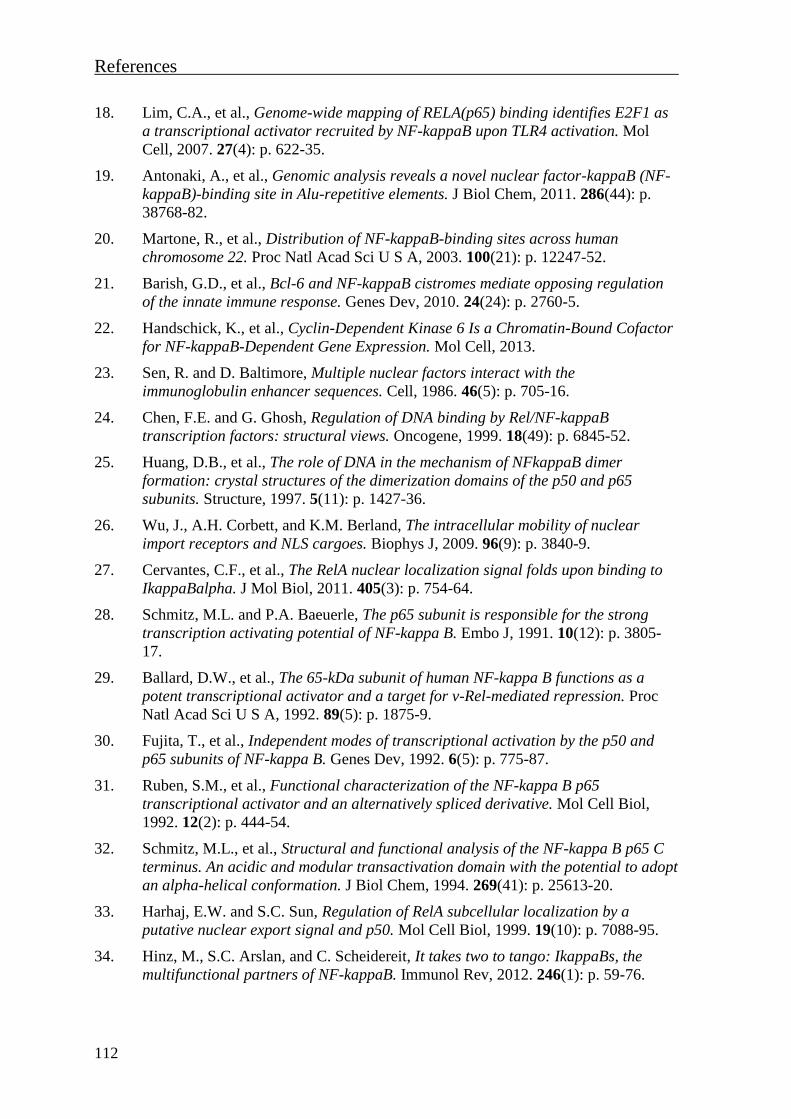
References
112
18. Lim, C.A., et al., Genome-wide mapping of RELA(p65) binding identifies E2F1 as
a transcriptional activator recruited by NF-kappaB upon TLR4 activation. Mol
Cell, 2007. 27(4): p. 622-35.
19. Antonaki, A., et al., Genomic analysis reveals a novel nuclear factor-kappaB (NF-
kappaB)-binding site in Alu-repetitive elements. J Biol Chem, 2011. 286(44): p.
38768-82.
20. Martone, R., et al., Distribution of NF-kappaB-binding sites across human
chromosome 22. Proc Natl Acad Sci U S A, 2003. 100(21): p. 12247-52.
21. Barish, G.D., et al., Bcl-6 and NF-kappaB cistromes mediate opposing regulation
of the innate immune response. Genes Dev, 2010. 24(24): p. 2760-5.
22. Handschick, K., et al., Cyclin-Dependent Kinase 6 Is a Chromatin-Bound Cofactor
for NF-kappaB-Dependent Gene Expression. Mol Cell, 2013.
23. Sen, R. and D. Baltimore, Multiple nuclear factors interact with the
immunoglobulin enhancer sequences. Cell, 1986. 46(5): p. 705-16.
24. Chen, F.E. and G. Ghosh, Regulation of DNA binding by Rel/NF-kappaB
transcription factors: structural views. Oncogene, 1999. 18(49): p. 6845-52.
25. Huang, D.B., et al., The role of DNA in the mechanism of NFkappaB dimer
formation: crystal structures of the dimerization domains of the p50 and p65
subunits. Structure, 1997. 5(11): p. 1427-36.
26. Wu, J., A.H. Corbett, and K.M. Berland, The intracellular mobility of nuclear
import receptors and NLS cargoes. Biophys J, 2009. 96(9): p. 3840-9.
27. Cervantes, C.F., et al., The RelA nuclear localization signal folds upon binding to
IkappaBalpha. J Mol Biol, 2011. 405(3): p. 754-64.
28. Schmitz, M.L. and P.A. Baeuerle, The p65 subunit is responsible for the strong
transcription activating potential of NF-kappa B. Embo J, 1991. 10(12): p. 3805-
17.
29. Ballard, D.W., et al., The 65-kDa subunit of human NF-kappa B functions as a
potent transcriptional activator and a target for v-Rel-mediated repression. Proc
Natl Acad Sci U S A, 1992. 89(5): p. 1875-9.
30. Fujita, T., et al., Independent modes of transcriptional activation by the p50 and
p65 subunits of NF-kappa B. Genes Dev, 1992. 6(5): p. 775-87.
31. Ruben, S.M., et al., Functional characterization of the NF-kappa B p65
transcriptional activator and an alternatively spliced derivative. Mol Cell Biol,
1992. 12(2): p. 444-54.
32. Schmitz, M.L., et al., Structural and functional analysis of the NF-kappa B p65 C
terminus. An acidic and modular transactivation domain with the potential to adopt
an alpha-helical conformation. J Biol Chem, 1994. 269(41): p. 25613-20.
33. Harhaj, E.W. and S.C. Sun, Regulation of RelA subcellular localization by a
putative nuclear export signal and p50. Mol Cell Biol, 1999. 19(10): p. 7088-95.
34. Hinz, M., S.C. Arslan, and C. Scheidereit, It takes two to tango: IkappaBs, the
multifunctional partners of NF-kappaB. Immunol Rev, 2012. 246(1): p. 59-76.
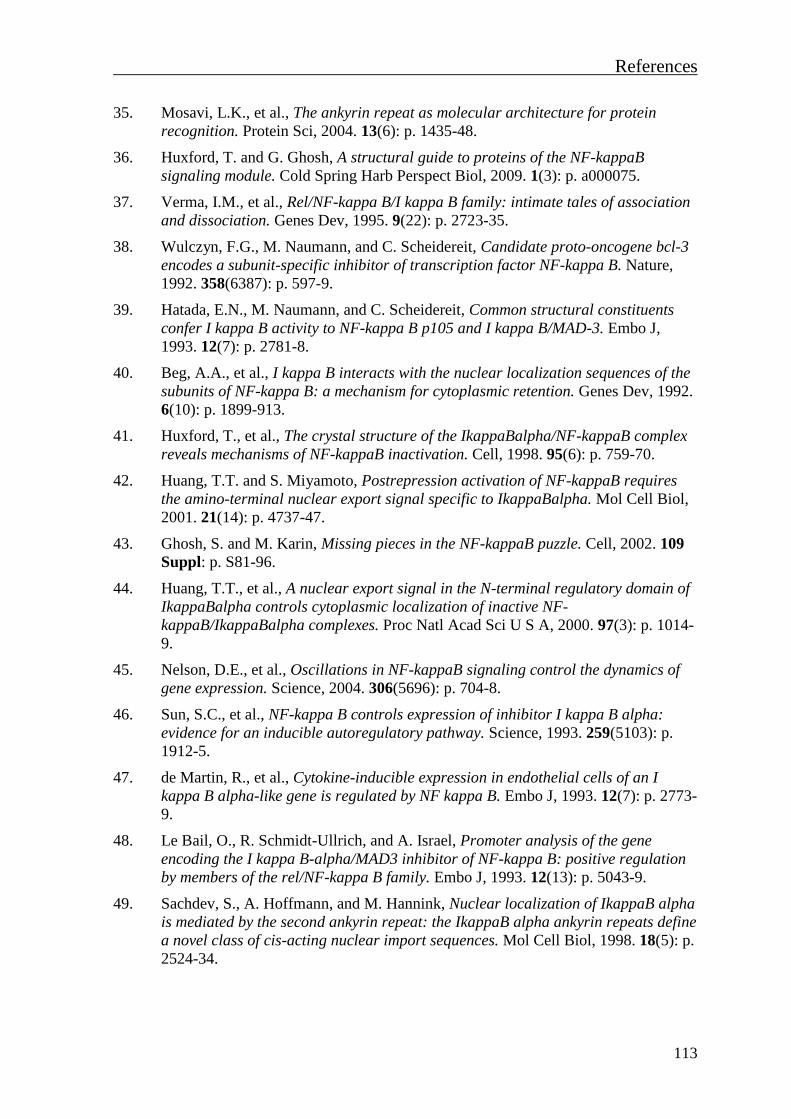
References
113
35. Mosavi, L.K., et al., The ankyrin repeat as molecular architecture for protein
recognition. Protein Sci, 2004. 13(6): p. 1435-48.
36. Huxford, T. and G. Ghosh, A structural guide to proteins of the NF-kappaB
signaling module. Cold Spring Harb Perspect Biol, 2009. 1(3): p. a000075.
37. Verma, I.M., et al., Rel/NF-kappa B/I kappa B family: intimate tales of association
and dissociation. Genes Dev, 1995. 9(22): p. 2723-35.
38. Wulczyn, F.G., M. Naumann, and C. Scheidereit, Candidate proto-oncogene bcl-3
encodes a subunit-specific inhibitor of transcription factor NF-kappa B. Nature,
1992. 358(6387): p. 597-9.
39. Hatada, E.N., M. Naumann, and C. Scheidereit, Common structural constituents
confer I kappa B activity to NF-kappa B p105 and I kappa B/MAD-3. Embo J,
1993. 12(7): p. 2781-8.
40. Beg, A.A., et al., I kappa B interacts with the nuclear localization sequences of the
subunits of NF-kappa B: a mechanism for cytoplasmic retention. Genes Dev, 1992.
6(10): p. 1899-913.
41. Huxford, T., et al., The crystal structure of the IkappaBalpha/NF-kappaB complex
reveals mechanisms of NF-kappaB inactivation. Cell, 1998. 95(6): p. 759-70.
42. Huang, T.T. and S. Miyamoto, Postrepression activation of NF-kappaB requires
the amino-terminal nuclear export signal specific to IkappaBalpha. Mol Cell Biol,
2001. 21(14): p. 4737-47.
43. Ghosh, S. and M. Karin, Missing pieces in the NF-kappaB puzzle. Cell, 2002. 109
Suppl: p. S81-96.
44. Huang, T.T., et al., A nuclear export signal in the N-terminal regulatory domain of
IkappaBalpha controls cytoplasmic localization of inactive NF-
kappaB/IkappaBalpha complexes. Proc Natl Acad Sci U S A, 2000. 97(3): p. 1014-
9.
45. Nelson, D.E., et al., Oscillations in NF-kappaB signaling control the dynamics of
gene expression. Science, 2004. 306(5696): p. 704-8.
46. Sun, S.C., et al., NF-kappa B controls expression of inhibitor I kappa B alpha:
evidence for an inducible autoregulatory pathway. Science, 1993. 259(5103): p.
1912-5.
47. de Martin, R., et al., Cytokine-inducible expression in endothelial cells of an I
kappa B alpha-like gene is regulated by NF kappa B. Embo J, 1993. 12(7): p. 2773-
9.
48. Le Bail, O., R. Schmidt-Ullrich, and A. Israel, Promoter analysis of the gene
encoding the I kappa B-alpha/MAD3 inhibitor of NF-kappa B: positive regulation
by members of the rel/NF-kappa B family. Embo J, 1993. 12(13): p. 5043-9.
49. Sachdev, S., A. Hoffmann, and M. Hannink, Nuclear localization of IkappaB alpha
is mediated by the second ankyrin repeat: the IkappaB alpha ankyrin repeats define
a novel class of cis-acting nuclear import sequences. Mol Cell Biol, 1998. 18(5): p.
2524-34.
References
114
50. Arenzana-Seisdedos, F., et al., Inducible nuclear expression of newly synthesized I
kappa B alpha negatively regulates DNA-binding and transcriptional activities of
NF-kappa B. Mol Cell Biol, 1995. 15(5): p. 2689-96.
51. Turpin, P., R.T. Hay, and C. Dargemont, Characterization of IkappaBalpha
nuclear import pathway. J Biol Chem, 1999. 274(10): p. 6804-12.
52. Bosisio, D., et al., A hyper-dynamic equilibrium between promoter-bound and
nucleoplasmic dimers controls NF-kappaB-dependent gene activity. Embo J, 2006.
25(4): p. 798-810.
53. Bergqvist, S., et al., Kinetic enhancement of NF-kappaBxDNA dissociation by
IkappaBalpha. Proc Natl Acad Sci U S A, 2009. 106(46): p. 19328-33.
54. Sue, S.C., et al., Detection of a ternary complex of NF-kappaB and IkappaBalpha
with DNA provides insights into how IkappaBalpha removes NF-kappaB from
transcription sites. Proc Natl Acad Sci U S A, 2011. 108(4): p. 1367-72.
55. Link, E., et al., Purified I kappa B-beta is inactivated upon dephosphorylation. J
Biol Chem, 1992. 267(1): p. 239-46.
56. Thompson, J.E., et al., I kappa B-beta regulates the persistent response in a
biphasic activation of NF-kappa B. Cell, 1995. 80(4): p. 573-82.
57. Hertlein, E., et al., RelA/p65 regulation of IkappaBbeta. Mol Cell Biol, 2005.
25(12): p. 4956-68.
58. Suyang, H., et al., Role of unphosphorylated, newly synthesized I kappa B beta in
persistent activation of NF-kappa B. Mol Cell Biol, 1996. 16(10): p. 5444-9.
59. Malek, S., et al., X-ray crystal structure of an IkappaBbeta x NF-kappaB p65
homodimer complex. J Biol Chem, 2003. 278(25): p. 23094-100.
60. Kearns, J.D., et al., IkappaBepsilon provides negative feedback to control NF-
kappaB oscillations, signaling dynamics, and inflammatory gene expression. J Cell
Biol, 2006. 173(5): p. 659-64.
61. Lee, S.H. and M. Hannink, Characterization of the nuclear import and export
functions of Ikappa B(epsilon). J Biol Chem, 2002. 277(26): p. 23358-66.
62. Hatada, E.N., et al., The ankyrin repeat domains of the NF-kappa B precursor p105
and the protooncogene bcl-3 act as specific inhibitors of NF-kappa B DNA binding.
Proc Natl Acad Sci U S A, 1992. 89(6): p. 2489-93.
63. Naumann, M., et al., NF-kappa B precursor p100 inhibits nuclear translocation
and DNA binding of NF-kappa B/rel-factors. Oncogene, 1993. 8(8): p. 2275-81.
64. Naumann, M., F.G. Wulczyn, and C. Scheidereit, The NF-kappa B precursor p105
and the proto-oncogene product Bcl-3 are I kappa B molecules and control nuclear
translocation of NF-kappa B. Embo J, 1993. 12(1): p. 213-22.
65. Lang, V., et al., betaTrCP-mediated proteolysis of NF-kappaB1 p105 requires
phosphorylation of p105 serines 927 and 932. Mol Cell Biol, 2003. 23(1): p. 402-
13.
66. Xiao, G., E.W. Harhaj, and S.C. Sun, NF-kappaB-inducing kinase regulates the
processing of NF-kappaB2 p100. Mol Cell, 2001. 7(2): p. 401-9.

References
115
67. Liang, C., M. Zhang, and S.C. Sun, beta-TrCP binding and processing of NF-
kappaB2/p100 involve its phosphorylation at serines 866 and 870. Cell Signal,
2006. 18(8): p. 1309-17.
68. Savinova, O.V., A. Hoffmann, and G. Ghosh, The Nfkb1 and Nfkb2 proteins p105
and p100 function as the core of high-molecular-weight heterogeneous complexes.
Mol Cell, 2009. 34(5): p. 591-602.
69. Hoffmann, A., et al., The IkappaB-NF-kappaB signaling module: temporal control
and selective gene activation. Science, 2002. 298(5596): p. 1241-5.
70. Ohno, H., G. Takimoto, and T.W. McKeithan, The candidate proto-oncogene bcl-3
is related to genes implicated in cell lineage determination and cell cycle control.
Cell, 1990. 60(6): p. 991-7.
71. Yamauchi, S., H. Ito, and A. Miyajima, IkappaBeta, a nuclear IkappaB protein,
positively regulates the NF-kappaB-mediated expression of proinflammatory
cytokines. Proc Natl Acad Sci U S A, 2010. 107(26): p. 11924-9.
72. Yamamoto, M., et al., Regulation of Toll/IL-1-receptor-mediated gene expression
by the inducible nuclear protein IkappaBzeta. Nature, 2004. 430(6996): p. 218-22.
73. Ghosh, S. and M.S. Hayden, New regulators of NF-kappaB in inflammation. Nat
Rev Immunol, 2008. 8(11): p. 837-48.
74. Wu, C.L., S.C. Kandarian, and R.W. Jackman, Identification of genes that elicit
disuse muscle atrophy via the transcription factors p50 and Bcl-3. PLoS One,
2011. 6(1): p. e16171.
75. Yang, J., R.S. Williams, and D.P. Kelly, Bcl3 interacts cooperatively with
peroxisome proliferator-activated receptor gamma (PPARgamma) coactivator
1alpha to coactivate nuclear receptors estrogen-related receptor alpha and
PPARalpha. Mol Cell Biol, 2009. 29(15): p. 4091-102.
76. Dechend, R., et al., The Bcl-3 oncoprotein acts as a bridging factor between NF-
kappaB/Rel and nuclear co-regulators. Oncogene, 1999. 18(22): p. 3316-23.
77. Keutgens, A., et al., The repressing function of the oncoprotein BCL-3 requires
CtBP, while its polyubiquitination and degradation involve the E3 ligase TBLR1.
Mol Cell Biol, 2010. 30(16): p. 4006-21.
78. Baek, S.H., et al., Exchange of N-CoR corepressor and Tip60 coactivator
complexes links gene expression by NF-kappaB and beta-amyloid precursor
protein. Cell, 2002. 110(1): p. 55-67.
79. Hacker, H. and M. Karin, Regulation and function of IKK and IKK-related kinases.
Sci STKE, 2006. 2006(357): p. re13.
80. Sil, A.K., et al., IkappaB kinase-alpha acts in the epidermis to control skeletal and
craniofacial morphogenesis. Nature, 2004. 428(6983): p. 660-4.
81. Tang, E.D., et al., Roles for homotypic interactions and transautophosphorylation
in IkappaB kinase beta IKKbeta) activation [corrected]. J Biol Chem, 2003.
278(40): p. 38566-70.
82. Wang, C., et al., TAK1 is a ubiquitin-dependent kinase of MKK and IKK. Nature,
2001. 412(6844): p. 346-51.
References
116
83. Chen, Z.J., V. Bhoj, and R.B. Seth, Ubiquitin, TAK1 and IKK: is there a
connection? Cell Death Differ, 2006. 13(5): p. 687-92.
84. Li, X.H., X. Fang, and R.B. Gaynor, Role of IKKgamma/nemo in assembly of the
Ikappa B kinase complex. J Biol Chem, 2001. 276(6): p. 4494-500.
85. Israel, A., The IKK complex, a central regulator of NF-kappaB activation. Cold
Spring Harb Perspect Biol, 2010. 2(3): p. a000158.
86. Shifera, A.S., Proteins that bind to IKKgamma (NEMO) and down-regulate the
activation of NF-kappaB. Biochem Biophys Res Commun, 2010. 396(3): p. 585-9.
87. Wajant, H., K. Pfizenmaier, and P. Scheurich, Tumor necrosis factor signaling.
Cell Death Differ, 2003. 10(1): p. 45-65.
88. Karin, M. and M. Delhase, The I kappa B kinase (IKK) and NF-kappa B: key
elements of proinflammatory signalling. Semin Immunol, 2000. 12(1): p. 85-98.
89. Perkins, N.D. and T.D. Gilmore, Good cop, bad cop: the different faces of NF-
kappaB. Cell Death Differ, 2006. 13(5): p. 759-72.
90. Zandi, E., et al., The IkappaB kinase complex (IKK) contains two kinase subunits,
IKKalpha and IKKbeta, necessary for IkappaB phosphorylation and NF-kappaB
activation. Cell, 1997. 91(2): p. 243-52.
91. Hayden, M.S. and S. Ghosh, Shared principles in NF-kappaB signaling. Cell, 2008.
132(3): p. 344-62.
92. Hayden, M.S. and S. Ghosh, Signaling to NF-kappaB. Genes Dev, 2004. 18(18): p.
2195-224.
93. Perkins, N.D., Post-translational modifications regulating the activity and function
of the nuclear factor kappa B pathway. Oncogene, 2006. 25(51): p. 6717-30.
94. Chen, L.F. and W.C. Greene, Shaping the nuclear action of NF-kappaB. Nat Rev
Mol Cell Biol, 2004. 5(5): p. 392-401.
95. Au, P.Y. and W.C. Yeh, Physiological roles and mechanisms of signaling by
TRAF2 and TRAF5. Adv Exp Med Biol, 2007. 597: p. 32-47.
96. Baud, V. and M. Karin, Signal transduction by tumor necrosis factor and its
relatives. Trends Cell Biol, 2001. 11(9): p. 372-7.
97. Bertrand, M.J., et al., cIAP1 and cIAP2 facilitate cancer cell survival by
functioning as E3 ligases that promote RIP1 ubiquitination. Mol Cell, 2008. 30(6):
p. 689-700.
98. Park, S.M., J.B. Yoon, and T.H. Lee, Receptor interacting protein is ubiquitinated
by cellular inhibitor of apoptosis proteins (c-IAP1 and c-IAP2) in vitro. FEBS Lett,
2004. 566(1-3): p. 151-6.
99. Christofferson, D.E., et al., A novel role for RIP1 kinase in mediating TNFalpha
production. Cell Death Dis, 2012. 3: p. e320.
100. Wertz, I.E., et al., De-ubiquitination and ubiquitin ligase domains of A20
downregulate NF-kappaB signalling. Nature, 2004. 430(7000): p. 694-9.
101. Dynek, J.N., et al., c-IAP1 and UbcH5 promote K11-linked polyubiquitination of
RIP1 in TNF signalling. Embo J, 2010. 29(24): p. 4198-209.
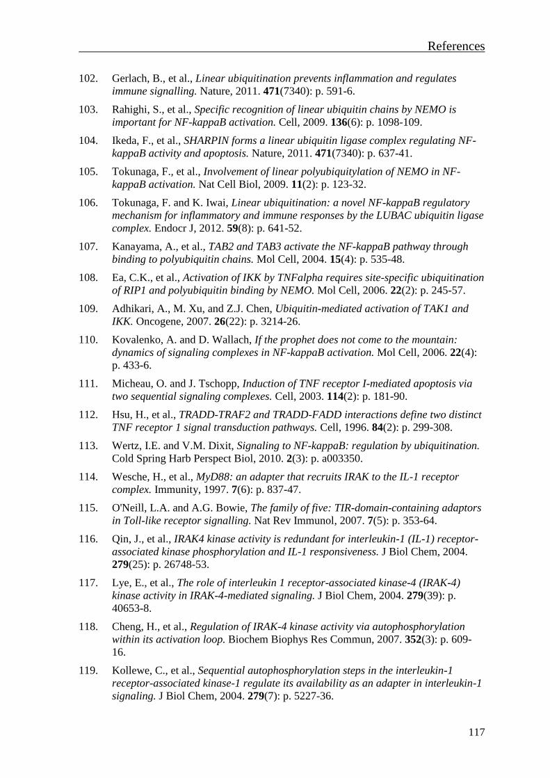
References
117
102. Gerlach, B., et al., Linear ubiquitination prevents inflammation and regulates
immune signalling. Nature, 2011. 471(7340): p. 591-6.
103. Rahighi, S., et al., Specific recognition of linear ubiquitin chains by NEMO is
important for NF-kappaB activation. Cell, 2009. 136(6): p. 1098-109.
104. Ikeda, F., et al., SHARPIN forms a linear ubiquitin ligase complex regulating NF-
kappaB activity and apoptosis. Nature, 2011. 471(7340): p. 637-41.
105. Tokunaga, F., et al., Involvement of linear polyubiquitylation of NEMO in NF-
kappaB activation. Nat Cell Biol, 2009. 11(2): p. 123-32.
106. Tokunaga, F. and K. Iwai, Linear ubiquitination: a novel NF-kappaB regulatory
mechanism for inflammatory and immune responses by the LUBAC ubiquitin ligase
complex. Endocr J, 2012. 59(8): p. 641-52.
107. Kanayama, A., et al., TAB2 and TAB3 activate the NF-kappaB pathway through
binding to polyubiquitin chains. Mol Cell, 2004. 15(4): p. 535-48.
108. Ea, C.K., et al., Activation of IKK by TNFalpha requires site-specific ubiquitination
of RIP1 and polyubiquitin binding by NEMO. Mol Cell, 2006. 22(2): p. 245-57.
109. Adhikari, A., M. Xu, and Z.J. Chen, Ubiquitin-mediated activation of TAK1 and
IKK. Oncogene, 2007. 26(22): p. 3214-26.
110. Kovalenko, A. and D. Wallach, If the prophet does not come to the mountain:
dynamics of signaling complexes in NF-kappaB activation. Mol Cell, 2006. 22(4):
p. 433-6.
111. Micheau, O. and J. Tschopp, Induction of TNF receptor I-mediated apoptosis via
two sequential signaling complexes. Cell, 2003. 114(2): p. 181-90.
112. Hsu, H., et al., TRADD-TRAF2 and TRADD-FADD interactions define two distinct
TNF receptor 1 signal transduction pathways. Cell, 1996. 84(2): p. 299-308.
113. Wertz, I.E. and V.M. Dixit, Signaling to NF-kappaB: regulation by ubiquitination.
Cold Spring Harb Perspect Biol, 2010. 2(3): p. a003350.
114. Wesche, H., et al., MyD88: an adapter that recruits IRAK to the IL-1 receptor
complex. Immunity, 1997. 7(6): p. 837-47.
115. O'Neill, L.A. and A.G. Bowie, The family of five: TIR-domain-containing adaptors
in Toll-like receptor signalling. Nat Rev Immunol, 2007. 7(5): p. 353-64.
116. Qin, J., et al., IRAK4 kinase activity is redundant for interleukin-1 (IL-1) receptor-
associated kinase phosphorylation and IL-1 responsiveness. J Biol Chem, 2004.
279(25): p. 26748-53.
117. Lye, E., et al., The role of interleukin 1 receptor-associated kinase-4 (IRAK-4)
kinase activity in IRAK-4-mediated signaling. J Biol Chem, 2004. 279(39): p.
40653-8.
118. Cheng, H., et al., Regulation of IRAK-4 kinase activity via autophosphorylation
within its activation loop. Biochem Biophys Res Commun, 2007. 352(3): p. 609-
16.
119. Kollewe, C., et al., Sequential autophosphorylation steps in the interleukin-1
receptor-associated kinase-1 regulate its availability as an adapter in interleukin-1
signaling. J Biol Chem, 2004. 279(7): p. 5227-36.

References
118
120. Cao, Z., W.J. Henzel, and X. Gao, IRAK: a kinase associated with the interleukin-1
receptor. Science, 1996. 271(5252): p. 1128-31.
121. Cui, W., et al., beta-TrCP-mediated IRAK1 degradation releases TAK1-TRAF6
from the membrane to the cytosol for TAK1-dependent NF-kappaB activation. Mol
Cell Biol, 2012. 32(19): p. 3990-4000.
122. Wooff, J., et al., The TRAF6 RING finger domain mediates physical interaction
with Ubc13. FEBS Lett, 2004. 566(1-3): p. 229-33.
123. Jiang, Z., et al., Interleukin-1 (IL-1) receptor-associated kinase-dependent IL-1-
induced signaling complexes phosphorylate TAK1 and TAB2 at the plasma
membrane and activate TAK1 in the cytosol. Mol Cell Biol, 2002. 22(20): p. 7158-
67.
124. Takaesu, G., et al., TAB2, a novel adaptor protein, mediates activation of TAK1
MAPKKK by linking TAK1 to TRAF6 in the IL-1 signal transduction pathway. Mol
Cell, 2000. 5(4): p. 649-58.
125. Neumann, M. and M. Naumann, Beyond IkappaBs: alternative regulation of NF-
kappaB activity. Faseb J, 2007. 21(11): p. 2642-54.
126. Verstrepen, L., et al., TLR-4, IL-1R and TNF-R signaling to NF-kappaB: variations
on a common theme. Cell Mol Life Sci, 2008. 65(19): p. 2964-78.
127. Yamamoto, M., et al., Essential role for TIRAP in activation of the signalling
cascade shared by TLR2 and TLR4. Nature, 2002. 420(6913): p. 324-9.
128. Horng, T., et al., The adaptor molecule TIRAP provides signalling specificity for
Toll-like receptors. Nature, 2002. 420(6913): p. 329-33.
129. Kagan, J.C., et al., TRAM couples endocytosis of Toll-like receptor 4 to the
induction of interferon-beta. Nat Immunol, 2008. 9(4): p. 361-8.
130. Cusson-Hermance, N., et al., Rip1 mediates the Trif-dependent toll-like receptor 3-
and 4-induced NF-{kappa}B activation but does not contribute to interferon
regulatory factor 3 activation. J Biol Chem, 2005. 280(44): p. 36560-6.
131. Bonizzi, G. and M. Karin, The two NF-kappaB activation pathways and their role
in innate and adaptive immunity. Trends Immunol, 2004. 25(6): p. 280-8.
132. Sun, S.C., The noncanonical NF-kappaB pathway. Immunol Rev, 2012. 246(1): p.
125-40.
133. Hostager, B.S., et al., Tumor necrosis factor receptor-associated factor 2 (TRAF2)-
deficient B lymphocytes reveal novel roles for TRAF2 in CD40 signaling. J Biol
Chem, 2003. 278(46): p. 45382-90.
134. Wallach, D. and A. Kovalenko, Self-termination of the terminator. Nat Immunol,
2008. 9(12): p. 1325-7.
135. Varfolomeev, E. and D. Vucic, (Un)expected roles of c-IAPs in apoptotic and
NFkappaB signaling pathways. Cell Cycle, 2008. 7(11): p. 1511-21.
136. Brzoska, K. and I. Szumiel, Signalling loops and linear pathways: NF-kappaB
activation in response to genotoxic stress. Mutagenesis, 2009. 24(1): p. 1-8.
References
119
137. Schoonbroodt, S., et al., Crucial role of the amino-terminal tyrosine residue 42 and
the carboxyl-terminal PEST domain of I kappa B alpha in NF-kappa B activation
by an oxidative stress. J Immunol, 2000. 164(8): p. 4292-300.
138. Franchi, L., et al., Function of Nod-like receptors in microbial recognition and host
defense. Immunol Rev, 2009. 227(1): p. 106-28.
139. Chen, G., et al., NOD-like receptors: role in innate immunity and inflammatory
disease. Annu Rev Pathol, 2009. 4: p. 365-98.
140. Vallabhapurapu, S. and M. Karin, Regulation and function of NF-kappaB
transcription factors in the immune system. Annu Rev Immunol, 2009. 27: p. 693-
733.
141. Kawai, T. and S. Akira, Toll-like receptor and RIG-I-like receptor signaling. Ann
N Y Acad Sci, 2008. 1143: p. 1-20.
142. Janssens, S. and J. Tschopp, Signals from within: the DNA-damage-induced NF-
kappaB response. Cell Death Differ, 2006. 13(5): p. 773-84.
143. Stilmann, M., et al., A nuclear poly(ADP-ribose)-dependent signalosome confers
DNA damage-induced IkappaB kinase activation. Mol Cell, 2009. 36(3): p. 365-78.
144. Hinz, M., et al., A cytoplasmic ATM-TRAF6-cIAP1 module links nuclear DNA
damage signaling to ubiquitin-mediated NF-kappaB activation. Mol Cell, 2010.
40(1): p. 63-74.
145. Wu, Z.H., et al., ATM- and NEMO-dependent ELKS ubiquitination coordinates
TAK1-mediated IKK activation in response to genotoxic stress. Mol Cell, 2010.
40(1): p. 75-86.
146. Bock, F.J., et al., Loss of PIDD limits NF-kappaB activation and cytokine
production but not cell survival or transformation after DNA damage. Cell Death
Differ, 2013. 20(4): p. 546-57.
147. McCool, K.W. and S. Miyamoto, DNA damage-dependent NF-kappaB activation:
NEMO turns nuclear signaling inside out. Immunol Rev, 2012. 246(1): p. 311-26.
148. Niu, J., et al., LUBAC regulates NF-kappaB activation upon genotoxic stress by
promoting linear ubiquitination of NEMO. Embo J, 2011. 30(18): p. 3741-53.
149. Hou, S., H. Guan, and R.P. Ricciardi, Phosphorylation of serine 337 of NF-kappaB
p50 is critical for DNA binding. J Biol Chem, 2003. 278(46): p. 45994-8.
150. Harris, J., et al., Nuclear accumulation of cRel following C-terminal
phosphorylation by TBK1/IKK epsilon. J Immunol, 2006. 177(4): p. 2527-35.
151. Wan, F. and M.J. Lenardo, Specification of DNA binding activity of NF-kappaB
proteins. Cold Spring Harb Perspect Biol, 2009. 1(4): p. a000067.
152. Vermeulen, L., et al., Transcriptional activation of the NF-kappaB p65 subunit by
mitogen- and stress-activated protein kinase-1 (MSK1). Embo J, 2003. 22(6): p.
1313-24.
153. Hochrainer, K., G. Racchumi, and J. Anrather, Hypo-phosphorylation leads to
nuclear retention of NF-kappaB p65 due to impaired IkappaBalpha gene synthesis.
FEBS Lett, 2007. 581(28): p. 5493-9.
References
120
154. Zhong, H., R.E. Voll, and S. Ghosh, Phosphorylation of NF-kappa B p65 by PKA
stimulates transcriptional activity by promoting a novel bivalent interaction with
the coactivator CBP/p300. Mol Cell, 1998. 1(5): p. 661-71.
155. Zhong, H., et al., The phosphorylation status of nuclear NF-kappa B determines its
association with CBP/p300 or HDAC-1. Mol Cell, 2002. 9(3): p. 625-36.
156. Nihira, K., et al., Pim-1 controls NF-kappaB signalling by stabilizing RelA/p65.
Cell Death Differ. 17(4): p. 689-98.
157. Zhong, H., et al., The transcriptional activity of NF-kappaB is regulated by the
IkappaB-associated PKAc subunit through a cyclic AMP-independent mechanism.
Cell, 1997. 89(3): p. 413-24.
158. Reber, L., et al., Ser276 phosphorylation of NF-kB p65 by MSK1 controls SCF
expression in inflammation. PLoS One, 2009. 4(2): p. e4393.
159. Dong, J., et al., Repression of gene expression by unphosphorylated NF-kappaB
p65 through epigenetic mechanisms. Genes Dev, 2008. 22(9): p. 1159-73.
160. Anrather, J., G. Racchumi, and C. Iadecola, cis-acting, element-specific
transcriptional activity of differentially phosphorylated nuclear factor-kappa B. J
Biol Chem, 2005. 280(1): p. 244-52.
161. Nowak, D.E., et al., RelA Ser276 phosphorylation is required for activation of a
subset of NF-kappaB-dependent genes by recruiting cyclin-dependent kinase
9/cyclin T1 complexes. Mol Cell Biol, 2008. 28(11): p. 3623-38.
162. Hochrainer, K., G. Racchumi, and J. Anrather, Site-specific phosphorylation of the
p65 protein subunit mediates selective gene expression by differential NF-kappaB
and RNA polymerase II promoter recruitment. J Biol Chem, 2013. 288(1): p. 285-
93.
163. Hoesel, B. and J.A. Schmid, The complexity of NF-kappaB signaling in
inflammation and cancer. Mol Cancer, 2013. 12: p. 86.
164. Ryo, A., et al., Regulation of NF-kappaB signaling by Pin1-dependent prolyl
isomerization and ubiquitin-mediated proteolysis of p65/RelA. Mol Cell, 2003.
12(6): p. 1413-26.
165. Duran, A., M.T. Diaz-Meco, and J. Moscat, Essential role of RelA Ser311
phosphorylation by zetaPKC in NF-kappaB transcriptional activation. Embo J,
2003. 22(15): p. 3910-8.
166. Moreno, R., et al., Specification of the NF-kappaB transcriptional response by p65
phosphorylation and TNF-induced nuclear translocation of IKK epsilon. Nucleic
Acids Res, 2010. 38(18): p. 6029-44.
167. Sakurai, H., et al., Tumor necrosis factor-alpha-induced IKK phosphorylation of
NF-kappaB p65 on serine 536 is mediated through the TRAF2, TRAF5, and TAK1
signaling pathway. J Biol Chem, 2003. 278(38): p. 36916-23.
168. Mattioli, I., et al., Transient and selective NF-kappa B p65 serine 536
phosphorylation induced by T cell costimulation is mediated by I kappa B kinase
beta and controls the kinetics of p65 nuclear import. J Immunol, 2004. 172(10): p.
6336-44.
References
121
169. Wittwer, T. and M.L. Schmitz, NIK and Cot cooperate to trigger NF-kappaB p65
phosphorylation. Biochem Biophys Res Commun, 2008. 371(2): p. 294-7.
170. Buss, H., et al., Constitutive and interleukin-1-inducible phosphorylation of p65
NF-{kappa}B at serine 536 is mediated by multiple protein kinases including
I{kappa}B kinase (IKK)-{alpha}, IKK{beta}, IKK{epsilon}, TRAF family member-
associated (TANK)-binding kinase 1 (TBK1), and an unknown kinase and couples
p65 to TATA-binding protein-associated factor II31-mediated interleukin-8
transcription. J Biol Chem, 2004. 279(53): p. 55633-43.
171. Jeong, S.J., et al., A novel NF-kappaB pathway involving IKKbeta and p65/RelA
Ser-536 phosphorylation results in p53 Inhibition in the absence of NF-kappaB
transcriptional activity. J Biol Chem, 2005. 280(11): p. 10326-32.
172. Bohuslav, J., et al., p53 induces NF-kappaB activation by an IkappaB kinase-
independent mechanism involving phosphorylation of p65 by ribosomal S6 kinase
1. J Biol Chem, 2004. 279(25): p. 26115-25.
173. Buss, H., et al., Cyclin-dependent kinase 6 phosphorylates NF-kappaB P65 at
serine 536 and contributes to the regulation of inflammatory gene expression.
PLoS One, 2012. 7(12): p. e51847.
174. Hoberg, J.E., et al., IkappaB kinase alpha-mediated derepression of SMRT
potentiates acetylation of RelA/p65 by p300. Mol Cell Biol, 2006. 26(2): p. 457-71.
175. Chen, L.F., et al., NF-kappaB RelA phosphorylation regulates RelA acetylation.
Mol Cell Biol, 2005. 25(18): p. 7966-75.
176. Lawrence, T., et al., IKKalpha limits macrophage NF-kappaB activation and
contributes to the resolution of inflammation. Nature, 2005. 434(7037): p. 1138-43.
177. Schwabe, R.F. and H. Sakurai, IKKbeta phosphorylates p65 at S468 in
transactivaton domain 2. Faseb J, 2005. 19(12): p. 1758-60.
178. Buss, H., et al., Phosphorylation of serine 468 by GSK-3beta negatively regulates
basal p65 NF-kappaB activity. J Biol Chem, 2004. 279(48): p. 49571-4.
179. Mattioli, I., et al., Inducible phosphorylation of NF-kappa B p65 at serine 468 by T
cell costimulation is mediated by IKK epsilon. J Biol Chem, 2006. 281(10): p.
6175-83.
180. Mukherjee, S.P., et al., Analysis of the RelA:CBP/p300 Interaction Reveals Its
Involvement in NF-kappaB-Driven Transcription. PLoS Biol, 2013. 11(9): p.
e1001647.
181. Bensinger, S.J. and P. Tontonoz, A Nurr1 pathway for neuroprotection. Cell, 2009.
137(1): p. 26-8.
182. Geng, H., et al., Phosphorylation of NF-kappaB p65 at Ser468 controls its
COMMD1-dependent ubiquitination and target gene-specific proteasomal
elimination. EMBO Rep, 2009. 10(4): p. 381-6.
183. Mao, X., et al., GCN5 is a required cofactor for a ubiquitin ligase that targets NF-
kappaB/RelA. Genes Dev, 2009. 23(7): p. 849-61.
184. Thoms, H.C., et al., Nucleolar targeting of RelA(p65) is regulated by COMMD1-
dependent ubiquitination. Cancer Res, 2010. 70(1): p. 139-49.
References
122
185. O'Shea, J.M. and N.D. Perkins, Thr435 phosphorylation regulates RelA (p65) NF-
kappaB subunit transactivation. Biochem J, 2010. 426(3): p. 345-54.
186. Rocha, S., et al., Regulation of NF-kappaB and p53 through activation of ATR and
Chk1 by the ARF tumour suppressor. Embo J, 2005. 24(6): p. 1157-69.
187. Campbell, K.J., et al., Cisplatin mimics ARF tumor suppressor regulation of RelA
(p65) nuclear factor-kappaB transactivation. Cancer Res, 2006. 66(2): p. 929-35.
188. Sabatel, H., et al., Phosphorylation of p65(RelA) on Ser(547) by ATM represses
NF-kappaB-dependent transcription of specific genes after genotoxic stress. PLoS
One, 2012. 7(6): p. e38246.
189. O'Mahony, A.M., et al., Human T-cell lymphotropic virus type 1 tax induction of
biologically Active NF-kappaB requires IkappaB kinase-1-mediated
phosphorylation of RelA/p65. J Biol Chem, 2004. 279(18): p. 18137-45.
190. Wang, D., et al., Tumor necrosis factor alpha-induced phosphorylation of RelA/p65
on Ser529 is controlled by casein kinase II. J Biol Chem, 2000. 275(42): p. 32592-
7.
191. Bae, J.S., et al., Phosphorylation of NF-kappa B by calmodulin-dependent kinase
IV activates anti-apoptotic gene expression. Biochem Biophys Res Commun, 2003.
305(4): p. 1094-8.
192. Yang, J., et al., Protein phosphatase 2A interacts with and directly
dephosphorylates RelA. J Biol Chem, 2001. 276(51): p. 47828-33.
193. Li, S., et al., RNAi screen in mouse astrocytes identifies phosphatases that regulate
NF-kappaB signaling. Mol Cell, 2006. 24(4): p. 497-509.
194. Chew, J., et al., WIP1 phosphatase is a negative regulator of NF-kappaB
signalling. Nat Cell Biol, 2009. 11(5): p. 659-66.
195. Yeh, P.Y., et al., Suppression of MEK/ERK signaling pathway enhances cisplatin-
induced NF-kappaB activation by protein phosphatase 4-mediated NF-kappaB p65
Thr dephosphorylation. J Biol Chem, 2004. 279(25): p. 26143-8.
196. Quivy, V. and C. Van Lint, Regulation at multiple levels of NF-kappaB-mediated
transactivation by protein acetylation. Biochem Pharmacol, 2004. 68(6): p. 1221-9.
197. Kiernan, R., et al., Post-activation turn-off of NF-kappa B-dependent transcription
is regulated by acetylation of p65. J Biol Chem, 2003. 278(4): p. 2758-66.
198. Chen, L.F., Y. Mu, and W.C. Greene, Acetylation of RelA at discrete sites regulates
distinct nuclear functions of NF-kappaB. Embo J, 2002. 21(23): p. 6539-48.
199. Rothgiesser, K.M., M. Fey, and M.O. Hottiger, Acetylation of p65 at lysine 314 is
important for late NF-kappaB-dependent gene expression. BMC Genomics, 2010.
11: p. 22.
200. Buerki, C., et al., Functional relevance of novel p300-mediated lysine 314 and 315
acetylation of RelA/p65. Nucleic Acids Res, 2008. 36(5): p. 1665-80.
201. Li, H., et al., Regulation of NF-kappaB activity by competition between RelA
acetylation and ubiquitination. Oncogene, 2012. 31(5): p. 611-23.
202. Ashburner, B.P., S.D. Westerheide, and A.S. Baldwin, Jr., The p65 (RelA) subunit
of NF-kappaB interacts with the histone deacetylase (HDAC) corepressors HDAC1
References
123
and HDAC2 to negatively regulate gene expression. Mol Cell Biol, 2001. 21(20): p.
7065-77.
203. Liu, Y., P.W. Smith, and D.R. Jones, Breast cancer metastasis suppressor 1
functions as a corepressor by enhancing histone deacetylase 1-mediated
deacetylation of RelA/p65 and promoting apoptosis. Mol Cell Biol, 2006. 26(23):
p. 8683-96.
204. Yeung, F., et al., Modulation of NF-kappaB-dependent transcription and cell
survival by the SIRT1 deacetylase. Embo J, 2004. 23(12): p. 2369-80.
205. Rothgiesser, K.M., et al., SIRT2 regulates NF-kappaB dependent gene expression
through deacetylation of p65 Lys310. J Cell Sci, 2010. 123(Pt 24): p. 4251-8.
206. Ziesche, E., et al., The coactivator role of histone deacetylase 3 in IL-1-signaling
involves deacetylation of p65 NF-kappaB. Nucleic Acids Res, 2012. 41(1): p. 90-
109.
207. Chen, L., et al., Duration of nuclear NF-kappaB action regulated by reversible
acetylation. Science, 2001. 293(5535): p. 1653-7.
208. Natoli, G. and S. Chiocca, Nuclear ubiquitin ligases, NF-kappaB degradation, and
the control of inflammation. Sci Signal, 2008. 1(1): p. pe1.
209. Saccani, S., et al., Degradation of promoter-bound p65/RelA is essential for the
prompt termination of the nuclear factor kappaB response. J Exp Med, 2004.
200(1): p. 107-13.
210. Maine, G.N., et al., COMMD1 promotes the ubiquitination of NF-kappaB subunits
through a cullin-containing ubiquitin ligase. Embo J, 2007. 26(2): p. 436-47.
211. Strebovsky, J., et al., Suppressor of cytokine signaling 1 (SOCS1) limits NFkappaB
signaling by decreasing p65 stability within the cell nucleus. Faseb J, 2011. 25(3):
p. 863-74.
212. Tanaka, T., M.J. Grusby, and T. Kaisho, PDLIM2-mediated termination of
transcription factor NF-kappaB activation by intranuclear sequestration and
degradation of the p65 subunit. Nat Immunol, 2007. 8(6): p. 584-91.
213. Yang, X.D., et al., Negative regulation of NF-kappaB action by Set9-mediated
lysine methylation of the RelA subunit. Embo J, 2009. 28(8): p. 1055-66.
214. Ea, C.K. and D. Baltimore, Regulation of NF-kappaB activity through lysine
monomethylation of p65. Proc Natl Acad Sci U S A, 2009. 106(45): p. 18972-7.
215. Lu, T., et al., Regulation of NF-kappaB by NSD1/FBXL11-dependent reversible
lysine methylation of p65. Proc Natl Acad Sci U S A, 2010. 107(1): p. 46-51.
216. Zhang, T., et al., PHF20 regulates NF-kappaB signalling by disrupting recruitment
of PP2A to p65. Nat Commun, 2013. 4: p. 2062.
217. Levy, D., et al., Lysine methylation of the NF-kappaB subunit RelA by SETD6
couples activity of the histone methyltransferase GLP at chromatin to tonic
repression of NF-kappaB signaling. Nat Immunol, 2011. 12(1): p. 29-36.
218. Chang, Y., et al., Structural basis of SETD6-mediated regulation of the NF-kB
network via methyl-lysine signaling. Nucleic Acids Res, 2011. 39(15): p. 6380-9.
References
124
219. Wei, H., et al., PRMT5 dimethylates R30 of the p65 subunit to activate NF-kappaB.
Proc Natl Acad Sci U S A, 2013. 110(33): p. 13516-21.
220. Bartlett, A.I. and S.E. Radford, An expanding arsenal of experimental methods
yields an explosion of insights into protein folding mechanisms. Nat Struct Mol
Biol, 2009. 16(6): p. 582-8.
221. Dunker, A.K., et al., Function and structure of inherently disordered proteins. Curr
Opin Struct Biol, 2008. 18(6): p. 756-64.
222. Hartl, F.U., A. Bracher, and M. Hayer-Hartl, Molecular chaperones in protein
folding and proteostasis. Nature, 2011. 475(7356): p. 324-32.
223. Jego, G., et al., Targeting heat shock proteins in cancer. Cancer Lett, 2010. 332(2):
p. 275-85.
224. Garrido, C., et al., Heat shock proteins 27 and 70: anti-apoptotic proteins with
tumorigenic properties. Cell Cycle, 2006. 5(22): p. 2592-601.
225. Lanneau, D., et al., Heat shock proteins: cell protection through protein triage.
ScientificWorldJournal, 2010. 10: p. 1543-52.
226. Parcellier, A., et al., HSP27 favors ubiquitination and proteasomal degradation of
p27Kip1 and helps S-phase re-entry in stressed cells. Faseb J, 2006. 20(8): p. 1179-
81.
227. Lanneau, D., et al., Apoptosis versus cell differentiation: role of heat shock proteins
HSP90, HSP70 and HSP27. Prion, 2007. 1(1): p. 53-60.
228. Onuoha, S.C., et al., Structural studies on the co-chaperone Hop and its complexes
with Hsp90. J Mol Biol, 2008. 379(4): p. 732-44.
229. Taipale, M., D.F. Jarosz, and S. Lindquist, HSP90 at the hub of protein
homeostasis: emerging mechanistic insights. Nat Rev Mol Cell Biol, 2010. 11(7):
p. 515-28.
230. McClellan, A.J., et al., Diverse cellular functions of the Hsp90 molecular
chaperone uncovered using systems approaches. Cell, 2007. 131(1): p. 121-35.
231. Pratt, W.B. and D.O. Toft, Regulation of signaling protein function and trafficking
by the hsp90/hsp70-based chaperone machinery. Exp Biol Med (Maywood), 2003.
228(2): p. 111-33.
232. Sharma, K., et al., Quantitative proteomics reveals that Hsp90 inhibition
preferentially targets kinases and the DNA damage response. Mol Cell Proteomics,
2012. 11(3): p. M111 014654.
233. Jaattela, M., Heat shock proteins as cellular lifeguards. Ann Med, 1999. 31(4): p.
261-71.
234. Tavaria, M., et al., A hitchhiker's guide to the human Hsp70 family. Cell Stress
Chaperones, 1996. 1(1): p. 23-8.
235. Kiang, J.G. and G.C. Tsokos, Heat shock protein 70 kDa: molecular biology,
biochemistry, and physiology. Pharmacol Ther, 1998. 80(2): p. 183-201.
236. Young, J.C., J.M. Barral, and F. Ulrich Hartl, More than folding: localized
functions of cytosolic chaperones. Trends Biochem Sci, 2003. 28(10): p. 541-7.
References
125
237. Mayer, M.P., S. Rudiger, and B. Bukau, Molecular basis for interactions of the
DnaK chaperone with substrates. Biol Chem, 2000. 381(9-10): p. 877-85.
238. Connell, P., et al., The co-chaperone CHIP regulates protein triage decisions
mediated by heat-shock proteins. Nat Cell Biol, 2001. 3(1): p. 93-6.
239. Jiang, J., et al., Chaperone-dependent regulation of endothelial nitric-oxide
synthase intracellular trafficking by the co-chaperone/ubiquitin ligase CHIP. J Biol
Chem, 2003. 278(49): p. 49332-41.
240. Dai, Q., et al., CHIP activates HSF1 and confers protection against apoptosis and
cellular stress. Embo J, 2003. 22(20): p. 5446-58.
241. Gannon, J.V., et al., Activating mutations in p53 produce a common
conformational effect. A monoclonal antibody specific for the mutant form. Embo J,
1990. 9(5): p. 1595-602.
242. Klenke, C., et al., Hsc70 Is a Novel Interactor of NF-kappaB p65 in Living
Hippocampal Neurons. PLoS One, 2013. 8(6): p. e65280.
243. Gao, T. and A.C. Newton, The turn motif is a phosphorylation switch that regulates
the binding of Hsp70 to protein kinase C. J Biol Chem, 2002. 277(35): p. 31585-92.
244. Nylandsted, J., K. Brand, and M. Jaattela, Heat shock protein 70 is required for the
survival of cancer cells. Ann N Y Acad Sci, 2000. 926: p. 122-5.
245. Gurbuxani, S., et al., Heat shock protein 70 binding inhibits the nuclear import of
apoptosis-inducing factor. Oncogene, 2003. 22(43): p. 6669-78.
246. Zhang, L., et al., Hsp70 inhibition induces myeloma cell death via the intracellular
accumulation of immunoglobulin and the generation of proteotoxic stress. Cancer
Lett, 2013. 339(1): p. 49-59.
247. Massey, A.J., et al., A novel, small molecule inhibitor of Hsc70/Hsp70 potentiates
Hsp90 inhibitor induced apoptosis in HCT116 colon carcinoma cells. Cancer
Chemother Pharmacol, 2010. 66(3): p. 535-45.
248. Chen, G., P. Cao, and D.V. Goeddel, TNF-induced recruitment and activation of
the IKK complex require Cdc37 and Hsp90. Mol Cell, 2002. 9(2): p. 401-10.
249. Broemer, M., D. Krappmann, and C. Scheidereit, Requirement of Hsp90 activity for
IkappaB kinase (IKK) biosynthesis and for constitutive and inducible IKK and NF-
kappaB activation. Oncogene, 2004. 23(31): p. 5378-86.
250. Lewis, J., et al., Disruption of hsp90 function results in degradation of the death
domain kinase, receptor-interacting protein (RIP), and blockage of tumor necrosis
factor-induced nuclear factor-kappaB activation. J Biol Chem, 2000. 275(14): p.
10519-26.
251. Sato, S., N. Fujita, and T. Tsuruo, Modulation of Akt kinase activity by binding to
Hsp90. Proc Natl Acad Sci U S A, 2000. 97(20): p. 10832-7.
252. Fisher, D.L., E. Mandart, and M. Doree, Hsp90 is required for c-Mos activation
and biphasic MAP kinase activation in Xenopus oocytes. Embo J, 2000. 19(7): p.
1516-24.
253. Citri, A., et al., Hsp90 recognizes a common surface on client kinases. J Biol
Chem, 2006. 281(20): p. 14361-9.
References
126
254. Qing, G., et al., Hsp90 regulates processing of NF-kappa B2 p100 involving
protection of NF-kappa B-inducing kinase (NIK) from autophagy-mediated
degradation. Cell Res, 2007. 17(6): p. 520-30.
255. Ran, R., et al., Hsp70 promotes TNF-mediated apoptosis by binding IKK gamma
and impairing NF-kappa B survival signaling. Genes Dev, 2004. 18(12): p. 1466-
81.
256. Parcellier, A., et al., HSP27 is a ubiquitin-binding protein involved in I-
kappaBalpha proteasomal degradation. Mol Cell Biol, 2003. 23(16): p. 5790-802.
257. Guzhova, I.V., et al., Major stress protein Hsp70 interacts with NF-kB regulatory
complex in human T-lymphoma cells. Cell Stress Chaperones, 1997. 2(2): p. 132-9.
258. Johnson, R.F., Witzel, II, and N.D. Perkins, p53-dependent regulation of
mitochondrial energy production by the RelA subunit of NF-kappaB. Cancer Res,
2011. 71(16): p. 5588-97.
259. Leung, T.H., A. Hoffmann, and D. Baltimore, One nucleotide in a kappaB site can
determine cofactor specificity for NF-kappaB dimers. Cell, 2004. 118(4): p. 453-
64.
260. Chen-Park, F.E., et al., The kappa B DNA sequence from the HIV long terminal
repeat functions as an allosteric regulator of HIV transcription. J Biol Chem, 2002.
277(27): p. 24701-8.
261. Zhang, T., et al., SPINE-D: accurate prediction of short and long disordered
regions by a single neural-network based method. J Biomol Struct Dyn, 2012.
29(4): p. 799-813.
262. Groban, E.S., A. Narayanan, and M.P. Jacobson, Conformational changes in
protein loops and helices induced by post-translational phosphorylation. PLoS
Comput Biol, 2006. 2(4): p. e32.
263. Zheng, H., et al., The prolyl isomerase Pin1 is a regulator of p53 in genotoxic
response. Nature, 2002. 419(6909): p. 849-53.
264. Schwabe, R.F., et al., Differential role of I kappa B kinase 1 and 2 in primary rat
hepatocytes. Hepatology, 2001. 33(1): p. 81-90.
265. Zabel, U., et al., Nuclear uptake control of NF-kappa B by MAD-3, an I kappa B
protein present in the nucleus. Embo J, 1993. 12(1): p. 201-11.
266. Gerritsen, M.E., et al., CREB-binding protein/p300 are transcriptional coactivators
of p65. Proc Natl Acad Sci U S A, 1997. 94(7): p. 2927-32.
267. Owen, H.R., et al., MYBBP1a is a novel repressor of NF-kappaB. J Mol Biol, 2007.
366(3): p. 725-36.
268. Owen, H.R., et al., Identification of novel and cell type enriched cofactors of the
transcription activation domain of RelA (p65 NF-kappaB). J Proteome Res, 2005.
4(4): p. 1381-90.
269. Kim, Y.E., et al., Molecular chaperone functions in protein folding and
proteostasis. Annu Rev Biochem, 2013. 82: p. 323-55.
270. Nakano, M., et al., Tumor necrosis factor-alpha-induced expression of heat shock
protein 72 in adult feline cardiac myocytes. Am J Physiol, 1996. 270(4 Pt 2): p.
H1231-9.
References
127
271. Dokladny, K., et al., LPS-induced cytokine levels are repressed by elevated
expression of HSP70 in rats: possible role of NF-kappaB. Cell Stress Chaperones,
2010. 15(2): p. 153-63.
272. Neckers, L., Hsp90 inhibitors as novel cancer chemotherapeutic agents. Trends
Mol Med, 2002. 8(4 Suppl): p. S55-61.
273. Roe, S.M., et al., Structural basis for inhibition of the Hsp90 molecular chaperone
by the antitumor antibiotics radicicol and geldanamycin. J Med Chem, 1999. 42(2):
p. 260-6.
274. Rebelo, S., et al., Monitoring "De Novo"APP synthesis by taking advantage of the
reversible effect of cycloheximide. Am J Alzheimers Dis Other Demen, 2008.
23(6): p. 602-8.
275. Costantino, P. and G. Attardi, Metabolic properties of the products of
mitochondrial protein synthesis in HeLa cells. J Biol Chem, 1977. 252(5): p. 1702-
11.
276. Mayer, M.P. and B. Bukau, Hsp70 chaperones: cellular functions and molecular
mechanism. Cell Mol Life Sci, 2005. 62(6): p. 670-84.
277. Richter, K. and J. Buchner, Hsp90: chaperoning signal transduction. J Cell
Physiol, 2001. 188(3): p. 281-90.
278. Toledano, M.B., et al., N-terminal DNA-binding domains contribute to differential
DNA-binding specificities of NF-kappa B p50 and p65. Mol Cell Biol, 1993. 13(2):
p. 852-60.
279. Ganchi, P.A., et al., I kappa B/MAD-3 masks the nuclear localization signal of NF-
kappa B p65 and requires the transactivation domain to inhibit NF-kappa B p65
DNA binding. Mol Biol Cell, 1992. 3(12): p. 1339-52.
280. Tian, B., D.E. Nowak, and A.R. Brasier, A TNF-induced gene expression program
under oscillatory NF-kappaB control. BMC Genomics, 2005. 6: p. 137.
281. Bren, G.D., et al., Transcription of the RelB gene is regulated by NF-kappaB.
Oncogene, 2001. 20(53): p. 7722-33.
282. Hannink, M. and H.M. Temin, Structure and autoregulation of the c-rel promoter.
Oncogene, 1990. 5(12): p. 1843-50.
283. Perkins, N.D., Achieving transcriptional specificity with NF-kappa B. Int J
Biochem Cell Biol, 1997. 29(12): p. 1433-48.
284. Barken, D., et al., Comment on "Oscillations in NF-kappaB signaling control the
dynamics of gene expression". Science, 2005. 308(5718): p. 52; author reply 52.
285. Davanian, H., et al., Signaling pathways involved in the regulation of TNFalpha-
induced toll-like receptor 2 expression in human gingival fibroblasts. Cytokine,
2012. 57(3): p. 406-16.
286. Phulwani, N.K., et al., TLR2 expression in astrocytes is induced by TNF-alpha- and
NF-kappa B-dependent pathways. J Immunol, 2008. 181(6): p. 3841-9.
287. Chen, C.C., et al., TNF-alpha-induced cyclooxygenase-2 expression in human lung
epithelial cells: involvement of the phospholipase C-gamma 2, protein kinase C-
alpha, tyrosine kinase, NF-kappa B-inducing kinase, and I-kappa B kinase 1/2
pathway. J Immunol, 2000. 165(5): p. 2719-28.
References
128
288. Zhou, A., et al., Identification of NF-kappa B-regulated genes induced by
TNFalpha utilizing expression profiling and RNA interference. Oncogene, 2003.
22(13): p. 2054-64.
289. Hayden, M.S. and S. Ghosh, NF-kappaB, the first quarter-century: remarkable
progress and outstanding questions. Genes Dev, 2012. 26(3): p. 203-34.
290. Nolan, G.P., et al., DNA binding and I kappa B inhibition of the cloned p65 subunit
of NF-kappa B, a rel-related polypeptide. Cell, 1991. 64(5): p. 961-9.
291. Liu, Y., et al., Phosphorylation of RelA/p65 promotes DNMT-1 recruitment to
chromatin and represses transcription of the tumor metastasis suppressor gene
BRMS1. Oncogene, 2012. 31(9): p. 1143-54.
292. Garza, A.M., S.H. Khan, and R. Kumar, Site-specific phosphorylation induces
functionally active conformation in the intrinsically disordered N-terminal
activation function (AF1) domain of the glucocorticoid receptor. Mol Cell Biol,
2010. 30(1): p. 220-30.
293. Mertens, C., et al., Dephosphorylation of phosphotyrosine on STAT1 dimers
requires extensive spatial reorientation of the monomers facilitated by the N-
terminal domain. Genes Dev, 2006. 20(24): p. 3372-81.
294. Wenta, N., et al., Tyrosine phosphorylation regulates the partitioning of STAT1
between different dimer conformations. Proc Natl Acad Sci U S A, 2008. 105(27):
p. 9238-43.
295. Brent, M.M., R. Anand, and R. Marmorstein, Structural basis for DNA recognition
by FoxO1 and its regulation by posttranslational modification. Structure, 2008.
16(9): p. 1407-16.
296. Yang, S.H., et al., The mechanism of phosphorylation-inducible activation of the
ETS-domain transcription factor Elk-1. Embo J, 1999. 18(20): p. 5666-74.
297. Sakaguchi, K., et al., DNA damage activates p53 through a phosphorylation-
acetylation cascade. Genes Dev, 1998. 12(18): p. 2831-41.
298. Turowski, P., et al., Differential methylation and altered conformation of
cytoplasmic and nuclear forms of protein phosphatase 2A during cell cycle
progression. J Cell Biol, 1995. 129(2): p. 397-410.
299. Finley, D., B. Bartel, and A. Varshavsky, The tails of ubiquitin precursors are
ribosomal proteins whose fusion to ubiquitin facilitates ribosome biogenesis.
Nature, 1989. 338(6214): p. 394-401.
300. Sasaki, M., L. Nie, and C.G. Maki, MDM2 binding induces a conformational
change in p53 that is opposed by heat-shock protein 90 and precedes p53
proteasomal degradation. J Biol Chem, 2007. 282(19): p. 14626-34.
301. Ulrich, H.D., SUMO modification: wrestling with protein conformation. Curr Biol,
2005. 15(7): p. R257-9.
302. Kim, J.W., et al., New molecular bridge between RelA/p65 and NF-kappaB target
genes via histone acetyltransferase TIP60 cofactor. J Biol Chem, 2012. 287(10): p.
7780-91.
303. Sen, N., et al., Hydrogen sulfide-linked sulfhydration of NF-kappaB mediates its
antiapoptotic actions. Mol Cell, 2012. 45(1): p. 13-24.
References
129
304. Fontana, A., et al., Correlation between sites of limited proteolysis and segmental
mobility in thermolysin. Biochemistry, 1986. 25(8): p. 1847-51.
305. Fontana, A., et al., Probing protein structure by limited proteolysis. Acta Biochim
Pol, 2004. 51(2): p. 299-321.
306. Iakoucheva, L.M., et al., The importance of intrinsic disorder for protein
phosphorylation. Nucleic Acids Res, 2004. 32(3): p. 1037-49.
307. Gao, J., et al., Musite, a tool for global prediction of general and kinase-specific
phosphorylation sites. Mol Cell Proteomics, 2010. 9(12): p. 2586-600.
308. Radivojac, P., et al., Identification, analysis, and prediction of protein
ubiquitination sites. Proteins, 2010. 78(2): p. 365-80.
309. Legros, Y., et al., Mutations in p53 produce a common conformational effect that
can be detected with a panel of monoclonal antibodies directed toward the central
part of the p53 protein. Oncogene, 1994. 9(12): p. 3689-94.
310. Antz, C., et al., Control of K+ channel gating by protein phosphorylation:
structural switches of the inactivation gate. Nat Struct Biol, 1999. 6(2): p. 146-50.
311. Nielsen, G. and H. Schwalbe, NMR spectroscopic investigations of the activated
p38alpha mitogen-activated protein kinase. Chembiochem, 2011. 12(17): p. 2599-
607.
312. Perez, Y., et al., Structural characterization of the natively unfolded N-terminal
domain of human c-Src kinase: insights into the role of phosphorylation of the
unique domain. J Mol Biol, 2009. 391(1): p. 136-48.
313. Tait, S., et al., Local control of a disorder-order transition in 4E-BP1 underpins
regulation of translation via eIF4E. Proc Natl Acad Sci U S A, 2010. 107(41): p.
17627-32.
314. Martenson, R.E., The use of gel filtration to follow conformational changes in
proteins. Conformational flexibility of bovine myelin basic protein. J Biol Chem,
1978. 253(24): p. 8887-93.
315. Karst, J.C., et al., Estimation of intrinsically disordered protein shape and time-
averaged apparent hydration in native conditions by a combination of
hydrodynamic methods. Methods Mol Biol, 2012. 896: p. 163-77.
316. Wales, T.E. and J.R. Engen, Hydrogen exchange mass spectrometry for the
analysis of protein dynamics. Mass Spectrom Rev, 2006. 25(1): p. 158-70.
317. Craig, A.L., et al., Novel phosphorylation sites of human tumour suppressor protein
p53 at Ser20 and Thr18 that disrupt the binding of mdm2 (mouse double minute 2)
protein are modified in human cancers. Biochem J, 1999. 342 ( Pt 1): p. 133-41.
318. Lee, C.W., et al., Graded enhancement of p53 binding to CREB-binding protein
(CBP) by multisite phosphorylation. Proc Natl Acad Sci U S A, 2010. 107(45): p.
19290-5.
319. Jabbur, J.R. and W. Zhang, p53 Antiproliferative function is enhanced by aspartate
substitution at threonine 18 and serine 20. Cancer Biol Ther, 2002. 1(3): p. 277-83.
320. Dumaz, N., et al., Critical roles for the serine 20, but not the serine 15,
phosphorylation site and for the polyproline domain in regulating p53 turnover.
Biochem J, 2001. 359(Pt 2): p. 459-64.
References
130
321. Nihira, K., et al., Pim-1 controls NF-kappaB signalling by stabilizing RelA/p65.
Cell Death Differ, 2010. 17(4): p. 689-98.
322. Brasier, A.R., et al., RelA Ser276 phosphorylation-coupled Lys310 acetylation
controls transcriptional elongation of inflammatory cytokines in respiratory
syncytial virus infection. J Virol, 2011. 85(22): p. 11752-69.
323. Seet, B.T., et al., Reading protein modifications with interaction domains. Nat Rev
Mol Cell Biol, 2006. 7(7): p. 473-83.
324. Plevin, M.J., M.M. Mills, and M. Ikura, The LxxLL motif: a multifunctional binding
sequence in transcriptional regulation. Trends Biochem Sci, 2005. 30(2): p. 66-9.
325. Ferreon, J.C., et al., Cooperative regulation of p53 by modulation of ternary
complex formation with CBP/p300 and HDM2. Proc Natl Acad Sci U S A, 2009.
106(16): p. 6591-6.
326. Okamura, H., et al., Concerted dephosphorylation of the transcription factor
NFAT1 induces a conformational switch that regulates transcriptional activity. Mol
Cell, 2000. 6(3): p. 539-50.
327. Gething, M.J., et al., Binding sites for Hsp70 molecular chaperones in natural
proteins. Cold Spring Harb Symp Quant Biol, 1995. 60: p. 417-28.
328. Marcinowski, M., et al., Conformational selection in substrate recognition by
Hsp70 chaperones. J Mol Biol, 2013. 425(3): p. 466-74.
329. Park, S.J., M. Kostic, and H.J. Dyson, Dynamic Interaction of Hsp90 with Its Client
Protein p53. J Mol Biol, 2011. 411(1): p. 158-73.
330. Schlecht, R., et al., Mechanics of Hsp70 chaperones enables differential interaction
with client proteins. Nat Struct Mol Biol, 2011. 18(3): p. 345-51.
331. Shi, Y., D.D. Mosser, and R.I. Morimoto, Molecular chaperones as HSF1-specific
transcriptional repressors. Genes Dev, 1998. 12(5): p. 654-66.
332. Dittmar, K.D., et al., Folding of the glucocorticoid receptor by the heat shock
protein (hsp) 90-based chaperone machinery. The role of p23 is to stabilize
receptor.hsp90 heterocomplexes formed by hsp90.p60.hsp70. J Biol Chem, 1997.
272(34): p. 21213-20.
333. Walerych, D., et al., Hsp90 chaperones wild-type p53 tumor suppressor protein. J
Biol Chem, 2004. 279(47): p. 48836-45.
334. Muller, L., et al., Hsp90 regulates the activity of wild type p53 under physiological
and elevated temperatures. J Biol Chem, 2004. 279(47): p. 48846-54.
335. Walerych, D., et al., ATP binding to Hsp90 is sufficient for effective chaperoning of
p53 protein. J Biol Chem, 2010. 285(42): p. 32020-8.
336. Fourie, A.M., et al., HSP70 binding sites in the tumor suppressor protein p53. J
Biol Chem, 1997. 272(31): p. 19471-9.
337. Dou, F., et al., Chaperones increase association of tau protein with microtubules.
Proc Natl Acad Sci U S A, 2003. 100(2): p. 721-6.
338. Thorn, D.C., et al., Amyloid fibril formation by bovine milk kappa-casein and its
inhibition by the molecular chaperones alphaS- and beta-casein. Biochemistry,
2005. 44(51): p. 17027-36.

References
131
339. Hegyi, H. and P. Tompa, Intrinsically disordered proteins display no preference for
chaperone binding in vivo. PLoS Comput Biol, 2008. 4(3): p. e1000017.
340. Suskiewicz, M.J., et al., Context-dependent resistance to proteolysis of intrinsically
disordered proteins. Protein Sci, 2011. 20(8): p. 1285-97.
341. Simister, P.C., et al., Self-organization and regulation of intrinsically disordered
proteins with folded N-termini. PLoS Biol, 2011. 9(2): p. e1000591.
342. Frydman, J., et al., Folding of nascent polypeptide chains in a high molecular mass
assembly with molecular chaperones. Nature, 1994. 370(6485): p. 111-7.
343. Yam, A.Y., et al., Defining the TRiC/CCT interactome links chaperonin function to
stabilization of newly made proteins with complex topologies. Nat Struct Mol Biol,
2008. 15(12): p. 1255-62.
344. Trinidad, A.G., et al., Interaction of p53 with the CCT complex promotes protein
folding and wild-type p53 activity. Mol Cell, 2013. 50(6): p. 805-17.
345. Shay, K.P., et al., Pim-1 kinase stability is regulated by heat shock proteins and the
ubiquitin-proteasome pathway. Mol Cancer Res, 2005. 3(3): p. 170-81.
346. Hoeflich, K.P., et al., Requirement for glycogen synthase kinase-3beta in cell
survival and NF-kappaB activation. Nature, 2000. 406(6791): p. 86-90.
347. Fang, X., et al., Phosphorylation and inactivation of glycogen synthase kinase 3 by
protein kinase A. Proc Natl Acad Sci U S A, 2000. 97(22): p. 11960-5.
348. Hernandez, F., et al., Regulation of GSK3 isoforms by phosphatases PP1 and
PP2A. Mol Cell Biochem, 2010. 344(1-2): p. 211-5.
349. Simard, J.P., et al., Overexpression of HSP70 inhibits cofilin phosphorylation and
promotes lymphocyte migration in heat-stressed cells. J Cell Sci, 2011. 124(Pt 14):
p. 2367-74.
350. Thompson, P.R., et al., Regulation of the p300 HAT domain via a novel activation
loop. Nat Struct Mol Biol, 2004. 11(4): p. 308-15.
351. Conway-Campbell, B.L., et al., The HSP90 molecular chaperone cycle regulates
cyclical transcriptional dynamics of the glucocorticoid receptor and its
coregulatory molecules CBP/p300 during ultradian ligand treatment. Mol
Endocrinol, 2011. 25(6): p. 944-54.
352. Sawarkar, R. and R. Paro, [email protected]: an emerging hub of a
networker. Trends Cell Biol, 2013. 23(4): p. 193-201.
353. Sawarkar, R., C. Sievers, and R. Paro, Hsp90 globally targets paused RNA
polymerase to regulate gene expression in response to environmental stimuli. Cell,
2012. 149(4): p. 807-18.
354. Floer, M., G.O. Bryant, and M. Ptashne, HSP90/70 chaperones are required for
rapid nucleosome removal upon induction of the GAL genes of yeast. Proc Natl
Acad Sci U S A, 2008. 105(8): p. 2975-80.
355. Dornan, D., et al., DNA-dependent acetylation of p53 by the transcription
coactivator p300. J Biol Chem, 2003. 278(15): p. 13431-41.

References
132
356. Rogatsky, I., et al., Alternate surfaces of transcriptional coregulator GRIP1
function in different glucocorticoid receptor activation and repression contexts.
Proc Natl Acad Sci U S A, 2002. 99(26): p. 16701-6.
357. Lefstin, J.A. and K.R. Yamamoto, Allosteric effects of DNA on transcriptional
regulators. Nature, 1998. 392(6679): p. 885-8.
358. Nwachukwu, J.C. and K.W. Nettles, The nuclear receptor signalling scaffold:
insights from full-length structures. Embo J, 2012. 31(2): p. 251-3.
359. Phillips, K. and B. Luisi, The virtuoso of versatility: POU proteins that flex to fit. J
Mol Biol, 2000. 302(5): p. 1023-39.
360. Pereira, J.H. and S.H. Kim, Structure of human Brn-5 transcription factor in
complex with CRH gene promoter. J Struct Biol, 2009. 167(2): p. 159-65.
361. Petty, T.J., et al., An induced fit mechanism regulates p53 DNA binding kinetics to
confer sequence specificity. Embo J, 2011. 30(11): p. 2167-76.
362. Zabel, U. and P.A. Baeuerle, Purified human I kappa B can rapidly dissociate the
complex of the NF-kappa B transcription factor with its cognate DNA. Cell, 1990.
61(2): p. 255-65.
363. Thakar, K., et al., Identification of CRM1-dependent Nuclear Export Cargos Using
Quantitative Mass Spectrometry. Mol Cell Proteomics, 2013. 12(3): p. 664-78.
364. Aguilera, C., et al., Efficient nuclear export of p65-IkappaBalpha complexes
requires 14-3-3 proteins. J Cell Sci, 2006. 119(Pt 17): p. 3695-704.
365. Mai, T., et al., Induction of activation-induced cytidine deaminase-targeting
adaptor 14-3-3gamma is mediated by NF-kappaB-dependent recruitment of CFP1
to the 5'-CpG-3'-rich 14-3-3gamma promoter and is sustained by E2A. J Immunol,
2013. 191(4): p. 1895-906.
366. Orphanides, G., T. Lagrange, and D. Reinberg, The general transcription factors of
RNA polymerase II. Genes Dev, 1996. 10(21): p. 2657-83.
367. Guermah, M., Y. Tao, and R.G. Roeder, Positive and negative TAF(II) functions
that suggest a dynamic TFIID structure and elicit synergy with traps in activator-
induced transcription. Mol Cell Biol, 2001. 21(20): p. 6882-94.
368. Mittler, G., et al., Novel critical role of a human Mediator complex for basal RNA
polymerase II transcription. EMBO Rep, 2001. 2(9): p. 808-13.
369. van Essen, D., et al., Two modes of transcriptional activation at native promoters
by NF-kappaB p65. PLoS Biol, 2009. 7(3): p. e73.
370. Naar, A.M., et al., Composite co-activator ARC mediates chromatin-directed
transcriptional activation. Nature, 1999. 398(6730): p. 828-32.
371. Perkins, N.D., et al., An interaction between the DNA-binding domains of
RelA(p65) and Sp1 mediates human immunodeficiency virus gene activation. Mol
Cell Biol, 1994. 14(10): p. 6570-83.
372. Perkins, N.D., et al., A cooperative interaction between NF-kappa B and Sp1 is
required for HIV-1 enhancer activation. Embo J, 1993. 12(9): p. 3551-8.
373. Thanos, D. and T. Maniatis, NF-kappa B: a lesson in family values. Cell, 1995.
80(4): p. 529-32.
References
133
374. Wolter, S., et al., c-Jun controls histone modifications, NF-kappaB recruitment,
and RNA polymerase II function to activate the ccl2 gene. Mol Cell Biol, 2008.
28(13): p. 4407-23.
375. Ramirez-Carrozzi, V.R., et al., Selective and antagonistic functions of SWI/SNF
and Mi-2beta nucleosome remodeling complexes during an inflammatory response.
Genes Dev, 2006. 20(3): p. 282-96.
376. Boekhoudt, G.H., et al., Communication between NF-kappa B and Sp1 controls
histone acetylation within the proximal promoter of the monocyte chemoattractant
protein 1 gene. J Immunol, 2003. 170(8): p. 4139-47.
377. Saccani, S. and G. Natoli, Dynamic changes in histone H3 Lys 9 methylation
occurring at tightly regulated inducible inflammatory genes. Genes Dev, 2002.
16(17): p. 2219-24.
378. Anest, V., et al., A nucleosomal function for IkappaB kinase-alpha in NF-kappaB-
dependent gene expression. Nature, 2003. 423(6940): p. 659-63.
379. Yamamoto, Y., et al., Histone H3 phosphorylation by IKK-alpha is critical for
cytokine-induced gene expression. Nature, 2003. 423(6940): p. 655-9.
380. Amato, S.F., et al., Transcriptional regulation of the junB promoter in mature B
lymphocytes. Activation through a cyclic adenosine 3',5'-monophosphate-like
binding site. J Immunol, 1996. 157(1): p. 146-55.
381. Leonard, W.J. and J.J. O'Shea, Jaks and STATs: biological implications. Annu Rev
Immunol, 1998. 16: p. 293-322.
382. Rao, A., C. Luo, and P.G. Hogan, Transcription factors of the NFAT family:
regulation and function. Annu Rev Immunol, 1997. 15: p. 707-47.
383. Grivennikov, S.I. and M. Karin, Dangerous liaisons: STAT3 and NF-kappaB
collaboration and crosstalk in cancer. Cytokine Growth Factor Rev, 2010. 21(1):
p. 11-9.
384. Saccani, S., S. Pantano, and G. Natoli, Modulation of NF-kappaB activity by
exchange of dimers. Mol Cell, 2003. 11(6): p. 1563-74.
385. Wang, V.Y., et al., The transcriptional specificity of NF-kappaB dimers is coded
within the kappaB DNA response elements. Cell Rep, 2012. 2(4): p. 824-39.
386. Gao, Z., et al., Coactivators and corepressors of NF-kappaB in IkappaB alpha gene
promoter. J Biol Chem, 2005. 280(22): p. 21091-8.
387. Negishi, H., et al., Essential contribution of IRF3 to intestinal homeostasis and
microbiota-mediated Tslp gene induction. Proc Natl Acad Sci U S A, 2012.
109(51): p. 21016-21.
388. van Tilborg, M.A., et al., Mutations in the glucocorticoid receptor DNA-binding
domain mimic an allosteric effect of DNA. J Mol Biol, 2000. 301(4): p. 947-58.
389. Lefstin, J.A., J.R. Thomas, and K.R. Yamamoto, Influence of a steroid receptor
DNA-binding domain on transcriptional regulatory functions. Genes Dev, 1994.
8(23): p. 2842-56.
390. Cogswell, P.C., et al., NF-kappa B and I kappa B alpha are found in the
mitochondria. Evidence for regulation of mitochondrial gene expression by NF-
kappa B. J Biol Chem, 2003. 278(5): p. 2963-8.

References
134
391. Scheller, K., et al., Localization of glucocorticoid hormone receptors in
mitochondria of human cells. Eur J Cell Biol, 2000. 79(5): p. 299-307.
392. Ray, A. and K.E. Prefontaine, Physical association and functional antagonism
between the p65 subunit of transcription factor NF-kappa B and the glucocorticoid
receptor. Proc Natl Acad Sci U S A, 1994. 91(2): p. 752-6.
135
Publications
Milanovic M, Kracht M, Schmitz ML. The TNF-induced conformational switch of
nuclear NF-κB p65 is mediated by p65 phosphorylations. Biochem J. 2014 Feb 1;
457(3):401-13.
de la Vega, Hornung J, Kremmer E, Milanovic M and Schmitz ML. Homeodomain
interacting protein kinase 2-dependent repression of myogenic differentiation is
relieved by its caspase-mediated cleavage. Nucleic Acids Res. 2013 Jun;
41(11):5731-45.
Saul VV, de la Vega L, Milanovic M, Krüger M, Braun T, Fritz-Wolf K, Becker
K, Schmitz ML. HIPK2 kinase activity depends on cis-autophosphorylation of its
activation loop. J Mol Cell Biol. 2013 Feb; 5(1):27-38.

136
Acknowledgements
I would like to express my sincere thanks to my supervisor Prof. Dr. M. Lienhard Schmitz,
who gave me the opportunity to come to Germany and join his research group. His
continuous guidance, expertise, enthusiasm, encouragement and support enormously
helped and motivated me to keep on with my project and contributed to my development
as a PhD student.
Many thanks also to Prof. Dr. Michael U. Martin for being my doctoral thesis supervisor at
the Faculty of Biology and to Prof. Dr. Michael Kracht and Prof. Dr. Reinhard Dammann
for joining the examination board and taking the time to go through this manuscript.
I would like to show my gratitude to my colleges and former lab members: Hilda Stekman,
Julian Rodriguez, Georgette Stovall, Sharmistha Dam, Markus Sibert, Inna Grishina,
Alfonso Rodriguez-Gil, Benedetto Giaimo, Francesca Ferrante, Laureano de la Vega,
Tobias Wittwer, Ylia Salazar, Mercedes Lopez, Daniela Stock, Stephanie Steitz, Amit
Shrestha for all the valuable suggestions and the nice discussion environment. Special
thanks to Vera Saul and Ines Höfliger for tremendous help, support and positive attitude.
My sincere thanks to the 129 lab crew: Juliane Hornung, Julia Stellzig, Jan Hagenbucher,
Lisa Dieterle, Carlis Farah and Rita Moreno for being there in good and bad times, dealing
and shearing the happiness and craziness with me, creating the amazing working
atmosphere.
I am extremely thankful to my family for the support and encouragement. Many, many
thanks to Dr. Dr. Dejan Arsic, Djula Gross, Mila Milosavljevic, Jovana Cvoric, Marko
Simic, Vanja Tomic, Jelena Bijelic and Svetlana Boskovic for being with me during these
years.