Reevaluation of Pluripotent Cytokine TGF- 3 in Immunity
15
International Journal of Molecular Sciences Review Reevaluation of Pluripotent Cytokine TGF-β3 in Immunity Toshihiko Komai 1 ID , Tomohisa Okamura 1,2,3, *, Mariko Inoue 1 ID , Kazuhiko Yamamoto 1,3,4 and Keishi Fujio 1, * 1 Department of Allergy and Rheumatology, Graduate School of Medicine, The University of Tokyo, Tokyo 113-8655, Japan; [email protected] (T.K.); [email protected] (M.I.); [email protected] (K.Y.) 2 Department of Functional Genomics and Immunological Diseases, Graduate School of Medicine, The University of Tokyo, Tokyo 113-8655, Japan 3 Max Planck-The University of Tokyo Center for Integrative Inflammology, The University of Tokyo, Tokyo 153-8505, Japan 4 Laboratory for Autoimmune Diseases, Center for Integrative Medical Sciences, RIKEN, Kanagawa 230-0045, Japan * Correspondence: [email protected] (T.O.); [email protected] (K.F.); Tel.: +81-3-3815-5411 Received: 10 July 2018; Accepted: 28 July 2018; Published: 1 August 2018 Abstract: Transforming growth factor (TGF)-βs are pluripotent cytokines with stimulatory and inhibitory properties for multiple types of immune cells. Analyses of genetic knockouts of each isoform of TGF-β have revealed differing expression patterns and distinct roles for the three mammalian isoforms of TGF-β. Considerable effort has been focused on understanding the molecular mechanisms of TGF-β1-mediated immune regulation, given its pivotal role in prohibiting systemic autoimmune disease. In recent years, functional similarities and differences between the TGF-β isoforms have delineated their distinct roles in the development of immunopathology and immune tolerance, with increased recent attention being focused on TGF-β3. In addition to the characteristic properties of each TGF-β isoform, recent progress has identified determinants of context-dependent functionality, including various cellular targets, cytokine concentrations, tissue microenvironments, and cytokine synergy, which combine to shape the physiological and pathophysiological roles of the TGF-βs in immunity. Controlling TGF-β production and signaling is being tested as a novel therapeutic strategy in multiple clinical trials for several human diseases. This review highlights advances in the understanding of the cellular sources, activation processes, contextual determinants, and immunological roles of TGF-β3 with comparisons to other TGF-β isoforms. Keywords: transforming growth factor-β3; transforming growth factor-β1; immune tolerance; regulatory T cell; autoimmune disease; immunometabolism; fibrosis 1. Introduction The transforming growth factor (TGF)-β superfamily comprises more than 40 members, including the TGF-βs, bone morphogenetic proteins (BMPs), growth and differentiation factors (GDFs), activins, and nodals [1–3]. TGF-β superfamily members share several biological functions, but also possess specific or opposite functions under some conditions [1]. The TGF-βs are involved in a variety of biological functions in cellular activities, fibrosis, and immune responses, in addition to their crucial roles in tissue homeostasis [1]. Three mammalian TGF-β isoforms (TGF-β1, TGF-β2, and TGF-β3) have been identified, with 70–82% amino acid homology [4]. TGF-β1 has been extensively investigated in immunity, because TGF-β1 is expressed Int. J. Mol. Sci. 2018, 19, 2261; doi:10.3390/ijms19082261 www.mdpi.com/journal/ijms
Transcript of Reevaluation of Pluripotent Cytokine TGF- 3 in Immunity
International Journal of
Molecular Sciences
Review
Reevaluation of Pluripotent Cytokine TGF-β3in Immunity
Toshihiko Komai 1 ID , Tomohisa Okamura 1,2,3,*, Mariko Inoue 1 ID , Kazuhiko Yamamoto 1,3,4
and Keishi Fujio 1,*1 Department of Allergy and Rheumatology, Graduate School of Medicine, The University of Tokyo,
Tokyo 113-8655, Japan; [email protected] (T.K.); [email protected] (M.I.);[email protected] (K.Y.)
2 Department of Functional Genomics and Immunological Diseases, Graduate School of Medicine,The University of Tokyo, Tokyo 113-8655, Japan
3 Max Planck-The University of Tokyo Center for Integrative Inflammology, The University of Tokyo,Tokyo 153-8505, Japan
4 Laboratory for Autoimmune Diseases, Center for Integrative Medical Sciences, RIKEN,Kanagawa 230-0045, Japan
* Correspondence: [email protected] (T.O.); [email protected] (K.F.); Tel.: +81-3-3815-5411
Received: 10 July 2018; Accepted: 28 July 2018; Published: 1 August 2018�����������������
Abstract: Transforming growth factor (TGF)-βs are pluripotent cytokines with stimulatory andinhibitory properties for multiple types of immune cells. Analyses of genetic knockouts of eachisoform of TGF-β have revealed differing expression patterns and distinct roles for the threemammalian isoforms of TGF-β. Considerable effort has been focused on understanding the molecularmechanisms of TGF-β1-mediated immune regulation, given its pivotal role in prohibiting systemicautoimmune disease. In recent years, functional similarities and differences between the TGF-βisoforms have delineated their distinct roles in the development of immunopathology and immunetolerance, with increased recent attention being focused on TGF-β3. In addition to the characteristicproperties of each TGF-β isoform, recent progress has identified determinants of context-dependentfunctionality, including various cellular targets, cytokine concentrations, tissue microenvironments,and cytokine synergy, which combine to shape the physiological and pathophysiological roles ofthe TGF-βs in immunity. Controlling TGF-β production and signaling is being tested as a noveltherapeutic strategy in multiple clinical trials for several human diseases. This review highlightsadvances in the understanding of the cellular sources, activation processes, contextual determinants,and immunological roles of TGF-β3 with comparisons to other TGF-β isoforms.
Keywords: transforming growth factor-β3; transforming growth factor-β1; immune tolerance;regulatory T cell; autoimmune disease; immunometabolism; fibrosis
1. Introduction
The transforming growth factor (TGF)-β superfamily comprises more than 40 members,including the TGF-βs, bone morphogenetic proteins (BMPs), growth and differentiation factors (GDFs),activins, and nodals [1–3]. TGF-β superfamily members share several biological functions, but alsopossess specific or opposite functions under some conditions [1].
The TGF-βs are involved in a variety of biological functions in cellular activities, fibrosis,and immune responses, in addition to their crucial roles in tissue homeostasis [1]. Three mammalianTGF-β isoforms (TGF-β1, TGF-β2, and TGF-β3) have been identified, with 70–82% amino acidhomology [4]. TGF-β1 has been extensively investigated in immunity, because TGF-β1 is expressed
Int. J. Mol. Sci. 2018, 19, 2261; doi:10.3390/ijms19082261 www.mdpi.com/journal/ijms

Int. J. Mol. Sci. 2018, 19, 2261 2 of 15
predominantly by various immune cells [5], and deficiency of Tgfb1 results in fatal systemicautoimmune disease [6,7]. TGF-β2, however, is thought to play an insignificant role in the immunesystem due to its low expression in immune cells [8]. Recent accumulating evidence on the expressionand function of TGF-β3 in immunity has revealed similarities and differences between TGF-β1and TGF-β3 [9,10]. In this review, we focus on the molecular characteristics, immunological roles,and future possibilities of TGF-β3 in comparison to TGF-β1.
2. TGF-β Superfamily Members and Immunity
TGF-β1, a prototypic TGF-β superfamily cytokine, was discovered in the early 1980sas an autocrine factor secreted by neoplastic cells to promote their transformation [11–13].Mature TGF-β1 forms a disulphide-linked dimer of two identical chains derived from the C-terminusby proteolytic cleavage [14]. A number of proteins that share sequence similarity have beenassigned to the TGF-β superfamily [14], and each TGF-β superfamily ligand signals by bindingheteromeric complexes of transmembrane type I receptors and type II serine/threonine kinasereceptors [1,15]. TGF-β superfamily members can be further subdivided into TGF-βs (TGF-β1, -β2,-β3), activins/inhibins/nodal proteins, and BMPs [15]. In this section, we discuss TGF-β superfamilymembers other than the TGF-βs and their impact on immunity.
Activins exist as either homo- or hetero-dimers of β-subunits, and the neonatal lethality withcraniofacial abnormalities in activin-βA knockout mice indicates the importance of activin A duringmammalian development [16]. Previous analyses demonstrate that activin A suppresses the generationand survival of B cells [17], cytokine and chemokine production by dendritic cells (DCs) [18], and theproliferation of thymocytes [19] and peripheral blood lymphocytes [20]. In T cells, activin A promotesTGF-β1-depdendent conversion of CD4+CD25− T cells into forkhead box P3 (Foxp3)-expressingregulatory T cells (Tregs) [21], which play critical roles in immune homeostasis [22,23], in additionto inducing antigen-specific Tregs [24]. Although these findings suggest that activin A functions asa potent anti-inflammatory cytokine, activin A also has pro-inflammatory effects, and its systemicrole in vivo has not been clearly demonstrated [25]. It has been reported that activin A is increased insynovial fluid of rheumatoid arthritis (RA) patients [26], and that activin A induces the proliferation offibroblast-like synoviocytes [27]. Further studies are needed in order to characterize activin A activityin vivo and its influence on disease pathogenesis in humans.
BMPs were initially discovered as a factor that induces bone formation, and a number of crucialroles in embryogenesis, development, the skeletal system, and tissue homeostasis have since beenidentified and assigned to the BMPs. BMP2 and BMP4 knockout mice result in embryonic lethality,and BMP1, BMP7, and BMP11 knockout mice die shortly after birth [28]. BMPs are reported toregulate diverse immune cell types, such as DCs, macrophages, natural killer (NK) cells, B cells,and T cells [1]. BMPs generally enhance anti-inflammatory activities, and BMP2 and BMP4 supportthymic organogenesis [1]. Since BMPs regulate neuronal and glial lineage cells as well as impactingimmune responses, BMPs are potential therapeutic targets for the neurodegenerative autoimmunedisease multiple sclerosis (MS) [29]. For example, injected neuronal precursor cells amelioratethe pathologies of experimental autoimmune encephalomyelitis (EAE), an animal model of MS,in a BMP4-dependent manner [30]. Furthermore, T cell secretion of BMP2, BMP4 and BMP5 iselevated in MS patients [31]. Although BMP5 expression is increased in mesenchymal stem cells fromsystemic lupus erythematosus (SLE) patients [32], BMP4 and BMP5 expression in synovial tissues fromosteoarthritis and RA patients [33], and BMP2 and BMP6 expression in the arthritic synovium fromRA and spondyloarthropathy patients [34] were decreased. Despite the intriguing expression datafrom a variety of autoimmune disorders, the immunological importance of BMPs in relation to thesesystemic autoimmune diseases has not been clearly demonstrated [1].
Mounting recent evidence suggests that activin A and BMPs have immunological roles, but onlylimited studies, especially in systemic autoimmune diseases, have been reported, in contrast to the
Int. J. Mol. Sci. 2018, 19, 2261 3 of 15
TGF-βs [1]. Further investigation of TGF-β superfamily members in immune responses is needed inorder to guide future strategies to develop therapies for systemic autoimmune diseases.
3. Expression of TGF-βs in Immune Cells
Immune cells are major sources of TGF-βs for regulating immune responses [35]. Since TGF-β1is the predominantly expressed isoform in the immune system [5], the production and function ofTGF-β1 has been preferentially investigated.
Conditional deletion of Tgfb1 in T cells results in systemic autoimmune inflammation,demonstrating the fact that TGF-β1 produced by T cells helps maintain immune homeostasis [36].Tregs are known to be one of the major producers of TGF-β1 [5,37]. Thymus-derived CD4+CD25+ Tregsproduce high levels of TGF-β1 in a cell surface-bound form [38], and Tregs developed in the periphery,such as T regulatory 1 (Tr1) cells, secrete TGF-β1 [39,40]. Although FoxP3+ Treg-derived TGF-β1 couldbe redundant in Treg-mediated immune tolerance in some conditions [35,41], TGF-β plays a crucialrole in Treg-cell-mediated suppression of T cells in vivo [42]. In other T cell subsets, activated C-X-Cchemokine receptor type 5 (CXCR5) expressing follicular helper T (TFH) cells are reported to produceTGF-β1 [43]. CXCR5+ T cell subsets include both TFH cells and recently identified follicular regulatoryT (TFR) cells [44,45]; thus, further evaluation of the cellular source of TGF-β1 among CXCR5+ T cellsubsets is required.
It is well known that lymphocytes other than T cells also produce TGF-β. Lipopolysaccharide(LPS)-activated B cells produce surface TGF-β1 that exerts inhibitory effects on CD8+ T cells [46],and neuroinflammation was exacerbated when the Tgfb1 gene was conditionally deleted in B cells,highlighting the important regulatory roles of B cell-derived TGF-β1 [47]. TGF-β1 production fromB cells is reduced upon co-engagement of the B cell receptor (BCR) and Toll-like receptor (TLR) 9,and the suppression of B cell-derived TGF-β1 with stimulation could lead to a breakdown in immunetolerance [48]. Another lymphocyte subset, NK cells, also produce TGF-β1 [49], which is important foranti-tumor NK cell activity [50].
Myeloid cells including macrophages, dendritic cells (DCs), mast cells, and eosinophils alsoproduce TGF-β1. Myeloid cell-specific deletion of Tgfb1 attenuated progression of heterotropicossification induced by Achilles tendon puncture [51]. More specifically, LPS-stimulated macrophagesproduce TGF-β1, which induces B cells to secrete IgA antibody [52], and bone marrow-derivedimmature DCs rather than mature DCs produce TGF-β1 [53]. Mast cells are unique in thatthey co-secrete latent TGF-β1 and the activating enzyme chymase 1 that activates TGF-β1 [54].In granulocytes, TGF-β1 derived from tissue-resident eosinophils in airway [55] and intestine [56]plays important roles for homeostasis in peripheral tissues [57]. Furthermore, recently identified innatelymphoid cells (ILCs) resident in the intestine produce TGF-β1, which expands regulatory ILCs duringinflammation in an autocrine manner [58].
In contrast to TGF-β1, according to the literature currently available, the amount of TGF-β2 inthe immune system is negligible, and the production of TGF-β3 from immune cells has only recentlybeen recognized [9,10]. TGF-β3 mRNA is reported to be expressed in lymphocytes such as CD4+ Tcells, CD8+ T cells, γδT cells, and B cells [10]. At the protein level, TGF-β3 is highly produced fromCD4+CD25−LAG3+ Tregs (LAG3+ Tregs), with lesser amounts of TGF-β3 produced by Th1 cells andTh17 cells [59]. We have previously reported that LAG3+ Tregs suppress systemic humoral immuneresponses in a TGF-β3-dependent manner [59], and that TGF-β3 production from LAG3+ Tregs issignificantly reduced when the expression of transcriptional factors early growth response gene 2(Egr2) and Egr3 is deficient or if Fas is mutated [59,60]. Another group reported that Egr2-deficientmice develop lupus-like autoimmune disease [61]. We previously verified that polymorphisms inEGR2 are associated with susceptibility to SLE [62], and that Egr2 is characteristically expressed inLAG3+ Tregs [63,64], which have the potential to ameliorate lupus pathology [59]. TGF-β3’s role asa regulatory molecule of LAG3+ Tregs and its inhibitory effect on systemic autoimmune diseases,including SLE, warrants further investigation.
Int. J. Mol. Sci. 2018, 19, 2261 4 of 15
4. Synthesis of TGF-β3
The biological activities of TGF-βs are regulated at multiple steps, including synthesis, proteolyticprocessing, secretion, and activation [65]. All three TGF-β isoforms are initially synthesized as aninactive pre-pro-TGF-β precursor, and the removal of the signal peptide, homo dimerization ofpro-TGF-β, and subsequent cleavage by furin convertase forms a small latent complex in which themature TGF-β ligand and its latency-associated peptide (LAP) are non-covalently connected [65].The LAP renders the biological activities of TGF-βs latent by shielding the receptor-binding epitopesin the mature ligand [65,66]. Subsequently, the small latent complex covalently binds to latent TGF-βbinding protein (LTBP) to form the large latent complex, which interacts with components of theextracellular matrix (ECM) [10,65].
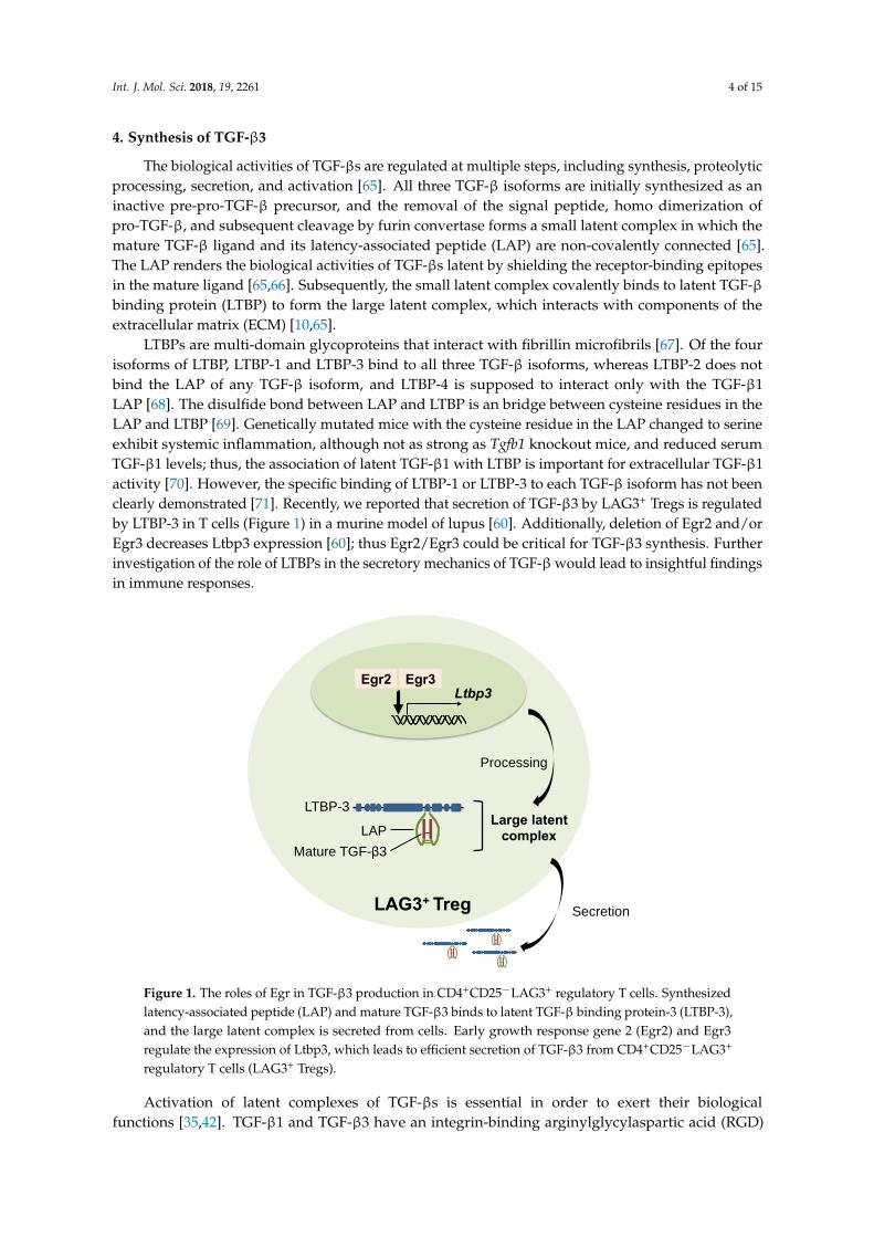
LTBPs are multi-domain glycoproteins that interact with fibrillin microfibrils [67]. Of the fourisoforms of LTBP, LTBP-1 and LTBP-3 bind to all three TGF-β isoforms, whereas LTBP-2 does notbind the LAP of any TGF-β isoform, and LTBP-4 is supposed to interact only with the TGF-β1LAP [68]. The disulfide bond between LAP and LTBP is an bridge between cysteine residues in theLAP and LTBP [69]. Genetically mutated mice with the cysteine residue in the LAP changed to serineexhibit systemic inflammation, although not as strong as Tgfb1 knockout mice, and reduced serumTGF-β1 levels; thus, the association of latent TGF-β1 with LTBP is important for extracellular TGF-β1activity [70]. However, the specific binding of LTBP-1 or LTBP-3 to each TGF-β isoform has not beenclearly demonstrated [71]. Recently, we reported that secretion of TGF-β3 by LAG3+ Tregs is regulatedby LTBP-3 in T cells (Figure 1) in a murine model of lupus [60]. Additionally, deletion of Egr2 and/orEgr3 decreases Ltbp3 expression [60]; thus Egr2/Egr3 could be critical for TGF-β3 synthesis. Furtherinvestigation of the role of LTBPs in the secretory mechanics of TGF-β would lead to insightful findingsin immune responses.
Int. J. Mol. Sci. 2018, 19, 2261 4 of 15
dimerization of pro-TGF-β, and subsequent cleavage by furin convertase forms a small latent
complex in which the mature TGF-β ligand and its latency-associated peptide (LAP) are
non-covalently connected [65]. The LAP renders the biological activities of TGF-βs latent by
shielding the receptor-binding epitopes in the mature ligand [65,66]. Subsequently, the small latent
complex covalently binds to latent TGF-β binding protein (LTBP) to form the large latent complex,
which interacts with components of the extracellular matrix (ECM) [10,65].
LTBPs are multi-domain glycoproteins that interact with fibrillin microfibrils [67]. Of the four
isoforms of LTBP, LTBP-1 and LTBP-3 bind to all three TGF-β isoforms, whereas LTBP-2 does not
bind the LAP of any TGF-β isoform, and LTBP-4 is supposed to interact only with the TGF-β1 LAP
[68]. The disulfide bond between LAP and LTBP is an bridge between cysteine residues in the LAP
and LTBP [69]. Genetically mutated mice with the cysteine residue in the LAP changed to serine
exhibit systemic inflammation, although not as strong as Tgfb1 knockout mice, and reduced serum
TGF-β1 levels; thus, the association of latent TGF-β1 with LTBP is important for extracellular TGF-β1
activity [70]. However, the specific binding of LTBP-1 or LTBP-3 to each TGF-β isoform has not been
clearly demonstrated [71]. Recently, we reported that secretion of TGF-β3 by LAG3+ Tregs is
regulated by LTBP-3 in T cells (Figure 1) in a murine model of lupus [60]. Additionally, deletion of
Egr2 and/or Egr3 decreases Ltbp3 expression [60]; thus Egr2/Egr3 could be critical for TGF-β3
synthesis. Further investigation of the role of LTBPs in the secretory mechanics of TGF-β would lead
to insightful findings in immune responses.
Egr3 Egr2 Ltbp3
LAG3+ Treg
LTBP-3 Large latent
complex LAP
Mature TGF-β3
Processing
Secretion
Figure 1. The roles of Egr in TGF-β3 production in CD4+CD25−LAG3+ regulatory T cells. Synthesized
latency-associated peptide (LAP) and mature TGF-β3 binds to latent TGF-β binding protein-3
(LTBP-3), and the large latent complex is secreted from cells. Early growth response gene 2 (Egr2)
and Egr3 regulate the expression of Ltbp3, which leads to efficient secretion of TGF-β3 from
CD4+CD25−LAG3+ regulatory T cells (LAG3+ Tregs).
Activation of latent complexes of TGF-βs is essential in order to exert their biological functions
[35,42]. TGF-β1 and TGF-β3 have an integrin-binding arginylglycylaspartic acid (RGD) motif, which
is recognized by αv integrins and is required for integrin-mediated activation [71,72]. One groups
showed that genetically modified mice harboring a nonfunctional variant of the RGD sequence in
the Tgfb1 gene develop fatal multiorgan inflammation identical to Tgfb1 knockout mice [73]. Among
the members of the integrin receptor family, integrin αvβ6 and αvβ8 have the capacity to bind and
activate pro-TGF-β1 and pro-TGF-β3 [74], and the combined genetic loss of αvβ6 and αvβ8 integrins
reproduces the phenotypes of Tgfb1 and Tgfb3 knockout mice [75]. Conditional deletion of the Igtb8
Figure 1. The roles of Egr in TGF-β3 production in CD4+CD25−LAG3+ regulatory T cells. Synthesizedlatency-associated peptide (LAP) and mature TGF-β3 binds to latent TGF-β binding protein-3 (LTBP-3),and the large latent complex is secreted from cells. Early growth response gene 2 (Egr2) and Egr3regulate the expression of Ltbp3, which leads to efficient secretion of TGF-β3 from CD4+CD25−LAG3+
regulatory T cells (LAG3+ Tregs).
Activation of latent complexes of TGF-βs is essential in order to exert their biologicalfunctions [35,42]. TGF-β1 and TGF-β3 have an integrin-binding arginylglycylaspartic acid (RGD)
Int. J. Mol. Sci. 2018, 19, 2261 5 of 15
motif, which is recognized by αv integrins and is required for integrin-mediated activation [71,72].One groups showed that genetically modified mice harboring a nonfunctional variant of the RGDsequence in the Tgfb1 gene develop fatal multiorgan inflammation identical to Tgfb1 knockout mice [73].Among the members of the integrin receptor family, integrin αvβ6 and αvβ8 have the capacity tobind and activate pro-TGF-β1 and pro-TGF-β3 [74], and the combined genetic loss of αvβ6 and αvβ8
integrins reproduces the phenotypes of Tgfb1 and Tgfb3 knockout mice [75]. Conditional deletion of theIgtb8 gene encoding integrin β8 in leukocytes results in systemic inflammation with autoantibodies [76].Further, deficiency of integrin αvβ8 in Tregs, which display less active TGF-β levels, abrogates theirsuppressive abilities on T cell responses [42]. The elaborate machinery of TGF-β synthesis is carefullyregulated by the immune system, and the recent findings of TGF-β isoform-specific function suggestthe importance of investigating further TGF-β isoform-specific synthesis mechanisms.
5. TGF-β3 Signal Transduction
TGF-βs signal through a hetero-tetrameric receptor complex composed of two type I receptorsand two type II receptors [77]. The constitutively active cytoplasmic domain of TGF-β type IIreceptors (TβRII) phosphorylates type I receptors on serine and threonine residues in response to thebinding of mature TGF-βs, after which the activated type I receptors canonically phosphorylate Smadproteins [77,78]. TβRII interacts with TGF-β type I receptors (TβRI), also called activin receptor likekinase (ALK)-5, which typically induces the phosphorylation of Smad2 and Smad3, and also interactswith ALK-1, which typically induces the phosphorylation of Smad1 and Smad5 [79,80]. In endothelialcells, ALK1 antagonizes ALK5-mediated Smad2/Smad3 signaling, and the ratio of ALK5 to ALK1determines the responsiveness to TGF-βs [79,81]. Further, type III receptors, such as endoglin andbetaglycan, regulate the access of TGF-βs to the type I and type II receptors [82]. Although TGF-β1 andTGF-β3 can interact with type II receptors even without type I receptors, TGF-β2 interacts very weaklywith type II receptors, and type I or type III receptors are required for binding [80,82]. In associationwith TβRII, TGF-β1 and TGF-β3, but not TGF-β2, interact with endoglin [82,83].
Like TGF-β1 [84], the initial report demonstrated that TGF-β3 dominantly drivesSmad2-dependent ALK-5 signaling in palatal fusion [85]. During palatogenesis, conditional deletion ofTrim33 and Smad4 in epithelium in concert with TGF-β activated kinase-1 (Tak1) inhibition phenocopiedthe palate defects observed in epithelium-specific Tgfb3-deficient mice; thus TGF-β3 is supposedto transduce signals via both canonical Smad-dependent and non-canonical Smad-independentsignaling [86]. In human B cells, we recently found that TGF-β3 induces phosphorylation ofSmad1/5 along with Smad2 and Smad3 [87], as previously reported in TGF-β1-treated B cells [88].The differential signal transduction observed for TGF-β1 and TGF-β3 is linked to different downstreamfunctions, at least for CD4+ T cells. TGF-β3 produces highly pathogenic Th17 cells and induces morephosphorylation of Smad1 and Smad5, but less Smad2 and Smad3 phosphorylation, than TGF-β1 [89],although further studies of the different downstream signaling pathways of TGF-β3 and TGF-β1are warranted.
6. TGF-β3 in Immune Responses
TGF-βs are pluripotent cytokines with both pro-inflammatory and anti-inflammatory effects,depending upon specific immune contexts such as cellular targets, cytokine concentrations,tissue microenvironments, and cytokine synergy [9,10,77,90]. In contrast to the previous reportsdescribing a variety of immunological roles for TGF-β1, a direct immunological role for TGF-β3 hasnot been described in detail until recently. Although Tgfb1-knockout mice develop fatal systemicautoimmune inflammation in heart, lungs, pancreas, colon and salivary glands [6,7], the immunologicalabnormalities in Tgfb3-knockout mice have not been reported due to death shortly after birth due tocleft palate formation and impaired lung development in Tgfb3-knockout mice [91,92].
Int. J. Mol. Sci. 2018, 19, 2261 6 of 15
6.1. Pro-Inflammatory Roles of TGF-β3
Similar to TGF-β1 [5,9,37], TGF-β3 has pro-inflammatory effects. Derepression of TGF-βs,especially Tgfb3 mRNA, results in the accumulation of Th17 cells and Foxp3+ T cells in T cell-specificdeletion of tripartite motif protein 28 (Trim28) [93]. Further analyses indicate an isoform-specificfunction of TGF-βs in Th17 cells in that a combination of TGF-β3 and IL-6 induces highly pathogenicTh17 cells, compared to the Th17 cells induced by TGF-β1 plus IL-6 [89].
In B cells, low concentrations of TGF-β1 enhance T cell-dependent sheep erythrocyte-induced Bcell proliferation [94]. In addition, TGF-β1 enhances IgG2b and IgA production from LPS-inducedB cells [95]. A direct pro-inflammatory role of TGF-β3 has not been reported, but our latestresearch indicates that antibody production in LPS-stimulated B cells is enhanced with TGF-β3 [96].TGF-β concentration is one of the determinants for its pro-inflammatory effects. TGF-β1 showsconcentration-dependent bifunctional effects in B cells [94], and we have verified that TGF-β3 also hasconcentration-dependent bifunctional effects [87,96]. Although a combination of TGF-β1 and IL-21generates mucosal-homing IgA-secreting plasmablasts [43], a role for TGF-β3 in mucosal immunityhas not been demonstrated. Since TGF-βs show context-dependent pro-inflammatory effects on B cells,different concentrations and a combination of cytokines should be utilized when investigating theirimmunoregulatory roles.
6.2. Anti-Inflammatory Roles of TGF-β3
Unlike TGF-β1 [5,37], direct anti-inflammatory roles for TGF-β3 in vivo had not been clearlydocumented until our recent reports. Targeted recombination of the coding sequence of matureTGF-β1 with a sequence from TGF-β3 partially prevented autoimmune diseases caused by TGF-β1deficiency, indicating that TGF-β3 is not fully interchangeable with TGF-β1 and that they have distinctimmunoregulatory roles [97]. In T cells, TGF-β3 inhibits the differentiation of Foxp3-expressing CD4+
T cells [98]. Moreover, treatment with a TGF-β3 neutralizing antibody prevented TGF-β3-expressingLAG3+ Treg-mediated suppression [59]. Although TGF-β1 is known to inhibit the differentiation ofTh1 [99] and Th2 cells [100], and suppress the activation of NK cells [101] and macrophages [102],the effects of TGF-β3 on these cell subsets has not yet been described.
B cell-specific deletion of TβRII results in B cell hyperresponsiveness and virtually completeserum IgA deficiency, indicating that TGF-βs could have a regulatory role in T cell-independent B cellhomeostasis [103]. TGF-β1 inhibits anti-IgM-simulated B cells by suppressing the phosphorylation ofspleen tyrosine kinase (Syk), and inhibits IL-4-stimulated B cells by suppressing the phosphorylationof signal transducer and activator of transcription-6 (STAT6) [104]. Similar to TGF-β1, TGF-β3inhibits B cell-proliferation and antibody production stimulated by anti-IgM, IL-4, or anti-CD40plus IL-4 by suppressing the phosphorylation of Syk, STAT6, or NF-κB p65, respectively [59].High-affinity antibodies are generated through the interaction of TFH cells with germinal centerB (GCB) cells [105,106], and TGF-β3, as well as TGF-β1, suppresses GCB cells in vitro [60]. Lastly,systemic administration of TGF-β3-expressing vectors, but not TGF-β1-expressing vectors, inhibited Tcell-dependent humoral immune responses by suppressing the development of GCB cells [96].
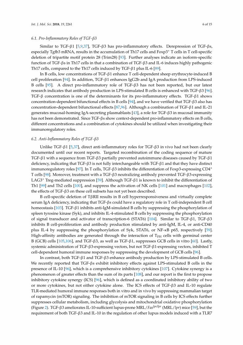
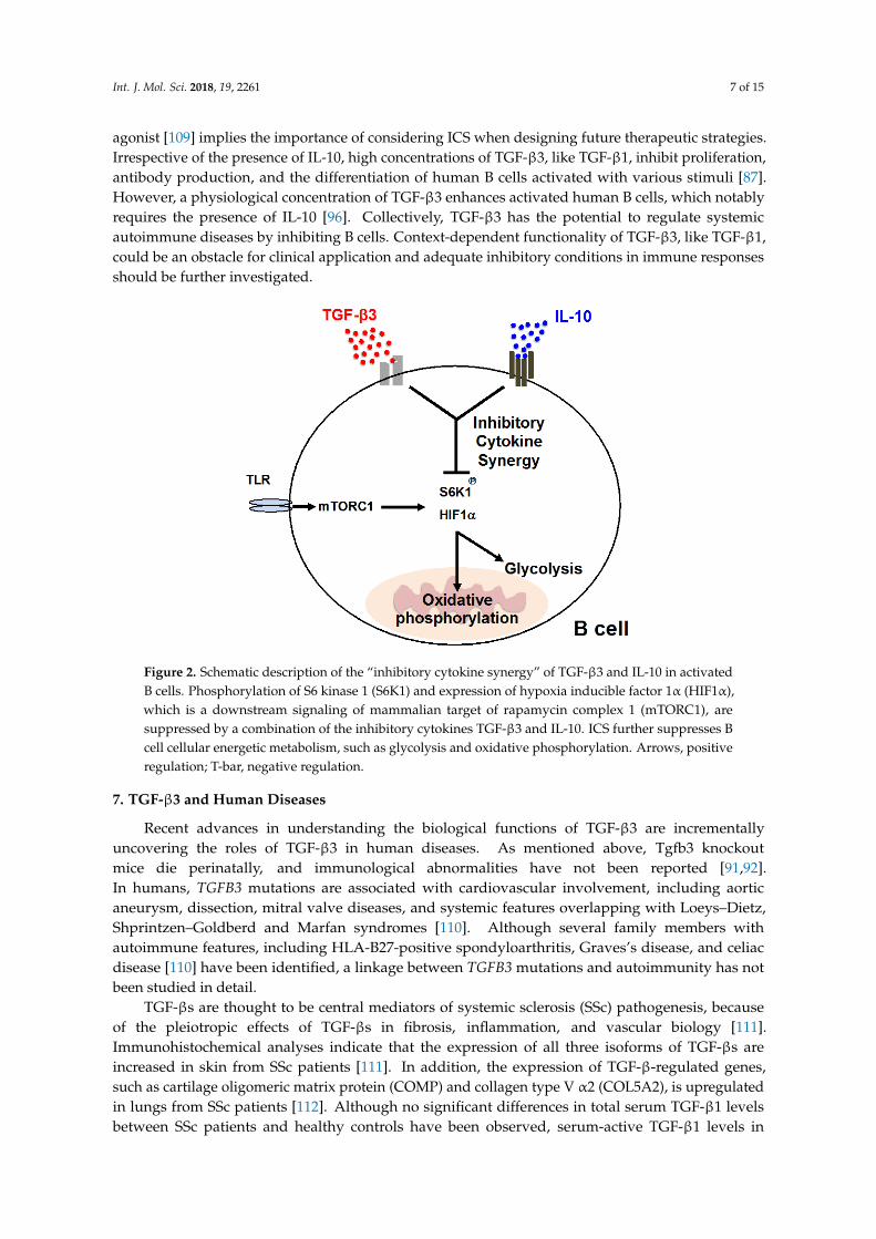
In contrast, both TGF-β1 and TGF-β3 enhance antibody production by LPS-stimulated B cells.We recently reported that TGF-βs exhibit inhibitory effects against LPS-stimulated B cells in thepresence of IL-10 [96], which is a comprehensive inhibitory cytokines [107]. Cytokine synergy is aphenomenon of greater effects than the sum of its parts [108], and our report is the first to proposeinhibitory cytokine synergy (ICS) [96], which is defined as a coordinated inhibitory ability of twoor more cytokines, but not either cytokine alone. The ICS effects of TGF-β3 and IL-10 regulateTLR-mediated humoral immune responses both in vitro and in vivo by suppressing mammalian targetof rapamycin (mTOR) signaling. The inhibition of mTOR signaling in B cells by ICS effects furthersuppresses cellular metabolism, including glycolysis and mitochondrial oxidative phosphorylation(Figure 2). TGF-β3 ameliorates IL-10-sufficient lupus-prone MRL/Faslpr/lpr (MRL/lpr) mice [59], but therequirement of both TGF-β3 and IL-10 in the regulation of other lupus models induced with a TLR7
Int. J. Mol. Sci. 2018, 19, 2261 7 of 15
agonist [109] implies the importance of considering ICS when designing future therapeutic strategies.Irrespective of the presence of IL-10, high concentrations of TGF-β3, like TGF-β1, inhibit proliferation,antibody production, and the differentiation of human B cells activated with various stimuli [87].However, a physiological concentration of TGF-β3 enhances activated human B cells, which notablyrequires the presence of IL-10 [96]. Collectively, TGF-β3 has the potential to regulate systemicautoimmune diseases by inhibiting B cells. Context-dependent functionality of TGF-β3, like TGF-β1,could be an obstacle for clinical application and adequate inhibitory conditions in immune responsesshould be further investigated.
Int. J. Mol. Sci. 2018, 19, 2261 7 of 15
the potential to regulate systemic autoimmune diseases by inhibiting B cells. Context-dependent functionality of TGF-β3, like TGF-β1, could be an obstacle for clinical application and adequate inhibitory conditions in immune responses should be further investigated.
Figure 2. Schematic description of the “inhibitory cytokine synergy” of TGF-β3 and IL-10 in activated B cells. Phosphorylation of S6 kinase 1 (S6K1) and expression of hypoxia inducible factor 1α (HIF1α), which is a downstream signaling of mammalian target of rapamycin complex 1 (mTORC1), are suppressed by a combination of the inhibitory cytokines TGF-β3 and IL-10. ICS further suppresses B cell cellular energetic metabolism, such as glycolysis and oxidative phosphorylation. Arrows, positive regulation; T-bar, negative regulation.
7. TGF-β3 and Human Diseases
Recent advances in understanding the biological functions of TGF-β3 are incrementally uncovering the roles of TGF-β3 in human diseases. As mentioned above, Tgfb3 knockout mice die perinatally, and immunological abnormalities have not been reported [91,92]. In humans, TGFB3 mutations are associated with cardiovascular involvement, including aortic aneurysm, dissection, mitral valve diseases, and systemic features overlapping with Loeys–Dietz, Shprintzen–Goldberd and Marfan syndromes [110]. Although several family members with autoimmune features, including HLA-B27-positive spondyloarthritis, Graves’s disease, and celiac disease [110] have been identified, a linkage between TGFB3 mutations and autoimmunity has not been studied in detail.
TGF-βs are thought to be central mediators of systemic sclerosis (SSc) pathogenesis, because of the pleiotropic effects of TGF-βs in fibrosis, inflammation, and vascular biology [111]. Immunohistochemical analyses indicate that the expression of all three isoforms of TGF-βs are increased in skin from SSc patients [111]. In addition, the expression of TGF-β-regulated genes, such as cartilage oligomeric matrix protein (COMP) and collagen type V α2 (COL5A2), is upregulated in lungs from SSc patients [112]. Although no significant differences in total serum TGF-β1 levels between SSc patients and healthy controls have been observed, serum-active TGF-β1 levels in diffuse cutaneous SSc patients are negatively correlated with skin score and lower than active TGF-β1 levels in limited cutaneous SSc patients and healthy controls [113]. In animal models with blocked TGF-β signaling, inhibition of ALK5 attenuates bleomycin-induced pulmonary fibrosis [114], and LAP prevents sclerodermatous graft-versus-host disease [115]. Metelimumab, which is a human monoclonal antibody that specifically targets TGF-β1, failed in phase I/II trials for SSc [116]. In contrast, fresolimumab, which is specific for TGF-β1 with high affinity in addition to TGF-β2 and TGF-β3, benefited SSc patients, and further studies for the safety and longer-term use effects are
Figure 2. Schematic description of the “inhibitory cytokine synergy” of TGF-β3 and IL-10 in activatedB cells. Phosphorylation of S6 kinase 1 (S6K1) and expression of hypoxia inducible factor 1α (HIF1α),which is a downstream signaling of mammalian target of rapamycin complex 1 (mTORC1), aresuppressed by a combination of the inhibitory cytokines TGF-β3 and IL-10. ICS further suppresses Bcell cellular energetic metabolism, such as glycolysis and oxidative phosphorylation. Arrows, positiveregulation; T-bar, negative regulation.
7. TGF-β3 and Human Diseases
Recent advances in understanding the biological functions of TGF-β3 are incrementallyuncovering the roles of TGF-β3 in human diseases. As mentioned above, Tgfb3 knockoutmice die perinatally, and immunological abnormalities have not been reported [91,92].In humans, TGFB3 mutations are associated with cardiovascular involvement, including aorticaneurysm, dissection, mitral valve diseases, and systemic features overlapping with Loeys–Dietz,Shprintzen–Goldberd and Marfan syndromes [110]. Although several family members withautoimmune features, including HLA-B27-positive spondyloarthritis, Graves’s disease, and celiacdisease [110] have been identified, a linkage between TGFB3 mutations and autoimmunity has notbeen studied in detail.
TGF-βs are thought to be central mediators of systemic sclerosis (SSc) pathogenesis, becauseof the pleiotropic effects of TGF-βs in fibrosis, inflammation, and vascular biology [111].Immunohistochemical analyses indicate that the expression of all three isoforms of TGF-βs areincreased in skin from SSc patients [111]. In addition, the expression of TGF-β-regulated genes,such as cartilage oligomeric matrix protein (COMP) and collagen type V α2 (COL5A2), is upregulatedin lungs from SSc patients [112]. Although no significant differences in total serum TGF-β1 levelsbetween SSc patients and healthy controls have been observed, serum-active TGF-β1 levels in
Int. J. Mol. Sci. 2018, 19, 2261 8 of 15
diffuse cutaneous SSc patients are negatively correlated with skin score and lower than activeTGF-β1 levels in limited cutaneous SSc patients and healthy controls [113]. In animal models withblocked TGF-β signaling, inhibition of ALK5 attenuates bleomycin-induced pulmonary fibrosis [114],and LAP prevents sclerodermatous graft-versus-host disease [115]. Metelimumab, which is a humanmonoclonal antibody that specifically targets TGF-β1, failed in phase I/II trials for SSc [116]. In contrast,fresolimumab, which is specific for TGF-β1 with high affinity in addition to TGF-β2 and TGF-β3,benefited SSc patients, and further studies for the safety and longer-term use effects are expected [117].A genome-wide association study (GWAS) of SSc patients in African Americans recently identifiedTGFB3 as a SSc susceptibility gene [118]. These results collectively suggest that TGF-β3 could be animportant mediator and drug target for SSc.
In SLE, the roles of TGF-βs are expected to be protective. For examples, TGF-β1 amelioratesmurine lupus [119,120], and serum TGF-β1 concentrations are low in active SLE patients [121,122].Additionally, an impaired response to a TGF-β1-mediated anti-proliferative effect against peripheralblood mononuclear cells from active SLE patients has been reported [123]. In relation to TGF-β3,mice with Egr2/Egr3 deficiency, which leads to the reduction of TGF-β3 secretion from LAG3+ Tregs,develop lupus-like diseases [60]. Furthermore, therapeutic effects of TGF-β3 in murine lupus [59,96],and the reduction of TGF-β3-secreting LAG3+ Tregs in SLE patients [59] imply potential preventiveroles of TGF-β3 in SLE.
TGF-β1 and TGF-β3, in combination with other cytokines, regulate both encephalitogenic T cellsand regulatory T cells [9,89], although precise roles for TGF-βs in MS have not been elucidated. Micewith T cell-specific deletion of Tgfb1 are resistant to EAE and exhibit less Th17 cell differentiation [41],although the in vivo roles of TGF-β3 in a mouse model of EAE have not been reported. Since CD4+ Tcells from MS patients show reduced levels of TGF-β signaling components [124], the initial failureof a clinical trial using TGF-β2 for MS [125] could be due to the lowered responsiveness of TGF-βsin the pathologies of MS. Further evaluations of TGF-β signaling in MS that incorporates recentunderstandings of TGF-β3’s impact on pathogenic Th17 cells are to be expected [126].
Targeting TGF-β pathways as a therapeutic strategy has been clinically applied outside the fieldof immunology. TGF-β is considered to be a critical regulator of fibrosis; TGF-β1 and TGF-β2 havepro-fibrotic effects and promote fibroplasia, whereas TGF-β3 exerts anti-fibrotic effects and reducesscar formation [9]. A humanized monoclonal antibody directed against integrin αvβ6, which activatespro-TGF-β1 and pro-TGF-β3 [74], is currently in phase II trials in patients with idiopathic pulmonaryfibrosis [127]. Although human recombinant TGF-β3, avotermin, failed in phase III clinical trials [128],avotermin improved wound healing without serious adverse events in phase I/II clinical trials fora prophylactic anti-scarring therapy [129]. In the field of oncology, regulation of TGF-β activity intumor microenvironments and targeting TGF-β signaling as a novel immunotherapy has been in thenews [130,131]. The TβRI-specific inhibitors galunisertib and fresolimumab, in combination with otherdrugs, have been in clinical trials for a variety of tumors [130]. The systemic effects, immunologicalchanges, and safety with these novel medications that modulate TGF-β signaling provide insightfulinformation for future therapeutic strategies for systemic autoimmune diseases.
8. Concluding Remarks
The pluripotent TGF-β cytokines have been investigated in a variety of fields. Although thethree isoforms of TGF-β each have distinct biological roles, TGF-β1 has been paid particular attentionregarding its ability to regulate immunity. Recent identification of pro- and anti-inflammatory roles forTGF-β3 [9,10] highlights the importance of the evaluation of TGF-β isoform-specific immunologicalroles. Since TGF-β3—like TGF-β1—has a context-dependent functionality in immunity [90,96],considering concentrations and combination with other cytokines is important when evaluating thepathogenic and pathoprotective roles of TGF-β3. Investigations of TGF-β3 by conditionally mutatedmouse models, together with evaluations of the physiological and pathological roles of TGF-β3 inhumans, would be helpful for further progress in assessing TGF-β3-mediated immune responses.
Int. J. Mol. Sci. 2018, 19, 2261 9 of 15
Author Contributions: T.K., T.O., M.I., K.Y., and K.F. contributed to writing the manuscript.
Funding: This work was supported by grants from the Japan Society for the Promotion of Science, and the Ministryof Education, Culture, Sports, Science and Technology of Japan (Grant Numbers JP16K09918, JP16K15510).
Conflicts of Interest: Tomohisa Okamura received financial support or fees from Chugai and Bristol-MyersSquibb (BMS). Kazuhiko Yamamoto received financial support or fees from AbbVie, Astellas, BMS, Daiichi-Sankyo,Mitsubishi Tanabe, Pfizer, Sanofi, Santen, Takeda, Teijin, Boehringer Ingelheim, Chugai, Eisai, Ono, Taisho Toyama,UCB, ImmunoFuture, Asahi Kasei, Janssen, and NIPPON KAYAKU. Keishi Fujio received financial support orfees from Astellas, BMS, Daiichi-Sankyo, Mitsubishi Tanabe, Pfizer, Ayumi, Takeda, Chugai, Eisai, Taisho Toyama,UCB, Janssen, Eli Lilly and NIPPON KAYAKU. Toshihiko Komai, Tomohisa Okamura, Mariko Inoue, KazuhikoYamamoto, and Keishi Fujio received patent-licensing arrangements with Chugai.
References
1. Chen, W.; Ten Dijke, P. Immunoregulation by members of the TGFβ superfamily. Nat. Rev. Immunol. 2016,16, 723–740. [CrossRef] [PubMed]
2. Zhao, M.; Mishra, L.; Deng, C.X. The role of TGF-β/SMAD4 signaling in cancer. Int. J. Biol. Sci. 2018, 14, 123.[CrossRef] [PubMed]
3. Wakefield, L.M.; Hill, C.S. Beyond TGFβ: Roles of other TGFβ superfamily members in cancer.Nat. Rev. Cancer 2013, 13, 328–341. [CrossRef] [PubMed]
4. Meng, X.; Nikolic-Paterson, D.J.; Lan, H.Y. TGF-β: The master regulator of fibrosis. Nat. Rev. Nephrol. 2016,12, 325–338. [CrossRef] [PubMed]
5. Li, M.O.; Wan, Y.Y.; Sanjabi, S.; Robertson, A.K.; Flavell, R.A. Transforming growth factor-β regulation ofimmune responses. Annu. Rev. Immunol. 2006, 24, 99–146. [CrossRef] [PubMed]
6. Kulkarni, A.B.; Huh, C.G.; Becker, D.; Geiser, A.; Lyght, M.; Flanders, K.C.; Roberts, A.B.; Sporn, M.B.;Ward, J.M.; Karlsson, S. Transforming growth factor β1 null mutation in mice causes excessive inflammatoryresponse and early death. Proc. Natl. Acad. Sci. USA 1993, 90, 770–774. [CrossRef] [PubMed]
7. Shull, M.M.; Ormsby, I.; Kier, A.B.; Pawlowski, S.; Diebold, R.J.; Yin, M.; Allen, R.; Sidman, C.; Proetzel, G.;Calvin, D. Targeted disruption of the mouse transforming growth factor-β1 gene results in multifocalinflammatory disease. Nature 1992, 359, 693–699. [CrossRef] [PubMed]
8. Rubtsov, Y.P.; Rudensky, A.Y. TGFβ signalling in control of T-cell-mediated self-reactivity. Nat. Rev. Immunol.2007, 7, 443–453. [CrossRef] [PubMed]
9. Fujio, K.; Komai, T.; Inoue, M.; Morita, K.; Okamura, T.; Yamamoto, K. Revisiting the regulatory roles of theTGF-β family of cytokines. Autoimmun. Rev. 2016, 15, 917–922. [CrossRef] [PubMed]
10. Okamura, T.; Morita, K.; Iwasaki, Y.; Inoue, M.; Komai, T.; Fujio, K.; Yamamoto, K. Role of TGF-β3 in theregulation of immune responses. Clin. Exp. Rheumatol. 2015, 33, 63–69.
11. Roberts, A.B.; Anzano, M.A.; Lamb, L.C.; Smith, J.M.; Sporn, M.B. New class of transforming growth factorspotentiated by epidermal growth factor: Isolation from non-neoplastic tissues. Proc. Natl. Acad. Sci. USA1981, 78, 5339–5343. [CrossRef] [PubMed]
12. Wrana, J.L. Signaling by the TGF-β Superfamily. Cold Spring Harb. Perspect. Biol. 2013, 5, a011197. [CrossRef][PubMed]
13. Roberts, A.B.; Lamb, L.C.; Newton, D.L.; Sporn, M.B.; De Larco, J.E.; Todaro, G.J. Transforming growthfactors: Isolation of polypeptides from virally and chemically transformed cells by acid/ethanol extraction.Proc. Natl. Acad. Sci. USA 1980, 77, 3494–3498. [CrossRef] [PubMed]
14. Burt, D.W.; Law, A.S. Evolution of the transforming growth factor-β superfamily. Prog. Growth Factor Res.1994, 5, 99–118. [CrossRef]
15. Akhurst, R.J.; Hata, A. Targeting the TGFβ signalling pathway in disease. Nat. Rev. Drug Discov. 2012,11, 790–811. [CrossRef] [PubMed]
16. Matzuk, M.M.; Kumar, T.R.; Vassalli, A.; Bickenbach, J.R.; Roop, D.R.; Jaenisch, R.; Bradley, A. Functionalanalysis of activins during mammalian development. Nature 1995, 374, 354–356. [CrossRef] [PubMed]
17. Zipori, D.; Barda-Saad, M. Role of activin A in negative regulation of normal and tumor B lymphocytes.J. Leukoc. Biol. 2001, 69, 867–873. [PubMed]
18. Robson, N.C.; Phillips, D.J.; McAlpine, T.; Shin, A.; Svobodova, S.; Toy, T.; Pillay, V.; Kirkpatrick, N.;Zanker, D.; Wilson, K.; et al. Activin-A: A novel dendritic cell-derived cytokine that potently attenuatesCD40 ligand-specific cytokine and chemokine production. Blood 2008, 111, 2733–2743. [CrossRef] [PubMed]
Int. J. Mol. Sci. 2018, 19, 2261 10 of 15
19. Hedger, M.P.; Drummond, A.E.; Robertson, D.M.; Risbridger, G.P.; de Kretser, D.M. Inhibin and activinregulate [3H]thymidine uptake by rat thymocytes and 3T3 cells in vitro. Mol. Cell. Endocrinol. 1989,61, 133–138. [CrossRef]
20. Hedger, M.P.; Clarke, L. Isolation of rat blood lymphocytes using a two-step Percoll density gradient. Effectof activin (erythroid differentiation factor) on peripheral T lymphocyte proliferation in vitro. J. Immunol.Methods 1993, 163, 133–136. [CrossRef]
21. Huber, S.; Stahl, F.R.; Schrader, J.; Luth, S.; Presser, K.; Carambia, A.; Flavell, R.A.; Werner, S.; Blessing, M.;Herkel, J.; et al. Activin A Promotes the TGF-β-Induced Conversion of CD4+CD25− T Cells into Foxp3+
Induced Regulatory T Cells. J. Immunol. 2009, 182, 4633–4640. [CrossRef] [PubMed]22. Sakaguchi, S.; Wing, K.; Onishi, Y.; Prieto-Martin, P.; Yamaguchi, T. Regulatory T cells: How do they suppress
immune responses? Int. Immunol. 2009, 21, 1105–1111. [CrossRef] [PubMed]23. Miyara, M.; Gorochov, G.; Ehrenstein, M.; Musset, L.; Sakaguchi, S.; Amoura, Z. Human FoxP3+ regulatory
T cells in systemic autoimmune diseases. Autoimmun. Rev. 2011, 10, 744–755. [CrossRef] [PubMed]24. Semitekolou, M.; Alissafi, T.; Aggelakopoulou, M.; Kourepini, E.; Kariyawasam, H.H.; Kay, A.B.;
Robinson, D.S.; Lloyd, C.M.; Panoutsakopoulou, V.; Xanthou, G. Activin-A induces regulatory T cellsthat suppress T helper cell immune responses and protect from allergic airway disease. J. Exp. Med. 2009,206, 1769–1785. [CrossRef] [PubMed]
25. Hardy, C.L.; Rolland, J.M.; O’Hehir, R.E. The immunoregulatory and fibrotic roles of activin A in allergicasthma. Clin. Exp. Allergy 2015, 45, 1510–1522. [CrossRef] [PubMed]
26. Soler Palacios, B.; Estrada-Capetillo, L.; Izquierdo, E.; Criado, G.; Nieto, C.; Municio, C.; González-Alvaro, I.;Sánchez-Mateos, P.; Pablos, J.L.; Corbí, A.L.; et al. Macrophages from the synovium of active rheumatoidarthritis exhibit an activin A-dependent pro-inflammatory profile. J. Pathol. 2015, 235, 515–526. [CrossRef][PubMed]
27. Ota, F.; Maeshima, A.; Yamashita, S.; Ikeuchi, H.; Kaneko, Y.; Kuroiwa, T.; Hiromura, K.; Ueki, K.; Kojima, I.;Nojima, Y. Activin A induces cell proliferation of fibroblast-like synoviocytes in rheumatoid arthritis. ArthritisRheum. 2003, 48, 2442–2449. [CrossRef] [PubMed]
28. Wang, R.N.; Green, J.; Wang, Z.; Deng, Y.; Qiao, M.; Peabody, M.; Zhang, Q.; Ye, J.; Yan, Z.; Denduluri, S.;et al. Bone Morphogenetic Protein (BMP) signaling in development and human diseases. Genes Dis. 2014,1, 87–105. [CrossRef] [PubMed]
29. Eixarch, H.; Calvo-Barreiro, L.; Montalban, X.; Espejo, C. Bone morphogenetic proteins in multiple sclerosis:Role in neuroinflammation. Brain. Behav. Immun. 2018, 68, 1–10. [CrossRef] [PubMed]
30. Pluchino, S.; Zanotti, L.; Brambilla, E.; Rovere-Querini, P.; Capobianco, A.; Alfaro-Cervello, C.; Salani, G.;Cossetti, C.; Borsellino, G.; Battistini, L.; et al. Immune regulatory neural stem/precursor cells protectfrom central nervous system autoimmunity by restraining dendritic cell function. PLoS ONE 2009, 4, e5959.[CrossRef] [PubMed]
31. Mausner-Fainberg, K.; Urshansky, N.; Regev, K.; Auriel, E.; Karni, A. Elevated and dysregulatedbone morphogenic proteins in immune cells of patients with relapsing-remitting multiple sclerosis.J. Neuroimmunol. 2013, 264, 91–99. [CrossRef] [PubMed]
32. Tang, Y.; Ma, X.; Zhang, H.; Gu, Z.; Hou, Y.; Gilkeson, G.S.; Lu, L.; Zeng, X.; Sun, L. Gene expression profilereveals abnormalities of multiple signaling pathways in mesenchymal stem cell derived from patients withsystemic lupus erythematosus. Clin. Dev. Immunol. 2012, 2012, 826182. [CrossRef] [PubMed]
33. Bramlage, C.P.; Häupl, T.; Kaps, C.; Ungethüm, U.; Krenn, V.; Pruss, A.; Müller, G.A.; Strutz, F.;Burmester, G.R. Decrease in expression of bone morphogenetic proteins 4 and 5 in synovial tissue ofpatients with osteoarthritis and rheumatoid arthritis. Arthritis Res. Ther. 2006, 8, R58. [CrossRef] [PubMed]
34. Lories, R.J.; Derese, I.; Ceuppens, J.L.; Luyten, F.P. Bone morphogenetic proteins 2 and 6, expressed inarthritic synovium, are regulated by proinflammatory cytokines and differentially modulate fibroblast-likesynoviocyte apoptosis. Arthritis Rheum. 2003, 48, 2807–2818. [CrossRef] [PubMed]
35. Travis, M.A.; Sheppard, D. TGF-β Activation and Function in Immunity. Annu. Rev. Immunol. 2014, 32, 51–82.[CrossRef] [PubMed]
36. Li, M.O.; Wan, Y.Y.; Flavell, R.A. T cell-produced transforming growth factor-β1 controls T cell tolerance andregulates Th1- and Th17-cell differentiation. Immunity 2007, 26, 579–591. [CrossRef] [PubMed]
37. Wan, Y.Y.; Flavell, R.A. “Yin-Yang” functions of transforming growth factor-β and T regulatory cells inimmune regulation. Immunol. Rev. 2007, 220, 199–213. [CrossRef] [PubMed]
Int. J. Mol. Sci. 2018, 19, 2261 11 of 15
38. Nakamura, K.; Kitani, A.; Strober, W. Cell contact-dependent immunosuppression by CD4(+)CD25(+)regulatory T cells is mediated by cell surface-bound transforming growth factor β. J. Exp. Med. 2001,194, 629–644. [CrossRef] [PubMed]
39. Roncarolo, M.G.; Levings, M.K.; Traversari, C. Differentiation of T regulatory cells by immature dendriticcells. J. Exp. Med. 2001, 193, F5-9. [CrossRef] [PubMed]
40. Roncarolo, M.G.; Bacchetta, R.; Bordignon, C.; Narula, S.; Levings, M.K. Type 1 T regulatory cells.Immunol. Rev. 2001, 182, 68–79. [CrossRef] [PubMed]
41. Gutcher, I.; Donkor, M.K.; Ma, Q.; Rudensky, A.Y.; Flavell, R.A.; Li, M.O. Autocrine transforming growthfactor-β1 promotes in vivo Th17 cell differentiation. Immunity 2011, 34, 396–408. [CrossRef] [PubMed]
42. Worthington, J.J.; Kelly, A.; Smedley, C.; Bauché, D.; Campbell, S.; Marie, J.C.; Travis, M.A. Integrinαvβ8-Mediated TGF-β Activation by Effector Regulatory T Cells is Essential for Suppression ofT-Cell-Mediated Inflammation. Immunity 2015, 42, 903–915. [CrossRef] [PubMed]
43. Dullaers, M.; Li, D.; Xue, Y.; Ni, L.; Gayet, I.; Morita, R.; Ueno, H.; Palucka, K.A.; Banchereau, J.; Oh, S.AT cell-dependent mechanism for the induction of human mucosal homing immunoglobulin A-secretingplasmablasts. Immunity 2009, 30, 120–129. [CrossRef] [PubMed]
44. Sage, P.T.; Sharpe, A.H. T Follicular Regulatory Cells in the Regulation of B cell Responses. Trends Immunol.2015, 36, 410–418. [CrossRef] [PubMed]
45. Sage, P.T.; Sharpe, A.H. T follicular regulatory cells. Immunol. Rev. 2016, 271, 246–259. [CrossRef] [PubMed]46. Parekh, V.V.; Prasad, D.V.R.; Banerjee, P.P.; Joshi, B.N.; Kumar, A.; Mishra, G.C. B cells activated by
lipopolysaccharide, but not by anti-Ig and anti-CD40 antibody, induce anergy in CD8+ T cells: Role ofTGF-β1. J. Immunol. 2003, 170, 5897–5911. [CrossRef] [PubMed]
47. Bjarnadóttir, K.; Benkhoucha, M.; Merkler, D.; Weber, M.S.; Payne, N.L.; Bernard, C.C.; Molnarfi, N.;Lalive, P.H. B cell-derived transforming growth factor-β1 expression limits the induction phase ofautoimmune neuroinflammation. Sci. Rep. 2016, 6, 34594. [CrossRef] [PubMed]
48. Molnarfi, N.; Bjarnadóttir, K.; Benkhoucha, M.; Juillard, C.; Lalive, P.H. Activation of human B cells negativelyregulates TGF-β1 production. J. Neuroinflamm. 2017, 14, 13. [CrossRef] [PubMed]
49. Gray, J.D.; Hirokawa, M.; Ohtsuka, K.; Horwitz, D.A. Generation of an inhibitory circuit involving CD8+
T cells, IL-2, and NK cell-derived TGF-β: Contrasting effects of anti-CD2 and anti-CD3. J. Immunol. 1998,160, 2248–2254. [PubMed]
50. Esplugues, E.; Sancho, D.; Vega-Ramos, J.; Martínez, C.; Syrbe, U.; Hamann, A.; Engel, P.; Sánchez-Madrid, F.;Lauzurica, P. Enhanced antitumor immunity in mice deficient in CD69. J. Exp. Med. 2003, 197, 1093–1106.[CrossRef] [PubMed]
51. Wang, X.; Li, F.; Xie, L.; Crane, J.; Zhen, G.; Mishina, Y.; Deng, R.; Gao, B.; Chen, H.; Liu, S.; et al. Inhibitionof overactive TGF-β attenuates progression of heterotopic ossification in mice. Nat. Commun. 2018, 9, 551.[CrossRef] [PubMed]
52. Min, K.M.; Kim, P.H. Macrophage-derived TGF-β1 induces IgA isotype expression. Mol. Cells 2003,16, 245–250. [PubMed]
53. Morelli, A.E.; Zahorchak, A.F.; Larregina, A.T.; Colvin, B.L.; Logar, A.J.; Takayama, T.; Falo, L.D.;Thomson, A.W. Cytokine production by mouse myeloid dendritic cells in relation to differentiation andterminal maturation induced by lipopolysaccharide or CD40 ligation. Blood 2001, 98, 1512–1523. [CrossRef][PubMed]
54. Lindstedt, K.A.; Wang, Y.; Shiota, N.; Saarinen, J.; Hyytiäinen, M.; Kokkonen, J.O.; Keski-Oja, J.; Kovanen, P.T.Activation of paracrine TGF-β1 signaling upon stimulation and degranulation of rat serosal mast cells:A novel function for chymase. FASEB J. 2001, 15, 1377–1388. [CrossRef] [PubMed]
55. Kobayashi, T.; Iijima, K.; Kita, H. Marked airway eosinophilia prevents development of airwayhyper-responsiveness during an allergic response in IL-5 transgenic mice. J. Immunol. 2003, 170, 5756–5763.[CrossRef] [PubMed]
56. Chu, V.T.; Beller, A.; Rausch, S.; Strandmark, J.; Zänker, M.; Arbach, O.; Kruglov, A.; Berek, C. Eosinophilspromote generation and maintenance of immunoglobulin-A-expressing plasma cells and contribute to gutimmune homeostasis. Immunity 2014, 40, 582–593. [CrossRef] [PubMed]
57. Weller, P.F.; Spencer, L.A. Functions of tissue-resident eosinophils. Nat. Rev. Immunol. 2017, 17, 746–760.[CrossRef] [PubMed]
Int. J. Mol. Sci. 2018, 19, 2261 12 of 15
58. Wang, S.; Xia, P.; Chen, Y.; Qu, Y.; Xiong, Z.; Ye, B.; Du, Y.; Tian, Y.; Yin, Z.; Xu, Z.; et al. Regulatory InnateLymphoid Cells Control Innate Intestinal Inflammation. Cell 2017, 171, 201–216. [CrossRef] [PubMed]
59. Okamura, T.; Sumitomo, S.; Morita, K.; Iwasaki, Y.; Inoue, M.; Nakachi, S.; Komai, T.; Shoda, H.; Miyazaki, J.;Fujio, K.; et al. TGF-β3-expressing CD4(+)CD25(−)LAG3(+) regulatory T cells control humoral immuneresponses. Nat. Commun. 2015, 6, 6329. [CrossRef] [PubMed]
60. Morita, K.; Okamura, T.; Inoue, M.; Komai, T.; Teruya, S.; Iwasaki, Y.; Sumitomo, S.; Shoda, H.; Yamamoto, K.;Fujio, K. Egr2 and Egr3 in regulatory T cells cooperatively control systemic autoimmunity throughLtbp3-mediated TGF-β3 production. Proc. Natl. Acad. Sci. USA 2016, 113, E8131–E8140. [CrossRef][PubMed]
61. Zhu, B.; Symonds, A.L.; Martin, J.E.; Kioussis, D.; Wraith, D.C.; Li, S.; Wang, P. Early growth response gene 2(Egr-2) controls the self-tolerance of T cells and prevents the development of lupuslike autoimmune disease.J. Exp. Med. 2008, 205, 2295–2307. [CrossRef] [PubMed]
62. Myouzen, K.; Kochi, Y.; Shimane, K.; Fujio, K.; Okamura, T.; Okada, Y.; Suzuki, A.; Atsumi, T.; Ito, S.;Takada, K.; et al. Regulatory polymorphisms in EGR2 are associated with susceptibility to systemic lupuserythematosus. Hum. Mol. Genet. 2010, 19, 2313–2320. [CrossRef] [PubMed]
63. Okamura, T.; Fujio, K.; Shibuya, M.; Sumitomo, S.; Shoda, H.; Sakaguchi, S.; Yamamoto, K.CD4+CD25−LAG3+ regulatory T cells controlled by the transcription factor Egr-2. Proc. Natl. Acad. Sci. USA2009, 106, 13974–13979. [CrossRef] [PubMed]
64. Okamura, T.; Yamamoto, K.; Fujio, K. Early Growth Response Gene 2-Expressing CD4+LAG3+ Regulatory TCells: The Therapeutic Potential for Treating Autoimmune Diseases. Front. Immunol. 2018, 9. [CrossRef][PubMed]
65. Dijke, P.; Arthur, H.M. Extracellular control of TGFβ signalling in vascular development and disease.Nat. Rev. Mol. Cell Biol. 2007, 8, 857–869. [CrossRef] [PubMed]
66. Annes, J.P.; Munger, J.S.; Rifkin, D.B. Making sense of latent TGFβ activation. J. Cell Sci. 2003, 116, 217–224.[CrossRef] [PubMed]
67. Robertson, I.B.; Horiguchi, M.; Zilberberg, L.; Dabovic, B.; Hadjiolova, K.; Rifkin, D.B. Latent TGF-β-bindingproteins. Matrix Biol. 2015, 47, 44–53. [CrossRef] [PubMed]
68. Saharinen, J.; Keski-Oja, J. Specific sequence motif of 8-Cys repeats of TGF-β binding proteins, LTBPs, createsa hydrophobic interaction surface for binding of small latent TGF-β. Mol. Biol. Cell 2000, 11, 2691–2704.[CrossRef] [PubMed]
69. Chen, Y.; Ali, T.; Todorovic, V.; O’Leary, J.M.; Kristina Downing, A.; Rifkin, D.B. Amino Acid Requirementsfor Formation of the TGF-β-Latent TGF-β Binding Protein Complexes. J. Mol. Biol. 2005, 345, 175–186.[CrossRef] [PubMed]
70. Yoshinaga, K.; Obata, H.; Jurukovski, V.; Mazzieri, R.; Chen, Y.; Zilberberg, L.; Huso, D.; Melamed, J.;Prijatelj, P.; Todorovic, V.; et al. Perturbation of transforming growth factor (TGF)-β1 association with latentTGF-β binding protein yields inflammation and tumors. Proc. Natl. Acad. Sci. USA 2008, 105, 18758–18763.[CrossRef] [PubMed]
71. Rifkin, D.B.; Rifkin, W.J.; Zilberberg, L. LTBPs in biology and medicine: LTBP diseases. Matrix Biol. 2017.[CrossRef] [PubMed]
72. Shi, M.; Zhu, J.; Wang, R.; Chen, X.; Mi, L.; Walz, T.; Springer, T.A. Latent TGF-β structure and activation.Nature 2011, 474, 343–349. [CrossRef] [PubMed]
73. Yang, Z.; Mu, Z.; Dabovic, B.; Jurukovski, V.; Yu, D.; Sung, J.; Xiong, X.; Munger, J.S. Absence ofintegrin-mediated TGFβ1 activation in vivo recapitulates the phenotype of TGFβ1-null mice. J. Cell Biol.2007, 176, 787–793. [CrossRef] [PubMed]
74. Wang, J.; Dong, X.; Zhao, B.; Li, J.; Lu, C.; Springer, T.A. Atypical interactions of integrin αVβ8withpro-TGF-β1. Proc. Natl. Acad. Sci. USA 2017, 114, E4168–E4174. [CrossRef] [PubMed]
75. Aluwihare, P.; Mu, Z.; Zhao, Z.; Yu, D.; Weinreb, P.H.; Horan, G.S.; Violette, S.M.; Munger, J.S. Mice that lackactivity of αvβ6- and αvβ8-integrins reproduce the abnormalities of Tgfb1- and Tgfb3-null mice. J. Cell Sci.2009, 122, 227–232. [CrossRef] [PubMed]
76. Travis, M.A.; Reizis, B.; Melton, A.C.; Masteller, E.; Tang, Q.; Proctor, J.M.; Wang, Y.; Bernstein, X.; Huang, X.;Reichardt, L.F.; et al. Loss of integrin α(v)β8 on dendritic cells causes autoimmunity and colitis in mice.Nature 2007, 449, 361–365. [CrossRef] [PubMed]
77. Massague, J. TGFβ signalling in context. Nat. Rev. Mol. Cell Biol. 2012, 13, 616–630. [CrossRef] [PubMed]
Int. J. Mol. Sci. 2018, 19, 2261 13 of 15
78. Wrighton, K.H.; Lin, X.; Feng, X.H. Phospho-control of TGF-β superfamily signaling. Cell Res. 2009, 19, 8–20.[CrossRef] [PubMed]
79. Goumans, M.J.; Valdimarsdottir, G.; Itoh, S.; Lebrin, F.; Larsson, J.; Mummery, C.; Karlsson, S.; ten Dijke, P.Activin receptor-like kinase (ALK)1 is an antagonistic mediator of lateral TGFβ/ALK5 signaling. Mol. Cell2003, 12, 817–828. [CrossRef]
80. Derynck, R.; Zhang, Y.E. Smad-dependent and Smad-independent pathways in TGF-β family signalling.Nature 2003, 425, 577–584. [CrossRef] [PubMed]
81. Curado, F.; Spuul, P.; Egana, I.; Rottiers, P.; Daubon, T.; Veillat, V.; Duhamel, P.; Leclercq, A.; Gontier, E.;Genot, E. ALK5 and ALK1 Play Antagonistic Roles in Transforming Growth Factor β-Induced PodosomeFormation in Aortic Endothelial Cells. Mol. Cell. Biol. 2014, 34, 4389–4403. [CrossRef] [PubMed]
82. Goumans, M.J.; Liu, Z.; ten Dijke, P. TGF-β signaling in vascular biology and dysfunction. Cell Res. 2009,19, 116–127. [CrossRef] [PubMed]
83. Cheifetz, S.; Bellón, T.; Calés, C.; Vera, S.; Bernabeu, C.; Massagué, J.; Letarte, M. Endoglin is a componentof the transforming growth factor-β receptor system in human endothelial cells. J. Biol. Chem. 1992,267, 19027–19030. [PubMed]
84. Piek, E.; Moustakas, A.; Kurisaki, A.; Heldin, C.H.; ten Dijke, P. TGF-(β) type I receptor/ALK-5 and Smadproteins mediate epithelial to mesenchymal transdifferentiation in NMuMG breast epithelial cells. J. Cell Sci.1999, 112 Pt 24, 4557–4568. [PubMed]
85. Dudas, M.; Nagy, A.; Laping, N.J.; Moustakas, A.; Kaartinen, V. Tgf-β3-induced palatal fusion is mediatedby Alk-5/Smad pathway. Dev. Biol. 2004, 266, 96–108. [CrossRef] [PubMed]
86. Lane, J.; Yumoto, K.; Azhar, M.; Ninomiya-Tsuji, J.; Inagaki, M.; Hu, Y.; Deng, C.X.; Kim, J.; Mishina, Y.;Kaartinen, V. Tak1, Smad4 and Trim33 redundantly mediate TGF-β3 signaling during palate development.Dev. Biol. 2015, 398, 231–241. [CrossRef] [PubMed]
87. Tsuchida, Y.; Sumitomo, S.; Ishigaki, K.; Suzuki, A.; Kochi, Y.; Tsuchiya, H.; Ota, M.; Komai, T.; Inoue, M.;Morita, K.; et al. TGF-β3 Inhibits Antibody Production by Human B Cells. PLoS ONE 2017, 12, e0169646.[CrossRef] [PubMed]
88. Bakkebø, M.; Huse, K.; Hilden, V.I.; Smeland, E.B.; Oksvold, M.P. TGF-β-induced growth inhibition in B-celllymphoma correlates with Smad1/5 signalling and constitutively active p38 MAPK. BMC Immunol. 2010,11, 57. [CrossRef] [PubMed]
89. Lee, Y.; Awasthi, A.; Yosef, N.; Quintana, F.J.; Xiao, S.; Peters, A.; Wu, C.; Kleinewietfeld, M.; Kunder, S.;Hafler, D.A.; et al. Induction and molecular signature of pathogenic TH17 cells. Nat. Immunol. 2012,13, 991–999. [CrossRef] [PubMed]
90. David, C.J.; Massagué, J. Contextual determinants of TGFβ action in development, immunity and cancer.Nat. Rev. Mol. Cell Biol. 2018. [CrossRef] [PubMed]
91. Kaartinen, V.; Voncken, J.W.; Shuler, C.; Warburton, D.; Bu, D.; Heisterkamp, N.; Groffen, J. Abnormallung development and cleft palate in mice lacking TGF-β 3 indicates defects of epithelial-mesenchymalinteraction. Nat. Genet. 1995, 11, 415–421. [CrossRef] [PubMed]
92. Proetzel, G.; Pawlowski, S.A.; Wiles, M.V.; Yin, M.; Boivin, G.P.; Howles, P.N.; Ding, J.; Ferguson, M.W.;Doetschman, T. Transforming growth factor-β3 is required for secondary palate fusion. Nat. Genet. 1995,11, 409–414. [CrossRef] [PubMed]
93. Chikuma, S.; Suita, N.; Okazaki, I.M.; Shibayama, S.; Honjo, T. TRIM28 prevents autoinflammatory T celldevelopment in vivo. Nat. Immunol. 2012, 13, 596–603. [CrossRef] [PubMed]
94. McKams, S.C.; Letterio, J.J.; Kaminski, N.E. Concentration-dependent bifunctional effect of TGF-beta 1 onimmunoglobulin production: A role for Smad3 inIgA production in vitro. Int. Immunopharmacol. 2003,3, 1761–1774.
95. McIntyre, T.M.; Klinman, D.R.; Rothman, P.; Lugo, M.; Dasch, J.R.; Mond, J.J.; Snapper, C.M. Transforminggrowth factor β1 selectivity stimulates immunoglobulin G2b secretion by lipopolysaccharide-activatedmurine B cells. J. Exp. Med. 1993, 177, 1031–1037. [CrossRef] [PubMed]
96. Komai, T.; Inoue, M.; Okamura, T.; Morita, K.; Iwasaki, Y.; Sumitomo, S.; Shoda, H.; Yamamoto, K.; Fujio, K.Transforming growth factor-β and interleukin-10 synergistically regulate humoral immunity via modulatingmetabolic signals. Front. Immunol. 2018, 9, 1364. [CrossRef] [PubMed]
Int. J. Mol. Sci. 2018, 19, 2261 14 of 15
97. Hall, B.E.; Wankhade, U.D.; Konkel, J.E.; Cherukuri, K.; Nagineni, C.N.; Flanders, K.C.; Arany, P.R.; Chen, W.;Rane, S.G.; Kulkarni, A.B. Transforming growth factor-β3 (TGF-β3) knock-in ameliorates inflammation dueto TGF-β1 deficiency while promoting glucose tolerance. J. Biol. Chem. 2013, 288, 32074–32092. [CrossRef][PubMed]
98. Shah, S.; Qiao, L. Resting B cells expand a CD4+CD25+Foxp3+ Treg population via TGF-β3. Eur. J. Immunol.2008, 38, 2488–2498. [CrossRef] [PubMed]
99. Gorelik, L.; Constant, S.; Flavell, R.A. Mechanism of transforming growth factor β-induced inhibition of Thelper type 1 differentiation. J. Exp. Med. 2002, 195, 1499–1505. [CrossRef] [PubMed]
100. Gorelik, L.; Fields, P.E.; Flavell, R.A. Cutting edge: TGF-β inhibits Th type 2 development through inhibitionof GATA-3 expression. J. Immunol. 2000, 165, 4773–4777. [CrossRef] [PubMed]
101. Viel, S.; Marçais, A.; Guimaraes, F.S.; Loftus, R.; Rabilloud, J.; Grau, M.; Degouve, S.; Djebali, S.; Sanlaville, A.;Charrier, E.; et al. TGF-β inhibits the activation and functions of NK cells by repressing the mTOR pathway.Sci. Signal. 2016, 9, ra19. [CrossRef] [PubMed]
102. Werner, F.; Jain, M.K.; Feinberg, M.W.; Sibinga, N.E.; Pellacani, A.; Wiesel, P.; Chin, M.T.; Topper, J.N.;Perrella, M.A.; Lee, M.E. Transforming Growth Factor-β1 Inhibition of Macrophage Activation is Mediatedvia Smad3. J. Biol. Chem. 2000, 275, 36653–36658. [CrossRef] [PubMed]
103. Cazac, B.B.; Roes, J. TGF-β receptor controls B cell responsiveness and induction of IgA in vivo. Immunity2000, 13, 443–451. [CrossRef]
104. Roes, J.; Choi, B.K.; Cazac, B.B. Redirection of B cell responsiveness by transforming growth factor β receptor.Proc. Natl. Acad. Sci. USA 2003, 100, 7241–7246. [CrossRef] [PubMed]
105. Victora, G.D.; Schwickert, T.A.; Fooksman, D.R.; Kamphorst, A.O.; Meyer-Hermann, M.; Dustin, M.L.;Nussenzweig, M.C. Germinal center dynamics revealed by multiphoton microscopy with a photoactivatablefluorescent reporter. Cell 2010, 143, 592–605. [CrossRef] [PubMed]
106. Kräutler, N.J.; Suan, D.; Butt, D.; Bourne, K.; Hermes, J.R.; Chan, T.D.; Sundling, C.; Kaplan, W.; Schofield, P.;Jackson, J.; et al. Differentiation of germinal center B cells into plasma cells is initiated by high-affinityantigen and completed by Tfh cells. J. Exp. Med. 2017, 214, 1259–1267. [CrossRef] [PubMed]
107. Saxena, A.; Khosraviani, S.; Noel, S.; Mohan, D.; Donner, T.; Hamad, A.R. Interleukin-10 paradox: A potentimmunoregulatory cytokine that has been difficult to harness for immunotherapy. Cytokine 2015, 74, 27–34.[CrossRef] [PubMed]
108. Bartee, E.; McFadden, G. Cytokine synergy: An underappreciated contributor to innate anti-viral immunity.Cytokine 2013, 63, 237–240. [CrossRef] [PubMed]
109. Yokogawa, M.; Takaishi, M.; Nakajima, K.; Kamijima, R.; Fujimoto, C.; Kataoka, S.; Terada, Y.; Sano, S.Epicutaneous application of toll-like receptor 7 agonists leads to systemic autoimmunity in wild-type mice:A new model of systemic lupus erythematosus. Arthritis Rheumatol. 2014, 66, 694–706. [CrossRef] [PubMed]
110. Bertoli-Avella, A.M.; Gillis, E.; Morisaki, H.; Verhagen, J.M.A.; de Graaf, B.M.; van de Beek, G.; Gallo, E.;Kruithof, B.P.T.; Venselaar, H.; Myers, L.A.; et al. Mutations in a TGF-β Ligand, TGFB3, Cause SyndromicAortic Aneurysms and Dissections. J. Am. Coll. Cardiol. 2015, 65, 1324–1336. [CrossRef] [PubMed]
111. Lafyatis, R. Transforming growth factor β—At the centre of systemic sclerosis. Nat. Rev. Rheumatol. 2014,10, 706–719. [CrossRef] [PubMed]
112. Christmann, R.B.; Sampaio-Barros, P.; Stifano, G.; Borges, C.L.; de Carvalho, C.R.; Kairalla, R.; Parra, E.R.;Spira, A.; Simms, R.; Capellozzi, V.L.; et al. Association of Interferon- and transforming growth factorβ-regulated genes and macrophage activation with systemic sclerosis-related progressive lung fibrosis.Arthritis Rheumatol. 2014, 66, 714–725. [CrossRef] [PubMed]
113. Dziadzio, M.; Smith, R.E.; Abraham, D.J.; Black, C.M.; Denton, C.P. Circulating levels of active transforminggrowth factor β1 are reduced in diffuse cutaneous systemic sclerosis and correlate inversely with themodified Rodnan skin score. Rheumatology 2005, 44, 1518–1524. [CrossRef] [PubMed]
114. Higashiyama, H.; Yoshimoto, D.; Kaise, T.; Matsubara, S.; Fujiwara, M.; Kikkawa, H.; Asano, S.;Kinoshita, M. Inhibition of activin receptor-like kinase 5 attenuates bleomycin-induced pulmonary fibrosis.Exp. Mol. Pathol. 2007, 83, 39–46. [CrossRef] [PubMed]
115. Zhang, Y.; McCormick, L.L.; Gilliam, A.C. Latency-associated peptide prevents skin fibrosis in murinesclerodermatous graft-versus-host disease, a model for human scleroderma. J. Investig. Dermatol. 2003,121, 713–719. [CrossRef] [PubMed]
Int. J. Mol. Sci. 2018, 19, 2261 15 of 15
116. Denton, C.P.; Merkel, P.A.; Furst, D.E.; Khanna, D.; Emery, P.; Hsu, V.M.; Silliman, N.; Streisand, J.; Powell, J.;Åkesson, A.; et al. Scleroderma Clinical Trials Consortium Recombinant human anti-transforming growthfactor β1 antibody therapy in systemic sclerosis: A multicenter, randomized, placebo-controlled phase I/IItrial of CAT-192. Arthritis Rheum. 2007, 56, 323–333. [CrossRef] [PubMed]
117. Rice, L.M.; Padilla, C.M.; McLaughlin, S.R.; Mathes, A.; Ziemek, J.; Goummih, S.; Nakerakanti, S.; York, M.;Farina, G.; Whitfield, M.L.; et al. Fresolimumab treatment decreases biomarkers and improves clinicalsymptoms in systemic sclerosis patients. J. Clin. Investig. 2015, 125, 2795–2807. [CrossRef] [PubMed]
118. Gourh, P.; Remmers, E.F.; Satpathy, A.; Boyden, S.; Morgan, N.D.; Shah, A.A.; Adeyemo, A.; Bentley, A.;Carns, M.A.; Chandrasekharappa, S.C.; et al. Transforming Growth Factor β3 (TGFB3)—A Novel SystemicSclerosis Susceptibility Locus Involved in Fibrosis and Th17 Cell Development Identified By Genome-WideAssociation Study in African Americans from the Genome Research in African American SclerodermaPatients Consortium. Arthritis Rheumatol. 2017, 69 (Suppl. 10). [CrossRef]
119. Saxena, V.; Lienesch, D.W.; Zhou, M.; Bommireddy, R.; Azhar, M.; Doetschman, T.; Singh, R.R. Dualroles of immunoregulatory cytokine TGF-β in the pathogenesis of autoimmunity-mediated organ damage.J. Immunol. 2008, 180, 1903–1912. [CrossRef] [PubMed]
120. Kaplan, J.; Woodworth, L.; Smith, K.; Coco, J.; Vitsky, A.; McPherson, J. Therapeutic benefit of treatment withanti-thymocyte globulin and latent TGF-β1 in the MRL/LPR lupus mouse model. Lupus 2008, 17, 822–831.[CrossRef] [PubMed]
121. Hammad, A.M.; Youssef, H.M.; El-Arman, M.M. Transforming growth factor β 1 in children with systemiclupus erythematosus: A possible relation with clinical presentation of lupus nephritis. Lupus 2006,15, 608–612. [CrossRef] [PubMed]
122. Becker-Merok, A.; Eilertsen, G.Ø.; Nossent, J.C. Levels of transforming growth factor-β are low in systemiclupus erythematosus patients with active disease. J. Rheumatol. 2010, 37, 2039–2045. [CrossRef] [PubMed]
123. Elbeldi-Ferchiou, A.; Ben Ahmed, M.; Smiti-Khanfir, M.; Houman, M.H.; Abdeladhim, M.; Belhadj Hmida, N.;Cerf-Bensussan, N.; Louzir, H. Resistance to exogenous TGF-β effects in patients with systemic lupuserythematosus. J. Clin. Immunol. 2011, 31, 574–583. [CrossRef] [PubMed]
124. Severin, M.E.; Lee, P.W.; Liu, Y.; Selhorst, A.J.; Gormley, M.G.; Pei, W.; Yang, Y.; Guerau-de-Arellano, M.;Racke, M.K.; Lovett-Racke, A.E. MicroRNAs targeting TGFβ signalling underlie the regulatory T cell defectin multiple sclerosis. Brain 2016, 139, 1747–1761. [CrossRef] [PubMed]
125. Calabresi, P.A.; Fields, N.S.; Maloni, H.W.; Hanham, A.; Carlino, J.; Moore, J.; Levin, M.C.; Dhib-Jalbut, S.;Tranquill, L.R.; Austin, H.; et al. Phase 1 trial of transforming growth factor β2 in chronic progressive MS.Neurology 1998, 51, 289–292. [CrossRef] [PubMed]
126. Lee, P.W.; Severin, M.E.; Lovett-Racke, A.E. TGF-β regulation of encephalitogenic and regulatory T cells inmultiple sclerosis. Eur. J. Immunol. 2017, 47, 446–453. [CrossRef] [PubMed]
127. Aschner, Y.; Downey, G.P. Transforming Growth Factor-β: Master Regulator of the Respiratory System inHealth and Disease. Am. J. Respir. Cell Mol. Biol. 2016, 54, 647–655. [CrossRef] [PubMed]
128. Walton, K.L.; Johnson, K.E.; Harrison, C.A. Targeting TGF-β Mediated SMAD Signaling for the Preventionof Fibrosis. Front. Pharmacol. 2017, 8, 461. [CrossRef] [PubMed]
129. Ferguson, M.W.; Duncan, J.; Bond, J.; Bush, J.; Durani, P.; So, K.; Taylor, L.; Chantrey, J.; Mason, T.; James, G.;et al. Prophylactic administration of avotermin for improvement of skin scarring: Three double-blind,placebo-controlled, phase I/II studies. Lancet 2009, 373, 1264–1274. [CrossRef]
130. De Gramont, A.; Faivre, S.; Raymond, E. Novel TGF-β inhibitors ready for prime time in onco-immunology.Oncoimmunology 2017, 6, e1257453. [CrossRef] [PubMed]
131. Harjes, U. Immunotherapy: Tear down this wall. Nat. Rev. Immunol. 2018, 18, 221. [CrossRef] [PubMed]
© 2018 by the authors. Licensee MDPI, Basel, Switzerland. This article is an open accessarticle distributed under the terms and conditions of the Creative Commons Attribution(CC BY) license (http://creativecommons.org/licenses/by/4.0/).