Stem Cell Research Related Cytokine Products, Booklet · Thymosin-β4 .....32 TIGAR / TIGAR-TAT...
49
Bio-Connect B.V. T NL +31 (0)26 326 44 50 T BE +32 (0)2 503 03 48 Begonialaan 3a F NL +31 (0)26 326 44 51 F BE +32 (0)2 503 03 27 6851 TE Huissen E [email protected] The Netherlands W www.bio-connect.nl Stem Cell Research Related Cytokine Products, Booklet Interest in any of the products, request or order them at Bio-Connect.
Transcript of Stem Cell Research Related Cytokine Products, Booklet · Thymosin-β4 .....32 TIGAR / TIGAR-TAT...

Bio-Connect B.V. T NL +31 (0)26 326 44 50 T BE +32 (0)2 503 03 48Begonialaan 3a F NL +31 (0)26 326 44 51 F BE +32 (0)2 503 03 276851 TE Huissen E [email protected] Netherlands W www.bio-connect.nl
Stem Cell Research Related CytokineProducts, Booklet
Interest in any of the products,
request or order them at Bio-Connect.

Copyright © 2017 by PeproTech, Inc.
PeproTech, Inc.5 Crescent AvenueRocky Hill, NJ 08553-0275
Tel: (800) 436-9910 (609) 497-0253Fax: (609) 497-0321email: [email protected] [email protected] [email protected]
All rights reserved. No part of this publication may be reproduced, stored in a retrieval system, or transmitted, in any form or by any means, electronic, mechanical, photocopying, recording, or otherwise, without the prior written permission of the publisher. Printed in the United States of America.
PeproTech - Stem Cell Research-Related Cytokine Products
1
Table of Contents
Introduction ........................................ 2Embryonic Stem Cells ......................... 6Hematopoietic Stem Cells ................... 7Neural Stem Cells ............................... 8Mesenchymal Stem Cells .................... 9
Activin A ............................................. 10Activin B .............................................. 10AITRL ................................................... 10Amphiregulin ...................................... 10Artemin ................................................ 10BAFF .................................................... 10BAFF Receptor .................................... 11BD-1 / BD-2 / BD-3 / BD-4 / BD-5 ....... 11BDNF .................................................... 11Betacellulin .......................................... 11BMP-2 .................................................. 11BMP-3 .................................................. 11BMP-4 .................................................. 11BMP-5 .................................................. 12BMP-6 .................................................. 12BMP-7 .................................................. 12BMP-10 ................................................ 12BMP-13 (CDMP-2) ............................... 12Cardiotrophin-1 (CT-1) ........................ 13sCD34 ................................................... 13CDNF .................................................... 13CNTF .................................................... 13CTGF .................................................... 13 CYR61 .................................................. 14DKK-1 ................................................... 14DKK-2 / DKK-3 .................................... 14sDLL-1 .................................................. 14sDLL-4 .................................................. 15EGF ...................................................... 15EGF Receptor (EGFR) .......................... 15EGF-L7 ................................................. 15EG-VEGF .............................................. 15Epigen .................................................. 15Epiregulin ............................................ 16EPO ...................................................... 16FGF Superfamily ................................. 16FGF-acidic (FGF-1) .............................. 16FGF-basic (FGF-2) ............................... 16FGF-4 ................................................... 16FGF-5 ................................................... 16FGF-6 ................................................... 17FGF-7 (KGF) ......................................... 17FGF-8a / FGF-8b .................................. 17FGF-9 ................................................... 17FGF-10 ................................................. 17FGF-16 ................................................. 17FGF-17 ................................................. 17FGF-18 ................................................. 17FGF-19 ................................................. 18
FGF-20 ................................................. 18FGF-21 ................................................. 18FGF-23 ................................................. 18FGFR1a / FGFR2a / FGFR3 ................. 18Flt3-Ligand .......................................... 18Follistatin ............................................. 19sFRP-1 .................................................. 19Galectin-1 ............................................. 19GASP-1 ................................................. 19G-CSF ................................................... 19GDF-2 ................................................... 19GDF-3 ................................................... 19GDF-5 (BMP-14/CDMP-1) ................... 20GDF-11 ................................................. 20GDF-15 / MIC-1 ................................... 20GDNF ................................................... 20GM-CSF ................................................ 20GMF-β .................................................. 20Gremlin-1 ............................................. 21HB-EGF ................................................ 21Heregulinβ-1 (Neuregulin) .................. 21HGF ..................................................... 21IFN-γ ..................................................... 21IGF-I / IGF-I LR3 / IGF-II ..................... 22IL-1α / IL-1β ......................................... 22IL-2 ....................................................... 22IL-3 / IL-3β ........................................... 22IL-4 ....................................................... 22sIL-4 Receptor α .................................. 22IL-5 ....................................................... 23IL-6 ....................................................... 23IL-6 Receptor ....................................... 23IL-7 ....................................................... 23IL-10 ..................................................... 23IL-11 ..................................................... 23IL-12 ..................................................... 23IL-13 ..................................................... 24IL-15 ..................................................... 24IL-21 ..................................................... 24IL-31 ..................................................... 24IL-35 ..................................................... 24KLF4-TAT ............................................ 24Klotho .................................................. 25LIF ........................................................ 25Lin28-TAT ............................................ 25MANF .................................................. 25M-CSF .................................................. 26Mesothelin ........................................... 26Midkine ................................................ 26MPF ...................................................... 26Myostatin ............................................. 27Nanog / Nanog-TAT ............................. 27Neuropoietin ........................................ 27Neurturin ............................................. 27β-NGF ................................................... 27
Noggin .................................................. 28NOV ..................................................... 28NT-3 ..................................................... 28NT-4 ..................................................... 28Oncostatin M ....................................... 28p16-INK4a / p16-INK4a-TAT .............. 28PDGF-AA / PDGF-AB / PDGF-BB ....... 29PDGF-CC .............................................. 29Persephin ............................................. 29Pleiotrophin ......................................... 29PlGF-1 .................................................. 29PlGF-2 .................................................. 30PlGF-3 .................................................. 30Prolactin ............................................... 30ROR1 .................................................... 30R-Spondin-1 ......................................... 30R-Spondin-2 / R-Spondin-3 ................ 30SCF ....................................................... 31SCGF-α / SCGF-β ................................. 31SDF-1α (CXCL12) / SDF-1β (CXCL12) .... 31Sonic Hedgehog (Shh) ......................... 31Sox2 / Sox2-TAT .................................. 31TFF-1 / TFF-2 / TFF-3 ......................... 32TGF-α ................................................... 32TGF-β1 / TGF-β2 / TGF-β3 .................. 32Thymosin-β4 ........................................ 32TIGAR / TIGAR-TAT ........................... 32TIMP-1 / TIMP-2 ................................. 33TNF-α (TNFSF1A) ............................... 33TNF-β (TNFSF1B) ............................... 33sTNF Receptor Type I (TNFR1)........... 33sTNF Receptor Type II (TNFR2) ......... 33TPO ...................................................... 34Uteroglobin .......................................... 34VEGF-A ................................................ 34VEGF-B ................................................ 34VEGF-C ................................................ 34VEGF-D ................................................ 35Vitronectin ........................................... 35WISP-1 ................................................. 35Wnt-1 ................................................... 35Wnt-3a .................................................. 35Wnt-7a .................................................. 36Wnt-9b ................................................. 36
PeproGrow Human Mesenchymal Stem Cell Medium ..........................................37PeproGrow Human Embryonic Stem Cell Medium ................................................ 38PeproGrow Animal-Free Low Protein Human Embryonic Stem Cell Medium ...39PeproTech Premium Products ............ 40PeproGMP® Cytokines ......................... 41PeproTech Worldwide ......................... 42
PeproTech - Stem Cell Research-Related Cytokine Products
2
Stem Cells: Links to Human Cancer and Aging The human body develops from a single diploid cell called a zygote and contains at adulthood an estimated 85 trillion cells, of which more than 150 billion turn over every day. All of these cells originate from a tiny population of so-called “embryonic” and “adult” stem cells which uniquely possess a long-term self-renewal capacity and have the potential to differentiate into a variety of cell lineages. “Embryonic Stem Cells (ESC)” is a term commonly used to refer to a distinct cluster of pluripotent stem cells found in the inner cell mass of mammalian blastocysts (early-stage embryos). Their primary function is to give rise to cell lineages of all three germ layers. On the other hand, “Adult Stem Cells (ASC)” is one of several terms used to describe a diverse group of multipotent stem cells clustered in various niches throughout the body, particularly in loci with high cell turnover such as bone marrow, skin, and intestine, but also in sites with low cell turn over such as brain and pancreas. ASC, also known as somatic or tissue-specific stem cells, serve as a renewable source of specialized cells for tissue development, maintenance, and repair. Depending upon the prevailing conditions in their microenvironment, individual stem cells express distinct cell-surface proteins and display differentiation patterns which normally suit the needs of the tissue or organ in which they reside. As discussed later, such stem-cell specialization is enabled by a battery of epigenetic regulatory factors which provide the means not only to arrest and maintain a particular stem cell behavior, but also to modify it in response to changes in the cell’s microenvironment. Therefore, although ESC and the seemingly various kinds of ASC display different gene expression and differentiation patterns, it remains unclear whether these dissimilarities reflect different cellular entities or different manifestations of the same cellular entity.
Stem cells divide infrequently and tend to form and stay within distinct-size clusters. Detachment of cells from the cluster (e.g. as a result of differentiation) triggers rapid replication of the remaining cells until the original cluster size is restored. Upon differentiation, stem cells give rise to rapidly propagating transient progeny, which then differentiate into immature
tissue blastocytes. A successive series of proliferating progenitors, displaying steadily increasing lineage commitment, ultimately results in a large number of different mature cells. The distinction betweenstem cells and their progeny is based on the longevity of their self-renewal, which is commonly assessed by the number of times that the cells can be sub-cultivated in culture conditions before turning senescent (i.e. remain viable but unable to divide). However, since these in vitro assays may not reflect the in vivo actuality, the term “stem/progenitor cells” is often used to refer to primary cells that could be expanded multiple times in culture while maintaining their multilineage potential.
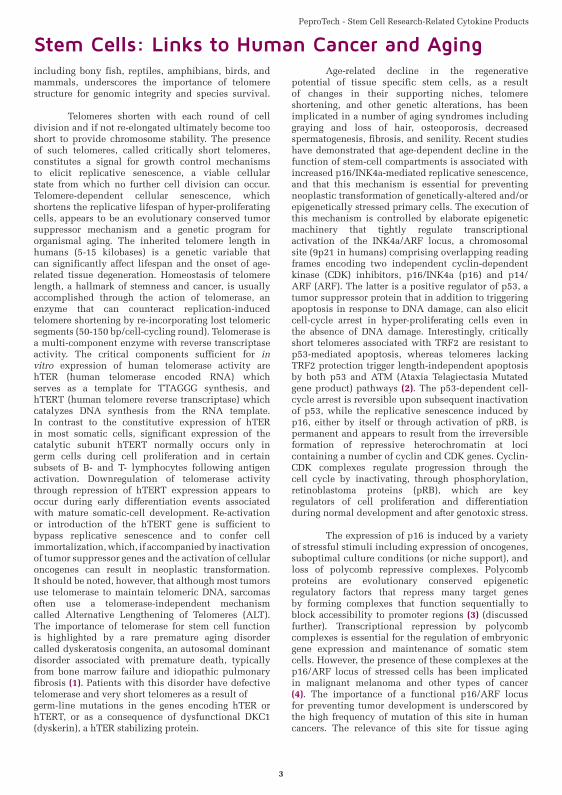
The replicative lifespan of individual primary cells depends on the mitotic history of the cell and its ability to maintain functional telomeres at both ends of all chromosomes. Telomeres comprise the ends of each eukaryotic chromosome and consist of several kilobases of repetitive non-coding DNA sequence associated with specialized proteins called telomeric repeat binding factors (TRF1 and TRF2) (Figure 1A). The presence of sufficiently long telomeres at chromosome termini enables complete replication of coding sequences and confers chromosomal stability by reducing the vulnerability of linear DNA ends to nucleolytic degradation and nonhomologous end joining. The conservation of the telomere sequence -TTAGGG-n in vertebrates,
Figure 1A - Schematic Illustration of a Eukaryotic Chromosome.
1
PeproTech - Stem Cell Research-Related Cytokine Products
3
Stem Cells: Links to Human Cancer and Agingincluding bony fish, reptiles, amphibians, birds, and mammals, underscores the importance of telomere structure for genomic integrity and species survival.
Telomeres shorten with each round of cell division and if not re-elongated ultimately become too short to provide chromosome stability. The presence of such telomeres, called critically short telomeres, constitutes a signal for growth control mechanisms to elicit replicative senescence, a viable cellular state from which no further cell division can occur. Telomere-dependent cellular senescence, which shortens the replicative lifespan of hyper-proliferating cells, appears to be an evolutionary conserved tumor suppressor mechanism and a genetic program for organismal aging. The inherited telomere length in humans (5-15 kilobases) is a genetic variable that can significantly affect lifespan and the onset of age-related tissue degeneration. Homeostasis of telomere length, a hallmark of stemness and cancer, is usually accomplished through the action of telomerase, an enzyme that can counteract replication-induced telomere shortening by re-incorporating lost telomeric segments (50-150 bp/cell-cycling round). Telomerase is a multi-component enzyme with reverse transcriptase activity. The critical components sufficient for in vitro expression of human telomerase activity are hTER (human telomerase encoded RNA) which serves as a template for TTAGGG synthesis, and hTERT (human telomere reverse transcriptase) which catalyzes DNA synthesis from the RNA template. In contrast to the constitutive expression of hTER in most somatic cells, significant expression of the catalytic subunit hTERT normally occurs only in germ cells during cell proliferation and in certain subsets of B- and T- lymphocytes following antigen activation. Downregulation of telomerase activity through repression of hTERT expression appears to occur during early differentiation events associated with mature somatic-cell development. Re-activation or introduction of the hTERT gene is sufficient to bypass replicative senescence and to confer cell immortalization, which, if accompanied by inactivation of tumor suppressor genes and the activation of cellular oncogenes can result in neoplastic transformation. It should be noted, however, that although most tumors use telomerase to maintain telomeric DNA, sarcomas often use a telomerase-independent mechanism called Alternative Lengthening of Telomeres (ALT). The importance of telomerase for stem cell function is highlighted by a rare premature aging disorder called dyskeratosis congenita, an autosomal dominant disorder associated with premature death, typically from bone marrow failure and idiopathic pulmonary fibrosis (1). Patients with this disorder have defective telomerase and very short telomeres as a result ofgerm-line mutations in the genes encoding hTER or hTERT, or as a consequence of dysfunctional DKC1 (dyskerin), a hTER stabilizing protein.
Age-related decline in the regenerative potential of tissue specific stem cells, as a result of changes in their supporting niches, telomere shortening, and other genetic alterations, has been implicated in a number of aging syndromes including graying and loss of hair, osteoporosis, decreased spermatogenesis, fibrosis, and senility. Recent studies have demonstrated that age-dependent decline in the function of stem-cell compartments is associated with increased p16/INK4a-mediated replicative senescence, and that this mechanism is essential for preventing neoplastic transformation of genetically-altered and/or epigenetically stressed primary cells. The execution of this mechanism is controlled by elaborate epigenetic machinery that tightly regulate transcriptional activation of the INK4a/ARF locus, a chromosomal site (9p21 in humans) comprising overlapping reading frames encoding two independent cyclin-dependent kinase (CDK) inhibitors, p16/INK4a (p16) and p14/ARF (ARF). The latter is a positive regulator of p53, a tumor suppressor protein that in addition to triggering apoptosis in response to DNA damage, can also elicit cell-cycle arrest in hyper-proliferating cells even in the absence of DNA damage. Interestingly, critically short telomeres associated with TRF2 are resistant to p53-mediated apoptosis, whereas telomeres lacking TRF2 protection trigger length-independent apoptosis by both p53 and ATM (Ataxia Telagiectasia Mutated gene product) pathways (2). The p53-dependent cell-cycle arrest is reversible upon subsequent inactivation of p53, while the replicative senescence induced by p16, either by itself or through activation of pRB, is permanent and appears to result from the irreversible formation of repressive heterochromatin at loci containing a number of cyclin and CDK genes. Cyclin-CDK complexes regulate progression through the cell cycle by inactivating, through phosphorylation, retinoblastoma proteins (pRB), which are key regulators of cell proliferation and differentiation during normal development and after genotoxic stress.
The expression of p16 is induced by a variety of stressful stimuli including expression of oncogenes, suboptimal culture conditions (or niche support), and loss of polycomb repressive complexes. Polycomb proteins are evolutionary conserved epigenetic regulatory factors that repress many target genes by forming complexes that function sequentially to block accessibility to promoter regions (3) (discussed further). Transcriptional repression by polycomb complexes is essential for the regulation of embryonic gene expression and maintenance of somatic stem cells. However, the presence of these complexes at the p16/ARF locus of stressed cells has been implicated in malignant melanoma and other types of cancer (4). The importance of a functional p16/ARF locus for preventing tumor development is underscored by the high frequency of mutation of this site in human cancers. The relevance of this site for tissue aging
PeproTech - Stem Cell Research-Related Cytokine Products
4
has been suggested through a series of recent studies showing that single nucleotide polymorphisms near this locus are associated with age-related pathologies including frailty, Type 2 diabetes mellitus, and vascular heart disease (reviewed in ref. 5).
The p16/ARF locus is epigenetically repressed in early life and then subjected to progressive activation, resulting in steadily increasing levels of p16 with age. The demonstration that p16 levels accumulate in stem cells of old mice suggests that these levels can constitute a good overall biomarker for aging (6). Age-dependent expression of p16 in stem cell compartments is associated with widespread tissue degeneration, whereas deficiency of p16 expression increases tissue regenerative potential accompanied with tumorigenesis (reviewed in ref. 6). These observations suggest that cancer prevention by p16-mediated cellular senescence might come at the expenseof accelerated tissue aging. This notion is further supported by recent studies on the link between p16 and ID-1, a helix-loop-helix protein that can specifically inhibit p16 expression but not that of ARF. ID-1 is a potent inhibitor of cell differentiation and plays an important role in the maintenance of many mammalian primary cells by coordinating cell division and differentiation. However, recent findings show that ID-1 can also promote cancer development by stimulating the proliferation, invasion, and survival of several types of human cancer cells. High expression of ID-1 has been observed in a large number of cancers including prostate, breast, ovary, thyroid, colorectal, liver, pancreas, and other tumors. Constitutive expression of ID-1 in cultured human primary melanocytes extends their lifespan in association with decreased expression of p16 but without notable
changes in cellular growth, migration, or telomere length. In contrast, ID1-null primary mouse embryo fibroblasts undergo premature senescence associated with increased expression of p16, but not ARF (7).
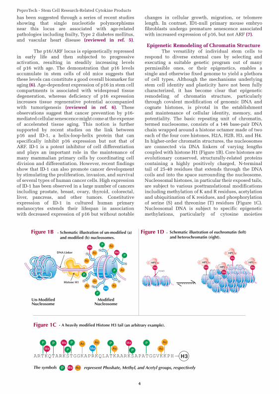
Epigenetic Remodeling of Chromatin Structure The versatility of individual stem cells to respond to diverse external cues by selecting and executing a suitable genetic program out of many permissible ones, or their epigenetics, enables a single and otherwise fixed genome to yield a plethora of cell types. Although the mechanisms underlying stem cell identity and plasticity have not been fully characterized, it has become clear that epigenetic remodeling of chromatin structure, particularly through covalent modification of genomic DNA and cognate histones, is pivotal in the establishment and maintenance of cellular identity, memory, and potentiality. The basic repeating unit of chromatin, termed nucleosome, consists of a 146 base-pair DNA chain wrapped around a histone octamer made of two each of the four core histones, H2A, H2B, H3, and H4. In higher-order chromatin structures, the nucleosomes are connected via DNA linkers of varying lengths coupled with histone H1 (Figure 1B). Core histones are evolutionary conserved, structurally-related proteins containing a highly positively charged, N-terminal tail of 25-40 residues that extends through the DNA coils and into the space surrounding the nucleosome. Nucleosomal histones, in particular their exposed tails, are subject to various posttranslational modifications including methylation of K and R residues, acetylation and ubiquitination of K residues, and phosphorylation of serine (S) and threonine (T) residues (Figure 1C). Nucleosomal DNA is subject to specific epigenetic methylations, particularly of cytosine moieties
Figure 1C - A heavily modified Histone H3 tail (an arbitrary example).
Figure 1D Schematic Illustration of euchromatin (left) and heterochromatin (right).
Figure 1B - Schematic illustration of un-modified (a) and modified (b) nucleosomes.
Figure 1B Figure 1D
Figure 1C A heavily modified Histone H3 tail (an arbitrary example).
Schematic illustration of euchromatin (left) and heterochromatin (right).
-
PeproTech - Stem Cell Research-Related Cytokine Products
5
within CpG dinucleotides. The functional relevance of the various covalent modifications of chromatin, which can be reversed through the action of specific nuclear enzymes, such as demethylases, deacetylases, and phosphatases, is a topic of current research and will be discussed here in general terms only.
Post-translational modifications of core histones trigger specific alterations in the spatial organization of chromatin, which in turn affect DNA-based processes including DNA repair, transcription, replication, and recombination. In the absence of histone modifications, the positively charged tails of core histones form stable salt bridges with negatively charged inter-nucleotide phosphate groups of adjacent DNA (Figure 1B). Such interactions can prevent, for example, interaction of RNA polymerase with promoter regions of genes whose expression is not needed by the cell. Modifications that reduce the net positive charge of histone tails, such as acetylation of K residues (neutralizes their positive charge) and phosphorylation of S and T residues (rendering them negatively charged), weaken tail-DNA interactions (Figure 1B) and are generally associated with transcriptionally active genes. Certain modifications, particularly methylation of specific K and R residues, generate docking sites for nuclear proteins that are involved in activation or repression of specific gene loci. For example, methylation of the K residue at position 4 of histone H3 produces binding site, H3K4Me*, for the chromatin remodeling protein Chd-1. Subsequent to its binding to H3K4Me*, Chd-1 recruits nucleosome remodeling enzymes such as acetylases and phosphokinases, whose action positively regulates DNA replication by disrupting H3 tail-DNA interactions. In contrast, the heterochromatin-associated protein HP-1 interacts with H3K9Me* and promotes formation and maintenance of replication-incompetent heterochromatin (Figure 1D). The symbol [Me*] denotes K residues that are either mono-, di-, or tri-methylated. Interestingly, high resolution profiling of histone methylations in the human genome has revealed that tri-methylated H3K27 signals were higher at silent promoters than active promoters, whereas an opposite trend was associated with monomethylated H3K27 (8). Tri-methylation of this residue, which produces H3K27Me3, is catalyzed by the polycomb repressive complex (PRC)-2 which contains the histone methyltransferase EZH2. H3K27Me3 serves as a docking site for the bulky, BMI1-containing PRC-1, whose presence at this site blocks accessibility to many gene loci including the p16/ARF locus (3). As already mentioned, sustained repression of the p16/ARF locus, which requires constitutive expression of PRC components such as EZH2 and BMI1 (an oncogenes), has been implicated in the development of various cancers. On the other hand, decreased expression of PRC components, particularly EZH2, in older or stressed cells has been suggested to be a major cause for the steady increase in p16 levels with age (3). Also, since p16 is a phosphokinase inhibitor, it is possible that p16-mediated inhibition of histone phosphorylation at the promoter region of EZH2 or other PRC components indirectly contributes to its own expression. Likewise, P16-induced cellular
senescence may result, in part, from p16 inhibition of histone phosphorylation at nucleosomes whose DNA transcription is required during mitosis and dependent upon such phosphorylation.
The Epigenome Recent advances in the development of analytical methods for determining histone methylation profiles across large genomic sequences have enabled closer examinations of the so called “epigenetic signature” or “epigenome” of somatic cell types (8, 9). The general picture emerging from these studies is that methylation of specific K and R residues of core histones is a fundamental mechanism for establishing and maintaining gene expression patterns that can carry epigenetic information through cell division. Theepigenome of Embryonic Stem Cells (ESC) has been found to be enriched for chromatin structures displaying histone methylation marks of both transcriptionally active and silent promoters (9). These chromatin structures, called “bivalent domains”, have been suggested to comprise transcriptionally repressed chromatin that is poised for selective activation by, for example, differentiation-inducing signaling pathway intermediates (9). Global silencing of developmentally important genes that can be selectively activated in response to environmental cues appears to be controlled by a small group of transcription factors (10-12). For example, it has been shown that retrovirus-mediated introduction of four transcription-factor genes, Oct4, Sox2, c-Myc, and Klf4, into adult fibroblasts selected for Nanog expression was sufficient to confer a pluripotent state upon the fibroblast genome (11). Analysis of the reprogrammed epigenome of such induced pluripotent cells revealed that it was almost indistinguishable from that of ESC (12). These results provide direct evidence that all chromatin modifications are reversible, and support the notion that targeted manipulation of the epigenome by agents that can induce reorganization of chromatin is a viable approach for the discovery of new therapeutic drugs for cancer treatment (13). Likewise, it should be interesting to determine the effects on the epigenome of dietary and physical regiments which appear to prolong lifespan or reverse the course of age-related pathologies, such as caloric restriction and boxing workout by Parkinson’s patients, respectively.
References(1) Aramnios M.Y. et al. New England J. Med., Vol. 356, 1317-1326 (2007)
(2) Karlsender J. Science, Vol. 283, 1321-1325 (1999)
(3) Bracken A.P. et al. Genes & Development, Vol. 21, 525-530 (2007)
(4) Bechmann I.M. et al. Modern Pathology, Advanced on line publication Feb 1 2008
(5) Bellantuono I. and Keith W.N. Expert. Rev.Mol. Med. Vol. 31, 1-20 (2007)
(6) Finkel T. et al. Nature, Vol. 448, 767-774 (2007)
(7) Cummings S.D. et al. Mol. Carcinog. (Epub Jan. 31, 2008)
(8) Barski A. et al. Cell, Vol. 129, 823-837 (2007)
(9) Berbstein B.E. et al. Cell, Vol. 125, 315-326 (2006)
(10) Takahashi K. and Yamanaka S. Cell, Vol. 126, 663-676 (2006)
(11) Okita K. et al. Nature, Vol. 448, 313-318 (2007)
(12) Maherali N. et al. Cell Stem Cell, Vol. 1, 55-70 (2007)
(13) Inche A.G. an La Thangue N.B. DDT, Vol. 11, 97-109 (2006)
PeproTech - Stem Cell Research-Related Cytokine Products
6
M
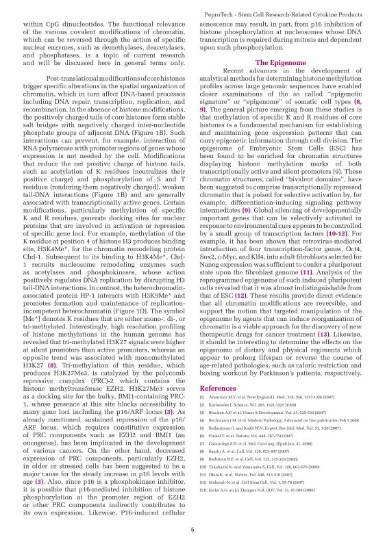
Fertilized egg
Totipotent stem cells
Blastocyst containing pluripotent stem cells
Isolated pluripotent SCs from inner cell mass
Cultured pluripotent SCs
Connective tissue, bones,
cartilage, etc.
Blood cells Cells of nervous system
Neural SCsTissue-specific SCs
Hematopoetic SCs Mesenchymal SCsNeural SCsTissue specific SCs
matopoetic SCCCc SCc SCHemmatopoeticemmatopoeticmmatopoetic MM al SCsMesenchymaal SCMesenchyma
Embryonic Stem Cells (ESCs) ESCs are pluripotent stem cells derived from the inner mast of a blastocyst that can
differentiate into the three primary germ layers; ectoderm, andoderm and mesoderm.
PeproTech - Stem Cell Research-Related Cytokine Products
7
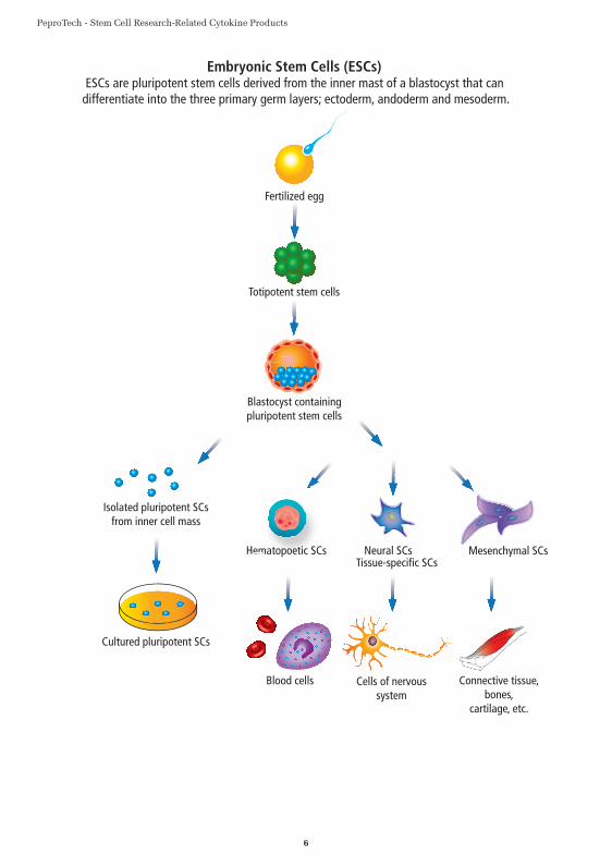
B-Lymphocyte/Plasma CellMonocyte/Macrophage T-LymphocyteMegakaryocyte/Plateletscyte/PlMegakaryockaryoc
Common LymphoidProgenitor
ommon LymphoididphoiphoiCoommon LymCoommon Lymmmon LLym
HematopoieticStem Cell
HematopoieticetictiHematopoieHematopoieH t i
dorr
Basophil/Mast CellEosinophilNeutrophil B hil/M C ll Erythrocyteh l BE i hil Lymphocyte/Plasma C T-Lymphocyte
Common MyeloidProgenitor
PeproTech - Stem Cell Research-Related Cytokine Products
8
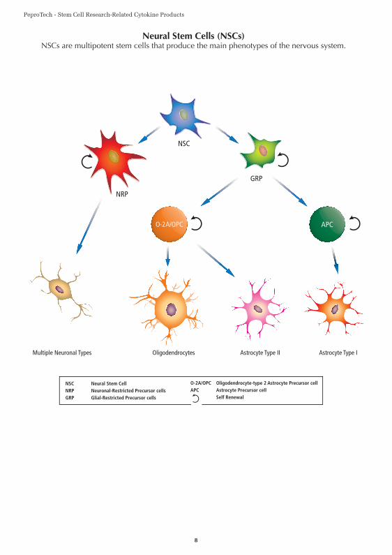
NSC
GRP
O-2A/OPC
Multiple Neuronal Types Oligodendrocytes Astrocyte Type IAstrocyte Type II
APC
NRP
NSC Neural Stem CellNRP Neuronal-Restricted Precursor cellsGRP Glial-Restricted Precursor cells
O-2A/OPC Oligodendrocyte-type 2 Astrocyte Precursor cellAPC Astrocyte Precursor cell Self Renewal
PeproTech - Stem Cell Research-Related Cytokine Products
9
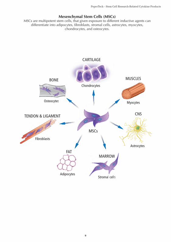
MSCs
CNS
MUSCLES
MARROWFAT
CARTILAGE
BONE
TENDON & LIGAMENT
Chondrocytes
Osteocytes
Fibroblasts
Myocytes
AdipocytesStromal cells
Astrocytes
MSCsMSC
M
Str
M
Str
W
sromal cells
MARROW
s
W
romal cells
MARROW
blastsFibrob
N & LIGAMEN
BONE
OOsteocytesOsteocytes
BONE
sOsteocytes
PeproTech - Stem Cell Research-Related Cytokine Products
10
Activin AActivin A is a TGF-β family member that exhibits a wide range of biological activities, including regulation of cellular proliferation and differentiation, and promotion of neuronal survival. Elevated levels of Activin A in human colorectal tumors and in postmenopausal women have been implicated in colorectal and breast cancers, respectively. The biological activities of Activin A can be neutralized by inhibins and by the diffusible TGF-β antagonist, follistatin. Activin A binds to the two forms of activin receptor type I (Act RI-A and Act RI-B) and two forms of activin receptor type II (Act RII-A and Act RII-B). Activins are homodimers or heterodimers of different β subunits. They are produced as precursor proteins with an amino terminal propeptide that is cleaved to release the C-terminal bioactive ligand.
Activin BActivin B is a TGF-β family member that exhibits a wide range of biological activities, including regulation of embryogenesis, osteogenesis, hematopoiesis, reproductive physiology and hormone secretion from the hypothalamic, pituitary and gonadal glands. Activin B, like certain other members of the TGF-β family, signals through the ActRII receptor (Activin Receptor type II). Activins are homodimers or heterodimers of different β subunits. They are produced as precursor proteins with an amino terminal propeptide that is cleaved to release the C-terminal bioactive ligand.
AITRLAITRL, a member of the TNF superfamily, is expressed in endothelial cells, and signals through the AITR receptor. AITRL regulates T-cell proliferation and survival, and effectuates the interaction between T lymphocytes and endothelial cells. The AITRL gene codes for a type II transmembrane protein comprised of 177 amino acids, including a 28 amino acid cytoplasmic region, a 21 amino acid transmembrane domain and a 128 amino acid extracellular domain.
AmphiregulinAmphiregulin is an EGF-related growth factor that signals through the EGF/TGF-α receptor, and stimulates growth of keratinocytes, epithelial cells and some fibroblasts. Amphiregulin also inhibits the growth of certain carcinoma cell lines. Synthesized as a transmembrane protein, Amphiregulin’s extracellular domain is proteolytically processed to release the mature protein. There are 6 conserved cysteine residues, which form 3 intramolecular disulfide bonds essential for biological activity.
ArteminArtemin is a disulfide-linked homodimeric neurotrophic factor structurally related to GDNF, Artemin, Neurturin and Persephin. These proteins belong to the cysteine knot superfamily of growth factors that assume stable dimeric protein structures. Artemin, GDNF, Persephin and Neurturin all signal through a multicomponent receptor system, composed of RET (receptor tyrosine kinase) and one of the four GFRα (α1-α4) receptors. Artemin prefers the receptor GFRα3-RET, but will use other receptors as an alternative. Artemin supports the survival of all peripheral ganglia, such as sympathetic, neural crest and placodally-derived sensory neurons, and dopaminergic midbrain neurons. The functional human Artemin ligand is a disulfide-linked homodimer of two 12.0 kDa polypeptide monomers. Each monomer contains seven conserved cysteine residues, one of which is used for interchain disulfide bridging and the others are involved in intramolecular ring formation known as the cysteine knot configuration.
BAFFBAFF, a member of the TNF superfamily of ligands, is expressed in T cells, macrophages, monocytes and dendritic cells. BAFF is involved in stimulation of B and T cell function, and is an important survival and maturation factor for peripheral B cells. BAFF signals through three different TNF receptors, TACI, BCMA and BAFF-R. The human BAFF gene codes for a 285 amino acid type II transmembrane protein containing a 46 amino acid cytoplasmic domain, a 21 amino acid transmembrane domain, and a 218 amino acid extracellular domain.
PeproTech - Stem Cell Research-Related Cytokine Products
11
BD-1 / BD-2 / BD-3 / BD-4 / BD-5Defensins (alpha and beta) are cationic peptides with a broad spectrum of antimicrobial activity that comprise an important arm of the innate immune system. The α-defensins are distinguished from the β-defensins by the pairing of their three disulfide bonds. To date, six human β-defensins have been identified; BD-1, BD-2, BD-3, BD-4, BD-5 and BD-6. β-defensins are expressed on some leukocytes and at epithelial surfaces. In addition to their direct antimicrobial activities, they can act as chemoattractants towards immature dendritic cells and memory T cells. The β-defensin proteins are expressed as the C-terminal portion of precursors, and are released by proteolytic cleavage of a signal sequence and, in some cases, a propeptide sequence. β-defensins contain a six-cysteine motif that forms three intra-molecular disulfide bonds.
BDNFBDNF is a member of the NGF family of neurotrophic growth factors. Like other members of this family, BDNF supports neuron proliferation and survival. BDNF can bind to a low affinity cell surface receptor called LNGFR, which also binds other neurotrophins such as NGF, NT-3 and NT-4. However, BDNF mediates its neurotrophic properties by signaling through a high affinity cell surface receptor called gp145/trkB. BDNF is expressed as the C-terminal portion of a 247 amino acid polypeptide precursor, which also contains a signal sequence of 18 amino acid residues and a propeptide of 110 amino acid residues.
BetacellulinBetacellulin is an EGF-related polypeptide growth factor that signals through the EGF receptor. It is produced in several tissues, including the pancreas, small intestine, and in certain tumor cells. Betacellulin is a potent mitogen for retinal pigment epithelial cells and vascular smooth muscle cells. Human Betacellulin is initially synthesized as a glycosylated 32.0 kDa transmembrane precursor protein, which is processed by proteolytic cleavage to produce the mature sequence.
BMP-2BMPs (Bone Morphogenetic Proteins) belong to the TGF-β superfamily of structurally related signaling proteins. BMP-2 is a potent osteoinductive cytokine, capable of inducing bone and cartilage formation in association with osteoconductive carriers such as collagen and synthetic hydroxyapatite. In addition to its osteogenic activity, BMP-2 plays an important role in cardiac morphogenesis, and is expressed in a variety of tissues, including lung, spleen, brain, liver, prostate ovary and small intestine. The functional form of BMP-2 is a 26 kDa protein composed of two identical 114 amino acid polypeptide chains linked by a single disulfide bond. Each BMP-2 monomer is expressed as the C-terminal part of a precursor polypeptide, which also contains a 23 amino acid signal sequence for secretion, and a 259 amino acid propeptide. After dimerization of this precursor, the covalent bonds between the propeptide (which is also a disulfide-linked homodimer) and the mature BMP-2 ligand are cleaved by a furin-type protease.
BMP-3TGF-β family members are key modulators of cell proliferation, differentiation, matrix synthesis, and apoptosis. As implied by their name, BMPs initiate, promote, and regulate the development, growth, and remodeling of bone and cartilage. In addition to this role, BMPs are also involved in prenatal development and postnatal growth, remodeling and maintenance of a variety of other tissues and organs. BMP-3 is abundantly found in adult bone and, to a lesser extent, fetal cartilage. BMP-3 inhibits osteogenesis and bone formation by activating a signaling cascade that antagonizes the signaling of pro-osteogenic BMPs.
BMP-4Bone morphogenetic proteins (BMPs) constitute a subfamily within the TGF-β superfamily of structurally related signaling proteins. Members of this superfamily are widely distributed throughout the body, and are involved in diverse physiological processes during both pre- and postnatal life. Like BMP-7, BMP-4 is involved in the development and maintenance of bone and cartilage. Reduced expression of BMP-4 is associated with a number of bone diseases, including the heritable disorder Fibrodysplasia Ossificans Progressiva.
PeproTech - Stem Cell Research-Related Cytokine Products
12
BMP-5TGF-β family members are key modulators of cell proliferation, differentiation, matrix synthesis, and apoptosis. As implied by their name, BMPs initiate, promote, and regulate the development, growth, and remodeling of bone and cartilage. In addition to this role, BMPs are also involved in prenatal development and postnatal growth, remodeling, and maintenance of a variety of other tissues and organs. BMP-5 is expressed in the nervous system, lungs and liver. It is a known regulator for dendritic growth in sympathetic neurons. BMP-5 is a 454 amino acid precursor protein that is cleaved to release the biologically active C-terminal mature protein.
BMP-6TGF-β family members are key modulators of cell proliferation, differentiation, matrix synthesis, and apoptosis. As implied by their name, BMPs initiate, promote, and regulate the development, growth, and remodeling of bone and cartilage. In addition to this role, BMPs are also involved in prenatal development and postnatal growth, remodeling, and maintenance of a variety of other tissues and organs. Increasing evidence indicates that BMP-Smad signaling has a tumor suppressing activity, and that BMPs can inhibit tumor growth. BMP-6 is abnormally expressed in breast cancer cell lines, however, its function in promoting breast cancer development is unknown. The mature and functional form of BMP-6 is a homodimer of two identical 139 amino acid polypeptide chains linked by a single disulfide bond. Each monomer is expressed as the C-terminal part of a precursor polypeptide, which contains a 20 amino acid signal peptide and a 354 amino acid propeptide. This precursor undergoes intracellular dimerization, and upon secretion it is processed by a furin-type protease.
BMP-7TGF-β family members are key modulators of cell proliferation, differentiation, matrix synthesis, and apoptosis. As implied by their name, BMPs initiate, promote, and regulate the development, growth, and remodeling of bone and cartilage. In addition to this role, BMPs are also involved in prenatal development and postnatal growth, remodeling, and maintenance of a variety of other tissues and organs. BMP-7, also known as osteogenic protein-1 or OP-1, is a potent bone inducing agent, which in the presence of an appropriate osteoconductive carrier (e.g. collagen sponge or synthetic hydroxyapatite) can be used in the treatment of bone defects. A bone-graft substitute, called OP-1TM implant, made of recombinant human BMP-7 associated with bovine bone-derived collagen, has recently been approved by the FDA as a device for treating critical-size bone fractures. The potential use of BMP-7 in dental reconstructive surgeries is currently under investigation.
BMP-10Bone morphogenetic proteins (BMPs) constitute a subfamily within the TGF-β superfamily of structurally related signaling proteins. Members of this superfamily are widely distributed throughout the body and are involved in diverse physiological processes during both pre- and postnatal life. BMP-10 plays a crucial role in the development of the embryonic heart by acting to stimulate and maintain cardiomyocyte proliferation. It can signal through various receptor complexes usually containing BMPR-1A, BMPR-1B, ALK1, ALK3, or ALK6. The interaction of BMP-10 with its specific receptors can induce signaling initiated by the phosphorylation of SMAD transcription factors, including SMAD1, SMAD5, or SMAD8, but can also induce SMAD independent processes. BMP-10 is structurally related to BMP-9, and both can inhibit endothelial cell proliferation and migration.
BMP-13 (CDMP-2)BMP-13 is expressed in hypertrophic chondrocytes during embryonic development of long bones. Continued postnatal expression of BMP-13 in articular cartilage suggests that it plays a regulatory role in the growth and maintenance of articular cartilage. Adenovirus-mediated BMP-13 gene transfer to rabbit bone marrow stem cells have been reported to augment periosteal repair of osteochondral defects. The functional form of BMP-13/CDMP-2 is a disulfide-linked homodimer of two 120 amino-acid polypeptide chains. This 27.5 kDa protein is obtained by proteolytic processing of a biologically inactive precursor protein of 97.7 kDa.
PeproTech - Stem Cell Research-Related Cytokine Products
13
Cardiotrophin-1 (CT-1)CT-1 is a member of the IL-6 family of cytokines which also includes LIF, CNTF, OSM (Oncostatin M), IL-11, IL-6 and possibly NNT-1/BSF-3. CT-1 is a pleiotropic cytokine which is expressed in various tissues including the adult heart, skeletal muscle, ovary, colon, prostate and fetal lung and signals through the LIF receptor and the gp130 receptor subunit. CT-1 has the ability to induce cardiac myocyte hypertrophy, and enhances the survival of cardiomyocyte and different neuronal populations. Biologically active human CT-1 is synthesized as a 201 amino acid polypeptide lacking a hydrophobic N-terminal secretion signal sequence.
sCD34CD34 is a highly glycosylated type I membrane protein that is selectively expressed on hematopoietic stem cells and vascular endothelium. It has been widely used as a molecular marker for the identification, isolation, and manipulation of hemopoietic stem cells and progenitors. CD34 can function as a regulator of hemopoietic cell adhesion by mediating the attachment of stem cells to bone marrow stromal cells or other bone marrow components. The full length human CD34 is a 385 amino acid protein, consisting of a 31 amino acid signal sequence, a 74 amino acid cytoplasmic domain, a 21 amino acid transmembrane domain and a 259 amino acid extracellular domain.
CDNFCDNF is a secreted neurotrophic factor that is expressed in brain, neuronal and certain non-neuronal tissues. It has been shown to promote survival, growth and function of dopamine-specific neurons. CDNF and its structural homolog, MANF, each contain an N-terminal saposin-like lipid binding domain, and a carboxyl-terminal domain, which is not homologous to previously characterized protein structures. CDNF and MANF can prevent 6-OHDA-induced degeneration of dopaminergic neurons by triggering survival pathways in a rat experimental model of Parkinson’s disease.
CNTFCNTF is a potent neural factor that was originally characterized as a vital factor for the survival of chick ciliary neurons in vitro. CNTF is also important for the survival of other neural cell types, including primary sensory neurons, motor neurons, basal forebrain neurons and type 2 astrocytes. CNTF is highly conserved across species and exhibits cross-species bioactivity.
CTGFCTGF is a member of the CCN family of secreted cysteine-rich regulatory proteins, and is the major mitogenic and chemoattractant protein produced by umbilical vein and vascular endothelial cells. CTGF stimulates the proliferation and differentiation of chondrocytes, induces angiogenesis, promotes cell adhesion of fibroblasts, endothelial and epithelial cells, and binds to IGF, TGF β1 and BMP-4. Cell migration and adhesion are signaled through binding to specific cell surface integrins and to heparin sulfate proteoglycans CTGF (98 a.a.), a lower molecular weight isoform containing the C-terminal portion of the full length CTGF protein, exerts full heparin binding, cell adhesion, and mitogenic CTGF activity. Mature Human CTGF is a 38.0 kDa secreted protein containing 323 amino acid residues. CTGF is comprised of four distinct structural domains (modules), which are identified as IGF binding protein (IGFBP), von Willebrand Factor C (VWFC), thrombospondin type-I (TSP type-I), and C-terminal cysteine knot-like (CTCK) domains. Full length CTGF can be proteolytically cleaved in certain tissues to yield N-terminal truncated isoforms, which, depending on the isoform, contain only the TSP type-I and CTCK domains or contain only the CTCK domain.
CYR61CYR61 is a member of the CCN family of secreted cysteine-rich regulatory proteins. CYR61 induces angiogenesis by stimulating the proliferation, migration, and adhesion of endothelial cells. Cell migration and adhesion are mediated through binding to specific cell surface integrins and to heparin sulfate proteoglycans. Increased expression of CYR61 is associated with several types of cancer, and correlates with the progression and estrogen independence of human breast cancers.
PeproTech - Stem Cell Research-Related Cytokine Products
14
DKK-1DKK-1 is a member of the DKK protein family which also includes DKK-2, DKK-3 and DKK-4. DKK-1 was originally identified as a Xenopus head-forming molecule that behaves as an antagonist for Wnt signaling. Subsequent studies have shown that DKK-1 and DKK-4 play an important regulatory role in the Wnt/β-catenin signaling pathway by forming inhibitory complexes with LDL receptor-related proteins 5 and 6 (LRP5 and LRP6), which are essential components of the Wnt/β-catenin signaling system. LPR5 and LPR6 are single-pass transmembrane proteins that appear to act as co-receptors for Wnt ligands involved in the Wnt/β-catenin signaling cascade. It has been suggested that by inhibiting Wnt/β-catenin signaling, which is essential for posterior patterning in vertebrates, DKK-1 permits anterior development. This notion is supported by the finding that mice deficient of DKK-1 expression lack head formation and die during embryogenesis.
DKK-2 / DKK-3The dickkopf (DKK)-related protein family is comprised of four central members, DKK-1 - 4, along with the distantly-related DKK family member DKK-11 (Soggy), which is thought to be a descendent of an ancestral DKK-3 precursor due to its unique sequence homology to DKK-3 and no other DKK family member. DKK family members, with the exception of the divergent Soggy, share two conserved cysteine-rich domains and show very little sequence similarity outside of these domains. Playing an important regulatory role in vertebrate development through localized inhibition of Wnt-regulated processes, including anterior-posterior axial patterning, limb development, somitogenesis, and eye formation, DKKs have also been implicated post-developmentally in bone formation, bone disease, cancer, and neurodegenerative diseases. DKK proteins typically play an important regulatory role in the Wnt/β-catenin signaling pathway by forming inhibitory complexes with LDL receptor-related proteins 5 and 6 (LRP5 and LRP6), which are essential components of the Wnt/β-catenin signaling system. LRP5 and LRP6 are single-pass transmembrane proteins that appear to act as co-receptors for Wnt ligands involved in the Wnt/β-catenin signaling cascade. DKK-2 has been shown to both inhibit and enhance canonical Wnt signaling; enhancing Wnt signaling through direct high-affinity binding of DKK-2 to LRP6 during LRP6 overexpression, while inhibiting Wnt signaling and promoting LRP6 internalization through the formation of a ternary complex between DKK-2, LRP6, and Kremen-2. DKK-3 has been shown to potentiate, rather than inhibit, Wnt signaling through interactions with the high-affinity, transmembrane co-receptors Kremen-1 (Krm1) and Kremen-2 (Krm2).
sDLL-1Human soluble DLL-1 comprises the extracellular signaling domain of DLL-1, a member of the Delta/Serrate/Lag-2 (DSL) family of single-pass type I trans-membrane proteins that serve as ligands for Notch receptors. It is expressed primarily in the heart, pancreas and epidermis. DLL-1 functions to specifically activate the Notch-1 and Notch-2 receptors. Proteolytic cleavage of DLL-1 produces a secreted extracellular domain, sDLL-1, that interacts with Notch receptors expressed on adjacent cells. Notch signaling plays an essential role in controlling cell fate decisions during prenatal development and postnatal stem cell renewal, and differentiation in many tissues. Human sDLL-1 blocks monocyte differentiation into macrophages, but permits differentiation into dendritic cells. In hematopoietic progenitor cells, hsDLL-1, suppresses differentiation into B-cells, while promoting differentiation into T-cells and NK cell precursors. In cell culture, human sDLL-1 has been shown to promote expansion of hematopoietic progenitor cells and suppress apoptosis by inhibiting differentiation. Overexpression of Notch receptors appears to inhibit differentiation in several mammalian cell lines, and increasing evidence suggests that Notch signaling is frequently downregulated in human malignancies. The human DLL-1 gene consists of a 528 amino acid extracellular domain with one DSL domain, eight EGF-like repeats, a 23 amino acid transmembrane domain, and a 155 amino acid cytoplasmic domain.
sDLL-4Human sDLL-4 comprises the extracellular signaling domain of DLL, a member of a structurally-related family of single-pass type I trans-membrane proteins that serve as ligands for Notch
PeproTech - Stem Cell Research-Related Cytokine Products
15
receptors. DLL-4 functions to specifically activate the Notch-1 and Notch-4 receptors. The Notch signaling pathway regulates endothelial cell differentiation, proliferation and apoptosis, and is essential for the development, maintenance and remodeling of the vascular system. Targeted deletion of the DLL-4 gene in mice resulted in severe vascular defects and death before birth. Up-regulation of DLL-4 expression has been implicated in the vascular development of certain tumors. The human DLL-4 gene consists of a 503 amino acid extracellular domain with one DSL domain, eight EGF-like repeats, a 21 a.a. transmembrane domain, and a 135 a.a. cytoplasmic domain.
EGFEGF is a potent growth factor that stimulates the proliferation of various epidermal and epithelial cells. Additionally, EGF has been shown to inhibit gastric secretion, and to be involved in wound healing. EGF signals through a receptor known as c-erbB, which is a class I tyrosine kinase receptor. This receptor also binds with TGF-a and VGF (vaccinia virus growth factor).
EGF Receptor (EGFR)EGF Receptor (EGFR, ErbB1) is a transmembrane protein that exerts tyrosine kinase activity upon ligand-induced activation. EGFR can be activated by binding EGF, or at least six other structurally related protein ligands, including TGFa, HB-EGF, Betacellulin (BTC), Amphiregulin, Epiregulin, and Epigen. Upon activation, EGFR initiates a signaling cascade, which includes dimerization and internalization, tyrosine phosphorylation, DNA synthesis of target genes and, ultimately, cell proliferation. EGFR signaling plays a role in the growth and differentiation of normal cells, but elevated EGFR activity is correlated with the development and pathogenesis of certain cancers.
EGF-L7EGF-L7 (Epidermal growth factor-like protein 7, Multiple EGF-like domains protein 7, VE-statin) is a multi-domain protein containing two EGF-like domains and one EMI domain. It is expressed almost exclusively in endothelial cells and functions to promote normal development of the vascular system, particularly tubulogenesis. EGF-L7 is capable of antagonistic binding to Notch receptors, resulting in the inhibition of Notch signaling in HUVEC and neural stem cells. In research models inducing hypoxia and subsequent reoxygenation (H/R), EGF-L7 can inhibit ICAM-1 expression and enhance the inhibition of NF-kB activation. Additionally, EGF-L7 can chemoattract endothelial cells and bind to the extracellular matrix. The overexpression of EGF-L7 is observed in various cancers, and is generally correlated with increased metastasis and a poor prognosis.
EGF-VEGFEG-VEGF is a secreted angiogenetic mitogen growth factor expressed in the steroidogenic glands, ovary, testis, adrenal gland, and placenta. EG-VEGF induces proliferation, migration, and fenestration (formation of membrane discontinuities) in capillary endothelial cells derived from endocrine glands. The human EG-VEGF gene codes for a 105 amino acid polypeptide containing an N-terminal signal sequence of 19 amino acids.
EpigenEpigen is an EGF-related polypeptide growth factor that signals through the ErbB receptor-1. It is produced in several tissues, including the testis, liver, and heart, as well as in certain tumor cells. Epigen is mitogenic for fibroblasts and epithelial cells. Human Epigen is initially synthesized as a glycosylated 14.7 kDa transmembrane precursor protein, which is processed by proteolytic cleavage to produce a mature soluble sequence.
PeproTech - Stem Cell Research-Related Cytokine Products
16
EpiregulinEpiregulin is an EGF-related growth factor that binds specifically to EGFR (ErbB1) and ErbB4, but not ErbB2 or ErbB3. It is expressed mainly in the placenta and peripheral blood leukocytes, as well as in certain carcinomas of the bladder, lung, kidney and colon. Epiregulin stimulates the proliferation of keratinocytes, hepatocytes, fibroblasts and vascular smooth muscle cells. It also inhibits the growth of several tumor-derived epithelial cell lines. Human Epiregulin is initially synthesized as a glycosylated 19.0 kDa transmembrane precursor protein, which is processed by proteolytic cleavage to produce a 6.0 kDa mature secreted sequence.
EPOErythropoietin (EPO) is a glycoprotein hormone that is principally known for its role in erythropoiesis, where it is responsible for stimulating proliferation and differentiation of erythroid progenitor cells. The differentiation of CFU-E (Colony Forming Unit-Erythroid) cells into erythrocytes can only be accomplished in the presence of EPO. Physiological levels of EPO in adult mammals are maintained primarily by the kidneys, whereas levels in fetal or neonatal mammals are maintained by the liver. EPO also can exert various non-hematopoietic activities, including vascularization and proliferation of smooth muscle, neural protection during hypoxia, and stimulation of certain B cells.
FGF SuperfamilyProteins of the FGF superfamily of growth factors manifest only a modest degree of primary sequence homology, yet share the ability to signal through one or more of four tyrosine kinase receptors called FGFR1 through FGFR4. The FGFs play a central role during prenatal development, postnatal growth and regeneration of a variety of tissues, by promoting cellular proliferation and differentiation. All members of the FGF superfamily bind, with varying degrees of affinity to heparin sulfate proteoglycans, which serve as extracellular storage sites and in some cases appear to be involved in the activation of the FGF receptors.
FGF-acidic (FGF-1)FGF-acidic is one of 23 known members of the FGF family. Proteins of this family play a central role during prenatal development, postnatal growth and regeneration of a variety of tissues, by promoting cellular proliferation and differentiation. FGF-acidic is a non-glycosylated heparin binding growth factor that is expressed in the brain, kidney, retina, smooth muscle cells, bone matrix, osteoblasts, astrocytes and endothelial cells. FGF-acidic has the ability to signal through all the FGF receptors.
FGF-basic (FGF2)FGF-basic is one of 23 known members of the FGF family. Proteins of this family play a central role during prenatal development, postnatal growth and regeneration of a variety of tissues, by promoting cellular proliferation and differentiation. FGF-basic is a non-glycosylated, heparin-binding growth factor that is expressed in the brain, pituitary, kidney, retina, bone, testis, adrenal gland, liver, monocytes, epithelial cells and endothelial cells. FGF-basic signals through FGFR 1b, 1c, 2c, 3c and 4.
FGF-4FGF-4 is a heparin-binding growth factor that is a member of the FGF family. Proteins of this family play a central role during prenatal development, postnatal growth and regeneration of a variety of tissues, by promoting cellular proliferation and differentiation. FGF-4 signals through the FGFR 1c, 2c, 3c, and 4.
FGF-5FGF-5 is a secreted, heparin-binding growth factor that belongs to the FGF family. Proteins of this family play a central role during prenatal development, postnatal growth and regeneration of a variety of tissues, by promoting cellular proliferation and differentiation. FGF-5 binds to FGFR 1c and 2c, and plays a regulatory role in the hair growth cycle.
PeproTech - Stem Cell Research-Related Cytokine Products
17
FGF-6FGF-6 is a secreted, heparin-binding growth factor that is a member of the FGF family. Proteins of this family play a central role during prenatal development, postnatal growth and regeneration of a variety of tissues, by promoting cellular proliferation and differentiation. FGF-6 is expressed in leukemia cell lines with platelet megakaryocytic differentiation potential. It signals through FGFR 1c, 2c, and 4.
FGF-7 (KGF)KGF (FGF-7) is one of 23 known members of the FGF family. Proteins of this family play a central role during prenatal development, postnatal growth, and the regeneration of a variety of tissues, by promoting cellular proliferation and differentiation. KGF (FGF-7) is a mitogen factor specific for epithelial cells and keratinocytes. KGF/FGF-7 signals through FGFR 2b. KGF (FGF-7) plays a role in kidney and lung development, as well as in angiogenesis and wound healing.
FGF-8a / FGF-8bFGF-8 is a heparin-binding growth factor belonging to the FGF family. Proteins of this family play a central role during prenatal development, postnatal growth and regeneration of a variety of tissues, by promoting cellular proliferation and differentiation. There are 4 known alternate spliced forms of FGF8; FGF-8a, FGF-8b, FGF-8e and FGF-8f. The human and murine FGF-8a and b are identical, unlike human and mouse FGF-8e and f, which are 98% identical. FGF-8 targets mammary carcinoma cells and other cells expressing the FGF receptors.
FGF-9FGF-9 is a heparin-binding growth factor that belongs to the FGF family. Proteins of this family play a central role during prenatal development, postnatal growth and regeneration of a variety of tissues, by promoting cellular proliferation and differentiation. FGF-9 targets glial cells, astrocytes cells and other cells that express the FGFR 1c, 2c, 3b, 3c, and 4.
FGF-10FGF-10 is a heparin-binding growth factor that belongs to the FGF family. Proteins of this family play a central role during prenatal development, postnatal growth and regeneration of a variety of tissues, by promoting cellular proliferation and differentiation. FGF-10 is most related to KGF/FGF-7, and is expressed during the development and, preferentially, in adult lungs. It signals through the FGFR 2b.
FGF-16FGF-16 is a heparin-binding growth factor that is a member of the FGF family. Proteins of this family play a central role during prenatal development, postnatal growth and regeneration of a variety of tissues, by promoting cellular proliferation and differentiation. FGF-16 signals through FGFR 2c and 3c. FGF-16 plays a role in the development of the central nervous system.
FGF-17FGF-17 is a heparin-binding growth factor that is a member of the FGF family. Proteins of this family play a central role during prenatal development, postnatal growth and regeneration of a variety of tissues, by promoting cellular proliferation and differentiation. FGF-17 signals through the FGFR 1c, 2c, 3c, and 4. FGF-17 signals induction and patterning of embryonic brain.
FGF-18FGF-18 is a heparin-binding growth factor that belongs to the FGF family. Proteins of this family play a central role during prenatal development, postnatal growth and regeneration of a variety of tissues, by promoting cellular proliferation and differentiation. FGF-18 is an essential regulator of long bone and calvarial development. FGF-18 signals through FGFR 1c, 2c, 3c, and 4.
PeproTech - Stem Cell Research-Related Cytokine Products
18
FGF-19The FGF family plays central roles during prenatal development and postnatal growth and regeneration of a variety of tissues, by promoting cellular proliferation and differentiation. FGF-19, a member of the FGF family, is a high-affinity heparin-dependent ligand for FGFR4. FGF-19 is expressed during brain development and embryogenesis.
FGF-20FGF-20 is a secreted, heparin-binding growth factor that is a member of the FGF family. Proteins of this family play a central role during prenatal development, postnatal growth and regeneration of a variety of tissues, by promoting cellular proliferation and differentiation. FGF-20 signals through the FGFR 2c and 3c, and is expressed during limb and brain development.
FGF-21FGF-21 is a secreted growth factor that is a member of the FGF family. Proteins of this family play a central role during prenatal development, postnatal growth and regeneration of a variety of tissues, by promoting cellular proliferation and differentiation. FGF-21, in the presence of b-Klotho as a protein cofactor, signals through the FGFR 1c and 4 receptors, and stimulates insulin-independent glucose uptake by adipocytes.
FGF-23The FGF family plays a central role during prenatal development, postnatal growth and regeneration of a variety of tissues, by promoting cellular proliferation and differentiation. FGF-23, FGF-21 and FGF-19 constitute an atypical FGF subfamily whose ligands act as circulating hormones and require the participation of asKlothosprotein as a co-receptor for their signaling. FGF-23 is a bone-derived hormone that acts in the kidney to regulate phosphate homeostasis and vitamin D metabolism. The signaling receptor for FGF-23, a Klotho-FGFR1 (IIIc) complex, is an essential regulator of the renal sodium phosphate co-transporter and key vitamin D-metabolizing enzymes CYP27B1 and CYP24A1.
FGFR1a / FGFR2a / FGFR3The FGF family plays a central role during prenatal development and postnatal growth, and the regeneration of a variety of tissues, by promoting cellular proliferation and differentiation. The FGF ligands bind to a family of type I transmembrane tyrosine kinase receptors, which leads to dimerization and activation by sequential autophosphorylation of specific tyrosine residues. Four genes encoding structurally related FGF receptors (FGFR-1 to -4) are known. Alternative splicing of the mRNAs generates numerous forms of FGFR-1 to -3. Alternate forms of FGF receptors can exhibit different specificities with respect to ligand binding. For example, the form designated as FGFR1a (IIc) interacts predominantly with FGF-acidic (FGF1) and FGF-basic (FGF2). A frequent splicing event involving FGFR-1 and -2 results in receptors containing all three Ig domains, referred to as the alpha isoform, or only IgII and IgIII, referred to as the beta isoform. Only the alpha isoform has been identified for FGFR-3 and FGFR-4. Additional splicing events for FGFR-1 to -3, involving the C-terminal half of the IgIII domain encoded by two mutually exclusive alternative exons, generate FGF receptors with alternative IgIII domains (IIIb and IIIc).
Flt3-LigandFlt3-Ligand is a growth factor that regulates proliferation of early hematopoietic cells. Flt3-Ligand binds to cells expressing the tyrosine kinase receptor Flt3. Flt3-Ligand, by itself does not stimulate proliferation of early hematopoietic cells, but synergizes with other CSFs and interleukins to induce growth and differentiation. Unlike SCF, Flt3-Ligand exerts no activity on mast cells. Multiple isoforms of Flt3-Ligand have been identified. The predominant biologically active form is anchored to the cell surface as the extracellular domain of a transmembrane protein (209 a.a.). The membrane-bound isoform can be proteolytically cleaved to generate a biologically active soluble isoform.
PeproTech - Stem Cell Research-Related Cytokine Products
19
FollistatinFollistatin is a secreted protein that binds to ligands of the TGF-b family and regulates their activity by inhibiting their access to signaling receptors. It was originally discovered as an activin antagonist whose activity suppresses expression and secretion of the pituitary hormone FSH (follicle stimulating hormone). In addition to being a natural antagonist, follistatin can inhibit the activity of other TGF-b ligands including BMP-2,-4,-6,-7, Myostatin, GDF-11, and TGF-b1. Follistatin is expressed in the pituitary, ovaries, decidual cells of the endometrium, and in some other tissues.
sFRP-1Secreted Frizzled Related Proteins (sFRPs) modulate WNT signaling by binding directly to WNT proteins in a manner that affects their receptor binding and signaling capabilities. sFRP-1 is a widely distributed protein that can bind directly to WNT1, WNT2, and possibly other WNT proteins, and generally exerts anti-proliferative effects consistent with activity as a WNT antagonist. It also inhibits apoptosis, and has been found to be down-regulated in many solid tumors, but up-regulated in uterine leiomyomas.
Galectin-1Lectins, of either plant or animal origin, are carbohydrate-binding proteins that interact with glycoprotein and glycolipids on the surface of animal cells. The Galectins are lectins that recognize and interact with beta-galactoside moieties. Galectin-1 is an animal lectin that has been shown to interact with CD3, CD4, and CD45. It induces apoptosis of activated T-cells and T-leukemia cell lines, and inhibits the protein phosphatase activity of CD45.
GASP-1Growth and differentiation factor-associated serum protein-1 (GASP-1) is a secreted inhibitory TGF-b binding protein that contains multiple protease inhibitor structural domains. It is expressed primarily in the ovary, testis, and brain, and can act as a potent soluble inhibitor of myostatin and GDF-11, but not Activin A. The GASP-1 gene encodes a 571 amino acid protein that contains a 29 amino acid secretion signal sequence, and multiple identifiable structural features, including a WAP domain, a follistatin/Kazal domain, an immunoglobulin domain, two tandem Kunitz domains, and a netrin domain.
G-CSFG-CSF is a hematopoietic growth factor that stimulates the development of committed progenitor cells to neutrophils and enhances the functional activities of the mature end-cell. It is produced in response to specific stimulation by a variety of cells, including macrophages, fibroblasts, endothelial cells and bone marrow stroma. G-CSF is being used clinically to facilitate hematopoietic recovery after bone marrow transplantation. Human and murine G-CSF is cross-species reactive.
GDF-2GDF-2 belongs to the TGF-b cytokine family, whose members play an important role during prenatal development and postnatal growth, and the remodeling and maintenance of a variety of tissues and organs. GDF-2 is expressed mainly in non-parenchymal cells of the liver, but is also found in other various cells and tissues. GDF-2 can signal through the ALK1 receptor, and has been implicated in a number of physiologic events including the regulation of the hepatic reticuloendothelial system, glucose homeostasis, iron homeostasis, and the inhibition of angiogenesis.
GDF-3GDF-3 is a member of the TGF-b superfamily of growth and differentiation factors, and is highly homologous to GDF-9. Unlike most TGF-b family members, GDF-3 and GDF-9 are not disulfide-linked dimers. GDF-3 is expressed in adult bone marrow, spleen, thymus, and adipose tissue. The expression of GDF-3 is upregulated in high-fat-fed wild-type FABP4/aP2 null mice and was associated with obesity, but not with the related hyperglycemia/hyperinsulinemia that characterizes Type 2 diabetes.
PeproTech - Stem Cell Research-Related Cytokine Products
20
GDF-5 (BMP-14/CDMP-1)GDF-5 is expressed in long bones during embryonic development and postnatally in articular cartilage. Mutations in the GDF-5 gene have been implicated in Hunter-Thompson type dwarfism and in Grebe Syndrome, which is characterized by short stature, extra digits, and short and deformed extremities. The mature and functional form of GDF-5 is a homodimer of two 120 amino-acid polypeptide chain (monomers) linked by a single disulfide bond. Each GDF-5 monomer is expressed as the C-terminal part of a precursor polypeptide, which also contains a 27 amino acid signal peptide and a 354 amino acid propeptide. This precursor undergoes intracellular dimerization, and upon secretion it is processed by a furin-type protease.
GDF-11GDF-11 is a myostatin-homologous protein that acts as an inhibitor of nerve tissue growth. GDF-11 has been shown to suppress neurogenesis through a myostatin-like pathway, which involves the arrest of the progenitor cell cycle in the G1 phase. Similarities between myostatin and GDF-11, which are 90% identical in their amino acid sequence, suggest that the regulatory mechanisms responsible for maintaining proper tissue size during neural and muscular development might be the same.
GDF-15 / MIC-1GDF-15 belongs to the TGF-b cytokine family, whose members play an important role during prenatal development and postnatal growth, and the remodeling and maintenance of a variety of tissues and organs. GDF-15 is expressed predominantly in the placenta and, to a much lesser extent, in various other tissues. The presence of GDF-15 in amniotic fluid and its elevated levels in the sera of pregnant women suggest GDF-15’s involvement in gestation and embryonic development. GDF-15 generally exerts tumor suppressive activities and is one of the predominant factors produced and secreted in response to activation of the p53 pathway. Interestingly, the serum level of GDF-15 is positively correlated with neoplastic progression of several tumor types, including certain colorectal, pancreatic, and prostate cancers.
GDNFGDNF is a disulfide-linked, homodimeric neurotrophic factor structurally related to Artemin, Neurturin and Persephin. These proteins belong to the cysteine-knot superfamily of growth factors that assume stable dimeric protein structures. GDNF signals through a multicomponent receptor system, composed of a RET and one of the four GFRa (a1-a4) receptors. GDNF specifically promotes dopamine uptake and survival, and morphological differentiation of midbrain neurons. Using a Parkinson’s disease mouse model, GDNF has been shown to improve conditions such as bradykinesia, rigidity, and postural instability. The functional human GDNF ligand is a disulfide-linked homodimer consisting of two 15 kDa polypeptide chains called monomers. Each monomer contains seven conserved cysteine residues, including Cys-101, which is used for inter-chain disulfide bridging, and others that are involved in the intramolecular ring formation known as the cysteine knot configuration.
GM-CSFGM-CSF is a hematopoietic growth factor that stimulates the development of neutrophils and macrophages, and promotes the proliferation and development of early erythroid megakaryocytic and eosinophilic progenitor cells. It is produced in endothelial cells, monocytes, fibroblasts and T lymphocytes. GM-CSF inhibits neutrophil migration and enhances the functional activity of the mature end-cells. The human and murine molecules are species-specific and exhibit no cross-species reactivity.
GMF-βGMF-b is a brain-specific protein that belongs to the actin-binding proteins ADF structural family. GMF-b appears to play a role in the differentiation, maintenance, and regeneration of the nervous system. It also supports the progression of certain auto-immune diseases, possibly through its ability to induce the production and secretion of various pro-inflammatory cytokines.
PeproTech - Stem Cell Research-Related Cytokine Products
21
Gremlin-1Gremlin-1 (isoform-1) belongs to a group of diffusible proteins that bind to ligands of the TGF-b family and regulate their activity by inhibiting their access to signaling receptors. The interplay between TGF-b ligands and their natural antagonists has major biological significance during development processes, in which cellular response can vary considerably depending upon the local concentration of the signaling molecule. Gremlin-1 is highly expressed in the small intestine, fetal brain, and colon; and is expressed at lower levels in the brain, prostate, pancreas, and in skeletal muscle. Gremlin-1 regulates multiple functions in early development by specifically binding to, and inhibiting the function of, BMP-2, -4, and -7. It also plays a role in carcinogenesis and kidney branching morphogenesis.
HB-EGFHB-EGF is an EGF-related growth factor that signals through the EGF receptor, and stimulates the proliferation of smooth muscle cells (SMC), fibroblasts, epithelial cells, and keratinocytes. HB-EGF is expressed in numerous cell types and tissues, including vascular endothelial cells, and vascular SMC, macrophages, skeletal muscle, keratinocytes, and certain tumor cells. The ability of HB-EGF to specifically bind heparin and heparin sulfate proteoglycans is distinct from other EGF-like molecules, and may be related to the enhanced mitogenic activity, relative to EGF, that HB-EGF exerts on smooth muscle cells. The human HB-EGF gene encodes a 208 amino acid transmembrane protein, which can be proteolytically cleaved to produce soluble HB-EGF.
Heregulinβ-1 (Neuregulin)Neuregulin/Heregulin is a family of structurally related polypeptide growth factors derived from alternatively spliced genes (NRG1, NRG2, NRG3 and NRG4). To date, there are over 14 soluble and transmembrane proteins derived from the NRG1 gene. Proteolytic processing of the extracellular domain of the transmembrane NRG1 isoforms releases soluble growth factors. HRG1-b1 contains an Ig domain and an EGF-like domain; the latter is necessary for direct binding to receptor tyrosine kinases erb3 and erb4. This binding induces erb3 and erb4 heterodimerization with erb2, stimulating intrinsic kinase activity that leads to tyrosine phosphorylation. Although HRG1-b1’s biological effects are still unclear, it has been found to promote motility and invasiveness of breast cancer cells, which may also involve up-regulation of expression and function of the autocrine motility-promoting factor (AMF).
HGFHGF is a potent, mesenchymally-derived mitogen for mature parenchymal hepatocytes, and acts as a growth factor for a broad spectrum of tissues and cell types. HGF signals through a transmembrane tyrosine kinase receptor known as MET. Activities of HGF include the induction of cell proliferation, motility, morphogenesis, inhibition of cell growth, and enhancement of neuron survival. HGF is a crucial mitogen for liver regeneration processes, especially after partial hepatectomy and other liver injuries. Human and murine HGF are cross-reactive. Human HGF is expressed as a linear, polypeptide-precursor glycoprotein containing 697 amino acid residues. Proteolytic processing of this precursor generates the biologically active heterodimeric form of HGF, which consists of two polypeptide chains (a-chain and b-chain) held together by a single disulfide bond resulting in formation of a biologically active heterodimer. The a-chain consists of 463 amino acid residues and four kringle domains. The b-chain consists of 234 amino acid residues.
IFN-γIFN-g is an acid-labile interferon produced by CD4 and CD8 T lymphocytes as well as activated NK cells. IFN-g receptors are present in most immune cells, which respond to IFN-g signaling by increasing the surface expression of class I MHC proteins. This promotes the presentation of antigen to T-helper (CD4+) cells. IFN-g signaling in antigen-presenting cells, and antigen-recognizing B and T lymphocytes, regulates the antigen-specific phases of the immune response. Additionally, IFN-g stimulates a number of lymphoid cell functions, including the anti-microbial and anti-tumor responses of macrophages, NK cells, and neutrophils. Human IFN-g is species-specific and is biologically active only in human and primate cells.
PeproTech - Stem Cell Research-Related Cytokine Products
22
IGF-I / IGF-I LR3 / IGF-IIThe IGFs are mitogenic, polypeptide growth factors that stimulate the proliferation and survival of various cell types, including muscle, bone, and cartilage tissue in vitro. IGFs are predominantly produced by the liver, although a variety of tissues produce the IGFs at distinctive times. The IGFs belong to the Insulin gene family, which also contains insulin and relaxin. The IGFs are similar to insulin by structure and function, but have a much higher growth-promoting activity than insulin. IGF-II expression is influenced by placenta lactogen, while IGF-I expression is regulated by growth hormone. Both IGF-I and IGF-II signal through the tyrosine kinase type I receptor (IGF-IR), but IGF-II can also signal through the IGF-II/Mannose-6-phosphate receptor. Mature IGFs are generated by proteolytic processing of inactive precursor proteins, which contain N-terminal and C-terminal propeptide regions. Recombinant Human IGF-I and IGF-II are globular proteins containing 70 and 67 amino acids, respectively, and 3 intra-molecular disulfide bonds. IGF-I LR3 is a recombinant analog of human IGF-I comprised of the complete IGF-I sequence, with an Arginine substitution for the third position Glutamic acid, and a 13 amino acid length N terminus peptide extension. Specifically engineered for higher biological potencysin vitro, IGF-I LR3 has an increased half-life and a binding aversion to native proteins within the body that make it ideal for both research and large-scale culturing.
IL-1α / IL-1βIL-1α is a non-secreted, proinflammatory cytokine produced in a variety of cells, including monocytes, tissue macrophages, keratinocytes, and other epithelial cells. Both IL-1α and IL-1β bind to the same receptor and have similar, if not identical, biological properties. These cytokines have a broad range of activities including the stimulation of thymocyte proliferation by inducing IL-2 release, B-cell maturation and proliferation, mitogenic FGF-like activity and the release of prostaglandin and collagenase from synovial cells. However, whereas IL-1β is a secreted cytokine, IL-1α is predominantly a cell-associated cytokine.
IL-2IL-2 is a powerful immunoregulatory lymphokine produced by T-cells in response to antigenic or mitogenic stimulation. IL-2/IL-2R signaling is required for T-cell proliferation and other fundamental functions that are essential for the immune response. IL-2 stimulates growth and differentiation of B-cells, NK cells, lymphokine-activated killer cells, monocytes, macrophages and oligodendrocytes.
IL-3 / IL-3βIL-3 is a hematopoietic growth factor that promotes the survival, differentiation and proliferation of committed progenitor cells of the megakaryocyte, granulocyte-macrophage, erythroid, eosinophil, basophil and mast cell lineages. Produced by T cells, mast cells and eosinophils, IL-3 enhances thrombopoiesis, phagocytosis, and antibody-mediated cellular cytotoxicity. Its ability to activate monocytes suggests that IL-3 may have additional immunoregulatory roles. Many of the IL-3 activities depend upon co-stimulation with other cytokines. IL-3 is a species-specific, variably glycosylated cytokine.
IL-4IL-4 is a pleiotropic cytokine that regulates diverse T and B cell responses including cell proliferation, survival and gene expression. Produced by mast cells, T cells and bone marrow stromal cells, IL-4 regulates the differentiation of naive CD4+ T cells into helper Th2 cells, characterized by their cytokine-secretion profile that includes secretion of IL-4, IL-5, IL-6, IL-10, and IL-13, which favor a humoral immune response. Another dominant function of IL-4 is the regulation of immunoglobulin class switching to the IgG1 and IgE isotypes. Excessive IL-4 production by Th2 cells has been associated with elevated IgE production and allergy.
sIL-4 Receptor αIL-4 can signal through type I and type II receptor complexes, which share a common gamma chain (gc). The type I receptor contains, in addition to the gc, an IL-4Ra subunit, whereas the type
PeproTech - Stem Cell Research-Related Cytokine Products
23
II receptor contains the IL-13Ra. The secreted extracellular domain of IL-4Ra, called sIL-4Ra, binds IL-4 and antagonizes its activity. It plays an important role in regulating the differentiation of naïve CD4+ T cells and class switching to IgG1 and IgE.
IL-5IL-5 is a hematopoietic growth factor that stimulates the proliferation and activation of eosinophils. Produced by mast cells, T cells, and eosinophils, IL-5 plays an important role in inducing cell-mediated immunity against parasitic infections and certain tumors. Elevated levels of IL-5 lead to Eosinophilia, which may result in the induction of asthma and other allergic diseases. Human and murine IL-5 are cross-species reactive.
IL-6IL-6 is a pleiotropic cytokine that plays an important role in host defense by regulating immune and inflammatory responses. Produced by T cells, monocytes, fibroblasts, endothelial cells and keratinocytes, IL-6 has diverse biological functions. It stimulates B cell differentiation and antibody production, synergizes with IL-3 in megakaryocyte development and platelet production, induces expression of hepatic acute-phase proteins, and regulates bone metabolism. IL-6 signals through the IL-6 receptor system that consists of two chains, IL-6Ra and gp130. Murine IL-6 is inactive on human cells, while both human and murine are equally active on murine cells.
IL-6 ReceptorIL-6 mediates its biological effects through the type I IL-6 receptor system that consists of two chains, IL-6Ra and gp130. While the IL-6Ra chain is the binding component specific to IL-6, the gp130 chain only transmits signals of IL-6 when bound to IL-6Ra. The gp130 can also transmit signals from LIF, OSM, CNTF, IL-11 and CT-1 in conjunction with other receptor subunits. The low-affinity binding site for IL-6 is composed of IL-6Ra alone. IL-6Ra is expressed in a wide range of cells, including T cells, fibroblasts and macrophages. Soluble IL-6Ra, which consists of only the extracellular domain of the IL-6Ra chain, acts as an agonist of IL-6 activity at low concentrations.
IL-7IL-7 is a hematopoietic growth factor that primarily affects early B and T cells. Produced by thymic stromal cells, spleen cells and keratinocytes, IL-7 can also co-stimulate the proliferation of mature T cells in combination with other factors, such as ConA and IL-2. Human and murine IL-7 are cross-species reactive.
IL-10IL-10 is an immunosuppressive cytokine produced by a variety of mammalian cell types including macrophages, monocytes, T cells, B cells and keratinocytes. IL-10 inhibits the expression of proinflammatory cytokines such as IL-1 and TNF-a. Like IL-4, IL-10 enhances humoral immune responses and attenuates cell-mediated immune reactions. Human IL-10 is active on murine cells, but murine IL-10 is inactive on human cells.
IL-11IL-11 is a multifunctional cytokine produced by stromal cells, such as fibroblasts, epithelial cells and osteoclasts. It is expressed in a wide variety of tissues, including thymus, lung, bone, connective tissue and central nervous system. IL-11 plays an important regulatory role in hematopoiesis by stimulating growth of myeloid, erythroid and megakaryocyte progenitor cells. It also regulates bone metabolism, inhibits production of proinflammatory cytokines, and protects against gastromucosal injury.
IL-12IL-12 is a potent regulator of cell-mediated immune responses and it induces IFN-g production by NK and T cells. It is produced by activated monocytes/macrophage cells, B lymphocytes and
PeproTech - Stem Cell Research-Related Cytokine Products
24
connective tissue-type mast cells. Among its biological activities, IL-12 promotes the growth and activity of activated NK, CD4+ and CD8+ cells, and induces the development of IFN-g-producing Th1 cells.
IL-13IL-13 is an immunoregulatory cytokine produced primarily by activated Th2 cells, and also by mast cells and NK cells. Targeted deletion of IL-13 in mice resulted in impaired Th2 cell development and indicated an important role for IL-13 in the expulsion of gastrointestinal parasites. IL-13 exerts anti-inflammatory effects on monocytes and macrophages and it inhibits the expression of inflammatory cytokines such as IL-1b, TNF-a, IL-6 and IL-8. IL-13 has also been shown to enhance B cell proliferation and to induce isotype switching, resulting in increased production of IgE. Blocking of IL-13 activity inhibits the pathophysiology of asthma. Human and murine IL-13 are cross-species reactive.
IL-15IL-15 is an immunomodulating cytokine that stimulates the proliferation of T lymphocytes and shares many biological properties with IL-2. IL-15 exerts its biological activities primarily on T cells. It is also essential in the development, survival and activation of NK cells. Increased expression of IL-15 has been linked to with rheumatoid arthritis, inflammatory bowel disease, and diseases affiliated with retroviruses HIV and HTLV-I. Human IL-15 is biologically active on mouse cells as measured by the dose-dependent stimulation of the proliferation of mouse CTLL cells.
IL-21IL-21 is a pleiotropic cytokine produced by CD4+ T cells in response to antigenic stimulation. Its action generally enhances antigen-specific responses of immune cells. The biological effects of IL-21 include: inducing the differentiation of T-cell-stimulated B-cells into plasma cells and memory B-cells; the stimulation of IgG production in conjunction with IL-4; and the induction of apoptotic effects in naïve B-cells and stimulated B-cells in the absence of T-cell signaling. Additionally, IL-21 promotes the anti-tumor activity of CD8+ T-cells and NK cells. IL-21 exerts its effect through binding to a specific type I cytokine receptor, IL-21R, which also contains the g chain (gc) found in other cytokine receptors, including IL-2, IL-4, IL-7, IL-9 and IL-15. The IL-21/IL-21R interaction triggers a cascade of events, which includes activation of the tyrosine kinases JAK1 and JAK3, followed by activation of the transcription factors STAT1 and STAT3.
IL-31IL-31 is a T cell-derived cytokine that shares several structural and functional characteristics with IL-6, Oncostatin M, LIF, and Cardiotrophin-1. It signals through a receptor complex comprised of GPL (also known as gp130-like receptor or IL-31RA) and Oncostatin-M receptor (OSMR). GPL/OSMR signaling is a strong activator of STAT3 and STAT5, and can also activate STAT1, JAK1, and JAK2 signaling pathways. IL-31-regulated immune responses have been implicated in skin physiology and inflammatory skin diseases.
IL-35Interleukin-35 is a glycosylated, disulfide-linked, heterodimeric protein consisting of the p35 subunit from IL-12 (IL-12a) and the b subunit from IL-27 (EBI3). IL-35 can be expressed by regulatory T-cells (Tregs), macrophages, and certain trophoblast and dendritic cells. It is induced in response to inflammation, and generally acts as an inflammation suppressor. IL-35 suppresses inflammation by exerting multiple activities, including the induction of regulatory T-cells and the suppression of Th17 cells.
KLF4-TATKLF4 is a member of the Kruppel-like factor (KLF) family of zinc finger transcription factors. Members of this family share 3 contiguous C2H2-type zinc fingers at the carboxyl terminus that comprise the DNA-binding domain. KLF4 is highly expressed in skin and gut epithelial
PeproTech - Stem Cell Research-Related Cytokine Products
25
tissues, but is also found in various other cells and tissues, including vascular endothelial cells, lymphocytes, lung, and testis. It is an important regulator of the cell cycle, transcription, and cell differentiation. Together with Sox2, Oct4, and cMyc, KLF4 can induce the reprogramming of primary human fibroblasts to a pluripotent state. KLF4 and other transcription factors can be introduced into cells by DNA transfection, viral infection, or microinjection. Protein transduction using TAT fusion proteins represents an alternative methodology for introducing transcription factors into primary, as well as transformed, cells.
KlothoKlotho is a glycosylated protein that plays an important role in the regulation of phosphate and calcium homeostasis. Human Klotho exists in both membrane bound and secreted forms, and is predominantly expressed in the kidney convoluted tubules, and, to a lesser extent, in the brain, reproductive organs, endocrine glands, urinary bladder, skeletal muscle, placenta, and colon. The full length transmembrane form has a large extracellular domain composed of two homologous subunits termed KL1 and KL2, which contain 516 and 439 amino acid residues, respectively. The predominant circulating form, which is derived from alternative RNA splicing, contains the KL1 subunit and constitutes the N-terminal sequence of transmembrane Klotho. A third Klotho protein of about 128 kDa has been identified in the blood and cerebrospinal fluid. This circulating protein arises from the action of an as yet unidentified protease, which cleaves transmembrane Klotho just above and/or within the plasma membrane. Klotho has been shown to play a key role in the signaling cascade of fibroblast growth factor-23 (FGF-23), a bone-derived hormone that acts in the kidney to inhibit phosphate reabsorption and vitamin D biosynthesis. Klotho promotes FGF-23 signaling through binding to FGFRI (IIIc) which converts this canonical FGF receptor into a specific receptor for FGF-23. In the absence of Klotho the function of FGF-23 is literally abolished.
LIFLIF is a pleiotrophic factor produced by multiple cell types, including T cells, myelomonocytic lineages, fibroblasts, liver, heart and melanoma. LIF promotes long-term maintenance of embryonic stem cells by suppressing spontaneous differentiation. Other activities include the stimulation of acute phase protein synthesis by hepatocytes, stimulation of differentiation of cholinergic nerves, and suppression of adipogenesis by inhibiting the lipoprotein lipase in adipocytes. While human LIF is active on mouse cells and is widely used in the maintenance of murine ESC to prevent spontaneous differentiation, mouse LIF is not active on human cells due to its inability to bind to the human LIF receptor.
Lin28-TATLin28 is a RNA-binding protein that belongs to a diverse family of structurally-related transcription factors. Lin28 is found abundantly in embryonic stem cells (ESCs), and to a lesser extent in placenta and testis. Lin28 has been shown to block let-7 microRNA processing and maturation, a necessary step in the differentiation of stem cells and certain cancer cell lines. Together with Sox2, Oct4, and Nanog, Lin28 can induce the reprogramming of primary human fibroblasts to a pluripotent state. Lin28 and other regulatory proteins can be introduced into cells by DNA transfection, viral infection, or microinjection. Protein transduction using TAT fusion proteins represents an alternative methodology for introducing proteins into primary, as well as transformed, cells.
MANFMANF is a secreted neurotrophic factor that is expressed in brain, neuronal and certain non-neuronal tissues. It has been shown to promote the survival, growth and function of dopamine-specific neurons. MANF and its structural homolog CDNF each contain an N-terminal, saposin-like, lipid-binding domain, and a carboxyl-terminal domain that is not homologous to previously characterized protein structures. MANF and CDNF can prevent 6-OHDA-induced degeneration of dopaminergic neurons by triggering survival pathways in a rat experimental model of Parkinson’s disease.
PeproTech - Stem Cell Research-Related Cytokine Products
26
M-CSFM-CSF is a potent hematopoietic factor produced by a variety of cells, including lymphocytes, monocytes, fibroblasts, endothelial cells, myoblasts and osteoblasts. It is a key regulator of cellular proliferation, differentiation, and survival for blood monocytes, tissue macrophages, and their respective progenitor cells. M-CSF has been shown to play important roles in modulating dermal thickness and fertility. M-CSF is clinically used in the treatment of infection, malignancies and atherosclerosis. It facilitates hematopoietic recovery after bone marrow transplantation. Human M-CSF is reactive in murine systems, but the murine molecule exhibits no activity on human cells.
MesothelinOriginally identified as a differentiation antigen of mesotheliomas, ovarian cystadenocarcinomas, and pancreatic adenocarcinomas, mesothelin is a glycosylphosphatidylinositol (GPI)-anchored, cell-surface glycoprotein predominantly secreted by cells of the mesothelium. Although mesothelin is expressed at restricted levels by normal mesothelial cells of the pleural, pericardial, and peritoneal membranes, aberrant expression has been documented in the aforementioned cancers, as well as in endometrioid uterine adenocarcinomas and squamous cell carcinomas of the esophagus, stomach, lung, and cervix. Proteolytic cleavage of mesothelin yields a soluble, polypeptide fragment-designated megakaryocyte-potentiating factor (MPF) based on its ability to stimulate megakaryocyte colony-forming activity of murine IL-3 in murine bone marrow cell cultures. Originally isolated from the HPC-Y5 pancreatic cell line, MPF has been suggested to play a role in the proliferation and differentiation of megakaryoctyes, and the regulation of resultant platelet production. While the biological functions of both mesothelin and MPF remain speculative, high levels of expression in cancerous tissues, compared to limited distribution in normal tissues, strongly suggests their involvement in tumorigenesis. Both have been demonstrated to promote tumor cell proliferation, migration, anchorage-independent growth, and tumor progression, demonstrating their involvement in heterotypic cell adhesion and the metastatic spread of cancer.
MidkineMidkine (MK) and its functionally-related protein pleiotrophin are heparin-binding neurotrophic factors that signal through the same receptor, known as anaplastic lymphoma kinase (ALK). MK plays an important regulatory role in epithelial-mesenchymal interactions during fetal development and in postnatal lung development. MK chemoattracts embryonic neurons, neutrophils and macrophages, and exerts angiogenic, growth and survival activities during tumorgenesis.
MPFOriginally identified as a differentiation antigen of mesotheliomas, ovarian cystadenocarcinomas, and pancreatic adenocarcinomas, Mesothelin is a glycosylphosphatidylinositol (GPI)-anchored, cell-surface glycoprotein predominantly secreted by cells of the mesothelium. Although Mesothelin is expressed at restricted levels by normal mesothelial cells of the pleural, pericardial, and peritoneal membranes, aberrant expression has been documented in the aforementioned cancers, as well as in endometriod uterine adenocarcinomas and squamous cell carcinomas of the esophagus, stomach, lung, and cervix. Proteolytic cleavage of Mesothelin yields a soluble, polypeptide fragment designated megakaryocyte potentiating factor (MPF) based on its ability to stimulate megakaryocyte colony-forming activity of murine interleukin-3 in murine bone marrow cell cultures. Originally isolated from the HPC-Y5 pancreatic cell line, MPF has been suggested to play a role in the proliferation and differentiation of megakaryoctyes, and the regulation of resultant platelet production. While the biological functions of both Mesothelin and MPF remain speculative, high-levels of expression in cancerous tissues compared to limited distribution in normal tissues strongly suggests their involvement in tumorigenesis. Both have been demonstrated to promote tumor cell proliferation, migration, anchorage-independent growth, and tumor progression; demonstrating their involvement in heterotypic cell adhesion and the metastatic spread of cancer.
PeproTech - Stem Cell Research-Related Cytokine Products
27
MyostatinMyostatin is a TGF-b family member that acts as an inhibitor of skeletal muscle growth. This muscle-specific cytokine interacts with Activin type I and type II receptors, and suppresses myoblast proliferation by arresting cell-cycle in the G1 phase. Suppression of myostatin activity facilitates muscle formation, and may be useful in reducing and/or preventing adiposity and type-2 diabetes. Myostatin activity can be blocked by the activin-binding protein follistatin, and by the propeptide of myostatin. The amino acid sequence of mature myostatin is extremely conserved across species, and is the same in murine, rat, chicken, turkey, porcine, and human. Myostatin is expressed as the C-terminal part of a precursor polypeptide, which also contains a short N-terminal signal sequence for secretion, and a propeptide of 243 amino acids. After dimerization of this precursor, the covalent bonds between the propeptide and the mature ligand are cleaved by furin-type proteases. However, the resulting two proteins remain associated through non-covalent interactions, and are secreted as a latent complex.
Nanog / Nanog-TATNanog is a regulatory protein that is associated with undifferentiated pluripotent cells. The expression of nanog, which is suppressed in all adult tissues, is restricted to embryonic stem cells and to certain pluripotent cancer cells. Decreased expression of nanog is strongly correlated with cell differentiation. Nanog, most likely, acts as an intracellular regulator, that helps maintain pluripotency and self renewal via a STAT3-independent pathway. The introduction of nanog, along with Sox2, Oct4, and Lin28, into primary human fibroblasts was sufficient to confer a pluripotent state upon the fibroblast genome. The reprogrammed cells thus obtained resemble ESC in morphology and gene expression. Protein transduction using TAT fusion proteins represents an alternative methodology for introducing transcription factors into primary, as well as transformed, cells.
NeuropoietinNeuropoietin is a newly identified member of the IL-6 cytokine family. Members of this family, including IL-6, IL-11, Oncostatin M, leukemia inhibitory factor (LIF), cardiotrophin-1 (CT-1), cardiotrophin-like cytokine, and CNTF, display a four-helix bundle structure, and signal through gp130-containing receptor complexes. Neuropoietin, which is predominantly expressed in neuroepithelia during embryonic life, acts through a receptor complex formed of a CNTF receptor-a component, gp 130, and LIF receptor. Like CNTF, it promotes the survival of embryonic motor neurons, and could increase the proliferation of neural precursor cells in the presence of EGF and FGF-2. Interestingly, the human neuropoietin gene has evolved toward a pseudogene, suggesting that alternative signaling via CNTF is an effective compensatory pathway.
NeurturinNeurturin is a disulfide-linked homodimer neurotrophic factor structurally related to GDNF, artemin, and persephin. These proteins belong to the cysteine-knot family of growth factors that assume stable dimeric structures. Neurturin signals through a multicomponent receptor system, composed of RET and one of four GFRa (a1-a4) receptors. Neurturin promotes the development and survival of sympathetic and sensory neurons by signaling through a receptor system composed of RET and GFRa2.
b-NGFb-NGF is a neurotrophic factor structurally related to BDNF, NT-3 and NT-4. These proteins belong to the cysteine-knot family of growth factors that assume stable dimeric structures. b-NGF is a potent neurotrophic factor that signals through its receptor b-NGFR, and plays a crucial role in the development and preservation of the sensory and sympathetic nervous systems. b-NGF also acts as a growth and differentiation factor for B lymphocytes, and enhances B-cell survival.
PeproTech - Stem Cell Research-Related Cytokine Products
28
NogginNoggin belongs to a group of diffusible proteins that bind to ligands of the TGF-β family, and regulate their activity by inhibiting their access to signaling receptors. The interplay between TGF-β ligands and their natural antagonists has major biological significance during development processes, in which cellular response can vary considerably depending upon the local concentration of the signaling molecule. Noggin was originally identified as a BMP-4 antagonist whose action was critical for proper formation of the head and other dorsal structures. Consequently, noggin has been shown to modulate the activities of other BMPs including BMP-2,-7,-13, and -14. Targeted deletion of Noggin in mice results in prenatal death, and a recessive phenotype displaying a severely malformed skeletal system. Conversely, transgenic mice over-expressing noggin in mature osteoblasts display impaired osteoblastic differentiation, reduced bone formation, and severe osteoporosis.
NOVNOV is a member of the CCN family of secreted, cysteine-rich regulatory proteins. The full length NOV protein contains four structural domains that confer distinct, and sometimes opposing, biological activities. Elevated expression of NOV is associated with certain tumors, including Wilm’s tumor and most nephroblastomas. However, in other tumor types and certain cancer cell lines, increased tumorgenicity and proliferation is correlated with decreased NOV expression. Additionally, NOV induces cell adhesion and cell migration by signaling through specific cell surface integrins, and by binding to heparin sulfate proteoglycans and to fibulin 1C. NOV has also been reported to exert proangiogenic activities.
NT-3NT-3 is a neurotrophic factor structurally related to b-NGF, BDNF, and NT-4. These proteins belong to the cysteine-knot family of growth factors that assume stable dimeric structures. NT-3 is expressed by neurons of the central nervous systems, and can signal through the trk receptors. NT-3 promotes the growth and survival of nerve and glial cells. The amino acid sequences of human, murine and rat NT-3 are identical.
NT-4NT-4 is a neurotrophic factor structurally related to b-NGF, BDNF, and NT-3. These proteins belong to the cysteine-knot family of growth factors that assume stable dimeric structures. NT-4 is expressed in the prostate, thymus, placenta, and skeletal muscle. NT-4 can signal through the LNGFR and trkB receptors, and promotes the survival of peripheral sensory sympathetic neurons.
Oncostatin MOncostatin M (OSM) is a growth and differentiation factor that participates in the regulation of neurogenesis, osteogenesis and hematopoiesis. Produced by activated T cells, monocytes and Kaposi’s sarcoma cells, OSM can exert both stimulatory and inhibitory effects on cell proliferation. It stimulates the proliferation of fibroblasts, smooth muscle cells and Kaposi’s sarcoma cells, but inhibits the growth of some normal and tumor cell lines. It also promotes cytokine release (e.g. IL-6, GM-CSF and G-CSF) from endothelial cells, and enhances the expression of low-density lipoprotein receptors in hepatoma cells. OSM shares several structural and functional characteristics with LIF, IL-6, and CNTF.
p16-INK4a / p16-INK4a-TATp16-INK4a is a nuclear protein that regulates the cell cycle by inhibiting cyclin-dependent kinase-4 (CDK4) and CDK6. p16-INK4a inhibits CDK activity by binding to the CDK molecules in a manner that interferes with their ability to interact with cyclin D. This activity has the effect of suppressing tumor formation and growth, and of inducing replicative senescence in various normal cells, including stem cells. The expression of p16-INK4a steadily increases with age, and tends to accumulate in stem cell compartments. The deletion, rearrangement, or mutation of the p16-INK4a gene is frequently found in melanomas, as well as in certain other types of cancer.
PeproTech - Stem Cell Research-Related Cytokine Products
29
p16-INK4a and other transcription factors have been introduced into cells by DNA transfection, viral infection, or microinjection. Protein transduction using TAT fusion proteins represents an alternative methodology for introducing transcription factors and other nuclear proteins into primary, as well as transformed, cells.
PDGF-AA / PDGF-AB / PDGF-BBPDGFs are disulfide-linked dimers consisting of two 12.0-13.5 kDa polypeptide chains, designated PDGF-A and PDGF-B chains. The three naturally occurring PDGFs, PDGF-AA, PDGF-BB and PDGF-AB, are potent mitogens for a variety of cell types, including smooth muscle cells, connective tissue cells, bone and cartilage cells, and some blood cells. The PDGFs are stored in platelet α-granules, and are released upon platelet activation. The PDGFs are involved in a number of biological processes, including hyperplasia, chemotaxis, embryonic neuron development, and respiratory tubule epithelial cell development. Two distinct signaling receptors used by PDGFs have been identified and named PDGFR-α and PDGFR-β. PDGFR-α is high-affinity receptor for each of the three PDGF forms. On the other hand, PDGFR-β interacts with only PDGF-BB and PDGF-AB.
PDGF-CCThe platelet-derived growth factor (PDGF) family of heparin-binding growth factors consists of five known members, denoted PDGF-AA, PDGF-BB, PDGF-AB, PDGF-CC and PDGF-DD. The mature and active form of these proteins, an anti-parallel, disulfide-linked dimer of two 12-14 kDa, polypeptide chains, is obtained through proteolytic processing of biologically inactive precursor proteins, which contain an N-terminal CUB domain and a PDGF/VEGF homologous domain. The PDGFs interact with two related protein tyrosine kinase receptors, PDGFR-a and PDGFR-b, and are potent mitogens for a variety of cell types, including smooth muscle cells, connective tissue cells, bone and cartilage cells, and certain tumor cells. They play an important role in a number of biological processes, including hyperplasia, chemotaxis, embryonic neuron development, and respiratory tubules’ epithelial cell development. Mature PDGFs are stored in platelet a-granules, and are released upon platelet activation. PDGF-AA, -AB, -BB and -CC signal primarily through the PDGF-Ra receptor, whereas PDGF-DD interacts almost exclusively with the PDGF-Rb receptor.
PersephinPersephin is a disulfide-linked, homodimeric, neurotrophic factor structurally related to GDNF, artemin, and neurturin. These proteins belong to the cysteine knot family of growth factors that assume stable dimeric structures. Persephin signals through a multicomponent receptor system, composed of RET and one of four GFR a (a1-a4) receptors. The GFRa4 was first identified in chicken, and was later shown to be the preferential binding subunit for persephin. Persephin promotes the survival of ventral midbrain dopaminergic neurons and motor neurons after sciatic nerve oxotomy, and, like GDNF, promotes ureteric bud branching. However, in contrast to GDNF and neurturin, persephin does not support the survival of peripheral neurons.
PleiotrophinPleiotrophin and Midkine are structurally related heparin-binding neurotrophic factors, whose expression is developmentally regulated. The expression pattern of these neurotrophic factors suggests function in neurogenesis, cell migration, secondary organogenetic induction, and mesoderm epithelial interaction. The expression of PTN increases during the process of brain embryogenesis, and reaches maximum levels at time of birth. The physiological roles of PTN and Midkine are largely unknown, but these neurotrophins have been implicated in the pathogenesis of neuroblastomas.
PlGF-1PlGF-1 is an angiogenic factor that belongs to the cysteine-knot superfamily of growth factors. PlGF-1 is expressed in placental tissues, the colon, and mammary carcinomas. It signals through the VEGFR-1/FLT1 receptor, and stimulates endothelial cell proliferation and migration.
PeproTech - Stem Cell Research-Related Cytokine Products
30
PlGF-2PlGF-2 is an angiogenic factor that belongs to the cysteine-knot superfamily of growth factors. PlGF-2 is expressed in umbilical vein endothelial cells and placenta. It signals through the VEGFR-1/FLT1 receptor, and stimulates endothelial cell proliferation and migration. PlGF-2 also signals through Neuropilin (NP-1), and can bind with high affinity to heparin.
PlGF-3PlGF-3 is an angiogenic factor that belongs to the cysteine-knot superfamily of growth factors. PlGF-3 is expressed exclusively in the placenta. It signals through the VEGFR-1/FLT1 receptor, and stimulates endothelial cell proliferation and migration. PlGF-3 lacks heparin binding affinity.
ProlactinProlactin is a neuroendocrine hormone secreted by the pituitary gland. Its primary function is to promote and maintain lactation during pregnancy and suckling. In addition, prolactin plays an immunoegulatory role by stimulating the activities of ornithine decarboxylase and protein kinase C, which are important for the proliferation, differentiation, and function of lymphocytes.
ROR1Receptor-tyrosine-kinase-like orphan receptor 1 (ROR1) is a tumor-associated, surface protein predominantly expressed during embryogenesis, where it is involved in organ morphogenesis, nervous system development, and neural progenitor cell maintenance and survival. Virtual absent from normal pediatric and adult tissues, with the exception of low-level expression in a subset of immature B-cell precursors known as hematogones and adipocytes, ROR1 is notably overexpressed, and considered a survival factor, in a number of B lymphoid and epithelial malignancies: most notably, but not exclusively, including chronic lymphocytic leukemia (CLL), mantle cell lymphoma (MCL), acute lymphoblastic leukemia (ALL), marginal zone lymphoma, lung adenocarcinoma. First identified during PCR-based cloning of a human neuroblastoma cell line in search of tyrosine kinases similar to tropomyosin-receptor-kinase (Trk) neurotropic receptors, ROR1, along with the related receptor tyrosine kinase (RTK) ROR2, was catalogued as an “orphan” receptor due to the fact its related ligand remained elusive. Wnt-5a has since been suggested as a candidate ligand for ROR1, and ROR1 has been implicated to function as a pseudokinase, promoting proliferation and resistance to apoptosis in cancer cells through interaction with Wnt-5a, and TCL1-co-activation of AKT. ROR1 is expressed as a glycoprotein containing extracellular immunoglobulin (Ig)-like, Frizzled, and Kringle domains, as well as an intracellular region containing a tyrosine kinase domain.
R-Spondin-1R-Spondin-1 (Rspo-1) belongs to the (Rspo) family of Wnt modulators. Currently, the family consists of four structurally related secreted ligands (Rspo 1-4), all containing the furin-like and thrombospondin structural domains. Rspo-1 is expressed in certain areas of the developing central nervous system, as well as in the adrenal glands, ovary, testis, thyroid, and trachea. Rspo can interact with the Frizzled/LRP6 receptor complex in a manner that stimulates the Wnt/beta-catenin signaling pathway.
R-Spondin-2 / R-Spondin-3The R-Spondin (Rspo) proteins belong to the Rspo family of Wnt modulators. Currently, the family consists of four structurally-related, secreted ligands (Rspo 1-4), all containing furin-like and thrombospondin structural domains. The Rspo proteins can interact with the Frizzled/LRP6 receptor complex in a manner that causes the stabilization, and resulting accumulation, of the intracellular signaling protein, b-catenin. This activity effectively activates and increases the subsequent nuclear signaling of b-catenin. R-Spondin can also bind to the previously discovered G-protein coupled receptors, LGR-4 and LGR-5. Rspo/b-catenin signaling can act as an inducer of the transformed phenotype, and can also regulate the proliferation and differentiation of certain stem cell populations.
PeproTech - Stem Cell Research-Related Cytokine Products
31
SCFSCF is a hematopoietic growth factor that exerts its activity by signaling through the c-Kit receptor. SCF and c-Kit are essential for the survival, proliferation and differentiation of hematopoietic cells committed to the melanocyte and germ cell lineages. Human SCF manifests low activity on murine cells, while murine and rat SCF are fully active on human cells. The human SCF gene encodes for a 273 amino acid transmembrane protein, which contains a 25 amino acid N-terminal signal sequence, a 189 amino acid extracellular domain, a 23 amino acid transmembrane domain, and a 36 amino acid cytoplasmic domain. The secreted soluble form of SCF is generated by proteolytic processing of the membrane anchored precursor.
SCGF-a / SCGF-bSCGF-α and -b are hematopoietic growth factors that exert their activity at early stages of hematopoiesis. The SCGFs are non-glycosylated, species-specific cytokines that can support growth of primitive hematopoietic cells, and, in combination with EPO or GM-CSF, promote proliferation of erythroid or myeloid progenitors, respectively.
SDF-1a (CXCL12) / SDF-1b (CXCL12)SDF-1α and b are stromal-derived, CXC chemokines that signal through the CXCR4 receptor. SDF-1α and b chemoattract B and T cells, and have been shown to induce migration of CD34+ stem cells. Additionally, the SDF-1 proteins exert HIV-suppressive activity in cells expressing the CXCR4 receptor. Human and murine SDF-1 proteins act across species. SDF-1α and b contain the four highly conserved cysteine residues present in CXC chemokines. The mature SDF-1α protein is the result of alternative splicing of the SDF-1 gene and contains 68 amino acid residues.
Sonic Hedgehog (Shh)Members of the Hedgehog (Hh) family are highly conserved proteins that are widely represented throughout the animal kingdom. The three known mammalian Hh proteins, Sonic (Shh), Desert (Dhh) and Indian (Ihh), are structurally related, and share a high degree of amino acid sequence identity (e.g. Shh and Ihh are 93% identical). The biologically active form of each Hh molecule is obtained by autocatalytic cleavage of their precursor proteins, and each corresponds to approximately one half of the N-terminal portion of the precursor molecule. Although Hh proteins have unique expression patterns and distinct biological roles within their respective regions of secretion, they use the same signaling pathway and can be substituted for one another in experimental systems.
Sox2 / Sox2-TATSox2 belongs to a diverse family of structurally-related transcription factors whose primary structure contains a 79-residue DNA-binding domain, called high mobility group (HMG) box. It plays an essential role in maintaining the pluripotency of embryonic stem cells (ESC) and the determination of cell fate. Microarray analysis showed that Sox2 regulates the expression of multiple genes involved in embryonic development, including FGF-4, YES1 and ZFP206. Sox2 acts as a transcriptional activator after forming a ternary complex with Oct3/4 and a conserved non-coding DNA sequence (CNS1) located approximately 2 kb upstream of the RAX promoter. The introduction of Sox2, Oct4, Myc, and Klf4 into human dermal fibroblasts isolated from a skin biopsy of a healthy research fellow was sufficient to confer a pluripotent state upon the fibroblast genome. The reprogrammed cells thus obtained resemble ESC in morphology, gene expression, and in their capacity to form teratomas in immune-deficient mice. Sox2 and other transcription factors have been introduced into cells by DNA transfection, viral infection, or microinjection. Protein transduction using TAT fusion proteins represents an alternative methodology for introducing transcription factors and other nuclear proteins into primary, as well as transformed, cells.
PeproTech - Stem Cell Research-Related Cytokine Products
32
TFF-1 / TFF-2 / TFF-3The Trefoil Factor peptides (TFF1, TFF2 and TFF3) are expressed in the gastrointestinal tract, and appear to play an important role in intestinal mucosal defense and repair. TFF1 is essential for normal differentiation of the antral and pyloric gastric mucosa, and functions as a gastric-specific tumor suppressor gene. TFF2 has been shown to inhibit gastrointestinal motility and gastric acid secretion. Recent data suggests a potential role for TFF2 in acute and chronic asthma. TFF-3 is expressed by goblet cells and in the uterus, and has also been shown to be expressed in certain cancers, including colorectal, hepatocellular, and in biliary tumors. TFF3 may be useful as a molecular marker for certain types of cancer, but its role, if any, in tumorigenesis is unknown. TFF3 also promotes airway epithelial cell migration and differentiation.
TGF-aTGF-α is an EGF-related polypeptide growth factor that signals through the EGF receptor, and stimulates the proliferation of a wide range of epidermal and epithelial cells. It is produced by monocytes, keratinocytes, and various tumor cells. TGF-α induces anchorage-independent transformation in cultured cells. Human, murine and rat TGF-α are cross-species reactive.
TGF-b1 / TGF-b2 / TGF-b3The three mammalian isoforms of TGF-b, TGF-b1, b2, and b3, signal through the same receptor and elicit similar biological responses. They are multifunctional cytokines that regulate cell proliferation, growth, differentiation and motility, as well as synthesis and deposition of the extracellular matrix. They are involved in various physiological processes, including embryogenesis, tissue remodeling and wound healing. They are secreted predominantly as latent complexes, which are stored at the cell surface and in the extracellular matrix. The release of the biologically active TGF-b isoform from a latent complex involves proteolytic processing of the complex and /or induction of conformational changes by proteins such as thrombospondin-1.TGF-b1 is the most abundant isoform secreted by almost every cell type. It was originally identified for its ability to induce phenotypic transformation of fibroblasts, and recently it has been implicated in the formation of skin tumors.TGF-b2 has been shown to exert suppressive effects on IL-2-dependent T-cell growth, and may also have an autocrine function in enhancing tumor growth by suppressing immuno-surveillance of tumor development.TGF-b3’s physiological role of still unknown, but its expression pattern suggests a role in the regulation of certain development processes. Thymosin-β4Thymosin-b4 is a small, actin-sequestering protein belonging to the thymosin-b family that is found at high concentrations within the spleen, thymus, and peritoneal macrophages, where it is most notably responsible for the organization of cytoskeletal structure. In mammalian tissues, this protein acts as a modulator for the polymerization/depolymerization of actin through the formation of a 1:1 complex with the monomer G (globular)-actin, and inhibits actin’s polymerization to form F (filamentous) actin, which together with other proteins binds microfilaments to construct the cytoskeleton. Commonly found at significant quantities within the brain, lungs, liver, kidneys, testes, and heart, Thymosin-b4 has also been shown to be synthesized by cells unrelated to the reticuloendothelial system, such as myoblasts and fibroblasts, and expressed at irregular levels by several hemopoietic cell lines, malignant lymphoid cells and myeloma cells. In addition to regulating actin polymerization, research has also found Thymosin-b4 to stimulate the secretion of hypothalamic luteinizing hormone-releasing hormone and luteinizing hormone, inhibit the migration of peritoneal macrophages, induce phenotypic changes in T cell lines during early host defense mechanisms, and inhibit the progression of hematopoietic pluripotent stem cells into the s-phase.
TIGAR / TIGAR-TATTIGAR is a p53-inducible enzyme that catalyzes the hydrolysis of fructose-2-6 bisphosphate (F-2-6-BP) to fructose-6-phosphate and inorganic phosphate. F-2-6-BP is a powerful activator
PeproTech - Stem Cell Research-Related Cytokine Products
33
of 6-phosphofructose-1 kinase, the rate limiting enzyme of glycolysis. By lowering the intracellular level of F-2-6-BP, TIGAR expression leads to increased glucose processing via the pentose phosphate pathway, the major cellular source for NADPH. NADPH plays a key role is maintaining the cellular redox state by regenerating reduced glutathione, which is critical for cellular protection against mitochondrial-derived reactive oxygen species (ROS). Consequently, TIGAR expression modulates p53-induced apoptosis in response to ROS-assocaited DNA damage. Since elevated levels of F-2-6-BP are required for cell growth and proliferation, p53-induces TIGAR expression prevents otugrowth of cells harboring damaged DNA. Protein transduction using TAT fusion proteins represents an alternative methodology for introducing transcription factors and other intracellular proteins into primary, as well as transformed, cells.
TIMP-1 / TIMP-2TIMP-1 is an extracellular inhibitor of MMPs, including MMP-1, -2, -3, -7, -8, -9, -10, -11, -12, -13, and -16. It belongs to the I35 (TIMP) family of irreversible protease inhibitors that function as key modulators of extracellular matrix degradation during tissue development and remodeling. TIMP-1 can also act through an MMP-independent mechanism to promote erythropoiesis by stimulating proliferation and differentiation of erythroid progenitors. TIMP-2 can also act through a MMP-independent mechanism inhibiting endothelial cell proliferationsin vitrosand demonstrates anti-angiogenic activities in vivo.
TNF-a (TNFSF1A)TNF-a is a pleiotropic pro-inflammatory cytokine secreted by various cells, including adipocytes, activated monocytes, macrophages, B cells, T cells and fibroblasts. It belongs to the TNF family of ligands, and signals through two receptors, TNFR1 and TNFR2. TNF-a is cytotoxic to a wide variety of tumor cells, and is an essential factor in mediating the immune response against bacterial infections. TNF-a also plays a role in the induction of septic shock, autoimmune diseases, rheumatoid arthritis, inflammation, and diabetes. Human and murine TNF-a demonstrate significant cross-species reactivity. TNF-a exists in two forms; a type II transmembrane protein, and a mature soluble protein. The TNF-a transmembrane protein is proteolytically cleaved to yield a soluble, biologically active, 17 kDa TNF-a, which forms a non-covalently linked homotrimer in solution.
TNF-b (TNFSF1B)TNF-b is a potent mediator of inflammatory and immune responses. It belongs to the TNF family of ligands, and signals through TNFR1 and TNFR2. TNF-b is produced by activated T and B lymphocytes, and has similar activities to TNF-a. Like TNF-a, TNF-b is involved in the regulation of various biological processes, including cell proliferation, differentiation, apoptosis, lipid metabolism, coagulation, and neurotransmission. TNF-b is secreted as a soluble polypeptide, but can form heterotrimers with lymphotoxin-b, which effectively anchors the TNF-b to the cell surface. TNF-b is cytotoxic to a wide range of tumor cells.
sTNF Receptor Type I (TNFR1)TNFRI belongs to the TNFR superfamily of transmembrane proteins, and is expressed in most cell types. Binding of either TNF-α or TNF-b to TNFRI initiates a signal transduction pathway that results in the activation of the transduction factor NF-kB, whose target genes are involved in the regulation of inflammatory responses, and, in certain cells induce apoptosis. Soluble TNF Receptor I (sTNFRI) is capable of inhibiting TNF-α and TNF-b activities by acting as a decoy receptor that serves as a sink for the TNF ligands. The human TNFRI gene encodes for a 455 amino acid type I transmembrane protein, which contains a 21 amino acid signal sequence, a 190 amino acid extracellular domain, a 23 amino acid transmembrane domain, and a 221 amino acid cytoplasmic domain.
sTNF Receptor Type II (TNFR2)TNFRII is a member of the TNFR family of transmembrane proteins, and is expressed in immune cells and certain endothelial cells. It is a high affinity receptor for TNF-α, but manifests
PeproTech - Stem Cell Research-Related Cytokine Products
34
a lower affinity to TNF-b. Signaling through this receptor regulates various biological processes, including cell proliferation, differentiation, apoptosis, lipid metabolism, coagulation, and neurotransmission. Soluble TNFRII is capable of inhibiting TNF-α-induced activities by acting as a decoy receptor. The human TNFRII gene encodes for a 461 amino acid type I transmembrane protein, which contains a 22 amino acid signal sequence, a 235 amino acid extracellular domain, a 30 amino acid transmembrane domain, and a 174 amino acid cytoplasmic domain.
TPOTPO is a lineage-specific growth factor produced in the liver, kidney and skeletal muscle. It stimulates the proliferation and maturation of megakaryocytes, and promotes increased circulating levels of platelets in vivo. TPO signals through the c-mpl receptor, and acts as an important regulator of circulating platelets. Human and murine TPO exhibit cross-species reactivity. The human TPO gene encodes for a 353 amino acid glycoprotein, which contains a 21 amino acid signal sequence, a 15 amino acid erythropoietin-like domain, and a highly glycosylated 179 amino acid C-terminal domain.
UteroglobinUteroglobin, which is a member of the Secretoglobin superfamily and is also known as Clara cell phospholipid-binding protein, is a multifunctional protein that can exert anti-inflammatory and anti-tumorigenic effects by binding small hydrophobic molecules such as phospholipids and prostaglandins. The small, non-glycosylated protein named for its high levels of expression in pre-implantation embryos, where it exhibits growth stimulatory effects, is produced and secreted by the non-ciliated, non-mucous Clara cells predominant in the epithelial surfaces of pulmonary airways, as well as other non-ciliated epithelia. Members of the Secretoglobin superfamily demonstrate a high level of structural conservation and are characterized as small, secretory homo- or heterodimers. In addition to sequestering pro-inflammatory mediators and carcinogens, Uteroglobin has been implicated in the inhibition of cell migration and invasion, platelet aggregation, and T cell differentiation.
VEGF-AVEGF is a potent growth and angiogenic cytokine. It stimulates proliferation and survival of endothelial cells, and promotes angiogenesis and vascular permeability. Expressed in vascularized tissues, VEGF plays a prominent role in normal and pathological angiogenesis. Substantial evidence implicates VEGF in the induction of tumor metastasis and intra-ocular neovascular syndromes. VEGF signals through three receptors; fms-like tyrosine kinase (flt-1), KDR gene product (the murine homolog of KDR is the flk-1 gene product) and the flt4 gene product.
VEGF-BVEGF-B, a member of the VEGF family, is a potent growth and angiogenic cytokine. It promotes DNA synthesis in endothelial cells, helps regulate angiogenesis and vascular permeability, and inhibits apoptosis in certain smooth muscle cells and neurons. VEGF-B is expressed in all tissues except the liver. It forms cell surface-associated, disulfide-linked homodimers, and can form heterodimers with VEGF-A. There are two known isoforms, formed by alternative splicing, which have been designated VEGF-B167 and VEGF-B186. Both forms have identical amino-terminal sequences encoding a cysteine knot-like structural motif, but differ in their carboxyl-terminal domains. Both VEGF-B isoforms signal only through the VEGFR1 receptor.
VEGF-CVEGF-C, a member of the VEGF/PDGF family of structurally related proteins, is a potent angiogenic cytokine. It promotes endothelial cell growth, promotes lymphangiogesis, and can also affect vascular permeability. VEGF-C is expressed in various tissues, but is not produced in peripheral blood lymphocytes. It forms cell surfaced-associated, non-covalent, disulfide-linked homodimers, and can bind and activate both VEGFR-2 (flk1) and VEGFR-3 (flt4) receptors. During embryogenesis, VEGF-C may play a role in the formation of the venous and lymphatic
PeproTech - Stem Cell Research-Related Cytokine Products
35
vascular systems. Both VEGF-C and VEGF-D are over-expressed in certain cancers, and the resulting elevated levels of VEGF-C or VEGF-D tend to correlate with increased lymphatic metastasis.
VEGF-DVEGF-D, a member of the VEGF/PDGF family of structurally related proteins, is a potent angiogenic cytokine. It promotes endothelial cell growth, promotes lymphangiogesis, and can also affect vascular permeability. VEGF-D is highly expressed in the lung, heart, small intestine and fetal lung, and at lower levels in the skeletal muscle, colon, and pancreas. It forms cell surface-associated, non-covalent, disulfide-linked homodimers, and can bind and activate both VEGFR-2 (flk1) and VEGFR-3 (flt4) receptors. During embryogenesis, VEGF-D may play a role in the formation of the venous and lymphatic vascular systems. It also participates in the growth and maintenance of differentiated lymphatic endothelium in adults. Both VEGF-C and VEGF-D are over-expressed in certain cancers, and the resulting elevated levels of VEGF-C or VEGF-D tend to correlate with increased lymphatic metastasis.
VitronectinVitronectin is a secreted glycoprotein that is synthesized in the liver. It circulates primarily in monomeric form, but can undergo conformational change to a structure that forms disulfide-linked multimers. The multimeric vitronectin can efficiently bind to, and incorporate into, the extracellular matrix. Within the matrix, vitronectin can support cell adhesion through binding to various integrins and other proteoglycans. Additionally, recombinant vitronectin can function as a chemically-defined matrix component in human embryonic stem cell renewal media.
WISP-1WISP-1 is a member of the CCN family of secreted, cysteine-rich regulatory proteins. It is expressed in the heart, kidney, lung, pancreas, placenta, ovary, small intestine and spleen. WISP-1 is a b-catenin-regulated protein that can contribute to tumorigenesis, and has also been shown to play a role in bone development and fracture repair.
Wnt-1Wnt-1 is a secreted protein that signals through the Frizzled family of cell surface receptors, and is required for normal embryonic development. Wnt-1 activation induces a complex signaling cascade that ultimately leads to the increased expression of over fifty genes. An important component of Wnt-1 signaling is the stabilization, and resulting accumulation, of the intracellular signaling protein, b-catenin. Wnt signaling induces and maintains the transformed phenotype, and, in certain embryonic cell lines, supports self-renewal in the absence of significant differentiation. Elevated levels of Wnt proteins are associated with tumorigenesis, and are present in numerous human breast cancers. Mature human Wnt-1 is a glycosylated protein containing 343 amino acid residues.
Wnt-3aWnt-3a belongs to the Wnt family of signaling proteins that play a key role in maintaining the integrity of embryonic and adult tissues. Expression of Wnt-3a occurs primarily along the dorsal midline across overlapping regions of the Central Nervous System (CNS). Wnt-3a signaling is essential for various morphogenetic events, including embryonic patterning, cell determination, cell proliferation, CNS development, and cytoskeletal formation. Like other members of this family, Wnt-3a contains a highly conserved lipid-modified, cysteine-rich domain that is essential for cell signaling. During a biochemical process called the canonical Wnt pathway, Wnt family members bind to and activate, seven-pass transmembrane receptors of the Frizzled family, ultimately leading to the disruption of b-catenin degradation. Intracellular accumulation of b-catenin increases translocation of the protein into the nucleus, where it binds to TCF/LEF transcription factors to promote gene expression. Lack of Wnt signaling disrupts transcriptional activation of tumor suppressor genes, and has been shown to result in neoplastic transformation, oncogenesis, and human degenerative diseases.
PeproTech - Stem Cell Research-Related Cytokine Products
36
Wnt-7aWnt-7a belongs to the Wnt family of signaling proteins that play a key role in maintaining the integrity of embryonic and adult tissues. It is expressed in placenta, kidney, testis, uterus, fetal lung, and fetal and adult brain. Most Wnt proteins can signal though a mechanism called the canonical Wnt pathway, in which Wnt proteins bind to and activate seven-pass transmembrane receptors of the Frizzled family, ultimately leading to the disruption of b-catenin degradation. Intracellular accumulation of b-catenin increases translocation of the protein into the nucleus, where it binds to TCF/LEF transcription factors to induce the expression of numerous genes. Increased Wnt/b-catenin signaling is associated with tumorigenesis in a diverse set of human cancers. However, Wnt-7a/Frizzled-9 signaling has been shown to act as a tumor suppressor in non-small cell lung cancers.
Wnt-9bFormerly known as Wnt-15 or Wnt-14b, Wnt-9b is a secreted glycoprotein belonging to the Wnt family of signaling proteins that are critically involved in maintaining the integrity of both embryonic and adult tissues. Wnt-9b is primarily expressed in adult kidneys and during late embryogenesis, and shares with other Wnt family members the same highly conserved lipid-modified, cysteine-rich domain essential for cell signaling. As is true for most Wnt family members, Wnt-9b functions through the biochemical process known as the canonical Wnt pathway; during which Wnt proteins bind to and activate seven-pass transmembrane receptors of the Frizzled family, and ultimately result in the disruption of b-catenin degradation. Intracellular accumulation of b-catenin increases translocation of the protein into the nucleus, where it binds to TCF/LEF transcription factors to promote the expression of numerous genes. In this manner, Wnt signaling induces and maintains transformed phenotype, and, in certain embryonic cell lines, supports self-renewal in the absence of significant differentiation. While increased Wnt/b-catenin signaling is associated with tumorigenesis in a diverse set of human cancers, lack of Wnt signaling disrupts transcriptional activation of tumor suppressor genes, and has been shown to result in neoplastic transformation, oncogenesis, and human degenerative diseases. Altered Wnt-9b expression has been shown to result in the underdevelopment of the kidneys, and incomplete lip and cleft fusion in mice.
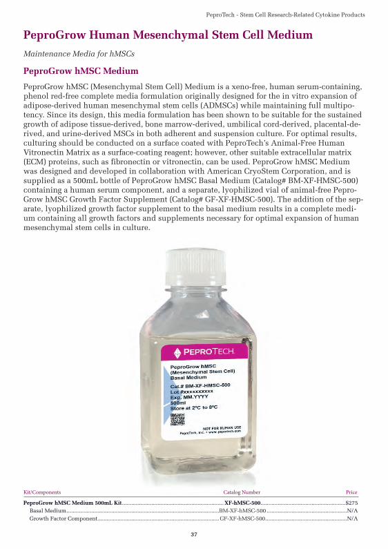
PeproTech - Stem Cell Research-Related Cytokine Products
37
PeproGrow Human Mesenchymal Stem Cell Medium
Maintenance Media for hMSCs
PeproGrow hMSC Medium
PeproGrow hMSC (Mesenchymal Stem Cell) Medium is a xeno-free, human serum-containing, phenol red-free complete media formulation originally designed for the in vitro expansion of adipose-derived human mesenchymal stem cells (ADMSCs) while maintaining full multipo-tency. Since its design, this media formulation has been shown to be suitable for the sustained growth of adipose tissue-derived, bone marrow-derived, umbilical cord-derived, placental-de-rived, and urine-derived MSCs in both adherent and suspension culture. For optimal results, culturing should be conducted on a surface coated with PeproTech’s Animal-Free Human Vitronectin Matrix as a surface-coating reagent; however, other suitable extracellular matrix (ECM) proteins, such as fibronectin or vitronectin, can be used. PeproGrow hMSC Medium was designed and developed in collaboration with American CryoStem Corporation, and is supplied as a 500mL bottle of PeproGrow hMSC Basal Medium (Catalog# BM-XF-HMSC-500) containing a human serum component, and a separate, lyophilized vial of animal-free Pepro-Grow hMSC Growth Factor Supplement (Catalog# GF-XF-HMSC-500). The addition of the sep-arate, lyophilized growth factor supplement to the basal medium results in a complete medi-um containing all growth factors and supplements necessary for optimal expansion of human mesenchymal stem cells in culture.
Kit/Components Catalog Number Price
PeproGrow hMSC Medium 500mL Kit .................................................................. XF-hMSC-500 .......................................................$275 Basal Medium ...................................................................................................BM-XF-hMSC-500 ....................................................N/A Growth Factor Component ............................................................................... GF-XF-hMSC-500 .....................................................N/A
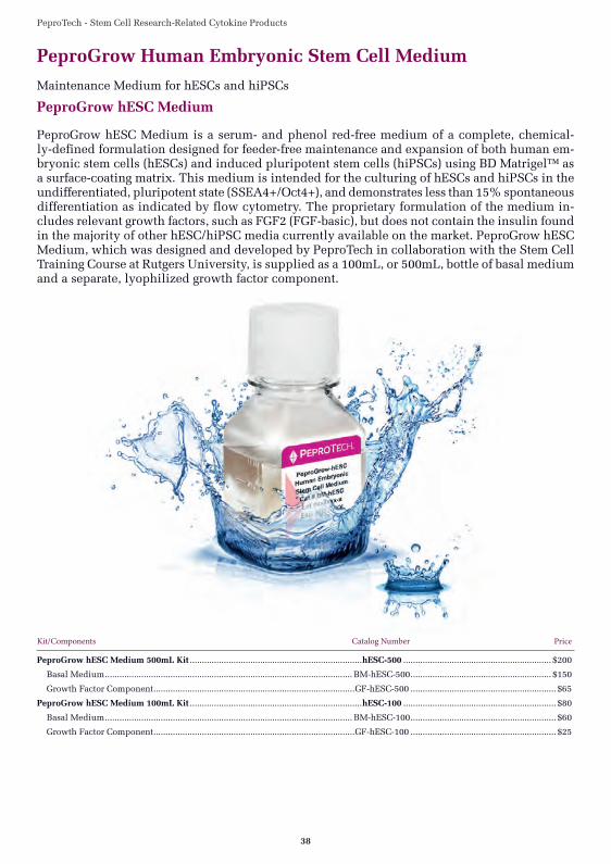
PeproTech - Stem Cell Research-Related Cytokine Products
38
PeproGrow Human Embryonic Stem Cell Medium
Maintenance Medium for hESCs and hiPSCs
PeproGrow hESC Medium
PeproGrow hESC Medium is a serum- and phenol red-free medium of a complete, chemical-ly-defined formulation designed for feeder-free maintenance and expansion of both human em-bryonic stem cells (hESCs) and induced pluripotent stem cells (hiPSCs) using BD Matrigel™ as a surface-coating matrix. This medium is intended for the culturing of hESCs and hiPSCs in the undifferentiated, pluripotent state (SSEA4+/Oct4+), and demonstrates less than 15% spontaneous differentiation as indicated by flow cytometry. The proprietary formulation of the medium in-cludes relevant growth factors, such as FGF2 (FGF-basic), but does not contain the insulin found in the majority of other hESC/hiPSC media currently available on the market. PeproGrow hESC Medium, which was designed and developed by PeproTech in collaboration with the Stem Cell Training Course at Rutgers University, is supplied as a 100mL, or 500mL, bottle of basal medium and a separate, lyophilized growth factor component.
Kit/Components Catalog Number Price
PeproGrow hESC Medium 500mL Kit .......................................................................hESC-500 ............................................................. $200
Basal Medium ...................................................................................................... BM-hESC-500 .......................................................... $150
Growth Factor Component ...................................................................................GF-hESC-500 ............................................................ $65
PeproGrow hESC Medium 100mL Kit .......................................................................hESC-100 ............................................................... $80
Basal Medium ...................................................................................................... BM-hESC-100 ............................................................ $60
Growth Factor Component ...................................................................................GF-hESC-100 ............................................................ $25
PeproTech - Stem Cell Research-Related Cytokine Products
39
PeproGrow Animal-Free Low Protein hESC Medium
Maintenance Medium for hESCs and hiPSCs
PeproGrow Animal-Free Low Protein hESC Medium
PeproGrow Animal-Free Low Protein hESC Medium is an animal component-free, serum- and phenol red-free medium of a complete, chemically-defined formulation designed for feeder-free maintenance and expansion of both human embryonic stem cells (hESCs) and human induced pluripotent stem cells (iPSCs). This medium formulation is best used with PeproTech’s Animal-Free Human Vitronectin Matrix as a surface-coating reagent; however, it can also be used with Corning Matrigel® or Synthemax® II-SC. The medium is intended for the culturing of hESCs and iPSCs in the undifferentiated, pluripotent state (Nanog+/Lin28+/Tra-1-60+ or SSEA4+/Oct4+), and demonstrates less than 15% spontaneous differentiation as indicated by immunofluorescent staining. Similar to the PeproGrow-hESC Medium, this pro-prietary medium formulation includes relevant growth factors, such as FGF2 (FGF-basic), and do not contain the insulin found in the majority of other hESC/iPSC media currently available on the market. PeproGrow Animal-Free Low Protein hESC Medium was designed and devel-oped by PeproTech in collaboration with the Stem Cell Training Course at Rutgers University. This medium contains minimal, essential components required for stem cell culture mainte-nance and are developed in the likeness of the E8® formulation developed by the laboratory of Dr. James Thomson at the University of Wisconsin, Madison.* This kit is supplied as a 100mL, or 500mL, bottle of basal medium, and a separate, lyophilized growth factor component. Ad-ditional companion products, such as the Animal-Free Human Vitronectin Matrix and Buffer Kit, and the Cell Passaging/Non-Enzymatic Detachment Buffer, are available separately. *E8 is a registered trademark of the Wisconsin Alumni Research Foundation.
Kit/Components Catalog Number Price
PeproGrow Animal-Free Low Protein hESC Medium 500mL Kit ......................... AF-LP-hESC-500 ...................................................$180 Low Protein Basal Medium ................................................................................AF-BM-LP-hESC-500 ................................................$130 Animal-Free Growth Factor Component .............................................................. AF-GF-hESC-500......................................................$75PeproGrow Animal-Free Low Protein hESC Medium 100mL Kit ......................... AF-LP-hESC-100 .....................................................$65 Low Protein Basal Medium ................................................................................AF-BM-LP-hESC-100 ..................................................$50 Animal-Free Growth Factor Component .............................................................. AF-GF-hESC-100......................................................$30
Companion Products for PeproGrow Animal-Free hESC Media
Kit/Components Catalog Number Price
Animal-Free Human Vitronectin Matrix and Buffer Kit ............................................AF-VMB-220 ...................................................... $60 Animal-Free Human Vitronectin Matrix (500Κg) .................................................................................................................................. N/A PBS + Kolliphor P 188 (220mL) ............................................................................................................................................................. N/A
Cell Passaging/Non-Enzymatic Detachment Buffer ........................................................CPD-125 ........................................................... $35 PBS+HEPES+EDTA (125mL) ................................................................................................................................................................. N/A
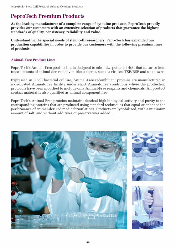
PeproTech - Stem Cell Research-Related Cytokine Products
40
As the leading manufacturer of a complete range of cytokine products, PeproTech proudly provides our customers with an extensive selection of products that guarantee the highest standards of quality, consistency, reliability and value.
Understanding the special needs of stem cell researchers, PeproTech has expanded our production capabilities in order to provide our customers with the following premium lines of products:
PeproTech Premium Products
Animal-Free Product Line:
PeproTech’s Animal-Free product line is designed to minimize potential risks that can arise from trace amounts of animal- derived adventitious agents, such as viruses, TSE/BSE and unknowns.
Expressed in E.coli bacterial culture, Animal-Free recombinant proteins are manufactured in a dedicated Animal- Free facility under strict Animal -Free conditions where the production protocols have been modified to include only Animal-Free reagents and chemicals. All product contact material is also qualified as animal component free.
PeproTech’s Animal-Free proteins maintain identical high biological activity and purity to the corresponding proteins that are produced using standard techniques that equal or enhance the performance of animal-derived media formulations. Products are lyophilized, with a minimum amount of salt, and without additives or preservatives added.
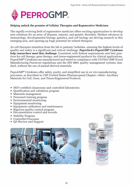
PeproTech - Stem Cell Research-Related Cytokine Products
41
Helping unlock the promise of Cellular Therapies and Regenerative Medicines
The rapidly evolving field of regenerative medicine offers exciting opportunities to develop new solutions for an array of diseases, injuries, and genetic disorders. Modern advances in immunology, developmental biology, genetics, and cell biology are driving research in this emerging area, and opening up huge potential for related therapies.
As cell therapies transition from the lab to patients’ bedsides, ensuring the highest levels of quality and safety is a significant and critical challenge. PeproTech’s PeproGMP® Cytokines help researchers meet that challenge. Consistent with federal requirements and best prac-tices for cell therapy, gene therapy, and tissue-engineered products for clinical applications, PeproGMP® Cytokines are manufactured and tested in compliance with US FDA GMP (Good Manufacturing Practices) regulations and the ISO 9001 quality management systems stan-dard, without the use of animal-derived materials.
PeproGMP® Cytokines offer safety, purity, and simplified use in ex vivo manufacturing processes, as described in USP (United States Pharmacopeia) Chapter <1043> Ancillary Materials for Cell, Gene, and Tissue-Engineered Products.
• ISO7-certified cleanrooms and controlled laboratories• Qualification and validation program• Materials management• Personnel training program• Environmental monitoring• Equipment monitoring• Equipment calibration and maintenance• Rigorous quality control program• Documentation control and records • Stability Program• Controlled Processes• QA Review and Support
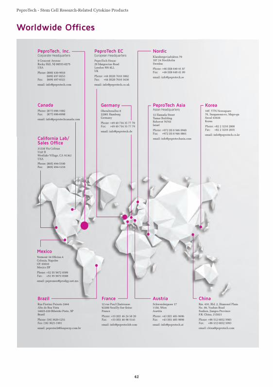
PeproTech - Stem Cell Research-Related Cytokine Products
42
Worldwide Offi ces

PeproTech - Stem Cell Research-Related Cytokine Products
43
Notes

PeproTech - Stem Cell Research-Related Cytokine Products
44
Notes