Anti-inflammatory effects of Rubus coreanus Miquel through ...
J Appl Oral Sci.
Abstract
Submitted: September 20, 2017Modification: December 15, 2017
Accepted: January 18, 2018
Pro-inflammatory and anti-inflammatory cytokine expression in post-treatment apical periodontitis
Objective: This study evaluated the expression of pro-inflammatory (IL-1β, IL-6, IFN-γ and TNF-α) and anti-inflammatory (IL-4 and TGF-β) cytokines in apical periodontitis lesions. Correlations between these cytokines and clinical and cone-beam computed tomographic (CBCT) data were also assessed. Material and Methods: Apical periodontitis lesions’ data were obtained from 27 patients subjected to periradicular surgery. Specimens were processed for histopathologic and immunohistochemical analysis. Sections were evaluated according to the amount of positive staining for each antibody. Expression levels of the target mediators were compared with clinical and CBCT data. Results: Twenty lesions were diagnosed as granuloma and 7 as cyst. In granulomas, IL-4 expression was significantly higher than IL-6 (p=0.001) and TNF-α (p=0.001). There was a significant relationship between high levels of TNF-α and lesions <5 mm (p=0.017). In cysts, IL-6 expression was significant lower than IL-4 (p=0.001) and IFN-γ (p=0.004). There was a significant relationship between high levels of TGF-β and endodontic treatment performed ≤4 years before (p=0.045). In general, IL-4 was the most expressed mediator in both cysts and granulomas. Conclusions: There was a balance between the expression of pro-inflammatory and anti-inflammatory cytokines associated with the chronic periradicular inflammatory process. TNF-α and TGF-β were related to some clinical and CBCT data.
Keywords: Periapical periodontitis. Periapical granuloma. Radicular cyst. Cytokines.
Nilton DESSAUNE NETO1
Mariana Teixeira Maneschy PORPINO1
Henrique dos Santos ANTUNES1
Renata Costa Val RODRIGUES1,2
Alejandro Ron PEREZ1
Fábio Ramôa PIRES1
José Freitas SIQUEIRA JR1
Luciana ARMADA1
Original Articlehttp://dx.doi.org/10.1590/1678-7757-2017-0455
1Universidade Estácio de Sá, Faculdade de Odontologia, Departamento de Endodontia, Rio de Janeiro, RJ, Brasil.2Universidade Veiga de Almeida, Faculdade de Odontologia, Departamento de Endodontia, Rio de Janeiro, RJ, Brasil.
Corresponding address:Fábio Ramôa Pires
Programa de Pós-Graduação em Odontologia - Universidade Estácio de Sá.
Av. Alfredo Baltazar da Silveira, 580 -cobertura - Recreio dos Bandeirantes -
Rio de Janeiro - RJ - 22790-710 - Brasil.Phone/Fax: + 55 21 2497-8988e-mail: [email protected]
2018;26:e201704551/8
J Appl Oral Sci. 2018;26:e201704552/8
Introduction
Apical periodontitis (AP) consists of a host tissue
inflammatory response to bacterial infection of the root
canal and serves the purpose of curbing the infection
advance towards the bone and other body parts23.
Bacteria cause direct damage to the periradicular
tissues by the action of exotoxins, enzymes and
metabolites24, but their main effects in the pathogenesis
of AP is highly likely to be related to cellular structural
components and other released factors that cause
indirect tissue damage by stimulating and modulating
the host inflammatory response22.
Bone destruction is a common feature of chronic
AP and results from the release of mediators that
induce osteoclast formation and activation during
inflammation13. The bone resorption process in the
periradicular region is modulated by pro-inflammatory
cytokines, such as interleukin (IL)-1β, IL-6, interferon
(IFN)-γ and tumor necrosis factor (TNF)-α, and anti-
inflammatory cytokines, such as transforming growth
factor (TGF)-β and IL-4. Most of these cytokines are
upregulated in response to bacterial infection and
the balance between pro- and anti-inflammatory
cytokines controls the extent and outcome of the
host immune response to antigen stimulation during
the chronic inflammatory process. Actually, pro-
inflammatory mechanisms must be properly controlled
to prevent excessive tissue destruction8,15. Studies
have indicated that a cytokine network is activated
in the periradicular tissues in response to bacterial
infection, in which pro-inflammatory pathways
predominate during active bone destruction, while
anti-inflammatory pathways prevail in phases of
lesion stabilization22. Numerous studies have detected
pro-inflammatory and anti-inflammatory cytokines
in AP lesions1,7-11,13-15, suggesting that destructive
and protective immune mechanisms must coexist in
the inflammatory periradicular response. There is a
scarcity of information correlating the expression of
pro- and anti-inflammatory cytokines with clinical and
imaging characteristics of AP. Therefore, the purpose
of this study was to evaluate the expression of pro-
inflammatory (IL-1β, IL-6, IFN-γ and TNF-α) and anti-
inflammatory (IL-4 and TGF-β) cytokines in human
AP lesions (granulomas and cysts), and evaluate any
relationship between their expression levels and clinical
and cone-beam computed tomography (CBCT) data.
Material and methods
The research project was approved by the
Institutional Research Ethics Committee (CAAE:
47669715.2.0000.5284). We have read the Helsinki
Declaration and followed its guidelines in this
investigation. Specimens consisted of AP lesions
obtained by periradicular surgery, all performed by the
same endodontist in a private practice (H.S.A.). Through
anamnesis and clinical examination, information was
gathered with reference to demographic characteristics
(age and gender), symptoms, presence of sinus
tracts, and the time elapsed since the root canal
treatment before surgery. CBCT scans were available
for all cases as requested for surgery planning and
were used to determine the location and size of the
AP lesion. The largest diameter of the lesions in the
3D CBCT analysis was classified as small when it was
<5 mm or large when it was ≥5 mm. Specimens
were excluded from this study when they were: of
insufficient size for histologic processing, from patients
with immunosuppressive diseases (such as diabetes
mellitus, acquired immunodeficiency syndrome or
auto-immune diseases), and from individuals who
made use of analgesic, anti-inflammatory and/or
antibiotic agents one month before surgery.
The specimens were obtained in one piece by
curettage during surgery. They were fixed in 10%
formalin medium and processed for histological
analysis. The histopathologic diagnosis was made
by two experienced evaluators by slides stained with
hematoxylin and eosin. Lesions that presented a cavity
partially or totally lined by epithelium were classified
as cysts. Therefore, 7 lesions were cysts and the other
20 were granulomas.
Histological sections were mounted on silanized
slides for performing the immunohistochemical
reactions according to a previously described protocol3,7.
The following antibodies were used: primary antibodies
for IL-1β (1:100, rabbit, sc-7884), IL-6 (1:500, mouse,
sc-130326), IFN-γ (1:200, rabbit, sc-8308), TGF-β
(1:100, rabbit, sc-146) and TNF-α (1:50, mouse,
sc-130349) from Santa Cruz Biotechnology (Dallas,
TX,USA); and the primary antibody for IL-4 (1:1000,
mouse, 25463) from RD Systems (Minneapolis, MN,
USA). The LSAB + HRP system (Dako K0690, DAKO
North America, Carpinteria, CA, USA) was used
as secondary antibody. Liquid DAB (Liquid DAB +
Substrate Chromogen System - Dako K3468, DAKO
Pro-inflammatory and anti-inflammatory cytokine expression in post-treatment apical periodontitis
J Appl Oral Sci. 2018;26:e201704553/8
North America, Carpinteria, CA, USA) was used to
stain the areas of cytokine expression. Positive and
negative controls were performed for each antibody
used, following the manufacturers’ instructions.
Images were independently analyzed by two blinded
evaluators, being previously calibrated with an optical
microscope (Leica DM500, Heerbrugg, Sweden). Each
specimen was divided into 5 fields and the epithelium
(cysts) and connective tissue were analyzed under
high-power view (400x); the expression values were
obtained from the number of positive cells in each
field. Each are observed was categorized according to
the percentage of positive staining and the following
scores were assigned: 0, negative/focal, if there were
no positive cells or <5% of the cells were positively
stained; 1, weak to moderate, if >5% to 50% of the
cells were positively stained; and 2, strong, if >50%
of the cells were positive. The mean score for the
whole specimen was calculated and the expression
of the target cytokines was ranked as negative/focal
(final mean ranging from 0 to 0.5), weak to moderate
(ranging from 0.6 to 1.2), and strong (ranging from
1.3 to 2.0). At the end of the evaluation, results were
compared. Discrepant cases were resolved by a third
and more experienced evaluator.
Comparative analysis of the data obtained was
performed using the SPSS program (Statistical Program
for Social Sciences, version 2.1, IBM, São Paulo,
SP, Brazil). For associations between the cytokine
expression levels and clinical/CBCT data, the Mann-
Whitney non-parametric test was used. Statistical
significance was set at p<0.05. For comparison of
expression levels between the target cytokines, the
Friedman and Wilcoxon multiple comparison tests
with Bonferroni correction were carried out. Statistical
significance was set at p<0.008.
Results
Demographic, clinical and CBCT data are displayed
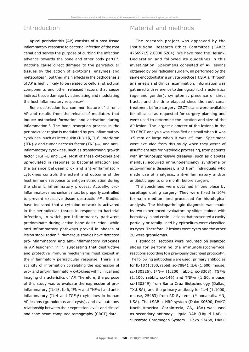
in Table 1. Evaluation of cytokine expression in AP
lesions revealed that focal expression was higher for
all cytokines, except for IL-4 (Table 2, Figure 1). IL-4
was the most intensely expressed cytokine in both
cysts and granulomas. No specimen exhibited negative
staining for the target cytokines.
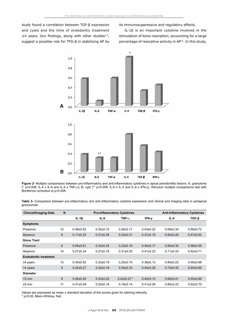
Comparison between the pro- and anti-inflammatory
cytokines in granulomas revealed that IL-4 expression
was significantly higher when compared with IL-6
(p=0.001) and TNF-α (p=0.001) (Figure 2A).
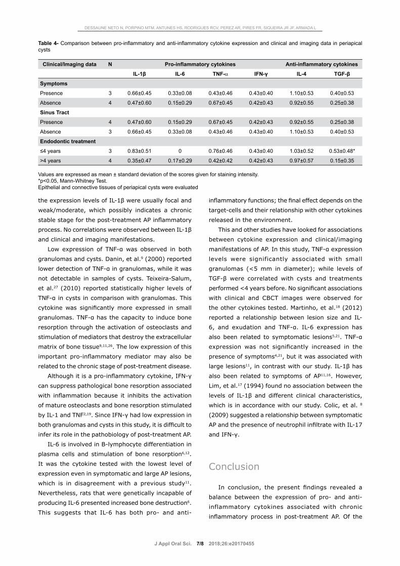
Evaluation of the cytokine expression in granulomas
associated with different clinical and CBCT data
showed a significant relationship only between high
levels of TNF-α and small <5 mm AP lesions (p=0.017)
(Table 3). There were no statistically significant
differences for all other comparisons involving the
cytokine expression levels and their relationship with
clinical/CBCT features (p>0.05).
Comparison between pro-inflammatory and anti-
inflammatory cytokines in cysts revealed that IL-6
expression was significant lower than IL-4 (p=0.001)
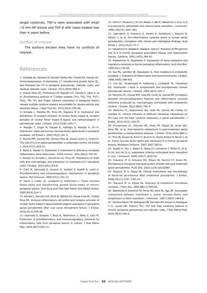
and IFN-γ (p=0.004) (Figure 2B). Evaluation of
cytokine expression in cysts associated with different
clinical and imaging data disclosed a significant
relationship only between high levels of TGF-β
expression and cases treated less than 4 years before
(p=0.045) (Table 4). All the other comparisons in cyst
Patient Data Apical periodontitis lesion/Teeth data
Age (years) Tooth location
Mean (SD) 51.7±14.3 Maxilla 18 (66.6%)
Range 32-78 Mandible 9 (33.4%)
Gender Tooth location in the arch
Female 20 (74%) Anterior 18 (66.6%)
Male 7 (26%) Posterior 9 (33.4%)
Symptoms Diameter
Presence 15 (55.5%) Small 9 (33.4%)
Absence 12 (44.5%) Large 18 (66.6%)
Sinus Tract Endodontic treatment
Presence 10 (37.1%) ≤ 4 years 15 (55.5%)
Absence 17 (63.9%) > 4 years 12 (44.5%)
Table 1- Demographic, clinical, and cone-beam computed tomographic data
DESSAUNE NETO N, PORPINO MTM, ANTUNES HS, RODRIGUES RCV, PEREZ AR, PIRES FR, SIQUEIRA JR JF, ARMADA L
J Appl Oral Sci. 2018;26:e201704554/8
lesions showed no statistical significance (p>0.05).
There was no comparison between the diameter of
the lesions and cytokine expression levels because all
cysts were classified as large.
Discussion
AP is a disease characterized by the accumulation
of members of both innate and adaptive immune
mechanisms in the periradicular tissues in response
to root canal infection20. This study used an
immunohistochemistry approach to detect the
expression of important pro-inflammatory and anti-
inflammatory cytokines in human post-treatment
AP lesions. Overall, our findings revealed that these
categories of cytokines co-existed in the lesions. In
addition, the expression levels of some cytokines were
related to clinical and imaging manifestations of AP.
Pro-inflammatory mechanisms must be tightly
counter-regulated to prevent excessive tissue
destruction23. The balance between pro-inflammatory
and anti-inflammatory cytokines controls the
extent of host responses to antigen stimulation
within chronic inflammatory processes. In AP, pro-
inflammatory cytokines are mostly produced by
TH1 cells, macrophages and neutrophils, and are
involved in the lesion expansion phases because of
bone destruction. In contrast, anti-inflammatory
cytokines, mostly released by TH2 and Treg cells, play
an important role in the healing process and restriction
of the immune response15.
Most cytokines tested in this study exhibited focal
expression levels (1 to 5% of the cells stained).
This may be justified by the nature of the samples
used. The chronic inflammatory AP lesions were
associated with teeth in which endodontic treatment
or retreatment had been performed at least one
year previously. Persistent AP lesions are usually
associated with an intraradicular microbiota composed
by fewer bacterial cells and species in comparison with
primary intraradicular infections25. This may result in
diverse antigenic load and type. In addition, given the
previous treatment, some areas of the lesion might
have been under a reparative process. These factors
may contribute to a more stable immune response,
characterized by a balance between pro- and anti-
inflammatory chemical mediators. This balance is
also very important to prevent excessive damage
associated with the immune response.
IL-4 had the highest expression levels among the
tested cytokines. This cytokine is mainly produced
by Th2 and mast cells and is known to stimulate the
humoral immune response and inhibit both Th1 pro-
inflammatory response and bone resorption2,8,19. IL-4
was not significantly related to any of the clinical and
CBCT data, which may be explained by the fact that
this cytokine occurred at high levels in most specimens.
IL-4 may have an important regulatory function in the
inflammatory process of post-treatment AP.
TGF-β mostly occurred in focal and weak/moderate
expression levels. This mediator can contribute
to tissue repair by inhibiting bone resorption and
stimulating collagen synthesis, neovascularization,
and fibroblast proliferation2. TGF-β can inhibit the
production of pro-inflammatory cytokines such as IL-
1β, TNF-α, and IL-6 by inflammatory cells isolated from
both symptomatic and asymptomatic AP lesions11. This
Staining intensity IL-1β IL-6 TNF-α IFN-γ IL-4 TGF-β
Granuloma
Focal 70% 85% 90% 75% 15% 70%
Weak/moderate 25% 15% 10% 25% 80% 10%
Strong 5% - - - 5% 20%
Cystic epithelium
Focal 57% 100% 43% 85.5% 14% 57%
Weak/moderate 28.5% - 57% 14.5% 57% 43%
Strong 14.5% - - - 29% -
Connective tissue of cysts
Focal 71% 86% 43% 57% 28.5% 71%
Weak/moderate - 14% 57% 43% 57% 14.5%
Strong 29% - - - 14.5% 14.5%
Table 2- Expression of pro-inflammatory and anti-inflammatory cytokines in post-treatment apical periodontitis
Pro-inflammatory and anti-inflammatory cytokine expression in post-treatment apical periodontitis
J Appl Oral Sci. 2018;26:e201704555/8
Figure 1- Apical periodontitis specimens with positive staining for pro-inflammatory and anti-inflammatory cytokines. A, IFN-γ (200x); B, IFN-γ (1000x); C, IL-1β (200x); D, IL-1β (1000x); E, IL-4 (200x); F, IL-4 (1000x); G, IL-6 (200x); H, IL-6 (1000x); I, TGF-β (200x); J, TGF-β (1000x); K, TNF-α (200x); L, TNF-α (1000x)
DESSAUNE NETO N, PORPINO MTM, ANTUNES HS, RODRIGUES RCV, PEREZ AR, PIRES FR, SIQUEIRA JR JF, ARMADA L
J Appl Oral Sci. 2018;26:e201704556/8
study found a correlation between TGF-β expression
and cysts and the time of endodontic treatment
≤4 years. Our findings, along with other studies1,9,
suggest a possible role for TFG-β in stabilizing AP by
its immunosuppressive and regulatory effects.
IL-1β is an important cytokine involved in the
stimulation of bone resorption, accounting for a large
percentage of resorptive activity in AP13. In this study,
Figure 2- Multiple comparisons between pro-inflammatory and anti-inflammatory cytokines in apical periodontitis lesions. A, granuloma (* p<0.008: IL-4 x IL-6 and IL-4 x TNF-α); B, cyst (** p<0.008: IL-6 x IL-4 and IL-6 x IFN-γ). Wilcoxon multiple comparisons test with Bonferroni correction to p<0.008
Clinical/Imaging Data N Pro-Inflammatory Cytokines Anti-Inflammatory Cytokines
IL-1β IL-6 TNF-α IFN-γ IL-4 TGF-β
Symptoms
Presence 12 0.48±0.52 0.30±0.10 0.26±0.17 0.43±0.22 0.68±0.34 0.58±0.72
Absence 8 0.17±0.22 0.27±0.28 0.32±0.31 0.37±0.19 0.90±0.26 0.47±0.62
Sinus Tract
Presence 6 0.56±0.61 0.33±0.24 0.23±0.19 0.40±0.17 0.90±0.30 0.56±0.59
Absence 14 0.27±0.34 0.27±0.16 0.31±0.25 0.41±0.22 0.71±0.33 0.52±0.71
Endodontic treatment
≤4 years 12 0.45±0.52 0.33±0.19 0.25±0.15 0.38±0.13 0.80±0.22 0.46±0.68
>4 years 8 0.22±0.27 0.22±0.16 0.35±0.33 0.45±0.29 0.72±0.45 0.65±0.66
Diameter
<5 mm 9 0.28±0.40 0.33±0.20 0.42±0.27 * 0.40±0.14 0.66±0.41 0.55±0.66
≥5 mm 11 0.41±0.49 0.25±0.18 0.18±0.14 0.41±0.26 0.85±0.22 0.52±0.70
Values are expressed as mean ± standard deviation of the scores given for staining intensity.* p<0.05, Mann-Whitney Test
Table 3- Comparison between pro-inflammatory and anti-inflammatory cytokine expression and clinical and imaging data in periapical granulomas
Pro-inflammatory and anti-inflammatory cytokine expression in post-treatment apical periodontitis
J Appl Oral Sci. 2018;26:e201704557/8
the expression levels of IL-1β were usually focal and
weak/moderate, which possibly indicates a chronic
stable stage for the post-treatment AP inflammatory
process. No correlations were observed between IL-1β
and clinical and imaging manifestations.
Low expression of TNF-α was observed in both
granulomas and cysts. Danin, et al.9 (2000) reported
lower detection of TNF-α in granulomas, while it was
not detectable in samples of cysts. Teixeira-Salum,
et al.27 (2010) reported statistically higher levels of
TNF-α in cysts in comparison with granulomas. This
cytokine was significantly more expressed in small
granulomas. TNF-α has the capacity to induce bone
resorption through the activation of osteoclasts and
stimulation of mediators that destroy the extracellular
matrix of bone tissue9,11,26. The low expression of this
important pro-inflammatory mediator may also be
related to the chronic stage of post-treatment disease.
Although it is a pro-inflammatory cytokine, IFN-γ
can suppress pathological bone resorption associated
with inflammation because it inhibits the activation
of mature osteoclasts and bone resorption stimulated
by IL-1 and TNF2,19. Since IFN-γ had low expression in
both granulomas and cysts in this study, it is difficult to
infer its role in the pathobiology of post-treatment AP.
IL-6 is involved in B-lymphocyte differentiation in
plasma cells and stimulation of bone resorption6,12.
It was the cytokine tested with the lowest level of
expression even in symptomatic and large AP lesions,
which is in disagreement with a previous study11.
Nevertheless, rats that were genetically incapable of
producing IL-6 presented increased bone destruction6.
This suggests that IL-6 has both pro- and anti-
inflammatory functions; the final effect depends on the
target-cells and their relationship with other cytokines
released in the environment.
This and other studies have looked for associations
between cytokine expression and clinical/imaging
manifestations of AP. In this study, TNF-α expression
levels were significantly associated with small
granulomas (<5 mm in diameter); while levels of
TGF-β were correlated with cysts and treatments
performed <4 years before. No significant associations
with clinical and CBCT images were observed for
the other cytokines tested. Martinho, et al.18 (2012)
reported a relationship between lesion size and IL-
6, and exudation and TNF-α. IL-6 expression has
also been related to symptomatic lesions5,21. TNF-α
expression was not significantly increased in the
presence of symptoms4,21, but it was associated with
large lesions11, in contrast with our study. IL-1β has
also been related to symptoms of AP11,16. However,
Lim, et al.17 (1994) found no association between the
levels of IL-1β and different clinical characteristics,
which is in accordance with our study. Colic, et al. 8
(2009) suggested a relationship between symptomatic
AP and the presence of neutrophil infiltrate with IL-17
and IFN-γ.
Conclusion
In conclusion, the present findings revealed a
balance between the expression of pro- and anti-
inflammatory cytokines associated with chronic
inflammatory process in post-treatment AP. Of the
Clinical/Imaging data N Pro-inflammatory cytokines Anti-inflammatory cytokines
IL-1β IL-6 TNF-α IFN-γ IL-4 TGF-β
Symptoms
Presence 3 0.66±0.45 0.33±0.08 0.43±0.46 0.43±0.40 1.10±0.53 0.40±0.53
Absence 4 0.47±0.60 0.15±0.29 0.67±0.45 0.42±0.43 0.92±0.55 0.25±0.38
Sinus Tract
Presence 4 0.47±0.60 0.15±0.29 0.67±0.45 0.42±0.43 0.92±0.55 0.25±0.38
Absence 3 0.66±0.45 0.33±0.08 0.43±0.46 0.43±0.40 1.10±0.53 0.40±0.53
Endodontic treatment
≤4 years 3 0.83±0.51 0 0.76±0.46 0.43±0.40 1.03±0.52 0.53±0.48*
>4 years 4 0.35±0.47 0.17±0.29 0.42±0.42 0.42±0.43 0.97±0.57 0.15±0.35
Values are expressed as mean ± standard deviation of the scores given for staining intensity.*p<0.05, Mann-Whitney Test. Epithelial and connective tissues of periapical cysts were evaluated
Table 4- Comparison between pro-inflammatory and anti-inflammatory cytokine expression and clinical and imaging data in periapical cysts
DESSAUNE NETO N, PORPINO MTM, ANTUNES HS, RODRIGUES RCV, PEREZ AR, PIRES FR, SIQUEIRA JR JF, ARMADA L
J Appl Oral Sci. 2018;26:e201704558/8
target cytokines, TNF-α were associated with small
<5 mm AP lesions and TGF-β with cases treated less
than 4 years before.
Conflict of interestThe authors declare they have no conflicts of
interest.
References1- Andrade AL, Nonaka CF, Gordón-Núñez MA, Freitas RA, Galvão HC. Immunoexpression of interleukin 17, transforming growth factor β1, and forkhead box P3 in periapical granulomas, radicular cysts, and residual radicular cysts. J Endod. 2013;39(8):990-4.2- Araujo-Pires AC, Francisconi CF, Biguetti CC, Cavalla F, Letra A, et al. Simultaneous analysis of T helper subsets (Th1, Th2, Th9, Th17, Th22, Tfh, Tr1 and Tregs) markers expression in periapical lesions reveals multiple cytokine clusters accountable for lesions activity and inactivity status. J Appl Oral Sci. 2014;22(4):336-46.3- Armada L, Marotta PS, Pires FR, Siqueira JF Jr. Expression and distribution of receptor activator of nuclear factor kappa B, receptor activator of nuclear factor kappa B ligand, and osteoprotegerin in periradicular cysts. J Endod. 2015;41(8):1281-7.4- Ataoğlu T, Ungör M, Serpek B, Haliloğlu S, Ataoğlu H, Ari H. Interleukin-1beta and tumour necrosis factor-alpha levels in periapical exudates. Int Endod J. 2002;35(2):181-5.5- Azuma MM, Samuel RO, Gomes-Filho JE, Dezan-Junior E, Cintra LT. The role of IL-6 on apical periodontitis: a systematic review. Int Endod J. 2014;47(7):615-21.6- Balto K, Sasaki H, Stashenko P. Interleukin-6 deficiency increases inflammatory bone destruction. Infect Immun. 2001;69(2):744-50.7- Bracks IV, Armada L, Goncalves LS, Pires FR. Distribution of mast cells and macrophages and expression of interleukin-6 in periapical cysts. J Endod. 2014;40(1):63-8.8- Colić M, Gazivoda D, Vucević D, Vasilijić S, Rudolf R, Lukić A. Proinflammatory and immunoregulatory mechanisms in periapical lesions. Mol Immunol. 2009;47(1):101-13.9- Danin J, Linder LE, Lundqvist G, Andersson L. Tumor necrosis factor-alpha and transforming growth factor-beta1 in chronic periapical lesions. Oral Surg Oral Med Oral Pathol Oral Radiol Endod. 2000;90(4):514-7.10- Estrela C, Decurcio DA, Silva JA, Batista AC, Souza Lima NC, Freitas Silva BS. Immune-inflammatory cell profile and receptor activator of nuclear factor kappa b ligand/osteoprotegerin expression in persistent apical periodontitis after root canal retreatment failure. J Endod. 2016;42(3):439-46.11- Gazivoda D, Dzopalic T, Bozic B, Tatomirovic Z, Brkic Z, Colic M. Production of proinflammatory and immunoregulatory cytokines by inflammatory cells from periapical lesions in culture. J Oral Pathol Med. 2009;38(7):605-11.
12- Ishimi Y, Miyaura C, Jin CH, Akatsu T, Abe E, Nakamura Y, et al. IL-6 is produced by osteoblasts and induces bone resorption. J Immunol. 1990;145(10):3297-303.13- Jakovljevic A, Knezevic A, Karalic D, Soldatovic I, Popovic B, Milasin J, et al. Pro-inflammatory cytokine levels in human apical periodontitis: correlation with clinical and histological findings. Aust Endod J. 2015;41(2):72-7.14- Kabashima H, Nagata K, Maeda K, Iijima T. Presence of IFN-gamma and IL-4 in human periapical granulation tissues and regeneration tissues. Cytokine. 2001;14(5):289-93.15- Kawashima N, Stashenko P. Expression of bone-resorptive and regulatory cytokines in murine periapical inflammation. Arch Oral Biol. 1999;44(1):55-66.16- Kuo ML, Lamster IB, Hasselgren G. Host mediators in endodontic exudates. I. Indicators of inflammation and humoral immunity. J Endod. 1998;24(9):598-603.17- Lim GC, Torabinejad M, Kettering J, Linkhardt TA, Finkelman RD. Interleukin 1-beta in symptomatic and asymptomatic human periradicular lesions. J Endod. 1994;20(5):225-7.18- Martinho FC, Chiesa WM, Leite FR, Cirelli JA, Gomes BP. Correlation between clinical/radiographic features and inflammatory cytokine networks produced by macrophages stimulated with endodontic content. J Endod. 2012;38(6):740-5.19- Martinho FC, Nascimento GG, Leite FR, Gomes AP, Freitas LF, Camões IC. Clinical influence of different intracanal medications on th1-type and th2-type cytokine responses in apical periodontitis. J Endod. 2015;41(2):169-75.20- Provenzano JC, Antunes HS, Alves FR, Rôças IN, Alves WS, Silva MR, et al. Host-bacterial interactions in post-treatment apical periodontitis: a metaproteome analysis. J Endod. 2016;42(6):880-5.21- Prso IB, Kocjan W, Simić H, Brumini G, Pezelj-Ribarić S, Borcić J, et al. Tumor necrosis factor-alpha and interleukin 6 in human periapical lesions. Mediators Inflamm. 2007;2007:38210.22- Sasaki H, Hou L, Belani A, Wang CY, Uchiyama T, Müller R, et al. IL-10, but not IL-4, suppresses infection-stimulated bone resorption in vivo. J Immunol. 2000;165(7):3626-30.23- Siqueira JF Jr, Antunes HS, Rôças IN, Rachid CT, Alves FR. Microbiome in the apical root canal system of teeth with post-treatment apical periodontitis. PLoS One. 2016:11(9):e0162887.24- Siqueira JF Jr, Rôças IN. Clinical implications and microbiology of bacterial persistence after treatment procedures. J Endod. 2008;34(11):1291-1301.e3.25- Siqueira JF Jr, Rôças IN. Diversity of endodontic microbiota revisited. J Dent Res. 2009;88(11):969-81.26- Stashenko P, Dewhirst FE, Peros WJ, Kent RL, Ago JM. Synergistic interactions between interleukin 1, tumor necrosis factor, and lymphotoxin in bone resorption. J Immunol. 1987;138(5):1464-8.27- Teixeira-Salum TB, Rodrigues DB, Gervásio AM, Souza CJ, Rodrigues V Jr, Loyola AM. Distinct Th1, Th2 and Treg cytokines balance in chronic periapical granulomas and radicular cysts. J Oral Pathol Med. 2010;39(3):250-6.
Pro-inflammatory and anti-inflammatory cytokine expression in post-treatment apical periodontitis