Process Optimization for the Production of β … Makwana, et al.pdf · Process Optimization for...
16
Int.J.Curr.Microbiol.App.Sci (2017) 6(8): 1454-1469 1454 Original Research Article https://doi.org/10.20546/ijcmas.2017.608.176 Process Optimization for the Production of β-Galactosidase Using Potential Lactobacillus Cultures Shrushti Makwana 1 , Subrota Hati 1* , Heena Parmar 1 and K. D. Aparnathi 2 1 Dairy Microbiology Department, 2 DairyChemistry Department, Anand Agricultural University, Anand-388110, Gujarat, India *Corresponding author ABSTRACT Introduction Lactose intolerance is due to lack of the enzyme lactase in the small intestines to break down the lactose into glucose and galactose (Deng et al., 2015). There are four types: primary, secondary, developmental and congenital. Primary lactose intolerance is when the amount of lactase declines as people age. Secondary lactose intolerance is due to injury to the small intestine such as from infection, inflammatory bowel disease, celiac disease, or other diseases (Berni et al., 2016). Developmental lactose intolerance may occur in the premature babies and usually improves over a short period of time. Congenital lactose intolerance is an extremely International Journal of Current Microbiology and Applied Sciences ISSN: 2319-7706 Volume 6 Number 8 (2017) pp. 1454-1469 Journal homepage: http://www.ijcmas.com Ten Lactobacillus cultures were considered for the study. Out of ten Lactobacillus cultures, V3 produced maximum β-galactosidase activity (3.381 O.D), followed by NK2 and NK9 up to 24h than other cultures during β-galactosidase activity. Extraction of β- galactosidase was carried out by disruption of Lactobacillus cells using sonication at 3, 4 and 5 minute (pulse 30 second on / 10 seconds off and 35 to 55 % amplitudes), lysozyme- EDTA (49 mg of lysozyme in 1.5 ml of TE buffer) treatment, toluene acetone (9:1 [toluene: acetone]) treatment, sodium dodecyl sulphate (SDS) - chloroform (250 μl chloroform and 100 ml of 0.1 % SDS) treatment. Out of 4 different treatments [sonication, lysozyme-EDTA, toluene-acetone and sodium dodecyl sulphate (SDS)-chloroform treatment] for β-galactosidase production, sonication treatment [V3 (1.093), NK2 (1.084) and NK9 (1.091)] was found to be best for production of β -galactosidase with optimized sonication period [5 min (pulse 15 sec off / 30 sec on at 55% amplitudes)]. Purification of β-galactosidase enzyme was carried out employing various t-butanol ratios (crude extract : t-butanol = 1:0.25, 1.0:0.5, 1.0:1.0, 1.0:1.5, 1.0:2.0) with a different ammonium sulfate saturation i.e. 30, 60 and 90%.After purification by treating with ratio of crude enzyme: t- butanol (R) and ammonium sulphate saturation (AS), 2 % of R (0.396 O.D) and 60 % of AS (0.292 O.D) showed maximum β-galactosidase activity for three selected Lactobacillus cultures i.e. V3, NK2 and NK9. The molecular mass of partially purified β- galactosidase (60% Ammonium sulphate saturation) from selected Lactobacillus cultures i.e. V3, NK2 and NK9 as determined by SDS-PAGE was approx. 69 kDa. Therefore, the study entailed to conclude that production and purification of β-galactosidase enzyme from safe Lactobacillus cultures is an alternative source of biological enzyme for the preparation of lactose hydrolysed milk for lactose intolerant population. Keywords β-galactosidase, Lactobacillus, Sonication, Partial purification. Accepted: 17 June 2017 Available Online: 10 August 2017 Article Info
Transcript of Process Optimization for the Production of β … Makwana, et al.pdf · Process Optimization for...
Int.J.Curr.Microbiol.App.Sci (2017) 6(8): 1454-1469
1454
Original Research Article https://doi.org/10.20546/ijcmas.2017.608.176
Process Optimization for the Production of β-Galactosidase
Using Potential Lactobacillus Cultures
Shrushti Makwana1, Subrota Hati
1*, Heena Parmar
1 and K. D. Aparnathi
2
1Dairy Microbiology Department,
2DairyChemistry Department, Anand Agricultural University,
Anand-388110, Gujarat, India *Corresponding author
A B S T R A C T
Introduction
Lactose intolerance is due to lack of
the enzyme lactase in the small intestines to
break down the lactose
into glucose and galactose (Deng et al.,
2015). There are four types: primary,
secondary, developmental and congenital.
Primary lactose intolerance is when the
amount of lactase declines as people age.
Secondary lactose intolerance is due to injury
to the small intestine such as from infection,
inflammatory bowel disease, celiac disease, or
other diseases (Berni et al.,
2016). Developmental lactose intolerance
may occur in the premature babies and
usually improves over a short period of time.
Congenital lactose intolerance is an extremely
International Journal of Current Microbiology and Applied Sciences ISSN: 2319-7706 Volume 6 Number 8 (2017) pp. 1454-1469 Journal homepage: http://www.ijcmas.com
Ten Lactobacillus cultures were considered for the study. Out of ten Lactobacillus
cultures, V3 produced maximum β-galactosidase activity (3.381 O.D), followed by NK2
and NK9 up to 24h than other cultures during β-galactosidase activity. Extraction of β-
galactosidase was carried out by disruption of Lactobacillus cells using sonication at 3, 4
and 5 minute (pulse 30 second on / 10 seconds off and 35 to 55 % amplitudes), lysozyme-
EDTA (49 mg of lysozyme in 1.5 ml of TE buffer) treatment, toluene acetone (9:1
[toluene: acetone]) treatment, sodium dodecyl sulphate (SDS) - chloroform (250 µl
chloroform and 100 ml of 0.1 % SDS) treatment. Out of 4 different treatments [sonication,
lysozyme-EDTA, toluene-acetone and sodium dodecyl sulphate (SDS)-chloroform
treatment] for β-galactosidase production, sonication treatment [V3 (1.093), NK2 (1.084)
and NK9 (1.091)] was found to be best for production of β-galactosidase with optimized
sonication period [5 min (pulse 15 sec off / 30 sec on at 55% amplitudes)]. Purification of
β-galactosidase enzyme was carried out employing various t-butanol ratios (crude extract :
t-butanol = 1:0.25, 1.0:0.5, 1.0:1.0, 1.0:1.5, 1.0:2.0) with a different ammonium sulfate
saturation i.e. 30, 60 and 90%.After purification by treating with ratio of crude enzyme: t-
butanol (R) and ammonium sulphate saturation (AS), 2 % of R (0.396 O.D) and 60 % of
AS (0.292 O.D) showed maximum β-galactosidase activity for three selected Lactobacillus
cultures i.e. V3, NK2 and NK9. The molecular mass of partially purified β-
galactosidase (60% Ammonium sulphate saturation) from selected Lactobacillus cultures
i.e. V3, NK2 and NK9 as determined by SDS-PAGE was approx. 69 kDa. Therefore, the
study entailed to conclude that production and purification of β-galactosidase enzyme from
safe Lactobacillus cultures is an alternative source of biological enzyme for the
preparation of lactose hydrolysed milk for lactose intolerant population.
K e y w o r d s
β-galactosidase,
Lactobacillus,
Sonication, Partial
purification.
Accepted:
17 June 2017
Available Online: 10 August 2017
Article Info
Int.J.Curr.Microbiol.App.Sci (2017) 6(8): 1454-1469
1455
rare genetic disorder in which no lactase is
made from birth (NIDDK, 2016). In the
family of β-galactosidase, lactase is one part
of them which is a glycoside hydrolase. It is
involved in the hydrolysis process of the
disaccharide lactose into galactose and
glucose monomers. Lactase is present in
predominantly along with the brush border
membrane of the differentiated enterocytes
lining the villi of the small intestine
(Skovbjerg et al., 1981).
Jelen et al., (1993) suggested that specific
microbial strains that are high β-galactosidase
producers could be used for lactose hydrolysis
after cell disruption but with minimum
additional purification. More recently,
proposed a process for lactose hydrolysis in
dairy systems using a crude enzymatic extract
(CEE) from the Lactobacilli culture, with
subsequent utilization of the fermentation
medium used for the culture propagation
(Vasiljevic and Jelen, 2001). Amongst lactic
acid bacteria, yogurt bacteria (Lactobacillus
bulgaricus and Streptococcus thermophilus)
are the highest β- galactosidase producers.
The β-galactosidase of these cultures has been
characterized, showing high stability and
activity at high temperatures. However, the β-
galactosidase from thermophilic LAB is an
intracellular enzyme. It is release from
microorganisms, which is obtained either by
mechanical disruption or by the chemical
permeabilization of the cell membrane. The
effectiveness of the various disruption
methods differs for different microbial genera
and strains. Generally, β-galactosidase
activity in the medium can be greatly
increased by rupturing cells using different
cell disruption methods. The literature tends
to focus on the disruption of yeasts with much
less information being available on the
disruption of lactobacilli. (Bury et al., 2001).
With this perspective, traditional yogurt
samples obtained from Toros mountain region
of Turkey with highly bio-diverse
environment were used as the source for the
isolation of LAB cultures. Almost 136
isolated strains were screened for β-
galactosidase activity based on their lactose
consumption and lactic acid producing
characteristics according to the method
described by Bury et al., (2001). Among these
isolates only three Lactobacillus delbrueckii
subsp. bulgaricus strains and three
Streptococcus thermophilus strains showed a
high potential. The activity and stability of β-
galactosidase is strongly influenced by the
enzyme origin and environmental conditions
of the enzyme production and the hydrolysis
process, such as temperature and pH optima,
as well as the presence of activators or
inhibitors. The use of CEEs instead of
purified β-galactosidase preparation may
complicate the final outcome of the hydrolysis
due to the presence of other microbial
enzymes, which may interfere with the lactose
hydrolysis.
Prasad et al., (2013) evaluated that the
production of β-galactosidase by
Bifidobacterium animalis ssp. lactis Bb12 and
Lactobacillus delbrueckii ssp. bulgaricus
ATCC 11842 in whey and the effect of 4
different extraction methods i.e. sonication,
acetone-toluene, SDS-chloroform and
lysozyme-EDTA treatment on enzyme
activity from these organisms. Among the
four methods used for β-galactosidase
extraction, sonication exhibited the best result
(6.80 Unit/mL) for B. animalis ssp. lactis
Bb12 while lysozyme-EDTA treatment was
also found to be the best (7.77 Unit/ mL) for
L. delbrueckii ssp. bulgaricus ATCC 11842.
Duman and Kaya, (2013) evaluated the
activity of β-galactosidase was completely
decreased at 60 % ammonium sulfate
saturation in aqueous phase, the best results of
recovery and the purification fold of β-
galactosidase (133 % and 10.1, respectively)
was obtained in the interfacial phase. Higher
salt concentrations from this point were
Int.J.Curr.Microbiol.App.Sci (2017) 6(8): 1454-1469
1456
resulted with reducing of recovery and
purification fold, which may be due to
irreversible denaturation of protein (Narayan
et al., 2008).
Approximately 75 % of Earth’s population is
lactose intolerant and in India (particularly
southern India) 70 % of the population are
lactose intolerance and they feel
uncomfortable whenever they consume milk
and milk products. To meet this challenge,
lactose hydrolysed milk is an alternative
solution for proposed study. However the
source of enzyme (β-galactosidase) from
biological material like LAB is an added
advantage from safety point of view as LAB
are having GRAS status.
Materials and Methods
Ten LAB cultures used in the present study
i.e. L. helveticus MTCC 5463 (V3),
L. rhamnosus (NK2), L. casei (NK9),
L. rhamnosus MTCC 25062 (NK10),
L. fermentum (M5), L. paracasei (M16),
L. rhamnosus (M31), L. plantarum (M38),
L. plantarum (M10), L. fermentum
TDS030603 (LBF) were obtained from the
Culture Collection of Dairy Microbiology
Department, SMC College of Dairy Science,
Anand Agricultural University, Anand. The
LAB cultures were propagated in sterilized
reconstituted skim milk (10% TS) and stored
at 5 ± 2ᵒC.
The transfer was given every week during the
course of the study. Most the bacteriological
media, molecular biology grade chemicals
and reagents were purchased either from Hi-
Media (India), Sigma (USA), SDFCL (India),
Chr. Hansen (Denmark) etc. During the entire
study, glass wares of Borosil brand (Borosil
Glass Works Ltd., Mumbai, India) and
analytical grade and molecular biology grade
chemicals were used. Glassware and other
materials were sterilized by usual procedures
viz. 160-180ᵒC for 2h in hot air oven,
whenever required.
Evaluation of pH, acidity and Lactobacillus
counts of fermented skim milk
All the cultures were activated by growing in
sterilized skim milk. The activated cultures
were added to 100 ml of skim milk flasks at
the rate of 2%. After mixing them thoroughly,
the cultures were incubated at 37ᵒC for
different intervals of 0, 6, 12, 18 and 24h.
Samples were taken out for determination of
pH, titratable acidity and viable counts after
each interval. pH of fermented skim milk
samples was measured using digital pH meter
(OAKTON pH700, India).
The titratable acidity was estimated by the
procedure described by (IS, 1960).
Lactobacilli counts of fermented skim milk
samples were determined as per the method
described by IDF standards (146:
2003).Typical colonies were calculated and
the counts were expressed as log cfu/ml.
Determination of β-galactosidase activity
All the cultures were activated by growing in
MRS broth. The activated cultures were
added to 100 ml of MRS broth at the rate of
2%. After mixing them thoroughly, the
culture flasks were incubated at 37ᵒC for
different intervals of 0, 24, 48 and 72h.
Samples were taken out for β-galactosidase
activity at each interval.
Enzyme extraction
After 0, 24, 48 and 72h of incubation, the
cells were harvested by centrifuging at 5000
rpm for 15 min at 4°C. The supernatant was
considered to be containing extracellular
enzymes. The cell pellet was crushed and
washed twice with a 0.05 M sodium
phosphate buffer (pH 6.8) and centrifuged at
Int.J.Curr.Microbiol.App.Sci (2017) 6(8): 1454-1469
1457
5000 rpm for 15 min at 4°C. The washed
pellets were resuspended in 10 mL of 0.05 M
phosphate buffer (pH 6.8) for intracellular
enzyme extraction using cell disintegration
methods listed below (Prasad et al., 2013):
Sonication treatment
The cell suspensions were sonicated for 30
min (pulse 30 seconds off / 30 seconds on and
61% amplitudes) in ice bath using sonicator
(LABMAN, India), according to the method
described by Prasad et al., (2013).
The extract was then centrifuged at 5000 rpm
at 4°C for 15 min and the supernatant
containing the crude enzyme extract was
stored at –20°C until used for enzyme assays.
Enzyme assay
The β-galactosidase was determined by the
reaction mixture was composed of 0.5 mL of
supernatant containing extracted enzyme and
2.0 mL of 15 mM O-nitrophenyl β-D-
galactopyranoside (ONPG) in 0.05 M sodium
phosphate buffer (pH 6.8). After incubation
for 20 min at 37°C, 0.5 mL of 0.1 M sodium
carbonate was added to the mixture to stop
the reaction.
Absorbance was measured at 420 nm with a
spectrophotometer (Systronics PC based
double beam spectrophotometer 2202, India).
Production, extraction and purification of
β-galactosidase from selected LAB
All the cultures were activated by growing in
MRS broth. The activated cultures were
added to 100 ml of MRS broth flasks at the
rate of 2%. After mixing them thoroughly, the
culture flasks were incubated at 37ᵒC for 24h.
Samples were taken out for β-galactosidase
activity.
Enzyme extraction
After 24h of incubation, the cells were
harvested by centrifuging at 5000 rpm for 15
min at 4°C. The supernatant was considered
to be containing extracellular enzymes. The
cell pellet was crushed and washed twice with
a 0.05 M sodium phosphate buffer (pH 6.8)
and centrifuged at 5000 rpm for 15 min at
4°C. The washed pellets were resuspended in
10 mL of 0.05 M phosphate buffer (pH 6.8)
for intracellular enzyme extraction using four
different cell disintegration methods listed
below (Prasad et al., 2013):
Sonication treatment
The cell suspensions were sonicated for 3, 4
and 5 minute intervals (pulse 15 seconds off /
30 seconds on as well as 35 and 55%
amplitudes) in ice bath using sonicator
(LABMAN, India), according to the method
mentioned by Prasad et al., (2013). The
extract was then centrifuged at 5000 rpm and
4°C for 15 min and the supernatant containing
the crude enzyme was stored at –20°C until
used for enzyme assays.
Lysozyme EDTA treatment
Lysozyme solution was prepared by
dissolving 49 mg of lysozyme (Hi-Media,
India) in 1.5 mL of TE (Tris-EDTA;
Ethylenediamine Tetraacetic Acid) buffer
containing 1 mM EDTA and 10 mM Tris-
HCl, adjusted to pH 8.0. The lysozyme
preparation was added to the cell suspension
at the rate of 150 μL per mL, incubated for 45
min at room temperature. Then supernatant
obtained was kept at -20ᵒC until enzyme
activity measurement (Prasad et al., 2013).
Toluene acetone treatment
10 ml cell suspension was ground for 10 min
in a pestle and mortar with 2.0 g alumina
Int.J.Curr.Microbiol.App.Sci (2017) 6(8): 1454-1469
1458
(SDFCL, India) and 0.2 mL of 9:1 mixture of
toluene (LOBA Chemie, India with 99.5%
purity) and acetone (LOBA Chemie, India
with 99.8% purity) solvents. The suspension
was extended in 8 mL phosphate buffer and
centrifuged at 5000 rpm for 15 min at 4°C.
The supernatant obtained was kept at -20ᵒC
until used for enzyme assay (Prasad et al.,
2013).
Sodium dodecyl sulphate (SDS) –
Chloroform treatment
Permeabilization of cell membrane was
carried out by vortexing 10 mL of the cell
suspension in the presence of 250 μL
chloroform and 100 μL 0.1% SDS solution
for 30 min at room temperature. The
suspension was centrifuged at 5000 rpm for
15 min at 4°C and the supernatant was kept at
-20ᵒC until needed for the enzyme assay
(Prasad et al., 2013).
Quantitative analysis of β-galactosidase
activity produced by selected Lactobacillus
cultures through X-Gal assay
Sonication treatment
The cell suspensions were sonicated for 5 min
(pulse 15 seconds off / 30 seconds on and
55% amplitudes) in ice bath using sonicator
(LABMAN, India), according to the method
described by Prasad et al., (2013).
The extract was then centrifuged at 5000 rpm
at 4°C for 15 min and the supernatant
containing the crude enzyme extract was
stored at –20°C until used for further study.
X-gal-IPTG treatment
2 ml of crude β-galactosidase enzyme was
added in the mixture of 0.5 ml of X-gal
(chromogenic substrate) and 0.5 ml of 0.1 [M]
isopropyl β-D-1-thiogalactopyrinoside (IPTG)
and incubated for 3h at 37ᵒC in water bath.
After incubation, the β-galactosidase activity
was measured using absorbance at 550 nm
with a spectrophotometer (Systronics PC
based double beam spectrophotometer 2202,
India) depending on the intensity of the
developed blue colour.
Purification of β-galactosidase enzyme
Purification of β-galactosidase enzyme was
carried out employing various t-butanol ratios
(crude extract: t-butanol = 1:0.25, 1.0:0.5,
1.0:1.0, 1.0:1.5, 1.0:2.0) with a different
ammonium sulfate saturation at 30, 60 and
90%. The mixture was mixed gently and then
allowed to stand for 30 min at 37ᵒC.
afterward, the mixture was centrifuged at
5000 rpm for 15 min at 4ᵒC to facilitate the
separation of phases. The upper t-butanol
phase was removed by the Pasteur pipette.
The lower aqueous phase and the interfacial
phase were separated carefully and each of
phases were analyzed for enzyme activity.
Different ammonium sulfate saturation effects
(30, 60 and 90%) (w/v) were investigated at
the best recovery activity of crude enzyme:t-
butanol ratio that is 1.0:1.0. The bottom phase
and interfacial phase were analyzed for
enzyme activity (Duman and Kaya, 2013).
Purification of β-galactosidase enzyme
through SDS-PAGE
The SDS-PAGE was performed following
method of Laemmli (1970); Carrasco-Castilla
et al., (2012) with some modifications to
identify the molecular weight of partially
purified β-galactosidase enzyme.
Results and Discussion
Growth behavior of Lactobacillus cultures
During the evaluation of β-galactosidase
activity, the Lactobacillus cultures were
Int.J.Curr.Microbiol.App.Sci (2017) 6(8): 1454-1469
1459
inoculated at the rate of 2 % in sterilized
reconstituted skim milk. Then pH, titratable
acidity (% Lactic acid) and Lactobacillus
counts were estimated at different time
intervals (0, 6, 12, 18 and 24h) at 37ᵒC. The
titratable acidity (% Lactic acid), pH and
Lactobacillus counts of individual
Lactobacillus culture were evaluated and
depicted in figures 1, 2 and 3 respectively.
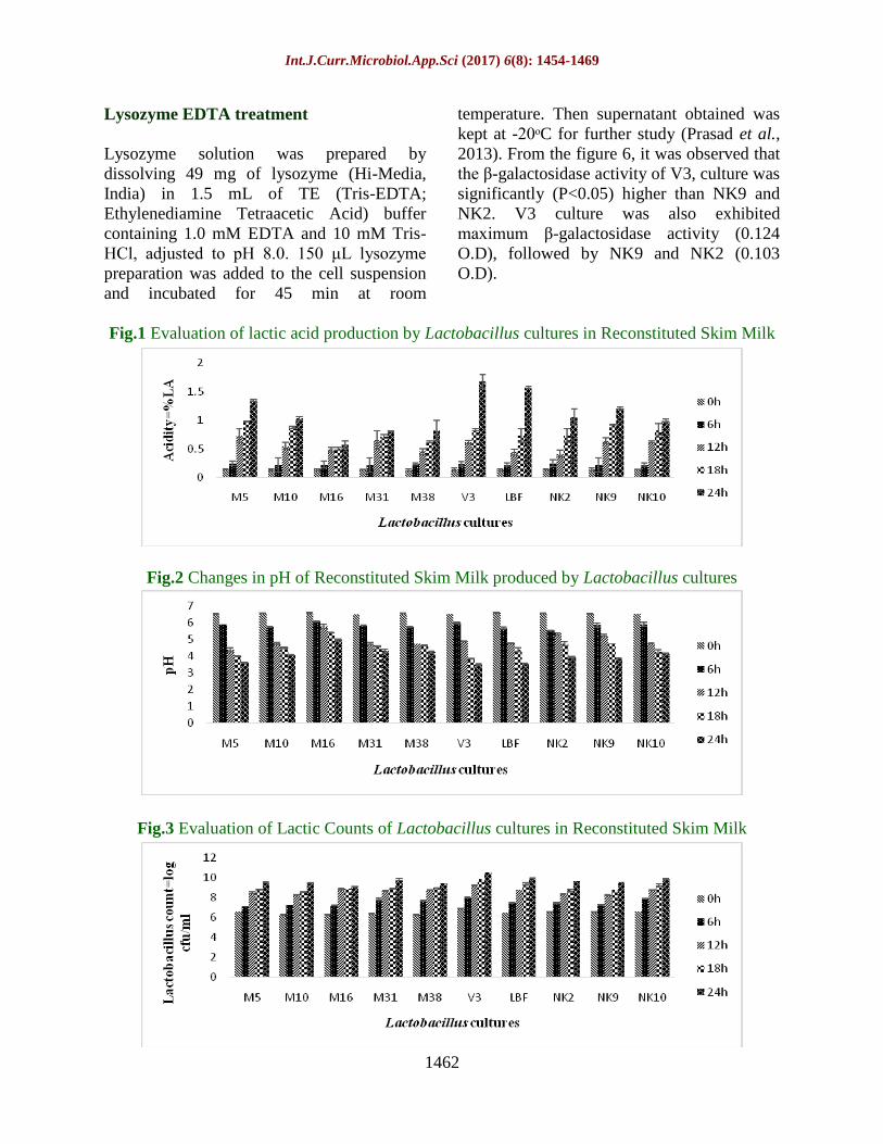
Titratable acidity was determined by
calculating the amount of titratable acidity
developed up to 24h of incubation. From the
figure 1, it was observed that the titratable
acidity of V3 was significantly (P<0.05)
increased up to 24h. The titratable acidity was
significantly higher at 24 h (1.109). Overall
titratable acidity was ranged from 0.141 %
LA (0h) to 1.109 % LA after 24h incubation
at 37ᵒC. During the growth in sterilized
reconstituted skim milk, V3 produced highest
titratable acidity (0.704 % LA), followed by
M5 (0.691%) and NK9 (0.630%) after 24h at
37ᵒC. From figure 2, it was also observed that
the pH of V3 was significantly (P<0.05)
decreased up to 24h of incubation period. The
pH was significantly lowered at 24h (4.04).
Overall pH was ranged from 6.56 (0h) to 4.04
after 24h at 37ᵒC. During the growth in
sterilized reconstituted skim milk, V3
produced highest reduction in pH (4.89),
followed by M5 (4.97) and NK9 (5.00) after
24h at 37ᵒC. From figure 3, it was observed
that the Lactobacillus counts (log cfu/ml) of
V3 was significantly (P<0.05) increased up to
24 h of incubation period. Lactobacillus
counts (log cfu/ml) were significantly
maximum at 24h (9.701). Overall
Lactobacillus counts (log cfu/ml) were ranged
from 6.486 log cfu/ml (0h) to 9.701 log
cfu/ml after 24h incubation at 37ᵒC. During
the growth in sterilized reconstituted skim
milk, V3 showed maximum Lactobacillus
counts (8.892 log cfu/ml), followed by NK10
(8.481 log cfu/ml) and LBF (8.425 log
cfu/ml) after 24h at 37 ᵒC.
Hati et al., (2015) studied the growth
performance of Lctobcillus rhamnosus (NS4
and NS6), Lactobacillus helveticus MTCC
5463 (V3), Lactobacillus delbruckii (09),
Enterococcus feacalis (ND3), Enterococcus
feacalis (ND11) and Lactobacillus rhamnosus
(SH8) by determining viable counts (log
cfu/ml) and production of Lactobacillus acid
measured by decline in pH in skim milk
inoculated at the rate of 1 % and incubated at
37 ᵒC for 12 h. It was observed that NS4
lowered down the pH at a maximum level
compared to V3, ND3 and SH8. However, it
was also observed that NS4 produced
maximum acidity compared to V3, ND3, SH8
and I4.
Viable counts of all the cultures were
measured after 12 h of incubation at 37 ᵒC.
From the study; it was found that NS4 gives
highest viable cell counts 10.68 log cfu/ml
than other bacterial isolates at this qrelatively
exhibited lesser bacterial counts compared to
other isolates in MRS agar medium. It was
also concluded that, viable cell counts, pH
and acidity varies due to the use of different
strains (Hati et al., 2015) as similar to our
study. In another study, Hati et al., (2017)
evaluated the influence of whey protein
concentrate on the production of antibacterial
peptides derived from fermented milk
by Lactic Acid Bacteria. They evaluated skim
milk supplemented with WPC 70 (@ 1.0, 1.5,
2%) was inoculated with 2% lactic cultures
i.e., S. thermophilus (MD2), L. helveticus
(V3), L. rhamnosus (NS4) and L. bulgaricus
(09). pH, titratable acidity, viable cell counts
and proteolytic activity were evaluated after
3, 6, 9, 12 and 24 h of fermentation at 37°C. It
has been previously reported that addition of
WPC 70 to milk followed by heat treatment
induced a decrease of fermentation time
(Antunes et al., 2005; Milanovic et al., 2009).
It has been also reported that addition of 1 or
2% WPC to a medium used for fermentation
with L. delbrueckii ssp. bulgaricus or S.
Int.J.Curr.Microbiol.App.Sci (2017) 6(8): 1454-1469
1460
thermophilus has produced significantly
higher bacterial counts and much faster
acidity (Burya et al., 1998).
Determination of β-galactosidase activity
Out of ten Lactobacillus cultures three
cultures were selected on the basis of β-
galactosidase activity. β-galactosidase activity
of Lactobacillus cultures were carried out in
MRS medium using sonicator (LABMAN,
India) for 30 min (30 seconds off / 30 seconds
on and 61 % amplitudes) in ice bath,
according to the method of Prasad et al.,
(2013). Individually each Lactobacillus
culture was statistically analyzed for the
production of β-galactosidase enzyme at
optimum incubation period. β-galactosidase
enzyme production of ten Lactobacillus
cultures in MRS medium was depicted in the
figure 4.
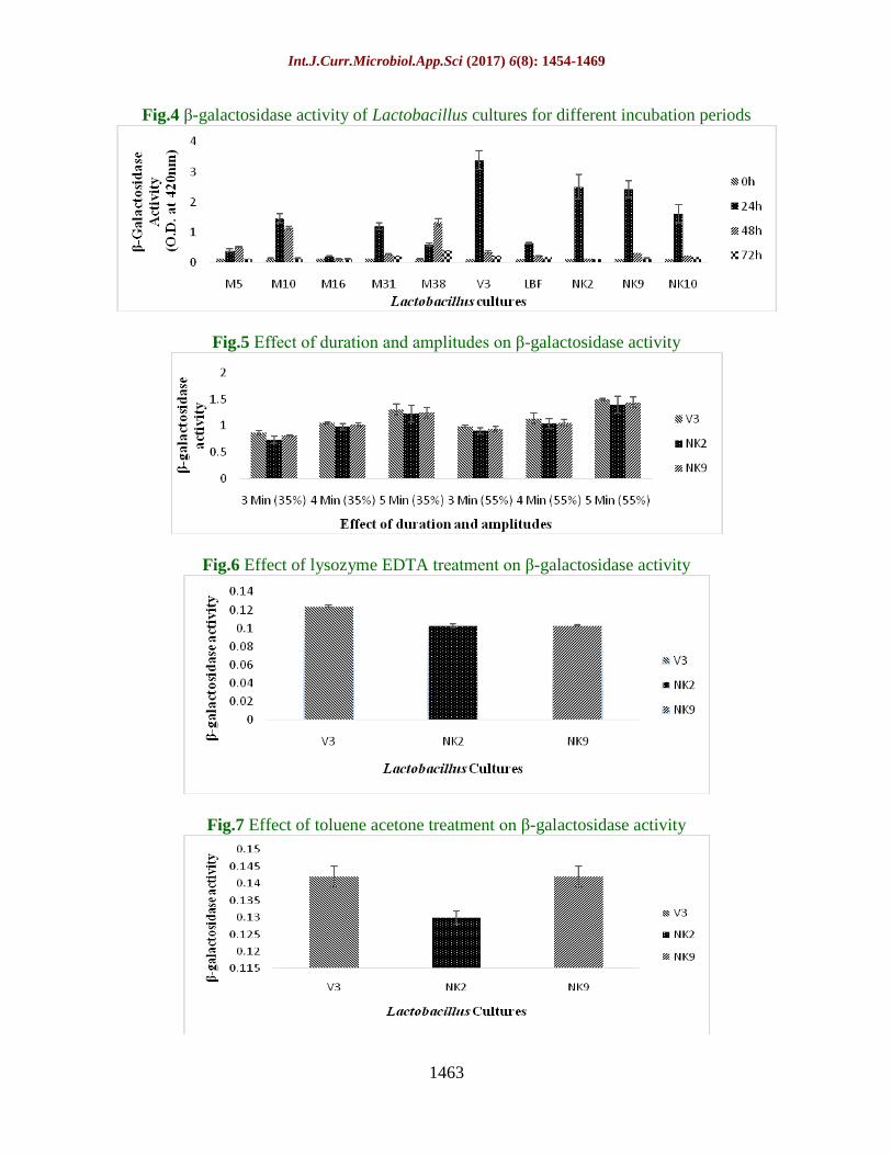
From the figure 4, it was observed that the β-
galactosidase activity of V3 was significantly
(P<0.05) increased up to 24h of incubation.
The β-galactosidase activity was significantly
higher at 24h (1.444 O.D.) for all the
Lactobacillus cultures compared to 48h
(0.467 O.D.) and 72h (0.193 O.D.). V3
showed highest (1.023 O.D.) β-galactosidase
activity at 24h. Overall β-galactosidase
activity was ranged from 0.113 O.D. (LBF at
0h) to 3.381 O.D. (V3 up to 24h). V3
produced the maximum β-galactosidase
activity at 24h (3.381 O.D.), followed by NK2
(2.502 O.D) and NK9 (2.417 O.D). Overall β-
galactosidase activity was ranged from M16
(0.149 O.D) to V3 (1.023 O.D).
Vikas et al., (2015) studied the efficiency of
different methods for disruption
of Streptococcus thermophilus cells, isolated
from different dairy products, to release β-
galactosidase and synthesis of Galacto
oligosaccharides (GOS) by extracted enzyme
using whey supplemented with different
concentrations of lactose as a substrate was
studied. Unlike most other studies on GOS
synthesis which used only one method of cell
disruption sonication (ultrasonics) and only
few microbial strains, they compared five
different cell disruption methods and used 30
strains of S. thermophilus in order to find out
the most effective method is sonication
(ultrasonics) and efficient strain for
production of β-galactosidase. This report had
a similar method from our study. In another
study, Prasad et al., (2013) studied the
activity of β-galactosidase from B. animalis
Bb12 and L. delbrueckii ssp. bulgaricus
ATCC 11842 in whey and its extraction using
various methods. L. delbrueckii ssp.
bulgaricus ATCC 11842 produced more
(p<0.05) intracellular β-galactosidase than B.
animalis ssp. lactis Bb12 with all extraction
methods, except sonication. There were
significant (p<0.05) differences in β-
galactosidase levels extracted from each
organism by the four extraction methods.
Sonication method was found to be more
effective for B. animalis Bb12 than the others
methods. This report had a similar
observation from our study. In another study,
Kara (2004) evaluated the release and
characterization of β-galactosidase
from Lactobacillus plantarum. She optimized
the lysozyme method, sonication method, and
liquid nitrogen method for protein release
from the cells of Lactobacillus plantarum.
Sonication was found to be the most effective
method. This report had a similar method
from our study.
The aim of this study was to screen different
Lactobacillus cultures on the basis of β-
galactosidase activity and to optimize the
growth conditions for the production of β-
galactosidase enzyme of Lactobacillus
cultures. Two important fermentation
variables i.e., ten Lactobacillus cultures (L.
helveticus MTCC 5463 (V3), L. rhamnosus
(NK2), L. casei (NK9), L. rhamnosus MTCC
Int.J.Curr.Microbiol.App.Sci (2017) 6(8): 1454-1469
1461
25062 (NK10), L. fermentum (M5), L.
paracasei (M16), L. rhamnosus (M31), L.
plantarum (M38), L. plantarum (M10), L.
fermentum TDS030603 (LBF)) and
incubation periods (0, 24, 48 and 72h) were
studied. As per statistical analysis, V3, NK2
and NK9 were found to be strong β-
galactosidase producer at 24h of incubation.
As, the degree of β-galactosidase activity was
found to depend on different strains of
Lactobacillus cultures and incubation period
i.e. 24 hours, V3, NK2 and NK9
Lactobacillus cultures were selected for the
production of β-galactosidase enzyme. β-
galactosidase activity being important factor
for the β-galactosidase production, the
variables studied were very relevant due to
their significance in improving the β-
galactosidase enzyme production from both
the microorganisms and optimum growth
temperature (24h).
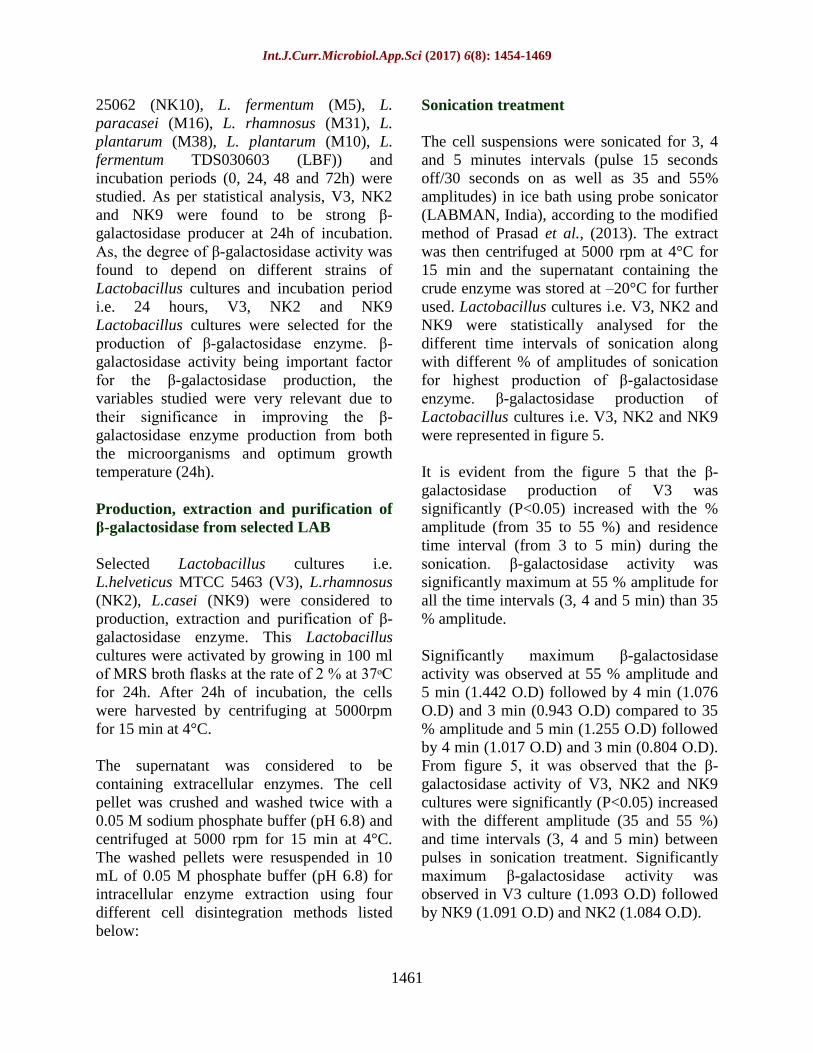
Production, extraction and purification of
β-galactosidase from selected LAB
Selected Lactobacillus cultures i.e.
L.helveticus MTCC 5463 (V3), L.rhamnosus
(NK2), L.casei (NK9) were considered to
production, extraction and purification of β-
galactosidase enzyme. This Lactobacillus
cultures were activated by growing in 100 ml
of MRS broth flasks at the rate of 2 % at 37ᵒC
for 24h. After 24h of incubation, the cells
were harvested by centrifuging at 5000rpm
for 15 min at 4°C.
The supernatant was considered to be
containing extracellular enzymes. The cell
pellet was crushed and washed twice with a
0.05 M sodium phosphate buffer (pH 6.8) and
centrifuged at 5000 rpm for 15 min at 4°C.
The washed pellets were resuspended in 10
mL of 0.05 M phosphate buffer (pH 6.8) for
intracellular enzyme extraction using four
different cell disintegration methods listed
below:
Sonication treatment
The cell suspensions were sonicated for 3, 4
and 5 minutes intervals (pulse 15 seconds
off/30 seconds on as well as 35 and 55%
amplitudes) in ice bath using probe sonicator
(LABMAN, India), according to the modified
method of Prasad et al., (2013). The extract
was then centrifuged at 5000 rpm at 4°C for
15 min and the supernatant containing the
crude enzyme was stored at –20°C for further
used. Lactobacillus cultures i.e. V3, NK2 and
NK9 were statistically analysed for the
different time intervals of sonication along
with different % of amplitudes of sonication
for highest production of β-galactosidase
enzyme. β-galactosidase production of
Lactobacillus cultures i.e. V3, NK2 and NK9
were represented in figure 5.
It is evident from the figure 5 that the β-
galactosidase production of V3 was
significantly (P<0.05) increased with the %
amplitude (from 35 to 55 %) and residence
time interval (from 3 to 5 min) during the
sonication. β-galactosidase activity was
significantly maximum at 55 % amplitude for
all the time intervals (3, 4 and 5 min) than 35
% amplitude.
Significantly maximum β-galactosidase
activity was observed at 55 % amplitude and
5 min (1.442 O.D) followed by 4 min (1.076
O.D) and 3 min (0.943 O.D) compared to 35
% amplitude and 5 min (1.255 O.D) followed
by 4 min (1.017 O.D) and 3 min (0.804 O.D).
From figure 5, it was observed that the β-
galactosidase activity of V3, NK2 and NK9
cultures were significantly (P<0.05) increased
with the different amplitude (35 and 55 %)
and time intervals (3, 4 and 5 min) between
pulses in sonication treatment. Significantly
maximum β-galactosidase activity was
observed in V3 culture (1.093 O.D) followed
by NK9 (1.091 O.D) and NK2 (1.084 O.D).
Int.J.Curr.Microbiol.App.Sci (2017) 6(8): 1454-1469
1462
Lysozyme EDTA treatment
Lysozyme solution was prepared by
dissolving 49 mg of lysozyme (Hi-Media,
India) in 1.5 mL of TE (Tris-EDTA;
Ethylenediamine Tetraacetic Acid) buffer
containing 1.0 mM EDTA and 10 mM Tris-
HCl, adjusted to pH 8.0. 150 μL lysozyme
preparation was added to the cell suspension
and incubated for 45 min at room
temperature. Then supernatant obtained was
kept at -20ᵒC for further study (Prasad et al.,
2013). From the figure 6, it was observed that
the β-galactosidase activity of V3, culture was
significantly (P<0.05) higher than NK9 and
NK2. V3 culture was also exhibited
maximum β-galactosidase activity (0.124
O.D), followed by NK9 and NK2 (0.103
O.D).
Fig.1 Evaluation of lactic acid production by Lactobacillus cultures in Reconstituted Skim Milk
Fig.2 Changes in pH of Reconstituted Skim Milk produced by Lactobacillus cultures
Fig.3 Evaluation of Lactic Counts of Lactobacillus cultures in Reconstituted Skim Milk
Int.J.Curr.Microbiol.App.Sci (2017) 6(8): 1454-1469
1463
Fig.4 β-galactosidase activity of Lactobacillus cultures for different incubation periods
Fig.5 Effect of duration and amplitudes on β-galactosidase activity
Fig.6 Effect of lysozyme EDTA treatment on β-galactosidase activity
Fig.7 Effect of toluene acetone treatment on β-galactosidase activity
Int.J.Curr.Microbiol.App.Sci (2017) 6(8): 1454-1469
1464
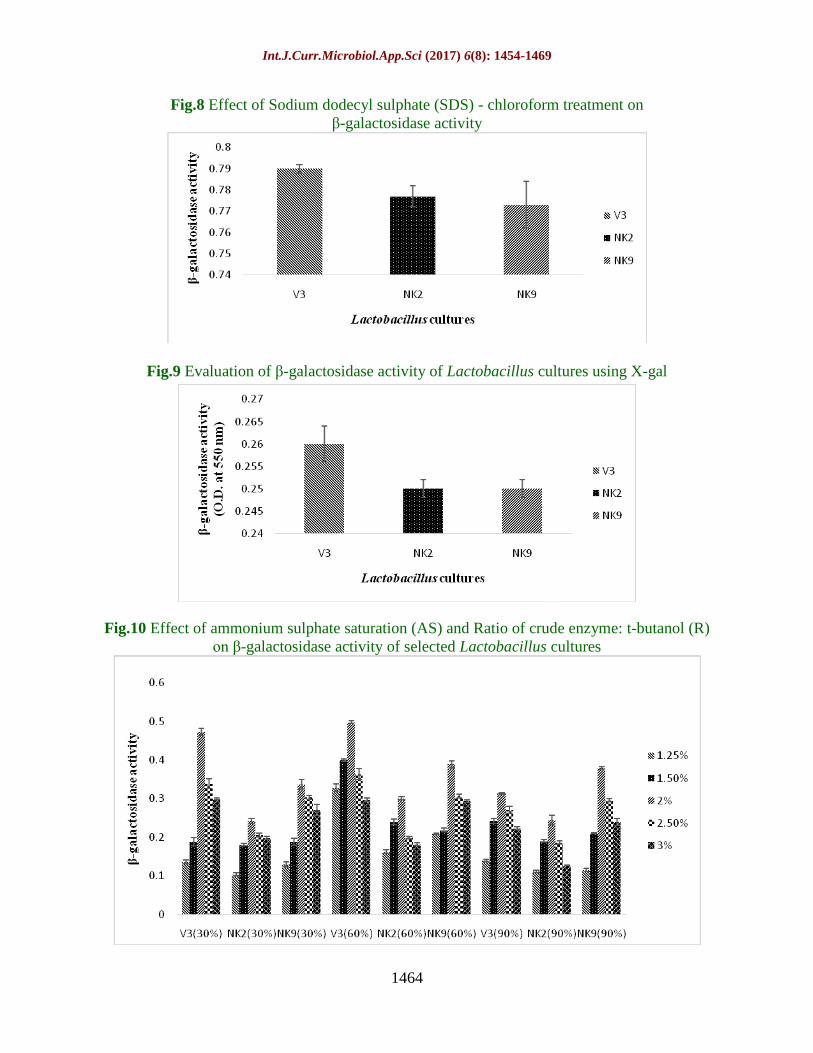
Fig.8 Effect of Sodium dodecyl sulphate (SDS) - chloroform treatment on
β-galactosidase activity
Fig.9 Evaluation of β-galactosidase activity of Lactobacillus cultures using X-gal
Fig.10 Effect of ammonium sulphate saturation (AS) and Ratio of crude enzyme: t-butanol (R)
on β-galactosidase activity of selected Lactobacillus cultures
Int.J.Curr.Microbiol.App.Sci (2017) 6(8): 1454-1469
1465
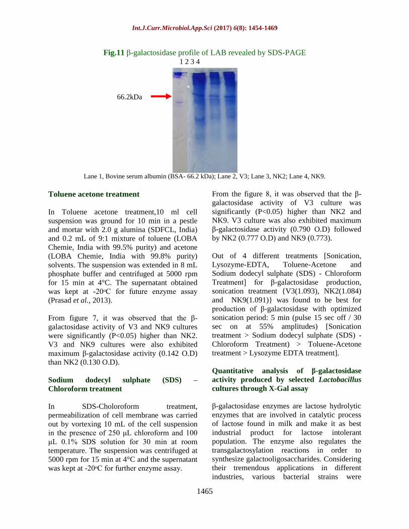
Fig.11 β-galactosidase profile of LAB revealed by SDS-PAGE
Lane 1, Bovine serum albumin (BSA- 66.2 kDa); Lane 2, V3; Lane 3, NK2; Lane 4, NK9.
Toluene acetone treatment
In Toluene acetone treatment,10 ml cell
suspension was ground for 10 min in a pestle
and mortar with 2.0 g alumina (SDFCL, India)
and 0.2 mL of 9:1 mixture of toluene (LOBA
Chemie, India with 99.5% purity) and acetone
(LOBA Chemie, India with 99.8% purity)
solvents. The suspension was extended in 8 mL
phosphate buffer and centrifuged at 5000 rpm
for 15 min at 4°C. The supernatant obtained
was kept at -20ᵒC for future enzyme assay
(Prasad et al., 2013).
From figure 7, it was observed that the β-
galactosidase activity of V3 and NK9 cultures
were significantly (P<0.05) higher than NK2.
V3 and NK9 cultures were also exhibited
maximum β-galactosidase activity (0.142 O.D)
than NK2 (0.130 O.D).
Sodium dodecyl sulphate (SDS) –
Chloroform treatment
In SDS-Choloroform treatment,
permeabilization of cell membrane was carried
out by vortexing 10 mL of the cell suspension
in the presence of 250 μL chloroform and 100
μL 0.1% SDS solution for 30 min at room
temperature. The suspension was centrifuged at
5000 rpm for 15 min at 4°C and the supernatant
was kept at -20ᵒC for further enzyme assay.
From the figure 8, it was observed that the β-
galactosidase activity of V3 culture was
significantly (P<0.05) higher than NK2 and
NK9. V3 culture was also exhibited maximum
β-galactosidase activity (0.790 O.D) followed
by NK2 (0.777 O.D) and NK9 (0.773).
Out of 4 different treatments [Sonication,
Lysozyme-EDTA, Toluene-Acetone and
Sodium dodecyl sulphate (SDS) - Chloroform
Treatment] for β-galactosidase production,
sonication treatment {V3(1.093), NK2(1.084)
and NK9(1.091)} was found to be best for
production of β-galactosidase with optimized
sonication period: 5 min (pulse 15 sec off / 30
sec on at 55% amplitudes) [Sonication
treatment > Sodium dodecyl sulphate (SDS) -
Chloroform Treatment) > Toluene-Acetone
treatment > Lysozyme EDTA treatment].
Quantitative analysis of β-galactosidase
activity produced by selected Lactobacillus
cultures through X-Gal assay
β-galactosidase enzymes are lactose hydrolytic
enzymes that are involved in catalytic process
of lactose found in milk and make it as best
industrial product for lactose intolerant
population. The enzyme also regulates the
transgalactosylation reactions in order to
synthesize galactooligosaccharides. Considering
their tremendous applications in different
industries, various bacterial strains were
66.2kDa
1 2 3 4
Int.J.Curr.Microbiol.App.Sci (2017) 6(8): 1454-1469
1466
isolated and screened for β-galactosidase
production (Kamaran et al., 2016).
2 ml of crude β-galactosidase enzyme was
added into the mixture of 0.5 ml of X-gal and
0.5 ml of 0.1 M IPTG and incubated for 3h at
37ᵒC. After incubation it was measured at 550
nm with a spectrophotometer (Systronics PC
based double beam spectrophotometer 2202,
India) of β-galactosidase activity of V3, NK2
and NK9 and was represented in figure 9 using
X-gal and IPTG. β-galactosidase activity
produced by Lactobacillus cultures was
determined in ranged from 0.25 O.D to 0.26
O.D. After 3h of incubation at 37ᵒC. V3 showed
maximum β-galactosidase activity (0.26 O.D)
compared to NK2 and NK9 (0.25 O.D).
Kamran et al., (2016) evaluating the qualitative
screening of bacterial isolates for β-
galactosidase production. Pure bacterial cultures
were grown at 37°C for 48h onto the lactose
agar medium, supplemented with 50 µg ml−1 X-
gal (chromogenic substrate) and 1.0 mM of
isopropyl β-d-1-thiogalactopyrinoside (IPTG) to
examine the capability of isolates for β-
galactosidase production (Jaturapiree et al.,
2012). Lactose agar medium contained (g L−1):
Lactose, 5.0; beef extract, 3.0; peptone, 5.0;
Agar, 15.0 with pH-7.0. After 48 h of
incubation period, blue colored colonies were
examined. Selected isolates were stored on
lactose medium slant and in 40% glycerol for
short and long term preservation, respectively.
The purified bacterial strains were screened for
β-galactosidase production by growing on
lactose agar medium (supplemented with X-gal
and IPTG) at 37°C for 48h (Jaturapiree et al.,
2012). Among them, blue colored colonies of
Bacillus strain B-2 were prominently observed
on lactose agar medium that indicates the
production of β-galactosidase into medium after
cleaving the chromogenic substrate (X-gal)
which was supplemented with lactose into
culture medium plate. But in our study, we had
developed a novel quantitative method to screen
the isolates on β-galactosidase production using
spectrophotometry method in X-Gal and IPTG
solution.
Purification of β-galactosidase enzyme
Purification of β-galactosidase enzyme was
carried out employing various t-butanol ratios
(crude extract: t-butanol = 1:0.25, 1.0:0.5,
1.0:1.0, 1.0:1.5, 1.0:2.0) with a different
ammonium sulfate saturation at 30, 60 and
90%. The mixture was mixed gently and then
allowed to stand for 30 min at 37ᵒC. afterward,
the mixture was centrifuged at 5000 rpm for 15
min at 4ᵒC to facilitate the separation of phases.
The upper t-butanol phase was removed by the
Pasteur pipette. The lower aqueous phase and
the interfacial phase were separated carefully
and each of phases was analyzed for enzyme
activity. Different ammonium sulfate saturation
effects (30, 60 and 90%) (w/v) were
investigated at the best recovery activity of
crude enzyme: t-butanol ratio that is 1.0:1.0.
The bottom phase and interfacial phase were
analyzed for enzyme activity (Duman and Kaya,
2013).The enzyme assay evaluates the β-
galactosidase enzyme, which was present in
selected Lactobacillus cultures. Lactobacillus
cultures i.e. V3, NK2 and NK9 were
statistically analysed for the different
ammonium sulphate saturation effects, different
sample size of crude enzyme: t-butanol ratio
and highest production of β-galactosidase
enzyme. β-galactosidase enzyme production of
Lactobacillus cultures i.e. V3, NK2 and NK9
were presented in figure 10. In figure 10, β-
galactosidase activity of V3 culture was
significantly (P<0.05) increased at 60%
ammonium sulphate saturation and ratio of
crude enzyme: t-butanol (1:1; 2%). β-
galactosidase activity was significantly highest
at 60 % ammonium sulphate saturation for all
the three Lactobacillus cultures (V3, NK2 and
NK9) followed by 30 and 90%. Significantly
maximum β-galactosidase production was also
observed at 60% ammonium sulphate saturation
in case of V3 culture (0.377O.D), followed by
NK9 (0.283 O.D) and NK2 (0.216 O.D)
compared to 30% ammonium sulphate
saturation of V3 (0.287 O.D) followed by NK9
(0.246 O.D) and NK2 (0.186 O.D) and
subsequently also compared with 90 %
ammonium sulphate saturation for NK9 (0.248
Int.J.Curr.Microbiol.App.Sci (2017) 6(8): 1454-1469
1467
O.D), followed by V3 (0.238 O.D) and NK2
(0.172 O.D).From figure 10, it was observed
that the β-galactosidase activity of V3, NK2 and
NK9 cultures were significantly (P<0.05)
increased with the different ammonium sulphate
saturations (30 and 60 %) and various crude
enzyme:t-butanol ratios (1:0.25;1.25, 1:0.5;1.5
and 1:1;2) during purification process. But β-
galactosidase activity were significantly
decreased at ammonium sulphate saturation
effect (90 %) and various crude enzyme: t-
butanol ratios (1:1.5; 2.5 and 1:2:3 %).
Significantly maximum β-galactosidase activity
was observed at (1:1:2 %) crude enzyme: t-
butanol ratio (0.353 O.D), followed by 1:1.5;
2.5 % (0.274 O.D), 1:2:3 % (0.236 O.D), 1:0.5;
1.5 % (0.228 O.D) and 1:0.25; 1.25 % (0.159
O.D) during purification process.
Prasad et al., (2013) evaluated that the
production of β-galactosidase by
Bifidobacterium animalis ssp. lactis Bb12 and
Lactobacillus delbrueckii ssp. bulgaricus
ATCC 11842 in whey and the effect of 4
different extraction methods i.e. sonication,
acetone-toluene, SDS-chloroform and
lysozyme-EDTA treatment on enzyme activity
from these organisms. Both organisms were
grown in deproteinised whey containing yeast
extract (3.0 g/L), peptone (5.0 g/L) and glucose
(10.0 g/L) for 18 h, at 37ºC for B. animalis ssp.
lactis Bb12 and at 45ºC for L. delbrueckii ssp.
bulgaricus ATCC 11842. The optimum
intracelluar β-galactosidase activity on 15 mM
o-nitrophenyl β-D-galactopyranoside (ONPG)
assay was at pH 6.8 for both organisms
irrespective of the method of extraction used.
Also, the effect of temperature on enzyme
activity was studied at various temperatures (30,
35, 40, 45 and 50°C). At 35°C and 40°C, B.
animalis ssp. lactis Bb12 evaluated more
intracellular β-galactosidase activity extracted
by sonication than other temperatures and
methods. However, L. delbrueckii ssp.
bulgaricus ATCC 11842 had more intracellular
β-gal activity at 35°C and 45°C when extracted
by lysozyme-EDTA treatment. Among the four
methods used for β-galactosidase extraction,
sonication exhibited the best result (6.80
Unit/mL) for B. animalis ssp. lactis Bb12 while
lysozyme-EDTA treatment was also found to be
the best (7.77 Unit/ mL) for L. delbrueckii ssp.
bulgaricus ATCC 11842. These treatments are
similar as our study. Duman and Kaya, (2013)
evaluated the activity of β-galactosidase was
completely decreased at 60% ammonium
sulfate saturation in aqueous phase, the best
results of recovery and the purification fold of
β-galactosidase (133% and 10.1, respectively)
was obtained in the interfacial phase. Probably,
this saturation was caused by the precipitation
of whole target protein molecule in the
interfacial phase. Generally, researchers started
with minimum salt concentration of 20% (w/v)
to optimize the partitioning conditions
(Dennison et al., 1997). In that study also
evaluated the minimum salt saturations for the
beginning and the increasing of salt
concentrations resulted with the increasing of
recovery and purification fold in the interphase
up to 60% (w/v) saturations. Higher salt
concentrations from this point were resulted
with reducing of recovery and purification fold,
which may be due to irreversible denaturation
of protein (Narayan et al., 2008). This results
obtained as similar data found in our study.
Purification of β-galactosidase enzyme
through SDS-PAGE
β-galactosidase enzyme extracts from different
Lactobacillus cultures were subjected to SDS-
PAGE analysis along with Bovine serum
albumin (BSA- 66.2 kDa) as a marker (Fig. 11).
The molecular mass of partially purified β-
galactosidase (60% Ammonium sulphate
saturation) from selected Lactobacillus cultures
i.e. V3, NK2 and NK9 as determined by SDS-
PAGE was approx. 69 kDa (Fig. 11). Presence
of high molecular weight protein band nearer to
BSA in LAB showed Mol. Weight of β-
galactosidase. The protein band of 62 kDa
corresponds to β-galactosidase enzyme as
reported by previous workers (Pal et al., 2013).
These results raised the expectation of finding
β-galactosidase enzyme in LAB. Out of ten
Lactobacillus cultures, three cultures i.e. V3,
NK2 and NK9 showed highest production of β-
Int.J.Curr.Microbiol.App.Sci (2017) 6(8): 1454-1469
1468
galactosidase after 24 h at 37ᵒC. Sonication
treatment was also found best for the maximum
production of β-galactosidase under the
optimized growth condition. 60 % Ammonium
sulphate and crude enzyme:t-butanol=1:1 also
produced maximum partially purified β-
galactosidase enzyme for further study. These
three Lactobacillus cultures could be used as β-
galactosidase producer.
Conflicts of interest
There is no conflict of interest in the study
among the authors.
Authors’ contributions
All the authors contributed equally to the
writing of this paper. They were also involved
in the overall work of experiments.
References
Antunes A. E. C., Cazetto T. F. and Bolini H.
M. A., Viability of probiotic
microorganisms during storage, post
acidification and Fig. 12 MS/MS
spectrum of fraction E1 inspected in
MASCOT database (Identified
‘ETVPYMFEN’ amino acid sequence as
‘lactoferrin’)sensory analysis of fat-free
yoghurts with added whey protein
concentrate, International Journal of
Dairy Technology, 58,169–173 (2005).
Berni Canani R., Pezzella V., Amoroso A. et
al., Diagnosing and treating intolerance to
carbohydrates in children, Nutrients, 8,
155-157 (2016).
Bury, T. and Jelen, P., Lactose hydrolysis in
milk as affected by neutralizers used for
the preparation of crude β-galactosidase
extracts from Lactobacillus bulgaricus
11842, Innovative Food Science and
Emerging Technologies, 3, 175-188
(2001).
Burya D., Jelena P., Kimura K., Whey protein
concentrateas a nutrient supplement for
lactic acid bacteria, International Dairy
Journal, 8,149–151 (1998).
Carrasco-Castilla J., Hernandez A., Lvarez A. J.
et al., Antioxidant and metal chelating
activities of Phaseolus vulgaris L. var.
Jamapa protein isolaes, phaseolin and
lectin hydrolysates. Food chemistry, 131,
1157-1164 (2012).
Deng Y., Misselwitz B., Dai N. and Fox
M., Lactose intolerance in adults:
biological mechanism and dietary
management, Nutrients, 7, 8020–8035
(2015)
Dennison C. and Lovrein R., Three phase
partitioning: concentration and
purification of proteins, Protein
Expression and Purification, 11, 149–161
(1997)
Duman Y. and Kaya E. Purification, recovery,
and characterization of chick pea (Cicer
arietinum) b-galactosidase in single step
by three phase partitioning as a rapid and
easy technique, Journal of Dairy Science,
91, 155–160 (2013)
Hati S., Patel N., Sakure A. and Mandal S.
Influence of whey protein concentrate
on the production of antibacterial peptides
derived from fermented milk by lacticacid
bacteria, International Journal of Peptide
Research and Therapeutics, (2017)
Hati S., Sreeja V., Solank, J. and Prajapati J. B.
Significance of proteolytic
microorganisms on ACE-inhibitory
activity and release of bioactive peptides
during fermentation of milk, Indian
Journal of Dairy Science, 68, 584-591
(2015)
IDF 146, Yogurt-Identification of characteristic
microorganisms (Lactobacillus
delbrueckii subsp. bulgaricus and
Streptococcus thermophilus),
International Standard, (2003)
Indian Standards., Methods of test for dairy
industry part-Irapid examination of milk,
Indian Standards Institution, New Delhi
(1479) (1960)
Jaturapiree P., Phuengjayaeam S., Seangsawang
S. et al., Isolation and production of novel
β-galactosidase from a newly isolated,
moderate thermophile, Bacillus sp. strain
B1, Journal of Food Science and
Engineering, 2, 395–402 (2012)
Int.J.Curr.Microbiol.App.Sci (2017) 6(8): 1454-1469
1469
Jelen P., Lactose hydrolysis using sonicated
dairy cultures, In Lactose hydrolysis, IDF
Brussels Belgium Bulletin, 289, 54-56
(1993)
Kamran A., Bibi Z., and Aman A. et al.,
Lactose hydrolysis approach: Isolation
and production of β-galactosidase from
newly isolated Bacillus strain B-2,
Biocatalysis and Agricultural
Biotechnology, 5, 99–103 (2016)
Kara F., Release and characterization of beta-
galactosidase from Lactobacillus
plantarum, Biotechnology, METU
(2004).
Laemmli U. K., Cleavage of structural proteins
during assembly of the head of
bacteriophage T4, Nature, 227, 680-685
(1970)
Milanovic S., Ilicic M., Duric M., Caric M.,
Effect of transglutaminase and whey
protein concentrate on textural
characteristicsof low fat probiotic
yoghurt, Milchwissenschaft, 64, 388–392
(2009)
Narayan A. V., Madhusudhan M. C. and
Raghavarao S. M., Extraction and
purification of ipomea peroxidase
employing three-phase partitioning,
Applied Biochemistry and Biotechnology,
151, 263–272 (2008)
NIDDK, Lactose Intolerance, National Institute
of Diabetes and Digestive and Kidney
Diseases (NIDDK), pp: 12-16 (2016)
Pal A., Lobo M. and Khanum F., Extraction,
purification and thermodynamic
characterization of almond (Amygdalus
communis) β-galactosidase for the
preparation of delactosed milk, Food
Technology and Biotechnology, 51, 53–61
(2013)
Prasad L. N., Ghosh B. C., Sherkat F. et al.,
Extraction and characterisation of β-
galactosidase produced by
Bifidobacterium animalis spp. lactis Bb12
and Lactobacillus delbrueckii spp.
bulgaricus ATCC 11842 grown in whey,
International Food Research Journal, 20,
487-494 (2013)
Skovbjerg H., Sjostrom H. and Noren O.,
Purification and characterization of
amphiphilic lactase/phlorizin hydrolase
from human small intestine, European
Journal of Biochemistry, 114, 653–661
(1981)
Vasiljevic T. and Jelen P., Production of β-
galactosidase for lactose hydrolysis in
milk and dairy products using
thermophilic lactic acid bacteria,
Innovative Food Science and Emerging
Technologies, 2, 75-85 (2001)
Vikas S., Sudhir K., Tomar B. A., Ram R. B.
and Ashish K. S. Production of β-
galactosidase from Streptococcus
thermophilus for galactooligosaccharides
synthesis, Journal of Food Science and
Technology, 52, 4206–4215 (2015)
How to cite this article:
Shrushti Makwana, Subrota Hati, Heena Parmar and Aparnathi, K.D. 2017. Process Optimization
for the Production of β-Galactosidase Using Potential Lactobacillus Cultures.
Int.J.Curr.Microbiol.App.Sci. 6(8): 1454-1469. doi: https://doi.org/10.20546/ijcmas.2017.608.176