
Ocular Onchocerciasis The of Toll Like Receptor 2 and ...hss.ulb.uni-bonn.de/2010/2052/2052.pdf ·...
115
Ocular Onchocerciasis – The Role of TollLike Receptor2 and Interferonγ Dissertation zur Erlangung des Doktorgrades (Dr. rer. nat.) der Mathematisch‐Naturwissenschaftlichen Fakultät der Rheinischen Friedrich‐Wilhelms‐Universität Bonn vorgelegt von Dr. med. Katrin Gentil aus Suhl Bonn 2009
Transcript of Ocular Onchocerciasis The of Toll Like Receptor 2 and ...hss.ulb.uni-bonn.de/2010/2052/2052.pdf ·...
Ocular Onchocerciasis – The Role of TollLike Receptor2
and Interferonγ
Dissertation
zur
Erlangung des Doktorgrades (Dr. rer. nat.)
der
Mathematisch‐Naturwissenschaftlichen Fakultät
der
Rheinischen Friedrich‐Wilhelms‐Universität Bonn
vorgelegt von
Dr. med. Katrin Gentil
aus Suhl
Bonn 2009

Gedruckt mit Genehmigung
der mathematisch‐naturwissenschaftlichen Fakultät
der Rheinischen‐Friedrich‐Wilhelms‐Universität Bonn
Referent: Prof. W. Kolanus
Koreferent: Prof. A. Hoerauf
Tag der mündlichen Prüfung: 23.02.2010
Erscheinungsjahr: 2010
Ocular Onchocerciasis - the Role of Toll-Like Receptor 2 and Interferon-γ
Abstract
by
KATRIN GENTIL
River Blindness is still highly prevalent in subsaharan Africa with millions of people
suffering from the infection with Onchocerca volvulus. This filarial worm harbors the
endosymbiont Wolbachia which was found to be essential for worm survival. The im-
mune system of chronically infected individuals is constantly stimulated with worm
proteins, lipids, carbohydrates and released Wolbachia from dying worms. In this study,
I investigated the activation of the adaptive immune system by O. volvulus and Wol-
bachia in a mouse model of onchocercal keratitis.
Toll-like receptors (TLRs) are innate immune receptors that are activated by con-
served bacterial and viral products. Lately, it was also demonstrated that fungal and
helminth products can activate selected TLRs. During filarial infection, stimulation oc-
curs through both bacterial and parasitic products, I therefore focused on the role of
TLR2 and TLR4 in the pathogenesis of adaptive immune responses and keratitis devel-
opment.
Using a mouse model of onchocercal keratitis, I immunized C57BL/6, TLR2-/- and
TLR4-/- mice with O. volvulus protein extract and examined granulocyte migration to
the corneal stroma following local injection of this protein extract. I found that TLR2
was required for IFNγ production by splenocytes, CXC chemokine production in the
iii
cornea and neutrophil migration to the corneal stroma, but not for IL-5 production or
eosinophil migration.
IFNγ was shown to increase surface expression of TLR2 on macrophages, priming
them for further activation via TLR2 agonists like onchocercal protein extract. The pro-
inflammatory cytokines produced by macrophages induced chemokine production by
corneal fibroblasts. This is one pathway of IFNγ enhanced neutrophil migration to the
corneal stroma.
Other parts of this study showed that pro-inflammatory Th1 responses were pre-
dominantly induced by the endosymbiont Wolbachia rather than the filarial worm itself,
whereas Th2 responses were mainly induced by the worm.
One wolbachial protein that has been previously described and acts as immuno-
activator is the Wolbachia surface protein. I characterized TLR2-dependent activation of
primary macrophages and splenocytes and identified the region 121-240 of the protein
as the main immunostimulatory domain.
iv

Acknowledgements
For their support of my work during the last four years, I would like to thank the following
people:
Eric Pearlman - thank you for accepting me in your lab, providing me with a place to
learn about filarial research and giving me the freedom to develop my own ideas. Thank
you for useful discussions and scientific support.
Achim Hörauf - thank you for encouraging me to go for a Ph.D. and supporting me
through the final stages.
Amy G. Hise - thank you for valuable input on experimental designs, new ideas and
for being a great friend all along. Thank you to your family as well.
Michelle Lin and Angela Johnson - thank you for being great labmates, helpfull with
discussions and friends outside the lab.
Eugenia Diaconu - thank you for helping with all those mouse injections, I will not
count how many we did over the years.
Maria Mackroth, Jose-Andres Portillo-Christen and everyone else at the Department
of Ophthalmology and the Center for Global Health and Diseases at Case Western Re-
serve University - thank you for being a great group to work with.
Meinen Eltern Sabine und Ulrich Dähnel - Danke, dass ihr mir den Freiraum gegeben
habt, mich zu dem Menschen zu entwickeln, der ich heute bin. Danke auch, dass ihr
meine diversen Auslandsaufenthalte immer unterstützt habt, auch wenn es für euch
nicht immer leicht war.
Johannes - Danke für die letzten vier Jahre mit dir! Du bist das Wichtigste, was ich
aus Cleveland mit nach Deutschland zurückgebracht habe!
v

This work was supported in part by a grant by the Deutsche Forschungsgemeinschaft
(DÄ1024/1-1)
vi
Table of Contents
Acknowledgements v
List of Tables ix
List of Figures x
Chapter 1. Introduction 1
River Blindness 1
Wolbachia 2
Cornea 3
Mouse Model of River Blindness 4
Antigen-presenting cells 9
Toll-like Receptors 13
Interferon-γ 16
Aims 17
Chapter 2. Materials and Methods 18
Reagents 18
Mice 22
In vivo studies 22
Cell culture 23
FACS 26
Immunohistochemistry 27
ELISA 29
Statistical analysis 29
vii
Chapter 3. Results 30
In vitro activation of antigen-presenting cells 30
The role of TLR2 and TLR4 in the generation of adaptive immune responses 40
The role of TLR2 and TLR4 in corneal inflammation 41
The role of IFN-γ in the generation of adaptive immune responses 47
IFN-γ induced cytokine responses in macrophages and fibroblasts 50
Activation of macrophages, neutrophils and fibroblasts by pro-inflammatory
cytokines. 55
The role of IL-1R1, IL-6 and TNFα in T cell responses and corneal inflammation 58
In vivo responses to Wolbachia 61
In vitro responses to rWSP 66
Chapter 4. Discussion 73
Activation of antigen-presenting cells 73
TLR2 in the adaptive immune response 75
IFN-γ in the adaptive immune response 77
Wolbachia 80
BmWSP 81
Revised mouse model of Onchocerciasis 85
Chapter 5. Summary 87
References 89
viii

List of Tables
2.1 Primers used in this study. 20
2.2 Antibodies used in this study. 27
ix
List of Figures
1.1 Ocular structure 4
1.2 Corneal structure 5
1.3 Sequence of events 8
3.1 Cytokine Production by Dendritic Cells 31
3.2 Relative Expression of Dendritic Cell Surface Activation Markers 32
3.3 Histogram of Dendritic Cell Surface Activation Markers 33
3.4 Cytokine Production by Macrophages 34
3.5 Release of Myeloperoxide by Neutrophils 35
3.6 Production of MIP-2 by Neutrophils 36
3.7 Production of Nitric Oxide by Neutrophils 37
3.8 Production of Reactive Oxygen Species by Neutrophils 38
3.9 Expression of Neutrophil Surface Activation Markers 39
3.10 Splenocyte responses of C57BL/6, TLR2-/- and TLR4-/- mice 41
3.11 Levels of IgE in C57BL/6, TLR2-/- and TLR4-/- mice 42
3.12 Levels of IgG1 and IgG2c in C57BL/6, TLR2-/- and TLR4-/- mice 42
3.13 Cross Section of the Cornea 43
3.14 Neutrophil Migration to the Cornea of C57BL/6, TLR2-/- and TLR4-/-
mice 44
3.15 Eosinophil Migration to the Cornea of C57BL/6, TLR2-/- and TLR4-/-
mice 44
x
3.16 PECAM-1 Expression on Limbal Vessels 46
3.17 CXC Chemokine Levels in the Cornea 46
3.18 Splenocyte Responses of C57BL/6 and IFN-γ-/- Mice 47
3.19 Granulocyte Migration to the Corneas of C57BL/6 and IFN-γ-/- Mice 48
3.20 PECAM-1 Expression in the Corneas of C57BL/6 and IFN-γ-/- Mice 49
3.21 IFN-γ-induced Activation of Macrophages 51
3.22 Priming of Macrophages by IFN-γ 52
3.23 Chemokine Production by Macrophages 53
3.24 IFN-γ-induced Neutrophil Activation 53
3.25 IFN-γ-induced Activation of MK/T-1 Cells 54
3.26 Surface TLR2 Expression by Macrophages 55
3.27 Macrophage Activation by Pro-inflammatory Cytokines 56
3.28 Neutrophil Activation by Pro-inflammatory Cytokines 57
3.29 Activation of MK/T-1 Cells by Stimulation with Pro-inflammatory
Cytokines 57
3.30 Splenocyte Responses of C57BL/6 and IL-1R1-/- Mice 59
3.31 Splenocyte Responses of C57BL/6 and IL-6-/- Mice 59
3.32 Splenocyte Responses of C57BL/6 and TNFαR-/- Mice 60
3.33 Granulocyte Migration to the Corneas of C57BL/6 and IL-1R1-/- Mouse
Corneas 61
3.34 Granulocyte Migration to the Corneas of C57BL/6 and IL-6-/- Mouse
Corneas 62
xi
3.35 Granulocyte Migration to the Corneas of C57BL/6 and TNFαR-/- Mouse
Corneas 62
3.36 Granulocyte Migration to the Cornea after Injection of Wolbachia
Depleted Extract 64
3.37 Splenocyte Responses to A. viteae and Wolbachia 65
3.38 Granulocyte Migration after A. viteae and Wolbachia Injection 65
3.39 Gene Sequence BmWSP 67
3.40 Amino Acid Sequence BmWSP 68
3.41 HEK Cell Stimulation with rWSP 69
3.42 Macrophage Stimulation with rWSP 70
3.43 Splenocyte Responses to rWSP 71
3.44 BmWSP-Specific IgG1 Levels in C57BL/6 Mice 72
4.1 Sequence of Events - Systemic Responses 85
4.2 Sequence of Events - IFN-γ-Dependent Responses 86
xii

1
1 Introduction
1.1 River Blindness
Filarial nematodes cause the debilitating diseases river blindness and lymphatic
filariasis. Lymphatic filariasis is caused by Brugia malayi or Wuchereria bancrofti. It
is characterized by immense swelling of mainly the lower limbs and the scrotum. An
estimated 120 million people are affected by this disease, the majority of them in India
and Africa (1).
Onchocerca volvulus is the causative agent of river blindness. River blindness is
mainly prevalent in Africa and distributed along rivers. O. volvulus is transmitted by
Simulium species. Upon a blood meal on infected individuals, first stage larvae are
taken up. The larvae undergo two molting steps before reaching the infectious L3 stage.
During the next blood meal, L3 larvae are transmitted into humans. They undergo 2 fur-
ther moltings and mature into adult male and female worms. Female worms produce
L1 microfilariae thus completing the life cycle.
Infections with O. volvulus are classically treated with ivermectin, a microfilaricidal
drug, which is also used for mass drug administration to decrease transmission of O.
volvulus. Treatment with the filaricidal drug diethylcarbamazine, especially for mass
Introduction 2
drug administration, is now contraindicated due to severe adverse reaction, especially
increased corneal inflammation (2, 3).
1.2 Wolbachia
Intracellular bacteria in filarial worms were first described by Kozek et al. in 1977 (4). At
this time, bacteria showing three distinct developmental forms were found in the lateral
chords and larvae, and were suspected to be more closely related to Chlamydiae than
to Rickettsiae. Sironi et al (5) could demonstrate by phylogenetic analysis of 16S rDNA
that these bacteria belong to the α-Proteobacteria, order Rickettsiales with Wolbachia
pipientis of arthropods as the closest relative.
Wolbachia are present in nematodes throughout larval development with microfi-
lariae having the lowest Wolbachia load and increasing numbers during development
to L4 larvae in the human host (6, 7). The levels of Wolbachia in nematodes correlate
with severity of disease (8). It has also been shown that neutrophils accumulate around
Wolbachia-containing worms, but not around worms that are Wolbachia-depleted (9).
Treatment with ivermectin or diethylcarbamazine causes microfilarial death. Upon
worm death, microfilariae are released into the blood and cause pro-inflammatory im-
mune responses: fever, tachycardia, and hypotension. Exacerbated corneal inflamma-
tion was also found during post-treatment reactions (10). These increased inflammatory
responses are shown to be correlated with wolbachial DNA in the blood (10, 11).
Alternatives to the traditional treatment with ivermectin and diethylcarbamazine
now include tetracyclines that kill Wolbachia. It was found that bacterial death is asso-
ciated with decreased worm fecundity and growth retardation (12). Other studies found
Introduction 3
decreased embryogenesis after treatment with rifampicin (13) and macrofilaricidal ef-
fects with longer dose regimens of doxycycline (14).
1.3 Cornea
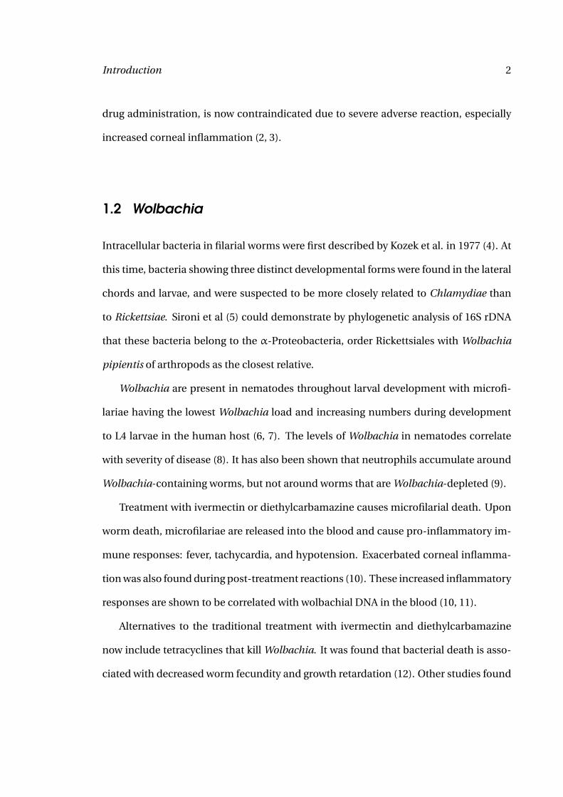
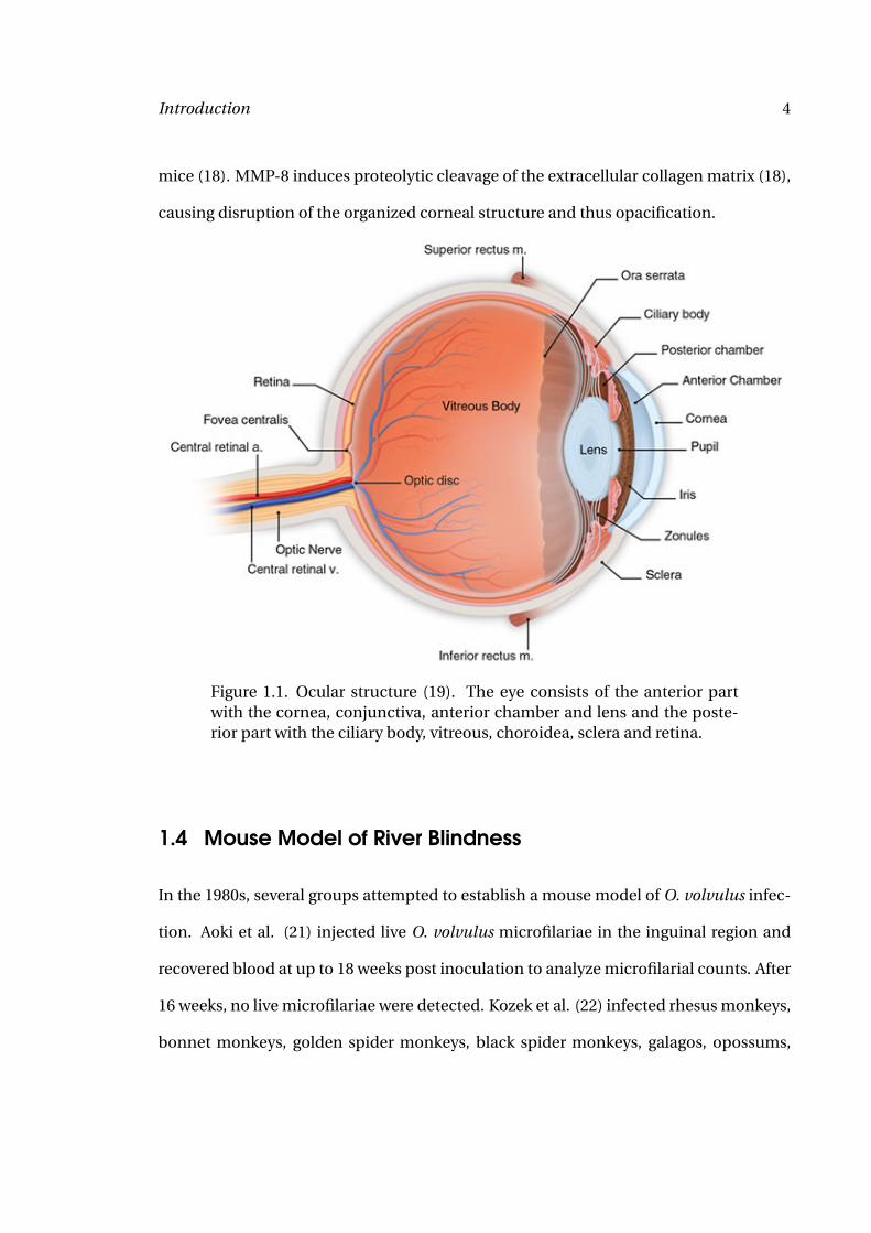
The cornea is the transparent outer part of the eye (Figure 1.1). Together with the lens,
it forms the optical apparatus. The cornea consists of three layers: the inner single layer
endothelium, the 450 µm thick corneal stroma and the outer multilayer epithelium (Fig-
ure 1.2). Corneal transparency is essential for intact vision and depends on the orga-
nized structure of the corneal matrix, mainly made up of collagen fibrils. Disruption
of the collagen fibril structure results in opacification of the cornea, visual impairment
and eventual blindness. Infection or inflammation of the cornea usually leads to the in-
filtration of inflammatory cells (15). During O. volvulus infection, eosinophils and neu-
trophils migrate to the corneal stroma. The granulocytes release cytokines and granular
components, disrupting the corneal structure and leading to visual impairment.
Lipopolysaccharide-induced corneal inflammation has been studied in more detail.
Neutrophils extravasate from limbal vessels and migrate through the corneal stroma,
depending on the presence of TLR4 and PECAM-1 (16). Migration of neutrophils is de-
pending on the presence of the neutrophil chemokines KC, MIP-2 and LIX with corneal
fibroblasts as the most probable source for KC and LIX while resident macrophages are
producing MIP-2 (17). Activated neutrophils in the cornea release additional MIP-2 (17).
Neutrophils also express MMP-8 to facilitate migration through the corneal stroma as
there is no MMP-8 expression in CXCR2-/- corneas where neutrophils are absent fol-
lowing LPS challenge and neutrophil migration to the cornea is decreased in MMP-8-/-
Introduction 4
mice (18). MMP-8 induces proteolytic cleavage of the extracellular collagen matrix (18),
causing disruption of the organized corneal structure and thus opacification.
Figure 1.1. Ocular structure (19). The eye consists of the anterior partwith the cornea, conjunctiva, anterior chamber and lens and the poste-rior part with the ciliary body, vitreous, choroidea, sclera and retina.
1.4 Mouse Model of River Blindness
In the 1980s, several groups attempted to establish a mouse model of O. volvulus infec-
tion. Aoki et al. (21) injected live O. volvulus microfilariae in the inguinal region and
recovered blood at up to 18 weeks post inoculation to analyze microfilarial counts. After
16 weeks, no live microfilariae were detected. Kozek et al. (22) infected rhesus monkeys,
bonnet monkeys, golden spider monkeys, black spider monkeys, galagos, opossums,
Introduction 5
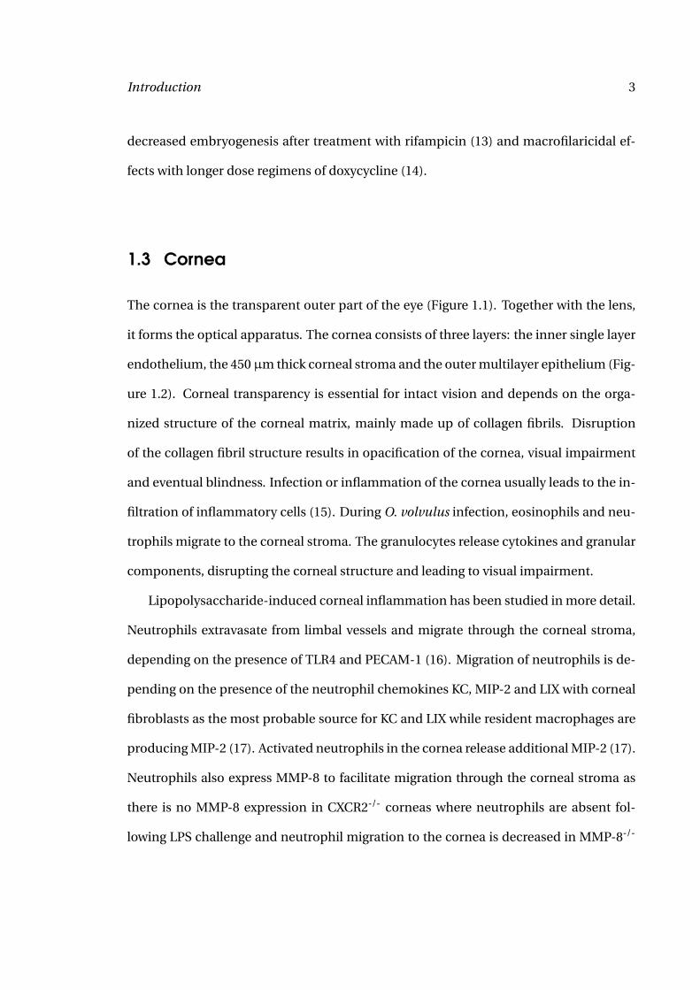
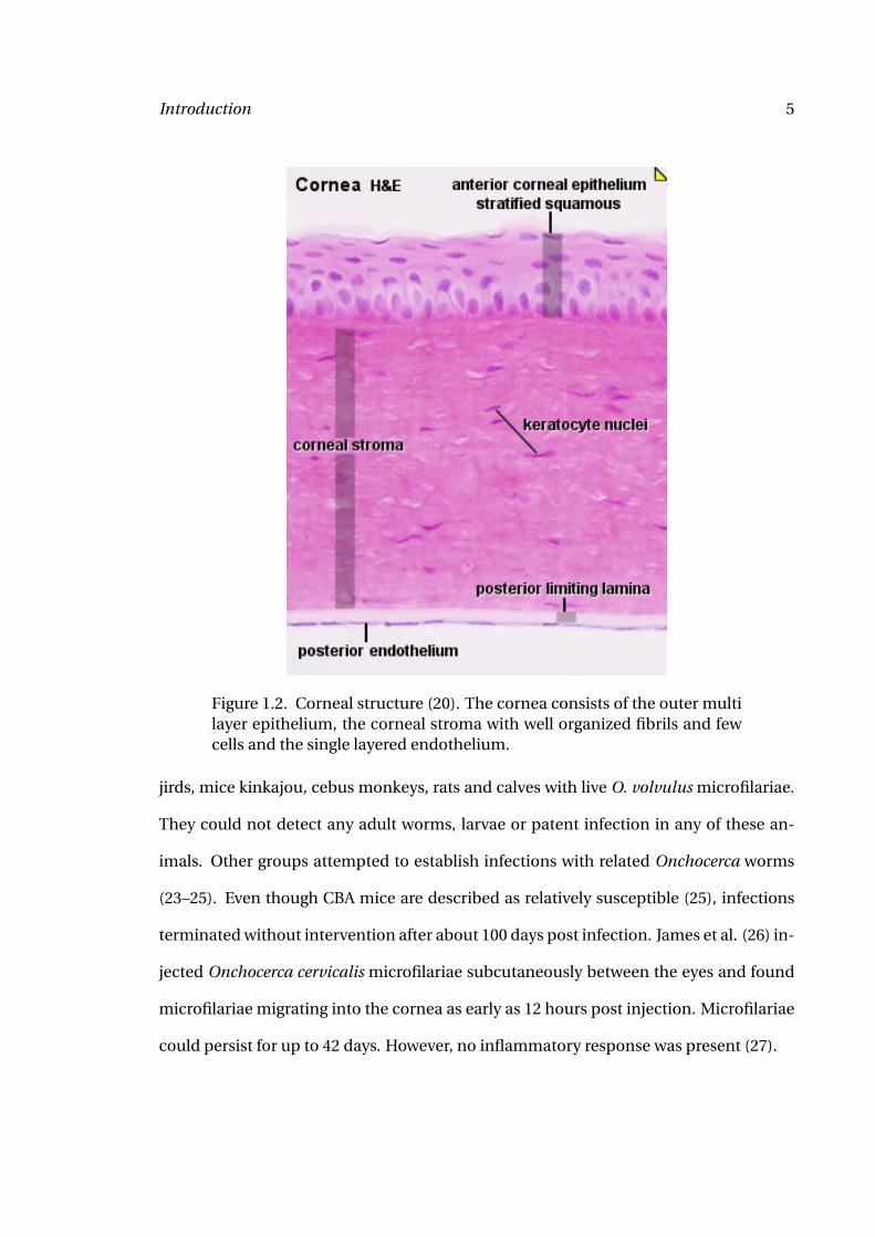
Figure 1.2. Corneal structure (20). The cornea consists of the outer multilayer epithelium, the corneal stroma with well organized fibrils and fewcells and the single layered endothelium.
jirds, mice kinkajou, cebus monkeys, rats and calves with live O. volvulus microfilariae.
They could not detect any adult worms, larvae or patent infection in any of these an-
imals. Other groups attempted to establish infections with related Onchocerca worms
(23–25). Even though CBA mice are described as relatively susceptible (25), infections
terminated without intervention after about 100 days post infection. James et al. (26) in-
jected Onchocerca cervicalis microfilariae subcutaneously between the eyes and found
microfilariae migrating into the cornea as early as 12 hours post injection. Microfilariae
could persist for up to 42 days. However, no inflammatory response was present (27).
Introduction 6
Therefore an artificial mouse model of onchocercal keratitis has been developed.
Mice immunized with soluble O. volvulus extract (OvAg) in either complete Freund’s
(CFA) or squalene based adjuvant (STP) develop corneal inflammation. The differ-
ent adjuvants cause differences in cellular corneal infiltrates: CFA-immunized mice
show strong neutrophil infiltration whereas in STP-immunized mice eosinophils are
predominant (27). Recent studies mainly used STP as adjuvant. In current studies,
mice are immunized with OvAg-STP three times in weekly intervals, inducing adap-
tive immune responses. One week after the third immunization, corneas are injected
with OvAg and eyes are excised at various time points. Corneal changes include edema,
neovascularization, disruption of the corneal matrix (collagen fibrils) and infiltration of
neutrophils and eosinophils (28). Whereas neutrophil migration to the corneal stroma
can also be induced by the injection of OvAg into unimmunized mice (29), the migration
of eosinophils depends on the presence of adaptive immunity (30).
1.4.1 Immune responses in the mouse model of river blindness
Splenocytes produce increased levels of IL-4, IL-5 and IFN-γ during in vitro stimula-
tion with filarial extract when previously immunized with filarial extracts (Brugia malayi
adult or whole microfilariae, O. volvulus extract) (31). Lymph nodes and splenocytes
from OvAg immunized mice showed higher levels of IL-4 and IL-5 production upon re-
stimulation in vitro compared with PPD (Mycobacterium tuberculosis purified protein
derivative) immunized mice, indicating a preference for Th2 responses (28). This cy-
tokine profile was found to depend on the presence of CD4+ T cells and could be trans-
ferred by the adoptive transfer of splenocytes from immunized mice (28).
Introduction 7
The preference for Th2-responses can be skewed by the injection of rIL-12 during
the immunization. In this case, IFN-γ production and IgG1 levels in the serum are in-
creased compared with normally immunized mice (32). The shift towards Th1 responses
is associated with decreased eosinophil infiltration into the lung and expression of ma-
jor basic protein (32). However, although eosinophil migration to the lung is decreased
in IL-12 injected mice, eosinophil infiltration into the corneal stroma is increased after
IL-12 injection (33).
Following injection with filarial extracts, neutrophils and eosinophils migrate to the
corneal stroma. At seven days post injection, infiltration of inflammatory cells is re-
solved in unimmunized mice, and the corneas appear normal (28). The infiltration was
found to be specific for filarial injection as compared with injections of Mycobacterium
tuberculosis purified protein derivative (PPD) (28).
Even though eosinophils are usually associated with parasite infections and were
found to infiltrate the corneas, studies using IL-5-/- could demonstrate that while
eosinophils are absent from the corneas of these mice, the disease manifestations
(corneal opacification and neovascularization) are identical to that of wild-type mice
(32). This was found to be due to neutrophil infiltration at earlier time points. Neutro-
phil as well as eosinophil infiltration was dependent on the presence of B cells, anti-
bodies (34) and Fcγ (35). Splenocytes from B cell deficient µMT mice also showed de-
creased IL-5 production after OvAg stimulation. However, decreased levels of IL-5 did
not correspond to diminished blood eosinophilia (34). Migration of eosinophils to the
cornea is depending on the presence of CD4+ T cells (36), P-selectin (37) and ICAM-1
(38), while neutrophil migration is decreased in PECAM-1 depleted mouse corneas (38)
Introduction 8
and CXCR2-/- mice (39). ICAM-1 expression is regulated by IL-4 and IL-13 (40). Using IL-
4-/- mice, it was determined that corneal opacification and neovascularization is depen-
dent on the presence of functional IL-4 (28). IL-4-/- mice also showed reduced number
of eosinophils in the corneal stroma (30).
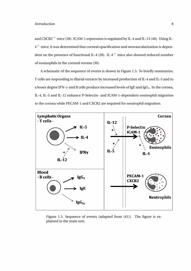
A schematic of the sequence of events is shown in Figure 1.3. To briefly summarize,
T cells are responding to filarial extracts by increased production of IL-4 and IL-5 and to
a lesser degree IFN-γ and B cells produce increased levels of IgE and IgG1. In the cornea,
IL-4, IL-5 and IL-12 enhance P-Selectin- and ICAM-1-dependent eosinophil migration
to the cornea while PECAM-1 and CXCR2 are required for neutrophil migration.
Figure 1.3. Sequence of events (adapted from (41)). The figure is ex-plained in the main text.
Introduction 9
1.5 Antigen-presenting cells
Traditionally, only dendritic cells, macrophages and B cells were considered antigen-
presenting cells. However, lately it has been shown that neutrophils and eosinophils
are capable of presenting antigen to T cells in a contact-dependent manner (42–46). In
this section, I will give background information on the cell types relevant for this study:
dendritic cells, macrophages and neutrophils.
1.5.1 Dendritic cells
Dendritic cells are the most efficient of the antigen-presenting cells. Upon recogni-
tion of microbial products, dendritic cells begin maturation. This is characterized by
the upregulation of MHCII and the co-stimulatory molecules CD40, CD80 and CD86
as well as the production of cytokines and chemokines (47). Antigen presented on
MHC molecules provides the first specific signal of T cell activation. The co-stimulatory
molecules provide the second activatory signal to T cells, thus making dendritic cells ef-
ficient antigen-presenting cells (48). A third signal is provided by the cytokines secreted
by activated dendritic cells (49).
Depending on the nature of the antigen (bacterial, viral, helminth origin), dendritic
cells are primed towards a DC1 cells or DC2 cells type cell. DC1 induce T cells via IL-12
and IL-23 to differentiate towards Th1 (50, 51). This is mainly found during bacterial
and viral infection. During helminth infection, DC2 cells dominate, producing IL-10
and TGF-β (51, 52) and inducing a Th2 and regulatory T cell response (50, 53).
Introduction 10
Typical Th1 responses include the production of IFN-γ and IL-2, activation of
macrophages, IgG2 antibody production. Neutrophils are also associated with Th1 im-
mune responses (54). Th2 responses are characterized by the production of the cy-
tokines IL-4, IL-5 and IL-10 as well as a predominant eosinophil response (55). Inter-
estingly, during filarial infection, we find mixed Th1 and Th2 responses. This is thought
to be due to the Wolbachia endosymbionts inducing Th1 type responses with the worm
itself inducing Th2 type responses.
1.5.2 Macrophages
Macrophages are important in the innate immune responses as well as for antigen-
presentation and induction of adaptive immune responses. Precursor cells are gener-
ated in the bone marrow entering the blood stream as monocytes. Following circulation
for about 18 hours, monocytes migrate into tissues and differentiate into macrophages
(56).
Activation of macrophages is tightly regulated by cytokines. Exposure to Th1 cy-
tokines IFN-γ, Tumor-necrosis factor (TNF) or lipopolysaccharide induces the classi-
cal type 1 activation of macrophages. This pathway is usually activated by intracellular
pathogens (56) and is characterized by activation of the interferon regulatory factors
(IRF). In response, pro-inflammatory cytokines are produced.
If macrophages are instead activated via the alternative pathway, including expo-
sure to Th2 cytokines IL-4 and IL-13, type 2 activation occurs and requires mast cell and
eosinophil involvement, and granuloma formation. This is usually found in helminth
infections or in response to extracellular pathogens (56). Production of macrophage-
derived chemokine (MDC) and thymus and activation-regulated chemokine (TARC) is
Introduction 11
promoted, while production of reactive oxygen species and pro-inflammatory cytokines
is inhibited (56).
Priming of macrophages ensures survival of the cells and prepares the cells for expo-
sure to a secondary signal. Primed macrophages respond to lower doses of antigen, but
are not yet activated. Classically, IFN-γ primes macrophages (56).
1.5.3 Neutrophils
For a long time, neutrophils were only regarded as responder cells in the innate im-
mune system. Recent publications have demonstrated a role for neutrophils in antigen-
presentation (43), including the expression of MHCII and co-stimulatory molecules on
neutrophils (42).
Neutrophils contain secretory vesicles, gelatinase (tertiary) granules, specific (sec-
ondary) granules and azurophil (primary) granules. Each granule type shows a specific
content: secretory vesicles contain only albumin and heparin binding protein, gelati-
nase granules contain gelatinase (matrix metalloproteinase 9), specific granules con-
tain CD66b in the granule wall and azurophil granules are characterized by myeloper-
oxidase content (57). Upon stimulation, granule proteins are released and take part in
neutrophil extravasation and migration (gelatinase granules) and antimicrobial activity
(specific granules, azurophil granules) (58).
Myeloperoxidase (MPO) is found in neutrophils where it is stored in azurophil gran-
ules. It has a central role in the regulation of inflammation. MPO catalyzes the pro-
duction of hypochlorous acid and serves as a marker for respiratory burst (57, 59). If
released by the cell, it contributes to the pathogenesis of the disease (59).
Introduction 12
Previous studies demonstrated down-regulation of L-Selectin on the neutrophil sur-
face in response to activation (60). L-Selectin is an adhesion molecule found on leuko-
cytes and functions as homing receptor for leukocytes that are extravasating in lym-
phoid tissues through the high endothelial venules. L-Selectin expression increases
rolling of neutrophils along the endothelium and thus helps extravasation. In addition
to adhesion to endothelial cells, L-Selectin also functions in neutrophil activation. Upon
L-Selectin activation, neutrophils become less deformable (60, 61). L-Selectin is usually
shed by neutrophils in response to stimulation and activation. It is hypothesized that the
now soluble L-Selectin can bind to the L-Selectin receptor and prevent neutrophil bind-
ing at subacute sites of inflammation (60). Increased serum levels of L-Selectin have
been associated with disease, e.g. AIDS (60).
CD18 is found colocalized with L-Selectin (61) but is upregulated upon stimulation.
CD11b often forms complexes with CD18. These surface molecules affect aggregation
and adhesive properties of neutrophils.
Matrix metalloproteinases (MMPs) are Ca2+-dependent Zn-endopeptidases that de-
grade the extracellular matrix (62). MMPs are regulated by hormones and cytokines
among other factors (63). They are produced as pro-form and stored inside granules
(57). Upon activation, they are cleaved and excreted (63). MMP-9 (gelatinase B) and
MMP-2 (gelatinase A) are both gelatinases important in corneal remodeling (64). They
contain a fibronectin-like domain which binds the gelatin (65). MMP-2 is not expressed
by neutrophils, but on keratinocytes (64, 65). MMP-8 (collagenase-2) is expressed by
neutrophils, corneal epithelial cells and keratinocytes (among others) (62). In contrast
to MMP-2 and MMP-9, it digests a variety of substrates: collagen, chemokines, protease
Introduction 13
inhibitors (62). It has been described as a regulator of neutrophil infiltration in LPS-
induced keratitis (18).
Neutrophils respond to chemotactic gradients of keratinocyte-derived chemokine
(KC, CXCL1), monocyte-inhibitory protein-2 (MIP-2, CXCL2) and LPS-induced
chemokine (LIX, CXCL5). Neutrophils only contribute to MIP-2 production, whereas
KC and MIP-2 are produced by macrophages and KC and LIX by corneal fibroblasts (17).
1.6 Toll-like Receptors
Pattern recognition receptors (PRRs) recognize pattern associated molecular patterns
(PAMPs) in microbes. PAMPs are highly conserved among different microbes, absent
from eukaryotes and essential for the survival of the microbe. The family of PRRs con-
sists of Toll-like receptors (TLRs), Dectin-1 (a C-type lectin) and Nod-like receptors
(NLRs). Other less studied receptors include NALP3, ISD sensor and RIG-I/MDA5 (66).
TLRs are germ-line encoded receptors recognizing a great variety of microbial
products. The receptors were first characterized in Drosophila. The family currently
consists of at least 10 TLRs in humans (67) and 13 TLRs in mice (68, 69). Some studies
refer to 11 human TLRs (70), however the original article identifying the 11th human TLR
could not be identified. The receptors recognize conserved microbial PAMPs, although
chemical agonists (e.g. imiquomid) have also been described (71, 72). Host proteins
like heat-shock proteins were shown to bind TLR2 and TLR4 (73–75). The receptors
contain an extracellular leucin-rich repeat domain and an intracellular domain. The
intracellular domain is homologous to the IL-1 receptor and therefore referred to as the
Toll/IL-1R (TIR) domain (76). Signaling through TLRs recruits the adaptor molecules
Introduction 14
myeloid differentiation primary response gene 88 (MyD88) or TIR-domain-containing
adapter-inducing interferon-β (TRIF). MyD88 is involved in the signaling of every TLR
except TLR3 while only TLR3 and (partly) TLR4 signal through TRIF. MyD88 is recruited
by Mal, then signals through IRAK and NFκB activation to induce production of pro-
inflammatory cytokines.
TLR2, TLR4 and TLR9 recognize bacterial products. TLR2 specifically interacts with
acylated lipoproteins, TLR4 with lipopolysaccharides and TLR9 with methylated DNA.
TLR3, TLR7 and TLR9 are specific receptors for viral products, TLR5 interacts with flag-
ellin. TLR2 is the most promiscuous receptor. Depending on coreceptor usage (TLR1,
TLR6 and TLR10) different bacterial products are recognized. TLR1 and TLR2 together
specifically interact with triacylated lipoprotein, TLR2 and TLR6 interact with diacylated
lipoproteins, ligands for TLR2/TLR10 are not yet described.
TLR2, TLR4 and TLR11 have been implicated in the activation of immune cells by
Leishmania major, Trypanosoma cruzi and Toxoplasma gondii (77–79). In contrast to
bacterial or viral activation of TLRs, activation via TLRs by eukaryotic parasites pre-
vented immunopathology (77–79).
TLRs are not only important during the generation of innate immune responses, but
cells activated via TLR can also induce adaptive immune responses. TLRs have been
shown to increase the antigen uptake by dendritic cells (80), induce maturation of and
cytokine production by dendritic cells thus increasing the capability to present antigen.
B cell proliferation also increases after stimulation with TLR ligands either directly or
indirectly via activation of dendritic cells (81). Activation of dendritic cells via Pam3CSK
or LPS induced T cell proliferation and differentiation to Th1 and Th2 cells, respectively
(82).
Introduction 15
1.6.1 Toll-like Receptors in Filarial Infection
Studies examining the role of TLRs in filarial infection focused on TLR2, TLR4 and TLR9.
It was reasoned that the most likely agonist for these receptors was not the worm itself
but its endosymbiont Wolbachia. It was therefore thought that bacteria specific TLRs
would be used. In a mouse model of O. volvulus keratitis, involvement of Wolbachia
and MyD88 in corneal inflammation was demonstrated (29). Further investigation iden-
tified TLR2 as the receptor activated by Wolbachia. No role for TLR4 or TLR9 could be
identified (83), although early studies showed TLR4-dependent neutrophil migration to
the corneal stroma (84) and TLR4-dependent activation of dendritic cells by Wolbachia
surface protein (WSP) (85). It was demonstrated that TLR2 required TLR6 as corecep-
tor in neutrophil and macrophage migration to the cornea in response to O. volvulus,
Wolbachia and recombinant Wolbachia lipoprotein (86, 87).
Other groups showed filaria-dependent expression of TLRs. Expression of TLRs was
down-regulated in filaria-infected individuals corresponding to decreased cytokine pro-
duction after stimulation with TLR ligands (88, 89). Repeated exposure to filarial ex-
tracts also decreased macrophage responsiveness to B. malayi and Wolbachia in a TLR2-
dependent manner (90). Finally, secreted products of the nematode Acanthocheilonema
viteae are able to activate dendritic cells in a TLR4-dependent manner (91). Activation of
macrophages and dendritic cells via this TLR4 and MyD88-dependent pathway results
in decreased responsiveness of these cells to TLR2 and TLR9 agonists (91).
Introduction 16
1.7 Interferon-γ
Interferonγ (IFN-γ) is the classical Th1 cytokine. It is produced only at low levels during
filarial infection. It has been described in human biliary epithelial cells and in human
monocytes from human rheumatoid arthritis patients that TLR2 is upregulated in re-
sponse to stimulation with IFN-γ (92, 93). Harada et al. (93) showed upregulation of
TLR1-5 by measuring mRNA levels and confirming synergistic effects of IFN-γ and LPS
and peptidoglycan stimulation. Similar results were recently shown for murine CD11b+
cells where TLR2 expression was increased after IFN-γ stimulation (94).
IFN-γ also affects differentiation of monocytes. It was shown that human precur-
sor cells developed into macrophages rather than dendritic cells if IFN-γ was supplied
in addition to the normal growth factors granulocyte-macrophage colony stimulating
factor (GM-CSF) and IL-4 used for dendritic cell differentiation. This effect could only
be observed in the early differentiation stages and was dependent on the production of
macrophage colony stimulating factor (M-CSF) and IL-6 (95).
In addition, as has been described in the section on macrophages, IFN-γ activates
macrophages via the classical pathway rather than the alternative pathway that is in-
duced by IL-4 and IL-13.
IFN-γ has been shown to regulate expression of platelet endothelial cell adhesion
molecule 1 (PECAM-1) in a mouse model of Herpes simplex keratitis. Blockade of IFN-γ
reduced neutrophil infiltration into the cornea and expression of PECAM-1 on corneal
vessels (96). It has been shown by other groups that PECAM-1 expression enhances
directional neutrophil migration (97). Our group previously demonstrated a role for
PECAM-1 in neutrophil migration during O. volvulus keratitis (38).

Introduction 17
Additionally, IFN-γ induces the production of neutrophil chemoattractant CXC
chemokines in Staphylococcus aureus infected tissues (98).
1.8 Aims
Previous studies described Pam3CSK and LPS as differential inducers of T cell responses
via TLR2 and TLR4, respectively (82). It was also shown that TLR2 and TLR4 are impli-
cated in the recognition of filarial products (84, 85). I therefore reasoned, that filarial
products (in addition to Wolbachia surface protein) might activate TLR2 and TLR4 and
this might lead to a skewing of the adaptive immune resonses.
In this study, I first wanted to characterize TLR-dependent activation of dendritic
cells by O. volvulus. After I demonstrated a role for TLR2 in the activation of dendritic
cells, I examined the role of TLRs in the adaptive arm of the immune responses in a
mouse model of river blindness. The aim was to show that the initial requirement for
TLR2 in the innate immune response was reflected by altered adaptive immune re-
sponses. I was interested in the generation of systemic immune responses (T cell ac-
tivation, serum antibody production) and in the local migration of antigen-presenting
cells to the cornea.
With the surprising result of TLR2 being required only for Th1-like responses IFN-γ
production and neutrophil migration, the next goal was to identify a mechanism, linking
systemic IFN-γ production to neutrophil infiltration into the cornea.
18
2 Materials and Methods
2.1 Reagents
2.1.1 OvAg
During previous studies (Eric Pearlman, personal communication), Onchocercomata
were excised from patients from the Ivory Coast. Nodules were stored frozen at -80◦C
until further processing. Then, nodules were thawed overnight at 4◦C in digestion solu-
tion (HBSS/1 % Penicillin+Streptomycin/50 µg/ml Gentamicin) and washed with sterile
ice cold HBSS. 1 % collagenase, 0.5 % elastase and 0,5 % trypsin were added to the diges-
tion solution (enzyme solution). Nodules were incubated in enzyme solution for 8 hours
at 37◦C until worms were released from the nodules. Worms were collected in a sterile
tube and transferred onto 40 µM filter. Worms were then washed six times with sterile
digestion solution, followed by four washes with sterile HBSS. Worms were processed in
1 ml HBSS in glass mortars for 30 minutes. Protein extracts were sonicated three times
for 5 minutes, then centrifuged for 10 minutes at 1000 xg. Soluble extract was used for in
vitro stimulation and in vivo intrastromal injection. Unsoluble proteins were dissolved
in 1 ml HBSS and used for immunizations. Protein concentrations in both extracts were
Materials and Methods 19
determined by BCA protein assay (Pierce). WSP and filarial 5S were quantified by quan-
titative PCR, 40 cycles of 30 seconds extension at 95◦CC, 30 seconds annealing at 60◦C
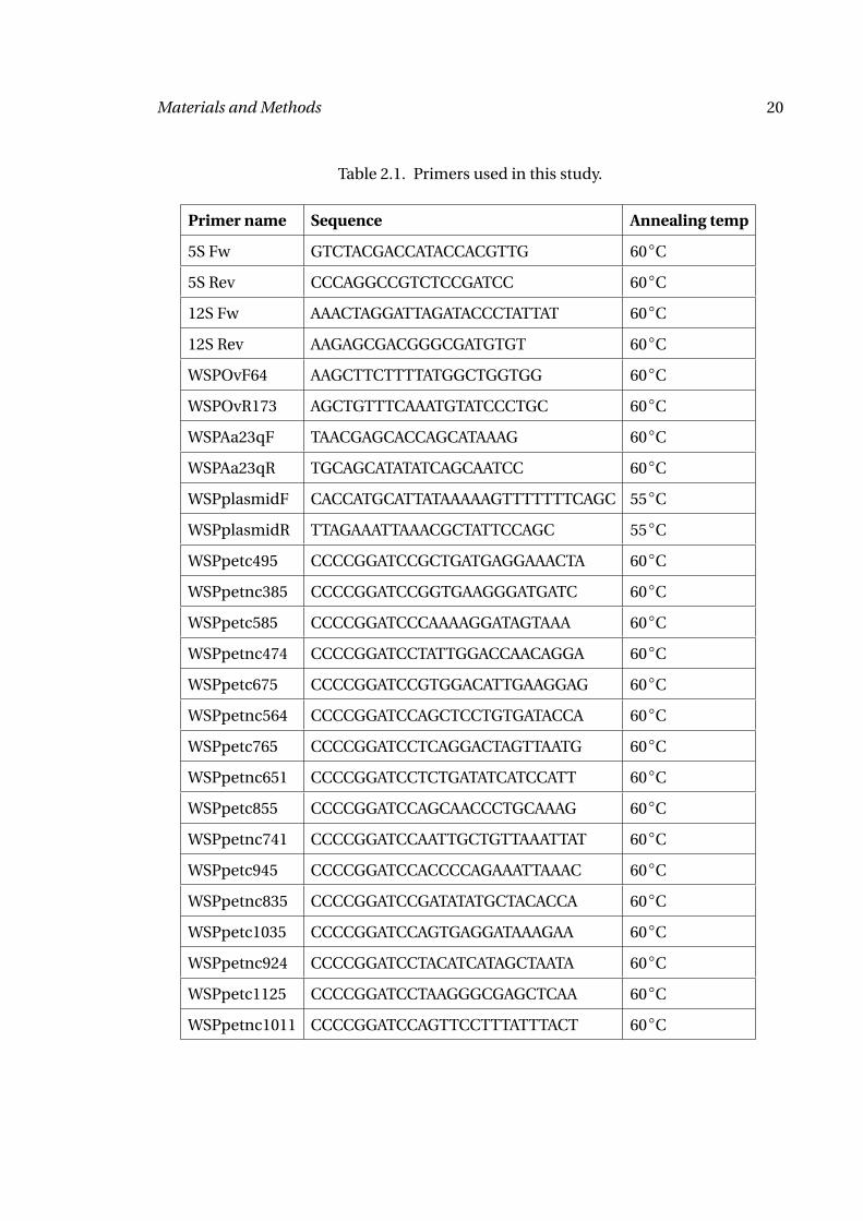
and 30 seconds elongation at 72◦C. Primers used in this study are shown in table 2.1.
2.1.2 Wolbachia
Wolbachia infected SF9 insect cells were kindly provided by Dr. Mark Taylor (Liver-
pool School of Tropical Medicine, Liverpool, Great Britain). Cells were maintained
under sterile conditions at 27◦C, no CO2. Insect cell medium contained 50 % Mit-
suhashi Maramorosh Medium, 50 % Schneider’s Insect Medium, 10 % FBS and 1 % Peni-
cillin+Streptomycin. Wolbachia infected SF9 cells were harvested, centrifuged at 2000 xg
for 10 minutes and the cells were disrupted by sonication for 2 minutes. Cell debris was
then pelleted at 600 xg for 5 minutes. Supernatants were transferred into a clean tube
and pelleted again. This was repeated four times to remove cellular debris. Wolbachia
were then pelleted at 16000 xg for 10 minutes. For controls, tetracycline treated SF9 cells
were processed identically.
2.1.3 Wolbachia Surface Protein
We obtained Wolbachia Surface Protein (WSP) cloned into the pET100 TOPO vector
from Alan Scott (Johns Hopkins University, Baltimore, MD). The gene was cloned with-
out the signal sequence (first 30 amino acids). Deletion mutants were generated by re-
verse PCR. Briefly, primers (table 2.1) were designed at both ends of the sequence to
be deleted. PCR reactions were run amplifying the full plasmid minus the deleted se-
quence. PCR amplification consisted of 2 minutes at 95◦C, followed by 10 cycles of
Materials and Methods 20
Table 2.1. Primers used in this study.
Primer name Sequence Annealing temp
5S Fw GTCTACGACCATACCACGTTG 60◦C
5S Rev CCCAGGCCGTCTCCGATCC 60◦C
12S Fw AAACTAGGATTAGATACCCTATTAT 60◦C
12S Rev AAGAGCGACGGGCGATGTGT 60◦C
WSPOvF64 AAGCTTCTTTTATGGCTGGTGG 60◦C
WSPOvR173 AGCTGTTTCAAATGTATCCCTGC 60◦C
WSPAa23qF TAACGAGCACCAGCATAAAG 60◦C
WSPAa23qR TGCAGCATATATCAGCAATCC 60◦C
WSPplasmidF CACCATGCATTATAAAAAGTTTTTTTCAGC 55◦C
WSPplasmidR TTAGAAATTAAACGCTATTCCAGC 55◦C
WSPpetc495 CCCCGGATCCGCTGATGAGGAAACTA 60◦C
WSPpetnc385 CCCCGGATCCGGTGAAGGGATGATC 60◦C
WSPpetc585 CCCCGGATCCCAAAAGGATAGTAAA 60◦C
WSPpetnc474 CCCCGGATCCTATTGGACCAACAGGA 60◦C
WSPpetc675 CCCCGGATCCGTGGACATTGAAGGAG 60◦C
WSPpetnc564 CCCCGGATCCAGCTCCTGTGATACCA 60◦C
WSPpetc765 CCCCGGATCCTCAGGACTAGTTAATG 60◦C
WSPpetnc651 CCCCGGATCCTCTGATATCATCCATT 60◦C
WSPpetc855 CCCCGGATCCAGCAACCCTGCAAAG 60◦C
WSPpetnc741 CCCCGGATCCAATTGCTGTTAAATTAT 60◦C
WSPpetc945 CCCCGGATCCACCCCAGAAATTAAAC 60◦C
WSPpetnc835 CCCCGGATCCGATATATGCTACACCA 60◦C
WSPpetc1035 CCCCGGATCCAGTGAGGATAAAGAA 60◦C
WSPpetnc924 CCCCGGATCCTACATCATAGCTAATA 60◦C
WSPpetc1125 CCCCGGATCCTAAGGGCGAGCTCAA 60◦C
WSPpetnc1011 CCCCGGATCCAGTTCCTTTATTTACT 60◦C
Materials and Methods 21
1 minute at 95◦C, 1 minute at 60◦C and 7 minutes at 72◦C, then 30 cycles of 30 sec-
onds at 95◦C, 1 minute at 60◦C and 7 minutes at 72◦C, followed by final extension at
72◦C for 10 minutes. Primers contained a BamH1 site to allow religation after digestion.
Circular plasmids were then transformed into competent BL21 cells.
WSP and WSP deletion mutants were purified using the Qiagen NiNTA fast start kit
with the native purification protocol. Cells were grown for 3 hours at 37◦C on shaking
incubator. Cells were then induced with 1 mM IPTG for 5 hours. Cells were harvested at
4000 xg for 20 minutes and frozen overnight at -20◦C. Cells were then thawed and lysed
in Native Lysis Buffer containing 1 µg/ml lysozyme and 25 U/ml benzonase nuclease for
30 minutes on ice. The lysate was then centrifuged at 14,000 xg for 30 minutes at 4◦C.
The supernatant was applied to NiNTA Fast Start Column. The column was then washed
twice in Native Wash Buffer and the protein was eluted in 1 ml Native Elution Buffer.
Purity was determined by Western Blot and Coomassie stain and protein concentration
was determined by BCA protein assay (Pierce).
2.1.4 Control reagents
To determine activation of dendritic cells, macrophages, neutrophils, MK/T-1 cells and
HEK cells depending on the TLR profile these cells expressed, I used synthetic TLR lig-
ands. For TLR2 activation, I used Peptidoglycan (Invivogen), Pam3CSK (Invivogen) and
FSL-1 (Invivogen), for TLR3 activation, Poly(I:C) (Invivogen) was used, for TLR4 acti-
vation ultrapure E. coli LPS (Invivogen) and for TLR9 activation CpG-DNA (Invivogen).
Positive control for T cell activation was anti-CD3 (kind gift by Dr. Thomas Forsthuber,
now at the University of Texas at San Antonio, TX).
Materials and Methods 22
2.1.5 Other reagents
STP adjuvant: 10 % squalene (Sigma, St. Louis, MO, USA), 0.4 % Polyoxyethylenesorbi-
tanmonooleate (Tween 80) (Sigma, St. Louis, MO, USA), 1 % Pluronic L (BASF, Germany)
in PBS
Recombinant cytokines: IFN-γ, IL-1α, IL-1β, IL-6, TNFα, GM-CSF (Peprotech,
Rocky Hill, NJ, USA)
2.2 Mice
C57BL/6 mice were bred at the Wolstein Animal Resource Center at Case Western Re-
serve University (Cleveland, OH) or purchased from Jackson Laboratories (Bar Harbor,
ME). TLR2-/-, TLR4-/-, TLR2/4-/-, MyD88+/+ and MyD88-/- mice were bred at the Wol-
stein Animal Resource Center. IFN-γ-/- mice were a gift by Dr. Tom McCormick (Case
Western Reserve University, Cleveland, OH), IL-1R-/- mice were kindly provided by Dr.
Susan Mohr (Case Western Reserve University, Cleveland, OH) and TNFαR-/- mice were
kindly provided by Dr. Claire M. Doerschuk (Case Western Reserve University, Cleve-
land, OH). All animal protocols were approved by the IACUC office at Case Western Re-
serve University.
2.3 In vivo studies
2.3.1 Innate immune responses
Mice were anaesthetized with tribromoethanol (TBE). Corneas of mice were rinsed, then
carefully scratched with a 27 gauge needle. A 33 gauge needle (Hamilton Company,
Materials and Methods 23
Switzerland) was inserted into the cornea through the scratch in the corneal epithelium
and 4 µl of experimental and control reagents were injected. Eyes were taped closed with
loosely adherent tape that mice could remove themselves upon awakening. 24 hours
post injection, mice were euthanized using CO2 and corneal haze was examined by con-
focal microscopy. Whole eyes were removed for sectioning and immunohistochemistry,
the corneas were excised carefully with a 2 mm trephine for FACS analysis of the cellular
infiltrate or ELISA analysis of chemokines present in the cornea.
2.3.2 Adaptive immune responses
Mice were immunized subcutaneously three times in weekly intervals with 10 µg
insoluble OvAg extract in a 1:1 dilution in STP adjuvant. One week past the final im-
munization, mice were immunized and injected into the corneal stroma as described
under innate immune responses. Mice were then euthanized at 24 or 72 hours post in-
trastromal injection and eyes removed for sectioning and immunohistochemistry. In
some experiments, corneas were excised for ELISA analysis of chemokines.
2.4 Cell culture
2.4.1 Dendritic cells
C57BL/6, TLR2-/-, TLR4-/-, TLR2/4-/-, MyD88+/+ and MyD88-/- mice were euthanized.
Femurs and tibias were dissected aseptically. Bones were cleaned under sterile condi-
tions, then opened at the distal and proximal ends. For harvesting bone marrow cells,
the opened bones were centrifuged for 30 seconds at 6000 xg in a sterile 0.5 ml tube with
a hole in the bottom, set into a sterile 1.5 ml tube. Cells were resuspended in HBSS.
Materials and Methods 24
Red blood cells were lysed with RBC lysis buffer (eBioscience, San Diego, CA, USA). Cell
numbers and viability were determined under trypan blue exclusion. Viability was gen-
erally greater than 90 %. Cells were plated at a concentration of 1x10⁶ per ml in six-well
plates. DC medium contained RPMI (BioWhittaker), 10 % fetal bovine serum (Cellgro,
Herndon, VA, USA), 1 % sodium pyruvate (BioWhittaker), 1 % HEPES (BioWhittaker),
1 % L-glutamine (BioWhittaker), 1 % 50/50 penicillin/streptomycin (BioWhittaker) and
1 mM 2-mercaptoethanol (Sigma, St. Louis, MO, USA). Medium was supplemented with
GM-CSF (Peprotech, Rocky Hill, NJ, USA) at a final concentration of 20 ng/ml. Fresh
medium was added on days 3 and 5. Cells were harvested on day 7 by carefully removing
non-adherent cells (dead cells and mature dendritic cells) and then dislodging loosely
adherent cells with HBSS (immature dendritic cells). Viability was greater than 90 %.
Dendritic cells were purified with CD11c magnetic beads according to the manu-
facturer’s instructions (MACS, Miltenyi Biotech, Auburn, CA, USA). Dendritic cells were
enriched to about 90-95 %.
1x10⁶ cells were plated in 96-well plates and stimulated for 24 hours with the fol-
lowing antigens: OvAg at different concentrations, 50 µg/ml Pam3CSK (InvivoGen, San
Diego, CA, USA), 100 ng/ml lipopolysaccharide (LPS) (Invivogen, San Diego, CA, USA)
or 20 µg/ml CpG-DNA (Invivogen, San Diego, CA, USA). Supernatants were analyzed for
cytokine production by ELISA and cells were examined by flow cytometry.
2.4.2 Macrophages
C57BL/6 and TLR2-/- mice were injected intraperitoneally with 4 % thioglycollate
(Remel, Lenexa, KS, USA) at 4-5 days prior to euthanization. Mice were euthanized by
CO2. The peritoneum was lavaged with 10 ml HBSS. Red blood cells were lysed with
Materials and Methods 25
RBC lysis buffer (eBioscience, San Diego, CA, USA) and cells were plated at 1x10⁵ per
well. After overnight rest, cells were stimulated with medium, OvAg, Pam3CSK, IFN-γ
and OvAg + IFN-γ.
2.4.3 Neutrophils
9 % Casein solution was prepared in 0.9 mM CaCl2 and 0.5 mM MgCl. 1 ml Casein
solution was injected at 18 hrs and 3 hours prior to euthanizing the mice. Cells were
harvested from the peritoneal cavity, washed with HBSS and purified on 90 % Percoll
gradient at 32000 xg for 20 minutes. Neutrophil fraction was harvested and washed to
remove residual Percoll. 1x10⁵ cells were stimulated with medium, OvAg and Pam3CSK.
2.4.4 MK/T-1 cells
The corneal fibroblast cell line MK/T-1 (99) was maintained by the Ophthalmology
Tissue Culture Core Facility at Case Western Reserve University. Cells were maintained
in DMEM( high glucose), 10 % FBS and 50 µg/ml hygromycine. Cells were stimulated at
70 % confluency for 18 hours with medium, OvAg, rIFN-γ, rIL-1α, rIL-1β, rIL-6, rTNFα
and Pam3CSK.
2.4.5 HEK cells
HEK293/hTLR2 8.10 CFP, HEK293/hTLR3 flag and HEK/hTLR4 MD2 1.6 were a kind
gift by Dr. Amy G. Hise (Case Western Reserve University, Cleveland, OH). Cells
were maintained in DMEM (high glucose), 10 % low endotoxin FBS, 10 µg/ml
ciprofloxacin with 5 µg/ml geneticin added every 4th passage. HEK293/hTLR1/hTLR2
Materials and Methods 26
and HEK293/hTLR2/hTLR6 were a kind gift by Dr. Clifford Harding (Case Western Re-
serve University, Cleveland, OH). These cells were maintained in DMEM (high glucose),
10 % FBS, 10 µg/ml blasticidin (Invivogen). 1x10⁵ cells were stimulated with medium
alone, OvAg, rWSP, Pam3CSK, Poly(I:C) or LPS for 18 hours. Activation was determined
by hIL-8 ELISA.
2.4.6 T cells
Spleens were dissected from C57BL/6, TLR2-/-, TLR4-/-, TLR2/4-/-, IFN-γ-/-, IL-6-/-, IL-
1R1/2-/- and TNFαR-/- mice. Single cell suspensions were prepared and 1x10⁶ cells were
stimulated with medium, OvAg, BmAg or anti-CD3 for 72 hours. For measuring mIL-4
production, soluble anti-IL-4 was added to prevent re-uptake of mIL-4 by the spleno-
cytes. Cell activation was measured by mIL-4, mIL-5 and mIFN-γ ELISA.
2.5 FACS
Following stimulation, cells were harvested and washed with FACS buffer (HBSS without
Ca2+ or Mg2+, 1 % FBS, 1 mM EDTA) and blocked with Fcγ for 30 minutes on ice. Cells
were then stained with 1 µg antibody per 1x10⁶ cells. Antibodies used are shown in
table 2.2.
Materials and Methods 27
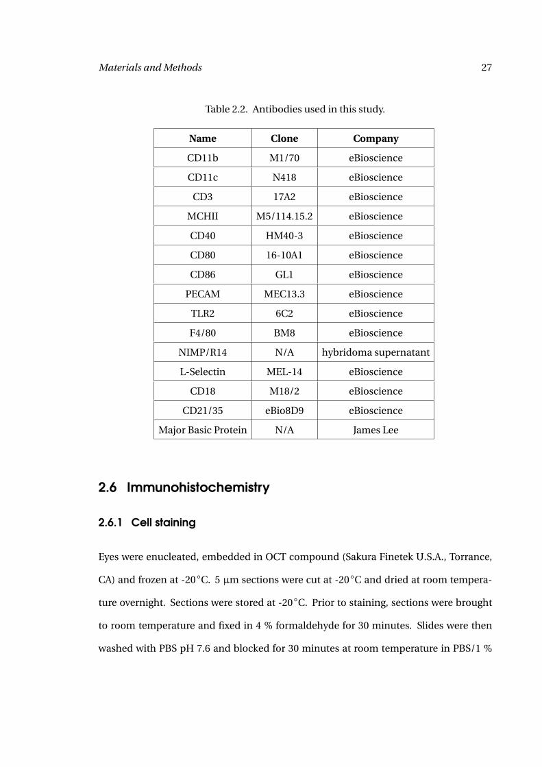
Table 2.2. Antibodies used in this study.
Name Clone Company
CD11b M1/70 eBioscience
CD11c N418 eBioscience
CD3 17A2 eBioscience
MCHII M5/114.15.2 eBioscience
CD40 HM40-3 eBioscience
CD80 16-10A1 eBioscience
CD86 GL1 eBioscience
PECAM MEC13.3 eBioscience
TLR2 6C2 eBioscience
F4/80 BM8 eBioscience
NIMP/R14 N/A hybridoma supernatant
L-Selectin MEL-14 eBioscience
CD18 M18/2 eBioscience
CD21/35 eBio8D9 eBioscience
Major Basic Protein N/A James Lee
2.6 Immunohistochemistry
2.6.1 Cell staining
Eyes were enucleated, embedded in OCT compound (Sakura Finetek U.S.A., Torrance,
CA) and frozen at -20◦C. 5 µm sections were cut at -20◦C and dried at room tempera-
ture overnight. Sections were stored at -20◦C. Prior to staining, sections were brought
to room temperature and fixed in 4 % formaldehyde for 30 minutes. Slides were then
washed with PBS pH 7.6 and blocked for 30 minutes at room temperature in PBS/1 %
Materials and Methods 28
fetal bovine serum (PBS/1 % FBS). Generally, sections were stained with primary anti-
body diluted 1:100 in PBS/1 % FBS for 2 hours at room temperature in a humid chamber.
Slides were washed in PBS and stained with 1:1000 dilution of Alexa Fluor 488 labeled
secondary antibody (except for eosinophil staining where FITC labeled secondary anti-
body at 1:200 dilution was used) at room temperature in a humid chamber in the dark.
After the final wash, slides were mounted in anti-fade medium (Vector Laboratories,
Burlingame CA). The number of cells per section was determined by counting stained
cells on a fluorescence microscope in a 400x magnification from limbus to limbus.
Primary antibody for neutrophil staining was anti-NIMP-R14 and for eosinophil
staining anti-major basic protein (anti-MBP). Secondary antibody for neutrophil
staining was rabbit-anti-rat antibody in PBS/1 % FBS, goat-anti-rabbit antibody for
eosinophil staining.
For staining with PECAM-1, I used FITC-conjugated PECAM-1 antibody.
2.6.2 PECAM-1 analysis
Images of PECAM-1 stained corneal sections were taken and vessels identified. Inten-
sity of PECAM-1 staining was analyzed using ImagePro software. Vessels were marked
with this software and the intensity of fluorescence per vessel was calculated and based
on these numbers, the average fluorescent intensity of vessels per eye as described (38).
Average vessel fluorescent intensity per eye was graphed and analyzed for statistical dif-
ferences.
Materials and Methods 29
2.7 ELISA
2.7.1 Cytokine ELISA
All cytokine ELISA kits were commercially obtained from R&D Systems and used accord-
ing to the manufacturer’s direction. Briefly, Immulon4 plates were coated with primary
antibody in PBS overnight at 4◦C, then blocked in PBS/5 % bovine serum albumin (BSA).
Standards and samples were applied overnight at 4◦C, followed by 2 hours incubation
with detection antibody and 20 minutes with HRP-streptavidin. HRP was detected with
3,3’,5,5’-tetramethylbenzidine (TMB), the reaction was stopped with 20 % H2SO4 and
absorption measured at 450 nm.
2.7.2 Antibody ELISA
Immulon4 plates were coated with 10 µg/ml filarial extract in carbonate buffer overnight
at 4◦C. Plates were blocked with PBS/1 % FBS for 2 hours. Serum dilutions ranging from
1:100 to 1:100 000 were then incubated for 2 hours at room temperature, followed by
1 hour detection with biotinylated anti-IgG1 or anti-IgG2c antibodies. Antibodies were
detected with HRP-streptavidin and TMB. The color reaction was stopped with 1 N HCl
and absorption was measured at 450 nm.
2.8 Statistical analysis
Experiments were analyzed by student’s t-test and p-values of less than 0.05 were con-
sidered significant. Data are shown as mean with standard error of the mean.
30
3 Results
3.1 In vitro activation of antigen-presenting cells
3.1.1 Dendritic cells
Bone marrow was harvested from C57BL/6, TLR2-/-, TLR4-/- and TLR2/4-/- mice and
myeloid dendritic cells were differentiated in GM-CSF enriched culture for 7 days. Cells
were purified with anti-CD11c magnetic beads and purity was generally greater than
90 % compared with dendritic cells without CD11c enrichment, that were 70 % CD11c
positive cells. I then stimulated dendritic cells to determine which TLRs are involved in
activation by filarial extracts.
1x10⁶ cells were stimulated for 24 hours with O. volvulus extract (OvAg), or with
defined TLR agonists, peptidoglycan (PGN) (TLR2), Pam3CSK (TLR2), LPS (TLR4), and
CpG-DNA (TLR9); and IL-6 and RANTES were measured by ELISA. I could not show IL-6
or RANTES production in response to Poly(I:C). Other groups also showed only absent
or low levels of IL-10, IL-12p40, IL-12p70 and TNFα upon stimulation with Poly(I:C)
(100). Surprisingly, no increased levels of mIL-12p70 were detected after stimulation
with worm antigen or TLR ligands (data not shown). Association of IL-12p70 with Th1
responses and IL-6 with Th2 responses were shown previously (50).
Results 31
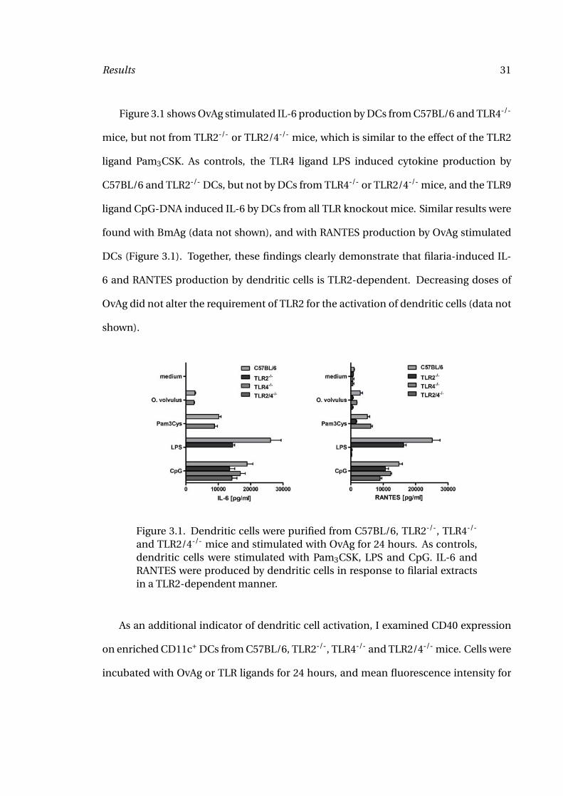
Figure 3.1 shows OvAg stimulated IL-6 production by DCs from C57BL/6 and TLR4-/-
mice, but not from TLR2-/- or TLR2/4-/- mice, which is similar to the effect of the TLR2
ligand Pam3CSK. As controls, the TLR4 ligand LPS induced cytokine production by
C57BL/6 and TLR2-/- DCs, but not by DCs from TLR4-/- or TLR2/4-/- mice, and the TLR9
ligand CpG-DNA induced IL-6 by DCs from all TLR knockout mice. Similar results were
found with BmAg (data not shown), and with RANTES production by OvAg stimulated
DCs (Figure 3.1). Together, these findings clearly demonstrate that filaria-induced IL-
6 and RANTES production by dendritic cells is TLR2-dependent. Decreasing doses of
OvAg did not alter the requirement of TLR2 for the activation of dendritic cells (data not
shown).
Figure 3.1. Dendritic cells were purified from C57BL/6, TLR2-/-, TLR4-/-
and TLR2/4-/- mice and stimulated with OvAg for 24 hours. As controls,dendritic cells were stimulated with Pam3CSK, LPS and CpG. IL-6 andRANTES were produced by dendritic cells in response to filarial extractsin a TLR2-dependent manner.
As an additional indicator of dendritic cell activation, I examined CD40 expression
on enriched CD11c+ DCs from C57BL/6, TLR2-/-, TLR4-/- and TLR2/4-/- mice. Cells were
incubated with OvAg or TLR ligands for 24 hours, and mean fluorescence intensity for
Results 32
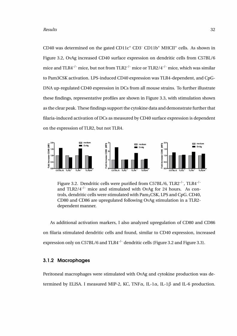
CD40 was determined on the gated CD11c+ CD3- CD11b+ MHCII+ cells. As shown in
Figure 3.2, OvAg increased CD40 surface expression on dendritic cells from C57BL/6
mice and TLR4-/- mice, but not from TLR2-/- mice or TLR2/4-/- mice, which was similar
to Pam3CSK activation. LPS-induced CD40 expression was TLR4-dependent, and CpG-
DNA up-regulated CD40 expression in DCs from all mouse strains. To further illustrate
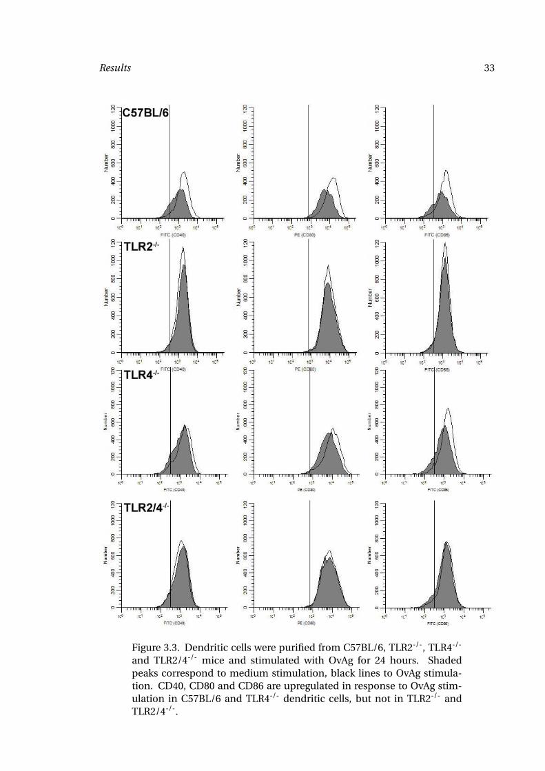
these findings, representative profiles are shown in Figure 3.3, with stimulation shown
as the clear peak. These findings support the cytokine data and demonstrate further that
filaria-induced activation of DCs as measured by CD40 surface expression is dependent
on the expression of TLR2, but not TLR4.
Figure 3.2. Dendritic cells were purified from C57BL/6, TLR2-/-, TLR4-/-
and TLR2/4-/- mice and stimulated with OvAg for 24 hours. As con-trols, dendritic cells were stimulated with Pam3CSK, LPS and CpG. CD40,CD80 and CD86 are upregulated following OvAg stimulation in a TLR2-dependent manner.
As additional activation markers, I also analyzed upregulation of CD80 and CD86
on filaria stimulated dendritic cells and found, similar to CD40 expression, increased
expression only on C57BL/6 and TLR4-/- dendritic cells (Figure 3.2 and Figure 3.3).
3.1.2 Macrophages
Peritoneal macrophages were stimulated with OvAg and cytokine production was de-
termined by ELISA. I measured MIP-2, KC, TNFα, IL-1α, IL-1β and IL-6 production.
Results 33
Figure 3.3. Dendritic cells were purified from C57BL/6, TLR2-/-, TLR4-/-
and TLR2/4-/- mice and stimulated with OvAg for 24 hours. Shadedpeaks correspond to medium stimulation, black lines to OvAg stimula-tion. CD40, CD80 and CD86 are upregulated in response to OvAg stim-ulation in C57BL/6 and TLR4-/- dendritic cells, but not in TLR2-/- andTLR2/4-/-.
Results 34
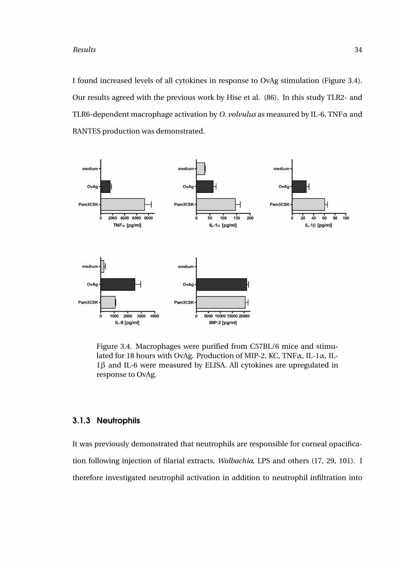
I found increased levels of all cytokines in response to OvAg stimulation (Figure 3.4).
Our results agreed with the previous work by Hise et al. (86). In this study TLR2- and
TLR6-dependent macrophage activation by O. volvulus as measured by IL-6, TNFα and
RANTES production was demonstrated.
Figure 3.4. Macrophages were purified from C57BL/6 mice and stimu-lated for 18 hours with OvAg. Production of MIP-2, KC, TNFα, IL-1α, IL-1β and IL-6 were measured by ELISA. All cytokines are upregulated inresponse to OvAg.
3.1.3 Neutrophils
It was previously demonstrated that neutrophils are responsible for corneal opacifica-
tion following injection of filarial extracts, Wolbachia, LPS and others (17, 29, 101). I
therefore investigated neutrophil activation in addition to neutrophil infiltration into
Results 35
the corneal stroma. Neutrophils were purified from the peritoneal cavity of Casein in-
jected mice. Cells were purified over a 90 % Percoll gradient. Purity was generally greater
than 95 %. 1x10⁵ cells were stimulated with OvAg or TLR ligands for 18 hours unless oth-
erwise noted. Cells and supernatants were analyzed for activation parameters.
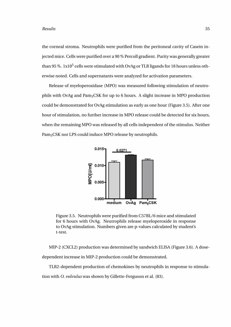
Release of myeloperoxidase (MPO) was measured following stimulation of neutro-
phils with OvAg and Pam3CSK for up to 6 hours. A slight increase in MPO production
could be demonstrated for OvAg stimulation as early as one hour (Figure 3.5). After one
hour of stimulation, no further increase in MPO release could be detected for six hours,
when the remaining MPO was released by all cells independent of the stimulus. Neither
Pam3CSK nor LPS could induce MPO release by neutrophils.
Figure 3.5. Neutrophils were purified from C57BL/6 mice and stimulatedfor 6 hours with OvAg. Neutrophils release myeloperoxide in responseto OvAg stimulation. Numbers given are p-values calculated by student’st-test.
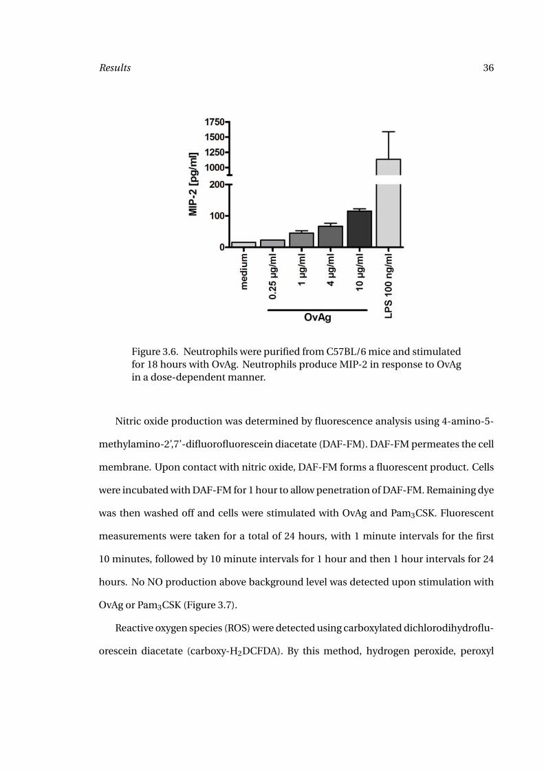
MIP-2 (CXCL2) production was determined by sandwich ELISA (Figure 3.6). A dose-
dependent increase in MIP-2 production could be demonstrated.
TLR2-dependent production of chemokines by neutrophils in response to stimula-
tion with O. volvulus was shown by Gillette-Ferguson et al. (83).
Results 36
Figure 3.6. Neutrophils were purified from C57BL/6 mice and stimulatedfor 18 hours with OvAg. Neutrophils produce MIP-2 in response to OvAgin a dose-dependent manner.
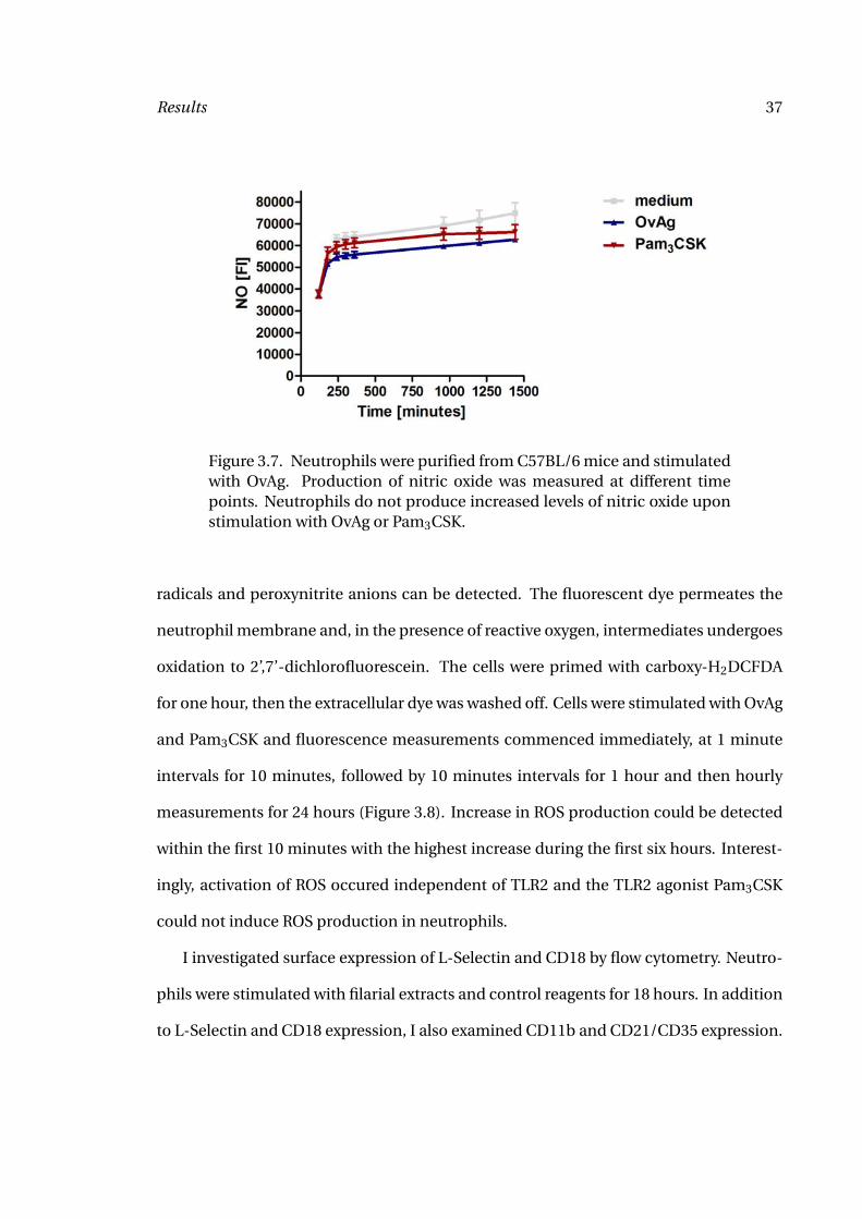
Nitric oxide production was determined by fluorescence analysis using 4-amino-5-
methylamino-2’,7’-difluorofluorescein diacetate (DAF-FM). DAF-FM permeates the cell
membrane. Upon contact with nitric oxide, DAF-FM forms a fluorescent product. Cells
were incubated with DAF-FM for 1 hour to allow penetration of DAF-FM. Remaining dye
was then washed off and cells were stimulated with OvAg and Pam3CSK. Fluorescent
measurements were taken for a total of 24 hours, with 1 minute intervals for the first
10 minutes, followed by 10 minute intervals for 1 hour and then 1 hour intervals for 24
hours. No NO production above background level was detected upon stimulation with
OvAg or Pam3CSK (Figure 3.7).
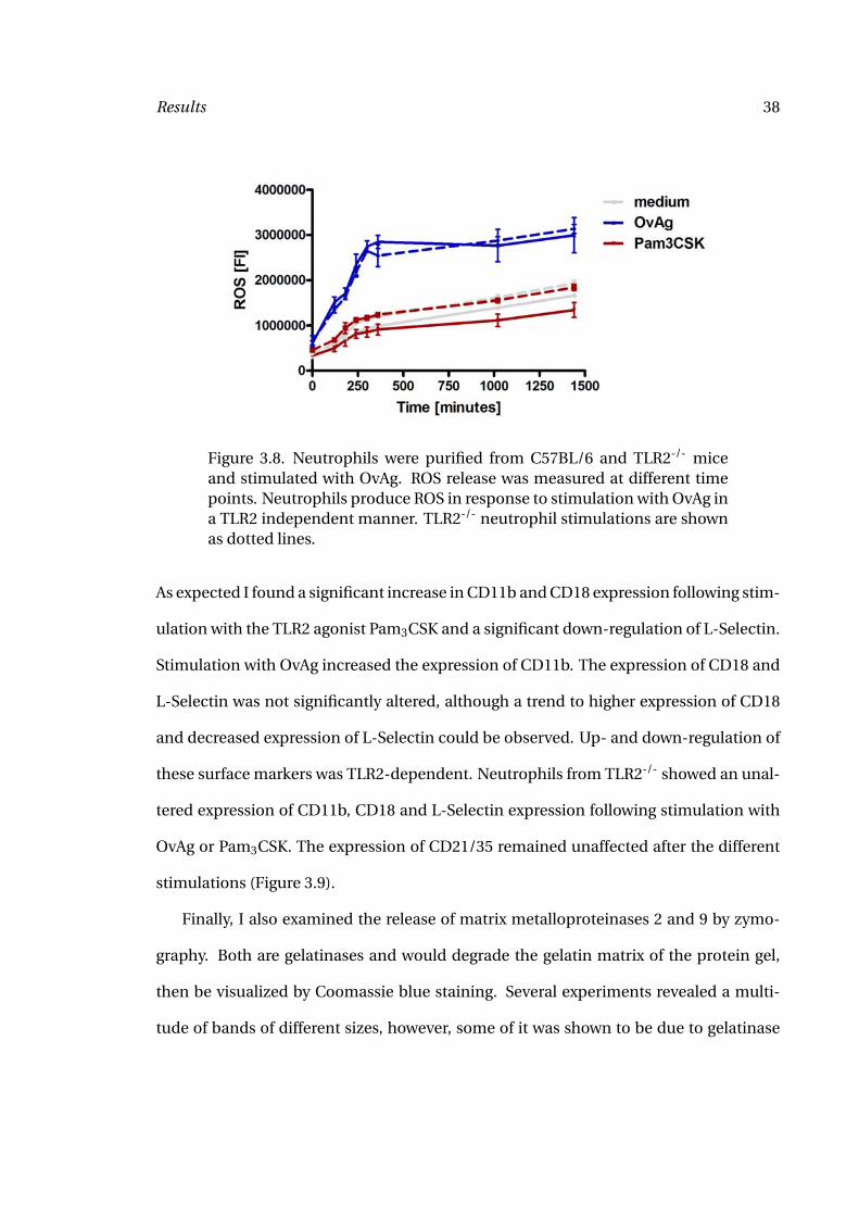
Reactive oxygen species (ROS) were detected using carboxylated dichlorodihydroflu-
orescein diacetate (carboxy-H2DCFDA). By this method, hydrogen peroxide, peroxyl
Results 37
Figure 3.7. Neutrophils were purified from C57BL/6 mice and stimulatedwith OvAg. Production of nitric oxide was measured at different timepoints. Neutrophils do not produce increased levels of nitric oxide uponstimulation with OvAg or Pam3CSK.
radicals and peroxynitrite anions can be detected. The fluorescent dye permeates the
neutrophil membrane and, in the presence of reactive oxygen, intermediates undergoes
oxidation to 2’,7’-dichlorofluorescein. The cells were primed with carboxy-H2DCFDA
for one hour, then the extracellular dye was washed off. Cells were stimulated with OvAg
and Pam3CSK and fluorescence measurements commenced immediately, at 1 minute
intervals for 10 minutes, followed by 10 minutes intervals for 1 hour and then hourly
measurements for 24 hours (Figure 3.8). Increase in ROS production could be detected
within the first 10 minutes with the highest increase during the first six hours. Interest-
ingly, activation of ROS occured independent of TLR2 and the TLR2 agonist Pam3CSK
could not induce ROS production in neutrophils.
I investigated surface expression of L-Selectin and CD18 by flow cytometry. Neutro-
phils were stimulated with filarial extracts and control reagents for 18 hours. In addition
to L-Selectin and CD18 expression, I also examined CD11b and CD21/CD35 expression.
Results 38
Figure 3.8. Neutrophils were purified from C57BL/6 and TLR2-/- miceand stimulated with OvAg. ROS release was measured at different timepoints. Neutrophils produce ROS in response to stimulation with OvAg ina TLR2 independent manner. TLR2-/- neutrophil stimulations are shownas dotted lines.
As expected I found a significant increase in CD11b and CD18 expression following stim-
ulation with the TLR2 agonist Pam3CSK and a significant down-regulation of L-Selectin.
Stimulation with OvAg increased the expression of CD11b. The expression of CD18 and
L-Selectin was not significantly altered, although a trend to higher expression of CD18
and decreased expression of L-Selectin could be observed. Up- and down-regulation of
these surface markers was TLR2-dependent. Neutrophils from TLR2-/- showed an unal-
tered expression of CD11b, CD18 and L-Selectin expression following stimulation with
OvAg or Pam3CSK. The expression of CD21/35 remained unaffected after the different
stimulations (Figure 3.9).
Finally, I also examined the release of matrix metalloproteinases 2 and 9 by zymo-
graphy. Both are gelatinases and would degrade the gelatin matrix of the protein gel,
then be visualized by Coomassie blue staining. Several experiments revealed a multi-
tude of bands of different sizes, however, some of it was shown to be due to gelatinase
Results 39
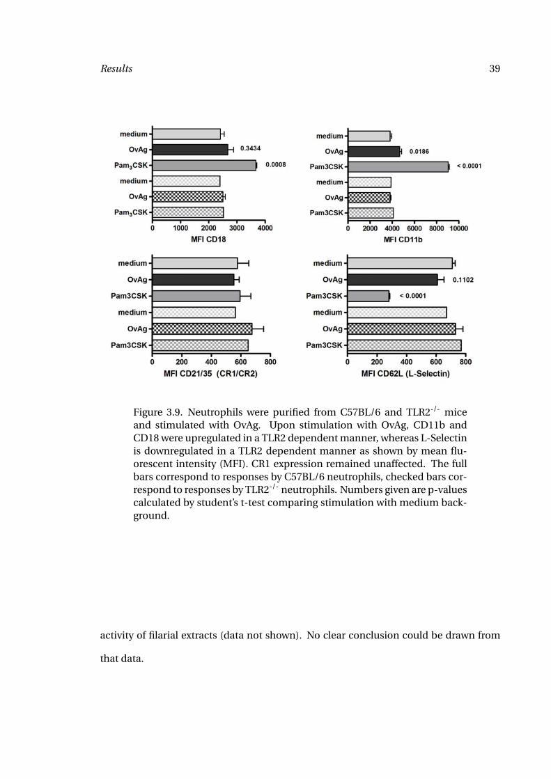
Figure 3.9. Neutrophils were purified from C57BL/6 and TLR2-/- miceand stimulated with OvAg. Upon stimulation with OvAg, CD11b andCD18 were upregulated in a TLR2 dependent manner, whereas L-Selectinis downregulated in a TLR2 dependent manner as shown by mean flu-orescent intensity (MFI). CR1 expression remained unaffected. The fullbars correspond to responses by C57BL/6 neutrophils, checked bars cor-respond to responses by TLR2-/- neutrophils. Numbers given are p-valuescalculated by student’s t-test comparing stimulation with medium back-ground.
activity of filarial extracts (data not shown). No clear conclusion could be drawn from
that data.
Results 40
3.2 The role of TLR2 and TLR4 in the generation of adaptive im-
mune responses
3.2.1 T cell activation
Previous studies demonstrated that immunization with BmAg or B. malayi microfilariae,
or with OvAg induced a predominant, but not exclusive Th2-like response (28, 102). To
examine the role of TLRs on the development of T cell phenotype, C57BL/6, TLR2-/-,
and TLR4-/- mice were immunized subcutaneously three times over three weeks with
OvAg. One week after the last immunization, mice were euthanized, splenocytes were
stimulated in vitro with OvAg, and IL-5 and IFN-γ were measured by ELISA. Figure 3.10
shows IFN-γ production by splenocytes from OvAg immunized C57BL/6 and TLR4-/-
mice, but not by TLR2-/- mice, indicating that filaria-induced IFN-γ production is TLR2-
dependent. In contrast, IL-5 production was similar in splenocytes from all mouse
strains. My findings indicate that TLR2 has a critical role in filaria-induced production
of Th1-associated IFN-γ, but not Th2-associated IL-5.
3.2.2 Antibody production
Previous studies demonstrated that Th2-associated isotypes IgE and IgG1 are predom-
inant after immunization with filarial extracts (34, 39). To determine the role of TLR2
and TLR4 on production of these isotypes, C57BL/6, TLR2-/-, and TLR4-/- mice were
immunized as described above, and total serum IgE and filaria-specific IgG1 were mea-
sured by ELISA. IgE was elevated in immunized C57BL/6 mice compared with unimmu-
nized mice (Figure 3.11); however, there was no decrease in IgE levels among immunized
TLR2-/- and TLR4-/- mice compared to C57BL/6 wildtype mice as shown in Figure 3.11.
Results 41
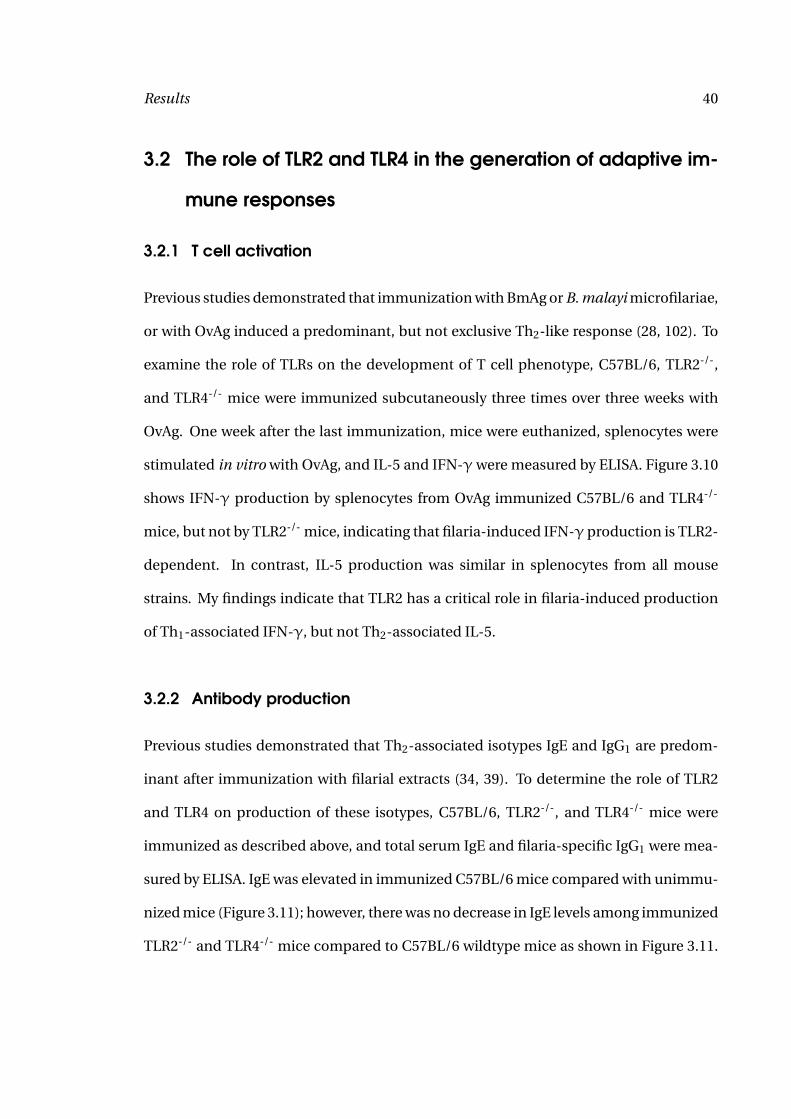
Figure 3.10. Splenocytes from immunized C57BL/6, TLR2-/-, and TLR4-/-
mice were stimulated for 72 hours with medium, OvAg and anti-CD3 andIL-5 and IFN-γ production was measured by ELISA. Splenocytes fromTLR2-/- mice produced significantly less IFN-γ when stimulated withOvAg.
The only difference I detected were increased levels of IgE in TLR2-/- mice. Similarly,
filaria-specific IgG1 in immunized C57BL/6 mice was detected at up to 1:100,000 di-
lution (filaria-specific IgG1 in unimmunized mice was undetectable); however, as with
IgE, there were no significant difference between C57BL/6 and TLR2-/- or TLR4-/- mice
(Figure 3.12). Previous studies also demonstrated that filaria-specific IgG2a, which is
associated with IFN-γ production (103–105), is produced at low levels compared with
IgG1 (106). Filaria-specific IgG2c levels were low in C57BL/6 mice, and there was no dif-
ference between these and TLR2-/- and TLR4-/- mice. These findings indicate that TLR2
and TLR4 have no major role in production of filaria-specific antibodies.
3.3 The role of TLR2 and TLR4 in corneal inflammation
Previous studies showed that neutrophils and eosinophils are recruited to the corneas of
immunized mice in a biphasic manner after injection of parasite extracts to the corneal
Results 42
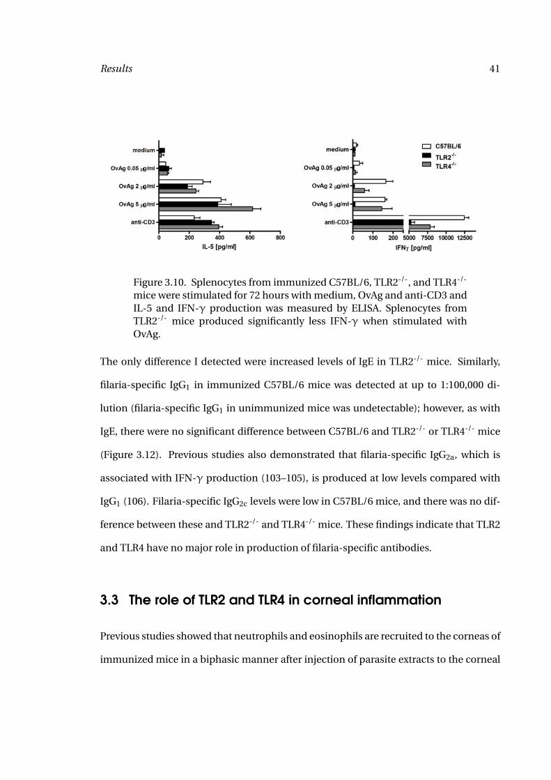
Figure 3.11. Serum levels of IgE in immunized C57BL/6, TLR2-/- andTLR4-/- mice were measured by ELISA. IgE was increased in immunizedmice compared with naive mice. TLR2-/- showed higher levels of IgE com-pared with C57BL/6 mice. Numbers given are p-values calculated by stu-dent’s t-test.
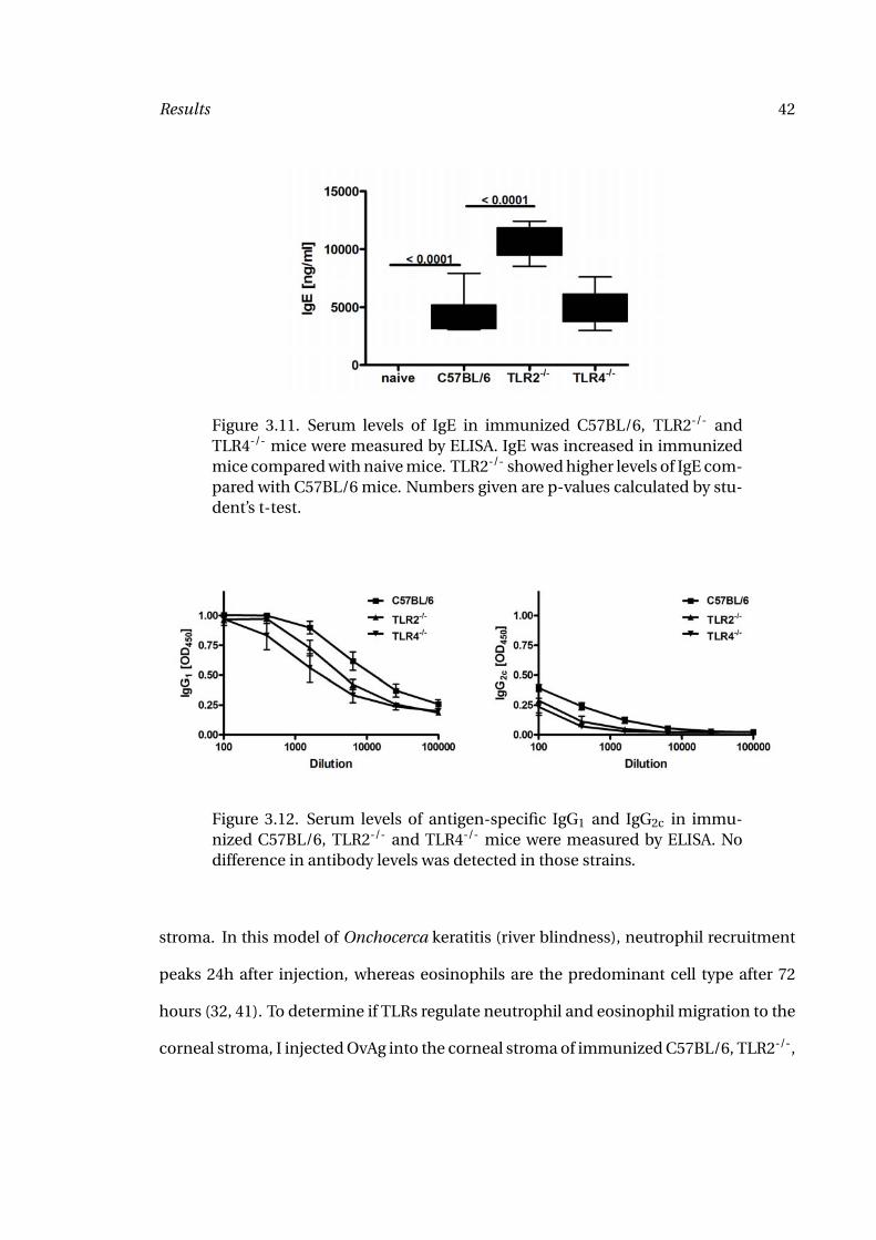
Figure 3.12. Serum levels of antigen-specific IgG1 and IgG2c in immu-nized C57BL/6, TLR2-/- and TLR4-/- mice were measured by ELISA. Nodifference in antibody levels was detected in those strains.
stroma. In this model of Onchocerca keratitis (river blindness), neutrophil recruitment
peaks 24h after injection, whereas eosinophils are the predominant cell type after 72
hours (32, 41). To determine if TLRs regulate neutrophil and eosinophil migration to the
corneal stroma, I injected OvAg into the corneal stroma of immunized C57BL/6, TLR2-/-,
Results 43
and TLR4-/- mice, and quantified the number of neutrophils in the corneal stroma after
24 hours, and the number of eosinophils after 72 hours.
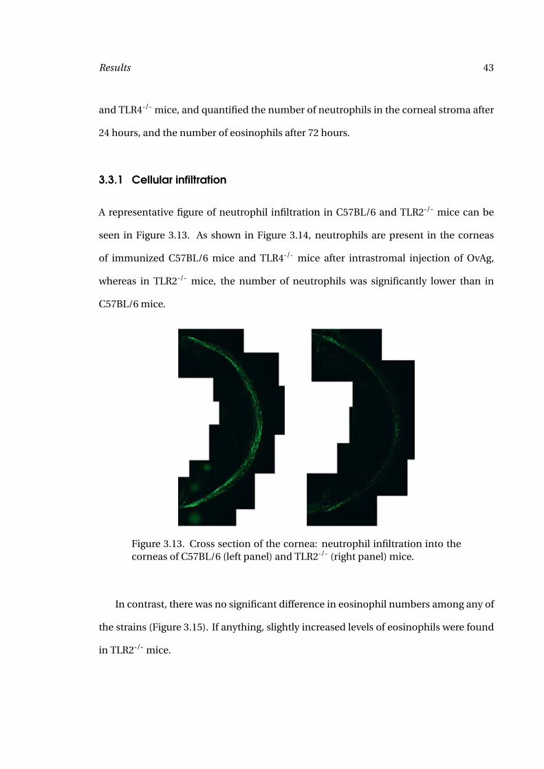
3.3.1 Cellular infiltration
A representative figure of neutrophil infiltration in C57BL/6 and TLR2-/- mice can be
seen in Figure 3.13. As shown in Figure 3.14, neutrophils are present in the corneas
of immunized C57BL/6 mice and TLR4-/- mice after intrastromal injection of OvAg,
whereas in TLR2-/- mice, the number of neutrophils was significantly lower than in
C57BL/6 mice.
Figure 3.13. Cross section of the cornea: neutrophil infiltration into thecorneas of C57BL/6 (left panel) and TLR2-/- (right panel) mice.
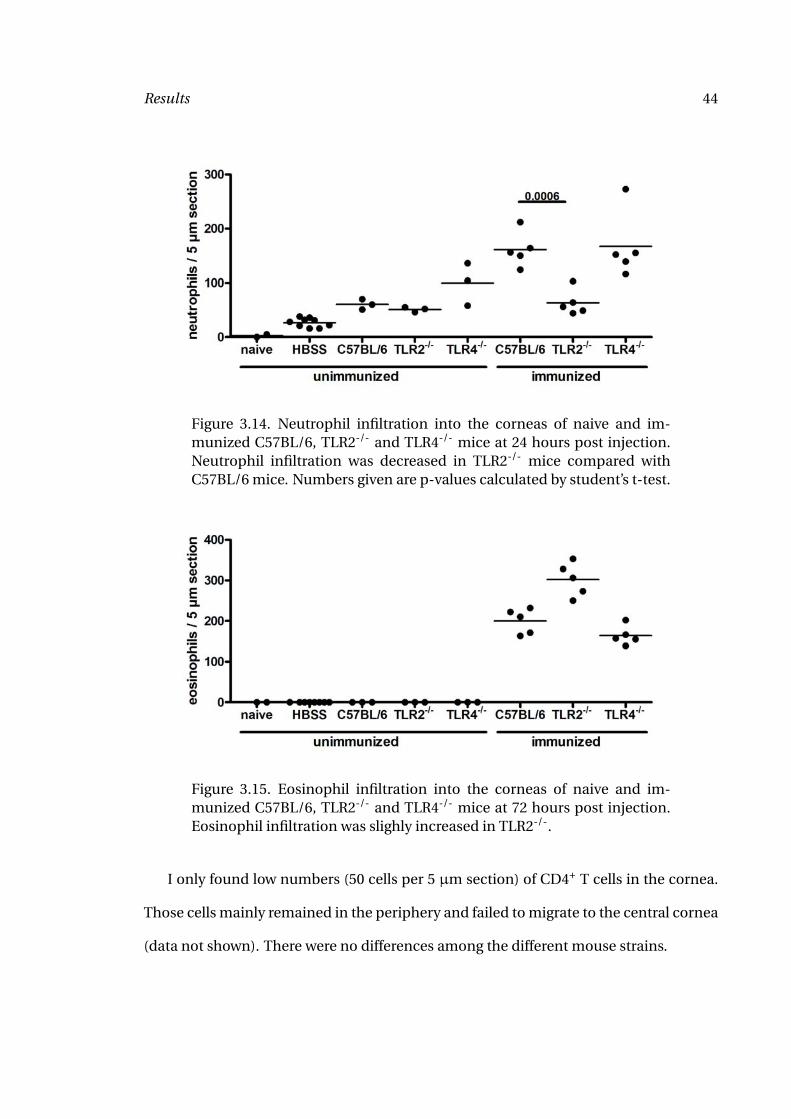
In contrast, there was no significant difference in eosinophil numbers among any of
the strains (Figure 3.15). If anything, slightly increased levels of eosinophils were found
in TLR2-/- mice.
Results 44
Figure 3.14. Neutrophil infiltration into the corneas of naive and im-munized C57BL/6, TLR2-/- and TLR4-/- mice at 24 hours post injection.Neutrophil infiltration was decreased in TLR2-/- mice compared withC57BL/6 mice. Numbers given are p-values calculated by student’s t-test.
Figure 3.15. Eosinophil infiltration into the corneas of naive and im-munized C57BL/6, TLR2-/- and TLR4-/- mice at 72 hours post injection.Eosinophil infiltration was slighly increased in TLR2-/-.
I only found low numbers (50 cells per 5 µm section) of CD4+ T cells in the cornea.
Those cells mainly remained in the periphery and failed to migrate to the central cornea
(data not shown). There were no differences among the different mouse strains.
Results 45
Macrophage numbers were quantified as F4/80+ and CD169+ positive cells. F4/80
is expressed on tissue macrophages, whereas CD169 is upregulated on activated
macrophages. I found decreased macrophage infiltration into the corneas of TLR2-/-
mice one day post injection (data not shown). This corresponds to findings in naive
mice injected with filarial extract, where macrophage migration was also dependent on
the presence TLR2 (86).
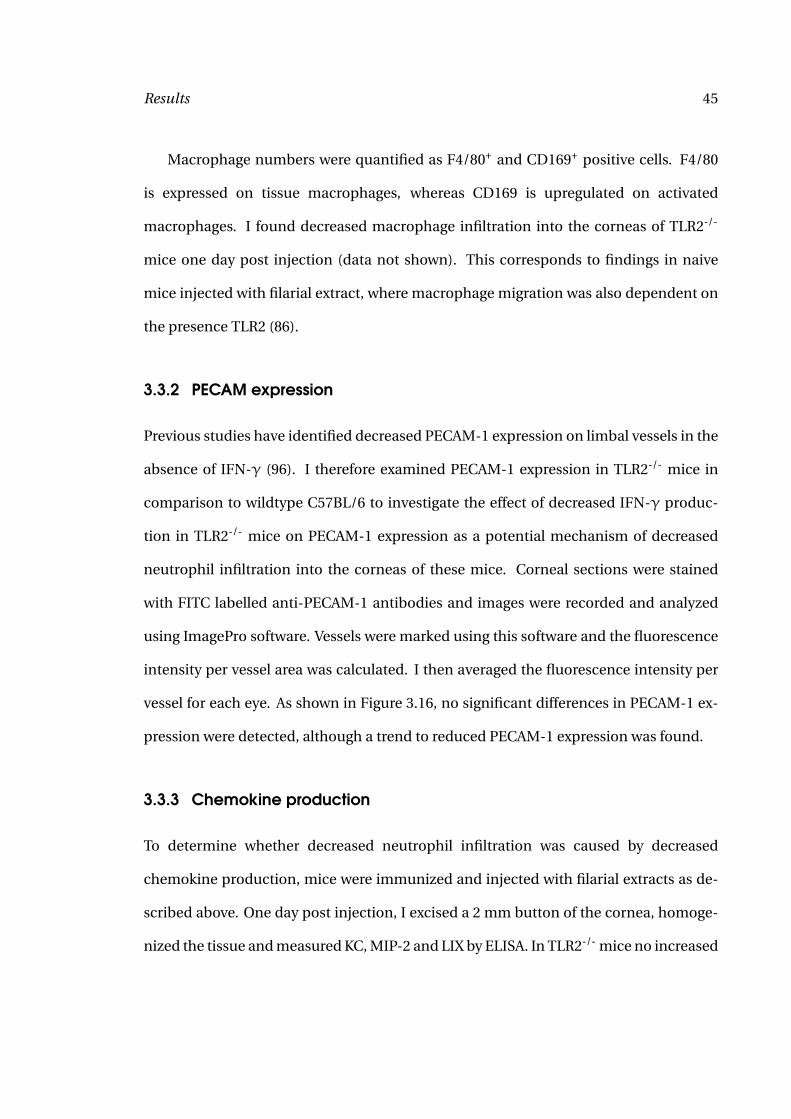
3.3.2 PECAM expression
Previous studies have identified decreased PECAM-1 expression on limbal vessels in the
absence of IFN-γ (96). I therefore examined PECAM-1 expression in TLR2-/- mice in
comparison to wildtype C57BL/6 to investigate the effect of decreased IFN-γ produc-
tion in TLR2-/- mice on PECAM-1 expression as a potential mechanism of decreased
neutrophil infiltration into the corneas of these mice. Corneal sections were stained
with FITC labelled anti-PECAM-1 antibodies and images were recorded and analyzed
using ImagePro software. Vessels were marked using this software and the fluorescence
intensity per vessel area was calculated. I then averaged the fluorescence intensity per
vessel for each eye. As shown in Figure 3.16, no significant differences in PECAM-1 ex-
pression were detected, although a trend to reduced PECAM-1 expression was found.
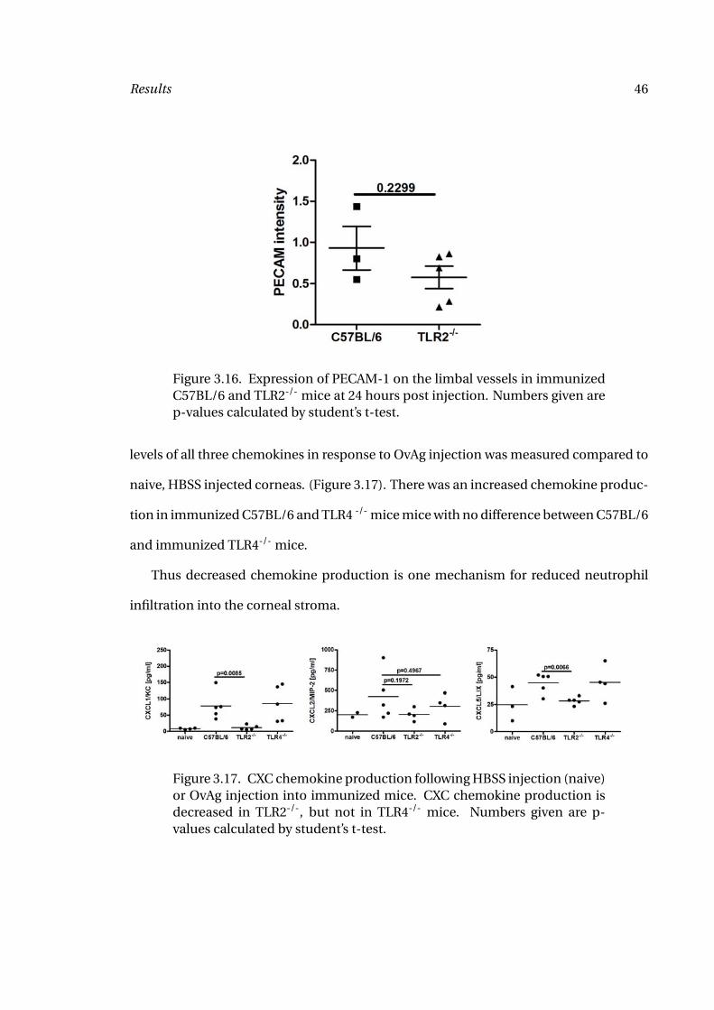
3.3.3 Chemokine production
To determine whether decreased neutrophil infiltration was caused by decreased
chemokine production, mice were immunized and injected with filarial extracts as de-
scribed above. One day post injection, I excised a 2 mm button of the cornea, homoge-
nized the tissue and measured KC, MIP-2 and LIX by ELISA. In TLR2-/- mice no increased
Results 46
Figure 3.16. Expression of PECAM-1 on the limbal vessels in immunizedC57BL/6 and TLR2-/- mice at 24 hours post injection. Numbers given arep-values calculated by student’s t-test.
levels of all three chemokines in response to OvAg injection was measured compared to
naive, HBSS injected corneas. (Figure 3.17). There was an increased chemokine produc-
tion in immunized C57BL/6 and TLR4 -/- mice mice with no difference between C57BL/6
and immunized TLR4-/- mice.
Thus decreased chemokine production is one mechanism for reduced neutrophil
infiltration into the corneal stroma.
Figure 3.17. CXC chemokine production following HBSS injection (naive)or OvAg injection into immunized mice. CXC chemokine production isdecreased in TLR2-/-, but not in TLR4-/- mice. Numbers given are p-values calculated by student’s t-test.
Results 47
3.4 The role of IFN-γ in the generation of adaptive immune re-
sponses
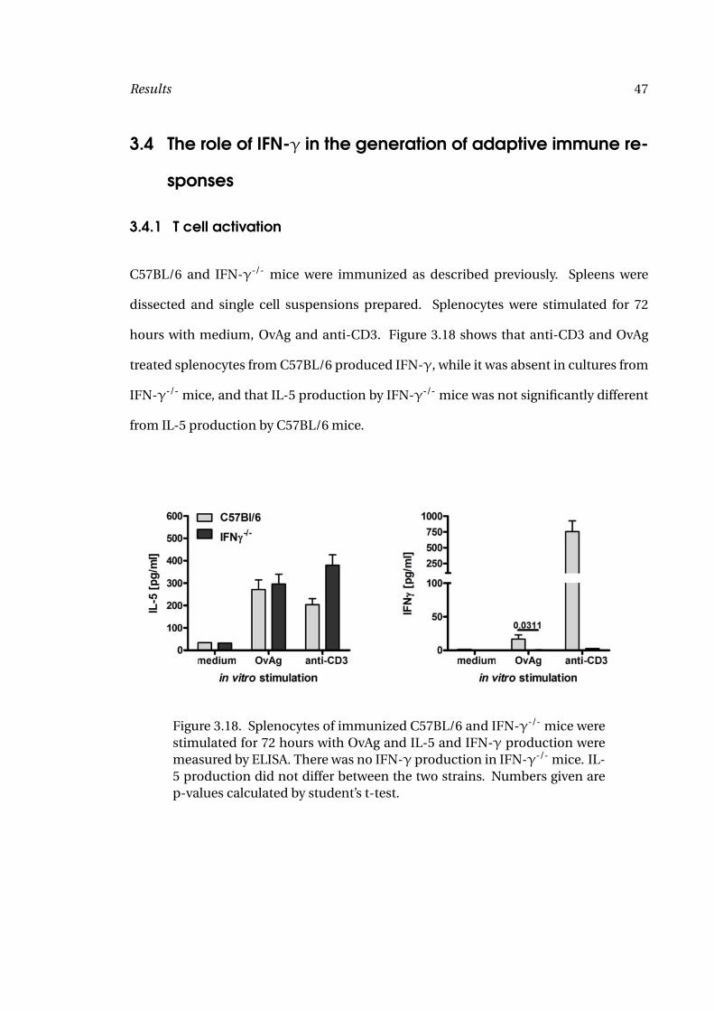
3.4.1 T cell activation
C57BL/6 and IFN-γ-/- mice were immunized as described previously. Spleens were
dissected and single cell suspensions prepared. Splenocytes were stimulated for 72
hours with medium, OvAg and anti-CD3. Figure 3.18 shows that anti-CD3 and OvAg
treated splenocytes from C57BL/6 produced IFN-γ, while it was absent in cultures from
IFN-γ-/- mice, and that IL-5 production by IFN-γ-/- mice was not significantly different
from IL-5 production by C57BL/6 mice.
Figure 3.18. Splenocytes of immunized C57BL/6 and IFN-γ-/- mice werestimulated for 72 hours with OvAg and IL-5 and IFN-γ production weremeasured by ELISA. There was no IFN-γ production in IFN-γ-/- mice. IL-5 production did not differ between the two strains. Numbers given arep-values calculated by student’s t-test.
Results 48
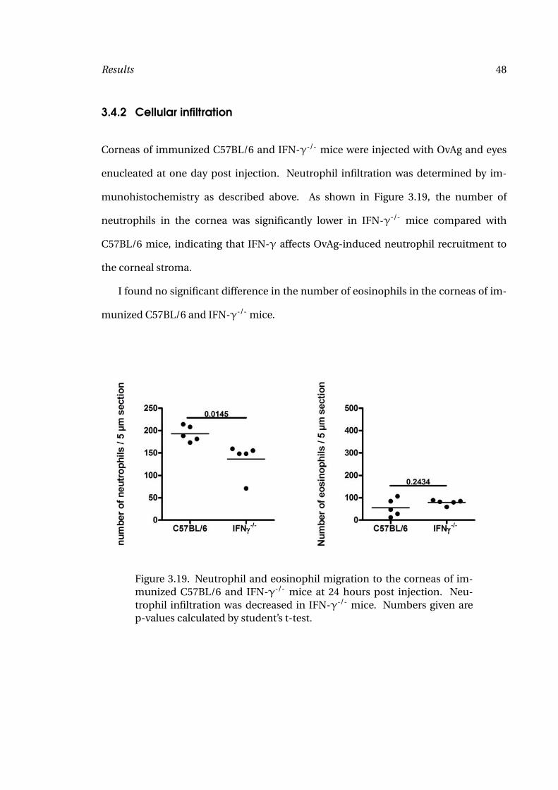
3.4.2 Cellular infiltration
Corneas of immunized C57BL/6 and IFN-γ-/- mice were injected with OvAg and eyes
enucleated at one day post injection. Neutrophil infiltration was determined by im-
munohistochemistry as described above. As shown in Figure 3.19, the number of
neutrophils in the cornea was significantly lower in IFN-γ-/- mice compared with
C57BL/6 mice, indicating that IFN-γ affects OvAg-induced neutrophil recruitment to
the corneal stroma.
I found no significant difference in the number of eosinophils in the corneas of im-
munized C57BL/6 and IFN-γ-/- mice.
Figure 3.19. Neutrophil and eosinophil migration to the corneas of im-munized C57BL/6 and IFN-γ-/- mice at 24 hours post injection. Neu-trophil infiltration was decreased in IFN-γ-/- mice. Numbers given arep-values calculated by student’s t-test.
Results 49
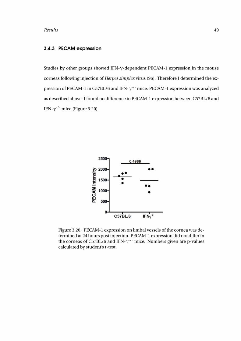
3.4.3 PECAM expression
Studies by other groups showed IFN-γ-dependent PECAM-1 expression in the mouse
corneas following injection of Herpes simplex virus (96). Therefore I determined the ex-
pression of PECAM-1 in C57BL/6 and IFN-γ-/- mice. PECAM-1 expression was analyzed
as described above. I found no difference in PECAM-1 expression between C57BL/6 and
IFN-γ-/- mice (Figure 3.20).
Figure 3.20. PECAM-1 expression on limbal vessels of the cornea was de-termined at 24 hours post injection. PECAM-1 expression did not differ inthe corneas of C57BL/6 and IFN-γ-/- mice. Numbers given are p-valuescalculated by student’s t-test.
Results 50
3.5 IFN-γ induced cytokine responses in macrophages and fi-
broblasts
3.5.1 Macrophages
To examine whether IFN-γ has a synergistic effect with OvAg on cytokine production,
macrophages isolated from the peritoneal cavity were stimulated for 18 hours with ei-
ther OvAg, rIFN-γ alone, or with both, then IL-1α, IL-1β, TNFα and IL-6 were mea-
sured by ELISA. Previous studies from our group showed macrophage activation with
filaria/Wolbachia antigens (86), and I found a dose-dependent increase of cytokine pro-
duction, and chose 2 µg/ml OvAg (half maximum cytokine production) as an optimal
dose. As shown in Figure 3.21, macrophages incubated with rIFN-γ alone did not in-
duce cytokine production, and incubation with OvAg alone produced increased levels
of IL-1α, IL-1β, TNFα and IL-6. In marked contrast, rIFN-γ added to macrophages to-
gether with OvAg stimulated greatly elevated levels of all four cytokines (Figure 3.21).
The synergistic effect of IFN-γ and OvAg was further increased if macrophages were in-
cubated for 6 hours with rIFN-γ prior to stimulation with OvAg (Figure 3.22). Priming
was most efficient for production of IL-6 and TNFα. Priming with OvAg followed by
IFN-γ stimulation did not have a similar effect.
As CXC chemokines are important for recruitment of neutrophils to the corneal
stroma, with macrophages producing CXCL1/KC and CXCL2/MIP-2 (17), I examined if
there is a synergistic effect of IFN-γ on OvAg-induced production of these chemokines.
Peritoneal macrophages were stimulated with OvAg, rIFN-γ or both, and CXCL1/KC
and CXCL2/MIP-2 were measured by ELISA. As shown in Figure 3.23, OvAg stimulated
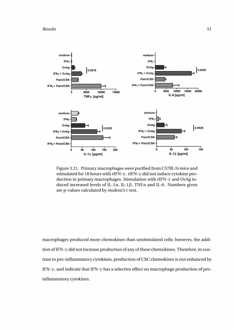
Results 51
Figure 3.21. Primary macrophages were purified from C57BL/6 mice andstimulated for 18 hours with rIFN-γ. rIFN-γ did not induce cytokine pro-duction in primary macrophages. Stimulation with rIFN-γ and OvAg in-duced increased levels of IL-1α, IL-1β, TNFα and IL-6. Numbers givenare p-values calculated by student’s t-test.
macrophages produced more chemokines than unstimulated cells; however, the addi-
tion of IFN-γ did not increase production of any of these chemokines. Therefore, in con-
trast to pro-inflammatory cytokines, production of CXC chemokines is not enhanced by
IFN-γ, and indicate that IFN-γ has a selective effect on macrophage production of pro-
inflammatory cytokines.
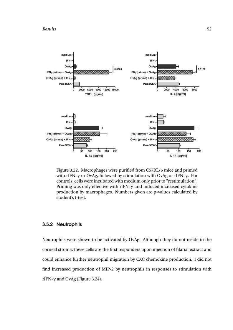
Results 52
Figure 3.22. Macrophages were purified from C57BL/6 mice and primedwith rIFN-γ or OvAg, followed by stimulation with OvAg or rIFN-γ. Forcontrols, cells were incubated with medium only prior to "restimulation".Priming was only effective with rIFN-γ and induced increased cytokineproduction by macrophages. Numbers given are p-values calculated bystudent’s t-test.
3.5.2 Neutrophils
Neutrophils were shown to be activated by OvAg. Although they do not reside in the
corneal stroma, these cells are the first responders upon injection of filarial extract and
could enhance further neutrophil migration by CXC chemokine production. I did not
find increased production of MIP-2 by neutrophils in responses to stimulation with
rIFN-γ and OvAg (Figure 3.24).
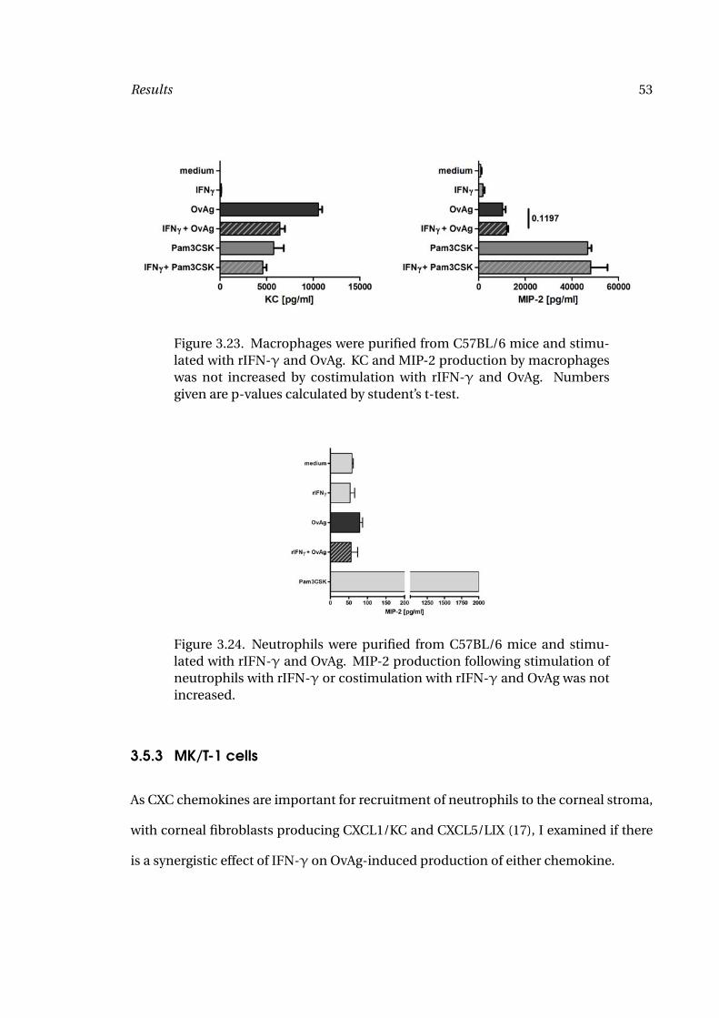
Results 53
Figure 3.23. Macrophages were purified from C57BL/6 mice and stimu-lated with rIFN-γ and OvAg. KC and MIP-2 production by macrophageswas not increased by costimulation with rIFN-γ and OvAg. Numbersgiven are p-values calculated by student’s t-test.
Figure 3.24. Neutrophils were purified from C57BL/6 mice and stimu-lated with rIFN-γ and OvAg. MIP-2 production following stimulation ofneutrophils with rIFN-γ or costimulation with rIFN-γ and OvAg was notincreased.
3.5.3 MK/T-1 cells
As CXC chemokines are important for recruitment of neutrophils to the corneal stroma,
with corneal fibroblasts producing CXCL1/KC and CXCL5/LIX (17), I examined if there
is a synergistic effect of IFN-γ on OvAg-induced production of either chemokine.
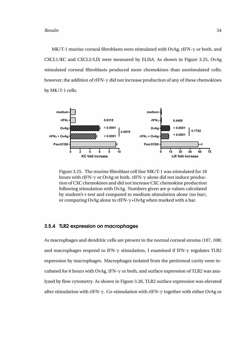
Results 54
MK/T-1 murine corneal fibroblasts were stimulated with OvAg, rIFN-γ or both, and
CXCL1/KC and CXCL5/LIX were measured by ELISA. As shown in Figure 3.25, OvAg
stimulated corneal fibroblasts produced more chemokines than unstimulated cells;
however, the addition of rIFN-γ did not increase production of any of these chemokines
by MK/T-1 cells.
Figure 3.25. The murine fibroblast cell line MK/T-1 was stimulated for 18hours with rIFN-γ or OvAg or both. rIFN-γ alone did not induce produc-tion of CXC chemokines and did not increase CXC chemokine productionfollowing stimulation with OvAg. Numbers given are p-values calculatedby student’s t-test and compared to medium stimulation alone (no bar),or comparing OvAg alone to rIFN-γ+OvAg when marked with a bar.
3.5.4 TLR2 expression on macrophages
As macrophages and dendritic cells are present in the normal corneal stroma (107, 108)
and macrophages respond to IFN-γ stimulation, I examined if IFN-γ regulates TLR2
expression by macrophages. Macrophages isolated from the peritoneal cavity were in-
cubated for 6 hours with OvAg, IFN-γ or both, and surface expression of TLR2 was ana-
lyzed by flow cytometry. As shown in Figure 3.26, TLR2 surface expression was elevated
after stimulation with rIFN-γ. Co-stimulation with rIFN-γ together with either OvAg or
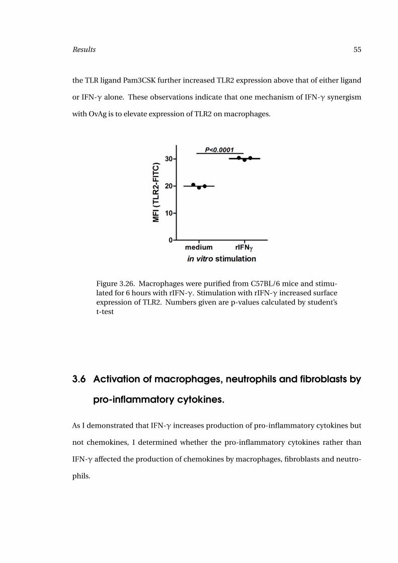
Results 55
the TLR ligand Pam3CSK further increased TLR2 expression above that of either ligand
or IFN-γ alone. These observations indicate that one mechanism of IFN-γ synergism
with OvAg is to elevate expression of TLR2 on macrophages.
Figure 3.26. Macrophages were purified from C57BL/6 mice and stimu-lated for 6 hours with rIFN-γ. Stimulation with rIFN-γ increased surfaceexpression of TLR2. Numbers given are p-values calculated by student’st-test
3.6 Activation of macrophages, neutrophils and fibroblasts by
pro-inflammatory cytokines.
As I demonstrated that IFN-γ increases production of pro-inflammatory cytokines but
not chemokines, I determined whether the pro-inflammatory cytokines rather than
IFN-γ affected the production of chemokines by macrophages, fibroblasts and neutro-
phils.
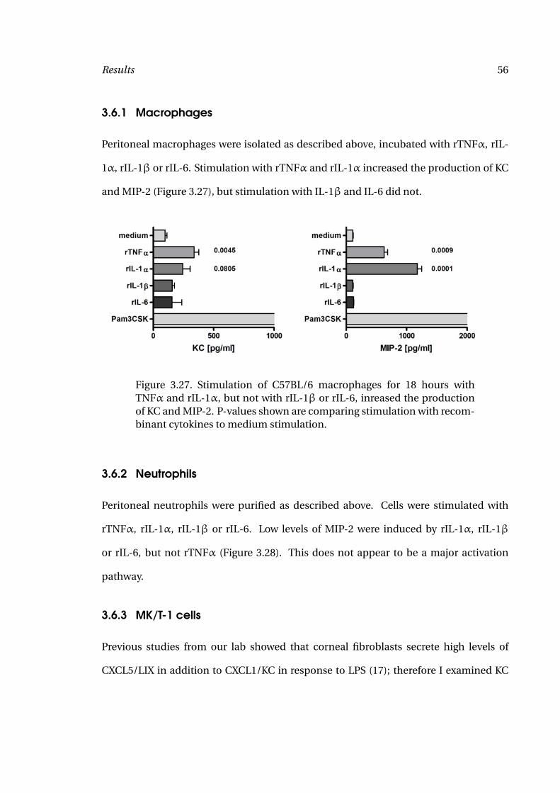
Results 56
3.6.1 Macrophages
Peritoneal macrophages were isolated as described above, incubated with rTNFα, rIL-
1α, rIL-1β or rIL-6. Stimulation with rTNFα and rIL-1α increased the production of KC
and MIP-2 (Figure 3.27), but stimulation with IL-1β and IL-6 did not.
Figure 3.27. Stimulation of C57BL/6 macrophages for 18 hours withTNFα and rIL-1α, but not with rIL-1β or rIL-6, inreased the productionof KC and MIP-2. P-values shown are comparing stimulation with recom-binant cytokines to medium stimulation.
3.6.2 Neutrophils
Peritoneal neutrophils were purified as described above. Cells were stimulated with
rTNFα, rIL-1α, rIL-1β or rIL-6. Low levels of MIP-2 were induced by rIL-1α, rIL-1β
or rIL-6, but not rTNFα (Figure 3.28). This does not appear to be a major activation
pathway.
3.6.3 MK/T-1 cells
Previous studies from our lab showed that corneal fibroblasts secrete high levels of
CXCL5/LIX in addition to CXCL1/KC in response to LPS (17); therefore I examined KC
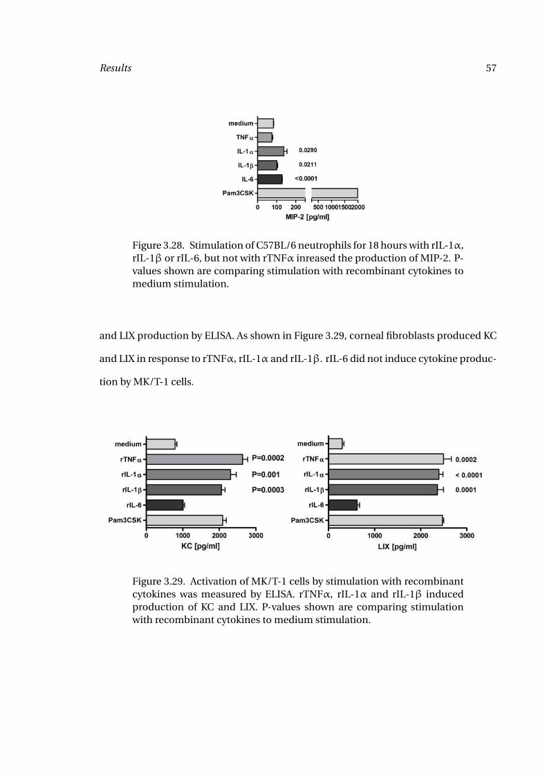
Results 57
Figure 3.28. Stimulation of C57BL/6 neutrophils for 18 hours with rIL-1α,rIL-1β or rIL-6, but not with rTNFα inreased the production of MIP-2. P-values shown are comparing stimulation with recombinant cytokines tomedium stimulation.
and LIX production by ELISA. As shown in Figure 3.29, corneal fibroblasts produced KC
and LIX in response to rTNFα, rIL-1α and rIL-1β. rIL-6 did not induce cytokine produc-
tion by MK/T-1 cells.
Figure 3.29. Activation of MK/T-1 cells by stimulation with recombinantcytokines was measured by ELISA. rTNFα, rIL-1α and rIL-1β inducedproduction of KC and LIX. P-values shown are comparing stimulationwith recombinant cytokines to medium stimulation.
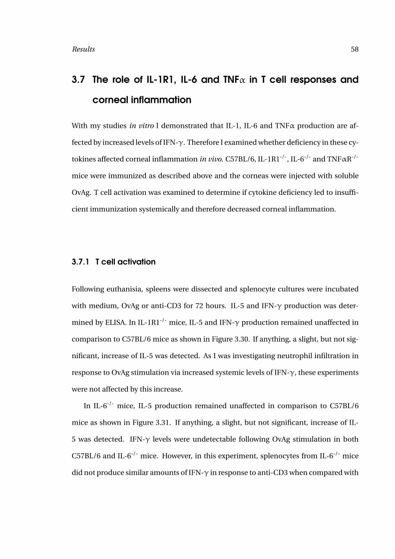
Results 58
3.7 The role of IL-1R1, IL-6 and TNFα in T cell responses and
corneal inflammation
With my studies in vitro I demonstrated that IL-1, IL-6 and TNFα production are af-
fected by increased levels of IFN-γ. Therefore I examined whether deficiency in these cy-
tokines affected corneal inflammation in vivo. C57BL/6, IL-1R1-/-, IL-6-/- and TNFαR-/-
mice were immunized as described above and the corneas were injected with soluble
OvAg. T cell activation was examined to determine if cytokine deficiency led to insuffi-
cient immunization systemically and therefore decreased corneal inflammation.
3.7.1 T cell activation
Following euthanisia, spleens were dissected and splenocyte cultures were incubated
with medium, OvAg or anti-CD3 for 72 hours. IL-5 and IFN-γ production was deter-
mined by ELISA. In IL-1R1-/- mice, IL-5 and IFN-γ production remained unaffected in
comparison to C57BL/6 mice as shown in Figure 3.30. If anything, a slight, but not sig-
nificant, increase of IL-5 was detected. As I was investigating neutrophil infiltration in
response to OvAg stimulation via increased systemic levels of IFN-γ, these experiments
were not affected by this increase.
In IL-6-/- mice, IL-5 production remained unaffected in comparison to C57BL/6
mice as shown in Figure 3.31. If anything, a slight, but not significant, increase of IL-
5 was detected. IFN-γ levels were undetectable following OvAg stimulation in both
C57BL/6 and IL-6-/- mice. However, in this experiment, splenocytes from IL-6-/- mice
did not produce similar amounts of IFN-γ in response to anti-CD3 when compared with
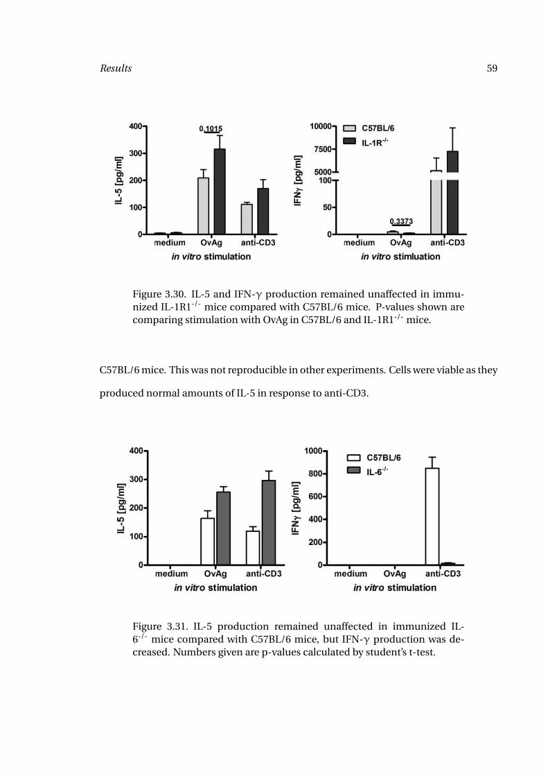
Results 59
Figure 3.30. IL-5 and IFN-γ production remained unaffected in immu-nized IL-1R1-/- mice compared with C57BL/6 mice. P-values shown arecomparing stimulation with OvAg in C57BL/6 and IL-1R1-/- mice.
C57BL/6 mice. This was not reproducible in other experiments. Cells were viable as they
produced normal amounts of IL-5 in response to anti-CD3.
Figure 3.31. IL-5 production remained unaffected in immunized IL-6-/- mice compared with C57BL/6 mice, but IFN-γ production was de-creased. Numbers given are p-values calculated by student’s t-test.
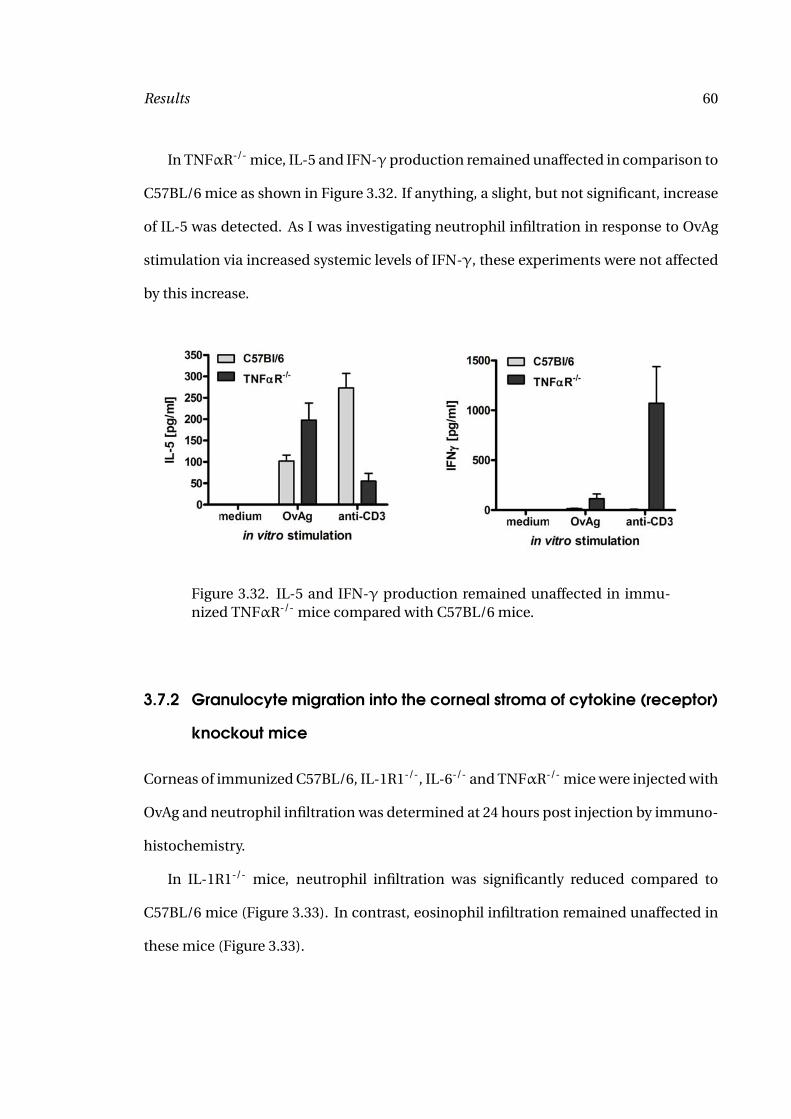
Results 60
In TNFαR-/- mice, IL-5 and IFN-γ production remained unaffected in comparison to
C57BL/6 mice as shown in Figure 3.32. If anything, a slight, but not significant, increase
of IL-5 was detected. As I was investigating neutrophil infiltration in response to OvAg
stimulation via increased systemic levels of IFN-γ, these experiments were not affected
by this increase.
Figure 3.32. IL-5 and IFN-γ production remained unaffected in immu-nized TNFαR-/- mice compared with C57BL/6 mice.
3.7.2 Granulocyte migration into the corneal stroma of cytokine (receptor)
knockout mice
Corneas of immunized C57BL/6, IL-1R1-/-, IL-6-/- and TNFαR-/- mice were injected with
OvAg and neutrophil infiltration was determined at 24 hours post injection by immuno-
histochemistry.
In IL-1R1-/- mice, neutrophil infiltration was significantly reduced compared to
C57BL/6 mice (Figure 3.33). In contrast, eosinophil infiltration remained unaffected in
these mice (Figure 3.33).

Results 61
Figure 3.33. Neutrophil infiltration into the corneal stroma is abro-gated in immunized IL-1R1-/- mice compared to C57BL/6 mice whileeosinophil infiltration remains unaffected. Numbers given are p-valuescalculated by student’s t-test.
In IL-6-/- mice, neutrophil infiltration was significantly reduced compared to
C57BL/6 mice (Figure 3.34). In contrast, eosinophil infiltration remained unaffected in
these mice, even though there was a slight trend towards decreased levels in IL-6-/- mice
(Figure 3.34).
In TNFαR-/- mice, migration of neutrophils and eosinophils to the corneal stroma
showed no difference to C57BL/6 mice (Figure 3.35).
3.8 In vivo responses to Wolbachia
3.8.1 Injection of Wolbachia depleted filarial extract into the cornea.
As previous studies suggested that pro-inflammatory immune responses are at least par-
tially induced by the endosymbiont Wolbachia (29, 84, 101), I next immunized C57BL/6
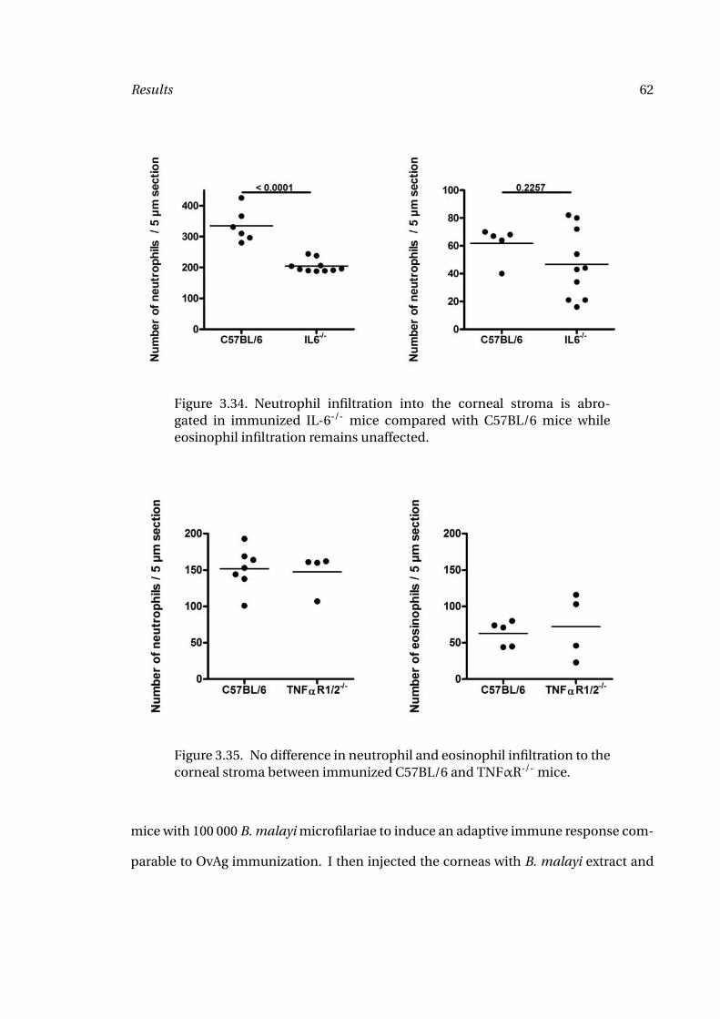
Results 62
Figure 3.34. Neutrophil infiltration into the corneal stroma is abro-gated in immunized IL-6-/- mice compared with C57BL/6 mice whileeosinophil infiltration remains unaffected.
Figure 3.35. No difference in neutrophil and eosinophil infiltration to thecorneal stroma between immunized C57BL/6 and TNFαR-/- mice.
mice with 100 000 B. malayi microfilariae to induce an adaptive immune response com-
parable to OvAg immunization. I then injected the corneas with B. malayi extract and
Results 63
extract from B. malayi worms that were treated with tetracycline (BmAgtet) for an ex-
tended period of time, thus depleting the Wolbachia. Following BmAgtet injection into
corneas of immunized mice, I found neutrophil infiltration into the corneal stroma sim-
ilar to that of unimmunized mice injected with BmAg, but significantly decreased com-
pared with immunized C57BL/6 mice injected with BmAg (Figure 3.36). This shows a
role for Wolbachia in the migration of neutrophils, in the presence of a full adaptive im-
mune response. In contrast to neutrophil infiltration, eosinophil infiltration remained
unaffected by the absence of Wolbachia from filarial extract.
3.8.2 Adaptive immune responses to A. viteae and Wolbachia
To determine Wolbachia-dependent granulocyte migration to the corneal stroma by a
second experimental design, I immunized mice with extract from A. viteae worms (AvAg)
that are naturally devoid of Wolbachia endobacteria or with Wolbachia purified from
insect cells. I then injected the corneas with AvAg, Wolbachia purified from insect cells
or a mixture of both.
After euthanasia, spleens were excised and single cell suspensions were stimulated
with AvAg, Wolbachia or both for 72 hours. IL-5 and IFN-γ production were determined
by ELISA. IL-5 production was dependent on the immunization and stimulation being
carried out with the same reagent, and AvAg as well as Wolbachia could induce similar
levels of IL-5. In contrast, IFN-γ was only induced when Wolbachia were present dur-
ing both the immunization and stimulation, indicating Wolbachia-dependent priming
events during immunization (Figure 3.37).
I then determined neutrophil and eosinophil infiltration into the corneal stroma fol-
lowing injection of AvAg or Wolbachia. Eosinophil infiltration was dependent on the
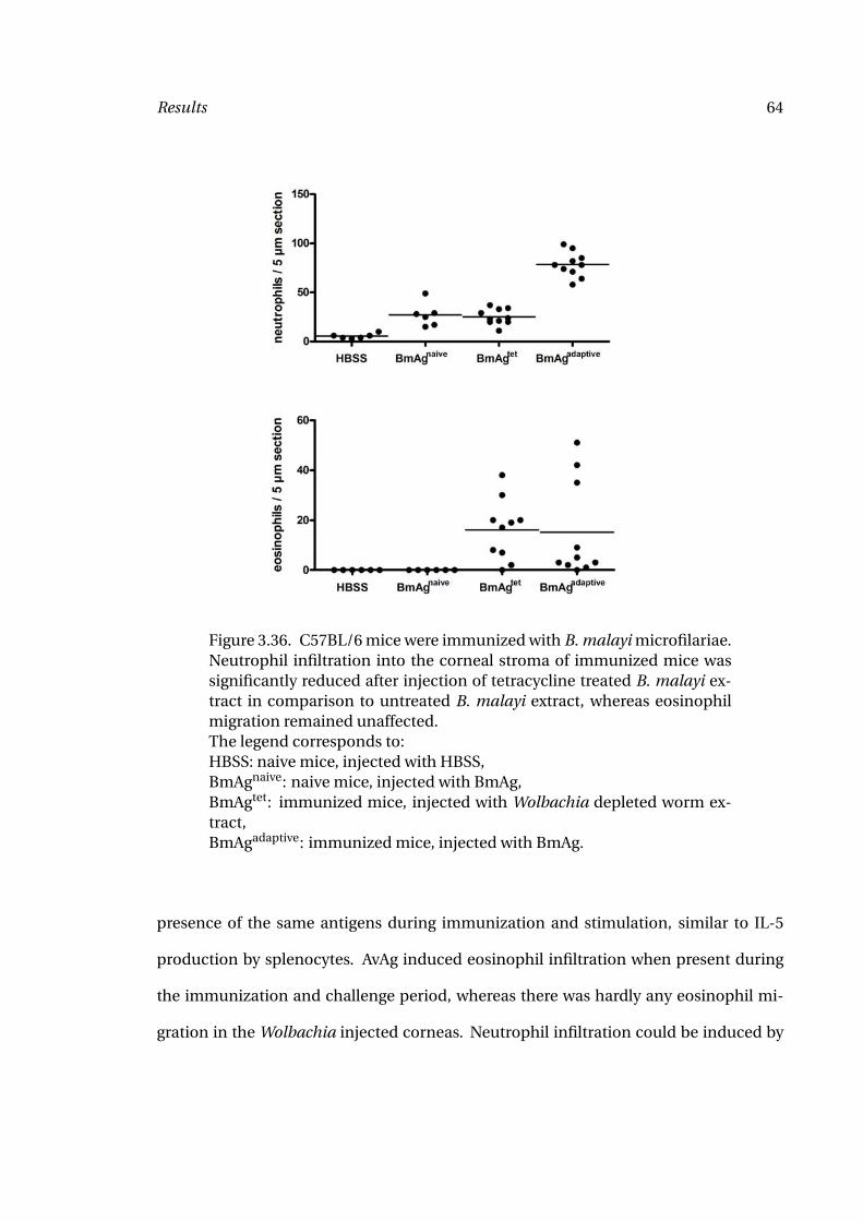
Results 64
Figure 3.36. C57BL/6 mice were immunized with B. malayi microfilariae.Neutrophil infiltration into the corneal stroma of immunized mice wassignificantly reduced after injection of tetracycline treated B. malayi ex-tract in comparison to untreated B. malayi extract, whereas eosinophilmigration remained unaffected.The legend corresponds to:HBSS: naive mice, injected with HBSS,BmAgnaive: naive mice, injected with BmAg,BmAgtet: immunized mice, injected with Wolbachia depleted worm ex-tract,BmAgadaptive: immunized mice, injected with BmAg.
presence of the same antigens during immunization and stimulation, similar to IL-5
production by splenocytes. AvAg induced eosinophil infiltration when present during
the immunization and challenge period, whereas there was hardly any eosinophil mi-
gration in the Wolbachia injected corneas. Neutrophil infiltration could be induced by
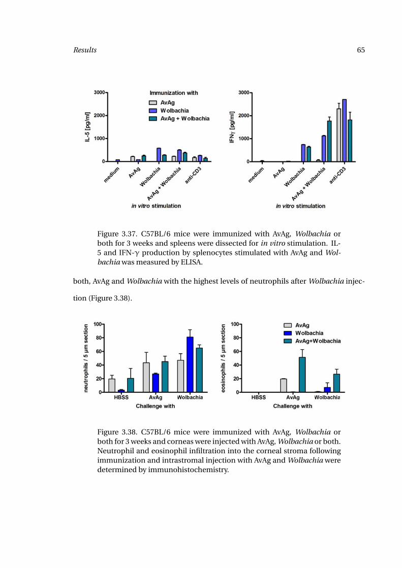
Results 65
Figure 3.37. C57BL/6 mice were immunized with AvAg, Wolbachia orboth for 3 weeks and spleens were dissected for in vitro stimulation. IL-5 and IFN-γ production by splenocytes stimulated with AvAg and Wol-bachia was measured by ELISA.
both, AvAg and Wolbachia with the highest levels of neutrophils after Wolbachia injec-
tion (Figure 3.38).
Figure 3.38. C57BL/6 mice were immunized with AvAg, Wolbachia orboth for 3 weeks and corneas were injected with AvAg, Wolbachia or both.Neutrophil and eosinophil infiltration into the corneal stroma followingimmunization and intrastromal injection with AvAg and Wolbachia weredetermined by immunohistochemistry.
Results 66
3.9 In vitro responses to rWSP
Previous studies indicated a role for the major surface protein of Wolbachia in the in-
duction of pro-inflammatory immune responses. Brattig (85) described a role for TLR2
and TLR4 in the activation of dendritic cells and macrophages by rWSP. To further char-
acterize the activation by rWSP, I generated deletion mutants of the surface protein of B.
malayi (BmWSP). BmWSP cloned into the pet100 vector was obtained from Alan Scott’s
laboratory (John Hopkins University, Baltimore, MD). The gene sequence of the cloned
BmWSP is shown in Figure 3.39. The amino acid sequence is depicted in Figure 3.40.
The full sequence contains 240 amino acids (720 base pairs). The 69 base pairs leader se-
quence (23 amino acids) was previously deleted. I generated sequential deletions from
the WSP gene by inverse PCR, replicating the complete plasmid minus the deleted se-
quence. The following deletion mutants were generated: BmWSP∆61-90, BmWSP∆91-
120, BmWSP∆121-150, BmWSP∆181-210 and BmWSP∆211-240. The wildtype and mu-
tant amino acid sequences are shown in Figure 3.40.
Proteins were purified from E. coli under native conditions.
3.9.1 HEK cell responses
To investigate activation of TLR2 by rWSP and deletion mutants, HEK cells transfected
with hTLR2 or hTLR3 were stimulated for 18 hours with the different proteins. hTLR4
transfected HEK cells were not used for this experiment as the recombinant proteins
were purified from E. coli and thus LPS contamination was very likely. For control
reagents, I used Pam3CSK (TLR2), Poly(I:C) (TLR3) and LPS (TLR4). Upon stimulation,
hTLR3 transfected HEK cells only produced IL-8 in response to Poly(I:C). All other stim-
ulations were at similar levels to the medium control. Stimulation of hTLR2 transfected
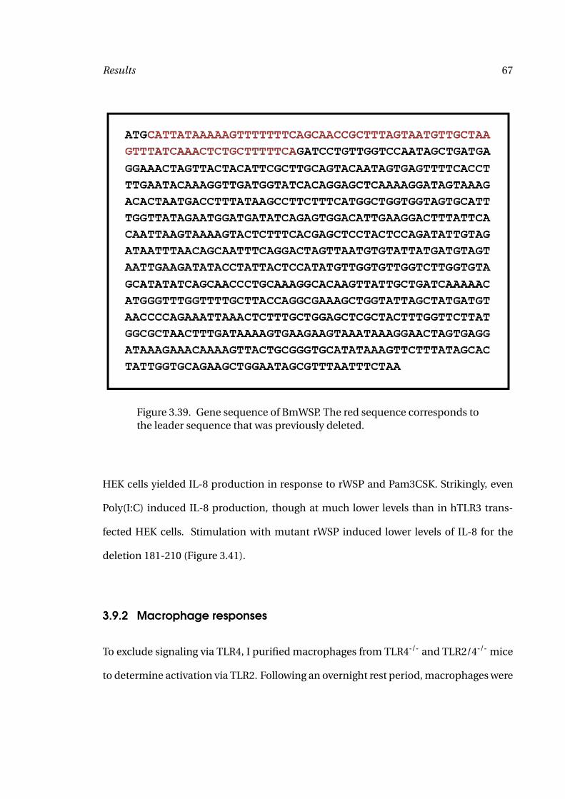
Results 67
Figure 3.39. Gene sequence of BmWSP. The red sequence corresponds tothe leader sequence that was previously deleted.
HEK cells yielded IL-8 production in response to rWSP and Pam3CSK. Strikingly, even
Poly(I:C) induced IL-8 production, though at much lower levels than in hTLR3 trans-
fected HEK cells. Stimulation with mutant rWSP induced lower levels of IL-8 for the
deletion 181-210 (Figure 3.41).
3.9.2 Macrophage responses
To exclude signaling via TLR4, I purified macrophages from TLR4-/- and TLR2/4-/- mice
to determine activation via TLR2. Following an overnight rest period, macrophages were
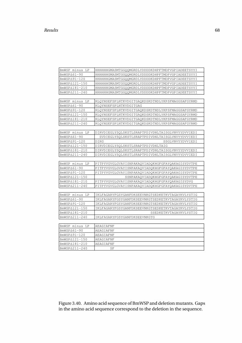
Results 68
BmWSP minus LP HHHHHHGMASMTGGQQMGRDLYDDDDKDHPFTMDPVGPIADEETSYYIBmWSPΔ61-90 HHHHHHGMASMTGGQQMGRDLYDDDDKDHPFTMDPVGPIADEETSYYIBmWSPΔ91-120 HHHHHHGMASMTGGQQMGRDLYDDDDKDHPFTMDPVGPIADEETSYYIBmWSPΔ121-150 HHHHHHGMASMTGGQQMGRDLYDDDDKDHPFTMDPVGPIADEETSYYIBmWSPΔ181-210 HHHHHHGMASMTGGQQMGRDLYDDDDKDHPFTMDPVGPIADEETSYYIBmWSPΔ211-240 HHHHHHGMASMTGGQQMGRDLYDDDDKDHPFTMDPVGPIADEETSYYI
BmWSP minus LP RLQYNSEFSPLNTKVDGITGAQKDSKDTNDLYKPSFMAGGSAFGYRMDBmWSPΔ61-90 RLQYNSEFSPLNTKVDGITGAGBmWSPΔ91-120 RLQYNSEFSPLNTKVDGITGAQKDSKDTNDLYKPSFMAGGSAFGYRMDBmWSPΔ121-150 RLQYNSEFSPLNTKVDGITGAQKDSKDTNDLYKPSFMAGGSAFGYRMDBmWSPΔ181-210 RLQYNSEFSPLNTKVDGITGAQKDSKDTNDLYKPSFMAGGSAFGYRMDBmWSPΔ211-240 RLQYNSEFSPLNTKVDGITGAQKDSKDTNDLYKPSFMAGGSAFGYRMD
BmWSP minus LP DIRVDIEGLYSQLSKSTLSRAPTPDIVDNLTAISGLVNVYYDVVIEDIBmWSPΔ61-90 SVDIEGLYSQLSKSTLSRAPTPDIVDNLTAISGLVNVYYDVVIEDIBmWSPΔ91-120 DIRG SSGLVNVYYDVVIEDIBmWSPΔ121-150 DIRVDIEGLYSQLSKSTLSRAPTPDIVDNLTAIG BmWSPΔ181-210 DIRVDIEGLYSQLSKSTLSRAPTPDIVDNLTAISGLVNVYYDVVIEDIBmWSPΔ211-240 DIRVDIEGLYSQLSKSTLSRAPTPDIVDNLTAISGLVNVYYDVVIEDI
BmWSP minus LP PITPYVGVGLGVAYISNPAKAQVIADQKHGFGFAYQAKAGISYDVTPEBmWSPΔ61-90 PITPYVGVGLGVAYISNPAKAQVIADQKHGFGFAYQAKAGISYDVTPEBmWSPΔ91-120 PITPYVGVGLGVAYISNPAKAQVIADQKHGFGFAYQAKAGISYDVTPEBmWSPΔ121-150 SSNPAKAQVIADQKHGFGFAYQAKAGISYDVTPEBmWSPΔ181-210 PITPYVGVGLGVAYISNPAKAQVIADQKHGFGFAYQAKAGISYDVGBmWSPΔ211-240 PITPYVGVGLGVAYISNPAKAQVIADQKHGFGFAYQAKAGISYDVTPE
BmWSP minus LP IKLFAGARYFGSYGANFDKSEEVNKGTSEDKETKVTAGAYKVLYSTIGBmWSPΔ61-90 IKLFAGARYFGSYGANFDKSEEVNKGTSEDKETKVTAGAYKVLYSTIGBmWSPΔ91-120 IKLFAGARYFGSYGANFDKSEEVNKGTSEDKETKVTAGAYKVLYSTIGBmWSPΔ121-150 IKLFAGARYFGSYGANFDKSEEVNKGTSEDKETKVTAGAYKVLYSTIGBmWSPΔ181-210 SSEDKETKVTAGAYKVLYSTIGBmWSPΔ211-240 IKLFAGARYFGSYGANFDKSEEVNKGTG
BmWSP minus LP AEAGIAFNFBmWSPΔ61-90 AEAGIAFNFBmWSPΔ91-120 AEAGIAFNFBmWSPΔ121-150 AEAGIAFNFBmWSPΔ181-210 AEAGIAFNFBmWSPΔ211-240 SF
Figure 3.40. Amino acid sequence of BmWSP and deletion mutants. Gapsin the amino acid sequence correspond to the deletion in the sequence.
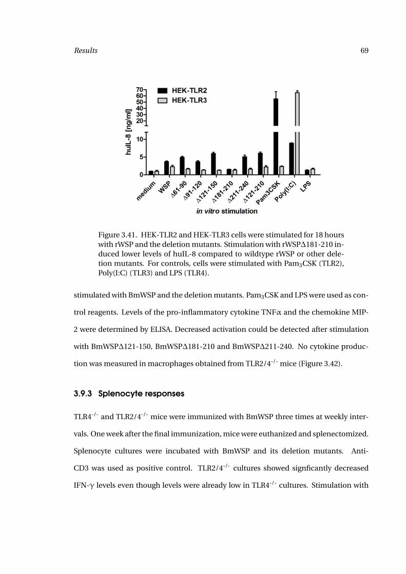
Results 69
Figure 3.41. HEK-TLR2 and HEK-TLR3 cells were stimulated for 18 hourswith rWSP and the deletion mutants. Stimulation with rWSP∆181-210 in-duced lower levels of huIL-8 compared to wildtype rWSP or other dele-tion mutants. For controls, cells were stimulated with Pam3CSK (TLR2),Poly(I:C) (TLR3) and LPS (TLR4).
stimulated with BmWSP and the deletion mutants. Pam3CSK and LPS were used as con-
trol reagents. Levels of the pro-inflammatory cytokine TNFα and the chemokine MIP-
2 were determined by ELISA. Decreased activation could be detected after stimulation
with BmWSP∆121-150, BmWSP∆181-210 and BmWSP∆211-240. No cytokine produc-
tion was measured in macrophages obtained from TLR2/4-/- mice (Figure 3.42).
3.9.3 Splenocyte responses
TLR4-/- and TLR2/4-/- mice were immunized with BmWSP three times at weekly inter-
vals. One week after the final immunization, mice were euthanized and splenectomized.
Splenocyte cultures were incubated with BmWSP and its deletion mutants. Anti-
CD3 was used as positive control. TLR2/4-/- cultures showed signficantly decreased
IFN-γ levels even though levels were already low in TLR4-/- cultures. Stimulation with
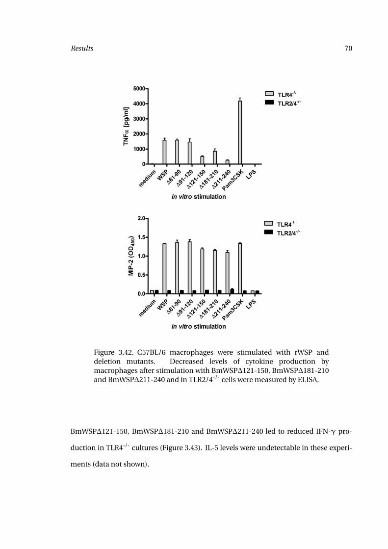
Results 70
Figure 3.42. C57BL/6 macrophages were stimulated with rWSP anddeletion mutants. Decreased levels of cytokine production bymacrophages after stimulation with BmWSP∆121-150, BmWSP∆181-210and BmWSP∆211-240 and in TLR2/4-/- cells were measured by ELISA.
BmWSP∆121-150, BmWSP∆181-210 and BmWSP∆211-240 led to reduced IFN-γ pro-
duction in TLR4-/- cultures (Figure 3.43). IL-5 levels were undetectable in these experi-
ments (data not shown).
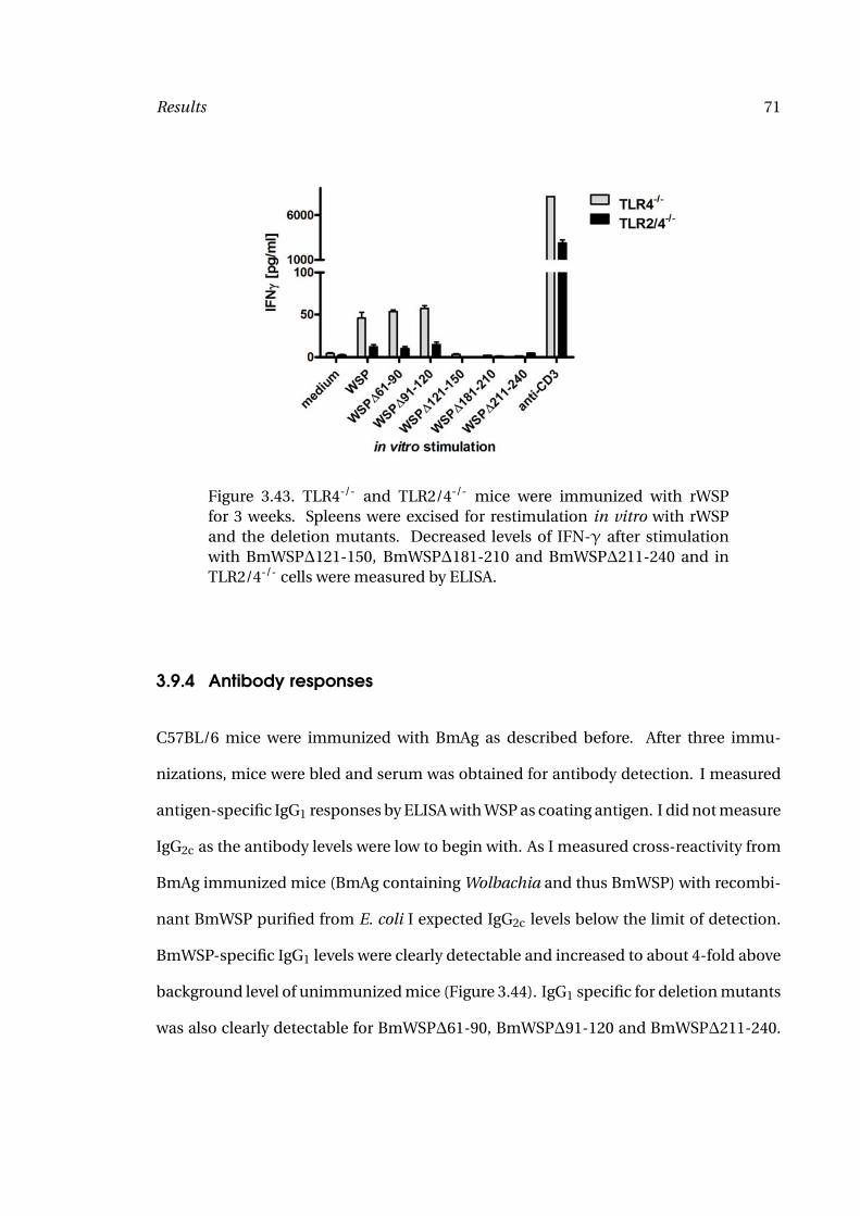
Results 71
Figure 3.43. TLR4-/- and TLR2/4-/- mice were immunized with rWSPfor 3 weeks. Spleens were excised for restimulation in vitro with rWSPand the deletion mutants. Decreased levels of IFN-γ after stimulationwith BmWSP∆121-150, BmWSP∆181-210 and BmWSP∆211-240 and inTLR2/4-/- cells were measured by ELISA.
3.9.4 Antibody responses
C57BL/6 mice were immunized with BmAg as described before. After three immu-
nizations, mice were bled and serum was obtained for antibody detection. I measured
antigen-specific IgG1 responses by ELISA with WSP as coating antigen. I did not measure
IgG2c as the antibody levels were low to begin with. As I measured cross-reactivity from
BmAg immunized mice (BmAg containing Wolbachia and thus BmWSP) with recombi-
nant BmWSP purified from E. coli I expected IgG2c levels below the limit of detection.
BmWSP-specific IgG1 levels were clearly detectable and increased to about 4-fold above
background level of unimmunized mice (Figure 3.44). IgG1 specific for deletion mutants
was also clearly detectable for BmWSP∆61-90, BmWSP∆91-120 and BmWSP∆211-240.
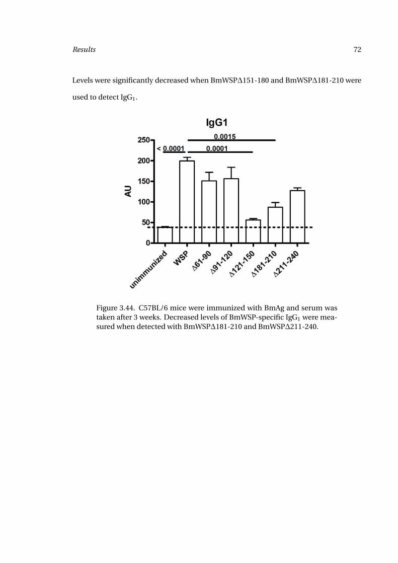
Results 72
Levels were significantly decreased when BmWSP∆151-180 and BmWSP∆181-210 were
used to detect IgG1.
Figure 3.44. C57BL/6 mice were immunized with BmAg and serum wastaken after 3 weeks. Decreased levels of BmWSP-specific IgG1 were mea-sured when detected with BmWSP∆181-210 and BmWSP∆211-240.
73
4 Discussion
4.1 Activation of antigen-presenting cells
With the results from this study and the experiments by colleagues, we could demon-
strate that dendritic cells, neutrophils and macrophages are all activated by filarial
extracts (83, 86). All three cell types require TLR2 expression for activation. For
macrophage activation, the requirement of TLR6, MyD88 and Mal was also shown. Ex-
pectedly, dendritic cells required MyD88 expression for activation by filarial extracts
(data not shown).
Dendritic cell and macrophage activation are essential for efficient induction of
adaptive immune responses. Dendritic cells can efficiently activate T cells when previ-
ously activated by TLR ligands (80). Upon activation, antigen uptake, membrane trans-
port and antigen presentation are described to be more efficient (80). Typical DC1 cy-
tokines are IL-12, IL-23 and typical DC2 cytokines are IL-10 and TGF-β (51), cytokines
more recently associated with the generation of regulatory T cells than in the generation
of Th2 cells, whose generation is characterized by low levels of IL-12 produced by den-
dritic cells (51). Myeloid dendritic cells in humans are capable of inducing Th1 or Th2
responses, depending on the production of IL-12 (109) whereas plasmacytoid dendritic
Discussion 74
cells induce Th1 cells. Initial studies with plasmacytoid dendritic cells did not show con-
sistent cytokine production (data not shown), so I focused on myeloid dendritic cells
instead. I initially measured IL-6, IL-12, IP10, TNFα and RANTES and found signficant
production of IL-6, RANTES and TNFα (data not shown). Missing production of IL-12
implicates differentiation towards Th2 which is the common pathway during filarial in-
fection. It has also been shown that activation of TLR2 does not induce IL-12 or IP10
production in human monocyte-derived dendritic cells (110).
Activation of the costimulatory molecules CD80 and CD86 will cause CD28 engage-
ment on the T cells (49). Interaction between CD80/CD86 and CD28 in combination
with T cell receptor activation via MHCII induces differentiation of naive T cells into Th1
or Th2 cells (111). I only found a slight increase of CD86, but higher levels of CD80. How-
ever, dendritic cells are differentially regulated and do not express all activation markers
upon each stimulation. Similar to cytokine production, the upregulation of costimula-
tory molecules was TLR2-dependent.
CD40 upregulation has been demonstrated early in the activation of dendritic cells.
Increased expression of CD40 can lead to higher MHCII expression upon CD40 ligand
engagement and thus more efficient antigen presentation (111). CD40-CD40 ligand in-
teractions were shown to be critical during dendritic cell responses to parasite antigens
(112). I found increased CD40 expression on wild-type dendritic cells, that was absent
in TLR2-/- cells.
To protect themselves from ROS produced by macrophages, neutrophils and
eosinophils, O. volvulus and B. malayi contain thioredoxin peroxidase (113–115). In-
creased production of ROS has been related to expulsion of the nematode Nippostrongy-
lus brasiliensis (116). Neutrophil activation by filarial extracts was investigated using
Discussion 75
several markers: myeloperoxide production, MIP-2 production, generation of nitric ox-
ide and reactive oxygen species, expression of CD11b, CD18 and L-Selectin and release
of MMPs. I found production of MPO and MIP-2, generation of ROS and upregulation of
CD11b and CD18, while L-Selectin was shed. No generation of NO or release of gelati-
nases could be clearly demonstrated. Expression of surface activation markers CD11b,
CD18 and L-Selectin was regulated TLR2-dependent as was the production of MIP-2.
ROS generation occured independent of TLR2 and could not be induced by TLR2 ago-
nists. This response could therefore be due to the filarial component more than to the
Wolbachia content of the protein extract. Future experiments may address this question
by stimulating neutrophils with the Wolbachia-free extract from A. viteae.
4.2 TLR2 in the adaptive immune response
This study examined the role of TLRs in the generation of eye disease. Previous studies
had shown (i) that neutrophil infiltration into the cornea was dependent on the pres-
ence of functional TLR4 and Wolbachia (84), and (ii) TLR2- and TLR4-dependent acti-
vation by Wolbachia surface protein (85). Studies carried out in our lab at the same time
could show neutrophil migration to the cornea depending on the presence of MyD88
(29) without previous immunizations. Further investigations revealed requirement of
TLR2, but not TLR4 for neutrophil infiltration (83). In this study, it was also shown by
using bone-marrow chimeric mice that TLR2 expression on either hematopoetic and
resident stromal cells can induce neutrophil infiltration, though not to the same levels
as in wildtype reconstituted bone-marrow chimeric mice.
Discussion 76
In this study I could also show dependence on TLR2 for neutrophil infiltration in the
presence of adaptive immune responses. Systemic responses as characterized by anti-
body responses were normal in TLR2-/- mice. As previous studies showed a requirement
for antibody production for neutrophil migration to the cornea (34), we anticipated nor-
mal neutrophil migration and were surprised that the requirement of the innate receptor
TLR2 was more important than the presence of antibodies. Neutrophil numbers in the
corneas of OvAg-injected immunized TLR2-/- mice were comparable to that of OvAg-
injected unimmunized wildtype mice. This indicates a role for the adaptive arm of the
immune system in further enhancing neutrophil migration. A potential pathway was
presented by TLR2-dependent IFN-γ production by splenocytes. Decreased levels of
systemic IFN-γ were hypothesized to affect the expression of PECAM on corneal vessels
as shown previously (96) and CXC chemokine production in the cornea.
Taken together, I found requirement for TLR2 at the stages: (i) activation of antigen-
presenting cells, (ii) IFN-γ production by splenocytes, (iii) CXC chemokine production
and (iv) neutrophil migration to the cornea. While decreased IFN-γ production could be
linked to decreased activation of dendritic cells and macrophages and decreased neu-
trophil migration is caused by decreased chemokine production in the cornea, there
is no obvious link between IFN-γ production and CXC chemokine production in the
cornea. This surprising finding resulted in the second part of this study, investigating
IFN-γ dependent neutrophil migration to the corneal stroma.
Discussion 77
4.3 IFN-γ in the adaptive immune response
I could also demonstrate increased neutrophil infiltration into the corneal stroma in
the presence of adaptive immune responses and showed that this increased neutrophil
infiltration is at least partially caused by IFN-γ.
Previous studies have shown strong IL-5 and weak IFN-γ production by splenocytes
in response to restimulation with filarial extracts (32, 117). IFN-γ levels were usually
only slightly above the limit of detection. I was therefore surprised that this low response
was even further reduced in the absence of TLR2. Previous studies on Herpes simplex
keratitis have identified IFN-γ as a regulator of PECAM-1 expression in the vascular en-
dothelium of limbal vessels (96). It was shown that the decreased expression of PECAM-
1 corresponded to decreased neutrophil infiltration into the corneal stroma (96). Studies
by our lab have also shown PECAM-1 expression being important for neutrophil migra-
tion during onchocercal keratitis (38). However, I could not show decreased PECAM-1
expression in TLR2-/- mice compared to C57BL/6 mice. I then wanted to investigate if
there was a connection between decreased systemical IFN-γ production and neutrophil
migration to the cornea. First, I investigated neutrophil migration in IFN-γ-/- mice and
found decreased levels of neutrophil infiltration in the absence of IFN-γ although neu-
trophil levels were still above background levels. However, neutrophils also migrate to
the corneas of naive mice injected into the corneal stroma in the absence of an adap-
tive immune response (29). It is therefore easily explained that neutrophil migration in
IFN-γ-/- mice reached similar levels compared to neutrophil migration in the absence
of adaptive immune responses. However, as in TLR2-/- mice, PECAM-1 expression in the
corneas of IFN-γ-/- mice was not different from C57BL/6 mice.
Discussion 78
Therefore I investigated alternative pathways of IFN-γ mediated neutrophil migra-
tion to the corneal stroma.
Resident cells in the cornea include keratinocytes (fibroblasts) and macrophages,
with a very low number of dendritic cells present (118). The first cells to reach
the cornea upon inflammation are neutrophils. I then investigated activation of fi-
broblasts (using the immortalized mouse corneal fibroblast cell line MK/T-1 (99)),
macrophages and neutrophils by IFN-γ and found no activation of either chemokines
or pro-inflammatory cytokines. As priming effects of IFN-γ stimulation in macrophages
have been demonstrated previously (56), I then investigated costimulation of fibrob-
lasts, macrophages and neutrophils with IFN-γ and filarial extract. I could demonstrate
significantly increased production of pro-inflammatory cytokines by macrophages. Nei-
ther fibroblasts nor neutrophils showed synergistic responses to IFN-γ and OvAg, nor
did macrophages produce increased levels of neutrophil attractant chemokines. How-
ever, pro-inflammatory cytokines alone will not increase neutrophil migration to the
cornea. I hypothesized autokrin or parakrin effects of those pro-inflammatory cytokines
on macrophages, fibroblasts or neutrophils. Stimulating fibroblasts with recombinant
IL-1α, IL-1β, IL-6 and TNF-α did indeed induce production of chemokines. No signifi-
cant effect on macrophages or neutrophils was observed.
I examined alternative pathways of macrophage priming by IFN-γ. Regulation
of TLR2 expression on biliary epithelial cells (93) and on rheumatoid synovial tis-
sues (92) by IFN-γ was previously shown. I therefore investigated TLR2 expression in
macrophages in response to recombinant IFN-γ and could show by Western Blot that
total TLR2 expression was increased and by FACS that surface expression of TLR2 was
upregulated as well.
Discussion 79
I attempted to investigate whether I could substitute the increased systemic levels
of IFN-γ that I achieve during immunization by local injection of rIFN-γ or rIL-12 into
the cornea. Injection of rIFN-γ had no effect on neutrophil migration (data not shown).
As rIFN-γ is known for a short half life, I then injected rIL-12 as an inducer of rIFN-γ.
I anticipated that rIL-12 would induce IFN-γ over an extended period of time and thus
efficiently prime corneal macrophages. However, repeated experiments produced only
inconclusive data with the occasional increase in neutrophil migration or without hav-
ing an effect on neutrophil migration (data not shown). The reason for this missing re-
sponse could be fast degradation of the recombinant cytokines or insufficient amounts
injected. I calculated the amount injected on other studies using rIL-12, however, in-
jection volume into the cornea is limited and non-physiological, high concentrations of
rIL-12 could have side effects like damaging the corneal matrix.
With this part of the study I clearly showed that even though IFN-γ is only pro-
duced at low levels by splenocytes, it does play an important role in the priming of
pro-inflammatory immune responses and in the development of Onchocerca keratitis.
Though it does induce higher levels of IL-1α, IL-1β, IL-6 and TNFα in macrophages,
only IL-1α and TNFα induce chemokine production in corneal fibroblasts. When exam-
ining the role of the two corresponding cytokine receptors, only IL-1R1 could be shown
to be involved in neutrophil migration, but not TNFαR1/2. Though I could show that
IL-6 affected neutrophil migration to the corneal stroma, this is likely to be due to a dif-
ferent mechanism.
Discussion 80
4.4 Wolbachia
Previous studies have shown a requirement of Wolbachia for neutrophil migration to
the corneal stroma (29, 84, 101). However, these studies only examined neutrophil infil-
tration in the absence of adaptive immune responses. Other studies showed that neu-
trophil infiltration into onchocercal skin nodules was depending on the presence of Wol-
bachia (9). Yet other studies implicated Wolbachia in the development of Th1 immune
responses (119). Wolbachia was also shown to cause severe pro-inflammatory responses
when released from worms upon treatment with diethylcarbamazine (11). I therefore
wanted to test the role of Wolbachia in the adaptive phase of the immune response.
I first generated a full adaptive immune response by immunizing mice with B. malayi
microfilariae. No difference could be detected in the cytokine and antibody responses
of mice then treated with BmAg or with BmAgtet. I then injected BmAg and BmAgtet
into the corneas. BmAgtet was obtained from worms of tetracycline treated Meriones
unguiculatus. Migration of neutrophils was found to be decreased in the absence of
Wolbachia while the migration of eosinophils was not affected. The decreased migration
of neutrophils is therefore likely to be due to the lack of Wolbachia.
In a second model, I immunized mice with A. viteae and/or Wolbachia, then injected
the corneas with A. viteae and/or Wolbachia. I used A. viteae as Wolbachia-free filarial
control. As before, the presence of Wolbachia increased neutrophil migration. In con-
trast, there was no eosinophil infiltration in response to Wolbachia alone. This all im-
plicates a role for Wolbachia in neutrophil migration while at the same time, Wolbachia
appears dispensable for the migration of eosinophils.
Discussion 81
A. viteae also induced neutrophil migration. This is in contrast to the results ob-
tained with Wolbachia-depleted B. malayi extract, however, this might be due to work-
ing with a related, but not identical organism that is naturally void of Wolbachia. Litera-
ture suggests that A. viteae may have harbored the endosymbiont Wolbachia, but lost it
during evolution (120) and incorporated essential genes into its genome.
Previous studies have shown Wolbachia dependent generation of Th1 immune re-
sponses (119). Similarly, I found IFN-γ production only when mice were immunized
and splenocytes challenged with Wolbachia. In contrast, I found IL-5 production in re-
sponse to both filarial and bacterial stimulation, but only when mice were previously
immunized with the same reagent.
With these findings, Wolbachia alone can induce a functional immune response at
the splenocyte level and neutrophil infiltration into the cornea. However, eosinophil
infiltration is caused by filaria alone.
4.5 BmWSP
BmWSP has been previously cloned by other laboratories (85, 121). I obtained the plas-
mid from Alan L. Scott (John Hopkins University, Baltimore).
Porksakorn et al. (121) demonstrated activation of the murine cell line RAW264.7 by
recombinant BmWSP. The cells presented increased expression of IL-1α, IL-1β, IL-6 and
TNFα at the RNA level. To exclude LPS contamination, purified proteins were (1) treated
with proteinase K and decreased macrophage activation was shown, implying a role for
the protein component in activating macrophages; and (2) treated with polymyxin B.
Treatment of LPS with polymyxin B decreased cytokine expression to background levels,
Discussion 82
while there was only a slight reduction of cytokine expression when BmWSP was treated
with polymyxin B.
BmWSP was purified under native and denaturing conditions. I obtained higher
protein levels from purification under denaturing conditions, however, the activation
of HEK cells and macrophages was decreased compared with native BmWSP (data not
shown). I therefore continued my studies with native BmWSP. I was especially interested
in activation of TLR2 by the recombinant BmWSP. Previous studies have also identified
TLR4 as receptor for WSP (85).
As I was purifying the proteins from E. coli, I assumed endotoxin contamination and
therefore TLR4 activation. For the studies comparing deletion mutants of BmWSP, I then
used TLR4-/- macrophages as controls and TLR2/4-/- macrophages as cells of interest. I
found activation of TLR4-/- macrophages that was completely abrogated in TLR2/4-/-
macrophages, clearly indicating involvement of TLR2 in the activation pathway. I con-
firmed activation of TLR2 by stimulation of TLR2-transfected HEK293 cells and again
found activation of TLR2-transfected HEK293 cells, but not TLR3-transfected HEK293
cells. As expected, TLR4-transfected HEK293 cells were activated (data not shown), at-
tributing this activation to endotoxin contamination.
I also stimulated splenocytes from BmWSP immunized mice and found increased
IFN-γ production in response to BmWSP compared with medium stimulation.
I then stimulated the same cells with deletion mutants of BmWSP. I consistently
showed wildtype cytokine production when cells were stimulated with BmWSP∆61-
90 and BmWSP∆91-120. Deletion mutants BmWSP∆181-210 consistently showed
decreased cytokine production in TLR2-transfected HEK293 cells, macrophages and
Discussion 83
splenocytes. This sequence is likely to be important for recognition by TLR2. Mu-
tants BmWSP∆121-150 and BmWSP∆211-240 showed decreased cytokine production
in some assays but not all. The role of those sequences will have to be further analyzed.
Finally, I tested antibody responses of BmAg immunized mice against recombinant
BmWSP. Serum from immunized mice could recognize recombinant BmWSP, showing
homology to the in vivo expressed proteins. Again, I could demonstrate clear decrease
in antibody responses for the mutants BmWSP∆121-150 and BmWSP∆181-210.
I extended previous studies to investigate the sole role of TLR2 in the activation pro-
cess.
When purifying recombinant proteins from E. coli, there will always be low level en-
dotoxin contamination. I therefore decided not to investigate the involvement of TLR4
in more detail especially since my previous studies as well as those of collegues could
not identify a role for this receptor in the mouse model of river blindness. While endo-
toxin effects can be minimalized by the addition of polymyxin B, I was more interested
in TLR2 activation.
BmWSP lacks signal sequences required for acylation. Usually TLR2 ligands are di-
or triacylated lipoproteins, depending on the coreceptor usage (122). There is only one
known non-acylated TLR2 ligand: PorinB of Neisseria meningitidis (123, 124). Direct
interaction between Neisseria porin and TLR2 was shown by an ELISA-like assay (124).
Coreceptor for activtion by this protein is TLR1.
Smaller studies also implicated porin of Shigella dysenteriae as a TLR2 ligand (125).
However, studies by this group never analyzed TLR2 activation in either TLR2-/- mice
or used single TLR-transfected HEK cells. Binding of porin to TLR2 was never demon-
strated by co-immunoprecipitation either. The requirement of TLR2 was concluded
Discussion 84
from upregulation of TLR2 upon stimulation of B cells and macrophages with porin and
from decreased activation (50 %) following anti-TLR2 treatment of macrophages. Fol-
lowing the same reasoning that upregulation of RNA levels of TLRs reflected TLR usage
by porins, this group also implicated TLR6 in the activation by Shigella porin.
Summarizing studies on those two porins, there is one confirmed TLR2 ligand that
requires TLR1 as coreceptor and the second porin might activate TLR2 with TLR6 as
coreceptor.
It will therefore be very interesting to confirm TLR2-dependent activation of
macrophages by BmWSP. Future studies will focus on purifying the protein from insect
cells to prevent LPS contamination. The protein will then be used for interaction stud-
ies with TLR2, TLR4, TLR1, TLR6 and CD14. Molecular interactions will be interesting
because of the lack of acylation. The binding will then be further analyzed by using the
here described deletion mutants. BmWSP is more likely to require TLR6 as coreceptor
as I am only showing TLR2- and TLR6-dependent activation when using whole filarial
extracts.
In addition to endotoxin contamination, there could also potentially be lipoprotein
contamination from E. coli. This problem will not be solved by expressing BmWSP in
insect cells as insect cells contain large numbers of lipoproteins. Lipoprotein activity
can be decreased by incubation of BmWSP with cleanascite.
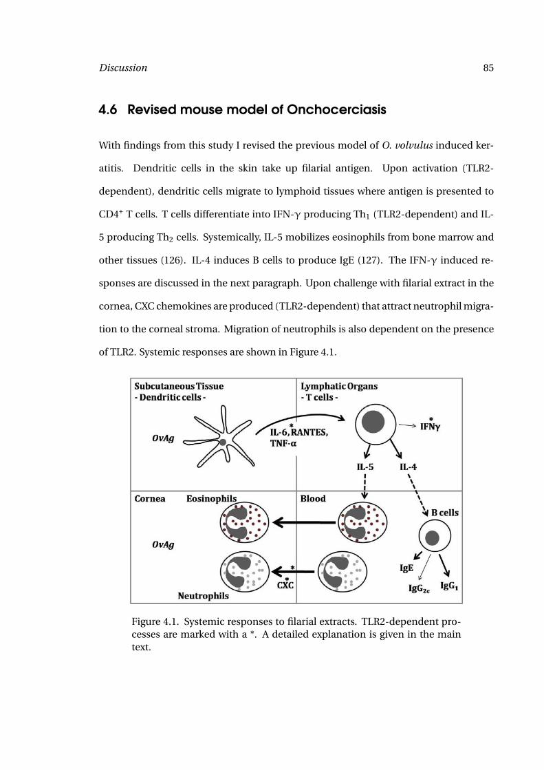
Discussion 85
4.6 Revised mouse model of Onchocerciasis
With findings from this study I revised the previous model of O. volvulus induced ker-
atitis. Dendritic cells in the skin take up filarial antigen. Upon activation (TLR2-
dependent), dendritic cells migrate to lymphoid tissues where antigen is presented to
CD4+ T cells. T cells differentiate into IFN-γ producing Th1 (TLR2-dependent) and IL-
5 producing Th2 cells. Systemically, IL-5 mobilizes eosinophils from bone marrow and
other tissues (126). IL-4 induces B cells to produce IgE (127). The IFN-γ induced re-
sponses are discussed in the next paragraph. Upon challenge with filarial extract in the
cornea, CXC chemokines are produced (TLR2-dependent) that attract neutrophil migra-
tion to the corneal stroma. Migration of neutrophils is also dependent on the presence
of TLR2. Systemic responses are shown in Figure 4.1.
Figure 4.1. Systemic responses to filarial extracts. TLR2-dependent pro-cesses are marked with a *. A detailed explanation is given in the maintext.
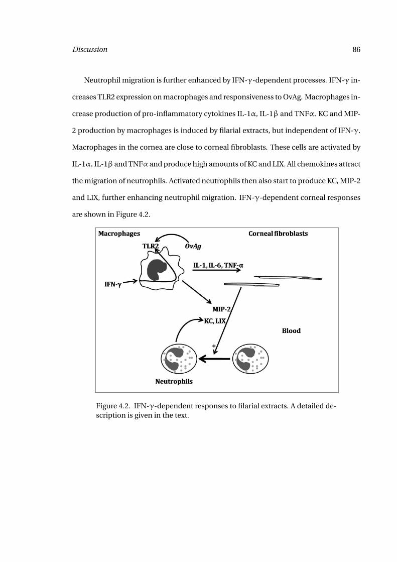
Discussion 86
Neutrophil migration is further enhanced by IFN-γ-dependent processes. IFN-γ in-
creases TLR2 expression on macrophages and responsiveness to OvAg. Macrophages in-
crease production of pro-inflammatory cytokines IL-1α, IL-1β and TNFα. KC and MIP-
2 production by macrophages is induced by filarial extracts, but independent of IFN-γ.
Macrophages in the cornea are close to corneal fibroblasts. These cells are activated by
IL-1α, IL-1β and TNFα and produce high amounts of KC and LIX. All chemokines attract
the migration of neutrophils. Activated neutrophils then also start to produce KC, MIP-2
and LIX, further enhancing neutrophil migration. IFN-γ-dependent corneal responses
are shown in Figure 4.2.
Figure 4.2. IFN-γ-dependent responses to filarial extracts. A detailed de-scription is given in the text.
87
5 Summary
In this study, I first identified a critical role for TLR2 in Th1 associated immune re-
sponses: decreased dendritic cell activation, decreased IFN-γ production by spleno-
cytes, decreased chemokine production in the cornea, and decreased neutrophil migra-
tion to the corneal stroma, whereas TLR4 does not appear to be involved in these steps.
Typical anti-helminth responses as IL-5 production and eosinophil migration were in-
dependent of TLR2.
Linking the systemic IFN-γ production to neutrophil migration to the corneal
stroma, I could show, that IFN-γ increases TLR2 expression and thus responsiveness to
the TLR2 ligand OvAg in macrophages. Primed macrophages do not produce increased
levels of chemokines, but proinflammatory cytokines that prime chemokine production
by corneal fibroblasts. The importance of these cytokines in neutrophil migration could
be confirmed in vivo where neutrophil migration was decreased in the absence of IL-
1R1, TNFαR1/2 and IL-6.
Examining the role of filarial proteins and the endosymbiont Wolbachia in the ac-
tivation of proinflammatory immune responses, I found that IFN-γ was only induced
by Wolbachia, but not by filarial extracts. Similarly, neutrophil migration to the corneal
stroma was induced by Wolbachia, although A. viteae could induce some neutrophil
Summary 88
migration as well. Eosinophil migration to the corneal stroma, in contrast, was only de-
pendent on the presence of filarial proteins.
Analyzing the recombinant proinflammatory protein BmWSP for its activation po-
tential, I found that I could activate TLR2-transfected HEK293 cells, primary murine
macrophages and splenocytes, all in a TLR2-dependent manner. Deletion mutants of
BmWSP identified region 121-240 as immunostimulatory part of BmWSP.
Summarizing, I identify Th2 responses as TLR2-independent, but filaria-dependent,
whereas Th1 responses are TLR2- and Wolbachia-dependent, with BmWSP as one can-
didate protein for the induction of proinflammatory responses via TLR2.
References 89
References
[1] WHO. Fact sheet nr 102: Lymphatic filariasis, 2000.
[2] Gladys Ozoh, Michel Boussinesq, Anne-Cécile Zoung-Kanyi Bissek, LéonKobangue, Maryvonne Kombila, Jean-Romain Mourou Mbina, Peter Enyong,Mounkaïla Noma, Azodoga Sékétéli, and Grace Fobi. Evaluation of the diethyl-carbamazine patch to evaluate onchocerciasis endemicity in central africa. TropMed Int Health, 12(1):123–129, Jan 2007.
[3] Achim Hoerauf. Filariasis: new drugs and new opportunities for lymphatic filaria-sis and onchocerciasis. Curr Opin Infect Dis, 21(6):673–681, Dec 2008.
[4] W. J. Kozek and H. F. Marroquin. Intracytoplasmic bacteria in onchocerca volvulus.Am J Trop Med Hyg, 26(4):663–678, Jul 1977.
[5] M. Sironi, C. Bandi, L. Sacchi, B. Di Sacco, G. Damiani, and C. Genchi. Molecularevidence for a close relative of the arthropod endosymbiont wolbachia in a filarialworm. Mol Biochem Parasitol, 74(2):223–227, Nov 1995.
[6] Helen F McGarry, Gillian L Egerton, and Mark J Taylor. Population dynamicsof wolbachia bacterial endosymbionts in brugia malayi. Mol Biochem Parasitol,135(1):57–67, May 2004.
[7] Katelyn Fenn and Mark Blaxter. Quantification of wolbachia bacteria in brugiamalayi through the nematode lifecycle. Mol Biochem Parasitol, 137(2):361–364,Oct 2004.
[8] Tarig B Higazi, Anthony Filiano, Charles R Katholi, Yankum Dadzie, Jan H Remme,and Thomas R Unnasch. Wolbachia endosymbiont levels in severe and mildstrains of onchocerca volvulus. Mol Biochem Parasitol, 141(1):109–112, May 2005.
[9] N. W. Brattig, D. W. Büttner, and A. Hoerauf. Neutrophil accumulation around on-chocerca worms and chemotaxis of neutrophils are dependent on wolbachia en-dobacteria. Microbes Infect, 3(6):439–446, May 2001.
[10] Paul B Keiser, Stacey M Reynolds, Kwablah Awadzi, Eric A Ottesen, Mark J Taylor,and Thomas B Nutman. Bacterial endosymbionts of onchocerca volvulus in thepathogenesis of posttreatment reactions. J Infect Dis, 185(6):805–811, Mar 2002.
[11] H. F. Cross, M. Haarbrink, G. Egerton, M. Yazdanbakhsh, and M. J. Taylor. Severereactions to filarial chemotherapy and release of wolbachia endosymbionts intoblood. Lancet, 358(9296):1873–1875, Dec 2001.
References 90
[12] A. Hoerauf, K. Nissen-Pähle, C. Schmetz, K. Henkle-Dührsen, M. L. Blaxter, D. W.Büttner, M. Y. Gallin, K. M. Al-Qaoud, R. Lucius, and B. Fleischer. Tetracycline ther-apy targets intracellular bacteria in the filarial nematode litomosoides sigmodon-tis and results in filarial infertility. J Clin Invest, 103(1):11–18, Jan 1999.
[13] Sabine Specht, Sabine Mand, Yeboah Marfo-Debrekyei, Alexander Yaw Debrah,Peter Konadu, Ohene Adjei, Dietrich W Büttner, and Achim Hoerauf. Efficacy of 2-and 4-week rifampicin treatment on the wolbachia of onchocerca volvulus. Para-sitol Res, 103(6):1303–1309, Nov 2008.
[14] Achim Hoerauf, Sabine Specht, Yeboah Marfo-Debrekyei, Marcelle Büttner,Alexander Yaw Debrah, Sabine Mand, Linda Batsa, Norbert Brattig, Peter Konadu,Claudio Bandi, Rolf Fimmers, Ohene Adjei, and Dietrich W Büttner. Efficacyof 5-week doxycycline treatment on adult onchocerca volvulus. Parasitol Res,104(2):437–447, Jan 2009.
[15] Eric C Carlson, Michelle Lin, Chia-Yang Liu, Winston W-Y Kao, Victor L Perez, andEric Pearlman. Keratocan and lumican regulate neutrophil infiltration and cornealclarity in lipopolysaccharide-induced keratitis by direct interaction with cxcl1. JBiol Chem, 282(49):35502–35509, Dec 2007.
[16] Saloni Khatri, Jonathan H Lass, Fred P Heinzel, W. Matthew Petroll, John Gomez,Eugenia Diaconu, Carolyn M Kalsow, and Eric Pearlman. Regulation of endotoxin-induced keratitis by pecam-1, mip-2, and toll-like receptor 4. Invest OphthalmolVis Sci, 43(7):2278–2284, Jul 2002.
[17] Michelle Lin, Eric Carlson, Eugenia Diaconu, and Eric Pearlman. Cxcl1/kc andcxcl5/lix are selectively produced by corneal fibroblasts and mediate neutrophilinfiltration to the corneal stroma in lps keratitis. J Leukoc Biol, 81(3):786–792, Mar2007.
[18] Michelle Lin, Patricia Jackson, Angus M Tester, Eugenia Diaconu, Christopher MOverall, J. Edwin Blalock, and Eric Pearlman. Matrix metalloproteinase-8 facili-tates neutrophil migration through the corneal stromal matrix by collagen degra-dation and production of the chemotactic peptide pro-gly-pro. Am J Pathol,173(1):144–153, Jul 2008.
[19] http://www.ohiovalleyeye.com/eyeinfo_anatomy.htm.
[20] http://www.lab.anhb.uwa.edu.au/mb140/corepages/eye/eye.htm.
[21] Y. Aoki, M. M. Recinos, and Y. Hashiguchi. Life span and distribution of on-chocerca volvulus microfilariae in mice. J Parasitol, 66(5):797–801, Oct 1980.
References 91
[22] W. J. Kozek and H. Figueroa Marroquin. Attempts to establish onchocerca volvulusinfection in primates and small laboratory animals. Acta Trop, 39(4):317–324, Dec1982.
[23] S. Townson and A. E. Bianco. Experimental infection of mice with the microfilariaeof onchocerca lienalis. Parasitology, 85 (Pt 2):283–293, Oct 1982.
[24] I. Beveridge, E. Kummerow, and P. Wilkinson. Experimental infection of laboratoryrodents and calves with microfilariae of onchocerca gibsoni. Tropenmed Parasitol,31(1):82–86, Mar 1980.
[25] C. K. Carlow, R. Muller, and A. E. Bianco. Further studies on the resistance to on-chocerca microfilariae in cba mice. Trop Med Parasitol, 37(3):276–281, Sep 1986.
[26] E. R. James, B. Smith, and J. Donnelly. Invasion of the mouse eye by onchocercamicrofilariae. Trop Med Parasitol, 37(4):359–360, Dec 1986.
[27] E. Pearlman. Experimental onchocercal keratitis. Parasitol Today, 12(7):261–267,Jul 1996.
[28] E. Pearlman, J. H. Lass, D. S. Bardenstein, M. Kopf, F. E. Hazlett, E. Diaconu, andJ. W. Kazura. Interleukin 4 and t helper type 2 cells are required for developmentof experimental onchocercal keratitis (river blindness). J Exp Med, 182(4):931–940,Oct 1995.
[29] Illona Gillette-Ferguson, Amy G Hise, Yan Sun, Eugenia Diaconu, Helen F Mc-Garry, Mark J Taylor, and Eric Pearlman. Wolbachia- and onchocerca volvulus-induced keratitis (river blindness) is dependent on myeloid differentiation factor88. Infect Immun, 74(4):2442–2445, Apr 2006.
[30] E. Pearlman, J. H. Lass, D. S. Bardenstein, E. Diaconu, F. E. Hazlett, J. Albright,A. W. Higgins, and J. W. Kazura. Onchocerca volvulus-mediated keratitis: cytokineproduction by il-4-deficient mice. Exp Parasitol, 84(2):274–281, Nov 1996.
[31] E. Pearlman, F. E. Hazlett, W. H. Boom, and J. W. Kazura. Induction of murinet-helper-cell responses to the filarial nematode brugia malayi. Infect Immun,61(3):1105–1112, Mar 1993.
[32] E. Pearlman, L. R. Hall, A. W. Higgins, D. S. Bardenstein, E. Diaconu, F. E. Hazlett,J. Albright, J. W. Kazura, and J. H. Lass. The role of eosinophils and neutrophils inhelminth-induced keratitis. Invest Ophthalmol Vis Sci, 39(7):1176–1182, Jun 1998.
References 92
[33] E. Pearlman, J. H. Lass, D. S. Bardenstein, E. Diaconu, F. E. Hazlett, J. Albright, A. W.Higgins, and J. W. Kazura. Il-12 exacerbates helminth-mediated corneal pathol-ogy by augmenting inflammatory cell recruitment and chemokine expression. JImmunol, 158(2):827–833, Jan 1997.
[34] L. R. Hall, J. H. Lass, E. Diaconu, E. R. Strine, and E. Pearlman. An essential role forantibody in neutrophil and eosinophil recruitment to the cornea: B cell-deficient(micromt) mice fail to develop th2-dependent, helminth-mediated keratitis. J Im-munol, 163(9):4970–4975, Nov 1999.
[35] L. R. Hall, E. Diaconu, and E. Pearlman. A dominant role for fc gamma receptors inantibody-dependent corneal inflammation. J Immunol, 167(2):919–925, Jul 2001.
[36] L. R. Hall, J. T. Kaifi, E. Diaconu, and E. Pearlman. Cd4(+) depletion selectivelyinhibits eosinophil recruitment to the cornea and abrogates onchocerca volvuluskeratitis (river blindness). Infect Immun, 68(9):5459–5461, Sep 2000.
[37] J. T. Kaifi, L. R. Hall, C. Diaz, J. Sypek, E. Diaconu, J. H. Lass, and E. Pearl-man. Impaired eosinophil recruitment to the cornea in p-selectin-deficient micein onchocerca volvulus keratitis (river blindness). Invest Ophthalmol Vis Sci,41(12):3856–3861, Nov 2000.
[38] J. T. Kaifi, E. Diaconu, and E. Pearlman. Distinct roles for pecam-1, icam-1, andvcam-1 in recruitment of neutrophils and eosinophils to the cornea in ocular on-chocerciasis (river blindness). J Immunol, 166(11):6795–6801, Jun 2001.
[39] L. R. Hall, E. Diaconu, R. Patel, and E. Pearlman. Cxc chemokine receptor 2 but notc-c chemokine receptor 1 expression is essential for neutrophil recruitment to thecornea in helminth-mediated keratitis (river blindness). J Immunol, 166(6):4035–4041, Mar 2001.
[40] Ravi B Berger, Nathan M Blackwell, Jonathan H Lass, Eugenia Diaconu, and EricPearlman. Il-4 and il-13 regulation of icam-1 expression and eosinophil recruit-ment in onchocerca volvulus keratitis. Invest Ophthalmol Vis Sci, 43(9):2992–2997,Sep 2002.
[41] E. Pearlman and L. R. Hall. Immune mechanisms in onchocerca volvulus-mediated corneal disease (river blindness). Parasite Immunol, 22(12):625–631,Dec 2000.
[42] Gavin P Sandilands, Zubir Ahmed, Nicole Perry, Martin Davison, Alison Lupton,and Barbara Young. Cross-linking of neutrophil cd11b results in rapid cell surfaceexpression of molecules required for antigen presentation and t-cell activation.Immunology, 114(3):354–368, Mar 2005.
References 93
[43] Shauna Culshaw, Owain R Millington, James M Brewer, and Iain B McInnes.Murine neutrophils present class ii restricted antigen. Immunol Lett, 118(1):49–54, Jun 2008.
[44] Huan-Zhong Shi. Eosinophils function as antigen-presenting cells. J Leukoc Biol,76(3):520–527, Sep 2004.
[45] Udaikumar M Padigel, James J Lee, Thomas J Nolan, Gerhard A Schad, and DavidAbraham. Eosinophils can function as antigen-presenting cells to induce pri-mary and secondary immune responses to strongyloides stercoralis. Infect Im-mun, 74(6):3232–3238, Jun 2006.
[46] Udaikumar M Padigel, Jessica A Hess, James J Lee, James B Lok, Thomas J Nolan,Gerhard A Schad, and David Abraham. Eosinophils act as antigen-presenting cellsto induce immunity to strongyloides stercoralis in mice. J Infect Dis, 196(12):1844–1851, Dec 2007.
[47] Akiko Iwasaki and Ruslan Medzhitov. Toll-like receptor control of the adaptive im-mune responses. Nat Immunol, 5(10):987–995, Oct 2004.
[48] J. Banchereau and R. M. Steinman. Dendritic cells and the control of immunity.Nature, 392(6673):245–252, Mar 1998.
[49] Olivier Joffre, Martijn A Nolte, Roman Spörri, and Caetano Reis e Sousa. Inflamma-tory signals in dendritic cell activation and the induction of adaptive immunity.Immunol Rev, 227(1):234–247, Jan 2009.
[50] Esther C de Jong, Pedro L Vieira, Pawel Kalinski, Joost H N Schuitemaker,Yuetsu Tanaka, Eddy A Wierenga, Maria Yazdanbakhsh, and Martien L Kapsen-berg. Microbial compounds selectively induce th1 cell-promoting or th2 cell-promoting dendritic cells in vitro with diverse th cell-polarizing signals. J Im-munol, 168(4):1704–1709, Feb 2002.
[51] Yonghai Li, Niansheng Chu, Abdolmohamad Rostami, and Guang-Xian Zhang.Dendritic cells transduced with socs-3 exhibit a tolerogenic/dc2 phenotype thatdirects type 2 th cell differentiation in vitro and in vivo. J Immunol, 177(3):1679–1688, Aug 2006.
[52] Vinoth K Latchumanan, Mumtaz Yaseen Balkhi, Aprajita Sinha, Balwan Singh,Pawan Sharma, and Krishnamurthy Natarajan. Regulation of immune responsesto mycobacterium tuberculosis secretory antigens by dendritic cells. Tuberculosis(Edinb), 85(5-6):377–383, 2005.
[53] Shivanthi P Manickasingham, Alexander D Edwards, Oliver Schulz, and Cae-tano Reis e Sousa. The ability of murine dendritic cell subsets to direct t helper
References 94
cell differentiation is dependent on microbial signals. Eur J Immunol, 33(1):101–107, Jan 2003.
[54] Klaas P J M van Gisbergen, Marta Sanchez-Hernandez, Teunis B H Geijtenbeek,and Yvette van Kooyk. Neutrophils mediate immune modulation of dendritic cellsthrough glycosylation-dependent interactions between mac-1 and dc-sign. J ExpMed, 201(8):1281–1292, Apr 2005.
[55] S. L. Constant and K. Bottomly. Induction of th1 and th2 cd4+ t cell responses: thealternative approaches. Annu Rev Immunol, 15:297–322, 1997.
[56] J. Ma, T. Chen, J. Mandelin, A. Ceponis, N. E. Miller, M. Hukkanen, G. F. Ma,and Y. T. Konttinen. Regulation of macrophage activation. Cell Mol Life Sci,60(11):2334–2346, Nov 2003.
[57] Mikkel Faurschou and Niels Borregaard. Neutrophil granules and secretory vesi-cles in inflammation. Microbes Infect, 5(14):1317–1327, Nov 2003.
[58] George Lominadze, David W Powell, Greg C Luerman, Andrew J Link, Richard AWard, and Kenneth R McLeish. Proteomic analysis of human neutrophil granules.Mol Cell Proteomics, 4(10):1503–1521, Oct 2005.
[59] Seymour J Klebanoff. Myeloperoxidase: friend and foe. J Leukoc Biol, 77(5):598–625, May 2005.
[60] T. A. Bennett, B. S. Edwards, L. A. Sklar, and S. Rogelj. Sulfhydryl regulationof l-selectin shedding: phenylarsine oxide promotes activation-independent l-selectin shedding from leukocytes. J Immunol, 164(8):4120–4129, Apr 2000.
[61] S. I. Simon, V. Cherapanov, I. Nadra, T. K. Waddell, S. M. Seo, Q. Wang, C. M. Do-erschuk, and G. P. Downey. Signaling functions of l-selectin in neutrophils: alter-ations in the cytoskeleton and colocalization with cd18. J Immunol, 163(5):2891–2901, Sep 1999.
[62] Philippe Van Lint and Claude Libert. Matrix metalloproteinase-8: cleavage can bedecisive. Cytokine Growth Factor Rev, 17(4):217–223, Aug 2006.
[63] Rajeshwar P Verma and Corwin Hansch. Matrix metalloproteinases (mmps):chemical-biological functions and (q)sars. Bioorg Med Chem, 15(6):2223–2268,Mar 2007.
[64] F. J. Ollivier, B. C. Gilger, K. P. Barrie, M. E. Kallberg, C. E. Plummer, S. O’Reilly,K. N. Gelatt, and D. E. Brooks. Proteinases of the cornea and preocular tear film.Vet Ophthalmol, 10(4):199–206, 2007.
References 95
[65] G. Opdenakker, P. E. Van den Steen, B. Dubois, I. Nelissen, E. Van Coillie, S. Masure,P. Proost, and J. Van Damme. Gelatinase b functions as regulator and effector inleukocyte biology. J Leukoc Biol, 69(6):851–859, Jun 2001.
[66] Noah W Palm and Ruslan Medzhitov. Pattern recognition receptors and control ofadaptive immunity. Immunol Rev, 227(1):221–233, Jan 2009.
[67] S. Akira, K. Takeda, and T. Kaisho. Toll-like receptors: critical proteins linking in-nate and acquired immunity. Nat Immunol, 2(8):675–680, Aug 2001.
[68] Kiyoshi Takeda and Shizuo Akira. Toll-like receptors in innate immunity. Int Im-munol, 17(1):1–14, Jan 2005.
[69] A. Phillip West, Anna Alicia Koblansky, and Sankar Ghosh. Recognition and signal-ing by toll-like receptors. Annu Rev Cell Dev Biol, 22:409–437, 2006.
[70] Angela C Johnson, Xiaoxia Li, and Eric Pearlman. Myd88 functions as a negativeregulator of tlr3/trif-induced corneal inflammation by inhibiting activation of c-jun n-terminal kinase. J Biol Chem, 283(7):3988–3996, Feb 2008.
[71] Osamu Takeuchi, Shintaro Sato, Takao Horiuchi, Katsuaki Hoshino, KiyoshiTakeda, Zhongyun Dong, Robert L Modlin, and Shizuo Akira. Cutting edge: roleof toll-like receptor 1 in mediating immune response to microbial lipoproteins. JImmunol, 169(1):10–14, Jul 2002.
[72] Sheila J Gibson, Jana M Lindh, Tony R Riter, Raymond M Gleason, Lisa M Rogers,Ashley E Fuller, JoAnn L Oesterich, Keith B Gorden, Xiaohong Qiu, Scott W McK-ane, Randy J Noelle, Richard L Miller, Ross M Kedl, Patricia Fitzgerald-Bocarsly,Mark A Tomai, and John P Vasilakos. Plasmacytoid dendritic cells produce cy-tokines and mature in response to the tlr7 agonists, imiquimod and resiquimod.Cell Immunol, 218(1-2):74–86, 2002.
[73] R. M. Vabulas, P. Ahmad-Nejad, C. da Costa, T. Miethke, C. J. Kirschning, H. Häcker,and H. Wagner. Endocytosed hsp60s use toll-like receptor 2 (tlr2) and tlr4 to acti-vate the toll/interleukin-1 receptor signaling pathway in innate immune cells. JBiol Chem, 276(33):31332–31339, Aug 2001.
[74] Ramunas M Vabulas, Sibylla Braedel, Norbert Hilf, Harpreet Singh-Jasuja, SylviaHerter, Parviz Ahmad-Nejad, Carsten J Kirschning, Clarissa Da Costa, Hans-Georg Rammensee, Hermann Wagner, and Hansjorg Schild. The endoplasmicreticulum-resident heat shock protein gp96 activates dendritic cells via the toll-like receptor 2/4 pathway. J Biol Chem, 277(23):20847–20853, Jun 2002.
[75] Ramunas M Vabulas, Parviz Ahmad-Nejad, Sanghamitra Ghose, Carsten JKirschning, Rolf D Issels, and Hermann Wagner. Hsp70 as endogenous stimulus of
References 96
the toll/interleukin-1 receptor signal pathway. J Biol Chem, 277(17):15107–15112,Apr 2002.
[76] Shizuo Akira and Hiroaki Hemmi. Recognition of pathogen-associated molecularpatterns by tlr family. Immunol Lett, 85(2):85–95, Jan 2003.
[77] Michael J de Veer, Joan M Curtis, Tracey M Baldwin, Joseph A DiDonato, AdrienneSexton, Malcolm J McConville, Emanuela Handman, and Louis Schofield. Myd88is essential for clearance of leishmania major: possible role for lipophosphoglycanand toll-like receptor 2 signaling. Eur J Immunol, 33(10):2822–2831, Oct 2003.
[78] Andre Bafica, Helton Costa Santiago, Romina Goldszmid, Catherine Ropert, Ri-cardo T Gazzinelli, and Alan Sher. Cutting edge: Tlr9 and tlr2 signaling togetheraccount for myd88-dependent control of parasitemia in trypanosoma cruzi infec-tion. J Immunol, 177(6):3515–3519, Sep 2006.
[79] Felix Yarovinsky, Sara Hieny, and Alan Sher. Recognition of toxoplasma gondiiby tlr11 prevents parasite-induced immunopathology. J Immunol, 181(12):8478–8484, Dec 2008.
[80] Michele A West, Robert P A Wallin, Stephen P Matthews, Henrik G Svensson,Rossana Zaru, Hans-Gustaf Ljunggren, Alan R Prescott, and Colin Watts. En-hanced dendritic cell antigen capture via toll-like receptor-induced actin remod-eling. Science, 305(5687):1153–1157, Aug 2004.
[81] Claudia R Ruprecht and Antonio Lanzavecchia. Toll-like receptor stimulation asa third signal required for activation of human naive b cells. Eur J Immunol,36(4):810–816, Apr 2006.
[82] Sudhanshu Agrawal, Anshu Agrawal, Barbara Doughty, Andrew Gerwitz, John Ble-nis, Thomas Van Dyke, and Bali Pulendran. Cutting edge: different toll-like recep-tor agonists instruct dendritic cells to induce distinct th responses via differentialmodulation of extracellular signal-regulated kinase-mitogen-activated protein ki-nase and c-fos. J Immunol, 171(10):4984–4989, Nov 2003.
[83] Illona Gillette-Ferguson, Katrin Daehnel, Amy G Hise, Yan Sun, Eric Carlson, Euge-nia Diaconu, Helen F McGarry, Mark J Taylor, and Eric Pearlman. Toll-like receptor2 regulates cxc chemokine production and neutrophil recruitment to the corneain onchocerca volvulus/wolbachia-induced keratitis. Infect Immun, 75(12):5908–5915, Dec 2007.
[84] Amélie v Saint André, Nathan M Blackwell, Laurie R Hall, Achim Hoerauf, Nor-bert W Brattig, Lars Volkmann, Mark J Taylor, Louise Ford, Amy G Hise, Jonathan HLass, Eugenia Diaconu, and Eric Pearlman. The role of endosymbiotic wolbachia
References 97
bacteria in the pathogenesis of river blindness. Science, 295(5561):1892–1895, Mar2002.
[85] Norbert W Brattig, Chiara Bazzocchi, Carsten J Kirschning, Norbert Reiling, Di-etrich W Büttner, Fabrizio Ceciliani, Frank Geisinger, Hubertus Hochrein, Mar-tin Ernst, Hermann Wagner, Claudio Bandi, and Achim Hoerauf. The major sur-face protein of wolbachia endosymbionts in filarial nematodes elicits immune re-sponses through tlr2 and tlr4. J Immunol, 173(1):437–445, Jul 2004.
[86] Amy G Hise, Katrin Daehnel, Illona Gillette-Ferguson, Eun Cho, Helen F McGarry,Mark J Taylor, Douglas T Golenbock, Katherine A Fitzgerald, James W Kazura, andEric Pearlman. Innate immune responses to endosymbiotic wolbachia bacteria inbrugia malayi and onchocerca volvulus are dependent on tlr2, tlr6, myd88, andmal, but not tlr4, trif, or tram. J Immunol, 178(2):1068–1076, Jan 2007.
[87] Joseph D Turner, R. Stuart Langley, Kelly L Johnston, Katrin Gentil, Louise Ford,Bo Wu, Maia Graham, Faye Sharpley, Barton Slatko, Eric Pearlman, and Mark JTaylor. Wolbachia lipoprotein stimulates innate and adaptive immunity throughtoll-like receptors 2 and 6 (tlr2/6) to induce disease manifestations of filariasis. JBiol Chem, May 2009.
[88] Subash Babu, Carla P Blauvelt, V. Kumaraswami, and Thomas B Nutman. Dimin-ished expression and function of tlr in lymphatic filariasis: a novel mechanism ofimmune dysregulation. J Immunol, 175(2):1170–1176, Jul 2005.
[89] Subash Babu, Carla P Blauvelt, V. Kumaraswami, and Thomas B Nutman. Cut-ting edge: diminished t cell tlr expression and function modulates the immuneresponse in human filarial infection. J Immunol, 176(7):3885–3889, Apr 2006.
[90] Joseph D Turner, R. Stuart Langley, Kelly L Johnston, Gill Egerton, Samuel Wanji,and Mark J Taylor. Wolbachia endosymbiotic bacteria of brugia malayi mediatemacrophage tolerance to tlr- and cd40-specific stimuli in a myd88/tlr2-dependentmanner. J Immunol, 177(2):1240–1249, Jul 2006.
[91] Helen S Goodridge, Fraser A Marshall, Kathryn J Else, Katrina M Houston,Caitlin Egan, Lamyaa Al-Riyami, Foo-Yew Liew, William Harnett, and Mar-garet M Harnett. Immunomodulation via novel use of tlr4 by the filarial nematodephosphorylcholine-containing secreted product, es-62. J Immunol, 174(1):284–293, Jan 2005.
[92] Timothy R D J Radstake, Mieke F Roelofs, Yvonne M Jenniskens, Birgitte Oppers-Walgreen, Piet L C M van Riel, Pilar Barrera, Leo A B Joosten, and Wim B van denBerg. Expression of toll-like receptors 2 and 4 in rheumatoid synovial tissue and
References 98
regulation by proinflammatory cytokines interleukin-12 and interleukin-18 viainterferon-gamma. Arthritis Rheum, 50(12):3856–3865, Dec 2004.
[93] K. Harada, K. Isse, and Y. Nakanuma. Interferon gamma accelerates nf-kappabactivation of biliary epithelial cells induced by toll-like receptor and ligand inter-action. J Clin Pathol, 59(2):184–190, Feb 2006.
[94] Stephan Spiller, Greg Elson, Ruth Ferstl, Stefan Dreher, Thomas Mueller, Ma-rina Freudenberg, Bruno Daubeuf, Hermann Wagner, and Carsten J Kirschning.Tlr4-induced ifn-gamma production increases tlr2 sensitivity and drives gram-negative sepsis in mice. J Exp Med, 205(8):1747–1754, Aug 2008.
[95] Yves Delneste, Peggy Charbonnier, Nathalie Herbault, Giovanni Magistrelli,Gersende Caron, Jean-Yves Bonnefoy, and Pascale Jeannin. Interferon-gammaswitches monocyte differentiation from dendritic cells to macrophages. Blood,101(1):143–150, Jan 2003.
[96] Q. Tang and R. L. Hendricks. Interferon gamma regulates platelet endothelial celladhesion molecule 1 expression and neutrophil infiltration into herpes simplexvirus-infected mouse corneas. J Exp Med, 184(4):1435–1447, Oct 1996.
[97] Yue Wu, Paul Stabach, Michael Michaud, and Joseph A Madri. Neutrophils lack-ing platelet-endothelial cell adhesion molecule-1 exhibit loss of directionality andmotility in cxcr2-mediated chemotaxis. J Immunol, 175(6):3484–3491, Sep 2005.
[98] Rachel M McLoughlin, Jean C Lee, Dennis L Kasper, and Arthur O Tzianabos. Ifn-gamma regulated chemokine production determines the outcome of staphylo-coccus aureus infection. J Immunol, 181(2):1323–1332, Jul 2008.
[99] R. L. Gendron, C. Y. Liu, H. Paradis, L. C. Adams, and W. W. Kao. Mk/t-1, an immor-talized fibroblast cell line derived using cultures of mouse corneal stroma. Mol Vis,7:107–113, May 2001.
[100] André Boonstra, Ricardo Rajsbaum, Mary Holman, Rute Marques, Carine Asselin-Paturel, João Pedro Pereira, Elizabeth E M Bates, Shizuo Akira, Paulo Vieira, Yong-Jun Liu, Giorgio Trinchieri, and Anne O’Garra. Macrophages and myeloid den-dritic cells, but not plasmacytoid dendritic cells, produce il-10 in response tomyd88- and trif-dependent tlr signals, and tlr-independent signals. J Immunol,177(11):7551–7558, Dec 2006.
[101] Illona Gillette-Ferguson, Amy G Hise, Helen F McGarry, Joseph Turner, AndrewEsposito, Yan Sun, Eugenia Diaconu, Mark J Taylor, and Eric Pearlman. Wolbachia-induced neutrophil activation in a mouse model of ocular onchocerciasis (riverblindness). Infect Immun, 72(10):5687–5692, Oct 2004.
References 99
[102] E. Pearlman, J. W. Kazura, F. E. Hazlett, and W. H. Boom. Modulation of murinecytokine responses to mycobacterial antigens by helminth-induced t helper 2 cellresponses. J Immunol, 151(9):4857–4864, Nov 1993.
[103] C. M. Snapper, C. Peschel, and W. E. Paul. Ifn-gamma stimulates igg2a secre-tion by murine b cells stimulated with bacterial lipopolysaccharide. J Immunol,140(7):2121–2127, Apr 1988.
[104] S. Huang, W. Hendriks, A. Althage, S. Hemmi, H. Bluethmann, R. Kamijo, J. Vil-cek, R. M. Zinkernagel, and M. Aguet. Immune response in mice that lack theinterferon-gamma receptor. Science, 259(5102):1742–1745, Mar 1993.
[105] Y. Kawano, T. Noma, and J. Yata. Regulation of human igg subclass production bycytokines. ifn-gamma and il-6 act antagonistically in the induction of human igg1but additively in the induction of igg2. J Immunol, 153(11):4948–4958, Dec 1994.
[106] R. K. Mehlotra, L. R. Hall, A. W. Higgins, I. A. Dreshaj, M. A. Haxhiu, J. W.Kazura, and E. Pearlman. Interleukin-12 suppresses filaria-induced pulmonaryeosinophilia, deposition of major basic protein and airway hyperresponsiveness.Parasite Immunol, 20(10):455–462, Oct 1998.
[107] Pedram Hamrah, Ying Liu, Qiang Zhang, and M. Reza Dana. The corneal stroma isendowed with a significant number of resident dendritic cells. Invest OphthalmolVis Sci, 44(2):581–589, Feb 2003.
[108] Pedram Hamrah and M. Reza Dana. Corneal antigen-presenting cells. Chem Im-munol Allergy, 92:58–70, 2007.
[109] Norimitsu Kadowaki. Dendritic cells: a conductor of t cell differentiation. AllergolInt, 56(3):193–199, Sep 2007.
[110] F. Re and J. L. Strominger. Toll-like receptor 2 (tlr2) and tlr4 differentially activatehuman dendritic cells. J Biol Chem, 276(40):37692–37699, Oct 2001.
[111] Gabriele Grunig, Alice Banz, and Rene de Waal Malefyt. Molecular regulation ofth2 immunity by dendritic cells. Pharmacol Ther, 106(1):75–96, Apr 2005.
[112] Andrew S MacDonald, Amy D Straw, Nicole M Dalton, and Edward J Pearce. Cut-ting edge: Th2 response induction by dendritic cells: a role for cd40. J Immunol,168(2):537–540, Jan 2002.
[113] W. Lu, G. L. Egerton, A. E. Bianco, and S. A. Williams. Thioredoxin peroxidase fromonchocerca volvulus: a major hydrogen peroxide detoxifying enzyme in filarialparasites. Mol Biochem Parasitol, 91(2):221–235, Mar 1998.
References 100
[114] S. Schrum, A. Bialonski, T. Marti, and P. F. Zipfel. Identification of a peroxidoxinprotein (ovpxn-2) of the human parasitic nematode onchocerca volvulus by se-quential protein fractionation. Mol Biochem Parasitol, 94(1):131–135, Jul 1998.
[115] I. Ghosh, S. W. Eisinger, N. Raghavan, and A. L. Scott. Thioredoxin peroxidasesfrom brugia malayi. Mol Biochem Parasitol, 91(2):207–220, Mar 1998.
[116] N. C. Smith and C. Bryant. The effect of antioxidants on the rejection of nip-postrongylus brasiliensis. Parasite Immunol, 11(2):161–167, Mar 1989.
[117] E. Pearlman, F. P. Heinzel, F. E. Hazlett, and J. W. Kazura. Il-12 modulation of thelper responses to the filarial helminth, brugia malayi. J Immunol, 154(9):4658–4664, May 1995.
[118] Takahiro Nakamura, Fumihiko Ishikawa, Koh hei Sonoda, Toshio Hisatomi, HongQiao, Jun Yamada, Mitsuhiro Fukata, Tatsuro Ishibashi, Mine Harada, and ShigeruKinoshita. Characterization and distribution of bone marrow-derived cells inmouse cornea. Invest Ophthalmol Vis Sci, 46(2):497–503, Feb 2005.
[119] C. Marcos-Atxutegi, L. H. Kramer, I. Fernandez, L. Simoncini, M. Genchi, G. Prieto,and F. Simón. Th1 response in balb/c mice immunized with dirofilaria immitissoluble antigens: a possible role for wolbachia? Vet Parasitol, 112(1-2):117–130,Feb 2003.
[120] Maurizio Casiraghi, Odile Bain, Ricardo Guerrero, Coralie Martin, Vanessa Pocac-qua, Scott L Gardner, Alberto Franceschi, and Claudio Bandi. Mapping the pres-ence of wolbachia pipientis on the phylogeny of filarial nematodes: evidence forsymbiont loss during evolution. Int J Parasitol, 34(2):191–203, Feb 2004.
[121] Chantima Porksakorn, Surang Nuchprayoon, Kiwon Park, and Alan L Scott. Proin-flammatory cytokine gene expression by murine macrophages in response to bru-gia malayi wolbachia surface protein. Mediators Inflamm, 2007:84318, 2007.
[122] Ute Buwitt-Beckmann, Holger Heine, Karl-Heinz Wiesmüller, Günther Jung,Roland Brock, Shizuo Akira, and Artur J Ulmer. Tlr1- and tlr6-independent recog-nition of bacterial lipopeptides. J Biol Chem, 281(14):9049–9057, Apr 2006.
[123] Paola Massari, Philipp Henneke, Yu Ho, Eicke Latz, Douglas T Golenbock, andLee M Wetzler. Cutting edge: Immune stimulation by neisserial porins is toll-likereceptor 2 and myd88 dependent. J Immunol, 168(4):1533–1537, Feb 2002.
[124] Paola Massari, Alberto Visintin, Jay Gunawardana, Kristen A Halmen, Carol A King,Douglas T Golenbock, and Lee M Wetzler. Meningococcal porin porb binds to tlr2and requires tlr1 for signaling. J Immunol, 176(4):2373–2380, Feb 2006.

References 101
[125] Amlan Biswas, Pallavi Banerjee, Gayatri Mukherjee, and Tapas Biswas. Porin ofshigella dysenteriae activates mouse peritoneal macrophage through toll-like re-ceptors 2 and 6 to induce polarized type i response. Mol Immunol, 44(5):812–820,Feb 2007.
[126] Kiyoshi Takatsu and Hiroshi Nakajima. Il-5 and eosinophilia. Curr Opin Immunol,20(3):288–294, Jun 2008.
[127] H. C. Oettgen. Regulation of the ige isotype switch: new insights on cytokinesignals and the functions of epsilon germline transcripts. Curr Opin Immunol,12(6):618–623, Dec 2000.
Curriculum vitae Scientific Career Since 02/2008 Postdoctoral Fellow in Prof. Achim Hoerauf’s group, University of
Bonn, Department of Medical Parasitology, Bonn, Germany Research Area: Interactions between the endosymbiont bacterium Wolbachia and its filarial host
01/2005 – 11/2007 Postdoctoral Fellow in Dr. Eric Pearlman’s lab, Case Western Reserve University, Cleveland, OH, USA
Research Area: Immunology of filarial infection
09/2000 – 09/2001 Graduate Student in Dr. Philip M Silverman’s lab, Oklahoma Medical Research Foundation, Oklahoma City, OK, USA
Research Area: Horizontal Gene Transfer in E. coli Bibliography Publications Original Publication
1. Daehnel, K., Harris, R., Maddera, L. and Silverman, P., Fluorescence assays for F‐ pili and their application. Microbiology 2005. 151:3541‐3548.
2. Hise, A. G., Daehnel, K., Gillette‐Ferguson, I., Cho, E., McGarry, H. F., Taylor, M. J., Golenbock, D. T., Fitzgerald, K. A., Kazura, J. W. and Pearlman, E., Innate Immune Responses to Endosymbiotic Wolbachia Bacteria in Brugia malayi and Onchocerca volvulus Are Dependent on TLR2, TLR6, MyD88, and Mal, but Not TLR4, TRIF, or TRAM. J Immunol 2007. 178:1068‐1076.
3. Daehnel, K., Gillette‐Ferguson, I., Hise, A. G., Diaconu, E., Harling, M. J., Heinzel, F. P. and Pearlman, E., Toll‐like Receptor (TLR) 2 mediates Filaria‐induced Dendritic Cell Activation and IFNγ production, but not IL‐5 production or eosinophil migration to the cornea in a murine model of river blindness. Parasite Immunol 2007. 29:463‐473.
4. Gillette‐Ferguson, I., Daehnel, K. (cofirst author), Hise, A. G., Sun, Y., Carlson, E., Diaconu, E., McGarry, H. F., Taylor, M. J., and Pearlman, E.. Toll‐like receptor 2 regulates CXC chemokine production and neutrophil recruitment to the cornea in Onchocerca volvulus/Wolbachia‐induced keratitis. Infect Immun 2007. 75:5908‐ 5915.
5. Portillo, J. A., Van Grol, J., Zheng, L., Okenka, G., Gentil, K., Garland, A., Carlson, E. C., Kern, T. S., and Subauste, C. S.. CD40 mediates retinal inflammation and neurovascular degeneration. J Immunol 2008. 181:8719‐8726.
6. Gentil, K., Pearlman, E. IFNγ mediates neutrophil infiltration into the corneal stroma in a mouse model of Onochcerca volvulus keratitis. Infect Immun 2009. 77:1606‐1612.
7. Turner J.D., Langley, R. S. Johnston, K. L., Gentil, K., Ford, L., Wu, B., Graham, M., Sharpley, F., Slatko, B., Pearlman, E., Taylor, M. J. Wolbachia lipoprotein stimulates innate and adaptive immunity through toll‐like receptors 2 and 6 (TLR2/6) to induce disease manifestations of filariasis. J Biol Chem 2009.
Reviews
1. Daehnel, K., Hise, A. G., Gillette‐Ferguson, I., and Pearlman, E., Wolbachia and Onchocerca volvulus: Pathogenesis of River Blindness. 2007. Bookchapter in Wolbachia: a but’s life in another bug. Hoerauf, A., Rao, R. U. (ed). (Karger press)
2. Ghedin, E., Daehnel, K., Foster, J., Slatko, B., Lustigman, S., The symbiotic relationship between filarial parasitic nematodes and their Wolbachia endosymbionts – a resource for a new generation of control measures. Symbiosis 2008.
Dissertation A mutagenic approach to F pilus structure and function. Universität Göttingen, Abteilung für Mikrobiologie, June 2002.
Honors and Awards 07/2009 International Eosinophil Society meeting, Brugge, Belgium Travel Award
02/2009 Symposium Infektion und Immunabwehr, Rothenfels, Germany Presentation Award
11/2006 Society of Leukocyte Biology meeting, San Antonio, TX, USA Finalist in the Young Investigator Award Competition
06/2006 – 05/2007 Scholarship German Research Foundation (DFG DA1024/1‐1), Project: “Role of Toll‐like receptors in dendritic cell maturation and T
cell activation in response to Wolbachia and filarial antigens.”
1999 – 2004 Fellow of the German National Merit Foundation
2003 Fellow of the Carlo‐Schmid‐Program for internships in International Organizations, funded by the German Academic Exchange Service and the German National Merit Foundation
Köln, 07.08.2009