miR-375 targets PDK1 and regulates glucose-induced...
21
miR-375 targets PDK1 and regulates glucose-induced biological responses in pancreatic β-cells Abdelfattah El Ouaamari, Nadine Baroukh, Geert A. Martens ψ , Patricia Lebrun, Daniel Pipeleers ψ and Emmanuel Van Obberghen* INSERM, U907, Nice, F-06107, France; Université de Nice-Sophia Antipolis, Faculté de Médecine, Institut de Génétique et Signalisation Moléculaire (IFR50), Nice, F-06107, France. ψ Diabetes Research Center, Brussels Free University-VUB, Laarbeeklaan 103, 1090 Brussels, Belgium * : corresponding author, E. Van Obberghen, INSERM, U907, Nice, F-06107, France; Université de Nice-Sophia Antipolis, Faculté de Médecine, Institut de Génétique et Signalisation Moléculaire (IFR50), Nice, F-06107,France. Email : [email protected] Received 15 November 2007 and accepted 20 June 2008 This is an uncopyedited electronic version of an article accepted for publication in Diabetes. The American Diabetes Association, publisher of Diabetes, is not responsible for any errors or omissions in this version of the manuscript or any version derived from it by third parties. The definitive publisher-authenticated version will be available in a future issue of Diabetes in print and online at http://diabetes.diabetesjournals.org. Diabetes Publish Ahead of Print, published online June 30, 2008 Copyright American Diabetes Association, Inc., 2008
Transcript of miR-375 targets PDK1 and regulates glucose-induced...
miR-375 targets PDK1 and regulates glucose-induced biological responses
in pancreatic β-cells
Abdelfattah El Ouaamari, Nadine Baroukh, Geert A. Martensψ, Patricia Lebrun, Daniel Pipeleersψ and Emmanuel Van Obberghen*
INSERM, U907, Nice, F-06107, France; Université de Nice-Sophia Antipolis, Faculté de
Médecine, Institut de Génétique et Signalisation Moléculaire (IFR50), Nice, F-06107, France. ψ Diabetes Research Center, Brussels Free University-VUB, Laarbeeklaan 103, 1090 Brussels,
Belgium
* : corresponding author, E. Van Obberghen, INSERM, U907, Nice, F-06107, France; Université de Nice-Sophia Antipolis, Faculté de Médecine, Institut de Génétique et Signalisation
Moléculaire (IFR50), Nice, F-06107,France. Email : [email protected]
Received 15 November 2007 and accepted 20 June 2008
This is an uncopyedited electronic version of an article accepted for publication in Diabetes. The American Diabetes Association, publisher of Diabetes, is not responsible for any errors or omissions in this version of the manuscript or any version derived from it by third parties. The definitive publisher-authenticated version will be available in a future issue of Diabetes in print and online at http://diabetes.diabetesjournals.org.
Diabetes Publish Ahead of Print, published online June 30, 2008
Copyright American Diabetes Association, Inc., 2008
miR-375 targets PDK1 in pancreatic β-cells
2
Objective: MicroRNAs are short non-coding RNAs that regulate gene expression. We hypothesized that the PI3kinase cascade known to be important in β-cell physiology could be regulated by microRNAs. Here, we focused on the pancreas-specific miR-375 as a potential regulator of its predicted target PDK1 and we analyzed its implication in the response of insulin-producing cells to elevation of glucose levels. Research Design and Methods: We used INS-1E cells to analyze the effects of miR-375 on : PDK1 protein level and downstream signaling using western blotting, glucose-induced insulin gene expression using qRT-PCR and DNA synthesis by measuring thymidine incorporation. Moreover, we analyzed the effect of glucose on miR-375 expression in both INS-1E cells and primary rat islets. Finally, miR-375 expression in isolated islets was analyzed in diabetic Goto-Kakizaki rats. Results: We found that miR-375 targets directly PDK1 and reduces its protein level resulting in decreased glucose-stimulatory action on insulin gene expression and DNA synthesis. Further, glucose leads to a decrease in miR-375 precursor level and a concomitant increase in PDK1 protein. Importantly, regulation of miR-375 expression by glucose occurs also in primary rat islets. Finally, miR-375 expression was found to be decreased in fed diabetic Goto-Kakizaki rat islets. Conclusions: Our findings provide evidence for a role of a pancreatic specific microRNA, miR-375, in the regulation of PDK1, a key molecule in PI3kinase signaling in pancreatic β-cells. The effects of glucose on miR-375 are compatible with the idea that miR-375 is involved in glucose regulation of insulin gene expression and β-cell growth.
miR-375 targets PDK1 in pancreatic β-cells
3
ype 2 diabetes mellitus affects currently more than 170 million people worldwide (1). The disease is
characterized by an inability of the functional β cell mass to meet chronically increased metabolic demands for insulin, as occurring under variable states of insulin resistance. The normal β-cell population can adapt to a sustained stimulation by recruiting β-cells into a higher translational and insulin synthetic activity (2; 3) and possibly by an expansion of its total cell number (4; 5). The molecular mechanisms involved in this chronic adaptation of the β-cell population are not completely understood. Several reports have highlighted the importance of PI3K signaling in β-cell physiology. For example, glucose stimulates the insulin gene promoter activity via a cascade involving PI3K. Indeed, glucose triggers phosphorylation of PDX-1 via the PI3K pathway, which induces nuclear translocation of PDX-1, the latter then increases insulin gene transcription (6; 7). Furthermore, glucose promotes β-cell survival through the PI3K/PKB cascade (8). Different actors of the PI3K cascade have been identified as critical control points in insulin signaling (9), amongst them PDK1. This kinase was initially recognized by its ability to phosphorylate in presence of lipid products generated by PI3K the activation loop of PKB on Thr308 (10; 11). The role of PDK1 in vivo has been addressed in different organisms by genetic deletion of PDK1 homologues. In short, knock-out studies revealed a central role of PDK1 in regulation of cell growth and organ development (12; 13). Importantly, PDK1 ablation in β-cells induces diabetes consecutively to a reduction in β-cell mass (14).
The discovery of microRNAs has opened an entirely new line of thoughts to grasp the regulation of signaling by growth factors and hormones, and its perturbations in situations associated to disease processes. MicroRNAs
are 21 to 25 nucleotide-long non-coding RNA molecules first identified as regulators of the level of several proteins in C. Elegans (15). Currently, hundreds of microRNAs have been cloned in mammalian species, i.e. 285 in rat, 442 in mouse and 533 in human. They are collectively annotated and indexed in the microRNA registry (http://microrna.sanger.ac.uk). Remarkably, some microRNAs are evolutionarily conserved heralding important biological functions (16; 17). Another feature of several microRNAs is their time- and tissue-specific expression, which points to their role in development and organ function. Since their discovery in 1993 by Lee et al. (15), a growing list of pleiotropic effects of microRNAs has appeared. Indeed, microRNAs have been shown to regulate development in C. Elegans (18) and metabolism in Drosophila (19). More recently, they have been implicated in mammals in adipocyte differentiation (20), lipid metabolism (21; 22), heart (23) and brain (24) development, and β-cell physiology (25-27).
Here we were interested in identifying microRNAs targeting molecules involved in insulin signaling in pancreatic β-cells. Using computational analysis we found that miR-375, previously described to decrease glucose-induced insulin secretion and which is characterized by pancreas-specific expression pattern (25), targets PDK1, a key player in the PI3Kinase cascade. By gain- and loss-of function experiments, we found that miR-375 regulates PDK1 protein level resulting in modulation of glucose-stimulatory action on insulin gene expression and DNA synthesis. We showed that miR-375 interacts directly with the 3’UTR of PDK1 mRNA. Further, exposure of either INS-1E cells or freshly isolated rat islets to glucose modulates endogenous miR-375 precursor levels suggesting its involvement in
T
miR-375 targets PDK1 in pancreatic β-cells
4
regulation of glucose responsiveness of β-cells. Finally, miR-375 expression is found to be decreased in diabetic GK rats compared to Wistar rats. Research design and methods: Cell culture and transfections INS-1E β-cells were maintained in RPMI-1640 medium containing 11 mM glucose supplemented with 10% (v/v) heat-inactivated fetal calf serum, 100 units/ml penicillin, 100 µg/ml streptomycin, 2 mM L-glutamine, 10 mM Hepes, 1 mM sodium-pyruvate and 50 µΜ β-mercaptoethanol in humidified 5% (v/v) CO2, 95% (v/v) air at 37ºC and used between passages 50 and 75. LipofectAMINE 2000 transfection reagent (Invitrogen, Life Technologies) was used to transfect INS-1E cells. A total of 3µg of pmiR-375 or pNeg and the indicated amounts of 2’-O-methyl-miR-375 or 2’-O-methyl-GFP antisense oligonucleotides and 2µl of lipofectAMINE 2000 were used per well containing 5x10⁵ cells (6-well plate). Islets preparation Islets were isolated by collagenase digestion, elutriation and manual handpicking from adult male Wistar rats (150-250g, Janvier, France). Animals were bred according to Belgian regulations of animal welfare and used in experiments that were approved by the local ethical committee. Islets were cultured in Ham’s F10 nutrient mixture (Gibco, Invitrogen Corporation, Carlsbad, California) supplemented with 0.5% BSA (w/v) (Cohn Analog, Sigma), 2 mM glutamine, penicillin (100 U/ml), streptomycin (0.1 mg/ml), 2% (v/v) FCS (Hyclone) and the indicated glucose concentration. Generation of DNA constructs Expression vector driving expression of miR-375 was prepared by introducing oligonucleotides corresponding to the murine precursor sequence of mir-375 into
pcDNA6.2 (pmiR-375) (Invitrogen). The oligonucleotide sequences were: sense: 5’-TGCTGCCCCGCGACGAGCCCC- TCGCACAAACCGGACCTGAGCGTTTTGTTCGTTCGGCTCGCGTGAGGC-3’ and antisense: 5’-CCTGGCCTCACGCGAGCCGAACGAACAAAACGCTCAGGTCCGGTTTGTG CGAGGGGCTCGTCGCGGGGC-3’. As negative control we used pNeg driving the expression of an unrelated known microRNA precursor (Invitrogen). The rat PDK1 3’UTR target site was cloned using the oligonucleotides: sense: 5’-ACCCAACCACACAAAGAACAAAA-3’ and antisense: 5’-TTTTGTTCTTTGTGTGGTTGGGT-3’ in the 3’UTR of the Renilla luciferase reporter vector, pmiR-Report luciferase (Ambion, Inc.), as described in (28). As a negative control response element, we used a mutated sequence by inserting the oligonucleotides : sense: 5’-ACCCAACCACACCCCTCCTGGGG-3’ and antisense 5’-CCCCAGGAGGGGTGTGGTT- GGGT-3’. Loss of function experiments were carried out using the following : 2’-O-methyl-375, UGCAUCACGCGAGCCGAACGAACAAAUAAGL, and 2’-O-methyl-eGFP, AAGGCAAGCUGACCCUGAAGUL. Luciferase assays INS-1E cells were cultured in 6-well plates and transfected with different reporter vectors: p-Luc -Empty, p-Luc 3’UTR PDK1 or p-Luc-3’UTR MUT PDK1 and co-transfected with pNeg or pmiR-375. Cells were assayed 48 h post-transfection with the dual-luciferase reporter assay system (Promega, Madison WI). Luciferase activity was normalized by β-galactosidase activity. RNA-reverse transcription (RT) and real time PCR RNA from transfected β-cells and rat islets was isolated using TRIzol reagent (Invitrogen), and its quality verified by
miR-375 targets PDK1 in pancreatic β-cells
5
Agilent Bioanalyzer (minimal cutoff RIN ≥ 8) . 1 µg of total RNA from each transfected well was reverse-transcribed. For microRNAs precursor detection, RT was performed as described (29). microRNA precursor primer sequences are described in (30). The following forward and reverse primers were used for amplification:
PDK1, reverse, 5’-
CCCACGTGATGGACTCAAAGA-3’ and forward, 5’-AAGGGTACGGGCCTCTCAAA-3’; Insulin-1, reverse, 5'-GTGCACCAACAGGGCCAT-3' and forward, 5'-CAGAGACCATCAGCAAGCAGG-3'; U6, reverse, 5’-AACGCTTCACGAATTTGCGT-3’ and forward, 5’-CTCGCTTCGGCAGCACA-3’; 36B4, reverse, 5’-ATGATCAGCCCGAAGGAGAAGG-3’ and forward, 5’-CCACGAAAATCTCCAGAGGCAC-3’. Analysis of total cell extracts and Western blotting Two days after transfection, INS-1E cells were washed
with ice-cold PBS and processed
for protein isolation. For Western blotting, total proteins were separated by electrophoresis and transferred to polyvinylidene difluoride membranes (Immobilon-P, Millipore, Bedford, MA, USA) followed by blotting. Immunodetection was performed using affinity purified polyclonal antibodies to PDK1 and to phosphoGSK3 (Cell Signaling, Beverly, MA, USA), to phosphoThr308PKB, to total PKB, or to total GSK3 (Santa Cruz, CA, USA). To assess the total protein amount, membranes were stripped and reprobed with antibody to β-tubulin (Sigma-Aldrich, St. Quentin-Fallavier, France). Cell viability assay Cells (2×10⁵ per well) seeded in 12-well plastic plates were transfected with pmiR-375 or pNeg and incubated at 37 °C. After 48h, cell viability was assessed by the ability of
metabolically active cells to reduce tetrazolium salt (XTT) to orange-colored formazan compounds. The absorbance of the samples was measured with a spectrophotometer reader (wavelength 450 nm). Data shown correspond to mean values from three independent experiments measured in sextuplicate. Measurement of DNA synthesis using [methyl-3H] thymidine incorporation Cells were plated in 6-well plates at a density of 5x10⁵
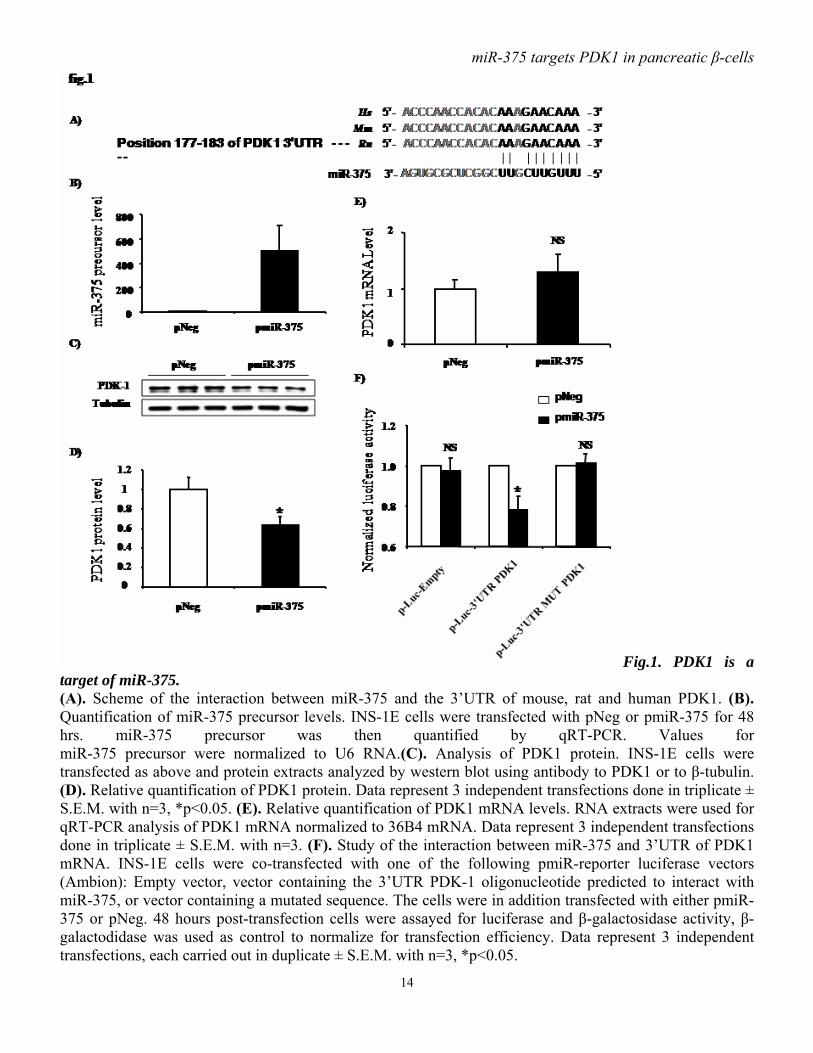
cells per well. After reaching 60% confluence, they were transfected with pmiR-375 or pNeg. 24 hours later, cells were starved in RPMI medium containing 0.5% FCS (v/v) for 24 hours and then replaced in RPMI 10% FCS (v/v). After 24 hours, DNA synthesis was assayed by adding 1µCi [methyl- 3H] thymidine/well and by incubating the cells for another 2 h. Then cells were washed twice with PBS, fixed with 10% (v/v) trichloroacetic acid for 30 min, and solubilized by adding 300 µl of 0.2 N NaOH to each well. Radioactivity, reflecting incorporation of [methyl-3H] thymidine into DNA, was measured by adding scintillation liquid and counting. Results: miR-375 regulates PDK1 protein level. To identify potential targets of miR-375, we used computational algorithms designed to predict mRNA targets of microRNAs. One of the predicted targets for miR-375 is 3’-phosphoinositide-dependent protein kinase-1, known as PDK1. The predicted miR-375 binding site in the 3’UTR of PDK1 mRNA (fig.1A) appears to be phylogenetically maintained. Hence this would suggest that the function of miR-375 as potential regulator of PDK1 has been conserved in mouse, rat and human. To investigate whether miR-375 affects PDK1, plasmids driving the expression of miR-375 precursor (pmiR-375)
miR-375 targets PDK1 in pancreatic β-cells
6
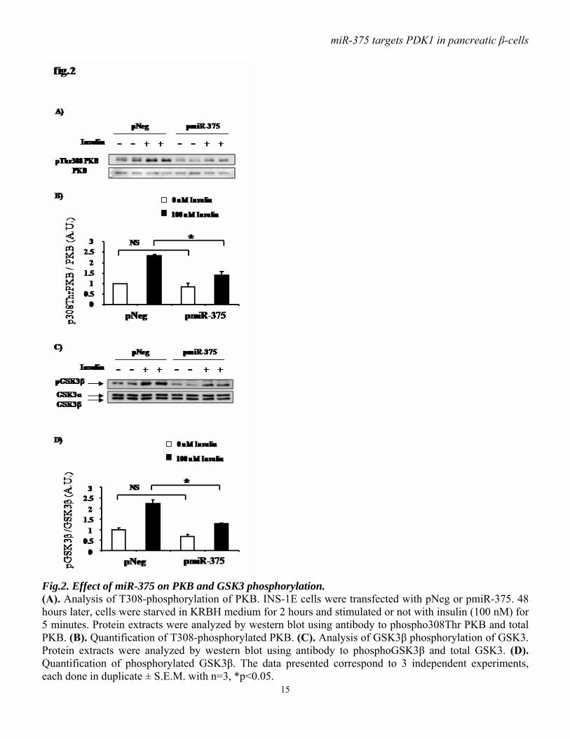
or control (pNeg) were transfected in INS-1E cells. pmiR-375 increased miR-375 precursor levels by approximately 500-fold, as assayed by real time PCR (fig.1B). Functional analysis shows that miR-375 precursor overexpression in INS-1E cells results in a reduction of approximately 40% of PDK1 protein (fig.1C, D) without affecting its mRNA level (fig.1E), indicating that miR-375 acts as a translational repressor. To evaluate whether the predicted miR-375 target site in the 3’UTR of PDK1 mRNA was directly involved in miR-375-induced reduction in PDK1 protein, we cloned the putative 3’UTR target site downstream of a luciferase reporter gene, and co-transfected this p-Luc-3’UTR PDK1 construct, into INS-1E cells with pmiR-375 or pNeg. Luciferase activity of cells transfected with pmiR-375 and p-Luc-3’UTR PDK1 was decreased by approximately 25 % compared to cells co-transfected with control pNeg and p-Luc-3’UTR PDK1 (fig.1F). With negative control constructs, p-Luc-Empty and p-Luc-3’UTR MUT PDK1, no reduced luciferase activity was observed when cells were co-transfected with pmiR-375 compared to pNeg. Taken together, our data argue for a direct interaction between miR-375 and PDK1 mRNA. miR-375 decreases insulin signaling downstream of PDK1. The PI3K/PDK1/PKB signaling pathway is used by insulin in pancreatic β-cells to elicit several of the hormone’s actions. It is generally believed that following insulin stimulation of cells, PDK1 is recruited to the plasma membrane and phosphorylates PKB on Thr308, which becomes activated and phosphorylates a series of substrates including GSK3α/β. Downregulation of PDK1 in β-cells is expected to cause a decrease in insulin-induced signaling dependent on this particular enzyme. To study the effect of miR-375 on insulin signaling, we examined the phosphorylation state of molecules
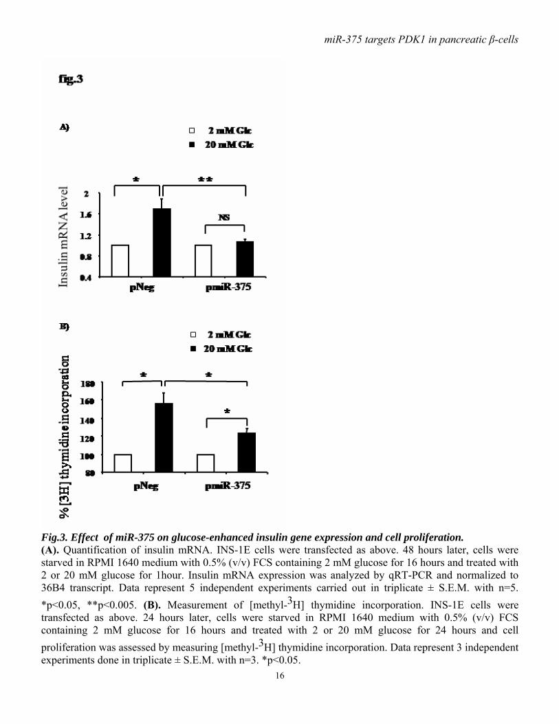
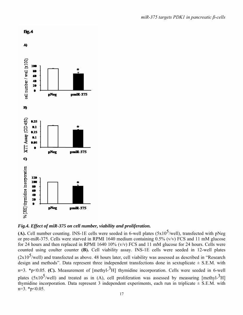
functioning downstream of PDK1. Immunoblot analysis showed that, in response to insulin, PKB phosphorylation on Thr308 is less abundant in cells overexpressing miR-375 precursor compared to control cells (fig.2 A, B). Consistent with this latter observation, ectopic expression of miR-375 precursor also reduces insulin-induced phosphorylation of GSK3β (fig.2 C, D). miR-375 decreases glucose-induced insulin gene expression and DNA synthesis. As PI3kinase signaling has been implicated in glucose-induced upregulation of insulin gene expression, and as we found that miR-375 decreases PDK1 protein, we examined the impact of miR-375 on insulin gene expression in response to glucose in INS-1E cells. We found that, as expected, high glucose concentration (20 mM) induced insulin gene expression in control cells, but this effect was lost in cells overexpressing pre-miR-375 (fig.3A). To test whether PDK1 is involved in the glucose-induced increase in cell proliferative activity, we analyzed the impact of miR-375 on cellular [methyl-3H] thymidine incorporation. As illustrated in fig.3B, the stimulatory effect of glucose on DNA synthesis was reduced by approximately 50% when miR-375 precursor was overexpressed. miR-375 attenuates cell viability and proliferation. As the PI3K/PKB cascade has been involved in cell survival and proliferation, we investigated the consequences of miR-375 expression on cell viability and proliferation. Thus, INS-1E cells were transfected with pmiR-375 or pNeg, and 48 hours after transfection cell number and viability were measured. We found that miR-375 overexpression reduced by approximately 25% cell number (fig.4A) and by approximately 20% cell viability (fig.4B). To further determine the effects of miR-375 on cell proliferation, DNA synthesis was measured using [methyl-3H] thymidine incorporation. As shown in fig.4C,
miR-375 targets PDK1 in pancreatic β-cells
7
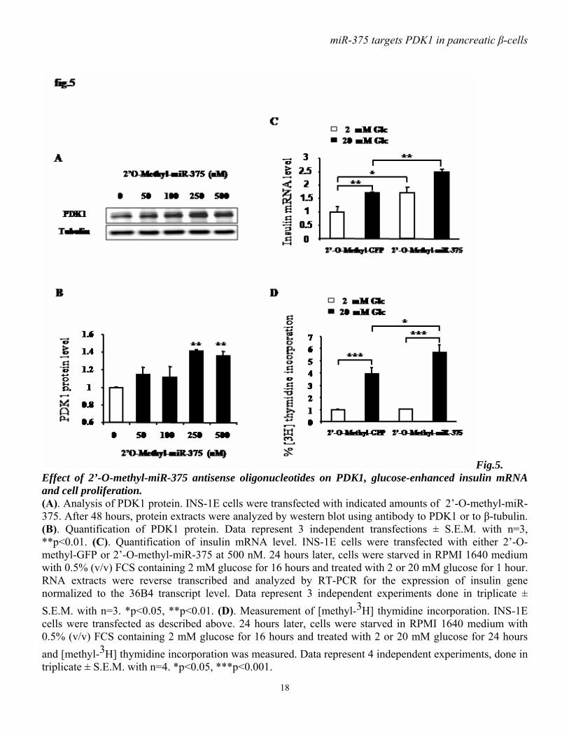
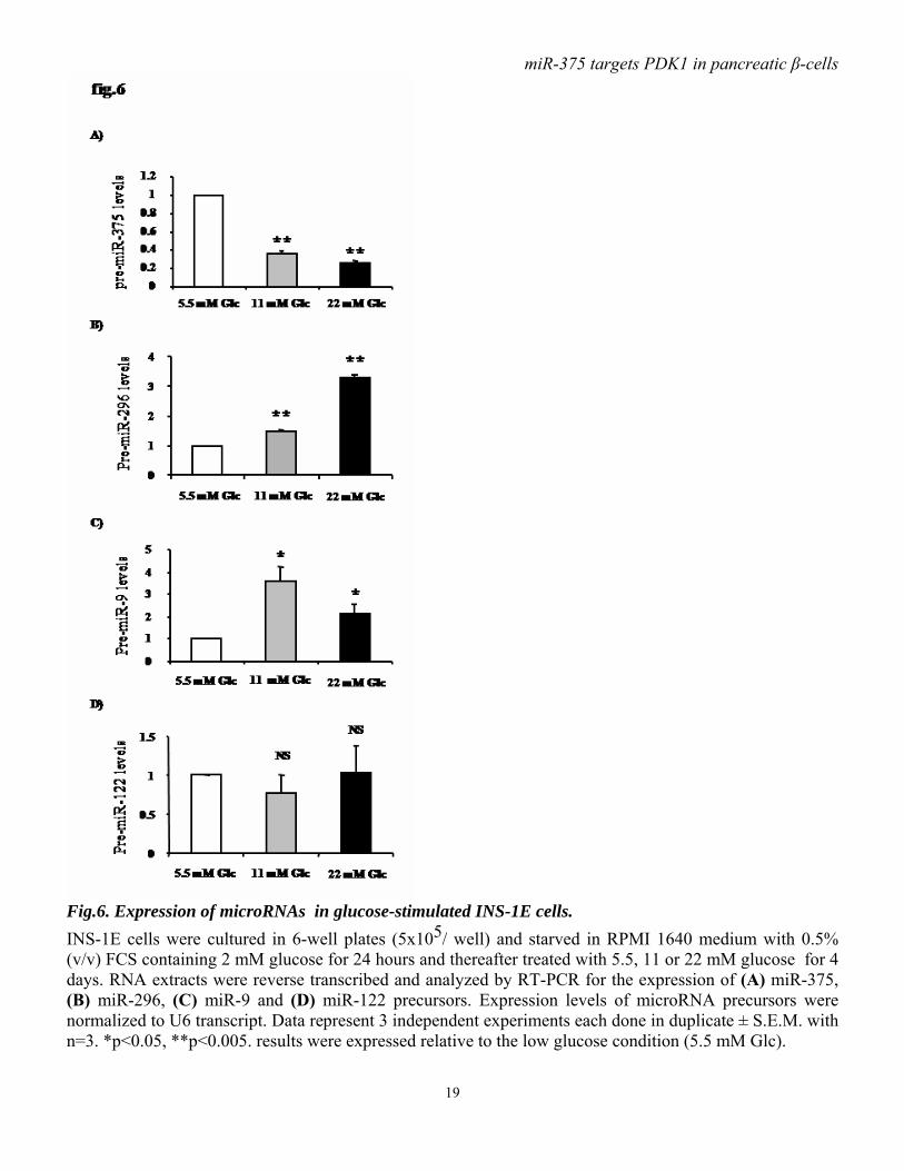
transfection with miR-375 precursor inhibited INS-1E cell proliferative activity by 20% compared to control cells. 2’-O-methyl-miR-375 increases PDK1 protein level and glucose-stimulatory action on insulin mRNA and DNA synthesis. As miR-375 targets PDK1 and impairs glucose-stimulated insulin gene expression and cell proliferation, we investigated whether antisense oligonucleotides of miR-375 induce effects opposite to those seen after miR-375 overexpression. Using 2’-O-methyl-miR-375 antisense oligonucleotides we found that blocking miR-375 augments PDK1 protein. This increase reaches approximately 40% when cells are transfected with either 250 or 500 nM of 2’-O-methyl-miR-375 (fig.5A-B). Importantly, we found that 2’-O-methyl-miR-375-induced miR-375 depletion increases both basal and glucose-enhanced insulin mRNA (fig.5C). Finally, as shown in (fig.5D), 2’-O-methyl-miR-375 increases the glucose stimulatory action on [3H] thymidine incorporation compared to 2’-O-methyl-GFP. Glucose specifically decreases miR-375 expression. To investigate whether glucose-induced responses in INS-1E cells could be mediated by microRNAs, we analyzed the expression of a series of microRNAs in cells maintained for 4 days with different glucose concentrations. Using qRT-PCR as described (29), we found that miR-375 precursor is negatively regulated by glucose. Indeed, 11 and 22 mM glucose induce a decrease of about 60 and 70%, respectively, in miR-375 precursor levels compared to 5.5 mM glucose (fig.6A). To look whether this glucose effect was limited or not to miR-375, we measured the level of three other microRNAs expressed in INS-1E cells. In contrast to miR-375, both miR-296 and miR-9 are positively regulated by prolonged glucose treatment. Indeed, glucose leads to a concentration-dependent increase in miR-296 precursor (fig.6B), while miR-9 precursor is robustly increased after
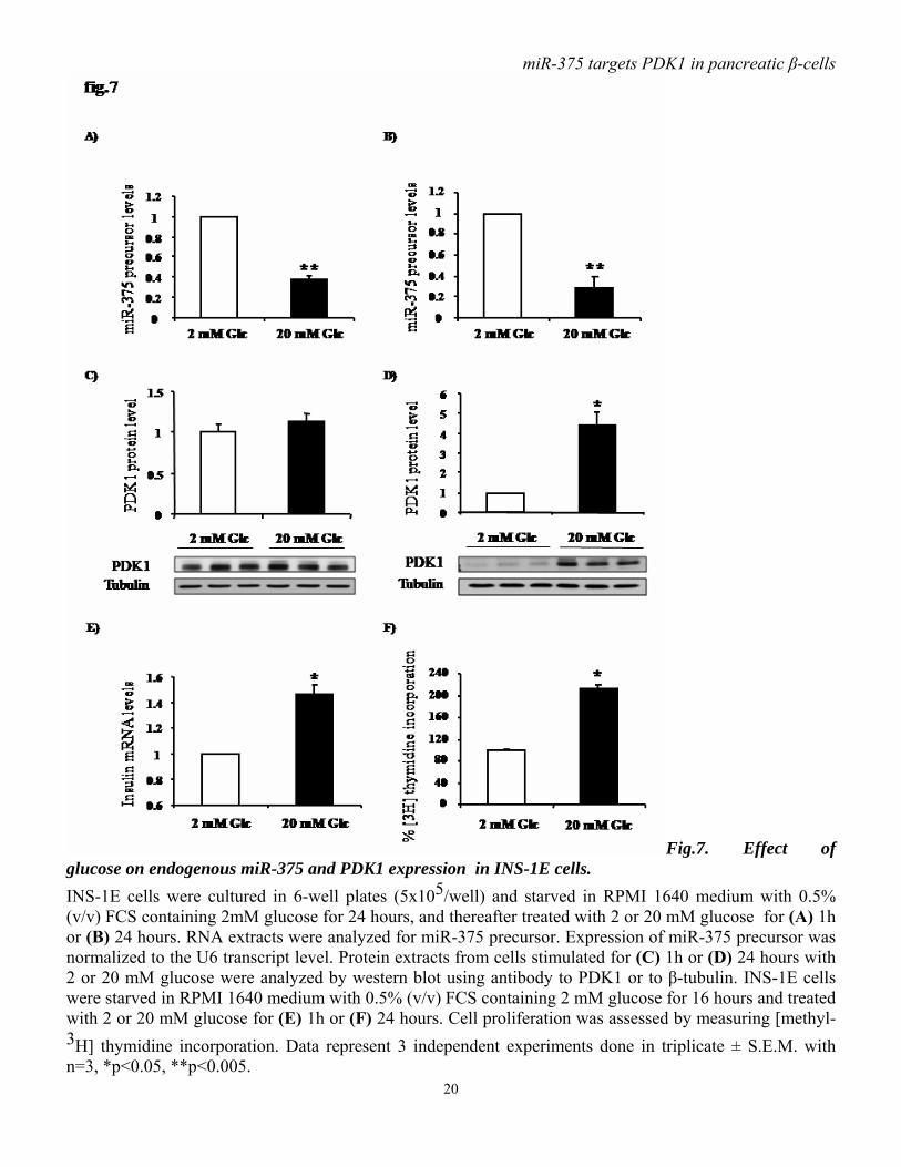
cell exposure to 11mM glucose. At higher glucose concentration, miR-9 expression tends to decline, but remains higher compared to low glucose (5.5 mM) condition (fig.6C). Finally, miR-122, which presents a specific liver expression, is detected in INS-1E cells, but glucose does not modulate its expression (fig.6D). The levels of miR-375 and PDK1 are inversely correlated in glucose-stimulated INS-1E cells. To further document the role of miR-375, we analyzed the expression of pre-miR-375 in INS-1E cells treated for 1 or 24 hours with 2 or 20 mM glucose. We found that glucose induces a robust decrease in miR-375 after 1 hour and 24 hours (fig.7A, B). Furthermore, immunoblot analysis show that PDK1 protein level is slightly, but not significantly, increased within one hour of glucose treatment (fig.7C). Remarkably, in INS-1E cells exposed for 24 hours to 20 mM glucose , PDK1 protein is increased (4-fold) and its level thus inversely correlates with that of miR-375 (fig.7D). Finally, decreased miR-375 without change in PDK1 level seen after 1 hour of glucose treatment occurs with enhanced insulin gene expression (fig7.E). Decreased miR-375 and increased PDK1 levels are associated with enhanced DNA synthesis as reflected by increased thymidine incorporation seen within 24 hours of glucose treatment (fig.7F). Glucose regulates miR-375 expression in freshly isolated rat pancreatic islets. To characterize the glucose-mediated miR-375 regulation, we analyzed the expression level of miR-375 precursor in isolated rat islets that had been exposed to 5, 10 or 20 mM of glucose for 2 or 72 hours, periods which respectively, represent conditions in which the acute and chronic influences of glucose can be studied. After 2 hours at stimulatory glucose concentrations (10 or 20 mM) miR-375 expression is lower than in the basal 5 mM condition (fig.8A).After 72 hours,
miR-375 targets PDK1 in pancreatic β-cells
8
a lower expression level was measured at 5 mM than at 10 and 20 mM whereby the level at 20 mM tended to be lower than at 10 mM (fig.8B). Reduced miR-375 expression in freshly isolated islets from diabetic Goto-Kakizaki rats. To study miR-375 expression under in vivo conditions of hyperglycemia, we analyzed islets from diabetic Goto-Kakizaki rats. We found that miR-375 is downregulated in diabetic Goto-Kakizaki rats compared to control Wistar rats (fig.8C). Further, in GK rats this decrease in miR-375 expression is associated with a modest, albeit not significant, increase in insulin mRNA level (fig.8E). Finally, the expression of miR-124a2, which has been involved in insulin mRNA expression (27), is upregulated in GK rats (fig.8D). Discussion. During the recent years microRNAs have emerged as important regulators of cell fate and metabolism. Although several microRNAs have been implicated in a variety of disease processes (31), only a few have been linked to insulin signaling and diabetes (32). Using either a gain- or loss- of-function approach, we show here in a pancreatic β-cell line that miR-375, previously described to reduce glucose-induced insulin secretion by inhibiting Mtpn protein (25), is able to downregulate PDK1 protein by interfering directly with its mRNA. By targeting PDK1, a key player in the PI3K signaling cascade, miR-375 decreases insulin-induced phosphorylation of PKB and GSK3, both acting downstream of PDK1.
It is interesting to mention that pancreatic β-cell specific PDK1 knockout in mice results in a reduced number and size of β-cells, and in decreased islet density (14). These data and ours are intriguing in the context of a report showing that miR-375 knockout by
morpholinos in zebrafish embryos leads to defects in the development of pancreatic islets (33).
Previous studies have shown that inhibition of the PI3K cascade dampens glucose-induced insulin gene expression (6). To document the biological role of miR-375 as an inhibitor of PI3K signaling downstream of PDK1, we analyzed the effect of miR-375 overexpression or depletion on glucose-induced insulin gene expression. Our findings that miR-375 controls insulin gene expression stimulated by glucose are consistent with the notion that the PI3K cascade is important for glucose stimulatory action on insulin gene expression. A chief observation of our study is that microRNA expression seems to be glucose sensitive. Importantly, the increased insulin gene expression seen in INS-1E cells exposed to high glucose is associated with reduced expression of miR-375 precursor. Our in silico analysis (data not shown) reveals that miR-375 is located in an intergenic region between cryba2 and Ccdc108 genes on the mouse chromosome 1. Comparison of genomic sequences (http://ecrbrowser.decode.org/) across human, mouse and rat shows that a 5 kilobase genomic region downstream of the miR-375 precursor contains highly conserved elements. Moreover, according to a recent publication, several regions located downstream and upstream of the miR-375 gene interact with NeuroD1/BETA2 and PDX-1 (34). Taken together, these facts suggest that the gene encoding for miR-375 may behave as a locus controlled by its own promoter.
While glucose decreased miR-375 expression within 1 hour in INS-1E cells, a longer exposure time was needed to observe the downstream stimulatory effect on PDK1 protein expression. This suggests that miR-375 suppresses synthesis of PDK1 protein. Interestingly, prolonged glucose exposure of INS-1E cells (35) has been shown to increase IRS-2 gene expression and protein
miR-375 targets PDK1 in pancreatic β-cells
9
localization to the plasma membrane as well as PKB phosphorylation. These observations together with ours indicate that in INS-1E cells glucose induces a decrease in miR-375 followed by an increase in PDK1 protein and an activation of the PI3kinase cascade, and hence β-cell proliferation.
Glucose was also found to regulate miR-375 expression in primary islet tissue. A 2 hour incubation was used to measure levels in freshly isolated rat islets at glucose concentrations known to have exerted dose-dependent stimulations of their metabolic, secretory and protein biosynthetic activities. We found miR-375 expression to be lower at 10 and 20 mM than at 5 mM glucose, which is similar to the effect seen in INS-1E cells. The 72 h culture condition compares expression in islets which have maintained this functional responsiveness ( 10 an 20 mM glucose during culture) with that in islets having lost this responsiveness and showing increased susceptibility to apoptosis ( 5mM glucose during culture ) (36-38). Expression of miR-375 is lower in islets showing viability consequences of prolonged glucose deprivation ; it is unknown whether this reduction participates in the apoptotic pathway and can be interpreted in light of a report showing that miR-375 knockout by morpholinos in zebrafish leads to defects in islet development (33). When comparing functionally competent islets cultured at control (10 mM) versus elevated (20 mM) glucose concentrations, miR-375 levels appeared lower after prolonged glucose hyperactivation (20 mM). Islets isolated from diabetic Goto-Kakizaki rats exhibited a lower miR-375 expression than islets from Wistar rats which can be considered as further evidence for the suppressing effect of supraphysiologic glucose concentrations. Such reduced miR-375 level is expected to be associated with increased insulin mRNA, but this was not the case in GK rats, suggesting a possible failure of the PDK1/PKB cascade to
increase insulin gene expression. On the other hand, expression of miR-124a2, previously described as inhibiting insulin gene expression (27), is upregulated in GK islets. Hence, we speculate that the antagonizing actions of miR-124a2 and miR-375 are counterbalanced resulting in an unchanged insulin mRNA level in GK islets. An alternative explanation is that the lower expression in GK islets results from a prolonged intracellular deprivation of glucose signals as in the case of the islets cultured in 5 mM glucose for 72 hours that progress to apoptosis.
According to recent studies (39; 40), total β-cell mass is reduced in patients and animals with type 2 diabetes and contributes to the disease process. Notably, β cell-specific PDK1 knockout in mice leads to diabetes as a result of a reduction in β cell mass (14). Another in vitro study reported that decreased PDK1 in human glioblastoma cells obtained with antisense oligonucleotides or with RNA interference blocks cell proliferation (41). Here we find that miR-375 downregulates PDK1 by targeting directly PDK1 mRNA and may therefore impact on cell proliferation given its key role in the PI3K/PKB cascade. In this context, several studies have revealed dysregulations in microRNA expression in human cancers (42), suggesting that microRNAs may act on cell proliferation. More directly related to our work, recent studies show that miR-375 is decreased in pancreatic cancer (43-45). Our results point to a similar anti-proliferative action of miR-375, as we found that miR-375 attenuates cell viability and proliferation.
A first key finding of our work is that miR-375 is regulated by glucose, which is the central molecule in islet metabolism and physiology. A second important observation is that miR-375 inhibits glucose-induced INS-1E cell proliferation. This result is particularly interesting in the context of diabetes. Indeed, while a distinct disease process is responsible
miR-375 targets PDK1 in pancreatic β-cells
10
for type 1 and type 2 diabetes, in both cases, β-cell failure occurs. Developing new approaches to induce β-cells to replicate is a supereminent goal in diabetes research. Our data presented here reveal the pancreatic islet-specific microRNA, miR-375, as an important regulator of glucose-stimulated insulin gene expression and proliferation of pancreatic β-cells. Taking into account the fact that miR-375 also decreases insulin secretion (25), miR-375 emerges as a target which should be prioritized to enhance islet function and to combat β-cell failure.
Acknowledgments:
We thank Professor Bernard Portha for providing us with the GK rats and Dr. Olivier Dumortier for help with rat islets isolation, Nadine Gautier, Chantal Filloux, Rachel Paul, Joseph Murdaca, Cyndie Raemdock and Elke De Vos for assistance. We are grateful to Drs Mourad Naïmi and Jean Giudicelli for discussions and advice. Our work in Nice was supported by INSERM, Université de Nice-Sophia-Antipolis, Conseil Régional PACA and Conseil Général des Alpes-Maritimes, by the European Community FP6 EUGENE2 (grant LSHM-CT-2004-512013) and the European Foundation for the Study of Diabetes (EFSD)/JDRF/Novo-Nordisk Program in Type 1 Diabetes (grant to Emmanuel Van Obberghen). Our research in Brussels was supported by Research Foundation Flanders ( FWO-G.0183.05) and the Inter-University Pole Attraction Program (IUAP P5/17) from the Belgian Science Policy (to D. Pipeleers). Abdelfattah El Ouaamari was supported by a PhD fellowship from la Ligue Nationale Contre le Cancer (France).
References: 1. Stumvoll M, Goldstein BJ, van Haeften TW: Type 2 diabetes: principles of pathogenesis and therapy. Lancet 365:1333-1346, 2005 2. Ling Z, Wang Q, Stange G, In't Veld P, Pipeleers D: Glibenclamide treatment recruits beta-cell subpopulation into elevated and sustained basal insulin synthetic activity. Diabetes 55:78-85, 2006 3. Pipeleers D, Kiekens R, Ling Z, Wilikens A, Schuit F: Physiologic relevance of heterogeneity in the pancreatic beta-cell population. Diabetologia 37 Suppl 2:S57-64, 1994 4. Hugl SR, White MF, Rhodes CJ: Insulin-like growth factor I (IGF-I)-stimulated pancreatic beta-cell growth is glucose-dependent. Synergistic activation of insulin receptor substrate-mediated signal transduction pathways by glucose and IGF-I in INS-1 cells. J Biol Chem 273:17771-17779, 1998 5. Alonso LC, Yokoe T, Zhang P, Scott DK, Kim SK, O'Donnell CP, Garcia-Ocana A: Glucose infusion in mice: a new model to induce beta-cell replication. Diabetes 56:1792-1801, 2007 6. Rafiq I, da Silva Xavier G, Hooper S, Rutter GA: Glucose-stimulated preproinsulin gene expression and nuclear trans-location of pancreatic duodenum homeobox-1 require activation of phosphatidylinositol 3-kinase but not p38 MAPK/SAPK2. J Biol Chem 275:15977-15984, 2000 7. MacFarlane WM, Read ML, Gilligan M, Bujalska I, Docherty K: Glucose modulates the binding activity of the beta-cell transcription factor IUF1 in a phosphorylation-dependent manner. Biochem J 303 ( Pt 2):625-631, 1994 8. Srinivasan S, Bernal-Mizrachi E, Ohsugi M, Permutt MA: Glucose promotes pancreatic islet beta-cell survival through a PI 3-kinase/Akt-signaling pathway. Am J Physiol Endocrinol Metab 283:E784-793, 2002 9. Taniguchi CM, Emanuelli B, Kahn CR: Critical nodes in signalling pathways: insights into insulin action. Nat Rev Mol Cell Biol 7:85-96, 2006 10. Stephens L, Anderson K, Stokoe D, Erdjument-Bromage H, Painter GF, Holmes AB, Gaffney PR, Reese CB, McCormick F, Tempst P, Coadwell J, Hawkins PT: Protein kinase B kinases that mediate phosphatidylinositol 3,4,5-trisphosphate-dependent activation of protein kinase B. Science 279:710-714, 1998 11. Alessi DR, James SR, Downes CP, Holmes AB, Gaffney PR, Reese CB, Cohen P: Characterization of a 3-phosphoinositide-dependent protein kinase which phosphorylates and activates protein kinase Balpha. In Curr Biol, 1997, p. 261-269 12. Rintelen F, Stocker H, Thomas G, Hafen E: PDK1 regulates growth through Akt and S6K in Drosophila. Proc Natl Acad Sci U S A 98:15020-15025, 2001 13. Lawlor MA, Mora A, Ashby PR, Williams MR, Murray-Tait V, Malone L, Prescott AR, Lucocq JM, Alessi DR: Essential role of PDK1 in regulating cell size and development in mice. Embo J 21:3728-3738, 2002 14. Hashimoto N, Kido Y, Uchida T, Asahara S, Shigeyama Y, Matsuda T, Takeda A, Tsuchihashi D, Nishizawa A, Ogawa W, Fujimoto Y, Okamura H, Arden KC, Herrera PL, Noda T, Kasuga M: Ablation of PDK1 in pancreatic beta cells induces diabetes as a result of loss of beta cell mass. Nat Genet 38:589-593, 2006 15. Lee RC, Feinbaum RL, Ambros V: The C. elegans heterochronic gene lin-4 encodes small RNAs with antisense complementarity to lin-14. Cell 75:843-854, 1993 16. Kim VN: MicroRNA biogenesis: coordinated cropping and dicing. Nat Rev Mol Cell Biol
miR-375 targets PDK1 in pancreatic β-cells
12
6:376-385, 2005 17. Bartel DP: MicroRNAs: genomics, biogenesis, mechanism, and function. Cell 116:281-297, 2004 18. Reinhart BJ, Slack FJ, Basson M, Pasquinelli AE, Bettinger JC, Rougvie AE, Horvitz HR, Ruvkun G: The 21-nucleotide let-7 RNA regulates developmental timing in Caenorhabditis elegans. Nature 403:901-906, 2000 19. Xu P, Vernooy SY, Guo M, Hay BA: The Drosophila microRNA Mir-14 suppresses cell death and is required for normal fat metabolism. Curr Biol 13:790-795, 2003 20. Esau C, Kang X, Peralta E, Hanson E, Marcusson EG, Ravichandran LV, Sun Y, Koo S, Perera RJ, Jain R, Dean NM, Freier SM, Bennett CF, Lollo B, Griffey R: MicroRNA-143 regulates adipocyte differentiation. J Biol Chem 279:52361-52365, 2004 21. Krutzfeldt J, Rajewsky N, Braich R, Rajeev KG, Tuschl T, Manoharan M, Stoffel M: Silencing of microRNAs in vivo with 'antagomirs'. Nature 438:685-689, 2005 22. Esau C, Davis S, Murray SF, Yu XX, Pandey SK, Pear M, Watts L, Booten SL, Graham M, McKay R, Subramaniam A, Propp S, Lollo BA, Freier S, Bennett CF, Bhanot S, Monia BP: miR-122 regulation of lipid metabolism revealed by in vivo antisense targeting. Cell Metab 3:87-98, 2006 23. Callis TE, Chen JF, Wang DZ: MicroRNAs in skeletal and cardiac muscle development. DNA Cell Biol 26:219-225, 2007 24. Makeyev EV, Zhang J, Carrasco MA, Maniatis T: The MicroRNA miR-124 promotes neuronal differentiation by triggering brain-specific alternative pre-mRNA splicing. Mol Cell 27:435-448, 2007 25. Poy MN, Eliasson L, Krutzfeldt J, Kuwajima S, Ma X, Macdonald PE, Pfeffer S, Tuschl T, Rajewsky N, Rorsman P, Stoffel M: A pancreatic islet-specific microRNA regulates insulin secretion. Nature 432:226-230, 2004 26. Plaisance V, Abderrahmani A, Perret-Menoud V, Jacquemin P, Lemaigre F, Regazzi R: MicroRNA-9 controls the expression of Granuphilin/Slp4 and the secretory response of insulin-producing cells. J Biol Chem 281:26932-26942, 2006 27. Baroukh N, Ravier MA, Loder MK, Hill EV, Bounacer A, Scharfmann R, Rutter GA, Van Obberghen E: MicroRNA-124a regulates Foxa2 expression and intracellular signaling in pancreatic beta-cell lines. J Biol Chem 282:19575-19588, 2007 28. Cheng AM, Byrom MW, Shelton J, Ford LP: Antisense inhibition of human miRNAs and indications for an involvement of miRNA in cell growth and apoptosis. Nucleic Acids Res 33:1290-1297, 2005 29. Schmittgen TD, Jiang J, Liu Q, Yang L: A high-throughput method to monitor the expression of microRNA precursors. Nucleic Acids Res 32:e43, 2004 30. Jiang J, Lee EJ, Gusev Y, Schmittgen TD: Real-time expression profiling of microRNA precursors in human cancer cell lines. Nucleic Acids Res 33:5394-5403, 2005 31. Chang TC, Mendell JT: microRNAs in vertebrate physiology and human disease. Annu Rev Genomics Hum Genet 8:215-239, 2007 32. He A, Zhu L, Gupta N, Chang Y, Fang F: Over-expression of miR-29, highly upregulated in diabetic rats, leads to insulin resistance in 3T3-L1 adipocytes. Mol Endocrinol, 2007 33. Kloosterman WP, Lagendijk AK, Ketting RF, Moulton JD, Plasterk RH: Targeted Inhibition of miRNA Maturation with Morpholinos Reveals a Role for miR-375 in Pancreatic Islet Development. PLoS Biol 5:e203, 2007 34. Keller DM, McWeeney S, Arsenlis A, Drouin J, Wright CV, Wang H, Wollheim CB, White
miR-375 targets PDK1 in pancreatic β-cells
13
P, Kaestner KH, Goodman RH: Characterization of pancreatic transcription factor Pdx-1 binding sites using promoter microarray and serial analysis of chromatin occupancy. J Biol Chem, 2007 35. Lingohr MK, Briaud I, Dickson LM, McCuaig JF, Alarcon C, Wicksteed BL, Rhodes CJ: Specific regulation of IRS-2 expression by glucose in rat primary pancreatic islet beta-cells. J Biol Chem 281:15884-15892, 2006 36. Hoorens A, Van de Casteele M, Kloppel G, Pipeleers D: Glucose promotes survival of rat pancreatic beta cells by activating synthesis of proteins which suppress a constitutive apoptotic program. J Clin Invest 98:1568-1574, 1996 37. Ling Z, Kiekens R, Mahler T, Schuit FC, Pipeleers-Marichal M, Sener A, Kloppel G, Malaisse WJ, Pipeleers DG: Effects of chronically elevated glucose levels on the functional properties of rat pancreatic beta-cells. Diabetes 45:1774-1782, 1996 38. Martens GA, Cai Y, Hinke S, Stange G, Van de Casteele M, Pipeleers D: Glucose suppresses superoxide generation in metabolically responsive pancreatic beta cells. J Biol Chem 280:20389-20396, 2005 39. Weir GC, Bonner-Weir S: Five stages of evolving beta-cell dysfunction during progression to diabetes. Diabetes 53 Suppl 3:S16-21, 2004 40. Butler AE, Janson J, Bonner-Weir S, Ritzel R, Rizza RA, Butler PC: Beta-cell deficit and increased beta-cell apoptosis in humans with type 2 diabetes. Diabetes 52:102-110, 2003 41. Bilanges B, Stokoe D: Direct comparison of the specificity of gene silencing using antisense oligonucleotides and RNAi. Biochem J 388:573-583, 2005 42. Lu J, Getz G, Miska EA, Alvarez-Saavedra E, Lamb J, Peck D, Sweet-Cordero A, Ebert BL, Mak RH, Ferrando AA, Downing JR, Jacks T, Horvitz HR, Golub TR: MicroRNA expression profiles classify human cancers. Nature 435:834-838, 2005 43. Szafranska AE, Davison TS, John J, Cannon T, Sipos B, Maghnouj A, Labourier E, Hahn SA: MicroRNA expression alterations are linked to tumorigenesis and non-neoplastic processes in pancreatic ductal adenocarcinoma. Oncogene 26:4442-4452, 2007 44. Lee EJ, Gusev Y, Jiang J, Nuovo GJ, Lerner MR, Frankel WL, Morgan DL, Postier RG, Brackett DJ, Schmittgen TD: Expression profiling identifies microRNA signature in pancreatic cancer. Int J Cancer 120:1046-1054, 2007 45. Bloomston M, Frankel WL, Petrocca F, Volinia S, Alder H, Hagan JP, Liu CG, Bhatt D, Taccioli C, Croce CM: MicroRNA expression patterns to differentiate pancreatic adenocarcinoma from normal pancreas and chronic pancreatitis. Jama 297:1901-1908, 2007
miR-375 targets PDK1 in pancreatic β-cells
14
Fig.1. PDK1 is a target of miR-375. (A). Scheme of the interaction between miR-375 and the 3’UTR of mouse, rat and human PDK1. (B). Quantification of miR-375 precursor levels. INS-1E cells were transfected with pNeg or pmiR-375 for 48 hrs. miR-375 precursor was then quantified by qRT-PCR. Values for miR-375 precursor were normalized to U6 RNA.(C). Analysis of PDK1 protein. INS-1E cells were transfected as above and protein extracts analyzed by western blot using antibody to PDK1 or to β-tubulin. (D). Relative quantification of PDK1 protein. Data represent 3 independent transfections done in triplicate ± S.E.M. with n=3, *p<0.05. (E). Relative quantification of PDK1 mRNA levels. RNA extracts were used for qRT-PCR analysis of PDK1 mRNA normalized to 36B4 mRNA. Data represent 3 independent transfections done in triplicate ± S.E.M. with n=3. (F). Study of the interaction between miR-375 and 3’UTR of PDK1 mRNA. INS-1E cells were co-transfected with one of the following pmiR-reporter luciferase vectors (Ambion): Empty vector, vector containing the 3’UTR PDK-1 oligonucleotide predicted to interact with miR-375, or vector containing a mutated sequence. The cells were in addition transfected with either pmiR-375 or pNeg. 48 hours post-transfection cells were assayed for luciferase and β-galactosidase activity, β-galactodidase was used as control to normalize for transfection efficiency. Data represent 3 independent transfections, each carried out in duplicate ± S.E.M. with n=3, *p<0.05.
miR-375 targets PDK1 in pancreatic β-cells
15
Fig.2. Effect of miR-375 on PKB and GSK3 phosphorylation. (A). Analysis of T308-phosphorylation of PKB. INS-1E cells were transfected with pNeg or pmiR-375. 48 hours later, cells were starved in KRBH medium for 2 hours and stimulated or not with insulin (100 nM) for 5 minutes. Protein extracts were analyzed by western blot using antibody to phospho308Thr PKB and total PKB. (B). Quantification of T308-phosphorylated PKB. (C). Analysis of GSK3β phosphorylation of GSK3. Protein extracts were analyzed by western blot using antibody to phosphoGSK3β and total GSK3. (D). Quantification of phosphorylated GSK3β. The data presented correspond to 3 independent experiments, each done in duplicate ± S.E.M. with n=3, *p<0.05.
miR-375 targets PDK1 in pancreatic β-cells
16
Fig.3. Effect of miR-375 on glucose-enhanced insulin gene expression and cell proliferation. (A). Quantification of insulin mRNA. INS-1E cells were transfected as above. 48 hours later, cells were starved in RPMI 1640 medium with 0.5% (v/v) FCS containing 2 mM glucose for 16 hours and treated with 2 or 20 mM glucose for 1hour. Insulin mRNA expression was analyzed by qRT-PCR and normalized to 36B4 transcript. Data represent 5 independent experiments carried out in triplicate ± S.E.M. with n=5. *p<0.05, **p<0.005. (B). Measurement of [methyl-3H] thymidine incorporation. INS-1E cells were transfected as above. 24 hours later, cells were starved in RPMI 1640 medium with 0.5% (v/v) FCS containing 2 mM glucose for 16 hours and treated with 2 or 20 mM glucose for 24 hours and cell proliferation was assessed by measuring [methyl-3H] thymidine incorporation. Data represent 3 independent experiments done in triplicate ± S.E.M. with n=3. *p<0.05.
miR-375 targets PDK1 in pancreatic β-cells
17
Fig.4. Effect of miR-375 on cell number, viability and proliferation. (A). Cell number counting. INS-1E cells were seeded in 6-well plates (5x10⁵/well), transfected with pNeg or pre-miR-375. Cells were starved in RPMI 1640 medium containing 0.5% (v/v) FCS and 11 mM glucose for 24 hours and then replaced in RPMI 1640 10% (v/v) FCS and 11 mM glucose for 24 hours. Cells were counted using coulter counter (B). Cell viability assay. INS-1E cells were seeded in 12-well plates (2x10⁵/well) and transfected as above. 48 hours later, cell viability was assessed as described in “Research design and methods”. Data represent three independent transfections done in sextuplicate ± S.E.M. with n=3. *p<0.05. (C). Measurement of [methyl-3H] thymidine incorporation. Cells were seeded in 6-well plates (5x10⁵/well) and treated as in (A), cell proliferation was assessed by measuring [methyl-3H] thymidine incorporation. Data represent 3 independent experiments, each run in triplicate ± S.E.M. with n=3. *p<0.05.
miR-375 targets PDK1 in pancreatic β-cells
18
Fig.5. Effect of 2’-O-methyl-miR-375 antisense oligonucleotides on PDK1, glucose-enhanced insulin mRNA and cell proliferation. (A). Analysis of PDK1 protein. INS-1E cells were transfected with indicated amounts of 2’-O-methyl-miR-375. After 48 hours, protein extracts were analyzed by western blot using antibody to PDK1 or to β-tubulin. (B). Quantification of PDK1 protein. Data represent 3 independent transfections ± S.E.M. with n=3, **p<0.01. (C). Quantification of insulin mRNA level. INS-1E cells were transfected with either 2’-O-methyl-GFP or 2’-O-methyl-miR-375 at 500 nM. 24 hours later, cells were starved in RPMI 1640 medium with 0.5% (v/v) FCS containing 2 mM glucose for 16 hours and treated with 2 or 20 mM glucose for 1 hour. RNA extracts were reverse transcribed and analyzed by RT-PCR for the expression of insulin gene normalized to the 36B4 transcript level. Data represent 3 independent experiments done in triplicate ± S.E.M. with n=3. *p<0.05, **p<0.01. (D). Measurement of [methyl-3H] thymidine incorporation. INS-1E cells were transfected as described above. 24 hours later, cells were starved in RPMI 1640 medium with 0.5% (v/v) FCS containing 2 mM glucose for 16 hours and treated with 2 or 20 mM glucose for 24 hours and [methyl-3H] thymidine incorporation was measured. Data represent 4 independent experiments, done in triplicate ± S.E.M. with n=4. *p<0.05, ***p<0.001.
miR-375 targets PDK1 in pancreatic β-cells
19
Fig.6. Expression of microRNAs in glucose-stimulated INS-1E cells. INS-1E cells were cultured in 6-well plates (5x10⁵/ well) and starved in RPMI 1640 medium with 0.5% (v/v) FCS containing 2 mM glucose for 24 hours and thereafter treated with 5.5, 11 or 22 mM glucose for 4 days. RNA extracts were reverse transcribed and analyzed by RT-PCR for the expression of (A) miR-375, (B) miR-296, (C) miR-9 and (D) miR-122 precursors. Expression levels of microRNA precursors were normalized to U6 transcript. Data represent 3 independent experiments each done in duplicate ± S.E.M. with n=3. *p<0.05, **p<0.005. results were expressed relative to the low glucose condition (5.5 mM Glc).
miR-375 targets PDK1 in pancreatic β-cells
20
Fig.7. Effect of glucose on endogenous miR-375 and PDK1 expression in INS-1E cells. INS-1E cells were cultured in 6-well plates (5x10⁵/well) and starved in RPMI 1640 medium with 0.5% (v/v) FCS containing 2mM glucose for 24 hours, and thereafter treated with 2 or 20 mM glucose for (A) 1h or (B) 24 hours. RNA extracts were analyzed for miR-375 precursor. Expression of miR-375 precursor was normalized to the U6 transcript level. Protein extracts from cells stimulated for (C) 1h or (D) 24 hours with 2 or 20 mM glucose were analyzed by western blot using antibody to PDK1 or to β-tubulin. INS-1E cells were starved in RPMI 1640 medium with 0.5% (v/v) FCS containing 2 mM glucose for 16 hours and treated with 2 or 20 mM glucose for (E) 1h or (F) 24 hours. Cell proliferation was assessed by measuring [methyl-3H] thymidine incorporation. Data represent 3 independent experiments done in triplicate ± S.E.M. with n=3, *p<0.05, **p<0.005.
miR-375 targets PDK1 in pancreatic β-cells
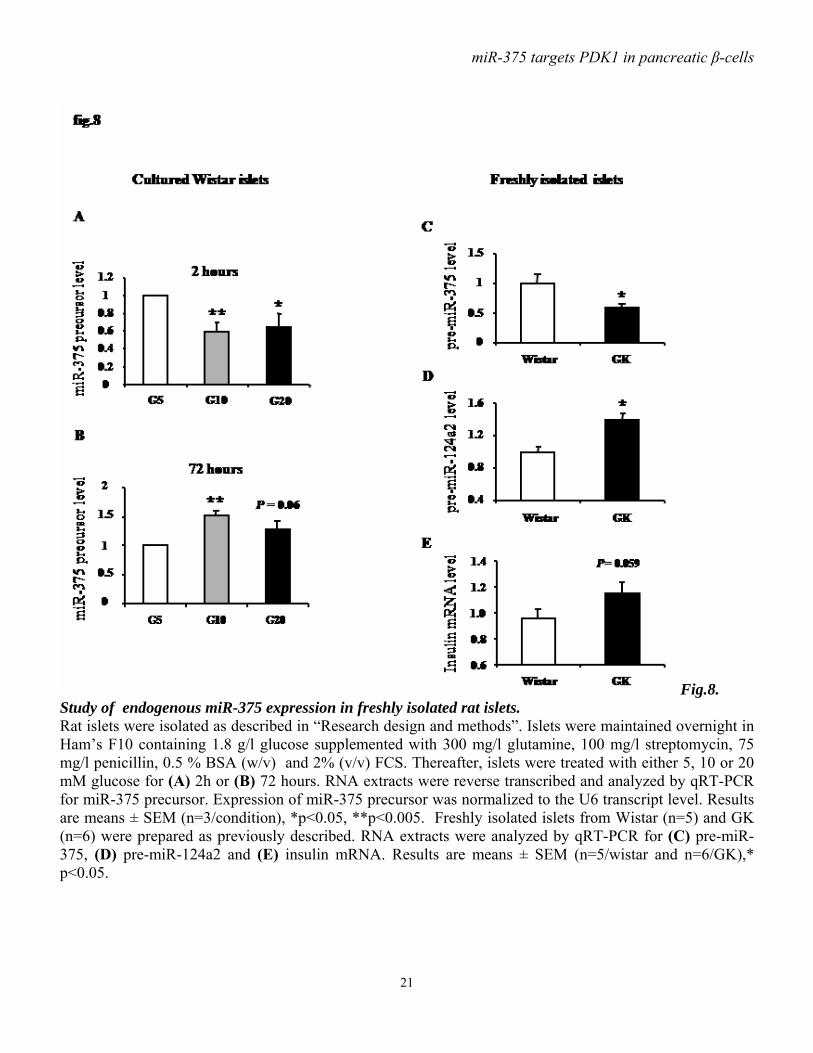
21
Fig.8. Study of endogenous miR-375 expression in freshly isolated rat islets. Rat islets were isolated as described in “Research design and methods”. Islets were maintained overnight in Ham’s F10 containing 1.8 g/l glucose supplemented with 300 mg/l glutamine, 100 mg/l streptomycin, 75 mg/l penicillin, 0.5 % BSA (w/v) and 2% (v/v) FCS. Thereafter, islets were treated with either 5, 10 or 20 mM glucose for (A) 2h or (B) 72 hours. RNA extracts were reverse transcribed and analyzed by qRT-PCR for miR-375 precursor. Expression of miR-375 precursor was normalized to the U6 transcript level. Results are means ± SEM (n=3/condition), *p<0.05, **p<0.005. Freshly isolated islets from Wistar (n=5) and GK (n=6) were prepared as previously described. RNA extracts were analyzed by qRT-PCR for (C) pre-miR-375, (D) pre-miR-124a2 and (E) insulin mRNA. Results are means ± SEM (n=5/wistar and n=6/GK),* p<0.05.